The Analogs of Furanyl Methylidene Rhodanine Exhibit Broad ...
Upload
rockefellerCategory
view
2download
0
Sequence and Structural Convergence of Broad and Potent HIVAntibodies That Mimic CD4 Binding
Johannes F. Scheid1,2, Hugo Mouquet1,*, Beatrix Ueberheide3,*, Ron Diskin4,*, FlorianKlein1, Thiago Y. K. Oliveira1, John Pietzsch1,5, David Fenyo3, Alexander Abadir1, KlaraVelinzon1, Arlene Hurley6, Sunnie Myung3, Farid Boulad7, Pascal Poignard8,9, Dennis R.Burton8,10, Florencia Pereyra10,11, David D. Ho12, Bruce D. Walker10,11,12, Michael S.Seaman14, Pamela J. Bjorkman4,12, Brian T. Chait3, and Michel C. Nussenzweig1,12,†
1Laboratory of Molecular Immunology, The Rockefeller University, New York, NY 10065, USA2Charite Universitätsmedizin, D-10117 Berlin, Germany3Laboratory of Mass Spectrometry and Gaseous Ion Chemistry, The Rockefeller University, NewYork, NY 10065, USA4Division of Biology, California Institute of Technology, 1200 East California Boulevard,Pasadena, CA 91125, USA5Department of Biology, Chemistry, and Pharmacy, Freie Universität Berlin, D14195 Berlin,Germany6Rockefeller University Hospital, The Rockefeller University, New York, NY10065, USA7Bone Marrow Transplant Service, Memorial Sloan-Kettering Cancer Center, New York,NY10065, USA8Department of Immunology and Microbial Science and IAVI Neutralizing Antibody Center, TheScripps Research Institute, 10550 North Torrey Pines Road, La Jolla, CA 92037, USA9International AIDS Vaccine Initiative, New York, NY 10028, USA10Ragon Institute of MGH, MIT, and Harvard, Cambridge, MA 02129, USA11Partners AIDS Research Center, Massachusetts General Hospital and Harvard Medical School,Charles-town, MA 02129, USA12Howard Hughes Medical Institute13Aaron Diamond Aids Research Center, New York, NY 10065, USA14Beth Israel Deaconess Medical Center, Boston, MA 02215, USA
AbstractPassive transfer of broadly neutralizing HIV antibodies can prevent infection, which suggests thatvaccines that elicit such antibodies would be protective. Thus far, however, few broadly
†To whom correspondence should be addressed. [email protected].*These authors contributed equally to this work.
Supporting Online Materialwww.sciencemag.org/cgi/content/full/science.1207227/DC1Materials and MethodsFigs. S1 to S14Tables S1 to S11References (38–51)
NIH Public AccessAuthor ManuscriptScience. Author manuscript; available in PMC 2012 May 15.
Published in final edited form as:Science. 2011 September 16; 333(6049): 1633–1637. doi:10.1126/science.1207227.
NIH
-PA Author Manuscript
NIH
-PA Author Manuscript
NIH
-PA Author Manuscript
neutralizing HIV antibodies that occur naturally have been characterized. To determine whetherthese antibodies are part of a larger group of related molecules, we cloned 576 new HIVantibodies from four unrelated individuals. All four individuals produced expanded clones ofpotent broadly neutralizing CD4-binding-site antibodies that mimic binding to CD4. Despiteextensive hypermutation, the new antibodies shared a consensus sequence of 68 immunoglobulinH (IgH) chain amino acids and arise independently from two related IgH genes. Comparison ofthe crystal structure of one of the antibodies to the broadly neutralizing antibody VRC01 revealedconservation of the contacts to the HIV spike.
Two to three years after infection, some HIV-infected patients develop serum antibodies thatcan neutralize a broad spectrum of HIV viruses (1–4). Among the naturally occurringmonoclonal antibodies, VRC01, an antibody cloned from memory B cells that targets theCD4-binding site (CD4bs) on the HIV envelope spike, is unusual in its potency and breadth(5, 6). As do other HIV antibodies obtained by the single-cell antigen-capture method (7),VRC01 shows high levels of somatic mutations that are essential for its activity (6–8). Thishigh frequency of mutation is a potential impediment to antibody identification, because themutated sequences may no longer be complementary to the primers used forimmunoglobulin gene amplification (9). To avert this potential problem, we developed anew primer set specifically designed to address this problem (the 5′ primer is set fartherupstream to avoid the mutated region) (fig. S1A and table S1) (10). The new strategy wastested by sorting single B cells from a patient with high titers of broadly neutralizingantibodies (Pt 8) (table S2) that bind to an HIV gp120 core glycoprotein stabilized in theCD4-bound conformation and lacking the variable (V loops 1 to 3) (2CC core) ( (fig. S1B)(11, 12). In contrast to the resurfaced protein used to clone VRC01, which was designed tofocus on antibodies to the CD4bs, the 2CC core should capture additional antibodiesincluding those specific to the CD4-induced co-receptor–binding site (CD4i) (11, 12).
In side-by-side comparisons, the new primer set increased recovery of immunoglobulin H(IgH) chains when compared with the original primer set (fig. S2, A and B) (9). Asexpected, the antibodies obtained with the new primer set were more mutated (fig. S2, A andC) (average 35.7 versus 19.8, P = 0.0013, and maximum 85 versus 50 for IgH ) and includedclones not found with the original primer set (fig. S2, A and B). Moreover, the new primersalso recovered VRC01-related antibodies from cDNA samples isolated from single cells thathad been sorted with the original YU2-gp140 trimer probe used to develop the single-cellantibody-cloning method (7, 13) (fig. S3, A and B).
Four unrelated HIV-infected individuals showing high titers of broadly neutralizingantibodies were examined by using the 2CC core (table S2 and fig. S4, A and B). Two ofthese individuals, Pt 1 and Pt 3, had been studied previously, but their cloned antibodiescould not account for their serologic activity (7). From single sorted B cells representing 200different B cell clones that were diversified by mutation in germinal centers, 576 antibodieswere obtained from a starting population of 1.5 × 10⁵ IgG+ memory B cells (Fig. 1A andtable S3). Representative members of each expanded B cell clone were tested for binding togp120 and were all positive (Fig. 1, B and C; fig. S5; and table S3). The site of antibodybinding on the envelope spike was mapped by using mutant proteins that interfere witheither the CD4bs [gp120(D368R)] (14–16), or the CD4i site [gp120(I420R)] (17). [Thesemutants have substitutions, respectively, Arg for Asp at position 368 and Arg for Ile atposition 420.] NIH45-46, which is a VRC01 variant, and antibodies 3BNC60, 8ANC131,and 12A12 (antibodies selected on the basis of neutralizing activity, see below) (Fig. 1C, fig.S5, and table S3) showed binding patterns similar to VRC01’s. Others, including 1B2530and 8ANC195, could not be classified precisely solely on the basis of enzyme-linkedimmunosorbent assay (ELISA). As expected from earlier studies on HIV envelope–specific
Scheid et al. Page 2
Science. Author manuscript; available in PMC 2012 May 15.
NIH
-PA Author Manuscript
NIH
-PA Author Manuscript
NIH
-PA Author Manuscript

antibodies (8), 65% of the antibodies isolated by using the 2CC core were polyreactive (fig.S6) compared with 22.7% polyreactivity in healthy control memory B cells (18) and 17.3%in gp140-negative B cells from HIV-positive controls (8). Somatic hypermutation was likelyrequired for development of high-affinity antigen binding and polyreactivity becausereversion of four representative antibodies to the corresponding germ line led to completeloss of binding to YU2-gp140 (13) (fig. S6B and fig. S7, A to C).
HIV-neutralizing activity was measured in vitro by using an initial panel of eight virusesincluding three tier 1 clade A, B, and C, and five tier 2 clade B envelope (Env) pseudovirusvariants (19, 20). The neutralizing activity of the antibodies was compared with VRC01 andpurified serum IgG from the donors (Fig. 2A, fig. S4, and table S4). A selection of 11representative antibodies showing high levels of neutralizing activity (Fig. 2A and table S5)were further tested on a panel of 15 additional tier 2 Env pseudovirus variants (Fig. 2B andtable S6), including five viruses that are resistant to VRC01 (Fig. 2B and table S6). Of all ofthe antibodies tested, 88% showed some neutralizing activity, and six clones (RU01, 08, 10,12, 16, and NIH45-46) contained antibodies that were highly potent and broad (Fig. 2 andtables S4 and S6). These clones were also the most abundant among those captured by 2CCcore in each of the four patients studied (Fig. 1A and table S3). Five antibodies representingfour different broadly neutralizing founder B cell clones [clone RU01 (3BNC117 and3BNC55), clone RU16 (12A12), clone RU12 (8ANC195), and NIH45-46] (table S5), weretested against an expanded panel of 118 tier 2 viral isolates from all known clades, including32 transmitted founder viruses (Fig. 2C and table S7) [VRC01 had previously been tested on82 of these (6).] The most impressive of the new antibodies, 3BNC117, belonging to a clonewith 85 members (RU01), showed an 80% inhibitory concentration (IC80) on a combinedgroup of 95 tier 2 viruses of 1.4 μg/ml (median and geometric mean IC80 values 0.3μg/ml,table S7B). When compared with the previously published VRC01 neutralization data, only17 of the viruses tested were more sensitive to VRC01 than 3BNC117, and 3BNC117showed greater breadth (Fig. 2, B and C, and tables S4, S6, and S7) (6). NIH45-46, a newvariant of VRC01, was more potent than VRC01 on 62 of the viruses tested but still lesspotent than 3BNC117 (Fig. 2, B and C, and tables S4, S6, and S7) (6). Together, the newantibodies neutralized 96% of the 118 viruses tested (table S7A). Finally, the best antibodieswere highly hypermutated, and this was essential for their breadth and potency (fig. S7D andtables S4, S6, and S7).
Our cloning strategy captures antibodies produced by antigen-binding memory B cells in theblood, but circulating antibodies are not produced by these cells; they originate instead fromplasma cells in the bone marrow (21). To determine whether the antibodies cloned frommemory B cells are also found in the bone marrow plasma cell compartment, we purifiedplasma cells from paired bone marrow samples from patients 3 and 8 (table S2 and fig. S8A)and used polymerase chain reaction (PCR) to specifically amplify IgVH genes from theclones RU01 and RU10 from memory B cells in these individuals (Fig. 2A, fig. S8A, andtable S3). Members of these clones and large numbers of additional variants were readilyidentified in the respective plasma cell samples (fig. S8, B and C). We also verified thatantibodies from clones RU01, RU08, and RU10 (Fig. 2A and table S3) are found in serumby mass spectrometry (fig. S9 and table S8).
To determine whether antibody affinity to gp120 is related to neutralizing activity, wecompared the binding of the highly active antibodies, selected clonal relatives, and germline–reverted progenitors by using surface plasmon resonance (SPR) (Fig. 3, A and B; figs.S7B and S10; and table S9). The top neutralizing antibodies (tables S4 to S7) showedaffinities (KA) ranging from ≃107 to 1012 (M−1) to YU2-gp140 trimers (13) and ≃107 to1011 (M−1) to the 2CC core (11) (Fig. 3, A and B, and tables S4, S6 and S9). Consistent withtheir decreased neutralizing potency and breadth, 3BNC66, 3BNC156, and 3BNC55
Scheid et al. Page 3
Science. Author manuscript; available in PMC 2012 May 15.
NIH
-PA Author Manuscript
NIH
-PA Author Manuscript
NIH
-PA Author Manuscript

displayed lower affinities to the YU2-gp140 trimer than 3BNC117, but surprisingly, theaffinities of these antibodies to 2CC core did not correlate with their neutralizing activity(Fig. 2, fig. S10, and tables S4 and S9). Finally, we were unable to detect binding by any ofthe germ line–reverted antibodies tested (Fig. 3B, fig. S7B, and table S9). We conclude thatthe antibodies captured by the 2CC core tend to show higher affinity to the YU2-gp140trimer than to the 2CC core.
When VRC01 binds to the HIV spike, it produces large conformational changes that mimicCD4 binding and expose the CD4i site (6). By contrast, the broadly neutralizing antibodyb12 (22) and many other known CD4bs antibodies do not (23). To determine whether theability to mimic CD4 is a shared feature of the most potent antibodies (tables S4 to S7), weexpressed two different HIV spikes (24) on the surface of human embryonic kidney–293(HEK 293T) cells and measured CD4i-antibody binding in the presence or absence of CD4or CD4bs antibodies (Fig. 3C). With one exception, all of the antibodies tested resembleCD4 and VRC01 in that they facilitate CD4i-antibody binding to one or both viral spikes(Fig. 3C and fig. S11). The only antibody tested that did not share this characteristic,8ANC195, was not a traditional CD4bs antibody in that it was equally sensitive to theD368R and I420R mutations (Fig. 1C and table S3), and it differed from the others in itsneutralization pattern (Fig. 2, A and B; and tables S4, S6, and S7).
To determine whether highly active CD4bs antibodies share common sequence features, wealigned the 10 best antibodies (table S5): two variants each from independently derivedantibody clones arising in each of the four patients studied and from NIH45 (Fig. 4, A andB; table S2; and fig. S3) (6). Comparison of the IgVH regions revealed a conservedconsensus sequence covering 68 IgVH residues (Fig. 4A). It is noteworthy that the IgVHconsensus contains seven VRC01-gp120 contact residues, including Arg 71VRC01, whichmimics the key interaction of Arg59CD4 and Asp368gp120 (Fig. 4A) (5, 25). All 10antibodies arise from only two closely related germline IgVH genes that conserve six contactresidues (Fig. 4A, fig. S12A, and table S3). The codons of the consensus residues are highlysomatically mutated in the 10 selected antibodies; however, the ratio of replacement to silentmutations in the consensus residues ranges from 0.7 to 1.7, whereas it is 3.5 to 22 in thenonconsensus residues, which together indicate that conservation of the consensus isstrongly selected (fig. S13 and table S10). The light chain of VRC01 makes fewer contactswith gp120 (5). Consistent with its more limited role, comparison of the light-chainsequences of the same antibodies uncovers a less-extensive consensus covering 53 residuesfrom the variable region of immunoglobulin light chain (VL residues), which includes threeVRC01-gp120 contact residues (Fig. 4B and fig. S3B). Finally, like the heavy chains, thelight chains arise from a limited set of germ-line genes: Two are derived from IgK1D-33,two from IgK3-11, and one from IgL1-47 (Fig. 4B, fig. S12B, and table S3) (5). Antibody8ANC195, which differed from the others in several important respects (Figs. 1C, 2B, and3C; fig. S11; and tables S4, S6, and S7), did not entirely conform to the consensus and didnot arise from related heavy or light chains (Fig. 4, A and B; fig. S14; and table S3). Thus,there is significant sequence convergence among highly active agonistic CD4bs antibodies(HAADs).
To determine whether the structure of the antibodies in different patients is also conserved,we solved the crystal structure of the 3BNC60 Fab to 2.65 Å resolution and compared itwith VRC01’s (26) (Fig. 4, C to E). Superimposition of domains in the variable region ofimmunoglobulin heavy chain (VH domains) from 3BNC60 and VRC01 in the VRC01-gp120 cocrystal structure (5) yields a root mean square deviation (RMSD) of 1.3 Å(calculated for 111 Cα atoms) with major differences confined to CDR2 residues 58 to 65(3BNC60 numbering). Superimposing the structures suggests conservation of therecognition interface with gp120 (Fig. 4D). For example, Arg723BNC60 adopts a similar
Scheid et al. Page 4
Science. Author manuscript; available in PMC 2012 May 15.
NIH
-PA Author Manuscript
NIH
-PA Author Manuscript
NIH
-PA Author Manuscript

conformation as Arg71VRC01, which mimics an important salt bridge normally formedbetween Arg59CD4 and Asp368gp120 (5, 25). Gln653BNC60, which corresponds toGln64VRC01, is within the residue segment (residues 58 to 65) that differs in structure fromVRC01. The conformation of this region of 3BNC60, which is involved in a lattice contactin the crystals, is likely to change upon binding gp120, as it would clash with the CD4-binding loop on gp120. Superimposing the 3BNC60 and VRC01 VL domains yields aRMSD of 0.9Å (calculated for 95 Cα atoms) and shows that some of gp120-contactingresidues are structurally conserved (Fig. 4E); Tyr913BNC60 and Glu903BNC60 adopt similarconformations as Tyr91VRC01 and Glu96VRC01, which engage loop D of gp120 via polarinteractions (Fig. 4E). The VH and VL domains of the two Fabs show similar interdomaininteractions and overall orientation, as super-imposition of the VH and VL domains of3BNC60 and VRC01 yields an RMSD of 1.4 Å (calculated for 206 Cα atoms). Overall,these structural comparisons suggest that 3BNC60 binds gp120 with the same architectureas observed for the binding of VRC01 (5).
Our experiments define a class of agonistic CD4bs antibodies, HAADs, that shares IgVHand IgVL consensus sequences including 10 of the contact residues between VRC01 and theHIV spike (Fig. 4, A and B). In five different donors, these antibodies originate from onlytwo closely related IgVH and three IgVL genes (Fig 4, A and B). Although we cannotexclude the possibility that by using the 2CC core as bait for single-cell sorting mightintroduce a selection bias, gp140 sorts performed on two of the donors did not revealadditional broadly neutralizing antibodies (tables S3, E and F, and S4), which suggests that asignificant fraction of the CD4bs-directed repertoire was captured in our experiments. Thus,the repertoire of antibodies that are available to solve the problem of broad and potent HIVneutralization by binding to the CD4bs appears to be somewhat restricted. Despite thisrestriction, HAADs account for a significant fraction of the memory B cell, and plasma cellcompartments, and the circulating IgGs in the patients tested. Therefore, HAADs are notrare, and once elicited, they contribute to the circulating antibody pool in patients thatneutralize the virus broadly. However, HAADs are not found in all patients with high titersof broad neutralizing activity (1, 27, 28). Moreover, broad neutralization can be achieved bytargeting epitopes other than the CD4bs (29–34) or by combinations of antibodies todifferent epitopes (7, 35), or even combination of different CD4bs antibodies (table S7).
The high levels of neutralizing activity displayed by HAADs appear to be correlated withbinding to a specific surface, which is similar, but not identical, to that contacted by CD4 (5,36). Why a specific type of binding that mimics CD4 appears to be required is not known(6); however, it has been suggested that binding of soluble CD4, or CD4 mimeticcompounds, to the HIV envelope trimer leads to the formation of a metastable intermediatethat decays rapidly and loses the ability to fuse (37). We speculate that HAAD bindingmimics soluble CD4 and destabilizes the trimer. Irrespective of their mechanism of action,HAADs might contribute to viremic control in a subset of HIV-infected individuals and maybe useful in HIV prevention, or possibly even therapy, because of the low concentrationsrequired for viral neutralization.
Supplementary MaterialRefer to Web version on PubMed Central for supplementary material.
AcknowledgmentsThe data reported in this paper are tabulated in the supporting online material, and the coordinates for the 3BNC60Fab as well as the antibody sequences have been deposited in the Protein Data Bank and EMBL-EBI database[accession codes: 3RPI (3BNC60 coordinates), HE584535-HE584554 (antibody sequences). We thank the HIV-positive donors for their support. We thank M. Jankovic for discussions and help with cloning of HIV envelope
Scheid et al. Page 5
Science. Author manuscript; available in PMC 2012 May 15.
NIH
-PA Author Manuscript
NIH
-PA Author Manuscript
NIH
-PA Author Manuscript
constructs; R. Wyatt for the plasmid of YU2-gp140; J. Mascola for the plasmid of 2CC-core and YU2-gp120mutants; and M. Connors for access to patient NIH45; C. Gaebler, M. Warncke, P. M. Marcovecchio, and H. Gaofor help with protein expression; K. Moss for patient coordination. We would like to thank Francine McCutchan,George Shaw, Beatrice Hahn, Joshua Baalwa, David Montefiori, Feng Gao, Michael Thomson, Julie Overbaugh,Ronald Swanstrom, Lynn Morris, Jerome Kim, Linqi Zhang, Dennis Ellenberger, and Carolyn Williamson forcontributing the HIV-1 Envelope plasmids used in our neutralization panel. We thank M. Simek, F. Priddy, and allthe study participants and research staff at each of the International AIDS Vaccine Initiative (IAVI) Protocol Gproject; clinical and site team members; and all of the Protocol G clinical investigators, specifically, G. Miiro, A.Pozniak, D. McPhee, O. Manigart, E. Karita, A. Inwoley, W. Jaoko, J. DeHovitz, L.-G. Bekker, P. Pitisuttithum, R.Paris, J. Serwanga, and S. Allen for samples from one broadly neutralizing serum donor from Protocol G. We thankL. Walker for providing data on the neutralizing specificity of the Protocol G donor. In connection with this work,M.C.N. and J.F.S. have a pending patent application with the U.S. Patent and Trademark Office, patent numberU.S. 61/486,960, entitled “Human Immunodeficiency Virus Neutralizing Antibodies and Methods of Use Thereof.”The reagents are available with a Materials Transfer Agreement. The work was supported by NIH grants, P01AI081677, RR00862, and RR022220, and by the Bill & Melinda Gates Foundation’s Collaboration for AIDSVaccine Discovery/Comprehensive Antibody–Vaccine Immune Monitoring Consortium, grant numbers 38619s and38660. We thank the Molecular Observatory at Caltech (supported by the Gordon and Betty Moore Foundation)and the Stanford Synchrotron Radiation Lightsource. F.K. was supported by the German Research Foundation(Deutsche Forschungsgemeinschaft, KL 2389/1-1). M.C.N., P.J.B, and B.D.W. are Howard Hughes MedicalInstitute Investigators.
References and Notes1. Walker LM, et al. PLoS Pathog. 2010; 6:e1001028. [PubMed: 20700449]
2. Doria-Rose NA, et al. J Virol. 2010; 84:1631. [PubMed: 19923174]
3. Gray ES, et al. J Virol. 2011; 85:4828. [PubMed: 21389135]
4. Mikell I, et al. PLoS Pathog. 2011; 7:e1001251. [PubMed: 21249232]
5. Zhou T, et al. Science. 2010; 329:811. [PubMed: 20616231]
6. Wu X, et al. Science. 2010; 329:856. [PubMed: 20616233]
7. Scheid JF, et al. Nature. 2009; 458:636. [PubMed: 19287373]
8. Mouquet H, et al. Nature. 2010; 467:591. [PubMed: 20882016]
9. Wardemann H, et al. Science. 2003; 301:1374. [PubMed: 12920303]
10. Nussenzweig A. M C Nussenzweig, Cell. 2010; 141:27.
11. Dey B, et al. PLoS Pathog. 2009; 5:e1000445. [PubMed: 19478876]
12. Chen B, et al. Nature. 2005; 433:834. [PubMed: 15729334]
13. Yang X. M Farzan, R Wyatt, J Sodroski, J Virol. 2000; 74:5716.
14. Pantophlet R, et al. J Virol. 2003; 77:642. [PubMed: 12477867]
15. Olshevsky U, et al. J Virol. 1990; 64:5701. [PubMed: 2243375]
16. Thali M, et al. J Virol. 1991; 65:6188. [PubMed: 1717717]
17. Thali M, et al. J Virol. 1993; 67:3978. [PubMed: 7685405]
18. Tiller T, et al. Immunity. 2007; 26:205. [PubMed: 17306569]
19. Seaman MS, et al. J Virol. 2010; 84:1439. [PubMed: 19939925]
20. Li M, et al. J Virol. 2005; 79:10108. [PubMed: 16051804]
21. Dörner T, Radbruch A. Immunity. 2007; 27:384. [PubMed: 17892847]
22. Burton DR, et al. Proc Natl Acad Sci USA. 1991; 88:10134. [PubMed: 1719545]
23. Moore JP. J Sodroski, J Virol. 1996; 70:1863.
24. Pietzsch J, et al. J Exp Med. 2010; 207:1995. [PubMed: 20679402]
25. Kwong PD, et al. Nature. 1998; 393:648. [PubMed: 9641677]
26. Materials and methods are available as supporting material on Science Online.
27. Li Y, et al. Nat Med. 2007; 13:1032. [PubMed: 17721546]
28. Dhillon AK, et al. J Virol. 2007; 81:6548. [PubMed: 17409160]
29. Walker LM, et al. Science. 2009; 326:285. [PubMed: 19729618]
30. Trkola A, et al. J Virol. 1996; 70:1100. [PubMed: 8551569]
31. Muster T, et al. J Virol. 1993; 67:6642. [PubMed: 7692082]
Scheid et al. Page 6
Science. Author manuscript; available in PMC 2012 May 15.
NIH
-PA Author Manuscript
NIH
-PA Author Manuscript
NIH
-PA Author Manuscript

32. Zwick MB, et al. J Virol. 2001; 75:10892. [PubMed: 11602729]
33. Purtscher M, et al. AIDS Res Hum Retroviruses. 1994; 10:1651. [PubMed: 7888224]
34. Buchacher A, et al. AIDS Res Hum Retroviruses. 1994; 10:359. [PubMed: 7520721]
35. Zwick MB, et al. J Virol. 2001; 75:12198. [PubMed: 11711611]
36. McClure MO, et al. Nature. 1987; 330:487. [PubMed: 2446142]
37. Haim H, et al. PLoS Pathog. 2009; 5:e1000360. [PubMed: 19343205]
Scheid et al. Page 7
Science. Author manuscript; available in PMC 2012 May 15.
NIH
-PA Author Manuscript
NIH
-PA Author Manuscript
NIH
-PA Author Manuscript

Fig. 1.The 2CC core captures CD4bs antibodies. (A and B) Top line indicates bait used for sorting,and below is the patient number. The number in the center of the pies denotes the number ofantibodies; slices are unique clones and are proportional to clone size. Membership of anantibody in a B cell clone is determined by sequence analysis, in particular CDR3s andshared V and J genes of paired heavy- and light-chain genes (table S3). (A) Pie charts showa summary of detected B cell clones irrespective of their binding epitopes. (★) Star indicatesclones found in both YU2-gp140 and 2CC core–sorted cells. (B) Pie charts show thedistribution of antibodies binding to CD4bs, CD4i, CD4ind. (equally affected by D368R andI420R), and core (not affected by either D368R or I420R). Dashed lines indicate antibodiesthat were cloned but could not be produced. (C) Representative ELISAs on YU2-gp120,mutants, and 2CC core.
Scheid et al. Page 8
Science. Author manuscript; available in PMC 2012 May 15.
NIH
-PA Author Manuscript
NIH
-PA Author Manuscript
NIH
-PA Author Manuscript

Fig. 2.Antibodies captured by 2CC core have broad and potent HIV-neutralizing activity. (A) HIV-neutralizing activity assayed on a limited panel of viruses. Top line indicates the donornumber (table S2). Antibody clones are grouped, and individual clonal relatives arerepresented by a column grouped as in table S4. Each row represents one virus as indicatedon the left. Colors indicate concentration of clones at the median inhibitory concentration(IC50): red, ≤0.1 μg/ml; orange, 0.1 to 1 μg/ml; yellow, 1 to 10 μg/ml; green, ≥10 μg/ml;and white, not neutralized at any concentration tested. (★) Star indicates the representativesselected for the extended virus panel (table S5). (B) HIV-neutralizing activity assayed on anextended panel of viruses (tables S5 and S6). (C) Neutralization summary graph comparingthe published IC50 values of VRC01 (6) with NIH45-46 and 3BNC117 (tables S5 and S7).Each color represents a different HIV clade (black corresponds to CRF01AE, blue to cladeB, green to clade G, pink to clade D, gray to clade CD, emerald to clade C, brown to cladeACD, magenta to clade AC, red to clade A, and orange to CRF02AG). Length of lines andsize of circles are inversely proportional to IC50. The distance between the outer and innercircle, as well as from the inner circle to the center of the spiders, in each span is two logs inIC50 concentration.
Scheid et al. Page 9
Science. Author manuscript; available in PMC 2012 May 15.
NIH
-PA Author Manuscript
NIH
-PA Author Manuscript
NIH
-PA Author Manuscript

Fig. 3.Binding properties of antibodies. (A) Representative SPR sensor-grams for binding to YU2-gp140 and 2CC core by 12A12, 12A21 (Fig. 2B and table S5), and 12A germ line (GL)–reverted antibodies. (B) Graph shows KA for representative antibodies (tables S5 and S9).(C) Graph shows mean fluorescence intensity of CD4i antibody 3-67 (7) binding to Bal-expressing 293T cells after incubation with the indicated antibodies. Table indicates whetheror not an antibody induces CD4i site accessibility (fig. S11).
Scheid et al. Page 10
Science. Author manuscript; available in PMC 2012 May 15.
NIH
-PA Author Manuscript
NIH
-PA Author Manuscript
NIH
-PA Author Manuscript
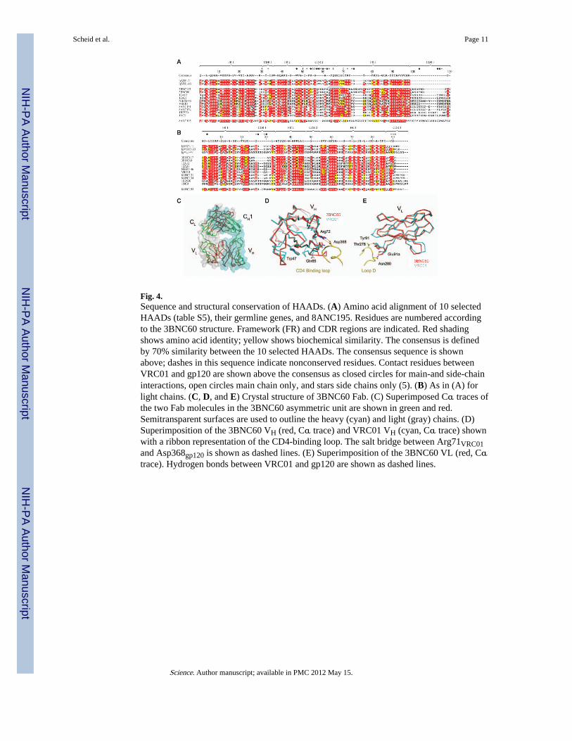
Fig. 4.Sequence and structural conservation of HAADs. (A) Amino acid alignment of 10 selectedHAADs (table S5), their germline genes, and 8ANC195. Residues are numbered accordingto the 3BNC60 structure. Framework (FR) and CDR regions are indicated. Red shadingshows amino acid identity; yellow shows biochemical similarity. The consensus is definedby 70% similarity between the 10 selected HAADs. The consensus sequence is shownabove; dashes in this sequence indicate nonconserved residues. Contact residues betweenVRC01 and gp120 are shown above the consensus as closed circles for main-and side-chaininteractions, open circles main chain only, and stars side chains only (5). (B) As in (A) forlight chains. (C, D, and E) Crystal structure of 3BNC60 Fab. (C) Superimposed Cα traces ofthe two Fab molecules in the 3BNC60 asymmetric unit are shown in green and red.Semitransparent surfaces are used to outline the heavy (cyan) and light (gray) chains. (D)Superimposition of the 3BNC60 VH (red, Cα trace) and VRC01 VH (cyan, Cα trace) shownwith a ribbon representation of the CD4-binding loop. The salt bridge between Arg71VRC01and Asp368gp120 is shown as dashed lines. (E) Superimposition of the 3BNC60 VL (red, Cαtrace). Hydrogen bonds between VRC01 and gp120 are shown as dashed lines.
Scheid et al. Page 11
Science. Author manuscript; available in PMC 2012 May 15.
NIH
-PA Author Manuscript
NIH
-PA Author Manuscript
NIH
-PA Author Manuscript
Copyright © 2022 FDOKUMEN