Annonaceous acetogenin mimic AA005 suppresses human ...
12
ARTICLE Annonaceous acetogenin mimic AA005 suppresses human colon cancer cell growth in vivo through downregulation of Mcl-1 Bing Han 1,2 , Yu-xia Cao 3 , Zhan-ming Li 1,2 , Zhao-xia Wu 1,2 , Yu-qin Mao 1,2 , Hui-ling Chen 1,2 , Zhu-jun Yao 4 and Li-shun Wang 1,2 Annonaceous acetogenins are a well-established family of natural products with significant bioactivities, especially high cytotoxic and antitumor activities. AA005 is an annonaceous acetogenin mimic that has shown significant cytotoxicity against a variety of cancer cell lines, but its in vivo antitumor effects have not been demonstrated so far, and its anticancer mechanisms remain ambiguous. In this study, we investigated the effects of AA005 on human colon cancer cell lines in vivo. Human colon carcinoma cell line SW620 xenograft nude mice were treated with AA005 (5 mg/kg/day, i.p.) for 21 days. AA005 administration markedly inhibited the tumor growth via promoting nuclear translocation of apoptosis-inducing factor (AIF) and inducing AIF-dependent cell death. Subsequent studies in human colon carcinoma cell lines SW620 and RKO in vitro revealed that after the colon cancer cells exposed to AA005, downregulation of a B-cell lymphoma 2 family protein, myeloid cell leukemia-1 (Mcl-1), was an early event due to the inhibition of Mcl-1 mRNA level and protein synthesis in a time-dependent manner. Intriguingly, knockdown of Mcl-1 using small interfering RNA markedly accelerated the nuclear translocation of AIF and upregulation of receptor interacting protein-1, and enhanced AA005-mediated lethality, whereas ectopic expression of Mcl-1 substantially attenuated AA005-mediated lethality in the colon cancer cells. Finally, silencing Mcl-1 expression markedly enhanced AA005-induced lethality in SW620 xenograft nude mice, demonstrating a pivotal role of Mcl-1 downregulation in mediating the in vivo antitumor effects of AA005. Taken together, this study demonstrates for the first time the anticancer effects of AA005 against human colon cancer cell lines in vivo, which is mediated through the downregulation of Mcl-1. Keywords: AA005; AIF; cell death; colon cancer; Mcl-1 Acta Pharmacologica Sinica (2019) 40:231–242; https://doi.org/10.1038/s41401-018-0025-7 INTRODUCTION Annonaceous acetogenins are a well-established family of natural products with significant bioactivities, especially high cytotoxic and antitumor activities [1, 2]. Over the past few years, Yao and co- workers [3] successfully developed a series of annonaceous acetogenin mimetics, and more interestingly, they found that some of these analogs have significant selectivity between human cancer cells and normal cells. In the current study, a new annonaceous acetogenin mimic, AA005, showed satisfactory selectivity and also the best antitumor activity against several human cancer cell lines among a set of mimetics of annonaceous acetogenin [4–7]. However, toxicity studies and the in vivo antitumor effects of AA005 have not been investigated thus far, creating an obstacle to its further development as a leading drug. Evidence supports that AA005 co-localizes with the mitochon- dria, can activate AMP-activated protein kinase and inhibit the mTOR complex 1 signal pathway, leading to growth inhibition and autophagy of cancer cells [8]. However, little is known of other signaling pathways involved in the regulation of AA005-induced cancer cell death. Interestingly, our recent study indicated that AA005 could induce apoptosis-inducing factor (AIF)-dependent but caspase-independent cell death [7]. However, the intrinsic cell death signaling that triggers AIF translocation to cause AA005- induced cell death remains ambiguous. In this work, we further clarify the molecular mechanisms of AA005 killing of cancer cells. Stress-induced cell death in mammalian cells is regulated by the B-cell lymphoma 2 (Bcl-2) family proteins through a series of orderly events in which mitochondrial outer membrane permea- bilization (MOMP) is critical. It appears that the pro-apoptotic family members Bax and Bak are crucial to inducing permeabiliza- tion of the outer mitochondrial membrane and the subsequent release of apoptogenic molecules such as cytochrome c, DIABLO, and AIF [9–12]. Anti-apoptotic family members such as Bcl-2 and B-cell lymphoma-extra large (Bcl-xL) inhibit Bax and Bak [13–15]. Myeloid cell leukemia-1 (Mcl-1) is also an anti-apoptotic Bcl-2 family protein frequently overexpressed or amplified in human cancers [16–18]. A number of recent studies suggest a critical role of Mcl-1 in tumor cell survival and therapeutic resistance [17, 19]. Selective overexpression of Mcl-1 in hematopoietic tissues of transgenic mice promotes the survival of hematopoietic cells and Received: 18 January 2018 Revised: 24 March 2018 Accepted: 28 March 2018 Published online: 19 June 2018 1 Minhang Branch, Zhongshan Hospital, Fudan University, 201199 Shanghai, China; 2 Institute of Fudan-Minhang Academic Health System, Minhang Hospital, Zhongshan Hospital, Fudan University, 201199 Shanghai, China; 3 Internal Medicine Department, Jinan Central Hospital, 250013 Jinan, China and 4 State Key Laboratory of Coordination Chemistry, School of Chemistry and Chemical Engineering, Nanjing University, 210093 Nanjing, China Correspondence: Zhu-jun Yao ([email protected]) or Li-shun Wang ([email protected]) www.nature.com/aps © CPS and SIMM 2018
-
Upload
khangminh22 -
Category
Documents
-
view
2 -
download
0
Transcript of Annonaceous acetogenin mimic AA005 suppresses human ...

ARTICLE
Annonaceous acetogenin mimic AA005 suppresses humancolon cancer cell growth in vivo through downregulation ofMcl-1Bing Han1,2, Yu-xia Cao3, Zhan-ming Li1,2, Zhao-xia Wu1,2, Yu-qin Mao1,2, Hui-ling Chen1,2, Zhu-jun Yao4 and Li-shun Wang1,2
Annonaceous acetogenins are a well-established family of natural products with significant bioactivities, especially high cytotoxicand antitumor activities. AA005 is an annonaceous acetogenin mimic that has shown significant cytotoxicity against a variety ofcancer cell lines, but its in vivo antitumor effects have not been demonstrated so far, and its anticancer mechanisms remainambiguous. In this study, we investigated the effects of AA005 on human colon cancer cell lines in vivo. Human colon carcinomacell line SW620 xenograft nude mice were treated with AA005 (5 mg/kg/day, i.p.) for 21 days. AA005 administration markedlyinhibited the tumor growth via promoting nuclear translocation of apoptosis-inducing factor (AIF) and inducing AIF-dependent celldeath. Subsequent studies in human colon carcinoma cell lines SW620 and RKO in vitro revealed that after the colon cancer cellsexposed to AA005, downregulation of a B-cell lymphoma 2 family protein, myeloid cell leukemia-1 (Mcl-1), was an early event dueto the inhibition of Mcl-1 mRNA level and protein synthesis in a time-dependent manner. Intriguingly, knockdown of Mcl-1 usingsmall interfering RNA markedly accelerated the nuclear translocation of AIF and upregulation of receptor interacting protein-1, andenhanced AA005-mediated lethality, whereas ectopic expression of Mcl-1 substantially attenuated AA005-mediated lethality in thecolon cancer cells. Finally, silencing Mcl-1 expression markedly enhanced AA005-induced lethality in SW620 xenograft nude mice,demonstrating a pivotal role of Mcl-1 downregulation in mediating the in vivo antitumor effects of AA005. Taken together, thisstudy demonstrates for the first time the anticancer effects of AA005 against human colon cancer cell lines in vivo, which ismediated through the downregulation of Mcl-1.
Keywords: AA005; AIF; cell death; colon cancer; Mcl-1
Acta Pharmacologica Sinica (2019) 40:231–242; https://doi.org/10.1038/s41401-018-0025-7
INTRODUCTIONAnnonaceous acetogenins are a well-established family of naturalproducts with significant bioactivities, especially high cytotoxicand antitumor activities [1, 2]. Over the past few years, Yao and co-workers [3] successfully developed a series of annonaceousacetogenin mimetics, and more interestingly, they found thatsome of these analogs have significant selectivity between humancancer cells and normal cells. In the current study, a newannonaceous acetogenin mimic, AA005, showed satisfactoryselectivity and also the best antitumor activity against severalhuman cancer cell lines among a set of mimetics of annonaceousacetogenin [4–7]. However, toxicity studies and the in vivoantitumor effects of AA005 have not been investigated thus far,creating an obstacle to its further development as a leading drug.Evidence supports that AA005 co-localizes with the mitochon-
dria, can activate AMP-activated protein kinase and inhibit themTOR complex 1 signal pathway, leading to growth inhibition andautophagy of cancer cells [8]. However, little is known of othersignaling pathways involved in the regulation of AA005-inducedcancer cell death. Interestingly, our recent study indicated that
AA005 could induce apoptosis-inducing factor (AIF)-dependentbut caspase-independent cell death [7]. However, the intrinsic celldeath signaling that triggers AIF translocation to cause AA005-induced cell death remains ambiguous. In this work, we furtherclarify the molecular mechanisms of AA005 killing of cancer cells.Stress-induced cell death in mammalian cells is regulated by the
B-cell lymphoma 2 (Bcl-2) family proteins through a series oforderly events in which mitochondrial outer membrane permea-bilization (MOMP) is critical. It appears that the pro-apoptoticfamily members Bax and Bak are crucial to inducing permeabiliza-tion of the outer mitochondrial membrane and the subsequentrelease of apoptogenic molecules such as cytochrome c, DIABLO,and AIF [9–12]. Anti-apoptotic family members such as Bcl-2 andB-cell lymphoma-extra large (Bcl-xL) inhibit Bax and Bak [13–15].Myeloid cell leukemia-1 (Mcl-1) is also an anti-apoptotic Bcl-2family protein frequently overexpressed or amplified in humancancers [16–18]. A number of recent studies suggest a critical roleof Mcl-1 in tumor cell survival and therapeutic resistance [17, 19].Selective overexpression of Mcl-1 in hematopoietic tissues oftransgenic mice promotes the survival of hematopoietic cells and
Received: 18 January 2018 Revised: 24 March 2018 Accepted: 28 March 2018Published online: 19 June 2018
1Minhang Branch, Zhongshan Hospital, Fudan University, 201199 Shanghai, China; 2Institute of Fudan-Minhang Academic Health System, Minhang Hospital, Zhongshan Hospital,Fudan University, 201199 Shanghai, China; 3Internal Medicine Department, Jinan Central Hospital, 250013 Jinan, China and 4State Key Laboratory of Coordination Chemistry,School of Chemistry and Chemical Engineering, Nanjing University, 210093 Nanjing, ChinaCorrespondence: Zhu-jun Yao ([email protected]) or Li-shun Wang ([email protected])
www.nature.com/aps
© CPS and SIMM 2018

enhances the outgrowth of myeloid cell lines [20]. Mcl-1 down-regulation is often sufficient to promote apoptosis in cancer cells,suggesting that Mcl-1 could be a potential therapeutic target inthe treatment of several human cancers [19, 21–23]. RIP kinasesconstitute a family of seven members, i.e., RIP1-7, that includesserine threonine kinases as crucial regulators of cell survival andcell death. Indeed, the kinase receptor interacting protein-1 (RIP-1)has been implicated in the regulation of apoptosis [24] andnecroptosis [25].In this study, we report for the first time that AA005 exhibits
in vivo antitumor activity by induction of cell death, which ismediated by downregulation of Mcl-1. The results indicate thatAA005 downregulates Mcl-1 expression through inhibition of thetranscriptional and translational mechanism. Furthermore, knock-down of Mcl-1 accelerates AIF translocation and upregulation ofRIP-1 and enhances AA005-mediated lethality, whereas ectopicexpression of Mcl-1 substantially attenuates AA005-mediatedlethality in these cells. Our in vivo results indicate that silencingof Mcl-1 expression markedly inhibits tumor growth and enhancesAA005-induced lethality, thus demonstrating a pivotal role of Mcl-1downregulation in mediating the antitumor effects of AA005. Thisstudy further elucidates the mechanism of AA005 as an antitumoragent and offers novel therapeutic clues for treating colon cancers.
MATERIALS AND METHODSChemicals and reagentsAnnonaceous acetogenin mimic AA005 (kindly supplied by theState Key Laboratory of Coordination Chemistry, Nanjing Uni-versity, Nanjing, China) was dissolved in dimethylsulfoxide as a 5mM stock solution and was stored at −80 °C. Antibodies againstAIF, Lamin B, Cox IV, Mcl-1, Bcl-2, Bcl-xL, Bax, and actin werepurchased from Cell Signaling Technology (Beverly, MA, USA).
Cell culture and treatmentsHuman colon carcinoma cell lines SW620 and RKO werepurchased from American Type Culture Collection (ATCC, Mana-ssas, VA, USA) and cultured in RPMI-1640 medium (Hyclone,Logan, UT, USA) supplemented with 10% heat-inactivated fetalbovine serum (Sigma-Aldrich, St. Louis, MO, USA) in a 5% CO2
humidified atmosphere at 37 °C. For experiments, cells wereseeded at 2–5 × 10⁵ cells/mL and incubated with or without theindicated concentrations of AA005.
Animal experimentsAnimal care and experiments were performed in strict accordancewith the “Guide for the Care and Use of Laboratory Animals” and“Principles for the Utilization and Care of Vertebrate Animals” andwere approved by the committee for humane treatment of animalsat Shanghai Jiao Tong University School of Medicine. Nude mice(5 weeks old) were purchased from Shanghai Model OrganismsCenter, Inc. (Shanghai, China). SW620 cells and shNC or shMcl-1SW620 cells (1 × 10⁶/0.2 mL per mouse) were suspended in sterilePBS and injected subcutaneously into the mice. Seven days aftertumor inoculation, the treatment group received AA005 (5mg/kg)intraperitoneally for 21 days. The control group received an equalvolume of solvent control. Tumor size and body weight weremeasured every day. Tumor volumes were determined by caliperand calculation according to the formula (width2 × length)/2. Allanimals were killed immediately after 21 days of drug exposure.
Immunohistochemical stainingImmunohistochemical (IHC) staining was applied to detect theprotein levels of Mcl-1 and subcellular localization of AIF betweendifferent tumor tissues with anti-Mcl-1 (Abcam, Cambridge, MA,USA, ab32087) or anti-AIF (Cell Signaling Technology, Danvers,MA, USA, #4642) antibodies. IHC staining was performed accord-ing to the manufacturer’s protocol.
Subcellular cell fractionation and mitochondria purificationCells were separated into different fractions according topreviously described methods [26, 27]. In brief, cells wereharvested and rinsed with mitochondria isolation buffer (MIB,0.25 M sucrose and 10 mM Tris–HCl, pH 7.4), resuspended inMIB supplemented with 1% protease inhibitor cocktail andhomogenized using a glass Dounce homogenizer (Kontes,Sigma-Aldrich) with 20 strokes at 4 °C. The homogenate wascentrifuged at 1000×g for 10 min to pellet the nucleus. Thesupernatant was centrifuged at 15,000×g for 20 min at 4 °C topellet the raw mitochondria. Cytoplasmic proteins in the post-mitochondria supernatant were precipitated with chloroformand methanol according to Klotz’s methods [28]. To furtherenrich the mitochondria, the pellet of raw mitochondria wasresuspended in 36% iodixanol (Sigma-Aldrich) and overlaidwith 30 and 10% iodixanol. The gradient was ultracentrifuged(80,000×g, 3 h) at 4 °C. The purified mitochondria ere collectedat the interface between 10 and 30% iodixanol and washedtwice with MIB. Samples of each fraction were prepared byaddition of sample buffer and subjected to immunoblotanalysis.
Western blotsCells were rinsed with ice-cold 1×PBS and lysed in non-reducing buffer containing 100 mM Tris–HCl, pH 6.8, 2% SDS,50 mM IAA, supplemented with PMSF and cocktail, or reducingbuffer containing an additional 100 mM DTT. Cell lysates wereseparated on a 8–12.5% SDS-polyacrylamide gel, transferred toa nitrocellulose membrane (Bio-Rad, Richmond, CA), blockedwith 5% nonfat milk in PBS, and immunoblotted with theindicated antibodies. After incubation with the horseradishperoxidase-linked second antibody (Cell Signaling, Beverly,MA, USA) at room temperature, detection was performedusing the SuperSignal West Pico Chemiluminescent SubstrateKit (Pierce, Rockford, IL) according to the manufacturer’sinstructions.
Apoptosis assayApoptotic cells were measured by staining with FITC-conjugatedAnnexin-V/propidium iodide (PI) (BD Pharmingen, San Diego, CA,USA) and determination via flow cytometer according to themanufacturer’s instructions, as described previously [29]. Bothearly apoptotic (Annexin-V-positive, PI-negative) and late apopto-tic (Annexin-V-positive and PI-positive) cells were included in celldeath determinations.
TUNEL assayCells were seeded in 6-well plates 1 day prior to treatments.Apoptosis in tumor tissue sections was determined using the InSitu Cell Death Detection kit (Roche, Mannheim, Germany). Inbrief, tumor tissue sections of formalin-fixed paraffin-embeddedspecimens were dewaxed in xylene and rehydrated in a gradedseries of ethanol. The tumor samples were incubated withproteinase K (2 mg/mL), and the TdT mediated dUTP-biotin nick-end labeling (TUNEL) staining was performed according to themanufacturer’s instructions.
Real-time quantitative RT-PCRTotal RNA from the cell lines were extracted with TRIzol reagent(Invitrogen, Carlsbad, CA) and treated with RNase-free DNase(Promega, Madison, WI, USA). Quantitative real-time PCR wasperformed using the SYBR Green PCR Master Mix (AppliedBiosystems, Foster City, CA, USA). The primers used are listed asfollows: Mcl-1-5′- GTGCCTTTGTGGCTAAACACT-3′(forward), 5′-AGT-CCCGTTTTGTCCTTACGA-3′(reverse), with β-actin-5′-CATCCTCAC-CCTGAAGTACCC-3′ (forward), 5′-AGCCTGGATAGCAACGTACATG-3′(reverse). Real-time RT-PCR was performed, and the data wereanalyzed according to a previous report [30].
AA005 suppresses cancer cell growth in vivo through Mcl-1B. Han et al.
232
Acta Pharmacologica Sinica (2019) 40:231 – 242
1234567890();,:

Plasmids and shRNAsPlasmids for knockdown were constructed by inserting thecorresponding shRNA sequences into the pGIPz plasmid (Dharma-con). The shRNA sequences are listed as follows: TAGCGGTCGCC-GAAATGTT (shAIF#1), CTGGTATCCGATCAGAGAG (shAIF#2),TACAAATACATTTACAAGC (shMcl-1#1), and TGTTAGCCATAATCCTCTT(shMcl-1#2). Plasmids expressing Flag-tagged Mcl-1 were generatedby inserting the corresponding coding sequence into a pBabe-Flagvector (Clontech, CA, USA). After co-transfection of lentiviral packagingplasmids into 293T cells, lentivirus-containing supernatants wereharvested 48 h after transfection. SW620 or RKO cells were infectedwith the lentiviral supernatant and selected by puromycin.
Statistical analysisAll in vitro experiments were repeated at least three times withsimilar results. The results are expressed as the mean ± S.D. TheP-values for comparison between groups were obtained viaStudent’s t-test. All statistical tests were two-sided, and P-value <0.05 (*) was considered to be statistically significant.
RESULTSAA005 exhibits antitumor activity in xenografts of colon cancerSW620 cellsTo examine the antitumor activity of AA005 in vivo, athymic nudemice bearing implanted xenografts of colon cancer cell lineSW620 were treated with AA005 (Fig. 1a) or vehicle control.Treatment with AA005 resulted in a dramatic suppression oftumor growth 13 days following drug exposure (Fig. 1b). Incontrast, no significant change in body weight was noted in acomparison of the vehicle control and AA005 regimen (Fig. 1c),indicating that no severe toxicity was observed. At the end of thegrowth period, the mean tumor volume was 3.7 times higher forthe control group than the AA005-treated group (1178 vs. 318mm3, P= 0.019; Fig. 1b, d and e), with a mean tumor weight thatwas three times higher for the control group than the AA005-treated group (0.6 vs. 0.2 g, P= 0.005; Fig. 1f). The averageinhibitory rate of tumor growth was 67.5% (Fig. 1g). Thustreatment with AA005 produced obvious tumor growth inhibitioneffects without toxicity to the animals.
Fig. 1 AA005 exhibits antitumor activity in the xenograft animal model. a Structure of compound AA005. b 5-week-old nude mice inoculatedsubcutaneously with SW620 cells (1 × 10⁶/0.2 mL per mouse). After 1 week, mice were treated with 5 mg/kg AA005 daily (intraperitoneally) orwith the vehicle control for 21 consecutive days, and the tumor volumes were calculated (6 mice per group). c Body weight of mice.d Photograph of tumors in nude mice at the end of the experiment. Scale bars, 1 cm. e Tumors of each group when nude mice were killed.Scale bars, 1 cm. f The average tumor weight of each group is expressed as the mean ± S.D. g The average inhibitory rate of tumor growthwas calculated. The inhibitory rate of tumor growth = (1− tumor weight of AA005 group/tumor weight of control group) × 100%. *P < 0.05;**P < 0.01 vs. the vehicle control group
AA005 suppresses cancer cell growth in vivo through Mcl-1B. Han et al.
233
Acta Pharmacologica Sinica (2019) 40:231 – 242

Treatment with AA005 results in cell death and translocation ofAIF in the xenograftsTo validate the antitumor effect of AA005, we first determined thecell death in the tumor tissue of the colon cancer xenograft usingTUNEL assay. The TUNEL-positive cells of the tumor sections weresignificantly increased in AA005-treated SW620 xenograft micecompared with the control group (Fig. 2a, b). In contrast, a weakreduction in cell proliferation was noted in the AA005-treatedgroup compared with the vehicle control, as indicated by Ki-67staining (Fig. 2c), suggesting that AA005 inhibited tumor growthprimarily through induction of cell death.Our previous findings suggested that AA005 triggered caspase-
3-independent cell death mediated by AIF in vitro [7], and thus weaimed to explore the effect of AIF on AA005-administered mice.Notably, nuclear translocation of AIF in tumor sections of SW620xenograft mice increased upon AA005 treatment, as shown by IHCstaining (Fig. 2d, e). Moreover, the increased level of AIF wasdetermined in the nucleus of tumor fractions derived from thenude mice treated with AA005 as observed in Western blots(Fig. 2f). Together, these results indicated that AIF potentiallycontributes to AA005-induced cell death in vivo.
Exposure of colon cancer cells to AA005 results in thedownregulation of Mcl-1To elucidate the mechanisms of AA005-induced cell death, weadministered AA005 to colon cancer cell line SW620, followedby cell viability analysis with trypan-blue exclusion assays. A
dose-dependent study revealed that the percentages of dead cellswere 22.62 ± 2.89%, 53.89 ± 1.79%, 66.73 ± 2.40%, and 75.00 ±1.33% when SW620 cells were treated for 48 h with 0.2, 1, 5, and25 μM AA005, respectively (Fig. 3a). Time-course analysis includingtreatment with AA005 for 12, 24, and 48 h showed that the celldeath-inducing activity of AA005 was also time-dependent(Fig. 3a). Furthermore, Annexin-V/PI double stain-based flowcytometry analysis indicated that although nearly 60% of cellshad died at 48 h with 1 μM AA005 treatment, no obvious Annexin-V+/PI− cells were detected throughout this process (Fig. 3b),indicating that AA005-induced cell death does not representclassical apoptosis. TUNEL analysis [31] of the 1 μM AA005treatment group showed increased TUNEL-positive cells, indicat-ing that AA005 induced DNA fragmentation (Fig. 3c). Subcellularfractionation assays also showed AIF translocation into thenucleus after treatment with AA005, which was consistent withthe results in vivo (Fig. 3d). Thus AA005 promoted AIF nucleartranslocation and triggered AIF-dependent cell death in vitro.However, the intrinsic cell death signaling that triggers AIF
translocation to cause AA005-induced cell death remains ambig-uous. We sought to further clarify the molecular mechanisms ofAA005 killing of cancer cells. Reportedly, anti-apoptotic Bcl-2family proteins play an important role in the regulation of celldeath by AIF [32]. The effects of AA005 on the expression of Bcl-2family proteins were examined in SW620 cells. Exposure of cells to1 μM AA005 for the indicated time resulted in a significant time-dependent decrease in levels of Mcl-1. Furthermore, translocation
Fig. 2 Treatment with AA005 results in cell death and translocation of AIF in the xenograft animal model. a Tumors were obtained from mice21 days after drug exposure. Tumors were fixed and stained with hematoxylin and eosin (H&E) and assayed via TUNEL and Ki-67. Scale bars,50 μm. b, c Image J software was used to quantify the TUNEL-positive cells (b) or Ki-67 positive cells (c) between the control and AA005-treated group tumors. All bar graphs are plotted as the mean ± S.D. P-values are calculated between linked groups. *P < 0.05; ***P < 0.001.d Representative images of IHC staining of AIF in colon xenografts derived from vehicle- or AA005-treated SW620 cells. Lower panels show anenlarged picture of the boxed area. Scale bars, 50 μm. e Image J software was used to quantify the AIF translocation cells between the controland AA005-treated group tumors. f Analysis of AIF translocation by subcellular fractionation. Western blots for the indicated proteins werederived from colon xenograft tumors with or without AA005 treatment. COX IV and Lamin B were used as mitochondrial and nuclear markerproteins, respectively
AA005 suppresses cancer cell growth in vivo through Mcl-1B. Han et al.
234
Acta Pharmacologica Sinica (2019) 40:231 – 242

of Bax from the cytosol to mitochondria was noted in cells treatedwith AA005. In contrast, exposure of cells to AA005 did notdiscernibly modify the expression of other Bcl-2 family proteins,including Bcl-2 and Bcl-xL (Fig. 3e).To determine whether the AA005-mediated lethality observed
in SW620 cells also occurs in other colon cancer cell lines,parallel studies were performed in RKO cells. Consistent with thefindings in SW620 cells, the same AA005 concentrations andexposure intervals resulted in a pronounced increase in celldeath (Fig. 4a) and non-canonical apoptotic cell death (Fig. 4b).Similarly, AA005 induced an increase in TUNEL-positive RKO cells(Fig. 4c). These events were also accompanied by nuclear AIFaccumulation (Fig. 4d), downregulation of Mcl-1 and transloca-tion of Bax (Fig. 4e). These findings indicated that AA005-induced cell death is in association with Mcl-1 downregulationin colon cancer cells.
Downregulation of Mcl-1 by AA005 proceeds through inhibition oftranscription and translationTo elucidate the mechanism underlying Mcl-1 downregulationby AA005, Mcl-1 mRNA expression in AA005-treated cells wasquantified using real-time RT-PCR. Notably, treatment of SW620cells with 1 μM AA005 resulted in a significant decrease in theexpression levels of Mcl-1 mRNA after 4 h of drug exposure(Fig. 5a). Similarly, the expression levels of Mcl-1 mRNA werereduced after AA005 treatment for 6 h in RKO cells
(Supplementary Figure S1). Such findings suggested thatAA005 downregulated Mcl-1 protein through inhibition oftranscription.To determine whether the downregulation of Mcl-1 mediated
by AA005 occurs through the proteasome pathway, SW620 cellswere pretreated with or without AA005 for 4 h, followed bytreatment with proteasome inhibitor MG132 for the indicatedtime (Fig. 5b), which demonstrated that treatment with MG132alone did not affect the protein level of Mcl-1 and that thecombination of AA005 and MG132 did not block the down-regulation of Mcl-1 mediated by AA005. These findings suggestthat downregulation of Mcl-1 mediated by AA005 does not occurthrough the proteasome pathway.We also investigated the effect of protein synthesis inhibitor
cycloheximide (CHX) on the downregulation of Mcl-1 mediated byAA005. The protein level of Mcl-1 was significantly decreased afterCHX treatment for 4 h, but no obvious changes were noted at 2 h.However, the combination treatment of AA005 and CHX showed asignificant decrease of Mcl-1 at 2 h, indicating that AA005 possiblyreduces the half-life of Mcl-1 by suppressing the synthesis of thisprotein (Fig. 5c).
Mcl-1 potentially contributes to AA005-induced AIF-dependentcell deathTo address whether Mcl-1 regulates AIF translocation induced byAA005, two pairs of shRNAs (shAIF#1 and shAIF#2) targeted
Fig. 3 AA005 induces AIF-dependent parthanatos in SW620 cells. a SW620 cells were treated with AA005 at the indicated concentrations. Celldeath was measured by trypan-blue exclusion assay. *P < 0.05; **P < 0.01; ***P < 0.001, vs. vehicle control group. b SW620 cells were treatedwith 1 μM AA005 for 24 or 48 h. Cells were stained with Annexin-V/PI, and the percentage of dead cells was determined using flow cytometry.c TUNEL analysis of SW620 cells treated with or without 1 μM AA005. Images were viewed using an Olympus BX-51 fluorescence microscope.Scale bars: 20 μm. **P < 0.01. d, e After treatment with AA005 for the indicated time, total cellular extracts and subcellular fractionations weresubjected to Western blot analysis using antibodies against AIF, Mcl-1, Bcl-2, Bcl-xL, and Bax. COX IV (mitochondrial fractions; M), Lamin B(nuclear extracts; N), and actin (cytosolic fractions; C) were used to ensure equivalent loading
AA005 suppresses cancer cell growth in vivo through Mcl-1B. Han et al.
235
Acta Pharmacologica Sinica (2019) 40:231 – 242

specifically against AIF were used to knockdown AIF expressiontogether with the non-specific scramble shRNA (shNC) as anegative control. shRNA but not shNC significantly silenced AIFexpression (Fig. 6a), whereas AIF knockdown failed to affect thedownregulation of Mcl-1 evoked by AA005 (Fig. 6a). In accor-dance, SW620 cells were stably transfected with a lentivirusharboring shNC or shRNA (shMcl-1#1) against Mcl-1. The absenceof Mcl-1 expression was confirmed by Western blot analysis(Fig. 6b). Notably, knockdown of Mcl-1 in SW620 cells acceleratedAIF translocation and a slight increase in Bax translocation to themitochondria after AA005 treatment compared with control cells(Fig. 6c, d). These observations implied that Mcl-1 downregulationwas required for AIF translocation from the mitochondria to thenucleus and that Mcl-1 was potentially a crucial upstreamregulator of AIF in AA005-induced cell death.
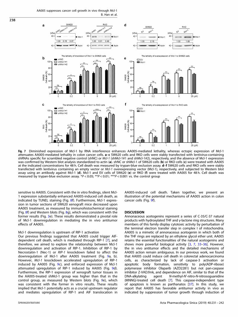
Diminished expression of Mcl-1 by RNA interference enhancesAA005-mediated lethalityTo further confirm the functional role of Mcl-1 in AA005-mediatedlethality in colon cancer cells, shRNAs targeted specifically againstMcl-1 were used to knockdown Mcl-1 expression together withshNC as a negative control in SW620 and RKO cells. shRNA but notshNC significantly silenced Mcl-1 expression (Fig. 7a). Knockdownof Mcl-1 in SW620 cells increased cell death by ~30% at each doseof AA005 compared with control cells (Fig. 7b), and similar
findings were observed in RKO cells (Fig. 7c). These data indicatedthat Mcl-1 had a critical role in AA005-mediated cell death.
Ectopic expression of Mcl-1 attenuates AA005-mediated lethalityTo determine whether downregulation of Mcl-1 has a functionalrole in AA005-induced cell death, SW620 and RKO cells that stablyoverexpressed Mcl-1 were selected (Fig. 7d). Overexpression ofMcl-1 in the two cell lines displayed more than two-fold increasesin Mcl-1 protein levels compared with the empty vector control(EV) (Fig. 7d). Significantly, enforced expression of Mcl-1 attenu-ated AA005-mediated cell death both in SW620 (Fig. 7e) and RKOcells (Fig. 7f). Taken together, these findings indicated that Mcl-1downregulation has a significant functional role in AA005-mediated lethality.
Mcl-1 downregulation mediates the in vivo antitumor effects ofAA005Given that Mcl-1 downregulation had a significant functional rolein AA005-mediated lethality in vitro, whether Mcl-1 downregula-tion also affected tumor growth in vivo was further investigated.To this end, we subcutaneously injected shNC or shMcl-1#1-infected SW620 cells into the flanks of nude mice and treatedthem with AA005 or vehicle control. Consistent with the antitumoreffects in colon cancer cells (Fig. 7b, c), the tumor growthinhibitory effect of AA005 was significantly enhanced in the silent
Fig. 4 AA005 induces AIF-dependent parthanatos in RKO cells. a RKO cells were treated with AA005 at the indicated concentrations for 12, 24,or 48 h. Cell death was measured by trypan-blue exclusion assay. **P < 0.01; ***P < 0.001 vs. the vehicle control group. b RKO cells were treatedwith 1 μM AA005. Cells were stained with Annexin-V/PI, and the percentage of dead cells was determined using flow cytometry. c TUNELanalysis of RKO cells treated with or without 1 μM AA005. Images were viewed using an Olympus BX-51 fluorescence microscope. Scale bars:20 μm. **P < 0.01. d, e After treatment with AA005 for the indicated time, total cellular extracts and subcellular fractionations were preparedand subjected to Western blot analysis using antibodies against AIF, Mcl-1 and Bax. COX IV (mitochondrial fractions; M), Lamin B (nuclearextracts; N), and actin (cytosolic fractions; C) were used to ensure equivalent loading
AA005 suppresses cancer cell growth in vivo through Mcl-1B. Han et al.
236
Acta Pharmacologica Sinica (2019) 40:231 – 242

Mcl-1 expression group compared with the control group (Fig. 8a).No significant change in body weight was noted compared withthe control group and the shMcl-1 group with or without AA005treatment (Fig. 8b), indicating that no severe toxicity to theanimals occurred.At the end of the growth period, the mean tumor volume was
5.4 times higher for the vehicle-treated shNC group than theAA005-treated shMcl-1 group (1446 vs. 267 mm3, P= 0.0001) and
2.5 times higher for the AA005-treated shNC group than theAA005-treated shMcl-1 group (663 vs. 267 mm3, P= 0.0026)(Fig. 8a, c). The mean tumor weight was 4.2 times higher for thevehicle-treated shNC group than the AA005-treated shMcl-1group (0.65 vs. 0.15 g, P= 0.0002) and three times higher for theAA005-treated shNC group than the AA005-treated shMcl-1 group(0.45 vs. 0.15 g, P= 0.0012) (Fig. 8d, e). Compared with the controltumors, silent Mcl-1 expression tumors were significantly more
Fig. 5 AA005 induces Mcl-1 downregulation through inhibition of transcription and translation in SW620 cells. a SW620 cells were treatedwith 1 μM AA005 at the designated intervals, after which total RNA were isolated, and Mcl-1 mRNA were quantified using real-time reversetranscription-polymerase chain reaction (RT-PCR). **P < 0.01 vs. control group. b, c SW620 cells were pretreated with or without AA005 (1 μM)for 4 h followed by MG132 (1 μM) or CHX (2 μM) treatment for the indicated time. Total cellular extracts were prepared and subjected toWestern blot assay using antibodies against Mcl-1 and actin
Fig. 6 Mcl-1 is an upstream regulator of AIF in AA005-induced cell death. a SW620 cells were stably transfected with a lentivirus-containingshRNA specific for scrambled negative control (shNC) or AIF (shAIF#1 and shAIF#2). After treatment with AA005 for the indicated time, totalcellular extracts were subjected to Western blot assay using antibodies against AIF and Mcl-1 and standardized to actin. b–d SW620 cells werestably transfected with lentivirus-containing shNC or Mcl-1 (shMcl-1#1), and the absence of Mcl-1 expression was confirmed by Western blotanalysis standardized to actin (b), shNC (c), and shMcl-1#1 (d). Cells were treated with 1 μM AA005 for the indicated time, and subcellularfractionations were subjected to Western blot assay using antibodies against AIF and Bax. COX IV (mitochondrial fractions; M), Lamin B(nuclear extracts; N), and actin (cytosolic fractions; C) were used to ensure equivalent loading
AA005 suppresses cancer cell growth in vivo through Mcl-1B. Han et al.
237
Acta Pharmacologica Sinica (2019) 40:231 – 242
sensitive to AA005. Consistent with the in vitro findings, silent Mcl-1 expression substantially enhanced AA005-induced cell death, asindicated by TUNEL staining (Fig. 8f). Furthermore, Mcl-1 expres-sion in tumor sections of SW620 xenograft mice decreased uponAA005 treatment, as measured by immunohistochemical staining(Fig. 8f) and Western blots (Fig. 8g), which was consistent with theformer results (Fig. 3e). These results demonstrated a pivotal roleof Mcl-1 downregulation in mediating the in vivo antitumoreffects of AA005.
Mcl-1 downregulation is upstream of RIP-1 activationOur previous findings suggested that AA005 could trigger AIF-dependent cell death, which is mediated through RIP-1 [7], andtherefore, we aimed to explore the relationship between Mcl-1downregulation and activation of RIP-1. Inhibition of RIP-1 byNecrostatin-1 (Nec-1) or RIP-1 knockdown failed to affect thedownregulation of Mcl-1 after AA005 treatment (Fig. 9a, b).However, Mcl-1 knockdown accelerated upregulation of RIP-1induced by AA005 (Fig. 9c), and enforced expression of Mcl-1attenuated upregulation of RIP-1 induced by AA005 (Fig. 9d).Furthermore, the RIP-1 expression of xenograft tumor tissues inthe AA005-treated shMcl-1 group was higher than that in thecontrol group, as measured by Western blots (Fig. 9e), whichwas consistent with the former in vitro results. These resultsimplied that Mcl-1 potentially acts as a crucial upstream regulatorand mediates upregulation of RIP-1 and AIF translocation in
AA005-induced cell death. Taken together, we present anillustration of the potential mechanisms of AA005 action in coloncancer cells (Fig. 9f).
DISCUSSIONAnnonaceous acetogenins represent a series of C-35/C-37 naturalproducts with hydroxylated THF and γ-lactone ring structures. Manymembers of this family display cytotoxic activity by perturbation ofthe terminal electron transfer step in complex I of mitochondria.AA005 is a mimetic of annonaceous acetogenin in which both ofthe THF rings are replaced by an ethylene glycol ether unit. AA005retains the essential functionalities of the natural acetogenins andshows more powerful biological activity [3, 7, 33–36]. However,the in vivo antitumor effects and the detailed mechanisms ofAA005 action remain ambiguous. In our previous work, we foundthat AA005 could induce cell death in colorectal adenocarcinomacells, as characterized by lack of caspase-3 activation orapoptotic body formation, sensitivity to poly(ADP-ribose)polymerase inhibitor Olaparib (AZD2281) but not pan-caspaseinhibitor Z-VAD.fmk, and dependence on AIF, similar to that of theDNA-alkylating agent N-methyl-N′-nitro-N-nitrosoguanidine(MNNG)-treated cell death [7]. This caspase-independent typeof apoptosis is known as parthanatos [37]. In this study, wereport that AA005 has favorable antitumor activity in vivo asindicated by suppression of tumor growth through induction of
Fig. 7 Diminished expression of Mcl-1 by RNA interference enhances AA005-mediated lethality, whereas ectopic expression of Mcl-1attenuates AA005-mediated lethality in colon cancer cells. a–c SW620 cells and RKO cells were stably transfected with lentivirus-containingshRNAs specific for scrambled negative control (shNC) or Mcl-1 (shMcl-1#1 and shMcl-1#2), respectively, and the absence of Mcl-1 expressionwas confirmed by Western blot analysis standardized to actin (a). shNC or shMcl-1 of SW620 cells (b) or RKO cells (c) were treated with AA005at the indicated concentrations for 48 h. Cell death was measured by trypan-blue exclusion assay. d–f SW620 cells and RKO cells were stablytransfected with lentivirus containing an empty vector or Mcl-1 overexpressing vector (Mcl-1), respectively, and subjected to Western blotassay using an antibody against Mcl-1 (d). Mcl-1 and EV cells of SW620 (e) or RKO (f) were treated with AA005 for 48 h. Cell death wasmeasured by trypan-blue exclusion assay. *P < 0.05; **P < 0.01; ***P < 0.001 vs. the control group
AA005 suppresses cancer cell growth in vivo through Mcl-1B. Han et al.
238
Acta Pharmacologica Sinica (2019) 40:231 – 242

AIF-dependent cell death. The results also strongly support ourrecent study [7].Directed induction of cell death could offer therapeutic benefits
for cancer treatment. Such treatments primarily target the caspasepathways to induce apoptosis [26, 38–40]. However, caspaseactivation might be dispensable for certain types of apoptosis, andincreasing attention has been drawn to key molecules involved in
non-apoptotic cell death or caspase-independent apoptosis [41–48].The mitochondrial protein AIF is a new therapeutic target involvedin most of the caspase-independent apoptosis systems, includingprogrammed necrosis [38, 49–52]. As a core executor in caspase-independent cell death, AIF has been intensively studied [41], butmany study results are highly controversial. We suggest that AA005is an effective chemical probe for examining the role of AIF.
Fig. 8 Mcl-1 downregulation mediates the in vivo antitumor effects of AA005. a 5-week-old nude mice were inoculated subcutaneouslywith shNC or shMcl-1#1-infected SW620 cells (1 × 10⁶/0.2 mL per mouse). After 1 week, mice were treated with 5 mg/kg AA005daily (intraperitoneally) or with the vehicle control for 21 consecutive days. The tumor volume was calculated (6 mice per group). *P < 0.05;**P < 0.01; ***P < 0.001 vs. the vehicle control group. b Body weight of mice during the 21 days of drug treatment. c Representative image oftumors in nude mice at the end of the experiment. Scale bars, 1 cm. d Tumors of each group when nude mice were killed (6 mice per group).Scale bars, 1 cm. e The average tumor weight of each group is expressed as the mean ± S.D. (6 mice per group). f Tumors were obtained frommice, fixed, and stained with hematoxylin and eosin (H&E), assayed via TUNEL, and the levels of MCL-1 were determined usingimmunohistochemistry. Scale bars, 50 μm. g Western blots for the expression levels of MCL-1 in each tumor group
AA005 suppresses cancer cell growth in vivo through Mcl-1B. Han et al.
239
Acta Pharmacologica Sinica (2019) 40:231 – 242

Furthermore, AA005 might represent the basis of a novel treatmentfor cancers that are resistant to classical apoptotic reagents.Notably, our results demonstrate for the first time that down-
regulation of Mcl-1 has an important role in AA005-mediatedlethality. One of the hallmarks of cancer is its resistance to apoptosis,which maintains survival of cells en route to oncogenic transforma-tion [53]. Overexpression or amplification of Mcl-1 is one of the mostfrequent alterations in human cancers [54]. Defective Mcl-1degradation allows tumor cells to evade the fate of death andunderlies development of therapeutic resistance. The developmentof anticancer agents that diminish Mcl-1 protein levels has been thefocus of intense interest [55, 56]. Indeed, a number of studies havedocumented Mcl-1 downregulation during apoptosis by a variety ofagents, including ultraviolet (UV) [57], kinase inhibitor BAY 43-9006[58], and growth factor withdrawal [59], among others. Our workmight offer new clues for cancer therapy and treatment thatcombines AA005 with other agents targeting Mcl-1.It has been shown that several cyclin-dependent kinase inhibitors
(including SU9516, flavopiridol, and seliciclib) downregulate Mcl-1expression in cancer cells through inhibition of RNA polymerase
(Pol) II, leading to transcriptional repression [60–62]. Furthermore,SU9516-induced inhibition of Mcl-1 transcription and phosphoryla-tion of RNA Pol II is reactive oxygen species (ROS) dependent. Ourprevious study showed that AA005 treatment resulted in a markedincrease in ROS production at the early stage, which was diminishedtogether with cell death by the free radical scavenger N-acetylcysteine [7]. On the basis of these data, we hypothesize thatAA005 kills colorectal adenocarcinoma cells sequentially throughinhibition of phosphorylation of the carboxyl-terminal domain ofRNA Pol II in association with oxidative damage and downregulationof Mcl-1 at the transcriptional level, culminating in mitochondrialinjury and cell death. In addition, it is highly useful to reveal whichprotein is directly regulated by AA005 during the cell death throughchemical proteomics, small molecule/protein crystal structureanalysis and/or bioinformatics.Cellular changes leading to inhibition of programmed cell death
have an essential role in tumor development. Moreover,parthanatos represents AIF-induced DNA damage-related celldeath. During this process, AIF is released from the mitochondriaby a mechanism implicating MOMP [63], ROS production [64], or
Fig. 9 Mcl-1 downregulation is upstream of RIP-1 activation and a sketch of mechanisms of AA005 action in colon cancer cells.a Immunoblotting analysis of the expressional level of Mcl-1 with 1 μM AA005 treatment for the indicated time after pretreatment with orwithout RIP-1 inhibitor Necrostatin-1 (Nec-1; 100 μM). b SW620 cells were stably transfected with lentivirus-containing small interfering RNA(siRNA) specific for the control (siControl) or RIP-1 (siRIP-1). After AA005 treatment, expression of the indicated proteins was confirmed byWestern blot analysis. c SW620 cells were stably transfected with lentivirus-containing siControl or siRNA specific for Mcl-1 (siMcl-1). Cells weretreated with 1 μM AA005 for the indicated time. Mcl-1 and RIP-1 were examined via Western blots standardized to actin. d SW620 cells werestably transfected with lentivirus containing the empty vector (EV) or Mcl-1 overexpressing vector (Mcl-1). After AA005 treatment, expressionof the indicated proteins was confirmed by Western blots. eWestern blots for the expression levels of RIP-1 in each group tumor standardizedto actin. f A sketch of the mechanisms of AA005 action in colon cancer cells
AA005 suppresses cancer cell growth in vivo through Mcl-1B. Han et al.
240
Acta Pharmacologica Sinica (2019) 40:231 – 242

RIP-1/TRAF2-mediated Jun N-terminal protein kinase 1 activation[65]. It appears that Bcl-2 family members are crucial to inducingMOMP and the subsequent release of apoptogenic molecules,thus leading to cell death. Bak resides on the outer mitochondrialmembrane in healthy cells, where it has been reported as boundto Mcl-1 [66] and Bcl-xL. With certain cytotoxic stimuli (such asAA005) induction, Mcl-1 is degraded or downregulated, and theMcl-1–Bak interactions are disrupted by BH3-only proteins such asNOXA, BIM, or BIK [67], which frees Bak. At the same time, Baxtranslocates from the cytosol to the mitochondria. Bax and Bakform pores in the membranes and facilitate the release of AIF fromthe mitochondrial intermembrane space to the cytosol. Once AIFtranslocates to the nucleus, it generates DNA breaks, chromatincondensation and the irreversible cell death of parthanatos(Fig. 9f). However, little is known of the upstream mechanismsthat regulate AIF release from mitochondria. Previous reports haveshown that calpain controls mitochondrial AIF release duringMNNG-induced death [68]. Our data suggest that this processrequires the cooperative upstream action of Mcl-1 and Bax. Mcl-1liberates Bak, yielding MOMP, and Bax facilitates the MOMPnecessary for AIF release. Consequently, knockdown of Mcl-1accelerated AIF translocation, increased Bax translocation tomitochondria, and enhanced AA005-mediated lethality (Fig. 6d).AIF-mediated and PARP-1-dependent parthanatos is caspase-
independent and lacks many morphological features of classicapoptosis [69]. However, unlike “accidental” necrosis, it has recentlybeen reported as regulated necrosis [70]. RIP-1 is known as a majormediator in regulation of necrotic cell death [71]. Consistent withthis view, data from our study suggest that caspase-independentcell death induced by AA005 also requires the function of RIP-1. Inthis work, we reveal that Mcl-1 and RIP-1 are upstream of AIF inAA005-induced parthanatos because AA005-induced nuclear AIFaccumulation was attenuated in Mcl-1–/– and RIP-1 inhibitor Nec-1-treated SW620 cells. Furthermore, Mcl-1 is upstream of RIP-1because it influences RIP-1 activity. Our previous work found thatAA005 treatment increased the intracellular concentrations of ROS.In fact, it has been reported that RIP-1 could modulate oxidativestress in AIF-dependent cell death. Therefore, we speculate thatAA005 might disrupt mitochondrial function by downregulation ofMcl-1, thus triggering ROS, RIP-1, and the AIF-dependent pathway.This work offers a new clue to the action of AA005.The mitochondria manage energy generation via citric acid cycle
and also play a key role in apoptosis regulation through release ofcytochrome c. Although much debate has focused on whethermitochondria contain defects in the oxidative phosphorylationpathway, they could offer a potential target for cancer therapy [72].Mitochondria complex I has been shown to be targeted byannonaceous acetogenin. On the basis of our findings in colon cancercells, we propose a model in which AA005 induces downregulation ofMcl-1 as an early event. Bax is translocated to the mitochondria andRIP-1 is upregulated, leading to the release of the apoptogenicmolecule AIF into the cytosol, followed by its translocation to thenucleus, where it contributes to large-scale DNA fragmentation andirreversible cell death (Fig. 9f). However, why mitochondria in cancercells are more prone to targeting by AA005 remains an open question.In summary, these findings demonstrate that AA005 exhibits
effective antitumor activity in vitro and in vivo by induction of celldeath, which occurs in association with downregulation of Mcl-1and the AIF-dependent signaling pathway. This study stronglysuggests that AA005 is a potential new agent for the treatment ofcolon cancer and deserves further preclinical and clinical studies.
ACKNOWLEDGEMENTSThis work was financially supported by the Fundamental Research Funds for MinhangHospital (Grant No. 2016MHJC01), Shanghai Sailing Program (Grant No.17YF1416700). This work is supported in part by grants from National Natural
Science Foundation of China (81472758, 31170783, U1302225, 21532002) andShanghai (E09013, SKLGE-1510).
AUTHOR CONTRIBUTIONSBH and L-sW conceived and designed the experiments; BH, Y-xC and Z-mL performedthe experiments; BH, Z-jY and L-sW analyzed the data; Z-xW, Y-qM, H-lC, and Z-jYcontributed reagents, materials, or analytical tools; BH and L-sW wrote the paper.
ADDITIONAL INFORMATIONThe online version of this article (https://doi.org/10.1038/s41401-018-0025-7)contains supplementary material, which is available to authorized users.
Competing interests: The authors declare no competing interests.
Publisher’s note: Springer Nature remains neutral with regard to jurisdictional claimsin published maps and institutional affiliations.
REFERENCES1. Alali FQ, Liu XX, McLaughlin JL. Annonaceous acetogenins: recent progress. J Nat
Prod. 1999;62:504–40.2. Chang FR, Wu YC. Novel cytotoxic annonaceous acetogenins from Annona
muricata. J Nat Prod. 2001;64:925–31.3. Zeng BB, Wu Y, Jiang S, Yu Q, Yao ZJ, Liu ZH, et al. Studies on mimicry of
naturally occurring annonaceous acetogenins: non-THF analogues leading toremarkable selective cytotoxicity against human tumor cells. Chemistry.2003;9:282–90.
4. Liu HX, Huang GR, Zhang HM, Jiang S, Wu JR, Yao ZJ. A structure-activity guidedstrategy for fluorescent labeling of annonaceous acetogenin mimetics and theirapplication in cell biology. Chembiochem. 2007;8:172–7.
5. Huang GR, Jiang S, Wu YL, Jin Y, Yao ZJ, Wu JR. Induction of cell death of gastriccancer cells by a modified compound of the annonaceous acetogenin family.Chembiochem. 2003;4:1216–21.
6. Jiang S, Li Y, Chen XG, Hu TS, Wu YL, Yao ZJ. Parallel fragment assembly strategytowards multiple-ether mimicry of anticancer annonaceous acetogenins. AngewChem Int Ed Engl. 2004;43:329–34.
7. Han B, Wang TD, Shen SM, Yu Y, Mao C, Yao ZJ, et al. Annonaceous acetogeninmimic AA005 induces cancer cell death via apoptosis inducing factor through acaspase-3-independent mechanism. BMC Cancer. 2015;15:139.
8. Liu YQ, Cheng X, Guo LX, Mao C, Chen YJ, Liu HX, et al. Identification of anannonaceous acetogenin mimetic, AA005, as an AMPK activator and autophagyinducer in colon cancer cells. PLoS ONE. 2012;7:e47049.
9. Newmeyer DD, Ferguson-Miller S. Mitochondria: releasing power for life andunleashing the machineries of death. Cell. 2003;112:481–90.
10. Ekert PG, Vaux DL. The mitochondrial death squad: hardened killers or innocentbystanders? Curr Opin Cell Biol. 2005;17:626–30.
11. Green DR, Kroemer G. The pathophysiology of mitochondrial cell death. Science.2004;305:626–9.
12. Arnoult D, Grodet A, Lee YJ, Estaquier J, Blackstone C. Release of OPA1 duringapoptosis participates in the rapid and complete release of cytochrome c andsubsequent mitochondrial fragmentation. J Biol Chem. 2005;280:35742–50.
13. Adams JM, Cory S. The Bcl-2 apoptotic switch in cancer development and ther-apy. Oncogene. 2007;26:1324–37.
14. Moldoveanu T, Follis AV, Kriwacki RW, Green DR. Many players in BCL-2 familyaffairs. Trends Biochem Sci. 2014;39:101–11.
15. Bhola PD, Letai A. Mitochondria-judges and executioners of cell death sentences.Mol Cell. 2016;61:695–704.
16. Thomas LW, Lam C, Edwards SW. Mcl-1; the molecular regulation of proteinfunction. FEBS Lett. 2010;584:2981–9.
17. Perciavalle RM, Opferman JT. Delving deeper: MCL-1’s contributions to normaland cancer biology. Trends Cell Biol. 2013;23:22–9.
18. Kaufmann SH, Karp JE, Svingen PA, Krajewski S, Burke PJ, Gore SD, et al. Elevatedexpression of the apoptotic regulator Mcl-1 at the time of leukemic relapse.Blood. 1998;91:991–1000.
19. Hussain SR, Cheney CM, Johnson AJ, Lin TS, Grever MR, Caligiuri MA, et al. Mcl-1 isa relevant therapeutic target in acute and chronic lymphoid malignancies:downregulation enhances rituximab-mediated apoptosis and complement-dependent cytotoxicity. Clin Cancer Res. 2007;13:2144–50.
20. Zhou P, Qian L, Bieszczad CK, Noelle R, Binder M, Levy NB, et al. Mcl-1 in trans-genic mice promotes survival in a spectrum of hematopoietic cell types andimmortalization in the myeloid lineage. Blood. 1998;92:3226–39.
AA005 suppresses cancer cell growth in vivo through Mcl-1B. Han et al.
241
Acta Pharmacologica Sinica (2019) 40:231 – 242

21. Moulding DA, Giles RV, Spiller DG, White MR, Tidd DM, Edwards SW. Apoptosis israpidly triggered by antisense depletion of MCL-1 in differentiating U937 cells.Blood. 2000;96:1756–63.
22. Opferman JT, Iwasaki H, Ong CC, Suh H, Mizuno S, Akashi K, et al. Obligate role ofanti-apoptotic MCL-1 in the survival of hematopoietic stem cells. Science.2005;307:1101–4.
23. Opferman JT, Letai A, Beard C, Sorcinelli MD, Ong CC, Korsmeyer SJ. Developmentand maintenance of B and T lymphocytes requires antiapoptotic MCL-1. Nature.2003;426:671–6.
24. Wang L, Du F, Wang X. TNF-alpha induces two distinct caspase-8 activationpathways. Cell. 2008;133:693–703.
25. Degterev A, Hitomi J, Germscheid M, Ch’en IL, Korkina O, Teng X, et al. Identification ofRIP1 kinase as a specific cellular target of necrostatins. Nat Chem Biol. 2008;4:313–21.
26. Yu Y, Wang LS, Shen SM, Xia L, Zhang L, Zhu YS, et al. Subcellular proteomeanalysis of camptothecin analogue NSC606985-treated acute myeloid leukemiccells. J Proteome Res. 2007;6:3808–18.
27. Morand JP, Macri J, Adeli K. Proteomic profiling of hepatic endoplasmicreticulum-associated proteins in an animal model of insulin resistance andmetabolic dyslipidemia. J Biol Chem. 2005;280:17626–33.
28. Klotz AV, Stegeman JJ, Walsh C. An alternative 7-ethoxyresorufin O-deethylaseactivity assay: a continuous visible spectrophotometric method for measurementof cytochrome P-450 monooxygenase activity. Anal Biochem. 1984;140:138–45.
29. Gao N, Budhraja A, Cheng S, Yao H, Zhang Z, Shi X. Induction of apoptosis inhuman leukemia cells by grape seed extract occurs via activation of c-Jun NH2-terminal kinase. Clin Cancer Res. 2009;15:140–9.
30. Zhao KW, Li X, Zhao Q, Huang Y, Li D, Peng ZG, et al. Protein kinase C delta mediatesretinoic acid and phorbol myristate acetate-induced phospholipid scramblase 1 geneexpression: its role in leukemic cell differentiation. Blood. 2004;104:3731–8.
31. Yang ZY, Qu Y, Zhang Q, Wei M, Liu CX, Chen XH, et al. Knockdown ofmetallopanstimulin-1 inhibits NF-kappaB signaling at different levels: the role ofapoptosis induction of gastric cancer cells. Int J Cancer. 2012;130:2761–70.
32. Youle RJ, Strasser A. The BCL-2 protein family: opposing activities that mediatecell death. Nat Rev Mol Cell Biol. 2008;9:47–59.
33. Xiao Q, Liu Y, Qiu Y, Zhou G, Mao C, Li Z, et al. Potent antitumor mimetics ofannonaceous acetogenins embedded with an aromatic moiety in the lefthydrocarbon chain part. J Med Chem. 2011;54:525–33.
34. Liu HX, Huang GR, Zhang HM, Wu JR, Yao ZJ. Annonaceous acetogenin mimicsbearing a terminal lactam and their cytotoxicity against cancer cells. Bioorg MedChem Lett. 2007;17:3426–30.
35. Xiao Q, Liu Y, Qiu Y, Yao Z, Zhou G, Yao ZJ, et al. Design, synthesis of symmetricalbivalent mimetics of annonaceous acetogenins and their cytotoxicities. BioorgMed Chem Lett. 2011;21:3613–5.
36. Yao ZJ, Wu HP, Wu YL. Polyether mimics of naturally occurring cytotoxic anno-naceous acetogenins. J Med Chem. 2000;43:2484–7.
37. Yu SW, Wang H, Poitras MF, Coombs C, Bowers WJ, Federoff HJ, et al. Mediationof poly(ADP-ribose) polymerase-1-dependent cell death by apoptosis-inducingfactor. Science. 2002;297:259–63.
38. Kim R. Recent advances in understanding the cell death pathways activated byanticancer therapy. Cancer. 2005;103:1551–60.
39. Song MG, Gao SM, Du KM, Xu M, Yu Y, Zhou YH, et al. Nanomolar concentrationof NSC606985, a camptothecin analog, induces leukemic-cell apoptosis throughprotein kinase C delta-dependent mechanisms. Blood. 2005;105:3714–21.
40. Gao YH, Wu ZX, Xie LQ, Li CX, Mao YQ, Duan YT, et al. VHL deficiency augmentsanthracycline sensitivity of clear cell renal cell carcinomas by down-regulatingALDH2. Nat Commun. 2017;8:15337.
41. Susin SA, Daugas E, Ravagnan L, Samejima K, Zamzami N, Loeffler M, et al. Twodistinct pathways leading to nuclear apoptosis. J Exp Med. 2000;192:571–80.
42. Zanna C, Ghelli A, Porcelli AM, Martinuzzi A, Carelli V, Rugolo M. Caspase-independentdeath of Leber’s hereditary optic neuropathy cybrids is driven by energetic failure andmediated by AIF and endonuclease G. Apoptosis. 2005;10:997–1007.
43. Susin SA, Lorenzo HK, Zamzami N, Marzo I, Snow BE, Brothers GM, et al. Molecularcharacterization of mitochondrial apoptosis-inducing factor. Nature. 1999;397:441–6.
44. Wsierska-Gadek J, Gueorguieva M, Wojciechowski J. MNNG induces dramaticDNA damage and non-apoptotic changes in cervical carcinoma HeLa cells. Ann NY Acad Sci. 2003;1010:278–82.
45. Wesierska-Gadek J, Gueorguieva M, Schloffer D, Uhl M, Wojciechowski J. Non-apoptogenic killing of hela cervical carcinoma cells after short exposure to thealkylating agent N-methyl-N’-nitro-N-nitrosoguanidine (MNNG). J Cell Biochem.2003;89:1222–34.
46. Wangpu X, Yang X, Zhao J, Lu J, Guan S, Lu J, et al. The metastasis suppressor,NDRG1, inhibits “stemness” of colorectal cancer via downregulation of nuclearbeta-catenin and CD44. Oncotarget. 2015;6:33893–911.
47. Hu N, Li ZM, Liu JF, Zhang ZZ, Wang LS. An overall and dose-response meta-analysis for thyrotropin and thyroid cancer risk by histological type. Oncotarget.2016;7:47750–9.
48. Li ZM, Wu ZX, Han B, Mao YQ, Chen HL, Han SF, et al. The association betweenBMI and gallbladder cancer risk: a meta-analysis. Oncotarget. 2016;7:43669–79.
49. Artus C, Boujrad H, Bouharrour A, Brunelle MN, Hoos S, Yuste VJ, et al. AIFpromotes chromatinolysis and caspase-independent programmed necrosis byinteracting with histone H2AX. EMBO J. 2010;29:1585–99.
50. Cabon L, Galan-Malo P, Bouharrour A, Delavallee L, Brunelle-Navas MN,Lorenzo HK, et al. BID regulates AIF-mediated caspase-independent necroptosisby promoting BAX activation. Cell Death Differ. 2012;19:245–56.
51. Zhao H, Wang C, Lu B, Zhou Z, Jin Y, Wang Z, et al. Pristimerin triggers AIF-dependent programmed necrosis in glioma cells via activation of JNK. CancerLett. 2016;374:136–48.
52. Liu Q, Mier JW, Panka DJ. Differential modulatory effects of GSK-3beta and HDM2on sorafenib-induced AIF nuclear translocation (programmed necrosis) in mela-noma. Mol Cancer. 2011;10:115.
53. Hanahan D, Weinberg RA. Hallmarks of cancer: the next generation. Cell.2011;144:646–74.
54. Beroukhim R, Mermel CH, Porter D, Wei G, Raychaudhuri S, Donovan J, et al. Thelandscape of somatic copy-number alteration across human cancers. Nature.2010;463:899–905.
55. Zhou T, Li G, Cao B, Liu L, Cheng Q, Kong H, et al. Downregulation of Mcl-1 throughinhibition of translation contributes to benzyl isothiocyanate-induced cell cyclearrest and apoptosis in human leukemia cells. Cell Death Dis. 2013;4:e515.
56. Rapino F, Naumann I, Fulda S. Bortezomib antagonizes microtubule-interferingdrug-induced apoptosis by inhibiting G2/M transition and MCL-1 degradation.Cell Death Dis. 2013;4:e925.
57. Nijhawan D, Fang M, Traer E, Zhong Q, Gao W, Du F, et al. Elimination of Mcl-1 isrequired for the initiation of apoptosis following ultraviolet irradiation. GenesDev. 2003;17:1475–86.
58. Rahmani M, Davis EM, Bauer C, Dent P, Grant S. Apoptosis induced by the kinaseinhibitor BAY 43-9006 in human leukemia cells involves downregulation of Mcl-1through inhibition of translation. J Biol Chem. 2005;280:35217–27.
59. Maurer U, Charvet C, Wagman AS, Dejardin E, Green DR. Glycogen synthasekinase-3 regulates mitochondrial outer membrane permeabilization and apop-tosis by destabilization of MCL-1. Mol Cell. 2006;21:749–60.
60. Gao N, Kramer L, Rahmani M, Dent P, Grant S. The three-substituted indolinonecyclin-dependent kinase 2 inhibitor 3-[1-(3H-imidazol-4-yl)-meth-(Z)-ylidene]-5-methoxy-1,3-dihydro-indol-2-one (SU9516) kills human leukemia cells viadownregulation of Mcl-1 through a transcriptional mechanism. Mol Pharmacol.2006;70:645–55.
61. Chen R, Keating MJ, Gandhi V, Plunkett W. Transcription inhibition by flavopiridol:mechanism of chronic lymphocytic leukemia cell death. Blood. 2005;106:2513–9.
62. MacCallum DE, Melville J, Frame S, Watt K, Anderson S, Gianella-Borradori A, et al.Seliciclib (CYC202, R-Roscovitine) induces cell death in multiple myeloma cells byinhibition of RNA polymerase II-dependent transcription and downregulation ofMcl-1. Cancer Res. 2005;65:5399–407.
63. Alano CC, Ying W, Swanson RA. Poly(ADP-ribose) polymerase-1-mediated celldeath in astrocytes requires NAD+depletion and mitochondrial permeabilitytransition. J Biol Chem. 2004;279:18895–902.
64. Kang YH, Yi MJ, Kim MJ, Park MT, Bae S, Kang CM, et al. Caspase-independent celldeath by arsenic trioxide in human cervical cancer cells: reactive oxygen species-mediated poly(ADP-ribose) polymerase-1 activation signals apoptosis-inducingfactor release from mitochondria. Cancer Res. 2004;64:8960–7.
65. Xu Y, Huang S, Liu ZG, Han J. Poly(ADP-ribose) polymerase-1 signaling to mito-chondria in necrotic cell death requires RIP1/TRAF2-mediated JNK1 activation. JBiol Chem. 2006;281:8788–95.
66. Cuconati A, Mukherjee C, Perez D, White E. DNA damage response and MCL-1destruction initiate apoptosis in adenovirus-infected cells. Genes Dev. 2003;17:2922–32.
67. Shimazu T, Degenhardt K, Nur EKA, Zhang J, Yoshida T, Zhang Y, et al. NBK/BIKantagonizes MCL-1 and BCL-XL and activates BAK-mediated apoptosis inresponse to protein synthesis inhibition. Genes Dev. 2007;21:929–41.
68. Yuste VJ, Moubarak RS, Delettre C, Bras M, Sancho P, Robert N, et al. Cysteineprotease inhibition prevents mitochondrial apoptosis-inducing factor (AIF)release. Cell Death Differ. 2005;12:1445–8.
69. Ha HC, Snyder SH. Poly(ADP-ribose) polymerase is a mediator of necrotic celldeath by ATP depletion. Proc Natl Acad Sci USA. 1999;96:13978–82.
70. Zong WX, Ditsworth D, Bauer DE, Wang ZQ, Thompson CB. Alkylating DNAdamage stimulates a regulated form of necrotic cell death. Genes Dev.2004;18:1272–82.
71. Holler N, Zaru R, Micheau O, Thome M, Attinger A, Valitutti S, et al. Fas triggers analternative, caspase-8-independent cell death pathway using the kinase RIP aseffector molecule. Nat Immunol. 2000;1:489–95.
72. Ben Sahra I, Laurent K, Giuliano S, Larbret F, Ponzio G, Gounon P, et al. Targetingcancer cell metabolism: the combination of metformin and 2-deoxyglucoseinduces p53-dependent apoptosis in prostate cancer cells. Cancer Res.2010;70:2465–75.
AA005 suppresses cancer cell growth in vivo through Mcl-1B. Han et al.
242
Acta Pharmacologica Sinica (2019) 40:231 – 242