Modulating lipid accumulation and composition in microalgae by biphasic nitrogen supplementation
Upload
independentCategory
view
0download
0
Molecular Dynamics Study of Substance P Peptides Partitioned in aSodium Dodecylsulfate Micelle
Troy Wymore and Tuck C. WongDepartment of Chemistry, University of Missouri, Columbia, Missouri 65211 USA
ABSTRACT Two neuropeptides, substance P (SP) and SP-tyrosine-8 (SP-Y8), have been studied by molecular dynamics(MD) simulation in an explicit sodium dodecylsulfate (SDS) micelle. Initially, distance restraints derived from NMR nuclearOverhauser enhancements (NOE) were incorporated in the restrained MD (RMD) during the equilibration stage of thesimulation. It was shown that when SP-Y8 was initially placed in an insertion (perpendicular) configuration, the peptideequilibrated to a surface-bound (parallel) configuration in ;450 ps. After equilibration, the conformation and orientation of thepeptides, the solvation of both the backbone and the side chain of the residues, hydrogen bonding, and the dynamics of thepeptides were analyzed from trajectories obtained from the RMD or the subsequent free MD (where the NOE restraints wereremoved). These analyses showed that the peptide backbones of all residues are either solvated by water or are hydrogen-bonded. This is seen to be an important factor against the insertion mode of interaction. Most of the interactions come fromthe hydrophobic interaction between the side chains of Lys-3, Pro-4, Phe-7, Phe-8, Leu-10, and Met-11 for SP, from Lys-3,Phe-7, Leu-10, and Met-11 in SP-Y8, and the micellar interior. Significant interactions, electrostatic and hydrogen bonding,between the N-terminal residues, Arg-Pro-Lys, and the micellar headgroups were observed. These latter interactions servedto affect both the structure and, especially, the flexibility, of the N-terminus. The results from simulation of the same peptidesin a water/CCl4 biphasic cell were compared with the results of the present study, and the validity of using the biphasic systemas an approximation for peptide-micelle or peptide-bilayer systems is discussed.
INTRODUCTION
Peptide-membrane interactions play important roles inmany biological processes (Schwyzer, 1992; White andWimley, 1994), and have been the subject of studies bymany experimental and computational techniques. Whenliquid-state (or high resolution) NMR techniques are usedfor investigating peptide-membrane interactions, micelleshave often been used as mimics of the membrane environ-ment (Opella, 1997). Micelles mimic the membrane envi-ronment by forming spherical aggregates where the hydro-phobic tails are located in the core with the polar headgroupon the surface (Gennis, 1989). Since micelles have a shortrotational correlation time (in nanoseconds), they are appro-priate for high resolution liquid-state NMR studies whereother membrane mimics are inappropriate because of theiranisotropic nature or size (Soderman et al., 1988). Micelleshave been shown to induce the same or similar secondarystructures in many small peptides and proteins, thoughmembrane-bound enzymes are rarely active in micellar me-dia (Sanders and Landis, 1995). Furthermore, the curvatureof the micelle surface is quite different from more planarlipid bilayers.
Often, knowledge of the secondary structure of the pep-tide induced by micelles is generated by the incorporation of
NMR-derived distance restraints into either a simulatedannealing (SA) or a distance geometry procedure. SA isusually done in vacuo or by using a distance-dependentdielectric constant. From this information deductions aremade as to the structure of the peptide-micelle complex andoccasionally about the dynamics of the peptide. However, inmost cases (for small peptides), the peptides are known notto possess the secondary structure observed in the micellarmedium in the absence of micelles. The nature of the actualmicellar environment (lipid core, interfacial region, andsurrounding solvent) and the effects of the environment onthe secondary structure and dynamics of the peptide cannotbe easily reproduced without the use of explicit solventmodels. Guba and Kessler (1994) have used a biphasicsimulation cell consisting of water and carbon tetrachloridefor molecular dynamics (MD) studies of peptides in mem-brane mimics to provide a better model of the hydrophilic/hydrophobic interface that more realistically includes theeffect of an interfacial environment on the properties of thepeptide. This model system gives an approximation aboutthe orientational/positional properties of the peptide withrespect to the membrane interface. The preceding paper alsodemonstrated the merits of this protocol. Yet, the biphasiccell leaves out the representation of the headgroup, withwhich interaction of the peptides may be significant. Fur-thermore, the interfacial region can be quite broad, possess-ing its own unique characteristics (Weiner and White, 1992)while the water/carbon tetrachloride interface was relativelysharp. In this work the results of MD simulations of twopeptides, substance P (SP) and its tyrosine-8 analog (SP-Y8), in an explicit sodium dodecylsulfate (SDS) micelle arereported. The SDS system is one of the two most commonly
Received for publication 12 June 1998 and in final form 24 November1998.
Address reprint requests to Dr. Tuck C. Wong, Department of Chemistry,University of Missouri, 123 Chemistry Building, Columbia, MO 65211.Tel.: 573-882-7725; Fax: 573-882-2754; E-mail: [email protected].
© 1999 by the Biophysical Society
0006-3495/99/03/1213/15 $2.00
1213Biophysical Journal Volume 76 March 1999 1213–1227

used micellar mimics for high resolution NMR studies. Theresults from these simulations can be compared to experi-ments for these peptides in the same micellar systems in amore direct fashion. At the time of this writing, there was noreport of any MD simulation of a peptide interacting with anexplicit micelle in the literature. A preliminary report(Woolf et al., 1998) on MD simulations of the transmem-brane portions of glycophorin, both the monomer and thedimer, in dodecylphosphocholine (DPC) micelles has beenpresented.
To understand the physical and structural properties ofmembrane-bound peptides and proteins and their relation-ship to the biological activities, MD simulations with ever-improving force fields and longer time scales have beenproviding molecular level details of such systems. MDsimulations on lipid bilayer spanning proteins with explicitrepresentation of the lipid bilayer have recently appeared inthe literature (Shen et al., 1997; Belohorcova et al., 1997;Woolf and Roux, 1996; Merz, Jr. and Roux, 1996). Forpeptides too short to span the membrane bilayer, the inter-actions with the membrane may be different. Damodaranand Merz (1995) presented MD simulations on the fusion-inhibiting peptide carbobenzoxy-D-Phe-L-Phe-Gly with aN-Me-DOPE bilayer that revealed a possible molecularmechanism for fusion inhibition. Damodaran et al. (1995)carried out MD simulations of the tripeptide Ala-Phe-Ala-O-tert-butyl with a dimyristoylphosphatidylcholine lipid bi-layer. The peptide-lipid interactions from the MD simula-tion were in agreement with experiment (Brown andHuestis, 1993; Jacobs and White, 1989). Huang and Loew(1995) reported simulations on residues 13–41 of the am-phipathic helical peptide corticotropin-releasing factor(CRF) in a DOPC bilayer. Zhou and Schulten (1996) inves-tigated the complex of phospholipase A2 on the surface of alipid membrane which provided explanations on the en-hanced activity of the enzyme once membrane-bound.Kothekar (1996) reported a MD simulation on the SP pep-tide in a lipid bilayer. The simulation was carried out withan initial configuration that had the peptide inserted into thelipid region perpendicular to the interface. The simulationtime of 260 ps was most likely insufficient to reveal theequilibrated position/orientation of the peptide if it wasinitially placed in an unfavorable orientation and position(see the Equilibration section).
SP is an 11-residue neuropeptide with the sequence RP-KPQQFFGLM-NH2. SP-Y8 has the single substitution ofTyr for Phe at the eighth residue (Fisher et al., 1976). Thebiological properties of SP and SP-Y8 have been describedin the preceding paper. SP has also been well studiedexperimentally in solution, in lipid bilayers, and in micellarmedia (see preceding paper) and therefore serves as a goodmodel to test our methods. In addition, the results obtainedfrom peptides placed in the biphasic cell and in the explicitSDS micelle will be compared and contrasted, and thevalidity and utility of the biphasic cell as an approximationfor micellar or bilayer systems are examined.
METHODS
MD simulation details
The CHARMM program (Brooks et al., 1983) version 24b2 was used forall minimizations and simulations of the explicit peptide-SDS micellesystem. The CHARMM all22 force field was used for the peptide (Mac-Kerell et al., 1998) and the lipids (Schlenkrich et al., 1996). The watermodel used was TIP3P (Jorgenson et al., 1983). All minimizations and MDsimulations were performed in theNVTensemble (see Discussion) with theapplication of periodic boundary conditions. Velocities were scaled if thetemperature was not within 10 of 300 K checked every 50 steps. Thisprocedure was never called for after 50 ps of equilibration. The integrationtime step was 1 fs with bonds to hydrogen atoms constrained to a fixedvalue by SHAKE (Ryckaert et al., 1977). The long-range forces werehandled by using a force switch from 8 to 10 Å. This method of handlinglong-range forces leaves short-range forces unaltered and damps forcesmonotonically to zero in the interval from ron to roff. The minima andbarriers introduced by force switching are considerably less pronouncedthan those caused by potential switching (Steinbach and Brooks, 1994). Inaddition, using this method for the nonbonded interactions does not requirethat we couple the solvents and the peptide to separate temperature bathsto produce uniform temperature (Oda et al., 1996). The nonbonded list wasupdated every 20 steps.
Construction of initial MD conditions
The initial coordinates for the solvated SDS micelle, which contains 60dodecylsulfate monomers, 60 sodium ions, and 4398 waters for a total of15,774 atoms resulting in a cubic system of 54.1 Å3, was kindly providedby Dr. A. MacKerell (MacKerell, 1995). Since our simulations wereperformed in theNVTensemble, the initial estimates of the dimensions anddensity of the membrane media must be accurate (Jakobbsen et al., 1996)to reproduce experimental aspects of the membrane structure. The numberof SDS monomers is in agreement with the experimental aggregationnumber of 62 (Croonen et al., 1983; Attwood and Florence, 1983). Thedensity profiles for carbon and sulfur are in good agreement with theexperimental paraffinic radius of 16.7 Å and the total radius of 22.3 Å fromx-ray scattering (Itri and Amaral, 1991) and from NMR (Soderman et al.,1988). Recent simulation results from our laboratory for another peptide ofsimilar size (adrenocorticotropin (1-10)) in a solvated SDS micelle done atconstant pressure and temperature suggest that the size of the cubic systemshould be;53.7 Å3, which is slightly contracted from 54.1 Å3. Therefore,the internal pressure of our system would be slightly negative but mostlikely still a reasonable value.
Results from simulations of SP and SP-Y8 in the biphasic cell clearlydemonstrated that the SP peptides equilibrated to the interface and becomeoriented parallel to the interface even if the peptide backbone was origi-nally inserted into the hydrophobic phase perpendicular to the interface(see preceding paper). In order to verify that the same resulting orientationwill prevail for a peptide inserted into themicellar core, the followingconfiguration was constructed. The SP-Y8 peptide was inserted into themicelle lipid core with Pro-4 located at the headgroup region in a second-ary structure from SA (see preceding paper for details). Residues precedingPro-4 in the sequence were mostly in contact with water. Residues follow-ing Pro-4 in the sequence were in contact with the lipids. Three lipids and42 waters were deleted to create space for the peptide. Six Na1 counterionsclosest to the peptide and three lipids were deleted to maintain electricalneutrality. The initial configuration of the peptide-micelle complex withthe peptide inserted into the lipid’s interior is shown in Fig. 1.
The MD of SP-Y8 inserted into the micelle core (carried out before theSP in SDS simulation) equilibrated to the surface of the micelle (seeResults). Based on this result and results from SP in the biphasic cell, SPwas placed on the surface of the micelle with a secondary structure fromSA (see preceding paper for details) to reduce the equilibration time.Residues that were in contact with the carbon tetrachloride throughout thebiphasic cell simulation were placed in contact with the lipids. The heter-
1214 Biophysical Journal Volume 76 March 1999

ogeneity of the micellar surface makes placing peptides at the micellarinterface a challenge, because the choice of water/lipid molecules to deletecan greatly affect the shape of the micelle (see Roux and Woolf, 1996 forconstruction of protein/lipid bilayer configurations). After placing SP onthe surface of the SDS micelle, overlapping waters and the three closestNa1 counterions were deleted due to the13 charge on SP. The system washeated to 300 K by scaling of the velocities over 3 ps, NOE restraints of 10kcal/(mol z Å) on the peptide. The simulation was continued for 40 ps atwhich time three lipids, which were somewhat separated from the micelle,were deleted along with the closest three Na1 counterions. The finalsystem contained about the same number of atoms from the original SDSmicelle system and the SP-Y8-micelle system. Deletion of three lipids wasrequired to maintain a similar density, yet deletion of even one lipid wouldhave an effect on the characteristics of the micelle unless volume adjust-ments are made by simulating in theNPT ensemble. The fact that threelipids became separated from the micelle to varying degrees made thechoice of which lipids to delete much easier and resulted in a morespherical micelle. Penetration of water into the micellar core was notobserved.
Both systems were minimized by 2000 steps of steepest descent usinga 10-Å nonbonded cutoff. The structure of the peptide was restrained byforce constants of 10 kcal/(molz Å) corresponding to strong (2.0–2.7),medium (2.0–3.0), and weak (2.0–3.3) NOE correlations in the two-dimensional (2D) NMR (see preceding paper). These restraints were keptthroughout the equilibration and beyond. The MD was started by heatingto 300 K in increments of 10 by scaling of the velocities over 3 ps.
MD simulations with no restraints on peptide
The unrestrained simulation of the SP/micelle system was started fromcoordinates taken at 95 ps of the restrained trajectory. The system washeated to 300 K as mentioned above with NOE restraints on the peptide.The restraints were removed and the trajectory was continued for 1.04 ns.
Removal of the restraints may cause some relaxation of the peptide andtherefore the first 40 ps of the unrestrained trajectory is neglected in theanalysis. Snapshots of the trajectory were taken every 250 fs. The SP-Y8simulation was carried out for 1 ns without restraints starting from the finalcoordinates of the restrained dynamics. The first 30 ps of the trajectorywere also neglected due to relaxation of the peptide from removing therestraints. Snapshots of the trajectory were taken every 250 fs.
RESULTS
Equilibration of the peptide-micelle complex
The SP/micelle system originally oriented parallel to themicellar surface was equilibrated for 120 ps. Analysis of therestrained dynamics was made over the subsequent 280 pswith snapshots of the trajectory taken every 500 fs. The totalsimulation time was 400 ps. Fig. 2A shows the typicalconfiguration of the SP peptide-micelle complex over therestrained simulation. The peptide remained almost parallelto the micellar surface. The hydrophobic side chains ofLys-3, Pro-4, Phe-7, Phe-8, Leu-10, and Met-11 are clearlyin contact with the interface or the hydrophobic core re-gions, whereas those of Arg-1, Pro-2, Gln-5, Gln-6, andGly-9 are in contact with water.
The SP-Y8 simulation with the initial insertion configu-ration showed virtually no change for;200 ps, at whichtime the peptide backbone began to orient at an angle withrespect to the local interface. By 400 ps, the SP-Y8 peptidebackbone had moved out of the micellar core and hadoriented parallel to the micelle surface except for the lastresidue, Met-11, which was still mostly in the micellar core.The carbonyl oxygen of Met-11 had formed an intramolec-ular hydrogen bond with the Leu-10 NH. This hydrogenbond was broken at 450 ps, when the backbone of Met-11was solvated by water and was positioned at the interfaceinstead of the micellar core. Fig. 2B shows the typicalconfiguration of the SP-Y8 peptide-micelle complex duringthe last 210 ps of the restrained dynamics. With the excep-tion of the side chains of Pro-4 and Tyr-8, the positioning ofthe side chains in the various regions of the micelle is quitesimilar to that of SP (this point will be discussed in moredetail later). The simulation was continued for another 300ps, for a total of 750 ps. The last 210 ps was used foranalysis of the restrained dynamics. Snapshots were takenevery 500 fs for analysis.
The equilibration of the SP-Y8 peptide to the surface ofthe micelle from an inserted configuration required over 400ps, and we observed virtually no change in the orientation ofthe peptide with respect to the micellar surface until after200 ps. The present result is consistent with the suggestionthat small peptides unable to traverse a lipid core region areusually less likely to be inserted into the bilayer because ofthe necessity of exposing some polar peptide bond to thehydrophobic interior (Ben-Tal et al., 1996; Wimley andWhite, 1996). Thus they should not be simulated in aninsertion mode unless there is specific experimental infor-mation to suggest that the peptide is inserted into the lipidregion or if one is willing to equilibrate for long periods,
FIGURE 1 Starting configuration of SP-Y8 partitioned in SDS micelle[SP-Y8 (blue), hydrocarbon chains (gray), sulfur (yellow), and oxygen(red)]. The peptide is inserted into the micelle hydrophobic core from Pro-4to Met-11. The hydrogen atoms on the lipids, TIP3P water, and sodiumcounterions have been deleted for clarity. A yellow ribbon traces thepeptide backbone.
Wymore and Wong MD of Substance P in SDS Micelle 1215

i.e., 500 ps or more. The resulting orientation of SP-Y8parallel to the micelle surface is in agreement with experi-mental results for SP (Young et al., 1994; Seelig and Mac-donald, 1989; Duplaa et al., 1992). However, Schwyzer(1992) suggested that SP was oriented perpendicular to themembrane surface with;7–8 residues inserted into thehydrophobic core. The degree of hydration of the bilayersmay have been a determining factor in Schwyzer’s studies
(Frey and Tamm, 1991). However, Schwyzer et al. basedtheir conclusions on results for SP in lipid bilayers of lowhydration (by ATR-IR), and SP in vesicles that have a highdegree of hydration (by photoaffinity labeling). There ap-peared to be no distinction made between the results fromthese two systems (Schwyzer, 1992).
The equilibration was followed by additional RMD andfree MD simulations, as described in the Methods section.The analyses of several important properties of the peptide-micelle system from the RMD or the free MD trajectoriesare presented as follows.
Conformation of SP peptides
The restrained simulation of SP was analyzed from 118 to398 ps. This structure consisted of two type Ib-turns(Wilmot and Thornton, 1990) from Pro-2 to Gln-5, fromLys-3 to Gln-6, and an extended turn that does not conformto any specific type ofb-turn from Gln-5 to Phe-8. Thesecondary structure of the midregion of the peptide, fromPro-4 through Phe-8, is in agreement with other studies ofSP (Williams and Weaver, 1990; Woolley and Deber, 1987;Keire and Fletcher, 1996). Our results extend the secondarystructure to Pro-2. The N-terminal residues, Pro-2 throughPro-4, have not previously been assigned a secondary struc-ture, perhaps due to the absence of the amide proton on theproline residues which limits the amount of data generatedfrom 2D NOESY experiments. Intramolecular hydrogenbonds with an acceptor-donor-hydrogen distance of,2.8 Åand an acceptor-hydrogen-donor angle.120° (Ravishankeret al., 1994) were present between Pro-2 oxygen and Gln-5/Gln-6 amide hydrogen, Lys-3 oxygen and Gln-6/Phe-7amide hydrogen, and between Phe-7 oxygen and Leu-10amide hydrogen. The donor for Pro-2 and Lys-3 oxygenfluctuated between the given acceptors. The 1-ns simulationof SP without restraints shows two conformations due to aconformational transition at;400 ps. The peptide duringthe first part of the simulation before the conformationaltransition adopts a distorted (due to Pro-4)a-helix fromLys-3 to Phe-7. This secondary structure is not far removedfrom the secondary structure obtained from RMD. Thedifference in thef-c values between consecutive type Ib-turns and ana-helix is minimal when the appreciableflexibility of small peptides is taken into consideration.After the conformational transitions forf of Gly-9 andc ofGln-6 and Phe-8, the only identifiable secondary structureconserved is the type Ib-turn from Pro-2 to Gln-5. Thissecondary structure does not resemble any reported in theliterature. However, such extensive MD simulations havenot been carried out previously for SP and it is not clear howlong such a secondary structural element persists and there-fore how much it will contribute to the time-averaged con-formation observed in experiments. Analysis of proton-proton distances corresponding to the medium range NOEsreveals that after the conformational transition in the freeMD, the Gln-5 Ha–Phe-8 NH and Gln-6 NH–Phe-8 NH
FIGURE 2 SP (A) and SP-Y8 (B) on the surface of the SDS micelle inan equilibrium configuration after 360 and 600 ps of simulation, respec-tively [hydrophobic residues (green), polar residues (red), sulfur/oxygen ofthe lipid headgroup (yellow/red), lipid methylene chains (gray)]. Thehydrogen atom on the lipids, TIP3P water, sodium counterions, and a fewlipids have been deleted for clarity. A yellow ribbon traces the peptidebackbone.
1216 Biophysical Journal Volume 76 March 1999

distances average 7.2 Å and 6.3 Å, respectively, apparentviolations of the NOE. The RMD averages for the distancesbetween these two proton pairs were 4.2 Å and 4.7 Å,respectively. Other RMD-MD differences were due to theincreased distances between the side chains of Gln-5 andPhe-8 and of Phe-7 and Leu-10 in the free MD. However,these side chain pairs remained in similar micellar environ-ments in the free MD and thus the amphipathic structureremained. Thef-c values and the root-mean-squared (rms)fluctuations are given for SP during the restrained MD andduring the last 450 ps of the free MD (after the conforma-tional transition) in Table 1. The carbonyl oxygen of Lys-3was mostly hydrogen-bonded either to Gln-5 or Gln-6amide hydrogen throughout the entire simulation, whileother intramolecular hydrogen bonds fluctuated over theunrestrained simulation, makingspecific analysis of hy-drogen-bonding partners less meaningful.
The restrained simulation of SP-Y8 was analyzed from530 to 750 ps. The secondary structure over this periodshows that residues Lys-3 through Phe-7 form ana-helix(see Table 2) with some distortion caused by Pro-4. Theseresults are in partial agreement with a study by Gao andWong (submitted for publication) who suggested, based on2D NMR, that SP-Y8 in SDS micelles is helical from Pro-4to Gly-9. However, the MD simulation results (both re-strained using similar NOE restraints and unrestrained)show the helix terminating at Tyr-8, which places it in bettercontact with the aqueous environment than would be thecase if Tyr-8 were part of the helix. The only significantRMD-MD differences in proton-proton NOE distances werebetween the side chain protons of Gln-5 and Tyr-8 andbetween Phe-7 and Leu-10, even though the side chain pairsremained in the aqueous and micellar core regions, respec-tively; thus the amphipathic structure remained. Intramolec-ular hydrogen bonds were present between Pro-2 oxygenand Gln-5/Gln-6 amide hydrogen, Lys-3 oxygen and Phe-7amide hydrogen, and between Gln-5 oxygen and Tyr-8amide hydrogen. The donor for Pro-2 oxygen fluctuated
between the given acceptors. The 1-ns free MD simulationof SP-Y8 showed that the peptide remains in this samesecondary structure. However, the fluctuations are quitelarge (see section on dynamics) with thec angle of Phe-7and thef angle of Tyr-8 moving toward more negativevalues. This secondary structure is slightly different than theone obtained with simulations in the biphasic cell, whichshowed only Gln-6 and Phe-7 havinga-helical values whilethe rest of the peptide existed in extended turn structures.The difference is most likely due to electrostatic interactionsthat place the charged segment of SP-Y8 in closer contactwith the interface and resulting in a more defined secondarystructure. No conformational transition was observed overthe Lys-3 through Tyr-8 section in the unrestrained simu-lation, but the Gly-9/Leu-10 backbone section is more flex-ible than the other residues based onf-c rms fluctuations.
One side chain-side chain interaction observed in thesimulations of SP and SP-Y8 in the SDS micelle was thatbetween Gln-5 Oe and protons bonded to the nitrogen atomsof the Arg-1 side chain. Though the proton donors fluctu-ated, in many snapshots this interaction was shown to be ina bifurcated configuration. This interaction could constrainthe motion of the Gln-5 side chain and reduce the T2 ofthe side chain protons. This may be the reason for theparticularly weak TOCSY signals for this residue for bothpeptides in the SDS micelles (Gao and Wong, submitted forpublication).
Peptide-solvent properties
The analysis of the solvation of the peptide backbone wasfacilitated by calculating the radial distribution functions(RDF), g(r), between the SP peptide backbone carbonyloxygen with the oxygen atoms of water (Fig. 3), the asso-ciated hydration numbers, and the position of the first hy-dration peak. The results are presented in Table 3. Since thepolarity of the peptide backbone stems largely from thecarbonyl group and the energetic cost of inserting the pep-
TABLE 1 Average f-c values for SP partitioned in an SDSmicelle during the RMD (118–398 ps) and free MD (the last450 ps) simulations
SP
RMD MD
f c f c
Arg-1 NA 149.2 (10.9) NA 158.2 (11.6)Pro-2 279.9 (7.1) 178.3 (5.8) 271.2 (8.6) 163.9 (13.1)Lys-3 273.5 (8.9) 232.4 (7.2) 259.0 (11.5) 234.6 (9.8)Pro-4 270.9 (7.2) 25.3 (7.4) 267.9 (7.5) 25.6 (10.1)Gln-5 2105.9 (8.4) 21.3 (9.5) 2108.4 (13.0) 261.2(9.5)Gln-6 273.6 (11.4) 247.9 (7.4) 279.7 (9.7) 72.4(12.7)Phe-7 292.2 (9.3) 29.6 (6.6) 275.2 (12.6) 249.7(14.3)Phe-8 269.3 (8.3) 3.2 (17.7) 278.7 (11.4) 155.5(19.9)Gly-9 2154.5 (19.1) 60.6 (8.9) 96.8(23.2) 31.7 (25.6)Leu-10 261.6 (8.4) 148.1 (9.4) 297.6(24.1) 177.3 (14.3)Met-11 90.6 (16.3) NA 90.7 (16.6) NA
Values in bold show changes from the RMD of over 30°; rms fluctuationsare in parentheses.
TABLE 2 Average f-c values for SP-Y8 partitioned in anSDS micelle during the RMD (530–750 ps) and free MD (thelast 1000 ps) simulations
SP-Y8
RMD MD
f c f c
Arg-1 NA 169.4 (7.3) NA 138.1(17.9)Pro-2 263.0 (6.4) 168.1 (6.4) 275.2 (9.1) 172.8 (8.2)Lys-3 243.5 (9.2) 249.4 (7.3) 250.3 (12.9) 249.0 (11.3)Pro-4 272.6 (7.0) 235.4 (7.2) 270.6 (7.8) 28.9 (11.0)Gln-5 280.6 (8.3) 223.3 (7.1) 295.0 (12.6) 249.7 (9.4)Gln-6 238.0 (9.6) 256.3 (8.2) 238.2 (12.1) 255.3 (11.2)Phe-7 275.2 (12.1) 268.6 (7.1) 278.4 (11.2) 270.2 (18.1)Tyr-8 225.1 (10.0) 112.9 (9.5) 275.5(18.5) 151.0(18.1)Gly-9 284.1 (11.0) 39.2 (8.8) 102.3(28.7) 8.1 (34.2)Leu-10 273.8 (9.1) 168.0 (7.5) 287.5 (23.7) 158.3 (12.4)Met-11 74.4 (9.9) NA 78.8 (12.7) NA
Values in bold show changes from the RMD of over 30°; rms fluctuationsare in parentheses.
Wymore and Wong MD of Substance P in SDS Micelle 1217

tide bond into the lipid region has been estimated to be 5–6kcal/mol (Roseman, 1988), these RDFs can be quite reveal-ing as to the nature of the peptide interactions with themicellar surface or hydrophobic core. The peptide solventRDFs were examined for the SP-SDS micelle system overthe restrained dynamics, the initial 356 ps, the final 400 ps,and over the entire unrestrained simulation of 1 ns. Thisprocedure was used because of the conformational transi-tion that took place at;400 ps. During the restrainedsimulation, the backbone of SP (except for Pro-4) was either
solvated or involved in intramolecular hydrogen bonds, aswas the case for simulations carried out in the biphasic cell.The only difference in the RDFs was that all the g(r) valueswere lower in the SDS simulation than in the biphasicsystem (compare Fig. 3 with Fig. 6 of preceding paper).This reduction in g(r) values was interpreted as being due tothe peptide residing in the interfacial region distinct fromeither the micelle core or the bulk solvent surrounding themicelle. This interfacial region is more heterogeneous thanthe relatively sharp interface found in the water/carbontetrachloride interface. During the first 350 ps of the unre-strained simulation, there are minor decreases in the valueof g(r) for the first solvent shell of Gln-6 and Phe-8 (resultsnot shown) from the restrained simulation. The change inPro-4 and Phe-7 during the first 350 ps showed both resi-dues developing a primary solvent shell (Fig. 4). This in-crease in the value of g(r) of the primary solvent shellcontinued throughout the simulation, as seen in the RDFover the last 400 ps of the trajectory (Fig. 4). It is not certainwhether these results indicate the peptide-micelle configu-ration is still equilibrating or these are slow fluctuations.Overall, the analysis showed that the backbone is eitherinvolved in intramolecular hydrogen bonds or is solvated bywater (Fig. 3 and Table 3). This can be considered as animportant factor favoring the orientation of the peptideparallel to the interface region. The side chain atoms that areimmersed in the hydrophobic part of the micelle are Pro-4,Phe-7, Phe-8, Leu-10, and Met-11 (Fig. 5). During the
FIGURE 3 RDFs of SP carbonyl oxygen with the oxygen atoms of water over restrained trajectory (120–400 ps). Residues not shown have an RDFsimilar to Phe-8, i.e., having a strong solvation peak, indicating the solvation of the carbonyl group by water.
TABLE 3 Hydration numbers for the peptide carbonyloxygen atoms with oxygen atoms of water from RDFs shownin Fig. 3
ResidueFirst Peak
Position (Å) Integrated to (Å) Hydration No.
Arg-1 3.1 4.4 1.19Pro-2 — 5.0 0.03Lys-3 — 5.0 0.09Pro-4 — 5.0 0.00Gln-5 2.8 3.4 0.42Gln-6 2.8 3.4 0.76Phe-7 — 5.0 0.13Phe-8 2.8 3.4 0.63Gly-9 2.8 3.4 0.82Leu-10 2.8 3.4 0.50Met-11 2.8 3.4 0.93
Residues in bold participate predominately in intramolecular hydrogenbonds.
1218 Biophysical Journal Volume 76 March 1999
simulation, the aromatic side chains moved away frombeing inserted into the micelle core to being directly at theinterface. Neither the Phe-7 or Phe-8 side chains develops aprimary solvent shell consistent with the hydrophobicity ofthese side chains (Wimley and White, 1996). The prefer-ence of aromatic residues for the membrane interfacialregion has been shown experimentally and also in anothersimulation (Wimley and White, 1996; Woolf, 1998) thoughthe exact physical interactions for this interfacial preferencehave not yet been well characterized. The methylenes ofLys-3 were shown to be in a hydrophobic environment in aconfiguration reminiscent of the “snorkel effect,” where themethylenes of Lys-3 make contact with the methylenes ofthe lipids and the charged terminal amino group of Lys-3curling up so as to be solvated by water (Segrest et al.,1990).
The RDFs, the associated hydration numbers, and theposition of the first hydration peak between the backbonecarbonyl oxygen and the oxygen atoms of water for SP-Y8
in SDS were examined over the restrained dynamics from531 to 750 ps (Fig. 6 and Table 4). When comparing theseRDFs with the respective RDFs for SP-Y8 in the biphasicsimulation cell, it was observed that the well-defined pri-mary solvent shell for the first three residues was absent dueto a major reduction in the g(r) values (compare Fig. 6 withFig. 9 of preceding paper). This is certainly due to thisinteraction between the charged segment and the chargedmicellar headgroup, which brings this section into the in-terfacial area. The detailed description of the interaction ofthese residues with the SDS headgroups will be presented inPeptide-headgroup interactions. Representation of thecharged headgroups is neglected in the biphasic cell model,and thus the effects of the interactions of the peptides withthe headgroups will not be revealed in the MD results.
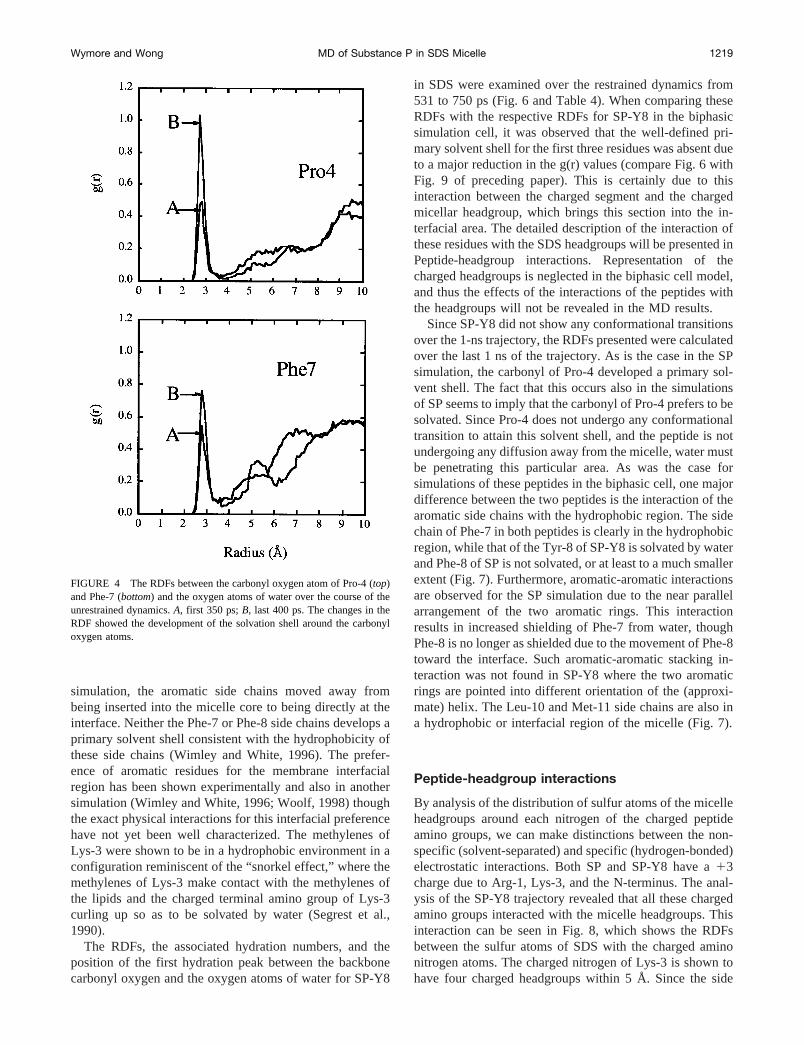
Since SP-Y8 did not show any conformational transitionsover the 1-ns trajectory, the RDFs presented were calculatedover the last 1 ns of the trajectory. As is the case in the SPsimulation, the carbonyl of Pro-4 developed a primary sol-vent shell. The fact that this occurs also in the simulationsof SP seems to imply that the carbonyl of Pro-4 prefers to besolvated. Since Pro-4 does not undergo any conformationaltransition to attain this solvent shell, and the peptide is notundergoing any diffusion away from the micelle, water mustbe penetrating this particular area. As was the case forsimulations of these peptides in the biphasic cell, one majordifference between the two peptides is the interaction of thearomatic side chains with the hydrophobic region. The sidechain of Phe-7 in both peptides is clearly in the hydrophobicregion, while that of the Tyr-8 of SP-Y8 is solvated by waterand Phe-8 of SP is not solvated, or at least to a much smallerextent (Fig. 7). Furthermore, aromatic-aromatic interactionsare observed for the SP simulation due to the near parallelarrangement of the two aromatic rings. This interactionresults in increased shielding of Phe-7 from water, thoughPhe-8 is no longer as shielded due to the movement of Phe-8toward the interface. Such aromatic-aromatic stacking in-teraction was not found in SP-Y8 where the two aromaticrings are pointed into different orientation of the (approxi-mate) helix. The Leu-10 and Met-11 side chains are also ina hydrophobic or interfacial region of the micelle (Fig. 7).
Peptide-headgroup interactions
By analysis of the distribution of sulfur atoms of the micelleheadgroups around each nitrogen of the charged peptideamino groups, we can make distinctions between the non-specific (solvent-separated) and specific (hydrogen-bonded)electrostatic interactions. Both SP and SP-Y8 have a13charge due to Arg-1, Lys-3, and the N-terminus. The anal-ysis of the SP-Y8 trajectory revealed that all these chargedamino groups interacted with the micelle headgroups. Thisinteraction can be seen in Fig. 8, which shows the RDFsbetween the sulfur atoms of SDS with the charged aminonitrogen atoms. The charged nitrogen of Lys-3 is shown tohave four charged headgroups within 5 Å. Since the side
FIGURE 4 The RDFs between the carbonyl oxygen atom of Pro-4 (top)and Phe-7 (bottom) and the oxygen atoms of water over the course of theunrestrained dynamics.A, first 350 ps;B, last 400 ps. The changes in theRDF showed the development of the solvation shell around the carbonyloxygen atoms.
Wymore and Wong MD of Substance P in SDS Micelle 1219

chain of Lys-3 is immobilized due to the “snorkel effect”(see above), the Lys-3 charged amino group was preventedfrom interacting as much with the solvent. To compensatefor the reduced solvent interaction, more sulfate groupsinteract with the Lys-3 amino group than the N-terminalamino group, which has more access to solvent. Specifichydrogen bonding between the Nz-H atoms and the oxygenatoms on the sulfate headgroup was observed. The hydrogenbonds fluctuate between different Nz-H atoms and the ox-ygen atoms on different sulfate headgroups of the SDSmolecules. Significant hydrogen bonding with the sulfateoxygen atoms was also observed for the Gln-6 NeH, theTyr-8 phenolic hydrogen, the Met-11 NH, and one of theamidated C-terminal hydrogen atoms. The preference oftyrosine for an interfacial region may be due to this hydro-gen bonding of the Tyr-8 phenolic hydrogen to the sulfateheadgroup or other hydrogen bond acceptors (Wimley andWhite, 1996; Woolf, 1998).
SP does not show the same overall interaction with thelipid headgroups. The interaction between Lys-3 and theheadgroup is the same as in SP-Y8. In addition, the Lys-3NH, Gln-5 Ne protons, Met-11 NH, and the C-terminalamide all show some hydrogen bonding with the micelleheadgroups. The Lys-3 NH chemical shift changes in goingfrom water to SDS micelles is 0.4 ppm, but only;0.02 ppmin DPC micelles for both SP (Keire and Fletcher, 1996) andSP-Y8 (Gao and Wong, submitted for publication), suggest-ing a specific interaction with the sulfate headgroup in SDS.A large chemical shift difference is also seen for the Met-11
NH in going from aqueous solution to SDS or DPC micellesfor both SP (Keire and Fletcher, 1998) and SP-Y8 (Gao andWong, submitted for publication). Since the C-terminalsection appears to lack any secondary structure, this chem-ical shift difference must be due to hydrogen bonding withthe headgroups. Though DPC is overall uncharged, electro-static interaction between the peptides with the headgroupscannot be completely ruled out. No other amide hydrogenatoms on the SP and SP-Y8 peptide backbone, with theexception of the amidated C-terminal, was observed tohydrogen-bond with the sulfate oxygen atoms throughoutthe entire 1-ns simulations, and their chemical shift differ-ences between aqueous solution and SDS micelles may thusbe attributed to secondary structure formation (see Confor-mation section). In Fig. 9, the RDF between the nitrogenatom in the backbone of Lys-3 and Met-11 and the sulfuratom on the SDS headgroup is shown. The peak in the RDFat 4.1 Å indicates the specific interaction, most probablyhydrogen bonding between the NH protons and the sulfuroxygen atoms. No other amide nitrogen atoms on the rest ofthe residues exhibit such specific interaction in the RDF.
Dynamics of the peptide
Examination of the times series for thef-c dihedral anglesand their rms fluctuations for both SP and SP-Y8 from theirrespective 1-ns unrestrained trajectories was carried out todetermine the flexibility of the various segments of the
FIGURE 5 RDFs of selected side chain atoms with the oxygen atoms of water from the trajectory over the last nanosecond for SP in the SDS micelle.
1220 Biophysical Journal Volume 76 March 1999

peptides. SP undergoes a conformational transition at;400ps; one may infer from that the Gln-6 through Gly-9 sectioninvolved in the transition is more flexible if the peptideundergoes further conformation transitions back to the orig-inal conformation during the course of the simulation. How-ever, since only one conformational transition is observedover the nanosecond trajectory, insufficient sampling doesnot permit such a conclusion. Fast fluctuations about thef-c dihedral angles were well sampled over this period bothbefore and after the conformational transition. Before theconformational transition, all the residues had roughly sim-ilar fluctuations of the dihedral angles. After the transition,
the c of Tyr-8 and thef of Gly-9 had larger rms fluctua-tions of 33.7° and 50.4°, respectively, while the other resi-dues had an average rms fluctuation about thef-c dihedralangles of 12.5° with a range of 9.5° to 20.9° (see Table 1).Glycine is known to be a residue that usually exhibits moreflexibility than other residues in proteins and peptides and isoften considered a helix breaker.
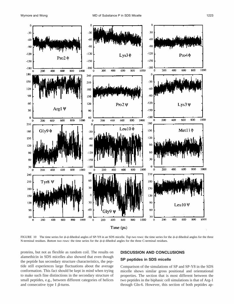
In SP-Y8, the Gly-9/Leu-10 region is also more flexiblethan the rest of the peptide. Thef angles of Gly-9 andLeu-10 have the two largest rms fluctuations with values of28.7° and 23.7°, respectively, while the average rms fluc-tuation inf of the other residues is 12.1° with a range from7.8° to 18.5°. The rms fluctuation inc of Gly-9 of 34.2° isalmost twice as large as that of any other residue (see Table2). Since the first three residues of SP have been proposedto be more flexible than the rest of the peptide (Convert etal., 1991), thef-c dihedral angles of Gly-9/Leu-10 and ofthe first three residues were examined and their time seriesare shown in Fig. 10. While Arg-1 is slightly more flexiblethan Pro-2 and Lys-3, the Gly-9/Leu-10 region is clearly themost flexible. Interestingly, this flexibility is not extendedto the Met-11f dihedral angle, perhaps because the Met-11side chain is in contact with the lipids (see Fig. 7) with theNH and the amidated C-terminal hydrogen bonded with theoxygen atoms of the sulfate headgroup, as discussed earlierin the Peptide-headgroup interaction section.
Convert et al. (1991) proposed that the flexibility of thethree N-terminal residues is a requirement for SP related
FIGURE 6 RDFs between the carbonyl oxygen atom with the oxygen atoms of water for SP-Y8 over the restrained trajectory (531–750 ps). Residuesnot shown have a RDF similar to Tyr-8, i.e., having a strong solvation peak, indicating the solvation of the carbonyl group by water.
TABLE 4 Hydration numbers for the SP-Y8 peptide carbonyloxygen atoms with oxygen atoms of water
ResidueFirst Peak
Position (Å) Integrated to (Å) Hydration No.
Arg-1 3.3 4.4 0.17Pro-2 — 5.0 0.06Lys-3 — 5.0 0.00Pro-4 — 5.0 0.14Gln-5 2.8 3.4 0.49Gln-6 2.8 3.4 0.59Phe-7 2.8 3.4 0.40Tyr-8 2.8 3.4 0.81Gly-9 2.8 3.4 0.81Leu-10 2.8 3.4 0.41Met-11 2.8 3.4 0.74
Residues in bold are predominately in intramolecular hydrogen bonds.
Wymore and Wong MD of Substance P in SDS Micelle 1221

peptides to be biologically active. If indeed the first threeresidues must remain flexible for receptor activation, thenthe interaction between the peptide and ananionic lipidsurface (the “natural membrane” contains;25% of anioniclipids) should greatly reduce the flexibility of the first threeresidues, Arg-1 through Lys-3, and would have violated thisrequirement. Interacting with the membrane lipids as thefirst step in binding to the receptor is a major part ofSchwyzer’s (1992) membrane-mediated mechanism of re-ceptor selection. The absence of intramolecular NOE cor-relations from 2D NMR for a certain segment of peptideshas often been interpreted as meaning that particular seg-ment of the peptide/protein is flexible. This inference maybe reasonable for peptides in solution, but is quite suspectfor peptides in micellar or similar environments. The inter-action of segments of the peptide with the micelle (head-group or hydrophobic regions) is not reflected in intramo-lecular NOE correlations, as demonstrated in this study. Thepresence of two prolines (Pro-2 and Pro-4) in SP peptides,and thus the lack of amide protons, near the N-terminusfurther reduces the chance of observing intramolecular NOE
and deriving a secondary structure. Thus, the conclusionthat the first three residues of SP in SDS micelles areflexible seems unwarranted from the experimental data andmay only be resolved by examining the NMR relaxationtimes of these segments. Our simulation results are consis-tent with experimental NMR relaxation results for alamethi-cin oriented on the surface of SDS micelles (Spyracopouloset al., 1996), which showed that the micelle tends to dampenout the flexibility of the peptide on the surface. Alamethicinwas shown not to be as constrained as the core regions of
FIGURE 7 RDFs of selected side chain atoms with the oxygen atoms of water from the trajectory over the last nanosecond for SP-Y8 in the SDS micelle.
FIGURE 8 RDFs of charged amino nitrogen atoms of SP-Y8 with thesulfur atoms of SDS. The close contact between the other charged N atomof Arg-1 with the sulfate oxygen atoms is very similar to the contactbetween the N-terminal nitrogen atoms with the sulfate oxygen. Due to thisoverlap, the latter RDF is not shown for clarity.
FIGURE 9 RDFs of amide nitrogen of peptide backbone residues Lys-3and Met-11 with the sulfur atom of the lipid headgroup. The large first peakin the RDF at a distance of 4.1 Å signifies hydrogen bonding betweenamide hydrogen and the sulfate oxygen atoms.
1222 Biophysical Journal Volume 76 March 1999
proteins, but not as flexible as random coil. The results onalamethicin in SDS micelles also showed that even thoughthe peptide has secondary structure characteristics, the pep-tide still experiences large fluctuations about the averageconformation. This fact should be kept in mind when tryingto make such fine distinctions in the secondary structure ofsmall peptides, e.g., between different categories of helicesand consecutive type Ib-turns.
DISCUSSION AND CONCLUSIONS
SP peptides in SDS micelle
Comparison of the simulations of SP and SP-Y8 in the SDSmicelle shows similar gross positional and orientationalproperties. The section that is most different between thetwo peptides in the biphasic cell simulations is that of Arg-1through Gln-6. However, this section of both peptides ap-
FIGURE 10 The time series forf-c dihedral angles of SP-Y8 in an SDS micelle.Top two rows:the time series for thef-c dihedral angles for the threeN-terminal residues.Bottom two rows:the time series for thef-c dihedral angles for the three C-terminal residues.
Wymore and Wong MD of Substance P in SDS Micelle 1223

pears very similar in the orientation of the backbone andposition of the side chains in SDS micelles. Both showedsimilar properties with regard to electrostatic interactionwith the headgroups, which forces this section of SP-Y8 tobe in the interfacial area. The side chain of Lys-3 seems toplay a large part in positioning of the peptide at the inter-face. Interestingly, this configuration was not part of theinitial configuration but developed over the equilibrationperiod of the simulation. SP and SP-Y8 differ the most inthe orientations of the two aromatic side chains. SP-Y8 hasthe two aromatic side chains on opposite faces with Phe-7 incontact with the methylenes of the hydrocarbon chain ofSDS and Tyr-8 well solvated by water. SP has both phe-nylalanines in contact with the hydrocarbon chains or in theheadgroup region. Furthermore, the faces of the aromaticside chains in SP are in close contact, giving rise to favor-able van der Waals (vdw) interactions (or aromatic stack-ing) between them. These favorable vdw interactions be-tween the two aromatic side chains are not seen in SP-Y8for either the biphasic cell or micelle simulations. Becauseof such a difference in the orientation of the backbone andthe position of the side chains between the two peptides wewould expect a difference in the hydrophobic interactionsand thus in theDGpart between the two peptides uponpartitioning in the SDS micelle. The experimental value of0.35 kcal/mol for the difference inDGpart for these twopeptides in DPC micelles (more negative for SP) (Wong andGao, 1998) is in agreement with this observation. Recentwork on the conformation of SP, its agonists, and antago-nists provided speculations on the significance of the rela-tive orientations of the two aromatic rings to provide aro-matic-aromatic stacking interactions for binding andreceptor recognition and activation (Desai et al., 1992;Huang et al., 1994; Josien et al., 1994; Grdadolnik et al.,1994). It is thus interesting to note that the relative orien-tations of the two aromatic rings for SP and SP-Y8 aredifferent when these two peptides are placed at the water-membrane interface. The reduction in the potency of SP-Y8as compared to SP (Fisher et al., 1976) may be a result ofthe partial loss of thep-p interaction between the twoaromatic rings (in residues 7 and 8) in SP-Y8 when it isbound to the membrane. However, it is also conceivable thatthe lower potency may be a direct result of the lower affinityof SP-Y8 for the membrane (Wong and Gao, 1998), if themembrane-mediated mechanism for receptor binding asproposed by Schwyzer is valid for these SP peptides (Schwy-zer, 1992).
The simulations of SP peptides in an explicit SDS micellehave allowed for the classification of a diverse range ofpeptide-lipid interactions. The orientation of the peptideparallel to the interface is due to the polarity of the peptidebackbone in that the peptide backbone should be solvatedby water or be intramolecularly hydrogen-bonded. The in-teraction of the peptide with the micelle is mainly throughthe hydrophobic interaction of the side chains of Pro-4,Phe-7, Leu-10, Met-11, and Phe-8. In addition, the N-terminal amino hydrogen atoms, the Arg-1 side chain, Lys-3
N-H and Nz hydrogen atoms, and Met-11 NH have signif-icant electrostatic and/or hydrogen bonding interactionswith the SDS headgroups. Even if SP forms ana-helix, thisconformation still leaves the final residues without intramo-lecular hydrogen bonds, which would be thermodynami-cally unstable if inserted into the micellar core (Ben-Tal etal., 1996, 1997). Despite the distinct charged and hydropho-bic segments (primary amphiphilicity as defined by Schw-yzer) of these peptides, Schwyzer’s model for the mem-brane-bound state of SP of insertion of the hydrophobicsegment (Schwyzer, 1992, 1995) seems unlikely. One ofDuplaa’s models (1992) for the membrane-bound state ofSP, in which both phenylalanine residues are embedded intothe membrane, agrees with our simulations of SP.
The results of Kothekar’s (1996) MD simulation of SP ina phospholipid bilayer for 260 ps using the insertion modelas the initial configuration are suspect. The results of thiswork show that it requires much more than 260 ps for thepeptide to equilibrate to the optimized orientation/positionwith respect to the water/membrane interface. Not surpris-ingly, the results of Kothekar’s work showed that SP was inan insertion mode in the bilayer that was basically the sameas the initial configuration.
This work is the first MD simulation of a peptide in amicelle that is most frequently used in high resolutionNMR, and it provides simulation results for direct compar-ison with and interpretation of experimental results for thesame systems. Our three-dimensional models of the SP/SP-Y8-micelle complex could serve as a good starting point forassessing the importance of particular residues to membranebinding and how membrane binding is related to activationof the NK1 receptor (Seelig et al., 1996). With the increas-ing use of supercomputers and parallel algorithm develop-ment, nanosecond simulations of peptide-membrane com-plexes will become more routine. Our simulations werebroken up into 80-ps sections that required;420 cpu hourson a Cray T3E at the Pittsburgh Supercomputing Center foreach section. Using 32 processors allows each of thesesections to be calculated in;13 h assuming that 1 cpu houris equal to 1 h wall time.
There are concerns that in theNVT simulation the con-stant volume may prevent the system from evolving to anincorrect density despite producing apparently good butspurious results. This could happen if the force field wasflawed (Jakobbsen et al., 1996). The CHARMM parametershave been tested in a number of examples (MacKerell et al.,1998; MacKerell, 1995; Feller et al., 1997) and have shownexcellent results. Recent results suggest that some form ofconstant pressure or constant surface tension is the mostappropriate way to simulate interfacial systems (Chiu et al.,1995; Feller et al., 1995; Tu et al., 1996) and future studieswill be carried out in such an ensemble. Studies are alsocurrently underway to quantitatively define the dynamics ofthe lipids and the peptide in the form of time correlationfunctions and to compare with NMR relaxation data.
1224 Biophysical Journal Volume 76 March 1999

Comparison of simulations in the biphasic cellversus an explicit SDS micelle
Certainly, constructing an environment that has propertiesof not only both hydrophilic and hydrophobic environmentsbut also an interfacial region will be useful in refiningstructures taken from SA treatments in vacuum (Chiche etal., 1989). A more complete picture of the properties ofpeptides and other amphipathic molecules at hydrophilic/hydrophobic interfaces is vitally important to the mem-brane-bound state of such molecules. The use of the bipha-sic cell in constructing models for the peptide-micellecomplex has been shown to reproduce many of the exper-imental properties of such a system, and reveal some de-tailed structural and dynamic properties of the peptides thatare not easily accessible experimentally (see preceding pa-per; Guba and Kessler, 1994; Guba et al., 1994). Yet water/hydrophobic interfaces are clearly not the same as micellesor lipid bilayers in that usually the interface is more distinct.Weiner and White (1992) have shown the interface of lipidbilayers to be quite heterogeneous and broad. Furthermore,the water/hydrophobic interface neglects the often very im-portant electrostatic interactions played by the headgroups.As a result of this work on SP peptides in an explicit SDSmicelle and the results from the preceding paper on thesame peptides in the biphasic system, we can make a moredefinite comparison between the two systems and commenton the merits of the biphasic system as an approximation fora more realistic explicit water/membrane mimicking sys-tem. Overall, the results seem to indicate that if the peptidehas a significant hydrophobic interaction with the micelle,the biphasic cell can reproduce most of the properties of thepeptide-micelle complex. If, in addition, the peptide hassignificant electrostatic interactions with the headgroup orcontains interfacial lysines that may interact both with themethylenes and the charged headgroup of the lipids, thenthe biphasic cell may give incomplete or misleading resultson both the structure and the dynamics of the segment(s) ofthe peptide having electrostatic interactions with the mi-celle. The present results also showed that the simulationsdone in the biphasic cell may have exaggerated the differ-ences between SP and SP-Y8 due to the absence of a broadheterogeneous interfacial area. For example, a hydrophobicside chain that is inserted into the micellar core may besubstituted for a more polar side chain that hydrogen-bondsto the headgroup. Both of these peptides may still retainsimilar orientations at the micellar surface, but simulationsin the biphasic cell would likely show the two side chains incompletely different environments (water versus carbon tet-rachloride), which may in turn cause the orientation of thewhole peptide to differ, as observed in the 1-6 segment ofthese two peptides in the biphasic simulation.
However, the important conclusion that can be drawnfrom the comparison of the results between the simulationsin these two systems is that the simulation in the biphasiccell can reproduce the gross orientational and positionalproperties of the peptide-micelles system (and presumably
peptide-bilayer systems as well). The distribution of thebackbones and side chains in respective phases and hydro-gen-bonding characteristics are also qualitatively similar inthese simulations. Since the biphasic system contains onlyabout one-third of the number of atoms of the peptide-micelles system, and we have demonstrated in this studythat the equilibration time for peptide in the former systemis ;200 ps vs. 450–500 ps for the latter, it is conceivablethat one could use the equilibration in the biphasic system toobtain the proper starting configuration for the peptide (withrespect to the water-membrane interface) in about one-fifthof the equilibration time required for the real peptide-mi-celle or peptide-bilayer system before starting to sample thedynamic trajectories for the real system of interest.
We thank Dr. Alexander MacKerell for providing the coordinates of theequilibrated SDS micelle, and gratefully acknowledge the support of Dr.Hossein Tahani and the University of Missouri Campus Computing.
This work was supported by Grant MCB950034P from the PittsburghSupercomputing Center, sponsored by the National Science Foundation(NSF), and by a grant from the Research Council of the University ofMissouri, Columbia.
REFERENCES
Attwood, D., and A. T. Florence. 1983.In Surfactant Systems. Chapmanand Hall, New York. 71.
Belohorcova, K., J. H. Davis, T. B. Woolf, and B. Roux. 1997. Structureand dynamics of an amphiphilic peptide in a lipid bilayer: a moleculardynamics study.Biophys. J.73:3039–3055.
Ben-Tal, N., A. Ben-Shaul, A. Nicholls, and B. Honig. 1996. Free-energydeterminants ofa-helix insertion into lipid bilayers.Biophys. J.70:1803–1812.
Ben-Tal, N., D. Sitkoff, I. Topol, A. Yang, S. Burt, and B. Honig. 1997.Free energy of amide hydrogen bond formation in vacuum, in water, andin liquid alkane solution.J. Phys. Chem. B.101:450–457.
Brooks, B. R., R. E. Bruccoleri, B. D. Olafson, D. J. States, S. Swami-nathan, and M. Karplus. 1983. CHARMM: a program for macromolec-ular energy, minimization, and dynamics calculations.J. Comp. Chem.4:187–217.
Brown, J., and W. Huestis. 1993. Structure and orientation of a bilayer-bound model tripeptide. A1H-NMR study. J. Phys. Chem.97:2967–2973.
Chiche, L., C. Gaboriaud, A. Heitz, J.-P. Mornon, B. Castro, and P.Kollman. 1989. Use of restrained molecular dynamics in water todetermine three-dimensional protein structure: prediction of the three-dimensional structure of ecballium elaterium trypsin inhibitor II.Proteins: Struct., Funct., Genet.6:405–417.
Chiu, S.-W., M. Clark, V. Balaji, S. Subramaniam, H. L. Scott, and E.Jakobsson. 1995. Incorporation of surface tension into molecular dy-namics simulation of an interface: a fluid phase lipid bilayer membrane.Biophys. J.69:1230–1245.
Convert, O., H. Duplaa, S. Lavielle, and G. Chassaing. 1991. Influence ofthe replacement of amino acid by its D-enantiomer in the sequence ofsubstance P. 2. Conformational analysis by nmr and energy calculations.Neuropeptides. 19:259–270.
Croonen, Y., E. Gelade, M. Van der Ziegel, H. Van der Auweraer, F. C.Vandendriessche, and F. C. DeSchryver. 1983. Influence of salt, deter-gent concentration, and temperature on the fluorescence quenching of1-methylpyrene in sodium dodecylsulfate with m-dichlorobenzene.J. Phys. Chem.87:1426.
Damodaran, K. V., and K. M. Merz. 1995. Interaction of the fusioninhibiting peptide carbobenzoxy-D-Phe-L-Phe-Gly with N-methyldio-leoylphosphatidylethanolamine lipid bilayers.J. Am. Chem. Soc.117:6561–6571.
Wymore and Wong MD of Substance P in SDS Micelle 1225

Damodaran, K. V., K. M. Merz, and B. P. Gaber. 1995. Interaction of smallpeptides with lipid bilayers.Biophys. J.69:1299–1308.
Desai, V. C., S. L. Lefkowitz, P. F. Thadeio, K. P. Longo, and R. M.Snider. 1992. Discovery of a potent substance P antagonist: recognitionof the key molecular determinant.J. Med. Chem.35:4911–4913.
Duplaa, H., O. Convert, A.-M. Sautereau, J.-F. Tocanne, and G. Chassaing.1992. Binding of substance P to monolayers and vesicles made ofphosphatidylcholine and/or phosphatidylserine.Biochim. Biophys. Acta.1107:12–22.
Feller, S. E., D. Yin, R. W. Pastor, and A. D. MacKerell, Jr. 1997.Molecular dynamics simulation of unsaturated lipid bilayers at lowhydration: parametrization and comparison with diffraction studies.Bio-phys. J.73:2269–2279.
Feller, S. E., Y. Zhang, R. W. Pastor, and B. R. Brooks. 1995. Constantpressure molecular dynamics simulation: the Langevin piston method.J. Chem. Phys.103:4613–4621.
Fisher, G., K. Folkers, B. Pernow, and C. Bowers. 1976. Synthesis andsome biological activities of the tyrosine-8 analog of substance P.J. Med. Chem.19:325–328.
Frey, S., and L. K. Tamm. 1991. Orientation of melittin in phospholipidbilayers: a polarized attenuated total reflection infrared study.Bio-phys. J.60:922–930.
Gennis, R. B. 1989. Biomembranes: Molecular Structure and Function.Springer-Verlag, New York.
Grdadolnik, S. G., D. F. Mierke, G. Byk, I. Zeltser, C. Gilon, and H.Kessler, 1994. Comparison of the conformation of active and nonactivebackbone cyclic analogs of substance P as a tool to elucidate features ofthe bioactive conformation: NMR and molecular dynamics in DMSOand water.J. Med. Chem.37:2145–2152.
Guba, W., R. Haessner, G. Breipohl, S. Henke, J. Knolle, V. Santagada,and H. Kessler. 1994. Combined approach of NMR and moleculardynamics within a biphasic membrane mimetic. Conformation and ori-entation of the bradykinin antagonist Hoe. 140.J. Am. Chem. Soc.116:7532–7540.
Guba, W., and H. Kessler. 1994. A novel computational mimetic ofbiological membranes in molecular dynamics simulations.J. Phys.Chem.98:23–27.
Huang, P., and G. H. Loew. 1995. Interaction of an amphiphilic peptidewith a phospho-lipid bilayer surface by molecular dynamics simulationstudy.J. Biomol. Struct. Dyn.12:937–956.
Huang, R. R. C., H. Yu, C. Strader, and T. M. Fong. 1994. Interaction ofsubstance P with the second and seventh transmembrane domains of theneurokinin-1 receptor.Biochemistry. 33:3007–3013.
Itri, R., and L. Q. Amaral. 1991. Distance distribution function of sodiumdodecylsulfate micelles by x-ray scattering.J. Phys. Chem.95:423–427.
Jacobs, R., and S. H. White. 1989. The nature of the hydrophobic bindingof small peptides at the bilayer interface: implications for the insertion oftransbilayer helices.Biochemistry. 28:3421–3437.
Jakobbsen, E., S. Subramaniam, and H. L. Scott. 1996. Strategic issues inmolecular dynamics simulations of membranes.In BiologicalMembranes: A Molecular Perspective from Computation and Experi-ment. K. M. Merz, Jr., and B. Roux, editors. Birkha¨user, Boston.105–123.
Jorgenson, W. L., R. W. Impey, J. Chandrasekhar, J. D. Madura, and M. L.Klein. 1983. Comparison of simple potential functions for simulatingliquid water.J. Chem. Phys.79:926–935.
Josien, H., S. Lavielle, A. Brunissen, M. Saffroy, Y. Torrens, J.-C. Beau-jouan, J. Glowinski, and G. Chassaing, 1994. Design and synthesis ofside-chain conformationally restricted phenylalanines and their use forstructure-activity studies on tachykinin NK-1 receptor.J. Med. Chem.37:1586–1601.
Keire, D., and T. Fletcher. 1996. The conformation of substance P in lipidenvironments.Biophys. J.70:1716–1727.
Kothekar, V. 1996. 260 ps molecular dynamics simulation of substance Pwith hydrated dimyristoylphosphatidyl choline bilayer.J. Biomol.Struct. Dyn.13:601–613.
MacKerell, Jr., A. D. 1995. Molecular dynamics simulation analysis of asodium dodecyl sulfate micelle in aqueous solution: decreased fluidity ofthe micelle hydrocarbon interior.J. Phys. Chem.99:1846–1855.
MacKerell, Jr., A. D., D. Bashford, M. Bellott, R. L. Dunbrack, Jr., J. D.Evanseck, M. J. Field, S. Fischer, J. Gao, H. Guo, S. Ha, D. Joseph-
McCarthy, L. Kuchnir, K. Kuczera, F. T. K. Lau, C. Mattos, S. Mich-nick, T. Ngo, D. T. Nguyen, B. Prodhom, W. E. Reiher III, B. Roux, M.Schlenkrich, J. C. Smith, R. Stote, J. Straub, M. Watanabe, J. Wiork-iewicz-Kuczera, D. Yin, and M. Karplus. 1998. All-atom empiricalpotential for molecular modeling and dynamics studies of proteins.J. Phys. Chem. B. 102:3586–3616.
Merz, Jr., K. M., and B. Roux, editors. 1996. Biological Membranes: AMolecular Perspective from Computation and Experiment. Birkha¨user,Boston.
Oda, K., H. Miyagawa, and K. Kitamura. 1996. How does the electrostaticforce cut-off generate non-uniform temperature distributions in proteins?Mol. Sim.16:167–177.
Opella, S. J. 1997. NMR and Membrane Proteins.Nat. Struct. Biol., NMRSuppl. 845–848.
Ravishanker, G., S. Vijakumar, and D. L. Beveridge. 1994. STRIPS: Analgorithm for generating two-dimensional hydrogen-bond topology dia-grams for proteins.In Modeling the Hydrogen Bond. The AmericanChemical Society, Washington, DC. 209–219.
Roseman, M. A. 1988. Hydrophobicity of the peptide C5O–H-N hydrogenbonded group.J. Mol. Biol. 201:621–625.
Roux, B., and T. B. Woolf. 1996. Molecular dynamics of Pf1 coat proteinin a phospholipid bilayer.In Biological Membranes: A Molecular Per-spective from Computation and Experiment. K. M. Merz, Jr., and B.Roux, editors. Birkha¨user, Boston. 555–587.
Ryckaert, J.-P., G. Cicotti, and H. J. C. Berendsen. 1977. Numericalintegration of the Cartesian equations of motion of a system withconstraints: molecular dynamics of n-alkanes.J. Comput. Phys.23:327–341.
Sanders, C. R., and G. C. Landis. 1995. Reconstitution of membraneproteins into lipid-rich bilayered mixed micelles for nmr studies.Bio-chemistry. 43:4030–4040.
Schlenkrich, M., J. Brickmann, A. D. MacKerell, Jr., and M. Karplus.1996. An empirical potential energy function for phospholipids: criteriafor parameter optimization and applications.In Biological Membranes:A Molecular Perspective from Computation and Experiment. K. M.Merz and B. Roux, editors. Birkha¨user, Boston.
Schwyzer, R. 1992. Conformations and orientations of amphiphilic pep-tides induced by artificial lipid membranes: correlations with biologicalactivity. Chemtracts–Biochem. and Mol. Biol.3:347–379.
Schwyzer, R. 1995. 100-Year lock-and-key concept: are peptide keysshaped and guided to their receptors by the target cell membrane?Biopolymers. 37:5–16.
Seelig, A., T. Alt, S. Lotz, and G. Ho¨lzemann. 1996. Binding of substanceP agonists to lipid membranes and to the neurokinin-1 receptor.Bio-chemistry. 35:4365–4374.
Seelig, A., and P. M. Macdonald. 1989. Binding of a neuropeptide, sub-stance P, to neutral and negatively charged lipids.Biochemistry. 28:2490–2496.
Segrest, J. P., H. De Loof, J. G. Dohlmann, C. G. Brouillette, and G. M.Anantharamaiah. 1990. Amphipathic helix motif: classes and properties.Proteins: Struct., Funct., Genet.8:103–117.
Shen, L., D. Bassolino, and T. Stouch. 1997. Transmembrane helix struc-ture, dynamics, and interactions: multi-nanosecond molecular dynamicssimulations.Biophys. J.73:3–20.
Soderman, O., G. Carlstrom, U. Olsson, and T. C. Wong. 1988. NMRrelaxation in micelles.2H relaxation at three field strengths of threepositions on the alkyl chain of sodium dodecyl sulfate.J. Chem. Soc.Faraday Trans. 1. 84:4475–4486.
Spyracopoulos, L., A. A. Yee, and J. D. J. O’Neil. 1996. Backbonedynamics of an alamethicin in methanol and aqueous detergent solutiondetermined by heteronuclear1H-15N NMR spectroscopy.J. Biomol.NMR. 7:283–294.
Steinbach, P., and B. R. Brooks. 1994. New spherical-cutoff methods forlong range forces in macromolecular simulation.J. Comp. Chem.15:667–683.
Tu, K., D. J. Tobias, J. K. Blasie, and M. L. Klein. 1996. Moleculardynamics investigation of the structure of a fully hydrated gel-phasedipalmitoylphosphatidylcholine bilayer.Biophys. J.70:595–608.
1226 Biophysical Journal Volume 76 March 1999

Weiner, M. C., and S. H. White. 1992. Structure of a fluid dioleoylphos-patidylcholine bilayer determined by joint refinement of x-ray andneutron diffraction data. III. Complete structure.Biophys J. 61:434–447.
White, S., and W. C. Wimley. 1994. Peptides in lipid bilayers: structuraland thermodynamic basis for partitioning and folding.Curr. Opin.Struct. Biol.4:79–86.
Williams, R. W., and J. L. Weaver. 1990. Secondary structure of substanceP bound to liposomes in organic solvents and in solution from Ramanand CD spectroscopy.J. Biol. Chem.265:2505–2513.
Wilmot, C. M., and J. M. Thorton. 1990.b-Turns and their distortions: aproposed new nomenclature.Protein Eng.3:479–493.
Wimley, W. C., and S. H. White. 1996. Experimentally determined hy-drophobicity scale for proteins at membrane interfaces.Nat. Struct. Biol.3:842–848.
Wong, T. C., and X. Gao. 1998. The temperature dependence and thermo-dynamic functions of partitioning of substance P peptides in dode-cylphosphocholine micelles.Biopolymers. 45:395–403.
Woolf, T. B. 1998. Molecular-dynamics simulations of individuala-heli-ces of bacteriorhodopsin in dimyristoylphosphatidylcholine. II. Interac-tion energy analysis.Biophys. J.74:115–131.
Woolf, T. B., A. Grossfield, K. MacKenzie, and D. Engelman. 1998.Helix:lipid interactions in glycophorin dimerization: molecular dynam-ics calculations.Biophys. J.74:A15.
Woolf, T. B. and B. Roux. 1996. Structure, energetic, and dynamics oflipid-protein interactions: a molecular dynamics study of the gramicidinA channel in a DMPC bilayer.Proteins: Struct., Funct., Genet.24:92–114.
Woolley, G. A., and C. M. Deber. 1987. Peptides in membranes: lipid-induced secondary structure of substance P.Biopolymers. 26:S109–S121.
Young, J. K., C. Anklin, and R. P. Hicks. 1994. NMR and molecularmodeling investigations of the neuropeptide substance P in the presenceof 15 mM sodium dodecyl sulfate micelles.Biopolymers. 34:1449–1462.
Zhou, F., and K. Schulten. 1996. Molecular dynamics study of phospho-lipase A2 on a membrane surface.Proteins: Struct., Funct., Genet.25:12–27.
Wymore and Wong MD of Substance P in SDS Micelle 1227
Copyright © 2022 FDOKUMEN