Human Papillomavirus 16 E6 Suppresses Transporter ... - MDPI
Upload
khangminh22Category
view
0download
0
JOURNAL OF VIROLOGY, June 1990, p. 3012-30180022-538X/90/063012-07$02.00/0Copyright C) 1990, American Society for Microbiology
The Adeno-Associated Virus rep Gene Suppresses Herpes SimplexVirus-Induced DNA Amplification
REGINE HEILBRONN,* ALEXANDER BURKLE, SONJA STEPHAN,AND HARALD ZUR HAUSEN
Deutsches Krebsforschungszentrum, Im Neuenheimer Feld 506,D-6900 Heidelberg, Federal Republic of Germany
Received 22 January 1990/Accepted 22 March 1990
Herpes simplex virus (HSV) induces within the host cell genome DNA amplification which can be suppressedby coinfection with adeno-associated virus (AAV). To characterize the AAV functions mediating this effect,cloned AAV type 2 wild-type or mutant genomes were transfected into simian virus 40 (SV40)-transformedhamster cells together with the six HSV replication genes (encoding UL5, UL8, major DNA-binding protein,DNA polymerase, UL42, and UL52) which together are necessary and sufficient for the induction of SV40 DNAamplification (R. Heilbronn and H. zur Hausen, J. Virol. 63:3683-3692, 1989). The AAV rep gene wasidentified as being responsible for the complete inhibition of HSV-induced SV40 DNA amplification. Likewise,rep inhibited origin-dependent HSV replication. rep neither killed the transfected host cells nor interfered withgene expression from the cotransfected amplification genes. This points to a specific interference withHSV-induced DNA amplification.
Adeno-associated viruses (AAVs) are members of theparvovirus family, a group of small single-stranded DNAviruses with unique replication properties. In contrast to theautonomous parvoviruses which can replicate independentlyin proliferating cells, the AAVs rely on helper viruses, eitheradenoviruses or herpesviruses, for efficient replication (for areview, see reference 4). However, cells treated with chem-ical or physical carcinogens can support low-level AAVreplication in the absence of a helper virus (13, 34, 45, 46).Thus, the replication defectiveness of AAV is not absolute.The autonomous and helper-dependent parvoviruses haveunique biological properties in common. Members of bothgroups efficiently suppress tumor growth in animals, irre-spective of the mode of tumor induction. Parvovirusesinhibit spontaneous tumor formation (37) and tumors in-duced by various oncogenic viruses as well as by chemicalcarcinogens (9, 10, 19, 28, 30, 39). In addition, there isevidence from seroepidemiological studies that high anti-body titers against the human AAV types 2, 3, and 5(AAV-2, AAV-3, and AAV-5) are associated with a reducedcancer incidence (11, 27, 35). The mechanisms, however, bywhich parvoviruses exert their oncosuppressive effect are
not yet understood. In an attempt to unravel the underlyingmolecular mechanisms, the effect of parvoviruses was stud-ied in various in vitro transformation systems. Minute virusof mice, an autonomous parvovirus, suppressed transforma-tion of mouse fibroblasts by simian virus 40 (SV40) (29).Likewise, AAV suppressed the transformation of hamsterfibroblasts by different adenovirus strains (5) and the trans-formation of the mouse fibroblast cell line C127 by bovinepapillomavirus. AAV-2 p78reP appeared to be responsible forthis effect (16).Three viral functions have been mapped on the 4.65-
kilobase (kb) AAV-2 genome (Fig. 1). The 145-base-pair (bp)terminal repeat structures serve as origins of replication. Theright-hand open reading frames (ORFs) encode the threecapsid proteins (cap), whereas the ORFs on the left-handside of the genome code for rep, a family of multifunctional
* Corresponding author.
nonstructural AAV proteins. The mRNAs coding for p78rePand the spliced p68reP start at the p5 promotor, and thosecoding for p52reP and the spliced p40reP start at the p1lpromotor. The rep proteins are required for AAV DNAreplication (17, 40). p78/68reP is responsible for the accumu-lation of replicative intermediates, whereas p52I40reP isrequired for the generation of single-stranded monomerAAV and for the packaging of the virus (6). The role ofp78168reP in the accumulation of replicative intermediates isalso reflected by their binding to the AAV origins of repli-cation (18). The rep proteins are required not only for AAVDNA replication but also for AAV gene regulation. Depend-ing on the presence or absence of helper adenovirus func-tions, they either activate or repress the AAV promotors intrans (3, 41). Although most of these experiments wereperformed with human cells, the different AAV mutantsexhibited the same replication phenotype in rodent cells (16;R. Heilbronn, unpublished data).We have shown previously that AAV can severely inhibit
herpesvirus- or carcinogen-induced DNA amplification (2,13, 34). DNA amplification plays a central role not only inthe development of drug resistance (20, 26, 36) but also in thecourse of tumor development, where amplified oncogenesequences are a frequent finding (for a review, see reference1). We therefore decided to analyze the AAV-mediatedinhibition of DNA amplification at the molecular level. Thiswas possible by cotransfecting cloned AAV mutants incombination with the recently identified set of herpes sim-plex virus (HSV) amplification genes (15), thus introducingthe amplification-inducing and -inhibiting functions into thesame fraction of cells. By the use of AAV wild-type (wt) ormutant genomes, we show here that rep alone is sufficient tocompletely suppress DNA amplification induced by the setof six HSV amplification genes.
MATERIALS AND METHODSRecombinant plasmid DNAs. The cloned AAV-2 wt ge-
nome (pAV2) was obtained from C. Laughlin (24). AAV-2was subcloned into the BamHI site of Bluescript (Strata-gene), resulting in plasmid pTAV2 (Fig. 1). ori mutants
3012
Vol. 64, No. 6
AAV rep SUPPRESSES HSV-INDUCED DNA AMPLIFICATION
p5 p19 p40
rep cap
pTAV-2
pTAV2-4
pTAV2-3
pTAV2-6
pTAV2-7
pTAV2-2
pTAV2-8
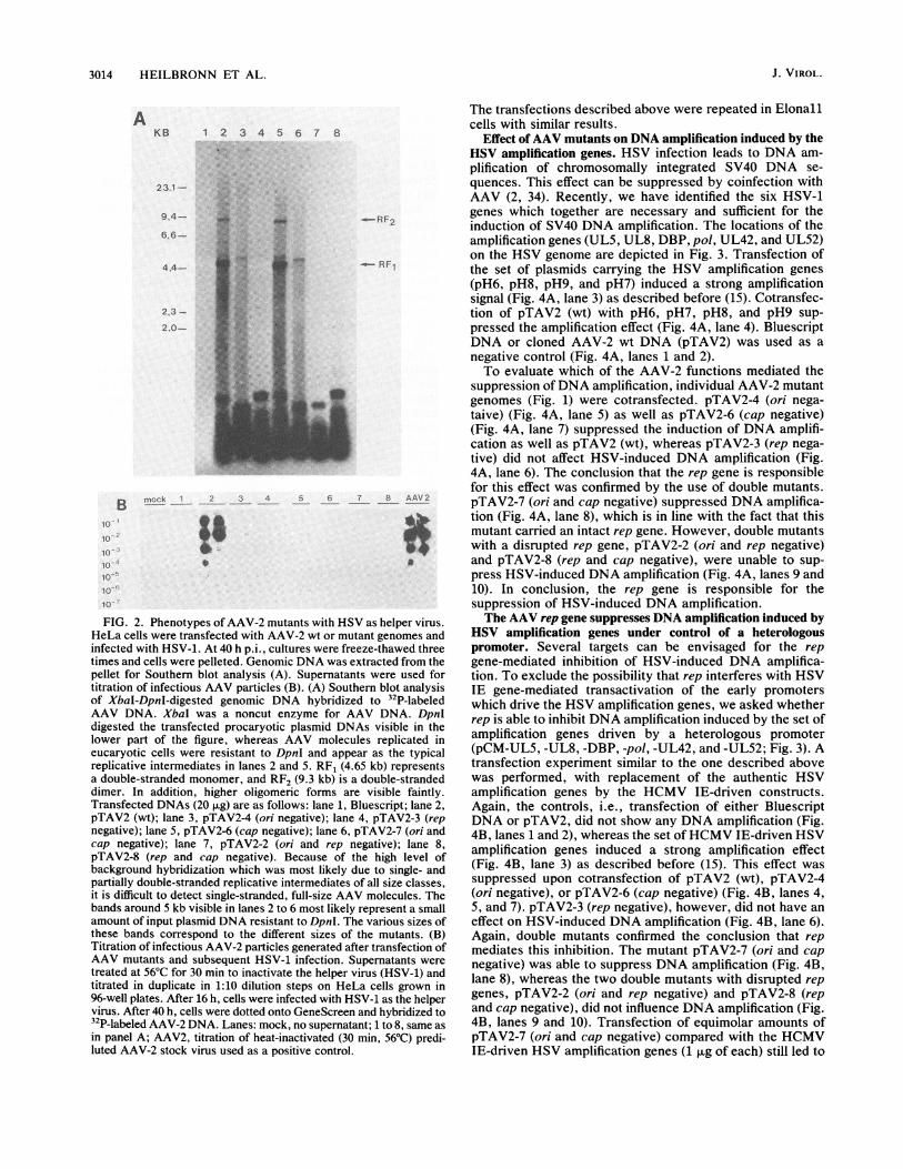
FIG. 1. Structure of AAV wt and mutant genomes. The AAV-2genome is represented schematically with the inverted repeatsserving as origins of replication at the ends of the genome (_). TheORFs encoding the nonstructural proteins (rep) and three capsidproteins (cap) are shown (O). Construction of the AAV mutantgenomes is explained in the text. Frameshift deletions (A) andinsertions (0) are indicated. Mutant phenotypes are indicated to theright of the plasmid designations.
(pTAV2-2, pTAV2-4, and pTAV2-7) were generated frompTAV2 by using the BalI sites at nucleotide positions 121and 4554. rep mutants were generated either by introducinga 4-bp frameshift insertion at position 1045 by using theunique BamHI site (pTAV2-3 and pTAV2-8) or by introduc-ing a 150-bp deletion encompassing the p1l promotor region(positions 814 to 964, pTAV2-2). Both mutations inactivatedthe rep genes starting from the promoters P5 and p19- cap
mutants (pTAV2-6, pTAV2-7, and pTAV2-8) were generatedby introducing a 164-bp deletion between positions 3326 and3490.The series of plasmids carrying the HSV amplification
genes pH6 (pol and DBP), pH7 (IE175 and IE110), pH8(UL42 and UL52), pH9 (UL5 and UL8), and the respectiveORFs expressed under the control of the human cytomega-lovirus (HCMV) immediate early (IE) promoter (-598 to+52) (pCM-UL5, -UL8, -UL9, -pol, -DBP, -UL42, and-UL52) have been described before (15). pHlO carries theHSV oris (15). pCMcat (kindly provided by Hubert Stop-pler) expresses the chloramphenicol acetyltransferase (CAT)gene under the control of the same HCMV IE promoterfragment (-598 to +52) as the pCM-UL series of HSVexpression constructs.
Cells and viruses. The Elonall cell line and propagation ofHSV-1 strain 17 have been described elsewhere (15, 33).AAV-2 was propagated in HeLa cells with adenovirus 2 asthe helper as described previously (46).
Transfection procedure. Calcium phosphate coprecipita-tion followed by a dimethyl sulfoxide shock was performedexactly as described before (15). Each transfection experi-ment was repeated at least three times with two differentplasmid preparations, leading to similar results.AAV replication assay. HeLa cells (3 x 10⁵) were plated
onto 6-cm-diameter dishes. The next day, cells were trans-fected with AAV-2 mutant plasmids which had been excisedfrom the vector with PvuII. After the dimethyl sulfoxide-shock, cells were infected with HSV-1 diluted in 1 ml ofmedium (multiplicity of infection, 10) and incubated at 37°Cfor 40 h. The cultures were lysed in situ by three freeze-thawcycles. The disrupted cells were pelleted and used for theanalysis of AAV DNA replication. Genomic DNA was
extracted by digestion with proteinase K, repeated extrac-tions with phenol and chloroform, digestion with RNase A,and dialysis against lx TE (15). The supernatants were
treated at 56°C for 30 min to inactivate the helper virus(HSV) and then used for the titration of infectious AAV in1:10 dilution steps on HeLa cells grown on 96-well plates.Cells were infected with HSV-1 as the helper virus 16 h later.At 40 h after HSV infection, cells were dotted onto Gene-Screen (Dupont, NEN Research Products, Boston, Mass.)and hybridized to 32P-labeled AAV-2 DNA.
Analysis of genomic DNA. For the analysis of AAV DNAreplication, total genomic DNA was doubly digested withXbaI and DpnI. A 5-,ug sample of each digest was run on0.7% agarose gels, blotted onto GeneScreen Plus (Dupont,NEN Research Products), and hybridized to a 32P-labeled,full-length AAV-2 probe. SV40 DNA amplification wasassayed on Southern blots of SacI-digested genomic DNAhybridized to 32P-labeled SV40 DNA, and HSV oris replica-tion was assayed with the DpnI assay as described before(15).CAT assays. Protein contents of freeze-thaw extracts were
determined by using the Bradford assay (Bio-Rad Laborato-ries, Munich, Federal Republic of Germany). CAT enzy-matic activity was assayed by using defined amounts ofheat-treated (10 min at 60°C) protein extract (8). Finalconcentrations in the assay were 1.25 ,uCi of "4C-chloram-phenicol per ml and 1 mmol of acetyl coenzyme A per liter.Ethyl acetate-extracted reaction products were analyzed bythin-layer chromatography. Spots representing unreactedchloramphenicol and acetylated chloramphenicol were ex-cised, and radioactivity was quantitated by liquid scintilla-tion counting.
RESULTS
Replication phenotype of AAV mutant plasmids. Single anddouble mutations were generated in cloned wt AAV-2 DNA(pTAV2) according to published genetic data (Fig. 1) (17,40). The phenotypes of this series of AAV plasmids wereverified by transfecting the individual mutants into HeLacells with HSV-1 as the helper virus. At 40 h after HSVinfection, cultures were processed for the determination ofAAV DNA replication and, in parallel, production of infec-tious viral particles (Fig. 2). With pTAV2 (wt), the typicalAAV replication intermediates appeared (Fig. 2A, lane 2).Titration of freeze-thaw supernatants of transfected cells onfresh HeLa cells in the presence of HSV-1 as the helper virusclearly demonstrated the rescue of infectious viral particlesfrom pTAV2 (Fig. 2B, lane 2). HSV-1 served as a helpervirus with an efficiency equal to that of adenovirus type 2(data not shown). The cap mutant pTAV2-6 gave rise toreplicative intermediates (Fig. 2A, lane 5) whose slightlyshorter lengths (compared with that of wt pTAV2) are due tothe 150-bp deletion within the cap ORF. The cap-negativephenotype of pTAV2-6 was documented by the inability ofthe mutant to generate infectious viral particles (Fig. 2B,lane 5). As expected, rep mutants did not give rise to eitherreplicative intermediates or infectious AAV particles (Fig.2A and B, lanes 4 [pTAV2-3], 7 [pTAV2-2], and 8 [pTAV2-8]). Transfection of ori mutants with deletions of the terminalrepeats but intact rep genes (pTAV2-4 and pTAV2-7) gaverise to a high AAV-2-specific background hybridizationsignal on the Southern blot without the typical replicationintermediates (Fig. 2A, lanes 3 and 6). The absence of similarhybridization signals in all the lanes with rep mutants (Fig.2A, lanes 4, 7, and 8) raises the possibility that rep geneexpression per se mediates unspecific initiation of DNAsynthesis on the AAV template in the absence of originsequences. However, no infectious particles were formed.
a
1%
A
-A
VOL. 64, 1990 3013
3014 HEILBRONN ET AL.
AKB 1 2 3 4 5 6 7 8
23.1
994-
666-
4.4-
2232.0-
B10
10'
1010,
lo
10
-RF1
nmock 2 _ 3 _4_ 7 8 AAV 2
FIG. 2. Phenotypes of AAV-2 mutants with HSV as helper virus.HeLa cells were transfected with AAV-2 wt or mutant genomes andinfected with HSV-1. At 40 h p.i., cultures were freeze-thawed threetimes and cells were pelleted. Genomic DNA was extracted from thepellet for Southern blot analysis (A). Supernatants were used fortitration of infectious AAV particles (B). (A) Southern blot analysisof XbaI-DpnI-digested genomic DNA hybridized to 32P-labeledAAV DNA. XbaI was a noncut enzyme for AAV DNA. DpnIdigested the transfected procaryotic plasmid DNAs visible in thelower part of the figure, whereas AAV molecules replicated ineucaryotic cells were resistant to DpnI and appear as the typicalreplicative intermediates in lanes 2 and 5. RF1 (4.65 kb) representsa double-stranded monomer, and RF2 (9.3 kb) is a double-strandeddimer. In addition, higher oligomeric forms are visible faintly.Transfected DNAs (20 p.g) are as follows: lane 1, Bluescript; lane 2,pTAV2 (wt); lane 3, pTAV2-4 (ori negative); lane 4, pTAV2-3 (repnegative); lane 5, pTAV2-6 (cap negative); lane 6, pTAV2-7 (ori andcap negative); lane 7, pTAV2-2 (ori and rep negative); lane 8,pTAV2-8 (rep and cap negative). Because of the high level ofbackground hybridization which was most likely due to single- andpartially double-stranded replicative intermediates of all size classes,it is difficult to detect single-stranded, full-size AAV molecules. Thebands around 5 kb visible in lanes 2 to 6 most likely represent a smallamount of input plasmid DNA resistant to DpnI. The various sizes ofthese bands correspond to the different sizes of the mutants. (B)Titration of infectious AAV-2 particles generated after transfection ofAAV mutants and subsequent HSV-1 infection. Supernatants weretreated at 56°C for 30 min to inactivate the helper virus (HSV-1) andtitrated in duplicate in 1:10 dilution steps on HeLa cells grown in96-well plates. After 16 h, cells were infected with HSV-1 as the helpervirus. After 40 h, cells were dotted onto GeneScreen and hybridized to32P-labeled AAV-2 DNA. Lanes: mock, no supernatant; 1 to 8, same asin panel A; AAV2, titration of heat-inactivated (30 min, 56°C) predi-luted AAV-2 stock virus used as a positive control.
The transfections described above were repeated in Elonallcells with similar results.
Effect of AAV mutants on DNA amplification induced by theHSV amplification genes. HSV infection leads to DNA am-plification of chromosomally integrated SV40 DNA se-quences. This effect can be suppressed by coinfection withAAV (2, 34). Recently, we have identified the six HSV-1genes which together are necessary and sufficient for theinduction of SV40 DNA amplification. The locations of theamplification genes (UL5, UL8, DBP, pol, UL42, and UL52)on the HSV genome are depicted in Fig. 3. Transfection ofthe set of plasmids carrying the HSV amplification genes(pH6, pH8, pH9, and pH7) induced a strong amplificationsignal (Fig. 4A, lane 3) as described before (15). Cotransfec-tion of pTAV2 (wt) with pH6, pH7, pH8, and pH9 sup-pressed the amplification effect (Fig. 4A, lane 4). BluescriptDNA or cloned AAV-2 wt DNA (pTAV2) was used as anegative control (Fig. 4A, lanes 1 and 2).To evaluate which of the AAV-2 functions mediated the
suppression of DNA amplification, individual AAV-2 mutantgenomes (Fig. 1) were cotransfected. pTAV2-4 (ori nega-taive) (Fig. 4A, lane 5) as well as pTAV2-6 (cap negative)(Fig. 4A, lane 7) suppressed the induction of DNA amplifi-cation as well as pTAV2 (wt), whereas pTAV2-3 (rep nega-tive) did not affect HSV-induced DNA amplification (Fig.4A, lane 6). The conclusion that the rep gene is responsiblefor this effect was confirmed by the use of double mutants.pTAV2-7 (ori and cap negative) suppressed DNA amplifica-tion (Fig. 4A, lane 8), which is in line with the fact that thismutant carried an intact rep gene. However, double mutantswith a disrupted rep gene, pTAV2-2 (ori and rep negative)and pTAV2-8 (rep and cap negative), were unable to sup-press HSV-induced DNA amplification (Fig. 4A, lanes 9 and10). In conclusion, the rep gene is responsible for thesuppression of HSV-induced DNA amplification.The AAV rep gene suppresses DNA amplification induced by
HSV amplification genes under control of a heterologouspromoter. Several targets can be envisaged for the repgene-mediated inhibition of HSV-induced DNA amplifica-tion. To exclude the possibility that rep interferes with HSVIE gene-mediated transactivation of the early promoterswhich drive the HSV amplification genes, we asked whetherrep is able to inhibit DNA amplification induced by the set ofamplification genes driven by a heterologous promoter(pCM-UL5, -UL8, -DBP, -pol, -UL42, and -UL52; Fig. 3). Atransfection experiment similar to the one described abovewas performed, with replacement of the authentic HSVamplification genes by the HCMV IE-driven constructs.Again, the controls, i.e., transfection of either BluescriptDNA or pTAV2, did not show any DNA amplification (Fig.4B, lanes 1 and 2), whereas the set ofHCMV IE-driven HSVamplification genes induced a strong amplification effect(Fig. 4B, lane 3) as described before (15). This effect wassuppressed upon cotransfection of pTAV2 (wt), pTAV2-4(ori negative), or pTAV2-6 (cap negative) (Fig. 4B, lanes 4,5, and 7). pTAV2-3 (rep negative), however, did not have aneffect on HSV-induced DNA amplification (Fig. 4B, lane 6).Again, double mutants confirmed the conclusion that repmediates this inhibition. The mutant pTAV2-7 (ori and capnegative) was able to suppress DNA amplification (Fig. 4B,lane 8), whereas the two double mutants with disrupted repgenes, pTAV2-2 (ori and rep negative) and pTAV2-8 (repand cap negative), did not influence DNA amplification (Fig.4B, lanes 9 and 10). Transfection of equimolar amounts ofpTAV2-7 (ori and cap negative) compared with the HCMVIE-driven HSV amplification genes (1 ,ug of each) still led to
J. VIROL.

AAV rep SUPPRESSES HSV-INDUCED DNA AMPLIFICATION
HSV1 UL us
_0 -_ _ 4 OULSUL8 UL9 dbp pol
pH9 pH6
Ip U I MIpI 1 11
pCM-UL5 pCM-ULS pCM-DBP pCM-POL
_ol _0UL42 UL52
i
pHs
IE110 IE175
I,
pH7
pCM-UL42 pCM-ULS2
FIG. 3. Structure of HSV-1 genome and cloned amplification genes. Schematic representation of the 150-kb HSV-1 genome with theunique long (UL) and unique short (Us) regions and the flanking inverted repeats (_ and =). The replication- and amplification-inducinggenes are indicated: UL5, -8, -9, -42, -52, dbp (single-stranded DNA-binding protein), and pOI (DNA polymerase). These genes are early geneswhich have to be transactivated by the HSV IE genes, IE110 (ICPO), and IE175 (ICP4), for efficient transcription. The arrows indicate thedirection of transcription: black arrows are genes necessary for both HSV DNA replication and SV40 DNA amplification (UL5, UL8, dbp,pol, UL42, UL52, and IE175); speckled arrows indicate genes required only for HSV DNA replication (UL9 and IE110). The set of plasmidscovering the individual genes are shown. Below, the pCM series of expression constructs is represented. These constructs are namedaccording to the ORF they express under the control of the HCMV IE promotor.
complete suppression of DNA amplification (data notshown). These experiments confirm that the rep gene func-tion alone mediates complete suppression of HSV-inducedDNA amplification. In addition, the HSV IE gene-mediatedtransactivation of early genes does not represent the targetfor this interference.
rep mediates suppression of DNA amplification by mecha-nisms other than nonspecific down regulation of expression ofHSV amplification genes. To monitor the transfection effi-ciency of the above described experiment, an HCMV IEpromoter-driven CAT construct (pCMcat) was cotransfected(at a molar ratio of 1:20 compared with pTAV2-7) and CATenzyme activity was assayed in parallel to the amplificationassay in Fig. 4B. CAT enzyme activity was in the same rangein all transfections (Fig. 4C), reflecting comparable transfec-tion efficiencies. In addition, this result excludes the possi-bility that the suppression ofDNA amplification in the rangeof 100-fold is due to a rep-mediated repression of the HCMVIE promoter, which drives the HSV amplification genes.However, some minor, two- to threefold reduction of pCM-cat expression was observed whenever AAV wt or mutantswith intact rep genes were cotransfected (Fig. 4C). To studythe influence of rep on pCMcat expression in more detail,pCMcat was transfected into Elonall cells together withincreasing amounts ofpTAV2-7 under conditions identical tothe ones used for the amplification studies described above.A 2-fold inhibition of CAT expression was observed withequimolar concentrations of pTAV2-7, and up to 3.5-foldinhibition was observed with a 25-fold molar excess ofpTAV2-7 versus pCMcat (Fig. 5). These results confirm theabove conclusion that this mild reduction of pCMcat expres-sion cannot explain the drastic inhibition of DNA amplifica-tion. Our results are in line with recent reports whichdescribe a moderate (about twofold) rep-mediated inhibitionof CAT expression from an SV40 or bovine papillomaviruspromoter (3, 16). We conclude that rep mediates inhibition ofHSV-induced SV40 DNA amplification by mechanismsother than down regulation of the expression of the HSVamplification genes.
rep inhibits origin-dependent HSV replication parallel to theinhibition of DNA amplification. The six HSV amplificationgenes encode proteins necessary but not sufficient for HSV
DNA replication. An additional gene coding for a HSVorigin-binding protein (UL9) is required for origin-dependentHSV replication (15, 44). We therefore asked whether repcould also interfere with origin-dependent HSV replication.The seven HSV replication genes under HCMV IE promotercontrol were transfected into Elonall cells together withincreasing amounts of pTAV2-7 and a plasmid carrying theHSV oris (pH10) to test for origin-dependent HSV replica-tion. oris replication was assayed with the DpnI assay (Fig.2A). oris replication induced by the seven HSV replicationgenes was suppressed by increasing amounts of pTAV2-7(ori and cap negative) (Fig. 6), thus paralleling the suppres-sion of SV40 DNA amplification which was measured in thesame experiment (data not shown). oris replication appearednot to be repressed as strongly as SV40 DNA amplification,but this might be due to a higher copy number of transfectedoris sequences compared with the single integrated copy ofSV40. In summary, rep markedly inhibits HSV oris replica-tion induced by the seven HSV replication genes in parallelto the inhibition of SV40 DNA amplification. At present, wedo not know which of the different rep proteins is responsi-ble for the above-described effects. We have generated aHCMV IE promoter-driven construct for the rep78 ORFwhich leads to suppression of DNA amplification and orisreplication similar to those of pTAV2-7 (ori and cap nega-tive) (data not shown). However, the rep52 ORF was em-bedded in the rep78 ORF, so an additional role of p52rePcannot be excluded.
DISCUSSION
AAV inhibits DNA amplification induced by HSV orcarcinogens. In this report, we show that the AAV rep geneis responsible for the inhibition of HSV-induced DNA am-plification.SV40 DNA amplification as a model system for the amplifi-
cation of authentic cellular genes. Two lines of evidencesupport the notion that DNA amplification plays an impor-tant role in tumor development. On the one hand, amplifi-cation of cellular or integrated viral DNA sequences can beinduced in cell lines by chemical or physical carcinogens, bya variety of chemotherapeutic drugs, and also by viruses like
VOL. 64, 1990 3015

3016 HEILBRONN ET AL.
A 2 3 4 5 6 7 8 9 10KB _ _*_
23.1-
9A4-
B 1 2 3 4 5 6 7 8 910KB
23.1-
9.4-
--J
---
_E__w_
--
-
'f4
3 _,
HSV (13, 15, 20, 25, 26, 33, 36). On the other hand, amplifiedoncogene sequences are frequently detected in tumor celllines as well as in primary tumors (1). The amplification ofcellular genes after carcinogen treatment has been welldocumented, as in the case of inducible dihydrofolate reduc-tase (DHFR) gene amplification, which leads to methotrex-ate resistance (20, 26, 36). Since inducible amplification ofcellular genes is a rare event whose detection requiresselection for the amplified phenotype, short-term assaysystems were developed. A variety of carcinogenic agentsand also viruses like HSV induce a high degree (over100-fold) ofDNA amplification of chromosomally integratedSV40 DNA sequences in SV40-transformed hamster cellswithin 2 to 5 days (2, 13, 15, 20, 25, 33). This made these celllines a convenient model system for analysis of the amplifi-cation-inducing or -inhibiting potential of many differentagents in a short-term assay system. Parallel analysis dem-onstrated that although SV40 DNA amplification occurred ata much higher rate than DHFR gene amplification, the twoevents exhibited the same dose responses and time coursesand, in addition, occurred in the same subpopulation of cells(20, 21). This parallelism suggested that similar intracellularevents lead to SV40 as well as DHFR gene amplification.Thus, SV40 DNA amplification can be considered to be asuitable model system for the molecular analysis of themechanisms leading to DNA amplification. This is furthersupported by the observation that AAV appears to interferewith both SV40 DNA amplification and DHFR gene ampli-fication: AAV severely inhibits not only carcinogen-inducedSV40 DNA amplification (34) but also carcinogen-inducedresistance against methotrexate, which has been associatedwith amplification of the DHFR gene (A. 0. Yalkinoglu, J.R. Schlehofer, and H. zur Hausen, submitted for publica-tion). It will be interesting to see whether the AAV rep geneis also responsible for these carcinogen-induced effects.
Targets for rep interference with DNA amplification. Threedifferent mechanisms can be envisaged for the rep-mediatedinterference with DNA amplification: an interference of repwith the expression of the cotransfected HSV amplificationgenes, a concentration-dependent toxicity of rep for the hostcell, and a specific interference of rep with DNA amplifica-tion.
Interference of rep with the expression of the cotrans-fected HSV amplification genes could be excluded. A minor
10-
lo- "
>~8
4
2
0
2 3 4 5 6 7 8 9 10FIG. 4. Effects of wt and mutant AAV genomes on HSV-induced
SV40 DNA amplification. (A) Southern blot of SacI-restrictedgenomic DNA of Elonall cells transfected with the set of HSVamplification-inducing genes (pH9, pH6, pH8, and pH7) and thedifferent AAV-2 mutant plasmids. The blot was probed with a
32P-labeled BstXI-KpnI fragment of SV40 DNA. The 15-kb (KB)band which is present in every lane corresponds to the genomicrestriction fragment carrying the integration locus of SV40, whereas
the bands around 40 kb represent amplified copies of this locus.Bands with intensities far below the level of 1 copy per cell alwaysbecame visible in all the lanes upon long exposure of the blots andmost probably represent cellular sequences cross-hybridizing withSV40 DNA. The following combinations of DNAs were transfected(see Fig. 1 and 3 for reference): lane 1, Bluescript DNA, 20 ,ug; lane2, pTAV2 (wt), 20 jig; lane 3, pH9, pH6, pH8, and pH7, 4 ,ug each;lane 4, same as lane 3 plus pTAV2 (wt), 10 ,Ig; lane 5, same as lane3 plus pTAV2-4 (ori negative), 10 jug; lane 6, same as lane 3 pluspTAV2-3 (rep negative), 10 ,ug; lane 7, same as lane 3 plus pTAV2-6(cap negative), 10 ,ug; lane 8, same as lane 3 plus pTAV2-7 (ori andcap negative), 10 jig; lane 9, same as lane 3 plus pTAV2-2 (ori andrep negative), 10 jig; lane 10, same as lane 3 plus pTAV2-8 (rep andcap negative), 10 jig. (B) As described for panel A, but with theHSV amplification genes under the control of a heterologous con-stitutive promotor, HCMV IE (pCM-UL5, -UL8, -DBP, -pol,-UL42, and -UL52; 1 jig each). For the CAT assay performed inparallel, 0.5 jig of pCMcat was cotransfected. Lanes correspond tothose in panel A. (C) CAT assays were performed in parallel to theamplification assay in panel B with 30 jig of protein extract for 1 h.CAT enzymatic activity is represented as percent CAT conversion.Numbers on the x axis correspond to lanes in panel A.
J. VIROL.
4m MO ow
14" !..
.i
,;ul .,
0-n

AAV rep SUPPRESSES HSV-INDUCED DNA AMPLIFICATION
pCMcat
16000~~~~~~,
46-
^- C C. 5 0 .0050.5 5
,.g pTAV2-7
FIG. 5. Influence of AAV rep gene on HCMV IE promoter-driven CAT gene expression. CAT assays were performed withextracts from Elonall cells transfected with 0.2 jig of pCMcat in thepresence of increasing amounts of pTAV2-7 (ori and cap negative).CAT enzymatic activity was assayed with 10 ,ug of protein extractfor 1 h as outlined in Materials and Methods. Autoradiograms of thethin-layer chromatographies are shown to the right. Quantitation ofthe enzyme activity is given to the left as picomoles of acetylchlo-ramphenicol generated per minute per milligram of total protein.
(2- to 3-fold) effect of rep on gene expression does not affectthis conclusion, since the repression ofDNA amplification isin the range of 100-fold. Moreover, to exclude competitionbetween the HCMV IE promoter driving the HSV amplifi-cation genes and the AAV promoters driving the rep genes,we cotransfected an HCMV IE promoter-driven constructfor the rep78 ORF which suppressed HSV-induced DNAamplification in a manner similar to that of pTAV2-7 (R.Heilbronn, unpublished data). Thus, the amplification-in-ducing HSV genes and the amplification-inhibiting AAV rep
genes behave similarly, irrespective of whether they are
expressed by their cognate or by a heterologous promoter(HCMV). This further argues against an interference at thelevel of gene expression under natural coinfection condi-tions.An alternative mechanism of rep interference with DNA
amplification could be a concentration-dependent toxicity ofrep for the host cell. Low-level rep expression is certainlynot toxic, since AAV is known to establish latency in cellcultures with high frequency (7, 12) in the absence of helperviruses. The AAV promoters driving rep are active withouta helper virus, though at a low level (3, 41, 42). However, it
- 10 0.1 g pTAV2-7
Dpn I + MpCM-series
-+ + + + + oriS
_0 _
_ * _ -ori5
marker transfection
FIG. 6. rep inhibits oris replication induced by HSV replicationgenes. Elonall cells were transfected with the set of seven HSVreplication genes (UL5, UL8, UL9, DBP, pol, UL42, and UL52; 1,ug of each) together with pHl0 (oris, 1 jig) and increasing amountsof the rep-expressing plasmid pTAV2-7 (0.1, 1, and 10 jg). ASouthern blot of EcoRI-HindIII-DpnI-digested genomic DNA wasprobed with a 32P-labeled oris fragment. -, Not present; +, present.
is well established that AAV infection alone already leads toa reduced plating efficiency of the infected cells (43; R.Heilbronn and A. Burkle, unpublished observation). Treat-ment of AAV-infected cells with carcinogens leads to AAVDNA replication and a concomitant drastic reduction ofplating efficiency and killing of the cells, irrespective ofwhether infectious progeny is produced (14, 43, 45, 46).Furthermore, overexpression of NS1, the rep gene homologof autonomous parvoviruses, leads to cell toxicity (31, 32).Toxicity of overexpressed rep has also been assumed be-cause it proved difficult if not impossible to generate celllines which constitutively express rep (23). In spite of all theaforementioned observations, toxicity of rep does not seemto play a role within the 48 h of the DNA amplification assay,because there is no major cell killing by rep. This can beconcluded from the amplification experiments described inthis report in which CAT expression from transfected pCM-cat was not significantly lowered by the expression of rep,whereas DNA amplification induced by the cotransfectedHSV amplification genes was completely suppressed. Wecan therefore exclude the possibility that rep leads to loss ofthe successfully transfected cell.From our data, we conclude that rep interferes with DNA
amplification by a specific mechanism. Many intracellulartargets can be envisaged. However, since rep interferes withboth SV40 DNA amplification and HSV oris replication, oneshould consider that rep might directly interact with theHSV replication and amplification complex. Recently, La-bow and Berns reported rep-mediated inhibition of hybridvirus genomes carrying AAV terminal repeats attached to anSV40 replicon (22). It is difficult to compare the two systems,but it appears that rep inhibits replication of the AAV/SV40hybrid virus through the AAV termini. This assumption isfurther supported by the recent demonstration that p78/68rePbinds to the AAV terminal repeats (18). Whatever intracel-lular target rep might use for the inhibition of inducible DNAamplification, further detailed analysis of rep gene interfer-ence with DNA amplification will hopefully lead to anunderstanding of AAV-mediated oncosuppression as well.
ACKNOWLEDGMENTSWe are grateful to M. Boshart, C. Laughlin, and H. Stoppler for
plasmids. We thank M. Marquard and G. Steinhauser for secretarialassistance during preparation of the manuscript. The stimulatingdiscussion and critical reading of the manuscript by M. Boshart andJ. Kleinschmidt is acknowledged.
Part of this work was supported by grant HE1598/1-1 from theDeutsche Forschungsgemeinschaft.
LITERATURE CITED1. Alitalo, K., and M. Schwab. 1986. Oncogene amplification in
tumor cells. Adv. Cancer Res. 47:235-281.2. Bantel-Schaal, U., and H. zur Hausen. 1988. Adeno-associated
viruses inhibit SV40 DNA amplification and replication ofherpes simplex virus in SV40-transformed hamster cells. Virol-ogy 164:64-74.
3. Beaton, A., P. Palumbo, and K. I. Berns. 1989. Expression fromthe adeno-associated virus p5 and p19 promoters is negativelyregulated in trans by the rep protein. J. Virol. 63:4450-4454.
4. Berns, K. I., and M. A. Labow. 1987. Parvovirus gene regula-tion. J. Gen. Virol. 68:601-614.
5. Casto, B. C., and C. R. Goodheart. 1972. Inhibition of adenovi-rus transformation in vitro by AAV-1. Proc. Soc. Exp. Med.Biol. 140:72-78.
6. Chejanovsky, N., and B. J. Carter. 1989. Mutagenesis of anAUG codon in the adeno-associated virus rep gene: effects onviral DNA replication. Virology 173:120-128.
7. Cheung, A. K. M., M. D. Hoggan, W. W. Hauswirth, and K. I.
VOL. 64, 1990 3017

3018 HEILBRONN ET AL.
Berns. 1980. Integration of the adeno-associated virus genomeinto cellular DNA in latently infected human Detroit 6 cells. J.Virol. 33:739-748.
8. Crabb,D. W., and J. E. Dixon. 1987. A method for increasingthe sensitivity of chloramphenicol acetyltransferase assays inextracts of transfected culture cells. Anal. Biochem. 163:88-92.
9. Cukor, G., N. R. Blacklow, S. Kilbrick, and I. C. Swan. 1975.Effect of adeno-associated virus on cancer expression by her-pesvirus-transformed hamster cells. J. Natl. Cancer Inst. 55:957-959.
10. De la Maza, L. M., and B. J. Carter. 1981. Inhibition ofadenovirus oncogenicity in hamsters by adeno-associated virusDNA. JNCI 67:1323-1326.
11. Georg-Fries, B., S. Biederlack, J. Wolf, and H. zur Hausen.1984. Analysis of proteins, helper dependence, and seroepide-miology of a new human parvovirus. Virology 134:64-71.
12. Handa, H., K. Shiroki, and H. Shimojo. 1977. Establishment andcharacterization of KB cell lines latently infected with adeno-associated virus type 1. Virology 82:84-92.
13. Heilbronn, R., J. R. Schlehofer, A. 0. Yalkinoglu, and H. zurHausen. 1985. Selective DNA amplification induced by carcin-ogens (initiators): evidence for a role of proteases and DNApolymerase alpha. Int. J. Cancer 36:85-91.
14. Heilbronn, R., J. R. Schlehofer, and H. zur Hausen. 1984.Selective killing of carcinogen-treated SV40-transformed Chi-nese hamster cells by a defective parvovirus. Virology 136:439-441.
15. Heilbronn, R., and H. zur Hausen. 1989. A subset of herpessimplex virus replication genes induces DNA amplificationwithin the host cell genome. J. Virol. 63:3683-3692.
16. Hermonat, P. L. 1989. The adeno-associated virus rep78 geneinhibits cellular transformation induced by bovine papillomavi-rus. Virology 172:253-261.
17. Hermonat, P. L., M. A. Labow, R. Wright, K. I. Berns, and N.Muzyczka. 1984. Genetics of adeno-associated virus: isolationand preliminary characterization of adeno-associated virus type2 mutants. J. Virol. 51:329-339.
18. Im, D.-S., and N. Muzyczka. 1989. Factors that bind to adeno-associated virus terminal repeats. J. Virol. 63:3095-3104.
19. Kirschstein, R. L., K. 0. Smith, and E. A. Peters. 1968.Inhibition of adenovirus 12 oncogenicity by adeno-associatedvirus. Proc. Soc. Exp. Biol. Med. 128:670-673.
20. Kleinberger, T., S. Etkin, and S. Lavi. 1986. Carcinogen-mediated methotrexate resistance and dihydrofolate reductaseamplification in Chinese hamster cells. Mol. Cell. Biol. 6:1958-1964.
21. Kleinberger, T., E. Sahar, and S. Lavi. 1988. Carcinogen-mediated co-activation of two independent genes in Chinesehamster cells. Carcinogenesis 9:979-985.
22. Labow, M. A., and K. I. Berns. 1988. The adeno-associatedvirus rep gene inhibits replication of an adeno-associated virus/simian virus 40 hybrid genome in cos-7 cells. J. Virol. 62:1705-1712.
23. Labow, M. A., L. H. Graf, and K. I. Berns. 1987. Adeno-associated virus gene expression inhibits cellular transformationby heterologous genes. Mol. Cell. Biol. 7:1320-1325.
24. Laughlin, C. A., J. D. Tratschin, H. Coon, and J. B. Carter.1983. Cloning of infectious adeno-associated virus genomes inbacterial plasmids. Gene 23:65-73.
25. Lavi, S. 1981. Carcinogen-mediated amplification of viral DNAsequences in simian virus 40-transformed Chinese hamsterembryo cells. Proc. Natl. Acad. Sci. USA 78:6144-6148.
26. Lee, T.-C., N. Tanaka, P. W. Lamb, T. M. Gilmer, and J. C.Barrett. 1988. Induction of gene amplification by arsenic. Sci-ence 241:79-81.
27. Mayor, H. D., S. Drake, J. Stahmann, and D. M. Mumford.1976. Antibodies to adeno-associated satellite virus and herpessimplex in sera from cancer patients and normal adults. Am. J.
Obstet. Gynecol. 126:100-104.28. Mayor, H. D., G. S. Houlditch, and D. M. Mumford. 1973.
Influence of adenoassociated satellite virus on adenovirus-induced tumors in hamsters. Nature (London) 241:44-46.
29. Mousset, S., and J. Rommelaere. 1982. Minute virus of miceinhibits cell transformation by simian virus 40. Nature (London)300:537-539.
30. Ostrove, J. M., D. H. Duckworth, and K. I. Berns. 1981.Inhibition of adenovirus-transformed cell oncogenicity by ad-eno-associated virus. Virology 113:521-533.
31. Ozawa, K., J. Ayub, S. Kajigya, T. Shimada, and N. Young.1988. The gene encoding the nonstructural protein of B19(human) parvovirus may be lethal in transfected cells. J. Virol.62:2884-2889.
32. Rhode, S. L., III. 1987. Construction of a genetic switch forinducible transactivation of gene expression in eucaryotic cells.J. Virol. 61:1448-1456.
33. Schlehofer, J. R., L. Gissmann, B. Matz, and H. zur Hausen.1983. Herpes simplex virus-induced amplification of SV40 se-quences in transformed Chinese hamster embryo cells. Int. J.Cancer 32:99-103.
34. Schlehofer, J. R., R. Heilbronn, B. Georg-Fries, and H. zurHausen. 1983. Inhibition of initiator-induced SV40 gene ampli-fication in SV40-transformed Chinese hamster cells by infectionwith a defective parvovirus. Int. J. Cancer 32:591-595.
35. Sprecher-Goldberger, S., L. Thiry, N. Lefebvre, D. Dekegel, andF. de Halleux. 1971. Complement-fixation antibodies to adeno-virus-associated viruses, adenoviruses, cytomegaloviruses andherpes simplex viruses in patients with tumors and in controlindividuals. Am. J. Epidemiol. 94:351-359.
36. Tlsty, T. D., P. C. Brown, and R. T. Schimke. 1984. UVradiation facilitates methotrexate resistance and amplification ofthe dihydrofolate reductase gene in cultured 3T6 mouse cells.Mol. Cell. Biol. 4:1050-1056.
37. Toolan, H. W. 1967. Lack of oncogenic effect of the H-virusesfor hamsters. Nature (London) 214:1036.
38. Toolan, H. W., S. L. Rhode III, and J. F. Gierthy. 1982.Inhibition of 7,12-dimethylbenz(a)anthracene-induced tumors inSyrian hamsters by prior infection with H-1 parvovirus. CancerRes. 42:2552-2555.
39. Toolan, H. W., and N. Ledinko. 1968. Inhibition by H-1 virus ofthe incidence of tumors produced by adenovirus 12 in hamsters.Virology 35:475-478.
40. Tratschin, J. D., I. L. Miller, and B. J. Carter. 1984. Geneticanalysis of adeno-associated virus: properties of deletion mu-tants constructed in vitro and evidence for an adeno-associatedvirus replication function. J. Virol. 51:611-619.
41. Tratschin, J. D., J. Tal, and B. J. Carter. 1986. Negative andpositive regulation in trans of gene expression from adeno-associated virus vectors in mammalian cells by a viral rep geneproduct. Mol. Cell. Biol. 6:2884-2894.
42. Trempe, J. P., and B. J. Carter. 1988. Regulation of adeno-associated virus gene expression in 293 cells: control of mRNAabundance and translation. J. Virol. 62:68-74.
43. Winocour, E., M. F. Callaham, and E. Huberman. 1988. Pertur-bation of the cell cycle by adeno-associated virus. Virology167:393-399.
44. Wu, C. A., N. J. Nelson, D. J. McGeoch, and M. D. Challberg.1988. Identification of herpes simplex virus type 1 genes re-quired for origin-dependent DNA synthesis. J. Virol. 62:435-443.
45. Yakobson, B., T. Koch, and E. Winocour. 1987. Replication ofadeno-associated virus in synchronized cells without the addi-tion of a helper virus. J. Virol. 61:972-981.
46. Yalkinoglu, A. O., R. Heilbronn, A. Burkle, J. R. Schlehofer,and H. zur Hausen. 1988. DNA amplification of adeno-associ-ated virus as a response to cellular genotoxic stress. CancerRes. 48:3123-3129.
J. VIROL.
Copyright © 2022 FDOKUMEN