Characterization of Adenovirus-Induced Inverted Terminal Repeat-Independent Amplification of...
10
2001, 75(1):375. DOI: 10.1128/JVI.75.1.375-383.2001. J. Virol. Avet-Loiseau, Philippe Moullier and Anna Salvetti Jacques Tessier, Gilliane Chadeuf, Pascale Nony, Hervé Sequences rep-cap Adeno-Associated Virus Amplification of Integrated Inverted Terminal Repeat-Independent Characterization of Adenovirus-Induced http://jvi.asm.org/content/75/1/375 Updated information and services can be found at: These include: REFERENCES http://jvi.asm.org/content/75/1/375#ref-list-1 at: This article cites 39 articles, 23 of which can be accessed free CONTENT ALERTS more» articles cite this article), Receive: RSS Feeds, eTOCs, free email alerts (when new http://journals.asm.org/site/misc/reprints.xhtml Information about commercial reprint orders: http://journals.asm.org/site/subscriptions/ To subscribe to to another ASM Journal go to: on March 30, 2014 by guest http://jvi.asm.org/ Downloaded from on March 30, 2014 by guest http://jvi.asm.org/ Downloaded from
Transcript of Characterization of Adenovirus-Induced Inverted Terminal Repeat-Independent Amplification of...

2001, 75(1):375. DOI: 10.1128/JVI.75.1.375-383.2001. J. Virol.
Avet-Loiseau, Philippe Moullier and Anna SalvettiJacques Tessier, Gilliane Chadeuf, Pascale Nony, Hervé Sequences
rep-capAdeno-Associated Virus Amplification of IntegratedInverted Terminal Repeat-Independent Characterization of Adenovirus-Induced
http://jvi.asm.org/content/75/1/375Updated information and services can be found at:
These include:
REFERENCEShttp://jvi.asm.org/content/75/1/375#ref-list-1at:
This article cites 39 articles, 23 of which can be accessed free
CONTENT ALERTS more»articles cite this article),
Receive: RSS Feeds, eTOCs, free email alerts (when new
http://journals.asm.org/site/misc/reprints.xhtmlInformation about commercial reprint orders: http://journals.asm.org/site/subscriptions/To subscribe to to another ASM Journal go to:
on March 30, 2014 by guest
http://jvi.asm.org/
Dow
nloaded from
on March 30, 2014 by guest
http://jvi.asm.org/
Dow
nloaded from

JOURNAL OF VIROLOGY,0022-538X/01/$04.0010 DOI: 10.1128/JVI.75.1.375–383.2001
Jan. 2001, p. 375–383 Vol. 75, No. 1
Copyright © 2001, American Society for Microbiology. All Rights Reserved.
Characterization of Adenovirus-Induced Inverted TerminalRepeat-Independent Amplification of Integrated
Adeno-Associated Virus rep-cap SequencesJACQUES TESSIER,1 GILLIANE CHADEUF,1 PASCALE NONY,1 HERVE AVET-LOISEAU,2
PHILIPPE MOULLIER,1* AND ANNA SALVETTI1*
Laboratoire de Therapie Genique1 and Laboratoire de Cytogenetique,2 CHU Hotel-Dieu,44035 Nantes Cedex 01, France
Received 20 July 2000/Accepted 3 October 2000
Stable packaging cell lines expressing the rep and cap genes for recombinant adeno-associated virus type 2(rAAV-2) assembly constitute an attractive alternative to transient transfection protocols. We recently char-acterized a stable HeLa rep-cap cell clone (HeRC32) and demonstrated that upon vector transfection andadenovirus infection, efficient rAAV assembly correlated with a 100-fold amplification of the integrated rep-capsequence with the inverted terminal repeats (ITRs) deleted. We now report a more detailed analysis of thisphenomenon and highlight the key cellular and viral factors involved. Determination of the rep-cap copynumber of HeRC32 cells indicated that maximum rep-cap amplification occurred between 24 and 48 h followingadenovirus infection. Analysis by pulsed-field gel electrophoresis of adenovirus-infected HeRC32 cells indi-cated that amplified rep-cap sequences were found in an extrachromosomal form. Amplification of the rep-capsequence with the ITRs deleted was not dependent on adenovirus replication and still occurred when the highlyspecific adenovirus polymerase was inactivated. In contrast, amplification was inhibited in the presence ofaphidicolin, indicating that cellular polymerases were needed. Our study also documented that among theadenovirus gene products, the DNA-binding protein (DBP) was essential, since rep-cap amplification wasseverely abrogated when HeRC32 cells were infected at a nonpermissive temperature with an adenovirusmutant encoding a thermosensitive DBP. Furthermore, expression of DBP alone in HeRC32 cells was sufficientto induce a sustained level of rep-cap amplification. Finally, immunofluorescence analysis showed that HeRC32cells expressing the DBP also simultaneously expressed the Rep proteins, suggesting a possible involvement ofthe latter in rep-cap amplification. Indeed, the lack of detectable amplification in an adenovirus-infected stablerep-cap HeLa cell clone unable to produce Rep proteins further supported that, among the viral gene products,both the DBP and Rep proteins are necessary to induce the targeted amplification of the integrated rep-capsequences in the absence of the AAV ITRs.
Adeno-associated virus type 2 (AAV-2) is a human parvo-virus that has attracted increasing interest because of its use asa gene transfer vector (23, 32). The viral genome consists of a4.7-kb single-stranded DNA molecule which is composed oftwo 145-base inverted terminal repeats (ITRs) flanking twoopen reading frames (ORFs), rep and cap. The ITRs constitutethe viral sequences required in cis for DNA replication andencapsidation. The rep ORF contains two promoters (p5 andp19) and encodes four regulatory Rep proteins (1). The twolarger Rep proteins, Rep 78 and Rep 68, are involved in allaspects of the viral life cycle, including regulation of geneexpression and DNA replication. They recognize a specificbinding site present in the ITRs (the Rep binding site), andthey can nick the origin of replication in a strand- and se-quence-specific fashion (7, 17, 27, 38). All of the Rep proteinsalso possess ATPase and helicase activities (31, 40, 41). Theseactivities are essential to the initiation of AAV DNA replica-tion. The two smaller Rep proteins, Rep 52 and Rep 40, arerequired for single-stranded DNA accumulation and encapsi-
dation (6, 11). The cap gene is regulated by the p40 promoterand encodes three structural proteins, VP1, VP2, and VP3,which constitute the capsid.
To undergo a productive infection, AAV requires the pres-ence of a helper virus, adenovirus or herpesvirus. The helpervirus, for instance, adenovirus, plays a role in nearly every stepof the AAV life cycle by promoting AAV gene expression andDNA replication. The critical adenovirus factors involved inthe helper effect are the products of the E1a, E1b, E4 (orf6),and E2a genes and the VA1 RNA (2). Among these earlyadenovirus proteins, the one encoded by the E2a gene, theDNA binding protein (DBP), was shown to be directly impli-cated in AAV DNA replication by stimulating the processivityof DNA polymerization (35), possibly by stabilizing single-strand templates for replication (36).
Recombinant AAV vectors (rAAV) used for gene therapyare derived from the wild-type virus by deleting the rep and capORFs and replacing them with the transgene and the tran-scriptional control elements. The only viral sequences retainedin the vector are the ITRs. To assemble rAAV, the rep and capgenes are usually provided in trans by transfecting cells with aplasmid harboring the AAV genome with the ITRs deletedtogether with the vector plasmid. Adenovirus helper activitiescan be provided either by adenovirus infection or by transfec-tion of a plasmid encoding the critical adenovirus gene prod-
* Corresponding author. Mailing address: Laboratoire de TherapieGenique, CHU Hotel-Dieu, Batiment Jean Monnet, 30 Avenue JeanMonnet, 44035 Nantes cedex 01, France. Phone: 33240087490. Fax:33240087491. E-mail for Philippe Moullier: [email protected]. E-mail for Anna Salvetti: [email protected].
375
on March 30, 2014 by guest
http://jvi.asm.org/
Dow
nloaded from

ucts (15). Several variations of this production scheme havebeen developed, including the use of herpesviruses to providehelper functions (10).
Recently, several studies reported the use of packaging celllines expressing the rep and cap genes for rAAV production.The cell lines previously described are all derived from HeLacells and harbor one to several copies of the AAV genome withthe ITRs deleted stably integrated in the chromosomes. rAAVis assembled following transfection of the AAV vector plasmidand adenovirus infection (8, 9, 18). Alternatively, the vectorcan be provided by an adenovirus with E1 deleted, which isthen used to infect the packaging cell line (13, 21).
We previously described a HeLa-derived packaging cell line(HeRC32) which harbors one copy of an AAV genome withthe ITRs deleted (3, 28). Upon vector transfection and wild-type adenovirus infection, we have found that efficient rAAVassembly correlated with a 100-fold amplification of the rep-cap genome (3). This observation was supported by a similarfinding reported by Liu et al. (21).
The present study was undertaken to further investigate thisphenomenon. Determination of the rep-cap copy number ofHeRC32 cells indicated that maximum rep-cap amplificationoccurred between 24 and 48 h following adenovirus infection.A more detailed analysis by pulsed-field gel electrophoresisindicated that amplified rep-cap sequences were found in anextrachromosomal form. Cellular, but not the adenovirus,polymerase activities were required for amplification to pro-ceed. We also documented that the DBP is the essential andsufficient adenovirus gene product, since expression of DBPalone in HeRC32 cells was able to induce rep-cap amplifica-tion. Finally, we also confirmed that Rep proteins were in-volved in the establishment of the phenomenon, since HeRC32expressing DBP alone also expressed Rep proteins. Further-more, rep-cap genome amplification was abrogated in a stableHeLa clone harboring a deleted rep-cap genome that was un-able to produce Rep proteins.
MATERIALS AND METHODS
Cell lines and viruses. HeRC32, 293RC21, and TERC21 cell clones wereobtained by cotransfecting plasmid pspRC, which harbors the rep-cap genomewith the ITRs deleted (bp 190 to 4484 of wild-type AAV), with plasmid PGK-Neo, conferring resistance to G418 on HeLa, 293, and TE671 (a human medul-loblastoma cell line) cells, respectively. The DRep-HeLa cell clone was obtainedusing the pRCtag/D plasmid, in which 350 bp located at the 59 end of the rep-capgenome (corresponding to nucleotides [nt] 191 to 540 of the wild-type AAV) wasdeleted. The isolation and characterization of HeRC32 and 293RC21 cells havebeen described elsewhere (3). TERC21 and DRep-HeLa cells were similarlycharacterized and shown to have one or less than one integrated rep-cap copy percell genome. The B50 cell line, kindly provided by J. Wilson (University ofPennsylvania), is a HeLa-derived cell clone harboring a stably integrated, rep-capgenome with the ITRs deleted (13). The adenoviruses used were wild-typeadenovirus type 5 (Ad5) (ATCC VR-5) and two thermosensitive strains, one witha mutation in the E2a gene (Ad.ts125) and one with a mutation in the E2b gene(Ad.ts149) (12). Adenoviruses were produced and titrated on 293 cells usingstandard procedures (14). The absence of revertants in the purified stock ofAd.ts125 and Ad.ts149 was tested at a nonpermissive temperature. The absenceof contaminating wild-type AAV in the three parental cell lines (HeLa, 293, andTE671) and the adenovirus stocks was determined by PCR analysis using repprimers.
Plasmids. To obtain the CMVDBP construct, plasmid pMSG-DBP-EN (19)was digested with KpnI, filled in with T4 polymerase, and subsequently digestedwith HindIII. The resulting band containing the E2a gene was gel purified andinserted into the blunt-ended pRC/CMV plasmid (Promega) which had beendigested with HindIII and XbaI. Plasmid pspRC (3) contained the AAV genome
with the ITRs deleted (nt 190 to 4484 of wild-type AAV) and was obtained byexcising the rep-cap fragment from plasmid psub201 by XbaI digestion (29) andby inserting it in the XbaI site of plasmid pSP72 (Promega). The pRCtag/Dplasmid contains a rep-cap sequence with 350 bp deleted (nt 191 to 540 of thewild-type AAV) followed at the 39 end of the AAV sequences by 404 bp fromfX174DNA.
Analysis of total genomic DNA by Southern blotting. Total DNA was extractedby lysing the cells in a 10 mM Tris-HCl (pH 7.5)–1 mM EDTA–100 mMNaCl–1% sodium dodecyl sulfate (SDS) solution containing 500 mg of proteinaseK (Boehringer Mannheim)/ml. After overnight digestion at 50°C, the DNA wasextracted twice with phenol-chloroform and precipitated.
For analysis, DNA was digested with the enzyme indicated, run on a 1%agarose gel, and transferred under alkaline conditions (NaOH at 0.4 N) to aHybond N1 membrane (Amersham). The membrane was hybridized to a fluo-rescein-labeled probe (Gene Images random prime labeling module; Amer-sham) and incubated overnight at 65°C. The following day, the membrane waswashed in 13 SSC (0.15 M NaCl plus 0.015 M sodium citrate) (ResearchOrganics)–0.1% SDS, and then in 0.13 SSC–0.1% SDS, for 15 min at 65°C eachtime. The membrane was then processed according to the manufacturer’s pro-tocol (Gene Images CDP-star detection module; Amersham) and exposed toautoradiography film.
Analysis of total genomic DNA by pulsed-field gel electrophoresis. Cells wereharvested by trypsinization, washed with phosphate-buffered saline (PBS) (KClat 2.5 mM, KH2PO4 at 1.5 mM, NaCl at 137 mM, Na2HPO4 at 8 mM [pH 7.4])at 37°C, resuspended at 4 3 107 cells/ml, and gently mixed with an equal volumeof a 1% solution of low-melting-point agarose (SeaPlaque; FMC Bioproducts) inMg21-Ca21-free PBS precooled at 50°C. The mixture was allowed to solidify inthe cold, and agarose-cell plugs were then treated with proteinase K (2 mg/ml)in the presence of 1% SDS. After washing, the plugs were stored at 4°C in 20 mMTris buffer–5 mM EDTA (pH 8.0). For digestion, the plugs were incubated for6 h at 37°C with 50 U of enzyme in a total volume of 300 ml per plug. Electro-phoresis was carried out using 1% agarose gels (SeaKem ME agarose [FMCBioproducts] in 0.53 TBE buffer [90 mM Tris, 90 mM borate, 2 mM EDTA [pH8.0]) at 6 V/cm for 14 to 20 h with a switching time of 50 to 90 s, usingrecirculating 0.53 TBE. After ethidium bromide (EtBr) staining and UV visu-alization, the DNA was transferred to a Hybond N1 membrane under alkalineconditions (NaOH at 0.4 N). The membrane was treated and hybridized asdescribed above.
Immunofluorescence analysis. Immunofluorescence analysis was performedon 5 3 104 cells seeded on glass slides. After being washed for 5 min in PBS, thecells were fixed in 4% paraformaldehyde in PBS for 20 min at room temperatureand then permeabilized with 2% Triton X-100 in PBS for 20 min at roomtemperature (RT). After a wash in PBS, the cells were incubated with 2% bovineserum albumin (BSA) in PBS for 20 min at RT and then incubated with theappropriate antibody. The primary antibody was diluted in PBS–0.1% Tweenand incubated for 1 h with the fixed cells at RT. The monoclonal anti-DBPmouse antibody (kindly provided by A. Levine [26]) was diluted 1/10, and thepolyclonal anti-Rep guinea pig antibodies (kindly provided by J. Kleinschmidt[39]) were diluted 1/100. Next, the slides were washed in PBS and then incubatedwith a fluoresceinated anti-mouse antibody (Amersham) and a rhodamine-con-jugated anti-guinea-pig antibody diluted 1/200 and 1/50, respectively, in PBS–0.1% Tween for 1 h at RT in the dark. After a wash in PBS, the cells wereembedded in Vectashield mounting medium (Vector Laboratories, Inc.) andanalyzed using a confocal Leica DMiRBE microscope.
Fluorescent in situ hybridization (FISH) analysis. To obtain metaphasespread, exponentially growing cells were treated with colcemid (40 ng/ml) for 1 hat 37°C. After trypsinization and centrifugation, the cell pellets were resus-pended in 75 mM KCl for 35 min at 37°C. After addition of a cold methanol-acetic acid (3:1) solution, cells were pelleted, then resuspended in the samefixative solution for 10 min at 4°C, and finally dropped onto slides. Slides were airdried, and the DNA was denatured in 70% formamide–23 SSC (pH 7.0) for 1min at 75°C. Slides were then dehydrated in an ice-cold ethanol series (70, 85,and 100% for 1 min each) and air dried. Hybridization was performed overnightat 37°C using a fluorescein-labeled probe according to the manufacturer’s pro-tocol (Nick Translation Reagent Kit; Vysis Inc.). Slides were then washed se-quentially in 23 SSC for 2 min at 75°C and in 23 SSC–0.1% Triton for 2 min atRT. After being air dried in the dark, slides were dehydrated and mounted withan antifade 49,69-diamidino-2-phenylindole (DAPI) solution. Hybridization sig-nals were visualized by using a Zeiss Axioplan 2 fluorescence microscope with aoil immersion objective.
376 TESSIER ET AL. J. VIROL.
on March 30, 2014 by guest
http://jvi.asm.org/
Dow
nloaded from

RESULTS
AAV rep-cap gene amplification is induced preferentially inadenovirus-infected HeLa-derived cell clones. The initial ob-servation underlying this study was made using a HeLa-derivedcell clone harboring one integrated copy of rep-cap genomewith an ITR deletion (HeRC32 cells) (3). When HeRC32 cellswere infected with wild-type adenovirus, the integrated rep-capcopies underwent a dramatic amplification, leading to a 100-fold increase in the rep-cap copy number, as evidenced bySouthern blot analysis of total DNA and hybridization with arep probe (Fig. 1). The determination of the rep-cap copynumber at different time points indicated that amplificationoccurred mainly between 24 and 48 h following adenovirusinfection. After the 48-h time point, no significant increase wasdetected. To exclude the possibility that this phenomenon wasdue to an intrinsic property of the HeRC32 cell clone, the sameanalysis was performed with another HeLa-derived rep-cap cellclone (B50), which harbors five integrated rep-cap copies (13).Despite the different origin of the B50 cells, rep-cap sequenceswere similarly amplified following adenovirus infection (Fig. 2,lanes 6 and 7). Interestingly, the number of rep-cap copiesfound in the B50 cells after adenovirus infection was similar tothat measured in HeRC32 cells, suggesting that the level ofamplification was not dependent upon the initial rep-cap copynumber (Fig. 2; compare lanes 5 and 7). The same results wereobtained using a cap probe (data not shown), indicating thatthe entire rep-cap genome had undergone amplification. Inaddition, other cellular or viral endogenous sequences such asthose corresponding to the elongation factor 1-a (EF1-a), bil-irubin glycuronyl transferase 1 (BGT1), and human papilloma-virus (HPV) genes were not found to be amplified upon ade-novirus infection (data not shown), suggesting that the
amplification phenomenon was restricted to rep-cap-containingsequences.
Further analyses were conducted to determine if rep-capamplification could also take place in other rep-cap stable cellclones derived from other cell backgrounds. For this purpose,two stable cell clones derived from low-passage-number 293(293RC21) and TE671 cells (TERC21) and harboring inte-grated rep-cap genomes were similarly analyzed by Southernblotting. Following adenovirus infection, the endogenous rep-cap sequences were amplified only two- to threefold in the293RC21 cells, a level much lower than that observed inHeRC32 and B50 cells (Fig. 2, lanes 8 and 9). In TERC21 cells,no rep-cap amplification was detected (Fig. 2, lanes 10 and 11).Overall, these analyses suggested that adenovirus-induced rep-cap amplification occurred preferentially in the HeLa-derivedcell clones analyzed.
Amplified rep-cap sequences are extrachromosomal. Thenext question concerned the status of the amplified rep-capsequences. We wished to determine if the amplified rep-capsequences are found in an integrated or in an extrachromo-somal form. For this, rep-cap sequences present in control andadenovirus-infected HeRC32 and B50 cells were analyzed byFISH. Metaphase spreads of uninfected cells confirmed thepresence of rep-cap sequences in an integrated state in bothcell clones (Fig. 3A and D). The analysis performed 48 hfollowing adenovirus infection showed an increase in the rep-cap signal, which appeared as a large dot (Fig. 3B and E). Thisresult illustrated the amplification phenomenon previously de-tected by Southern blotting. However, because of the growtharrest induced by the adenovirus infection, it was not possibleto visualize metaphases in these cells and thus to distinguish ifthe rep-cap signal following amplification colocalized with achromosomal structure. To try to visualize intermediate formsof amplification, HeRC32 cells were infected with wild-type
FIG. 2. Analysis of rep-cap amplification in different stable rep-capcell clones. The stable rep-cap cell clones analyzed are HeRC32, B50(derived from HeLa cells [13]), 293RC21 (derived from 293 cells), andTERC21 (derived from TE671 cells). rep-cap amplification was ana-lyzed as described in the legend to Fig. 1 following adenovirus infectionof the cells at an MOI of 50 (for HeLa-derived cells), 10 (for 293-derived cells), or 25 (for TE671-derived cells). Lanes 1 and 2, standardrep-cap genome copies; lane 3, DNA from adenovirus-infected HeLacells; lanes 4, 6, 8, and 10, DNA from noninfected HeRC32, B50,293RC21, and TERC21 cells, respectively; lanes 5, 7, 9, and 11, DNAfrom adenovirus-infected HeRC32, B50, 293RC21, and TERC21 cells,respectively.
FIG. 1. Kinetics of rep-cap amplification upon adenovirus infec-tion. HeRC32 cells were infected with Ad5 at an MOI of 50. Totalgenomic DNA extracted at 24, 48, and 72 h postinfection was digestedwith PstI and analyzed on a Southern blot using a rep probe (1.4 kb)obtained by digesting plasmid pspRC with PstI. The position of theexpected 1.4-kb rep band is indicated. The standard samples with 1, 10,and 100 rep-cap copies per cell were obtained by adding 36, 360, and3,600 pg, respectively, of plasmid pspRC to 10 mg of total genomicDNA from noninfected HeLa cells. Lane 1, DNA from adenovirus-infected HeLa cells; lanes 2, 3, and 4, standard rep-cap genome copies;lane 5, DNA from noninfected HeRC32 cells; lanes 6, 7, and 8, DNAextracted from HeRC32 cells 24, 48, and 72 h post-adenovirus infec-tion, respectively.
VOL. 75, 2001 ADENOVIRUS-INDUCED rep-cap GENE AMPLIFICATION 377
on March 30, 2014 by guest
http://jvi.asm.org/
Dow
nloaded from

adenovirus at a suboptimal multiplicity of infection (MOI) of1. In this case, different patterns could be observed. Particu-larly, some nuclei displayed a strong rep-cap signal, which wasnot concentrated in a single spot but was rather diffuse (Fig.3C). This result suggested that amplified rep-cap sequenceswere present in an extrachromosomal form.
To confirm this observation, total genomic DNA extractedfrom infected and uninfected HeRC32 cells, was analyzed bypulsed-field gel electrophoresis followed by Southern blotanalysis using a rep probe. Digestion of total DNA extractedfrom uninfected HeRC32 cells with NotI, which does not cutthe rep-cap DNA, released a unique high-molecular-weightband presumably containing the integrated rep-cap copies (Fig.4A, lane 2). Following adenovirus infection of HeRC32 cells,an additional, faster-migrating form was detected (Fig. 4A,lane 4). Neither of these signals was detected by using DNAfrom control or adenovirus-infected HeLa cells (Fig. 4A, lanes1 and 3). The highest-molecular-weight band seen with DNAfrom adenovirus-infected HeRC32 cells was not detected byusing undigested DNA (Fig. 4B, lane 3), highlighting the spec-ificity of the probe. Conversely, the faster-migrating band wasstill detected using by undigested DNA (Fig. 4B, lane 3), sug-gesting that this form corresponded to an extrachromosomalmolecule containing rep-cap sequences.
Cellular but not adenovirus polymerases are involved in theamplification process. The above results indicated that uponadenovirus infection, integrated rep-cap sequences were am-plified and extruded from the chromosomal structure. To fur-ther elucidate this phenomenon, it was important to determineif the amplification of rep-cap sequences resulted from theactivity of cellular or adenovirus polymerases. To answer thisquestion, rep-cap amplification was analyzed after infection of
HeRC32 cells with an adenovirus mutant harboring a thermo-sensitive mutation in the E2b gene encoding the viral polymer-ase (Ad.ts149). HeRC32 cells were infected with Ad.ts149 andmaintained for 48 h at either 32°C (the permissive tempera-ture) or 39°C (the nonpermissive temperature). Analysis oftotal DNA by Southern blotting and hybridization with a repprobe indicated that inactivation of the adenovirus polymeraseat 39°C did not inhibit rep-cap amplification, which reached alevel similar to that observed in cells infected at 32°C (Fig. 5A,lanes 6 and 7). This result indicated that the adenovirus poly-merase was not involved in the rep-cap amplification and fur-ther suggested the involvement of cellular polymerases in thisprocess.
To confirm this hypothesis, rep-cap amplification was ana-lyzed in the presence of an inhibitor of cellular polymerases.For this, HeRC32 cells were infected with wild-type adenovirusfor 2 h. After this period, the medium was changed and cellswere incubated with different concentrations of aphidicolin, adrug known to inhibit the activity of polymerases a, d, and ε(16, 20). Two days later, DNA was analyzed by dot blot andhybridized either to a rep probe, to monitor rep-cap amplifica-tion, or to an E2a probe, to monitor the effect of the drug onadenovirus replication. As shown in Fig. 5B, the addition ofaphidicolin strongly inhibited rep-cap amplification, with amaximum effect reached at a concentration of 2.5 mg/ml. Incontrast, aphidicolin did not inhibit adenovirus replication.Overall, these results indicated that a cellular polymerase(s)was involved in the amplification process.
rep-cap amplification can be induced in the presence of DBPand Rep proteins. Previous results indicated that the adenovi-rus E2b gene was not necessary for rep-cap amplification. Tofurther investigate the role of adenovirus, the same analysis
FIG. 3. FISH analysis of noninfected and adenovirus-infected HeRC32 and B50 cells. Cells were prepared for FISH analysis as described inMaterials and Methods, and were analyzed using a fluorescein-labeled rep-cap probe (4.5 kb) obtained by digesting pspRC with XbaI. (A)noninfected HeRC32 cells; (B) adenovirus-infected HeRC32 cells (MOI, 50); (C) adenovirus-infected HeRC32 cells (MOI, 1); (D) noninfectedB50 cells; (E) adenovirus-infected B50 cells (MOI, 50); (F) noninfected control HeLa cells. Magnification, 31,000.
378 TESSIER ET AL. J. VIROL.
on March 30, 2014 by guest
http://jvi.asm.org/
Dow
nloaded from
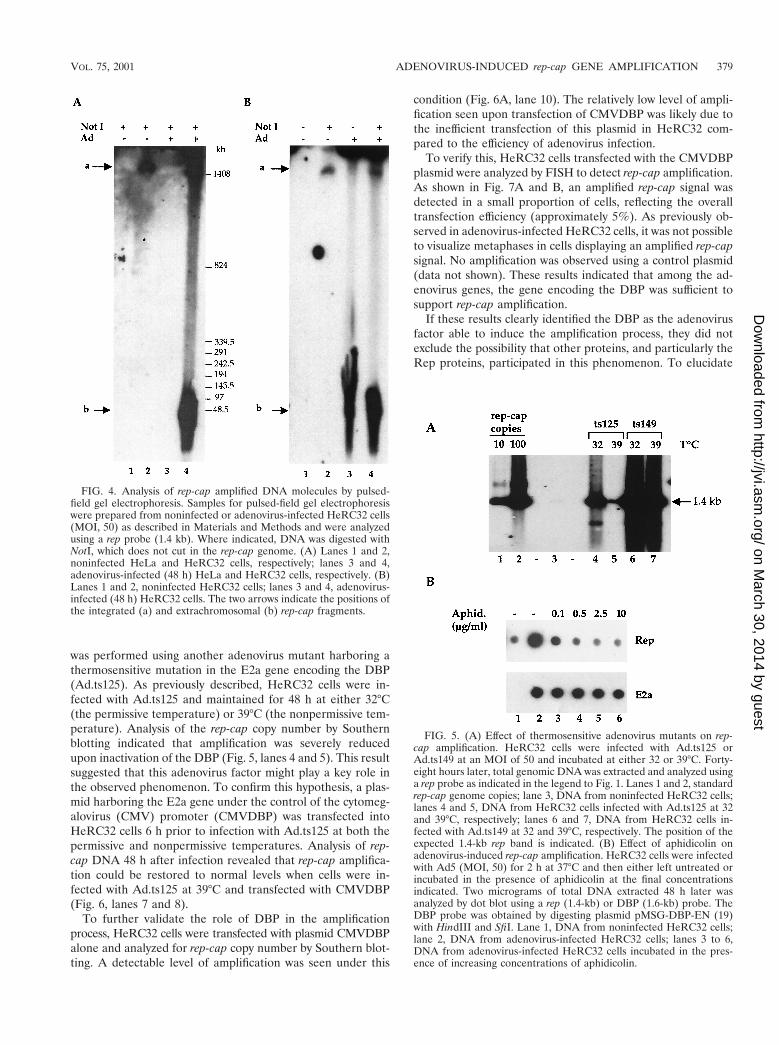
was performed using another adenovirus mutant harboring athermosensitive mutation in the E2a gene encoding the DBP(Ad.ts125). As previously described, HeRC32 cells were in-fected with Ad.ts125 and maintained for 48 h at either 32°C(the permissive temperature) or 39°C (the nonpermissive tem-perature). Analysis of the rep-cap copy number by Southernblotting indicated that amplification was severely reducedupon inactivation of the DBP (Fig. 5, lanes 4 and 5). This resultsuggested that this adenovirus factor might play a key role inthe observed phenomenon. To confirm this hypothesis, a plas-mid harboring the E2a gene under the control of the cytomeg-alovirus (CMV) promoter (CMVDBP) was transfected intoHeRC32 cells 6 h prior to infection with Ad.ts125 at both thepermissive and nonpermissive temperatures. Analysis of rep-cap DNA 48 h after infection revealed that rep-cap amplifica-tion could be restored to normal levels when cells were in-fected with Ad.ts125 at 39°C and transfected with CMVDBP(Fig. 6, lanes 7 and 8).
To further validate the role of DBP in the amplificationprocess, HeRC32 cells were transfected with plasmid CMVDBPalone and analyzed for rep-cap copy number by Southern blot-ting. A detectable level of amplification was seen under this
condition (Fig. 6A, lane 10). The relatively low level of ampli-fication seen upon transfection of CMVDBP was likely due tothe inefficient transfection of this plasmid in HeRC32 com-pared to the efficiency of adenovirus infection.
To verify this, HeRC32 cells transfected with the CMVDBPplasmid were analyzed by FISH to detect rep-cap amplification.As shown in Fig. 7A and B, an amplified rep-cap signal wasdetected in a small proportion of cells, reflecting the overalltransfection efficiency (approximately 5%). As previously ob-served in adenovirus-infected HeRC32 cells, it was not possibleto visualize metaphases in cells displaying an amplified rep-capsignal. No amplification was observed using a control plasmid(data not shown). These results indicated that among the ad-enovirus genes, the gene encoding the DBP was sufficient tosupport rep-cap amplification.
If these results clearly identified the DBP as the adenovirusfactor able to induce the amplification process, they did notexclude the possibility that other proteins, and particularly theRep proteins, participated in this phenomenon. To elucidate
FIG. 5. (A) Effect of thermosensitive adenovirus mutants on rep-cap amplification. HeRC32 cells were infected with Ad.ts125 orAd.ts149 at an MOI of 50 and incubated at either 32 or 39°C. Forty-eight hours later, total genomic DNA was extracted and analyzed usinga rep probe as indicated in the legend to Fig. 1. Lanes 1 and 2, standardrep-cap genome copies; lane 3, DNA from noninfected HeRC32 cells;lanes 4 and 5, DNA from HeRC32 cells infected with Ad.ts125 at 32and 39°C, respectively; lanes 6 and 7, DNA from HeRC32 cells in-fected with Ad.ts149 at 32 and 39°C, respectively. The position of theexpected 1.4-kb rep band is indicated. (B) Effect of aphidicolin onadenovirus-induced rep-cap amplification. HeRC32 cells were infectedwith Ad5 (MOI, 50) for 2 h at 37°C and then either left untreated orincubated in the presence of aphidicolin at the final concentrationsindicated. Two micrograms of total DNA extracted 48 h later wasanalyzed by dot blot using a rep (1.4-kb) or DBP (1.6-kb) probe. TheDBP probe was obtained by digesting plasmid pMSG-DBP-EN (19)with HindIII and SfiI. Lane 1, DNA from noninfected HeRC32 cells;lane 2, DNA from adenovirus-infected HeRC32 cells; lanes 3 to 6,DNA from adenovirus-infected HeRC32 cells incubated in the pres-ence of increasing concentrations of aphidicolin.
FIG. 4. Analysis of rep-cap amplified DNA molecules by pulsed-field gel electrophoresis. Samples for pulsed-field gel electrophoresiswere prepared from noninfected or adenovirus-infected HeRC32 cells(MOI, 50) as described in Materials and Methods and were analyzedusing a rep probe (1.4 kb). Where indicated, DNA was digested withNotI, which does not cut in the rep-cap genome. (A) Lanes 1 and 2,noninfected HeLa and HeRC32 cells, respectively; lanes 3 and 4,adenovirus-infected (48 h) HeLa and HeRC32 cells, respectively. (B)Lanes 1 and 2, noninfected HeRC32 cells; lanes 3 and 4, adenovirus-infected (48 h) HeRC32 cells. The two arrows indicate the positions ofthe integrated (a) and extrachromosomal (b) rep-cap fragments.
VOL. 75, 2001 ADENOVIRUS-INDUCED rep-cap GENE AMPLIFICATION 379
on March 30, 2014 by guest
http://jvi.asm.org/
Dow
nloaded from

this point, HeRC32 cells transfected with the CMVDBP plas-mid were first analyzed by immunofluorescence to detect Repprotein synthesis. As shown in Fig. 8, both spliced and un-spliced Rep proteins were detected in cells transfected with theCMVDBP plasmid alone. This result indicated that Rep pro-teins were expressed in cells transfected with the CMVDBPplasmid and further suggested their involvement in the ampli-fication process. To confirm this hypothesis, a stable cell cloneharboring a mutated rep-cap genome (DRep-HeLa), unable toproduce Rep proteins, was isolated. As expected, no amplifi-cation of integrated rep-cap sequences was detected followingwild-type adenovirus infection (Fig. 6B). Overall, these resultsstrongly suggested that the Rep proteins were implicated in theamplification process.
DISCUSSION
rep-cap amplification, first mentioned by Liu et al. (21), wasdescribed using the HeRC32 cell line (3). Using Southern blotanalysis we showed that 48 h after adenovirus infection, therep-cap copy number was increased at least 100-fold. This in-crease in the number of rep-cap genome copies correlated with
both a high level of Rep and Cap protein synthesis and rAAVassembly, thus supporting the idea that the newly amplifiedrep-cap copies were used as templates for rep and cap geneexpression.
In this study, we further investigated the mechanisms under-lying rep-cap amplification. First, by comparing different stablerep-cap cell lines, we found that among the various cell back-grounds examined, rep-cap amplification occurred preferen-tially in the HeLa-derived cell clones. rep-cap sequences inte-grated in the genome of 293 and TE671 cells were barelyamplified (Fig. 2). This observation suggests that the HeLa cellbackground is critical for this phenomenon, and it can berelated to the fact that, at least in our hands, this cell type isalso optimal for rAAV production (3). Interestingly, cellularextracts from uninfected HeLa cells have been reported to beable to support in vitro AAV replication in the presence ofRep proteins (24, 37). These characteristics might be related tothe presence in these cells of several copies of an HPV18genome in which E2 is deleted (22). Indeed, HPV has alsobeen reported to exert a helper activity for AAV replication(25, 34). Alternatively, these properties might be related to the
FIG. 6. (A) Effect of the adenovirus DBP on rep-cap amplification. HeRC32 cells were infected with Ad.ts125 (MOI, 50) at the indicatedtemperature, and total DNA was analyzed by Southern blotting using a rep probe (1.4 kb) as described in the legend to Fig. 1. Where indicated,the CMVDBP plasmid (10 mg) was transfected into 4 3 106 HeRC32 cells using Exgen (EuroMedex), either alone or 6 h prior to adenovirusinfection. In this case, the transfection was done at 37°C and the cells were switched to the indicated temperature immediately after adenovirusinfection. Lane 1, DNA from noninfected HeLa cells; lanes 2, 3, and 4, standard rep-cap genome copies; lane 5, DNA from HeRC32 cells infectedwith Ad.ts125 at 32°C; lane 6, DNA from HeRC32 cells transfected with CMVDBP and infected with Ad.ts125 at 32°C; lane 7, DNA from HeRC32cells infected with Ad.ts125 at 39°C; lane 8, DNA from HeRC32 cells transfected with CMVDBP and infected with Ad.ts125 at 39°C; lane 9, DNAfrom noninfected HeRC32 cells; lane 10, DNA from HeRC32 cells transfected with the CMVDBP plasmid. (B) Analysis of rep-cap amplificationin DRep-HeLa cells. Total DNA was extracted from uninfected (lane 1) and adenovirus-infected (lane 2) DRep-HeLa cells, digested with PstI, andanalyzed on a Southern blot as previously indicated. Since the deletion in the rep sequence removes one PstI site, the size of the expected bandis 3.8 kb.
FIG. 7. FISH analysis of HeRC32 cells transfected with the CMVDBP plasmid. A total of 4 3 106 HeRC32 cells were transfected with 10 mgof the CMVDBP plasmid using Exgen (EuroMedex). Forty-eight hours later, the cells were prepared for FISH analysis as indicated in Materialsand Methods. The samples were analyzed using a fluorescein-labeled rep-cap probe. Two typical examples of rep-cap amplification are shown. (A)Untransfected HeRC32 cells; (B and C) transfected HeRC32 cells. Magnification, 31,000.
380 TESSIER ET AL. J. VIROL.
on March 30, 2014 by guest
http://jvi.asm.org/
Dow
nloaded from

presence of a cell type-specific factor. We are currently testingthese hypotheses by examining if rep-cap amplification can alsooccur in stable rep-cap cell clones derived from SiHa cellswhich, like the HeLa cells, harbor the HPV genome (22).
Second, this study examined the status of the amplified rep-cap sequences. The data obtained by FISH analysis confirmedthe tremendous increase in the rep-cap copy number detectedby Southern blotting (Fig. 3). However, a clear-cut analysis ofthe status of these amplified sequences was obtained only afterpulsed-field gel electrophoresis of the DNA. Using thismethod, it was found that amplified DNA is present in anextrachromosomal form 48 h after adenovirus infection (Fig. 4).
Amplification of endogenous cellular genes, and particularlyoncogenes, has been extensively described as a common phe-nomenon occurring during tumor progression. Furthermore,cellular gene amplification can also occur as a response tovarious drugs such as DNA-damaging agents (30). Amplifiedsequences are found either integrated, under the form of ho-mogeneously staining regions (HSR), or extrachromosomally.In this case, amplified sequences are usually identified as dou-ble-minute chromosomes (DMs). These high-molecular-weight circular DNA molecules autonomously replicate using acellular replication origin, but, lacking centromeres, they donot segregate with chromosomes and as a consequence areusually lost upon cell division (33). A third class of amplifiedstructures has also been described as submicroscopic circularDNA molecules termed “episomes”. Although the precisemechanism of gene amplification is still unclear, it has beenproposed that DMs, which are the predominant cytogenic
manifestation of gene amplification, are derived from smallerepisomes which progressively enlarge and can lead to HSR byintegrating back in the chromosomal structure (33). The ex-trachromosomal rep-cap sequences detected in our modelmight be defined as episomal structures resembling those lead-ing to DMs. It should be noted that rep-cap amplification wasnot observed following treatment of the cells with DNA-dam-aging agents such as hydroxyurea, UV exposure, and the X-rayirradiation (data not shown). As such, rep-cap amplificationcould represent a unique model of gene amplification.
Third, this study aimed at identifying the minimal cellularand viral factors involved in rep-cap amplification. Using anadenovirus harboring a thermosensitive mutation in the E2bgene, we found that rep-cap amplification still occurred even inthe absence of a functional adenovirus polymerase (Fig. 5A).This result also indicated that adenovirus replication per sewas not required for rep-cap amplification. As shown in thecase of wild-type AAV DNA replication (24), we further dem-onstrated that rep-cap amplification can be completely abol-ished by treating the cells with aphidicolin (Fig. 5B), a drugknown to inhibit the activity of the cellular polymerases a, d,and ε (16, 20). The similarity to wild-type AAV replicationextends further to the requirement for a functional DBP. In-deed, by using an adenovirus harboring a thermosensitive mu-tation in the E2a gene, it was shown that the DBP was essentialfor rep-cap amplification (Fig. 5A and 6A). This protein is theonly adenovirus factor directly implicated in AAV DNA rep-lication. Ward et al. recently showed that DBP was essential invitro, to increase processing of DNA replication, presumably
FIG. 8. Detection of Rep and DBP proteins following transfection of the CMVDBP plasmid into HeRC32 cells. A total of 6 3 104 HeRC32cells grown on glass slides were transfected with 0.4 mg of the CMVDBP plasmid. Forty-eight hours later, the cells were fixed and analyzed byimmunofluorescence using an anti-DBP (26) and an anti-Rep 68/40 (A, B, and C) or anti-Rep 78/52 (D, E, and F) antibody (39). Cells werephotographed with either a fluorescein (A and D) or a rhodamine (B and E) filter. In panels C and F, the two images are superimposed.Magnification, 31,000.
VOL. 75, 2001 ADENOVIRUS-INDUCED rep-cap GENE AMPLIFICATION 381
on March 30, 2014 by guest
http://jvi.asm.org/
Dow
nloaded from

by stabilizing single-stranded templates (35, 36). The involve-ment of DBP in rep-cap amplification was further demon-strated by transfecting a plasmid encoding this protein intoHeRC32 cells and by showing that amplification events couldbe detected by Southern blotting and FISH analysis (Fig. 6Aand 7). Although these results do not exclude the implicationof other adenovirus factors in rep-cap amplification, theyclearly demonstrated that the DBP alone is sufficient.
The last question concerned the role of the AAV geneproducts and particularly the Rep proteins. We found that,upon transfection of the CMVDBP plasmid, both spliced andunspliced Rep proteins were detected (Fig. 8). This observa-tion, which is in agreement with a previous report by Changand Shenk, who demonstrated that DBP was able to trans-activate the p5 promoter (4), suggested the possible involve-ment of Rep proteins in the amplification process. Abolish-ment of Rep proteins in adenovirus-infected stable HeLa cellclones (DRep-HeLa) harboring a rep-cap genome unable toproduce Rep proteins also suggested that they are needed foramplification (Fig. 6B). Importantly, the fact that Rep 78 andRep 52 were still expressed in Ad.ts125-infected HeRC32 cellsat a nonpermissive temperature (data not shown), i.e., underconditions in which amplification no longer occurred (Fig. 5A),further confirmed that Rep proteins, and particularly Rep 78and Rep 52, were not sufficient alone and that a functionalDBP was also needed to induce rep-cap amplification. Finally,although the DBP is able to stimulate Rep protein synthesisalone (4), it is possible that a maximal level of amplificationrequires an optimal rate of rep gene expression that is obtainedonly in the presence of the E1a gene product (5).
Given these findings, we assume that rep-cap amplification isthe result of the activity of at least three main factors: DBP,cellular polymerases, and Rep proteins. It remains to be seenif rep-cap amplification results from the presence of a cellularorigin of replication or from one present in the viral genome.Analysis of stable rep-cap cell clones harboring critical dele-tions of the AAV rep-cap sequences will help resolve this issue.It is possible to envision that the combination of these trans(Rep, DBP, cellular polymerases, and presumably some un-known factor related to HeLa cells) and cis (a viral or cellularorigin of replication) elements generate unscheduled overrep-lication of rep-cap sequences. The fact that the endogenousintegrated rep-cap copies are still detected in adenovirus-in-fected HeRC32 cells (Fig. 4B) indicates that the original rep-cap sequences are not excised from the chromosome duringrep-cap amplification. Further analysis of these extrachromo-somal molecules together with the sequence of the integratedrep-cap genomes will help define the mechanism of amplifica-tion.
In conclusion, our observations constitute a first step towardthe elucidation of the mechanism underlying rep-cap amplifi-cation in HeLa cells. These findings have important implica-tions for the development of future generations of rep-cap celllines able to produce optimal levels of Rep and Cap proteinsupon adenovirus infection.
ACKNOWLEDGMENTS
We are grateful to Michael Linden and Matthew Weitzman forcritical reading of the manuscript. We thank Marie-Claire Devilderand Jean-Paul Moisan for technical assistance.
This work was supported by the Association Francaise contre lesMyopathies (AFM), Vaincre les Maladies Lysosomales (VML), theAssociation Nantaise de Therapie Genique (ANTG), and the Fonda-tion pour la Therapie Genique en Pays de la Loire. P.M. was supportedby a sponsored research agreement from Genopoietic Inc.
J. Tessier and G. Chadeuf contributed equally to this work.
REFERENCES
1. Berns, K. I., and C. Giraud. 1996. Biology of adeno-associated virus. Curr.Top. Microbiol. Immunol. 218:1–23.
2. Carter, B. J. 1990. Adeno-associated virus helper functions, p. 255–282. In P.Tijssen (ed.), Handbook of parvoviruses. CRC Press, Boca Raton, Fla.
3. Chadeuf, G., D. Favre, J. Tessier, N. Provost, P. Nony, J. Kleinschmidt, P.Moullier, and A. Salvetti. 2000. Efficient recombinant adeno-associated virusproduction by a stable rep-cap HeLa cell line correlates with adenovirus-induced amplification of the integrated rep-cap genome. J. Gene Med.2:260–268.
4. Chang, L.-S., and T. Shenk. 1990. The adenovirus DNA-binding proteinstimulates the rate of transcription directed by adenovirus and adeno-asso-ciated virus promoters. J. Virol. 64:2103–2109.
5. Chang, L.-S., Y. Shi, and T. Shenk. 1989. Adeno-associated virus p5 pro-moter contains an adenovirus E1A-inducible element and a binding site forthe major late transcription factor. J. Virol. 63:3479–3488.
6. Chejanovsky, N., and B. J. Carter. 1989. Mutagenesis of an AUG codon inthe adeno-associated virus rep gene: effects on viral replication. Virology173:120–128.
7. Chiorini, J. A., M. D. Weitzman, R. A. Owens, E. Urcelay, B. Safer, and R. M.Kotin. 1994. Biologically active Rep proteins of adeno-associated virus type2 produced as fusion proteins in Escherichia coli. J. Virol. 68:797–804.
8. Clark, K. R., X. Liu, J. P. McGrath, and P. R. Johnson. 1999. Highly purifiedrecombinant adeno-associated virus vectors are biologically active and freeof detectable helper and wild-type viruses. Hum. Gene Ther. 10:1031–1039.
9. Clark, K. R., F. Voulgaropoulou, D. M. Fraley, and P. R. Johnson. 1995. Celllines for the production of recombinant adeno-associated virus. Hum. GeneTher. 6:1329–1341.
10. Conway, J. E., C. M. J. ap Rhys, I. Zolotukhin, S. Zolotukhin, N. Muzyczka,G. S. Hayward, and B. J. Byrne. 1999. High-titer recombinant adeno-asso-ciated virus production utilizing a recombinant herpes simplex virus type Iexpressing AAV-2 rep and cap. Gene Ther. 6:986–993.
11. Dubielzig, R., J. A. King, S. Weger, A. Kern, and J. A. Kleinschmidt. 1999.Adeno-associated virus type 2 protein interactions: formation of pre-encap-sidation complexes. J. Virol. 73:8989–8998.
12. Ensinger, M. J., and H. S. Ginsberg. 1972. Selection and preliminary char-acterization of temperature-sensitive mutants of type 5 adenovirus. J. Virol.10:328–339.
13. Gao, G.-P., G. Qu, L. Z. Faust, R. K. Engdahl, W. Xiao, J. V. Hughes, P. W.Zoltick, and J. M. Wilson. 1998. High-titer adeno-associated viral vectorsfrom a rep/cap cell line and hybrid shuttle virus. Hum. Gene Ther. 9:2353–2362.
14. Graham, F. L., and L. Prevec. 1991. Manipulation of adenovirus vectors, p.109–128. In E. J. Murray (ed.), Gene transfer and protocol. The HumanaPress, Inc., Clifton, N.J.
15. Grimm, D., and J. A. Kleinschmidt. 1999. Progress in adeno-associated virustype 2 vector production: promises and prospects for clinical use. Hum. GeneTher. 10:2445–2450.
16. Ikegami, S., T. Taguchi, M. Ohashi, M. Oguro, H. Nagano, and Y. Mano.1978. Aphidicolin prevents mitotic cell division by interfering with the activ-ity of DNA polymerase-alpha. Nature 275:458–460.
17. Im, D. S., and N. Muzyczka. 1990. The AAV origin binding protein Rep68is an ATP-dependent site-specific endonuclease with DNA helicase activity.Cell 61:447–457.
18. Inoue, N., and D. W. Russel. 1998. Packaging cells based on inducible geneamplification for the production of adeno-associated virus vectors. J. Virol.72:7024–7031.
19. Klessig, D. F., D. E. Brough, and V. Cleghon. 1984. Introduction, stableintegration, and controlled expression of a chimeric adenovirus gene whoseproduct is toxic to the recipient human cell. Mol. Cell. Biol. 4:1354–1362.
20. Lee, M. Y. W. T., C.-K. Tan, K. M. Downey, and A. G. So. 1981. Structuraland functional properties of calf thymus DNA polymerase delta. Prog. Nu-cleic Acids Res. Mol. Biol. 26:83–96.
21. Liu, L., K. R. Clark, and P. R. Johnson. 1999. Production of recombinantadeno-associated virus vectors using a packaging cell line and a hybridrecombinant adenovirus. Gene Ther. 6:293–299.
22. Meissner, J. D. 1999. Nucleotide sequences and further characterization ofhuman papillomavirus DNA present in the CaSki, SiHa and HeLa cervicalcarcinoma cell lines. J. Gen. Virol. 80:1725–1733.
23. Muzyczka, N. 1992. Use of adeno-associated virus as a general transductionvector for mammalian cells. Curr. Top. Microbiol. Immunol. 158:97–129.
24. Ni, T. H., W. F. McDonald, I. Zolotukhin, T. Melendy, S. Waga, B. Stillman,and N. Muzyczka. 1998. Cellular proteins required for adeno-associated
382 TESSIER ET AL. J. VIROL.
on March 30, 2014 by guest
http://jvi.asm.org/
Dow
nloaded from

virus DNA replication in the absence of adenovirus coinfection. J. Virol.72:2777–2787.
25. Ogston, P., K. Raj, and P. Beard. 2000. Productive replication of adeno-associated virus can occur in human papillomavirus type 16 (HPV-16) epi-some-containing keratinocytes and is augmented by the HPV-16 E2 protein.J. Virol. 74:3494–3504.
26. Reich, N. C., P. Sarnow, E. Duprey, and A. J. Levine. 1983. Monoclonalantibodies which recognize native and denatured forms of the adenovirusDNA-binding protein. Virology 128:480–484.
27. Ryan, J. H., S. Zolotukhin, and N. Muzyczka. 1996. Sequence requirementsfor binding of Rep68 to the adeno-associated virus terminal repeats. J. Virol.70:1542–1553.
28. Salvetti, A., G. Chadeuf, S. Oreve, D. Favre, Y. Cherel, P. Champion-Arnaud, J. David-Ameline, and P. Moullier. 1998. Factors influencing re-combinant adeno-associated virus production. Hum. Gene Ther. 9:695–706.
29. Samulski, R. J., L. S. Chang, and T. Shenk. 1989. Helper-free stocks ofrecombinant adeno-associated viruses: normal integration does not requireviral gene expression. J. Virol. 63:3822–3828.
30. Schimke, R. T. 1988. Gene amplification in cultured cells. J. Biol. Chem.263:5989–5992.
31. Smith, R. H., and R. M. Kotin. 1998. The Rep52 gene product of adeno-associated virus is a DNA helicase with 39-to-59 polarity. J. Virol. 72:4874–4881.
32. Snyder, R. O. 1999. Adeno-associated virus-mediated gene delivery. J. GeneMed. 1:166–175.
33. Wahl, G. M. 1989. The importance of circular DNA in mammalian gene
amplification. Cancer Res. 49:1333–1340.34. Walz, C., A. Deprez, T. Dupressoir, M. Durst, M. Rabreau, and J. R. Schle-
hofer. 1997. Interaction of human papillomavirus type 16 and adeno-associ-ated virus type 2 co-infecting human cervical epithelium. J. Gen. Virol.78:1441–1452.
35. Ward, P., F. B. Dean, M. E. O’Donnell, and K. I. Berns. 1998. Role of theadenovirus DNA-binding protein in in vitro adeno-associated virus DNAreplication. J. Virol. 72:420–427.
36. Ward, P., and R. M. Linden. 2000. A role for single-stranded templates incell-free adeno-associated virus DNA replication. J. Virol. 74:744–754.
37. Ward, P., E. Urcelay, R. Kotin, B. Safer, and K. I. Berns. 1994. Adeno-associated virus DNA replication in vitro: activation by a maltose bindingprotein/Rep 68 fusion protein. J. Virol. 68:6029–6037.
38. Weitzman, M. D., S. R. Kyostio, R. M. Kotin, and R. A. Owens. 1994.Adeno-associated virus (AAV) Rep proteins mediate complex formationbetween AAV DNA and its integration site in human DNA. Proc. Natl.Acad. Sci. USA 91:5808–5812.
39. Wistuba, A., S. Weger, A. Kern, and J. A. Kleinschmidt. 1995. Intermediatesof adeno-associated virus type 2 assembly: identification of soluble com-plexes containing Rep and Cap proteins. J. Virol. 69:5311–5319.
40. Wonderling, R. S., S. R. Kyostio, and R. A. Owens. 1995. A maltose-bindingprotein/adeno-associated virus Rep68 fusion protein has DNA-RNA heli-case and ATPase activities. J. Virol. 69:3542–3548.
41. Zhou, X., I. Zolotukhin, D. S. Im, and N. Muzyczka. 1999. Biochemicalcharacterization of adeno-associated virus Rep68 DNA helicase and ATPaseactivities. J. Virol. 73:1580–1590.
VOL. 75, 2001 ADENOVIRUS-INDUCED rep-cap GENE AMPLIFICATION 383
on March 30, 2014 by guest
http://jvi.asm.org/
Dow
nloaded from