Apigenin suppresses insulin-like growth factor I receptor signaling in human prostate cancer: An in...
18
Apigenin Suppresses Insulin-like Growth Factor I Receptor Signaling in Human Prostate Cancer: An In Vitro and In Vivo Study Sanjeev Shukla 1 and Sanjay Gupta 1,2,3 1Department of Urology, Case Western Reserve University, Cleveland, OH 2University Hospitals Case Medical Center, Cleveland, OH 3Case Comprehensive Cancer Center, Cleveland, OH Abstract Deregulation of insulin-like growth factor (IGF)-I/IGF-IR signaling has been implicated in the development and progression of prostate cancer. Agents that can suppress the mitogenic activity of the IGF/IGF-IR growth axis may be of preventive or therapeutic value. We have previously demonstrated that apigenin, a plant flavone, modulates IGF signaling through upregulation of IGFBP-3. In this study, we investigated the mechanism(s) of apigenin action on the IGF/IGF-IR signaling pathway. Exposure of human prostate cancer DU145 cells to apigenin markedly reduced IGF-I-stimulated cell proliferation and induced apoptosis. Apigenin inhibited IGF-I-induced activation of IGF-IR and Akt in DU145 cells. Similar growth inhibitory and apoptotic responses were observed in PC-3 cells, which constitutively over-express this pathway. This effect of apigenin appears to be due partially to reduced autophosphorylation of IGF-IR. Inhibition of p-Akt by apigenin resulted in decreased phosphorylation of GSK-3β along with decreased expression of cyclin D1 and increased expression of p27/kip1. In vivo administration of apigenin to PC-3 tumor xenografts inhibited tumor growth, resulted in IGF-IR inactivation and dephosphorylation of Akt and its downstream signaling. These results suggest that inhibition of cell proliferation and induction of apoptosis by apigenin are mediated, at least in part, by its ability to inhibit IGF/IGF-IR signaling and the PI3K/Akt pathway. Keywords prostate cancer; apigenin; IGF-I; IGF-IR; PI3K-Akt; glycogen synthase kinase-3 INTRODUCTION The insulin-like growth factor (IGF) axis is an important modulator of growth and development [1,2]. It is comprised of six affinity binding proteins, several low-affinity binders, proteases and receptors [1-3]. The two IGF ligands (IGF-I and IGF-II) modulate a diverse range of biological activities including cell growth, differentiation and apoptosis; imbalance of this growth axis may preferentially favor uncontrolled cell proliferation and malignant transformation [2,3]. IGFs are mostly produced in the liver and in small amounts in the local tissues, where they act in an autocrine and paracrine manner. The biological functions of IGFs are mediated primarily by the type I IGF receptor (IGF-IR), which is a heterotetrameric transmembrane protein that is comprised of two subunits, α and β. The β subunit expresses Correspondence to: Sanjay Gupta, Ph.D., Department of Urology, The James & Eilleen Dicke Research Laboratory, Case Western Reserve University & University Hospitals Case Medical Center, 10900 Euclid Avenue, Cleveland, Ohio 44106 Phone: (216) 368-6162, Fax: (216) 368-0213, E-mail: [email protected]. NIH Public Access Author Manuscript Mol Carcinog. Author manuscript; available in PMC 2010 March 1. Published in final edited form as: Mol Carcinog. 2009 March ; 48(3): 243–252. doi:10.1002/mc.20475. NIH-PA Author Manuscript NIH-PA Author Manuscript NIH-PA Author Manuscript
-
Upload
independent -
Category
Documents
-
view
1 -
download
0
Transcript of Apigenin suppresses insulin-like growth factor I receptor signaling in human prostate cancer: An in...

Apigenin Suppresses Insulin-like Growth Factor I ReceptorSignaling in Human Prostate Cancer: An In Vitro and In Vivo Study
Sanjeev Shukla1 and Sanjay Gupta1,2,3
1Department of Urology, Case Western Reserve University, Cleveland, OH
2University Hospitals Case Medical Center, Cleveland, OH
3Case Comprehensive Cancer Center, Cleveland, OH
AbstractDeregulation of insulin-like growth factor (IGF)-I/IGF-IR signaling has been implicated in thedevelopment and progression of prostate cancer. Agents that can suppress the mitogenic activity ofthe IGF/IGF-IR growth axis may be of preventive or therapeutic value. We have previouslydemonstrated that apigenin, a plant flavone, modulates IGF signaling through upregulation ofIGFBP-3. In this study, we investigated the mechanism(s) of apigenin action on the IGF/IGF-IRsignaling pathway. Exposure of human prostate cancer DU145 cells to apigenin markedly reducedIGF-I-stimulated cell proliferation and induced apoptosis. Apigenin inhibited IGF-I-inducedactivation of IGF-IR and Akt in DU145 cells. Similar growth inhibitory and apoptotic responses wereobserved in PC-3 cells, which constitutively over-express this pathway. This effect of apigeninappears to be due partially to reduced autophosphorylation of IGF-IR. Inhibition of p-Akt by apigeninresulted in decreased phosphorylation of GSK-3β along with decreased expression of cyclin D1 andincreased expression of p27/kip1. In vivo administration of apigenin to PC-3 tumor xenograftsinhibited tumor growth, resulted in IGF-IR inactivation and dephosphorylation of Akt and itsdownstream signaling. These results suggest that inhibition of cell proliferation and induction ofapoptosis by apigenin are mediated, at least in part, by its ability to inhibit IGF/IGF-IR signaling andthe PI3K/Akt pathway.
Keywordsprostate cancer; apigenin; IGF-I; IGF-IR; PI3K-Akt; glycogen synthase kinase-3
INTRODUCTIONThe insulin-like growth factor (IGF) axis is an important modulator of growth and development[1,2]. It is comprised of six affinity binding proteins, several low-affinity binders, proteasesand receptors [1-3]. The two IGF ligands (IGF-I and IGF-II) modulate a diverse range ofbiological activities including cell growth, differentiation and apoptosis; imbalance of thisgrowth axis may preferentially favor uncontrolled cell proliferation and malignanttransformation [2,3]. IGFs are mostly produced in the liver and in small amounts in the localtissues, where they act in an autocrine and paracrine manner. The biological functions of IGFsare mediated primarily by the type I IGF receptor (IGF-IR), which is a heterotetramerictransmembrane protein that is comprised of two subunits, α and β. The β subunit expresses
Correspondence to: Sanjay Gupta, Ph.D., Department of Urology, The James & Eilleen Dicke Research Laboratory, Case WesternReserve University & University Hospitals Case Medical Center, 10900 Euclid Avenue, Cleveland, Ohio 44106 Phone: (216) 368-6162,Fax: (216) 368-0213, E-mail: [email protected].
NIH Public AccessAuthor ManuscriptMol Carcinog. Author manuscript; available in PMC 2010 March 1.
Published in final edited form as:Mol Carcinog. 2009 March ; 48(3): 243–252. doi:10.1002/mc.20475.
NIH
-PA Author Manuscript
NIH
-PA Author Manuscript
NIH
-PA Author Manuscript

intrinsic tyrosine kinase activity and is activated upon ligand binding to the α subunit. Tyrosinekinase activation results in autophosphorylation within the kinase domain, particularly atTyr1131, Tyr1135 and Tyr1136, leading to downstream signaling [4]. The activated IFG-IRphosphorylates an adaptor protein, IRS-1 which is involved in the activation ofphosphatidylinositol 3-kinase (PI3K) and mitogen-activated protein kinase (MAPK) pathways[5].
A number of laboratory and population-based studies have demonstrated that insulin-likegrowth factor (IGF) physiology plays a critical role in the development and progression ofprostate cancer [6,7]. Prospective case controlled studies have demonstrated a positivecorrelation between circulating IGF-I levels in healthy men and the risk of developing prostatecancer [8,9]. There is evidence that in the normal prostate, IGFs are produced by stromal cells,and it is known that normal epithelial cells express IGF-IR, suggesting a paracrine mode ofregulation [10]. Studies have shown that IGF may increase proliferation of prostate cancercells, whereas antisense-mediated inhibition of IGF-IR suppresses cell invasiveness and in vivotumor growth [11].Deregulated expression of IGF-I in prostatic epithelium leads to neoplasiain transgenic mice [12]. Upregulation of IGF-I expression has been found in neoplastic prostateepithelial cells; it has been postulated that this is an adaptive response that may contribute tothe evolution of androgen-independent prostate cancer [13]. Currently, IGF/IGFBP-3 plasmalevels are being evaluated as potential surrogate biomarkers in patients with a risk of prostatecancer [14]. Since IGF signaling has been shown to play an important role in prostate cancerprogression, the IGF-IR could be a potential target in the development of a new strategy forprostate cancer prevention and treatment.
Plant flavonoids have gained considerable attention as anticancer agents. Apigenin (4’, 5, 7,-trihydroxyflavone), a naturally occurring plant flavone abundantly present in common fruitsand vegetables, has been shown to possess cancer preventive and therapeutic properties [15and references therein]. We have previously shown that apigenin selectively inhibits cellgrowth and induces apoptosis in cancer cells without affecting normal cells [16]. Apigenin hasbeen shown to be effective in inhibiting growth in several different types of human cancer celllines including leukemia, and carcinomas of the breast, colon, lungs, skin, thyroid, and prostate[17-22]. Apigenin is a potent inhibitor of several protein kinases, including epidermal growthfactor receptor and src tyrosine kinase [23]. Apigenin has been shown to modulate theexpression of PI3K-Akt, MAPKs (ERK1/2, c-Jun-N-terminal kinase, and p38), casein kinase-2and other upstream kinases involved in the development and progression of cancer [24-27].Apigenin has also been shown to suppress angiogenesis in melanoma and carcinoma of thebreast, skin and colon [28-31]. We have demonstrated that apigenin induces apoptosis inprostate tumor xenografts through upregulation of IGFBP-3 [32]. Since apigenin inhibitsMAPK and PI3K-Akt kinases and induces IGFBP-3 in prostate cancer [33], we postulated thatthese inhibitory actions might be effected through the IGF-IR pathway. In the present studywe demonstrate the effect of apigenin in IGF/IGF-IR signaling both in cell culture and in anin vivo model of prostate cancer.
MATERIALS AND METHODSCell lines and treatments
Androgen-refractory human prostate cancer DU145 and PC-3 cells, obtained from AmericanType Culture Collection (Manassas, VA), were cultured in RPMI 1640 supplemented with 5%fetal bovine serum and 1% penicillin-streptomycin. Monolayer cultures of DU145 and PC-3cells were maintained at 37°C and 5% CO2 in a humid environment. The cells were treatedwith varying concentrations of apigenin dissolved in DMSO, which was provided to the controland treated groups with maximum final concentration of 0.1%, v/v.
Shukla and Gupta Page 2
Mol Carcinog. Author manuscript; available in PMC 2010 March 1.
NIH
-PA Author Manuscript
NIH
-PA Author Manuscript
NIH
-PA Author Manuscript

Cell cycle analysisHuman prostate cancer DU145 cells were seeded in a 100 mm dish at 40–50% confluence andincubated overnight. Cells were washed once with PBS and grown in serum-free medium. In24 h, the old medium was discarded and fresh serum-free medium containing 25ng/mL IGF-I was added with or without apigenin. The cells were incubated for 24 h. In another experimentasynchronized (70-80%) confluent PC-3 cells were treated with various doses of apigenin for24 h. After treatment cells were collected, washed twice with chilled PBS and spin in a coldcentrifuge at X 600g for 10 minutes. The pellet was fixed by re-suspending in 50 μl PBS and450 μl chilled methanol for 1 h at 4°C. The cells were washed twice with PBS at X 600g for5 min and again suspended in 500 μl PBS and incubated with 5 μl RNAse (20 μg/ml finalconcentration) for 30 min at 37°C. The cells were chilled over ice for 10 min and stained withpropidium iodide (50 μg/ml final concentration) for 1 h and analyzed by flow cytometry andevaluated using Cell Quest & ModFit cell cycle analysis software.
ImmunoblottingThe cell lysate was cleared by centrifugation at 13,000 rpm for 15 min at 4°C, and the proteinconcentration was measured in the supernatant by Bio-Rad assay using the manufacturer’sprotocol (Bio-Rad Laboratories, Hercules, CA). 20-40μg of supernatant proteins were resolvedby sodium-dodecyl sulfate–polyacrylamide gel electrophoresis (SDS–PAGE) using 4–20%Tris-glycine polyacrylamide gel and then transferred onto the nitrocellulose membrane eitherfor 2 h or overnight. The blots were blocked using 5% nonfat dry milk in Tris buffered salinecontaining 0.05% Tween-20 and probed using primary antibodies for IGF-IR (Cat#3022), p-IGF-IR Tyr1131 (Insulin Receptor, Tyr1146, Cat#3021), GSK-3β (Cat#9332), p-GSK-3α/βSer21/9 (Cat#9331), from Cell Signaling Technology (Beverly, MA), p-Akt Ser473
(Cat#SC-7985-R) from Santa Cruz, CA and Akt1/2 (Cat#SC-8312), cyclin D1 (Cat #MS-210),p27/kip1 (Cat # MS-256), from NeoMarkers (Fremont, CA) in blocking buffer either at roomtemperature for 1 h or overnight at 4°C. The membrane was then incubated with appropriatesecondary antibody and the immunoreactive bands were visualized using an enhancedchemiluminescence kit (ECL; Amersham Life Sciences Inc.). The membrane was furtherstripped and reprobed with anti-α-tubulin antibody (Santa Cruz, Cat#SC-8035) to ensure theequal protein loading.
Tumor xenograft studiesPC-3 tumors were grown subcutaneously in athymic nude mice. Approximately 1 million PC-3cells suspended in 0.05 ml of medium and mixed with 0.05 ml of Matrigel were subcutaneouslyinjected into the left and right flank of each mouse to initiate tumor growth. The animals wereequally divided in two groups. The first group received only 0.2 ml of vehicle material bygavage daily and served as a control group. The second group of animals received 50μg/mouse/day doses of apigenin in vehicle, respectively, for 8 weeks. These doses are comparable to thedaily consumption of flavonoid in humans as reported in previously published studies (29,30). Apigenin feeding was started 2 weeks after cell inoculation and was continued for 8 weeks.Animals were monitored daily, and their body weights were recorded weekly throughout thestudies. Once the tumors started growing, their sizes were measured twice weekly in twodimensions with calipers. Tumor volume was determined with the equation: volume =(width)2 × length × π/6. Tumor measurements were taken by one individual and performed induplicate to confirm measurements. At the termination of the experiment, tumors were excisedand weighed to record wet tumor weight. A portion of the tumors from control and treatedanimals was used for preparation of tumor lysate used in further experiments.
Shukla and Gupta Page 3
Mol Carcinog. Author manuscript; available in PMC 2010 March 1.
NIH
-PA Author Manuscript
NIH
-PA Author Manuscript
NIH
-PA Author Manuscript

Apoptosis by ELISAApoptosis was assessed by M30-Apoptosense™ ELISA kit (Alexis Biochemicals, San Diego,CA) according to the manufacturer’s protocol and color developed was read at 450-nm againstthe blank and values were plotted against standards provided and expressed as units per liter.
Statistical analysisChanges in tumor volume and body weight during the course of the experiments werevisualized by scatter plot. Differences in tumor volume (mm3) and body weight at thetermination of the experiment among two groups were examined using analysis of variance(ANOVA) followed by Tukey’s multiple comparison procedure. The statistical significanceof differences between control and treatment group was determined by simple ANOVAfollowed by multiple comparison tests. All tests were two-tailed and P values less than 0.05were considered to be statistically significant.
RESULTSSince IGF-I/IGF-IR axis plays an important role in cell growth and proliferation, we initiatedstudies to evaluate IGF-I mediated phosphorylation and its downstream signaling in humanprostate cancer DU145 cells. As shown in figure 1A, treatment of serum starved DU145 cellswith 25ng/mL IGF-I caused a rapid phosphorylation of IGF-IR which was assessed throughphospho-specific antibody directed against Tyr1131 and Ser1146. This phosphorylation wasattained as early as 10 min with highest levels attained at 30 min post IGF-I incubation, andthis phosphorylation was maintained up to 60 min. Similar phosphorylation response wasobserved in Akt at Ser473 in DU145 cells after IGF-I treatment.
Previous studies have demonstrated that knockdown of the IGF-IR receptor through siRNA orantisense approach affects the two major signaling pathways, the ERK and Akt pathways[11,34]. Since incubation with IGF-I cause increase in Akt phosphorylation at Ser473 in DU145cells (figure 1A), therefore, we next determined whether inhibition of Akt phosphorylationcauses a similar response to IGF-IR phosphorylation. For these studies we incubated DU145along with 25ng/mL IGF-I for 8 h with and without PI3K-Akt inhibitor LY294002. Exposureof cells to a 10μM concentration of LY294002 caused a decrease in phosphorylation of Akt atSer473 whereas no significant decrease in IGF-IR phosphorylation was observed in these cells(Figure 1B).
In a further series of experiments we tested whether or not apigenin could inhibit IGF-I inducedproliferative activity in prostate cancer DU145 cells. We pretreated serum starved cells withvarying concentrations of apigenin before stimulation with IGF-I. As shown in figure 2, cellcycle analysis of propidium iodide stained cells from the serum starved control revealed asignificant block of 76.6% in the G1-phase of the cell cycle. Treatment with 25ng/mL IGF-Iinduced almost a 2-fold increase in the number of cells in the S-phase along with a concomitantdecrease in the number of cells in the G1-phase (61.3%) of the cell cycle. These results are inagreement with previous reports that IGF-I can stimulate DNA synthesis and proliferation inDU145 cells [35]. We also determined the effects of LY294002 on cell cycle progression inDU145 cells stimulated with IGF-I. Treatment of cells with 10μM LY294002 in the presenceof IGF-I increased the percentage of cells in the G1-phase to 71.4% with a decreased percentageof cells in the S-phase (15.4%). Treatment of DU145 cells with apigenin caused a dose-dependent increase of cells in the G1-phase from 72.2% to 78.1% along with inhibition of theIGF-I induced progression of cells into the S-phase. Furthermore, treatment of cells withapigenin caused a dose-dependent increase of cells in the sub-G1 phase, a finding which isindicative of apoptosis.
Shukla and Gupta Page 4
Mol Carcinog. Author manuscript; available in PMC 2010 March 1.
NIH
-PA Author Manuscript
NIH
-PA Author Manuscript
NIH
-PA Author Manuscript

Next we evaluated the effects of apigenin on IGF-I/IGF-IR signaling after stimulation withIGF-I. Treatment of cells with apigenin significantly inhibited the IGF-IR phosphorylationinduced by IGF-I. This effect was observed with 5μM apigenin and was persistent in apigeninconcentrations up to 40μM. The blockade of IGF-IR activation by apigenin further resulted insuppression of IGF-I-induced activation of Akt (Figure 3). IGF-I activates PI3K-Akt, and theactivated Akt in turn activates its downstream signals to promote cell growth and proliferation,effects that are attributed in part to enhanced phosphorylation of glycogen synthase kinase(GSK)-3β and increased levels of cyclinD1 protein [4,5]. Therefore next we evaluated theeffects of apigenin on some of the downstream targets of Akt. As shown in figure 3, IGF-Iinduced phosphorylation of GSK-3β and increased cyclin D1 protein in DU145 cells. Apigenintreatment caused a dose-dependent decrease in the phosphorylation of GSK-3β and cyclinD1.We also evaluated the effect of apigenin on the expression of p27/kip1, the cyclin-dependentkinase inhibitor, which is downregulated during cell proliferation in prostate cancer cells[36]. Treatment of DU145 cells with apigenin caused a dose-dependent increase in the proteinlevels of p27/kip1, which was more pronounced at 10-40μM apigenin treatments (Figure 3).
It is of interest that DU145 cells have low basal levels of phosphorylated Akt, whereas twoother prostate cancer cell lines, PC-3 and LNCaP, have constitutively high basal levels ofphospho-Akt. This observation has been attributed to the loss of PTEN (phosphatase and tensinhomologue deleted on chromosome 10) function in both these cell lines [37,38]. Consequently,we investigated whether apigenin could suppress growth in PC-3 cells, which exhibit highconstitutive expression of phospho-Akt and IGF-IR. We treated log phase growing cells with10-40μM concentrations of apigenin. Exposure of PC-3 cells to apigenin resulted in anincreased accumulation of cells in the G1-phase from 64.8% and 69.4% at 10- and 20-μMdoses. A simultaneous reduction of the percentage of cells in the S-phase was observed afterapigenin treatment. Furthermore, apigenin treatment caused an increase in the sub-G1percentage of cells, which is indicative of apoptosis (Figure 4).
Next we evaluated the effects of apigenin on the inhibition of constitutively expressed IGF-IRand Akt phosphorylation in PC-3 cells. Treatment of cells with apigenin at doses of 5-40μMcaused a significant decrease in IGF-IR phosphorylation and phospho-Akt levels in a dose-dependent fashion. Furthermore, apigenin treatment caused a decrease in the phosphorylationof GSK-3β and cyclinD1, with concomitant increase in p27/kip1 levels, an effect earlierobserved in DU145 cells after IGF-I stimulation (Figure 5). These results indicate that apigeninis capable of suppressing both stimulated and constitutive IGF-IR and Akt phosphorylation,with resultant effects on the downstream signaling in prostate cancer cells.
Next we evaluated the effects of apigenin on PC-3 tumor xenografts in nude mice. As shownin figure 6A, growth of PC-3 tumors in nude mice was significantly inhibited by administrationof apigenin to these mice. The tumor volume was reduced by 51% (p<0.0001) and the wetweight of tumor was decreased by 40% (p < 0.001) in nude mice receiving apigenin. Noevidence of systemic toxicity was noted in these mice, as evidenced by their continued normalfood intake and their maintenance of body weight (data not shown). Earlier results in cellculture demonstrated that apigenin induces decreased proliferation of PC-3 cells [33]; thereforewe evaluated the effects of apigenin intake on the induction of apoptosis in tumor xenografts.As shown in figure 6B, oral administration of apigenin at doses of 50μg/mouse/day resultedin a marked induction of apoptosis in PC-3 tumor xenografts as shown by M-30 reactivitymeasured by ELISA assay. Compared to vehicle-treated controls, 2.86-fold increases(p<0.0001) in the induction of apoptosis were observed in PC-3 tumors after apigenintreatment. Furthermore, consistent with the findings in cell culture, apigenin administration tonude mice resulted in a marked decrease in the expression of IGF-IR, phospho-IGF-IR, p-AktSer473, p-GSK3β, cyclinD1, with concomitant increases in the p27/kip1 levels, compared tomice receiving vehicle treatment. Likewise, the expression of cyclinD1 and PCNA levels were
Shukla and Gupta Page 5
Mol Carcinog. Author manuscript; available in PMC 2010 March 1.
NIH
-PA Author Manuscript
NIH
-PA Author Manuscript
NIH
-PA Author Manuscript

also inhibited after apigenin treatment, indicating apigenin-mediated suppression of tumor cellproliferation (Figure 6C).
DISCUSSIONNumerous studies have demonstrated that mitogenic as well as cell survival signaling via theIGF/IGF-IR pathway is constitutively activated and that such signaling provides growth andsurvival advantages to prostate cancer cells [1-5]. It is widely accepted that the IGF-axisactivates anti-apoptotic signaling, which in turn upregulates the PI3K and MAPK pathways incancer cells [4,5]. The induction of these pathways occurs through the IGF-IR signalingpathway. Therefore, inhibition of IGF-IR signaling is critical to inhibition of prostate cancergrowth and survival. In our previous studies we demonstrated induction of IGFBP-3, thebinding protein for IGF-I, by apigenin, thereby reducing the amount of ligand available forinteraction with IGF-IR [32]. In this study, we demonstrate that apigenin blocks IGF-I regulatedevents and modulate IGF-I/IGF-IR signaling pathways in prostate cancer cells.
The IGF-IR is a glycoprotein complex consisting of two transmembrane β-subunits and twoextracellular α-subunits. The ligand binding specificity is conferred by the α-subunits, whereasthe β-subunits contain the tyrosine kinase. The expression of the receptor subunits appears tobe transcriptionally downregulated by IGF-I through a negative feedback inhibitionmechanism [39]. Binding of IGF-I to its receptor causes activation of the receptor tyrosinekinase and its autophosphorylation [40]. Our studies demonstrate that apigenin is capable ofsuppressing autophosphorylation in prostate cancer cells both in constitutively expressed celllines and after IGF-I stimulation. Furthermore, IRS-1 is a critical substrate for the IGF-Ireceptor tyrosine kinase and contains multiple phosphorylation sites [41]. IRS-1 functions asan adaptor protein for other SH2 proteins like PI3K [42]. IGF-IR not only mediates themitogenic and anti-apoptotic actions of IGF-I but also may stimulate differentiation, dependingon the cell type and other factors in the microenvironment [1-5]. Previously, we havedemonstrated that apigenin causes inhibition of IGF-I-induced IRS-1 phosphorylation inprostate cancer cells, an effect that may be one of the mechanisms that contribute to inhibitionof the prostate cancer cell proliferation cascade.
Evidence suggests that PI3K/Akt and MAPK are important pathways in transmitting IGF-Imitogenic and antiapoptotic signals [4,5]. Our previous studies using specific inhibitors ofMEK1/2 and p38 demonstrated an inhibition of PC-3 cell proliferation in parallel withinhibition of phophorylation of ERK1/2 and p38 [33]. Interestingly, high phosphorylation ofERK1/2 was observed after treatment of cells with apigenin which usually do not activate thedownstream signaling molecules that favors cell proliferation [33,43]. Akt is another majorinfluence in IGF-I signaling, and a number of factors regulated by Akt have been shown to beinvolved in regulating cell survival and proliferation [44]. In the present study we observedthat apigenin blocked constitutive as well as IGF-I induced activation of Akt.
Cyclin proteins are involved in regulating entry into different phases of the cell cycle, andcyclin D1 is necessary for progression through the G1 phase [33]. Studies have demonstratedthat cyclin D1 is regulated at a number of different levels, but one primary mechanism ofregulation is through protein degradation. Phosphorylation of cyclin D1 by GSK-3β has beendemonstrated to result in its exclusion from the nucleus to the cytoplasm and it’s targeting forubiquitinin degradation [45]. Akt is known to phosphorylate GSK-3β, inactivating it andfacilitating elevation of levels of cyclin D1 protein. Our results demonstrate that apigenininhibits the expression of cyclin D1 in association with reduced phosphorylation of GSK-3βin prostate cancer cells, and hence appears to influence cell cycle arrest and/or apoptosis.
Shukla and Gupta Page 6
Mol Carcinog. Author manuscript; available in PMC 2010 March 1.
NIH
-PA Author Manuscript
NIH
-PA Author Manuscript
NIH
-PA Author Manuscript

Another mechanism proposed for the involvement of Akt in cell cycle regulation is thephosphorylation and inactivation of forkhead transcription factors that can regulate the cellcycle kinase inhibitor, p27/kip1 [46]. Our studies demonstrate that apigenin caused increasedp27/kip1 levels in human prostate cancer cells, which corroborates our previous findings[47]. We did not investigate the effects of apigenin on various forkhead transcription factorsin prostate cancer cells; additional studies are required to further elucidate these mechanisms.
Although cell culture studies are informative in demonstrating the effects of apigenin uponIGF-I regulated mechanisms, it is critically important to determine whether these same effectsare operative in vivo. Our in vivo studies, providing an intake of 50μg/day of apigenin to micewith PC-3 tumor xenografts, demonstrate that apigenin intake significantly inhibits tumorproliferation and induces apoptosis, without any apparent signs of toxicity. These results areconsistent with inhibition of IFG-IR signaling in tumor xenografts. Since IGF-IRs are over-expressed in many human cancers, including prostate cancer, development of ways to blockadethe IGF-IR signaling pathways is a strategy that might be highly successful in the preventionand/or therapy of prostate cancer.
Our present studies clearly demonstrate that apigenin effectively blocks the IGF-IR signalingpathway both in cell cultures and in prostate cancer xenografts in vivo. The dose of 50μg/dayapigenin used in our studies corresponds to consumption of approximately 120μg/day offlavonoid by an adult human, an intake that results in effective physiologically attainable serumconcentrations in adult humans [48,49]. Our studies support the notion that apigenin may beworthy of further development as a chemopreventive or chemotherapeutic agent, exploitingits effectiveness in inhibiting IGF-IR signaling in prostate cancer.
AcknowledgementsThis research was supported by the Athymic Animal Core Facility of the Comprehensive Cancer Center of CaseWestern Reserve University and University Hospitals Case Medical Center (P30 CA43703). The authors are thankfulto Dr. Pingfu Fu for performing statistical evaluations for this study.
Financial Support: This work was supported by grants from United States Public Health Services RO1 CA108512,RO3 CA094248, RO3 CA099049 and partial support of funds from Cancer Research and Prevention Foundation toSG.
References1. Jones JI, Clemmons DR. Insulin-like growth factors and their binding proteins: biological actions.
Endocr Rev 1995;16:3–34. [PubMed: 7758431]2. Silha JV, Murphy LJ. Insulin-like growth factor binding proteins in development. Adv Exp Med Biol
2005;567:55–89. [PubMed: 16370136]3. Grimberg A, Cohen P. Role of insulin-like growth factors and their binding proteins in growth control
and carcinogenesis. J Cell Physiol 2000;183:1–9. [PubMed: 10699960]4. Baserga R. The contradictions of the insulin-like growth factor 1 receptor. Oncogene 2000;19:5574–
5581. [PubMed: 11114737]5. Kurihara S, Hakuno F, Takahashi S. Insulin-like growth factor-I-dependent signal transduction
pathways leading to the induction of cell growth and differentiation of human neuroblastoma cell lineSH-SY5Y: the roles of MAP kinase pathway and PI 3-kinase pathway. Endocr J 2000;47:739–751.[PubMed: 11228049]
6. Yu H, Rohan T. Role of the insulin-like growth factor family in cancer development and progression.J Natl Cancer Inst 2000;92:1472–1489. [PubMed: 10995803]
7. Djavan B, Waldert M, Seitz C, Marberger M. Insulin-like growth factors and prostate cancer. WorldJ Urol 2001;19:225–233. [PubMed: 11550779]
8. Wolk A, Mantzoros CS, Andersson SO, et al. Insulin-like growth factor 1 and prostate cancer risk: apopulation-based, case-control study. J Natl Cancer Inst 1998;90:911–915. [PubMed: 9637140]
Shukla and Gupta Page 7
Mol Carcinog. Author manuscript; available in PMC 2010 March 1.
NIH
-PA Author Manuscript
NIH
-PA Author Manuscript
NIH
-PA Author Manuscript

9. Stattin P, Bylund A, Rinaldi S, et al. Plasma insulin-like growth factor-I, insulin-like growth factor-binding proteins, and prostate cancer risk: a prospective study. J Natl Cancer Inst 2000;92:1910–1917.[PubMed: 11106682]
10. Li SL, Goko H, Xu ZD, et al. Expression of insulin-like growth factor (IGF)-II in human prostate,breast, bladder, and paraganglioma tumors. Cell Tissue Res 1998;291:469–479. [PubMed: 9477303]
11. Grzmil M, Hemmerlein B, Thelen P, Schweyer S, Burfeind P. Blockade of the type I IGF receptorexpression in human prostate cancer cells inhibits proliferation and invasion, up-regulates IGFbinding protein-3, and suppresses MMP-2 expression. J Pathol 2004;202:50–59. [PubMed:14694521]
12. DiGiovanni J, Kiguchi K, Frijhoff A, et al. Deregulated expression of insulin-like growth factor 1 inprostate epithelium leads to neoplasia in transgenic mice. Proc Natl Acad Sci U S A 2000;97:3455–3460. [PubMed: 10737798]
13. Nickerson T, Chang F, Lorimer D, Smeekens SP, Sawyers CL, Pollak M. In vivo progression ofLAPC-9 and LNCaP prostate cancer models to androgen independence is associated with increasedexpression of insulin-like growth factor I (IGF-I) and IGF-I receptor (IGF-IR). Cancer Res2001;61:6276–6280. [PubMed: 11507082]
14. Li L, Yu H, Schumacher F, Casey G, Witte JS. Relation of serum insulin-like growth factor-I (IGF-I) and IGF binding protein-3 to risk of prostate cancer (United States). Cancer Causes Control2003;14:721–726. [PubMed: 14674736]
15. Patel D, Shukla S, Gupta S. Apigenin and cancer chemoprevention: progress, potential and promise.Int J Oncol 2007;30:233–245. [PubMed: 17143534]review
16. Gupta S, Afaq F, Mukhtar H. Selective growth-inhibitory, cell-cycle deregulatory and apoptoticresponse of apigenin in normal versus human prostate carcinoma cells. Biochem Biophys ResCommun 2001;287:914–920. [PubMed: 11573952]
17. Wang IK, Lin-Shiau SY, Lin JK. Induction of apoptosis by apigenin and related flavonoids throughcytochrome c release and activation of caspase-9 and caspase-3 in leukaemia HL-60 cells. Eur JCancer 1999;35:1517–1525. [PubMed: 10673981]
18. Way TD, Kao MC, Lin JK. Apigenin induces apoptosis through proteasomal degradation of HER2/neu in HER2/neu-overexpressing breast cancer cells via the phosphatidylinositol-3’-kinase/Akt-dependent pathway. J Biol Chem 2004;279:4479–4489. [PubMed: 14602723]
19. Liu LZ, Fang J, Zhou Q, Hu X, Shi X, Jiang BH. Apigenin inhibits expression of vascular endothelialgrowth factor and angiogenesis in human lung cancer cells: implication of chemoprevention of lungcancer. Mol Pharmacol 2005;68:635–643. [PubMed: 15947208]
20. Abu-Yousif AO, Smith KA, Getsios S, Green KJ, Van Dross RT, Pelling JC. Enhancement of UVB-induced apoptosis by apigenin in human keratinocytes and organotypic keratinocyte cultures. CancerRes 2008;68:3057–3065. [PubMed: 18413777]
21. Yin F, Giuliano AE, Van Herle AJ. Signal pathways involved in apigenin inhibition of growth andinduction of apoptosis of human anaplastic thyroid cancer cells (ARO). Anticancer Res1999;19:4297–4303. [PubMed: 10628390]
22. Shukla S, Gupta S. Molecular mechanisms for apigenin-induced cell-cycle arrest and apoptosis ofhormone refractory human prostate carcinoma DU145 cells. Mol Carcinog 2004;39:114–126.[PubMed: 14750216]
23. Geahlen RL, Koonchanok NM, McLaughlin JL, Pratt DE. Inhibition of protein-tyrosine kinaseactivity by flavanoids and related compounds. J Nat Prod 1989;52:982–986. [PubMed: 2607357]
24. Lee WJ, Chen WK, Wang CJ, Lin WL, Tseng TH. Apigenin inhibits HGF-promoted invasive growthand metastasis involving blocking PI3K/Akt pathway and beta 4 integrin function in MDA-MB-231breast cancer cells. Toxicol Appl Pharmacol 2008;226:178–191. [PubMed: 17961621]
25. Gopalakrishnan A, Xu CJ, Nair SS, Chen C, Hebbar V, Kong AN. Modulation of activator protein-1(AP-1) and MAPK pathway by flavonoids in human prostate cancer PC3 cells. Arch Pharm Res2006;29:633–644. [PubMed: 16964758]
26. Hessenauer A, Montenarh M, Gotz C. Inhibition of CK2 activity provokes different responses inhormone-sensitive and hormone-refractory prostate cancer cells. Int J Oncol 2003;22:1263–1270.[PubMed: 12738992]
Shukla and Gupta Page 8
Mol Carcinog. Author manuscript; available in PMC 2010 March 1.
NIH
-PA Author Manuscript
NIH
-PA Author Manuscript
NIH
-PA Author Manuscript

27. Fang J, Xia C, Cao Z, Zheng JZ, Reed E, Jiang BH. Apigenin inhibits VEGF and HIF-1 expressionvia PI3K/AKT/p70S6K1 and HDM2/p53 pathways. FASEB J 2005;19:342–353. [PubMed:15746177]
28. Caltagirone S, Rossi C, Poggi A, et al. Flavonoids apigenin and quercetin inhibit melanoma growthand metastatic potential. Int J Cancer 2000;87:595–600. [PubMed: 10918203]
29. Birt DF, Mitchell D, Gold B, Pour P, Pinch HC. Inhibition of ultraviolet light induced skincarcinogenesis in SKH-1 mice by apigenin, a plant flavonoid. Anticancer Res 1997;17:85–91.[PubMed: 9066634]
30. Wang W, Heideman L, Chung CS, Pelling JC, Koehler KJ, Birt DF. Cell-cycle arrest at G2/M andgrowth inhibition by apigenin in human colon carcinoma cell lines. Mol Carcinog 2000;28:102–110.[PubMed: 10900467]
31. Chen D, Landis-Piwowar KR, Chen MS, Dou QP. Inhibition of proteasome activity by the dietaryflavonoid apigenin is associated with growth inhibition in cultured breast cancer cells and xenografts.Breast Cancer Res 2007;9:R80. [PubMed: 18300387]
32. Shukla S, Mishra A, Fu P, MacLennan GT, Resnick MI, Gupta S. Up-regulation of insulin-like growthfactor binding protein-3 by apigenin leads to growth inhibition and apoptosis of 22Rv1 xenograft inathymic nude mice. FASEB J 2005;19:2042–2044. [PubMed: 16230333]
33. Shukla S, Gupta S. Apigenin-induced cell cycle arrest is mediated by modulation of MAPK, PI3K-Akt, and loss of cyclin D1 associated retinoblastoma dephosphorylation in human prostate cancercells. Cell Cycle 2007;6:1102–1114. [PubMed: 17457054]
34. Fang J, Zhou Q, Shi XL, Jiang BH. Luteolin inhibits insulin-like growth factor 1 receptor signalingin prostate cancer cells. Carcinogenesis 2007;28:713–723. [PubMed: 17065200]
35. Connolly JM, Rose DP. Regulation of DU145 human prostate cancer cell proliferation by insulin-like growth factors and its interaction with the epidermal growth factor autocrine loop. Prostate1994;24:167–175. [PubMed: 7511802]
36. Fernández PL, Arce Y, Farré X, Martínez A, Nadal A, Rey MJ, Peiró N, Campo E, Cardesa A.Expression of p27/Kip1 is down-regulated in human prostate carcinoma progression. J Pathol1999;187:563–566. [PubMed: 10398122]
37. Vlietstra RJ, van Alewijk DC, Hermans KG, van Steenbrugge GJ, Trapman J. Frequent inactivationof PTEN in prostate cancer cell lines and xenografts. Cancer Res 1998;58:2720–2723. [PubMed:9661880]
38. Shukla S, Maclennan GT, Hartman DJ, Fu P, Resnick MI, Gupta S. Activation of PI3K-Akt signalingpathway promotes prostate cancer cell invasion. Int J Cancer 2007;121:1424–1432. [PubMed:17551921]
39. Yang Y, Hoeflich A, Butenandt O, Kiess W. Opposite regulation of IGF-I and IGF-I receptor mRNAand concomitant changes of GH receptor and IGF-II/M6P receptor mRNA in human IM-9lymphoblasts. Biochim Biophys Acta 1996;1310:317–324. [PubMed: 8599610]
40. Jackson JG, White MF, Yee D. Insulin receptor substrate-1 is the predominant signaling moleculeactivated by insulin-like growth factor-I, insulin, and interleukin-4 in estrogen receptor-positivehuman breast cancer cells. J Biol Chem 1998;273:9994–10003. [PubMed: 9545345]
41. Valverde AM, Lorenzo M, Pons S, White MF, Benito M. Insulin receptor substrate (IRS) proteinsIRS-1 and IRS-2 differential signaling in the insulin/insulin-like growth factor-I pathways in fetalbrown adipocytes. Mol Endocrinol 1998;12:688–697. [PubMed: 9605931]
42. Asano T, Yao Y, Shin S, McCubrey J, Abbruzzese JL, Reddy SA. Insulin receptor substrate is amediator of phosphoinositide 3-kinase activation in quiescent pancreatic cancer cells. Cancer Res2005;65:9164–9168. [PubMed: 16230374]
43. Llorens F, Miró FA, Casañas A, Roher N, Garcia L, Plana M, Gómez N, Itarte E. Unbalancedactivation of ERK1/2 and MEK1/2 in apigenin-induced HeLa cell death. Exp Cell Res 2004;299:15–26. [PubMed: 15302569]
44. Song G, Ouyang G, Bao S. The activation of Akt/PKB signaling pathway and cell survival. J CellMol Med 2005;9:59–71. [PubMed: 15784165]
45. Takahashi-Yanaga F, Sasaguri T. GSK-3beta regulates cyclin D1 expression: a new target forchemotherapy. Cell Signal 2008;20:581–589. [PubMed: 18023328]
Shukla and Gupta Page 9
Mol Carcinog. Author manuscript; available in PMC 2010 March 1.
NIH
-PA Author Manuscript
NIH
-PA Author Manuscript
NIH
-PA Author Manuscript

46. Dijkers PF, Medema RH, Pals C, et al. Forkhead transcription factor FKHR-L1 modulates cytokine-dependent transcriptional regulation of p27(KIP1). Mol Cell Biol 2000;20:9138–9148. [PubMed:11094066]
47. Shukla S, Gupta S. Molecular targets for apigenin-induced cell cycle arrest and apoptosis in prostatecancer cell xenograft. Mol Cancer Ther 2006;5:843–852. [PubMed: 16648554]
48. Hollman PC, Katan MB. Dietary flavonoids: intake, health effects and bioavailability. Food ChemToxicol 1999;37:937–942. [PubMed: 10541448]
49. Ross JA, Kasum CM. Dietary flavonoids: bioavailability, metabolic effects, and safety. Annu RevNutr 2002;22:19–34. [PubMed: 12055336]
AbbreviationsIGF
insulin-like growth factor
IGF-IR insulin-like growth factor I receptor
PI3K phosphatidylinositol 3-kinase
IRS insulin receptor substrate
PTEN phosphatase and tensin homologue deleted on chromosome 10
ERK extracellular signal-regulated kinase
JNK c-Jun-N-terminal kinase
MAPK mitogen-activated protein kinase
GSK3 glycogen synthase kinase-3
Shukla and Gupta Page 10
Mol Carcinog. Author manuscript; available in PMC 2010 March 1.
NIH
-PA Author Manuscript
NIH
-PA Author Manuscript
NIH
-PA Author Manuscript
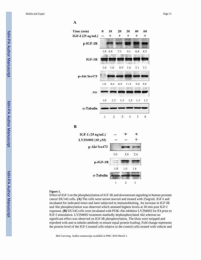
Figure 1.Effect of IGF-I on the phosphorylation of IGF-IR and downstream signaling in human prostatecancer DU145 cells. (A) The cells were serum starved and treated with 25ng/mL IGF-I andincubated for indicated times and later subjected to immunoblotting. An increase in IGF-IRand Akt phosphorylation was observed which attained highest levels at 30 min post IGF-Iexposure. (B) DU145 cells were incubated with PI3K-Akt inhibitor LY294002 for 8 h prior toIGF-I stimulation. LY294002 treatment markedly dephosphorylated Akt whereas nosignificant effect was observed on IGF-IR phosphorylation. The blots were stripped andreprobed with anti-α-tubulin antibody to ensure equal protein loading. Fold change representsthe protein level of the IGF-I treated cells relative to the control cells treated with vehicle and
Shukla and Gupta Page 11
Mol Carcinog. Author manuscript; available in PMC 2010 March 1.
NIH
-PA Author Manuscript
NIH
-PA Author Manuscript
NIH
-PA Author Manuscript

the resulting protein levels were then normalized to the α-tubulin protein. Details are describedin ‘Materials and Methods’.
Shukla and Gupta Page 12
Mol Carcinog. Author manuscript; available in PMC 2010 March 1.
NIH
-PA Author Manuscript
NIH
-PA Author Manuscript
NIH
-PA Author Manuscript

Figure 2.Effect of apigenin on IGF-I-induced proliferation in human prostate cancer DU145 cells. Thecells were fasted for 24 h following which they were treated with apigenin and IGF-I for 24 hat the indicated doses. (A) Control (no IGF-I or apigenin), (B) IGF-I (25ng/mL), (C) IGF-I(25ng/mL) + 10μM LY294002, (D) IGF-I (25ng/mL) + 10μM apigenin, (E) IGF-I (25ng/mL)+ 20μM apigenin, (F) IGF-I (25ng/mL) + 40μM apigenin. Cells were fixed in cold methanoland stained with propidium iodide in the presence of 5mg/mL RNase, subjected to flowcytometry, and evaluated using Cell Quest & ModFit cell cycle analysis software. Results arean average of two independent experiments run in duplicate.
Shukla and Gupta Page 13
Mol Carcinog. Author manuscript; available in PMC 2010 March 1.
NIH
-PA Author Manuscript
NIH
-PA Author Manuscript
NIH
-PA Author Manuscript

Figure 3.Effect of apigenin on IGF-IR signaling after IGF-I stimulation in human prostate cancer DU145cells. The cells were incubated with indicated doses of apigenin for 24 h and later incubatedwith 25ng/mL IGF-I for 30 min and subjected to immunoblotting. A decrease in IGF-IR, Aktand GSK-3β phosphorylation was observed along with decrease in cyclin D1 and increase inp27/kip1. No significant changes were noted in total Akt and IGF-IR levels. The blots werestripped and reprobed with anti-α-tubulin antibody to ensure equal protein loading.. Foldchange represents the protein level of the apigenin treated cells relative to the control cellstreated with vehicle and the resulting protein levels were then normalized to the α-tubulinprotein. Details are described in ‘Materials and Methods’.
Shukla and Gupta Page 14
Mol Carcinog. Author manuscript; available in PMC 2010 March 1.
NIH
-PA Author Manuscript
NIH
-PA Author Manuscript
NIH
-PA Author Manuscript

Figure 4.Effect of apigenin on cell proliferation in human prostate cancer PC-3 cells expressing highconstitutively expressing IGF/PI3K/Akt signaling. Log phase growing cells were treated with(A) vehicle alone, and (B-D) indicated doses of apigenin for 24 h. Cells were fixed in coldmethanol and stained with propidium iodide in the presence of 5mg/mL RNase, subjected toflow cytometry, and evaluated using Cell Quest & ModFit cell cycle analysis software. Resultsare an average of two independent experiments run in duplicate.
Shukla and Gupta Page 15
Mol Carcinog. Author manuscript; available in PMC 2010 March 1.
NIH
-PA Author Manuscript
NIH
-PA Author Manuscript
NIH
-PA Author Manuscript

Figure 5.Effect of apigenin on IGF-IR signaling in human prostate cancer PC-3 cells. The cells wereincubated with indicated doses of apigenin for 24 h and subjected to immunoblot analysis. Adecrease in IGF-IR, Akt and GSK-3β phosphorylation was observed along with decrease incyclin D1 and increase in p27/kip1. No significant changes were noted in total Akt and IGF-IR levels. The blots were stripped and reprobed with anti-α-tubulin antibody to ensure equalprotein loading. Fold change represents the protein level of the apigenin treated cells relativeto the control cells treated with vehicle and the resulting protein levels were then normalizedto the α-tubulin protein. Details are described in ‘Materials and Methods’.
Shukla and Gupta Page 16
Mol Carcinog. Author manuscript; available in PMC 2010 March 1.
NIH
-PA Author Manuscript
NIH
-PA Author Manuscript
NIH
-PA Author Manuscript

Figure 6.Effect of apigenin on PC-3 tumor growth inhibition in athymic nude mouse xenograft.Approximately 1 million cells were injected into both flanks of each mouse to initiate prostatetumor xenograft, and apigenin was provided to the animals 2 weeks after cell inoculation. Micewere fed ad libitum with Teklad 8760 autoclaved high-protein diet. Apigenin was providedwith 0.5% methyl cellulose and 0.025% Tween 20 as vehicle to these animals perorally on adaily basis. Group I, control, received 0.2 ml vehicle only, and the II group received 50μgapigenin per mouse in 0.2 ml vehicle daily for 8 weeks and experiment was terminated. Oncethe tumor xenografts started growing, their sizes were measured twice weekly in twodimensions throughout the study. (A) Apigenin inhibits growth of PC-3 tumor xenograft in
Shukla and Gupta Page 17
Mol Carcinog. Author manuscript; available in PMC 2010 March 1.
NIH
-PA Author Manuscript
NIH
-PA Author Manuscript
NIH
-PA Author Manuscript

athymic nude mice as measured by volume and wet weights. (B) Apoptosis assay as determinedby M30 reactivity. . Values are means ± SE, n = 6–8, repeated twice with similar results.Significantly different from control: **P<0.001. (C) Immunoblots for tumor lysates afterapigenin intake. Apigenin inhibited IGF/IGF-IR signaling pathway. Apigenin intake reducedtumor proliferation as shown by reduction in PCNA and IGF-IR, caused dephosphorylation ofIGF-IR, Akt, GSK-3β along with decrease in cyclin D1 and increase in p27/kip1. The blotswere stripped and reprobed with anti-α-tubulin antibody to ensure equal protein loading. Foldchange represents the protein level after apigenin administration relative to the control groupadministered with vehicle and the resulting protein levels were then normalized to the α-tubulinprotein. Details are described in ‘Materials and Methods’.
Shukla and Gupta Page 18
Mol Carcinog. Author manuscript; available in PMC 2010 March 1.
NIH
-PA Author Manuscript
NIH
-PA Author Manuscript
NIH
-PA Author Manuscript