The Role of the Transcription Factor SIM2 in Prostate Cancer
Upload
khangminh22Category
view
2download
0
AWARD NUMBER: W81XWH-18-1-0219
TITLE: Radiogenomic Characterization of Prostate Cancer: Distinguishing Aggressive From Indolent Disease
PRINCIPAL INVESTIGATOR: Simpa S. Salami, MD, MPH
CONTRACTING ORGANIZATION: University of Michigan, Ann Arbor, MI
REPORT DATE: October 2021
TYPE OF REPORT: Annual
PREPARED FOR: U.S. Army Medical Research and Materiel Command Fort Detrick, Maryland 21702-5012
DISTRIBUTION STATEMENT: Approved for Public Release; Distribution Unlimited
The views, opinions and/or findings contained in this report are those of the author(s) and should not be construed as an official Department of the Army position, policy or decision unless so designated by other documentation.

REPORT DOCUMENTATION PAGE Form Approved
OMB No. 0704-0188 Public reporting burden for this collection of information is estimated to average 1 hour per response, including the time for reviewing instructions, searching existing data sources, gathering and maintaining the data needed, and completing and reviewing this collection of information. Send comments regarding this burden estimate or any other aspect of this collection of information, including suggestions for reducing this burden to Department of Defense, Washington Headquarters Services, Directorate for Information Operations and Reports (0704-0188), 1215 Jefferson Davis Highway, Suite 1204, Arlington, VA 22202-4302. Respondents should be aware that notwithstanding any other provision of law, no person shall be subject to any penalty for failing to comply with a collection of information if it does not display a currently valid OMB control number. PLEASE DO NOT RETURN YOUR FORM TO THE ABOVE ADDRESS. 1. REPORT DATEOctober 2021
2. REPORT TYPEAnnual
3. DATES COVERED 30Sep2020-29Sep2021
4. TITLE AND SUBTITLE 5a. CONTRACT NUMBER W81XWH-18-1-0219
Radiogenomic Characterization of Prostate Cancer: Distinguishing Aggressive From Indolent Disease
5b. GRANT NUMBER PC170717 5c. PROGRAM ELEMENT NUMBER
6. AUTHOR(S): Simpa S. Salami, MD, MPH
Simpa
5d. PROJECT NUMBER AWD006494
5e. TASK NUMBER
E-Mail: [email protected]
5f. WORK UNIT NUMBER
7. PERFORMING ORGANIZATION NAME(S) AND ADDRESS(ES):REGENTS OF THE UNIVERSITY OF MICHIGAN, 503 THOMPSON ST, ANN ARBOR, MI 48109-1340
8. PERFORMING ORGANIZATION REPORTNUMBER
9. SPONSORING / MONITORING AGENCY NAME(S) AND ADDRESS(ES) 10. SPONSOR/MONITOR’S ACRONYM(S)
U.S. Army Medical Research and Development Command Fort Detrick, Maryland 21702-5012 11. SPONSOR/MONITOR’S REPORT
NUMBER(S)
12. DISTRIBUTION / AVAILABILITY STATEMENT
Approved for Public Release; Distribution Unlimited
13. SUPPLEMENTARY NOTESNone 14. ABSTRACT: Over the past several years, there has been growing utilization of multi-parametric magnetic resonance imaging (mpMRI) to detectaggressive or high-grade PCa. Even though close to 20 % of aggressive PCa are missed by mpMRI, this imaging modality is currently being used to guidetreatment decisions, such as for active surveillance and delineating areas for focal therapy. The goal of this project is to improve the detection of aggressive(Gleason ≥7) PCa by combining mpMRI and urinary biomarkers. Building on our prior work on using PCA3 and TMPRSS2:ERG to detect aggressive PCa, wehave developed a novel urine-based targeted next generation sequencing (NGS) assay to detect PCa. We hypothesize that aggressive PCa harbors uniquemolecular alterations that impact detection by mpMRI or a urine-based sequencing assay. To test this hypothesis, we propose the following Specific Aims: 1):To assess the accuracy of a novel urine-based NGS assay for the detection of high-grade PCa. We will perform targeted DNA/RNA NGS on already collectedpre-biopsy post-DRE urine specimens in patients who underwent radical prostatectomy (RP) at the University of Michigan (U-M). The molecular profile ofpatients with high-grade (cases) versus low-grade (controls) PCa will be compared. 2): To comprehensively characterize the genomic and transcriptomicalterations associated with cancer visibility on mpMRI. We will collect formalin-fixed paraffin-embedded (FFPE) radical prostatectomy (RP) specimens withmultiple foci of cancer in men who had mpMRI prior to RP, with an emphasis on those with multiple Gleason grades. Where available, corresponding matchedpelvic lymph node specimens with metastasis will also be collected. Targeted multiplexed PCR-based NGS will be performed to characterize and compare themolecular profiles of visible and invisible PCa foci on mpMRI. 3): Determine and compare tissue versus a urine-based prognostic assays to predict upgradingat RP. Targeted DNA/RNA NGS will be performed on post-DRE urine and biopsy tissue obtained prior to RP. We will assess and compare the performance ofthe novel urine-based assay with tissue-based prognostic assays to predict Gleason upgrading at RP.15. SUBJECT TERMS None listed.
16. SECURITY CLASSIFICATION OF: 17. LIMITATIONOF ABSTRACT
18. NUMBEROF PAGES
19a. NAME OF RESPONSIBLE PERSON USAMRMC
a. REPORT
Unclassified
b. ABSTRACT
Unclassified
c. THIS PAGE
Unclassified Unclassified
46 19b. TELEPHONE NUMBER (include area code)
Standard Form 298 (Rev. 8-98) Prescribed by ANSI Std. Z39.18

TABLE OF CONTENTS
Page
1. Introduction 4
2. Keywords 5
3. Accomplishments 6
4. Impact 13
5. Changes/Problems 13
6. Products 15
7. Participants & Other Collaborating Organizations 18
8. Special Reporting Requirements 23
9. Appendices 24

4
1. INTRODUCTION:
The goal of this project is to improve the detection of aggressive (Gleason ≥7) prostate cancer by combining multiparametric MRI and urinary biomarkers.

5
2. KEYWORDS:
Prostate cancer, urinary biomarkers, MRI, aggressive, genomics, transcriptomics

6
3. ACCOMPLISHMENTS:
What were the major goals of the project?
Major Task 1: Training and educational development in prostate cancer research
1 – 48 Months
Research-Specific Tasks: Specific Aim 1: To assess the accuracy of a novel urine-based NGS assay for the detection of high-grade PCa. 0 – 20 months
Major Task 1: Test the capacity of a novel urine-based NGS assay to detect aggressive PCa at first biopsy. 0 – 14 months
Major Task 2: Determine the differential performance characteristics of a novel urine-based NGS assay to detect aggressive PCa in the setting of a – vs. + prostate mpMRI.
10 – 20 months
Specific Aim 2: To comprehensively characterize the genomic and transcriptomic alterations associated with cancer visibility on mpMRI
Major Task 3: Interrogate specific molecular changes associated with high-grade PCa visibility on mpMRI.
16 – 33 months
Major Task 4: Elucidate the molecular profile of low-grade fusion biopsy cores obtained from PIRADS 4 and 5 lesions and correlate these findings with RP pathology.
28 – 38 months
Specific Aim 3: Determine and compare tissue versus a urine-based prognostic assays to predict Gleason upgrading at RP.
Major Task 5: Assess tissue-based and novel urine-based prognostic scores in biopsy core and urine respectively in patients with Gleason upgrading
34 – 48 months

7
What was accomplished under these goals?
We continue to make progress in our project specific aims as described below. It is worth mentioning, however, my lab is behind due to the ongoing COVID-19 pandemic and supply chain issues which have severely hampered our planned research activities.
Major Task 1: Training and educational development in prostate cancer research: Training accomplishments:
- Completion of R01 boot camp program at University of Michigan (UM). This grantwriting training program culminated in the submission of an R01 grant application thatwas reviewed and triaged in June 2020; I revised the grant application based on thereviewer comments and resubmitted in February 2021, discussed and scored but notwithin fundable range. I will be submitting another grant application in 2022.
- I have continued to attend (virtually) the American Urological Association Annualmeetings, Society of Urologic Oncology annual meeting, and Prostate CancerFoundation annual meetings. I have presented results from this work at some of thesemeetings. Continue as instructor/faculty of medical school/graduate courses.
- Co-lead of a Prostate SPORE project at UM.
- Awarded the AUA Rising Stars in Urologic Research.
Conferences/journal clubs: - Attend monthly prostate cancer seminars- Meet with mentors (Drs. Palapattu and Tomlins) regularly to discuss research
progress/career development- Continue leadership/active roles in Urology Grand Rounds, GU tumor board, Prostate
SPORE collaborative conferences.- Participated in multiple study sections, including American Cancer Society and American
Urological Association. Now standing member of American Cancer Society Committee onClinical Cancer Research and Epidemiology Study Section.
- Named Review Editor for Urologic Oncology of Frontiers in Urology.
Clinical responsibilities: - Continue Urologic oncology clinic.- Continue operative schedules.
Professional accomplishments: - Member of the AUA and NCCN Prostate Cancer Early Detection guidelines panels.Member of the Society of Nuclear Medicine and Molecular Imaging Appropriateness UseCriteria for PSMA PET imaging in prostate cancer.
8
Specific Aim 1: To assess the accuracy of a novel urine-based NGS assay for the detection of high-grade PCa.
We published the initial paper describing the Urnie NGS-assay this year. The findings are detailed in:
Cani AK, Hu K, Liu CJ, Siddiqui J, Zheng Y, Han S, Nallandhighal S, Hovelson DH, Xiao L, Pham T, Eyrich NW, Zheng H, Vince R Jr, Tosoian JJ, Palapattu GS, Morgan TM, Wei JT, Udager AM, Chinnaiyan AM, Tomlins SA, Salami SS. Development of a Whole-urine, Multiplexed, Next-generation RNA-sequencing Assay for Early Detection of Aggressive Prostate Cancer. Eur Urol Oncol. 2021 Mar 31:S2588-9311(21)00046-8. doi: 10.1016/j.euo.2021.03.002. Online ahead of print.PMID: 33812851
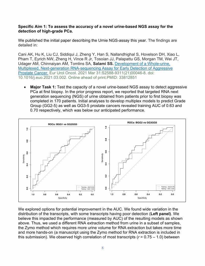
• Major Task 1: Test the capacity of a novel urine-based NGS assay to detect aggressivePCa at first biopsy. In the prior progress report, we reported that targeted RNA nextgeneration sequencing (NGS) of urine obtained from patients prior to first biopsy wascompleted in 170 patients. Initial analyses to develop multiplex models to predict GradeGroup (GG2-5) as well as GG3-5 prostate cancers revealed training AUC of 0.63 and0.70 respectively, which was below our anticipated performance.
We explored options for potential improvement in the AUC. We found wide variation in the distribution of the transcripts, with some transcripts having poor detection (Left panel). We believe this impacted the performance (measured by AUC) of the resulting models as shown above. Thus, we used a different RNA extraction method from urine in a subset of samples, the Zymo method which requires more urine volume for RNA extraction but takes more time and more hands-on (a manuscript using the Zymo method for RNA extraction is included in this submission). We observed high correlation of most transcripts (r = 0.75 – 1.0) between
ROCs: BGG1 vs GG2GG5
1.0 0.8 0.6 0.4 0.2 0.0
0.0
0.2
0.4
0.6
0.8
1.0
Specificity
Sens
itivi
ty
1.0 0.8 0.6 0.4 0.2 0.0
0.0
0.2
0.4
0.6
0.8
1.0
Training − AUC:0.63Testing − AUC: 0.66
ROCs: BGG2 vs GG3GG5
1.0 0.8 0.6 0.4 0.2 0.0
0.0
0.2
0.4
0.6
0.8
1.0
Specificity
Sens
itivi
ty
1.0 0.8 0.6 0.4 0.2 0.0
0.0
0.2
0.4
0.6
0.8
1.0
Training − AUC:0.70Testing − AUC: 0.64

9
both RNA extraction methods. However, we also observed poor correlation in some transcripts between the two methods (Right panel).
• We have explored the following experimental and analytic approaches:o Introduced clinical variables for predicting aggressive disease in model
development but with no improvement in performance.o Since these experiments were performed on patients who underwent their first
prostate biopsies, we are evaluating the possibility of biopsy unersampling as aconfounder by collecting follow up biopsy or prostatectomy information to retrainthe model.
o Optimizing RNA extraction in a subset of urine samples with low transcriptsdetection/expression. Our challenge here was exhaustion of urine specimensgiven that large volumes of urine was required for RNA extraction using theZymo method.
o Earlier this year, our prostate SPORE program in prostate cancer increased theamount of urine collection from patients to accommodate multiple assaycomparisons and the goal is to utilize some these specimens in the future tofurther optimize our assay.
• Major Task 2: Determine the differential performance characteristics of a novelurine-based NGS assay to detect aggressive PCa in the setting of a – vs. +prostate mpMRI.Next generation sequencing and bioinformatics analysis to evaluate the performance ofthe novel urine based assay independent of mpMRI in this sub-aim is currently ongoing.This aspect of the study is on hold pending further optimization of our urine assay.
●
●
●
●
●
● ●●●●●
●
●
●
●
●
●
●
●
●
●
●●●●●
●●
●
●●●
●●●●
●
●●●●
●
●●●●●●●●
●
●
●
●
●
●●
●
●
●
●
●
0
5
10
15
20
PCA3.E2E3
ERG.E1E2
RP11.314O
13.1.E3E4
KLK2.E3E4
PDLIM5.E6E7
HMGN2P_C
15ORF21.ET
V1.H2E8
NKX
3.1.E1E2
TMPR
SS2.ER
G.T3E4.COSF
130
SPOP.E5E6
HPN
.E10E11
RP11.314O
13.1.E2E3
ACPP
.E2E3
SLC45A3.ETV
1.S1E7.COSF
1119
TMPR
SS2.ET
V1.T1E4.COSF
115
DLX1.E1E2
TMPR
SS2.ET
V5.T1bE2
RP11_ES
T14.ET
V1.R1E7
CAN
T1.ETV
4.C2E5.COSF
1155
gene
log2rpm
ids
high
low
−0.5
0.0
0.5
1.0
AC009478.1.E3E4
ACPP
.E2E3
AR−FL5−1−2.E1E2
AR−V7−3−CE3.E1E2
DLX1.E1E2
ERG.E1E2
FOLH
1.E5E6
FOXP
1−ET
V1.F11E8
G053084_T230577.E3E4
G053084_T230586.E2E3
HMGN2P_C
15ORF21−ET
V1.H2E8
HOX
B13.E1
HOX
C6.E1E2
HPN
.E10E11
KLK2.E3E4
KLK3.E2E3
NDRG1−ER
G.N3E4.COSF
1133
NKX
3−1.E1E2
OR51E2.E2E3
PCA3.E2E3
PCAT4.E1E2
PDLIM5.E6E7
RP11_ES
T14−ET
V1.R1E8
RP11−314O
13.1.E2E3
RP11−314O
13.1.E3E4
SCHLAP1.E1E2
SLC45A3−BRAF.S1B9.COSF
871
SPOP.E5E6
TDRD1.E16E17
TMPR
SS2−ER
G.EF194202
TMPR
SS2−ER
G.T1E2.COSF
123
TMPR
SS2−ER
G.T1E4.COSF
125
TMPR
SS2−ER
G.T1E5.COSF
26TM
PRSS
2−ER
G.T1E6.COSF
36TM
PRSS
2−ER
G.T1EIIIc_4
TMPR
SS2−ER
G.T2E2.COSF
127
TMPR
SS2−ER
G.T2E4.COSF
128
TMPR
SS2−ER
G.T2E5.COSF
129
TMPR
SS2−ER
G.T2EIIIc_4
TMPR
SS2−ER
G.T3E4.COSF
130
TMPR
SS2−ER
G.T4E4.COSF
18TM
PRSS
2−ER
G.T4E5.COSF
17TM
PRSS
2−ER
G.T5E4.COSF
116
TMPR
SS2−ER
G.T5E5
TMPR
SS2−ET
V5.T1bE2
Gene
correlation

10
Specific Aim 2: To comprehensively characterize the genomic and transcriptomic alterations associated with cancer visibility on mpMRI
• Major Task 3: Interrogate specific molecular changes associated with high-gradePCa visibility on mpMRI.We performed targeted NGS in 26 samples of mpMRI visible and invisible prostatecancer. Bioinformatics analysis to compare the molecular profile of mpMRI visibleversus invisible lesions indicates that under expression of cellular organization andstructure underlies mpMRI invisibility (see publication, Appendix A).
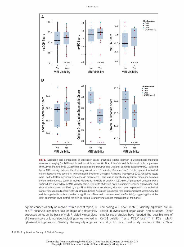
• We also compared derived commercially available tissue-based prognostic biomarkerassays (Myriad Prolaris cell cycle progression (mxCCP), OncotypeDX genomic prostate score (mxGPS), and Decipher genomic classifier (mxGC) between mpMRI visible and invisible lesions and found no significant difference in the scores.
Development a 9-gene signature to predict mpMRI visible tumors. Annotated heatmap of differentially expressed genes between mpMRI visible and invisible PCa which were identified with the EdgeR package using a Benjamini-Hochberg procedure (BH) adjusted false discovery rate (FDR) cutoff of < 0.05. Targets and samples were clustered using hierarchical clustering based on Euclidian distances. Comparisons of observed (first row) vs. predicted (second row) mpMRI visibility using the 9-gene signature is shown.
Figure 2A
B
Observed VisibilityNoYes
Predicted VisibilityNoYes
Grade GroupGG1GG2GG3GG4GG4 (minor)GG5
Patient IDMRI!1MRI!2MRI!3MRI!4MRI!5MRI!6MRI!8MRI!9MRI!10
í�
í�
0
�
�
PR!472_RNA
PR!452_RNA
PR!456_RNA
PR!463_RNA
PR!475_RNA
PR!466_RNA
PR!462_RNA
PR!465_RNA
PR!460_RNA
PR!471_RNA
PR!454_RNA
PR!464_RNA
PR!458_RNA
PR!457_RNA
PR!459_RNA
PR!474_RNA
PR!473_RNA
PR!476_RNA
PR!470_RNA
PR!455_RNA
PR!453_RNA
PR!467_RNA
PR!461_RNA
PR!477_RNA
TPM2GSNDSTSRD5A2SGK1S100A6MYLKKLK2AMACR
Observed VisibilityPredicted VisibilityGrade GroupPatient ID
0.00
0.25
0.50
0.75
1.00
0.000.250.500.751.00
Speci"city
Sens
itivi
ty
Discovery cohort (UM); AUC = 0.89
Validation cohort (CSMC); AUC = 0.88

11
• Major Task 4: Elucidate the molecular profile of low-grade fusion biopsy coresobtained from PIRADS 4 and 5 lesions and correlate these findings with RPpathology.This experiments are ongoing.
• Major Task 5: Assess tissue-based and novel urine-based prognostic scores inbiopsy core and urine respectively in patients with Gleason upgradingThis analysis is scheduled to be performed in the 4th year of the award.
Derivation and comparison of expression-based prognostic scores between mpMRI visible and invisible lesions. A) Boxplots of derived Myriad Prolaris cell cycle progression (mxCCP), OncotypeDX genomic prostate score (mxGPS), and Decipher genomic classifier (mxGC) stratified by mpMRI visibility status in our preliminary cohort (n= 10 patients, 26 cancer foci). Points represent individual cancer focus colored according to ISUP Grade Group. Unpaired t-tests were used to test for significant differences in mean score. There was no statistically significant difference between the derived prognostic scores of mpMRI-visible and -invisible lesions (p > 0.05). B) Comparisons of derived mxGPS submodules stratified by mpMRI visibility status. Boxplots of derived mxGPS Androgen, Cellular Organization, and Stromal submodules stratified by mpMRI visibility status are shown. Unpaired t-tests were used to compare mean sub-component scores. Only the Cellular Organization submodule had a significant difference in mean expression (p = 0.014).

12
What opportunities for training and professional development has the project provided? Nothing to Report
How were the results disseminated to communities of interest? Findings were disseminated in the form of publications in journals(Journal of Clinical Oncology Precision Oncology and European Urology Oncology) as well as in talks and interviews.
What do you plan to do during the next reporting period to accomplish the goals?
Major Task 1: Optimize experimental and analytic efforts o Optimizing RNA extraction and assay in ongoing urine sample collection.
Major Task 2: Determine the differential performance characteristics of our novel urine-based NGS assay to detect aggressive PCa in the setting of a – vs. + prostate mpMRI. This will be achieved once we optimize our urine RNA extraction.
Major Task 5: Assess tissue-based and novel urine-based prognostic scores in biopsy core and urine respectively in patients with Gleason upgrading. This analysis is scheduled to be performed in the 4th year of the award.

13
4. IMPACT:
What was the impact on the development of the principal discipline(s) of the project?The finding that mpMRI invisible prostate cancer are just as important biologically as visibleones indicates that we should not use mpMRI alone for determining which patients shouldundergo focal therapy or active surveillance.
The potential impact of the ongoing analyses will delineate the utility of using a urine test tosupplement mpMRI for detecting clinically significant prostate cancer.
What was the impact on other disciplines?Nothing to report
What was the impact on technology transfer?Nothing to report
What was the impact on society beyond science and technology?Nothing to report
5. CHANGES/PROBLEMS:
Changes in approach and reasons for changeSee Major task 1 above.
Actual or anticipated problems or delays and actions or plans to resolve themThis project was severely impacted by lab closures and temporary furloughs due to theCOVID-19 pandemic. Most recently, we have suffered issues imposed by supply chainshortages. Regarding exhaustion of urine samples necessary for urine RNA extractionoptimization, we plan to use ongoing urine sample collection in our prostate cancer SPOREproject.
We will need to apply for a no-cost extension of the grant to completed our stated work.
Changes that had a significant impact on expendituresNothing to report.
Significant changes in use or care of human subjects, vertebrate animals, biohazards,and/or select agentsNot applicable.
Significant changes in use or care of human subjectsNothing to report
Significant changes in use or care of vertebrate animals

14
Not applicable
Significant changes in use of biohazards and/or select agents Not applicable

15
6. PRODUCTS:
• Publications, conference papers, and presentations
Journal publications.
Salami SS, Kaplan JB, Nallandhinghal S, Takhar M, Tosoian JJ, Lee M, Yoon J,Hovelson DH, Plouffe KR, Kaffenberger SD, Schaeffer EM, Karnes R, Lotan TL,Morgan TM, George, AK, Montgomery JS, Davenport MS, You S, Tomlins SA, CurciNE, Kim HL, Spratt DE, Udager AM, Palapattu GS. Biologic Significance of MRIInvisibility in Localized Prostate Cancer (JCO Precision oncology, 2019, in press,acknowledgement of federal support – yes)
Cani AK, Hu K, Liu CJ, Siddiqui J, Zheng Y, Han S, Nallandhighal S, Hovelson DH, XiaoL, Pham T, Eyrich NW, Zheng H, Vince R Jr, Tosoian JJ, Palapattu GS, Morgan TM,Wei JT, Udager AM, Chinnaiyan AM, Tomlins SA, Salami SS. Development of a Whole-urine, Multiplexed, Next-generation RNA-sequencing Assay for Early Detection ofAggressive Prostate Cancer. Eur Urol Oncol. 2021 Mar 31:S2588-9311(21)00046-8.doi: 10.1016/j.euo.2021.03.002. Online ahead of print.PMID: 33812851
Books or other non-periodical, one-time publications.Nothing to report
Other publications, conference papers and presentations.
Eyrich NW, Wei JT, Niknafs YS, Siddiqui J, Ellimoottil C, Salami SS, Palapattu GS, Mehra R, Kunju LP, Tomlins SA, Chinnaiyan AM, Morgan TM, Tosoian JJ. Association of MyProstateScore (MPS) with prostate cancer grade in the radical prostatectomy specimen. Urol Oncol. 2021 Nov 6:S1078-1439(21)00433-6. doi: 10.1016/j.urolonc.2021.09.007. Online ahead of print.PMID: 34753659
Stensland KD, Kaffenberger SD, George AK, Morgan TM, Miller DC, Salami SS, Dunn RL, Palapattu GS, Montgomery JS, Hollenbeck BK, Skolarus TA. Prostate cancer clinical trial completion: The role of geography. Contemp Clin Trials. 2021 Oct 19;111:106600. doi: 10.1016/j.cct.2021.106600. Online ahead of print.PMID: 34673273
Jadvar H, Calais J, Fanti S, Feng F, Greene KL, Gulley JL, Hofman M, Koontz BF, Lin DW, Morris MJ, Rowe SP, Royce TJ, Salami S, Savir-Baruch B, Srinivas S, Hope TA. Appropriate Use Criteria for Prostate-Specific Membrane Antigen PET Imaging. J Nucl Med. 2021 Sep 30:jnumed.121.263262. doi: 10.2967/jnumed.121.263262. Online ahead of print.PMID: 34593595
Stangl-Kremser J, Rasul S, Tosoian JJ, Salami SS, Zaslavsky A, Udager A, Mazal P, Kain R, Comperat E, Hacker M, Haug A, Mitterhauser M, Pozo-Salido C, Steinbach C, Hassler MR, Kramer G, Shariat SF, Palapattu GS. Single-lesion Prostate-specific
16
Membrane Antigen Protein Expression (PSMA) and Response to [177Lu]-PSMA-ligand Therapy in Patients with Castration-resistant Prostate Cancer. Eur Urol Open Sci. 2021 Jun 30;30:63-66. doi: 10.1016/j.euros.2021.06.007. eCollection 2021 Aug.PMID: 34337549
Tosoian JJ, Dunn RL, Niknafs YS, Saha A, Vince RA Jr, St Sauver JL, Jacobson DJ, McGree ME, Siddiqui J, Groskopf J, Jacobsen SJ, Tomlins SA, Kunju LP, Morgan TM, Salami SS, Wei JT, Chinnaiyan AM, Sarma AV. Association of Urinary MyProstateScore, Age, and Prostate Volume in a Longitudinal Cohort of Healthy Men: Long-term Findings from the Olmsted County Study. Eur Urol Open Sci. 2021 May 25;29:30-35. doi: 10.1016/j.euros.2021.04.009. eCollection 2021 Jul.PMID: 34337531
Shankar PR, Ellimoottil C, George AK, Hadj-Moussa M, Modi PK, Salami S, Tosoian JJ, Wei JT, Davenport MS. Testing-Related Health Impact of Transrectal and Transperineal Prostate Biopsy as Assessed by Health Utilities. J Urol. 2021 Dec;206(6):1403-1410. doi: 10.1097/JU.0000000000002118. Epub 2021 Jul 21.PMID: 34288719
Sessine MS, Das S, Park B, Salami SS, Kaffenberger SD, Kasputis A, Solorzano M, Luke M, Vince RA, Kaye DR, Borza T, Stoffel EM, Cobain E, Merajver SD, Jacobs MF, Milliron KJ, Caba L, van Neste L, Mondul AM, Morgan TM. Initial Findings from a High Genetic Risk Prostate Cancer Clinic. Urology. 2021 Oct;156:96-103. doi:10.1016/j.urology.2021.05.078. Epub 2021 Jul 17.PMID: 34280438
Cricco-Lizza E, Wilcox Vanden Berg RN, Laviana A, Pantuck M, Basourakos SP, Salami SS, Hung AJ, Margolis DJ, Hu JC, McClure TD. Comparative Effectiveness and Tolerability of Transperineal MRI-Targeted Prostate Biopsy under Local versus Sedation. Urology. 2021 Sep;155:33-38. doi: 10.1016/j.urology.2021.06.023. Epub 2021 Jul 2.PMID: 34217762
Gaffney C, Liu D, Cooley V, Ma X, Angulo C, Robinson B, Khani F, Cai P, Salami S, Nallandhighal S, Shoag J, Barbieri C. Tumor size and genomic risk in localized prostate cancer. Urol Oncol. 2021 Jul;39(7):434.e17-434.e22. doi: 10.1016/j.urolonc.2021.01.020. Epub 2021 Feb 6.PMID: 33563537
Jairath NK, Dal Pra A, Vince R Jr, Dess RT, Jackson WC, Tosoian JJ, McBride SM, Zhao SG, Berlin A, Mahal BA, Kishan AU, Den RB, Freedland SJ, Salami SS, Kaffenberger SD, Pollack A, Tran P, Mehra R, Morgan TM, Weiner AB, Mohamad O, Carroll PR, Cooperberg MR, Karnes RJ, Nguyen PL, Michalski JM, Tward JD, Feng FY, Schaeffer EM, Spratt DE. A Systematic Review of the Evidence for the Decipher Genomic Classifier in Prostate Cancer. Eur Urol. 2021 Mar;79(3):374-383. doi: 10.1016/j.eururo.2020.11.021. Epub 2020 Dec 5.PMID: 33293078 Review.
Tosoian JJ, Trock BJ, Morgan TM, Salami SS, Tomlins SA, Spratt DE, Siddiqui J, Kunju LP, Botbyl R, Chopra Z, Pandian B, Eyrich NW, Longton G, Zheng Y, Palapattu GS, Wei JT, Niknafs YS, Chinnaiyan AM. Use of the MyProstateScore Test to Rule Out
17
Clinically Significant Cancer: Validation of a Straightforward Clinical Testing Approach. J Urol. 2021 Mar;205(3):732-739. doi: 10.1097/JU.0000000000001430. Epub 2020 Oct 20.PMID: 33080150
Salami SS, Tosoian JJ, Nallandhighal S, Jones TA Jr, Brockman S, Elkhoury FF, Bazzi S, Plouffe KR, Siddiqui J, Liu CJ, Kunju LP, Morgan TM, Natarajan S, Boonstra PS, Sumida L, Tomlins SA, Udager AM, Sisk AE Jr, Marks LS, Palapattu GS. Serial Molecular Profiling of Low-grade Prostate Cancer to Assess Tumor Upgrading: A Longitudinal Cohort Study. Eur Urol. 2021 Apr;79(4):456-465. doi: 10.1016/j.eururo.2020.06.041. Epub 2020 Jul 3.PMID: 32631746
• Website(s) or other Internet site(s)https://www.urotoday.com/categories-media/1748-centers-of-excellence/advanced-prostate-cancer-coe/1248-use-of-mri-to-risk-stratify-patients-with-prostate-cancer-simpa-salami.htmlThis was an interview with urotoday to discuss the Biologic Significance of MagneticResonance Imaging Invisibility in Localized Prostate Cancer.
https://www.urotoday.com/recent-abstracts/pelvic-health-reconstruction/neurogenic-bladder/1956-video-transcripts/130463-transcript-simpa-salami-recent-eau-article.htmlThis was an interview with urotoday to discuss the application of the urine nextgeneration sequencing assay for the detection of aggressive prostate cancer.
• Technologies or techniquesNothing to report
• Inventions, patent applications, and/or licensesNothing to report
• Other ProductsNothing to report

18
7. PARTICIPANTS & OTHER COLLABORATING ORGANIZATIONS
What individuals have worked on the project?
Name: Simpa S. Salami Project Role: PI Researcher Identifier (e.g. ORCID ID): 0000-0001-7461-7079 Nearest person month worked: 6 Contribution to Project: Providing scientific and administrative oversight, data
interpretation, manuscript writing Funding Support: DOD
Name: Sri Nallanghighal, MS Project Role: Bioinformatician Researcher Identifier (e.g. ORCID ID): Nearest person month worked: 4 Contribution to Project: Mr. Nallandhighal has performed bioinformatic
analysis of the data generated in Aim 2. Funding Support: DOD, NIH SPORE
Name: Kevin Hu Project Role: Graduate Student Researcher Identifier (e.g. ORCID ID): Nearest person month worked: 5 Contribution to Project: Mr. Hu has performed bioinformatic analysis of the
data generated Funding Support: UM Department of Pathology Training award
Name: Trinh Pham Project Role: Laboratory technologist Researcher Identifier (e.g. ORCID ID): Nearest person month worked: 5 Contribution to Project: Ms. Pham has performed the laboratory experiments
– DNA/RNA extraction from urine and Nextgeneration sequencing
Funding Support: Department of Urology funds

19
Has there been a change in the active other support of the PD/PI(s) or senior/key personnel since the last reporting period?
Current DOD PRA AWARD PC170717 (Salami) 10/01/2018-09/30/2022 2.40 Calendar Months Department of Defense Source Country: USA Annual Directs: Total Award: Radiogenomic Characterization of Prostate Cancer: Distinguishing Aggressive from Indolent Disease The successful completion of the proposed project will improve our understanding of the molecular basis of PCa visibility on mpMRI and guide treatment decisions based on mpMRI findings. Role: PI
Other Awards P50 CA186786 (Chinnaiyan) 09/01/2019-08/31/2024 1.80 Calendar Months NIH/NCI Source Country: USA Annual Directs: Total Award: SPORE Project 2: Michigan Prostate SPORE The Prostate SPORE program continues to place premiums on rigorous scientific review of its translational research programs, pairing of basic and clinical investigators, drawing on expertise of scientists from within and from outside the prostate cancer field, and utilizing flexibility to fund promising new research approaches. Role: Co-Lead
AWD017037 (Salami) 01/01/2021-12/31/2024 4.38 Calendar Months Robert Wood Johnson Foundation Source Country: USA Annual Directs: Total Award Evolution of Kidney Cancer Metastases: Implications for Surveillance and Targeted Therapy The goals of this project will define the molecular profile of the kidney cancer clone most likely to metastasize and develop a molecular signature for RCC associated with recurrence/metastasis and/or mortality. Role: PI
Palapattu (PI) 10/24/2017-06/30/2022 0.01 Calendar Months Joint Institute for Translational and Clinical Research Source Country: USA Annual Directs: Total Award: Comprehensive molecular profiling of renal cell carcinoma The long-term goal of our proposal is to improve the health of men diagnosed with renal cell carcinoma (RCC) Role: Co-Investigator *Funds used for lab
20
16YOUN17 (Salami) 06/27/2016-06/27/2022 0.01 Calendar Months Prostate Cancer Foundation Source Country: USA Annual Directs: Total Award: Molecular Characterization of the Biologically Dominant Nodule in Multifocal Prostate Cancer with N1 disease Goals: To determine and compare the molecular profile of each cancer focus in multifocal prostate cancer; ii) To characterize the biologically dominant nodule or index tumor in multifocal prostate cancer with lymph node (LN) metastasis; and iii) To evaluate the prognostic accuracy of Oncotype DX™, Prolaris™ and Decipher™ scores in predicting LN metastasis. Role: PI *Funds used for lab research
PENDING 20-PAF08652 (Zaslavsky) 09/01/2020-08/31/2022 0.12 Calendar Months Karmanos Cancer Institute/Prostate Cancer Foundation Source Country: USA Annual Directs Total Award Tissue and Plasma Biomarker Validation and Refinement and Early Drug Compound Development to Inhibit Pro-inflammatory Cytokines and Chemokines in African and European American Men We anticipate that validation and refinement of our biomarkers will ultimately be more predictive and independent of clinical outcomes than clinical markers in a future prospective analysis Role: Co-Investigator
20-PAF08526 (Udager) 06/01/2021-05/31/2024 0.60 Calendar Months DOD Source Country: USA Annual Directs: Total Award: Integrative molecular profiling of whole urine in African-American men with aggressive prostate cancer The goal of this project is to: 1) evaluate the performance of a novel whole urine NGS assay for the detection of high-grade prostate cancer in African-American men; and, 2) validate and apply a high-throughput NGS genomic profiling method for whole urine to identify African-American men with aggressive prostate cancer. Role: Co-Investigator
21-PAF03379 (Zaslavsky) 07/01/2021-06/30/2024 0.12 Calendar Months Barbara Ann Karmanos Cancer Institute/NIH Source Country: USA Annual Directs: Total Award: Tissue and Plasma Biomarker Validation and Refinement and Early Drug Compound Development to Inhibit Pro-inflammatory Cytokines and Chemokines in African and European American Men We anticipate that validation and refinement of our biomarkers will ultimately be more predictive and independent of clinical outcomes than clinical markers in a future prospective analysis Role: Co-Investigator
21-PAF01820 (Salami) 09/01/2021-08/31/2026 2.40 Calendar Months
21
NIH Source Country: USA Annual Directs: Total Award: Defining the Biological Arc of Grade Group 2 Prostate Cancer The long-term goal of this project is to improve the early detection of aggressive prostate cancer and reduce racial disparities in this disease Role: PI
22-PAF02096 (Wang) 09/01/2022-08/31/2026 1.19 calendar monthsPenn State/NIH Source Country: USAAnnual Directs: Total Award: Transrectal ultrasound and photoacoustic dual imaging guided biopsy of prostate cancerAim 1: Develop and validate a clinical grade transrectal ultrasound and photoacoustic (TRUSPA) system for image- guided targeted biopsy of prostate cancer.Aim 2: Via the studies on clinically relevant TRAMP mouse model of PCa, develop and validate TRUSPA for PCa detection and grading by quantitatively assessing a list of structural and functional imaging biomarkers.Aim 3: Via the studies on human subjects, examine the clinical feasibility of TRUSPA, by assessing the imaging biomarkers validated in aim 2, in detecting and grading PCa tumors for guiding needle biopsy. The team at PSU will take the lead in Aim1 in assembling the clinical grade TRUSPA system, while the proposed studies on animal models in Aim 2 and the experiments on human subjects in Aim 3 will be conducted in parallel at PSU and UM. It is understood by all parties that this is a research project and that the work will be conducted on a best effort basis to achieve these objectives on this time line. Role: Co-Investigator
OVERLAP If any of the pending grants are awarded funding will be revised as need be.

What other organizations were involved as partners? Nothing to report

23
8. SPECIAL REPORTING REQUIREMENTS
COLLABORATIVE AWARDS: Not applicable
QUAD CHARTS: Not applicable

24
9. APPENDICES:
a. Appendix A (Published Manuscript): Biologic Significance of MagneticResonance Imaging Invisibility in Localized Prostate Cancer.
b. Appendix B (Published Manuscrupt): Development of a Whole-urine,Multiplexed, Next-generation RNA-sequencing Assay for Early Detection ofAggressive Prostate Cancer.

originalreport
Biologic Significance of Magnetic ResonanceImaging Invisibility in Localized Prostate CancerSimpa S. Salami, MD, MPH1,2; Jeremy B. Kaplan1; Srinivas Nallandhighal, MS1; Mandeep Takhar, MS3; Jeffrey J. Tosoian, MD, MPH1;
Matthew Lee, MD1; Junhee Yoon, MS4; Daniel H. Hovelson, PhD1; Komal R. Plouffe, MS1; Samuel D. Kaffenberger, MD1,2;
Edward M. Schaeffer, MD, PhD5; R. Jeffrey Karnes, MD6; Tamara L. Lotan, MD7; Todd M. Morgan, MD1,2; Arvin K. George, MD1,2;
Jeffrey S. Montgomery, MD, MHSA1,2; Matthew S. Davenport, MD1; Sungyong You, PhD4; Scott A. Tomlins, MD, PhD1,2;
Nicole E. Curci, MD1; Hyung L. Kim, MD4; Daniel E. Spratt, MD2,1; Aaron M. Udager, MD, PhD1,2; and Ganesh S. Palapattu, MD1,2,8
abstract
PURPOSE Multiparametric magnetic resonance imaging (mpMRI) is used widely for prostate cancer (PCa)evaluation. Approximately 35% of aggressive tumors, however, are not visible on mpMRI. We sought to identifythe molecular alterations associated with mpMRI-invisible tumors and determine whether mpMRI visibility isassociated with PCa prognosis.
METHODS Discovery and validation cohorts included patients who underwent mpMRI before radical prosta-tectomy and were found to harbor both mpMRI-visible (Prostate Imaging and Reporting Data System 3 to 5) and-invisible (Prostate Imaging and Reporting Data System 1 or 2) foci on surgical pathology. Next-generationsequencing was performed to determine differential gene expression between mpMRI-visible and -invisible foci.A genetic signature for tumor mpMRI visibility was derived in the discovery cohort and assessed in an in-dependent validation cohort. Its association with long-term oncologic outcomes was evaluated in a separatetesting cohort.
RESULTS The discovery cohort included 10 patients with 26 distinct PCa foci on surgical pathology, of which 12(46%) were visible and 14 (54%) were invisible on preoperative mpMRI. Next-generation sequencing detectedprioritized genetic mutations in 14 (54%) tumor foci (n = 8 mpMRI visible, n = 6 mpMRI invisible). A nine-genesignature (composed largely of cell organization/structure genes) associated with mpMRI visibility was derived(area under the curve = 0.89), and the signature predicted MRI visibility with 75% sensitivity and 100%specificity (area under the curve = 0.88) in the validation cohort. In the testing cohort (n = 375, median follow-up8 years) there was no significant difference in biochemical recurrence, distant metastasis, or cancer-specificmortality in patients with predicted mpMRI-visible versus -invisible tumors (all P . .05).
CONCLUSION Compared with mpMRI-invisible disease, mpMRI-visible tumors are associated with under-expression of cellular organization genes. mpMRI visibility does not seem to be predictive of long-term canceroutcomes, highlighting the need for biopsy strategies that detect mpMRI-invisible tumors.
JCO Precis Oncol. © 2019 by American Society of Clinical Oncology
INTRODUCTION
Distinguishing aggressive from indolent clinically lo-calized prostate cancer (PCa) continues to posea significant clinical challenge. Recent efforts toovercome this have involved the development andoptimization of several diagnostic strategies, includingmultiparametric magnetic resonance imaging (mpMRI).mpMRI permits visual identification of areas that aresuggestive for intermediate to high-grade cancer. Theemergence of various MRI/ultrasound fusion biopsyplatforms has led to increased detection of aggressive PCaby facilitating targeted biopsy of visible lesions.1-6 Asa result, mpMRI is now widely used in guiding treatmentdecisions in men with clinically localized disease, espe-cially when selecting patients suitable for active surveil-lance or potentially focal therapy.7-10 The prevailing view isthat only mpMRI-visible cancers require clinical action.
However, use of mpMRI in the evaluation of men withPCa is limited by cancer multifocality and interfocaldisease heterogeneity. Individual patients are knownto harbor multiple spatially distinct PCa foci withvarying clinical, radiographic, and pathologiccharacteristics.11-15 Up to 55% of all PCa foci and 35%of clinically significant foci are not visible onmpMRI.3,16,17 Furthermore, more than 35% of lesions1 cm or larger are missed by mpMRI.17 Although somestudies have demonstrated that up to 50% of mpMRI-invisible PCa may harbor relevant genomic alterations,the clinical and prognostic significance of mpMRI-invisible PCa remains unknown.18 An improved un-derstanding of the molecular characteristics andclinical trajectories of mpMRI-visible and -invisiblecancers could facilitate more optimal treatment allo-cation. For example, if mpMRI-invisible foci are found
ASSOCIATEDCONTENT
Data Supplement
Author affiliationsand supportinformation (ifapplicable) appear atthe end of thisarticle.
Accepted on April 22,2019 and published atascopubs.org/journal/po on June 12, 2019:DOI https://doi.org/10.1200/PO.19.00054
1
Downloaded from ascopubs.org by 68.49.104.219 on June 10, 2020 from 068.049.104.219Copyright © 2020 American Society of Clinical Oncology. All rights reserved.
to be biologically indolent, those with a known diagnosis oflow-grade disease and a negative mpMRI could be directedtoward active surveillance. Similarly, those with a singlelesion detected on mpMRI could be more confidently di-rected toward focal therapy, with low concern for missinga clinically relevant lesion. We herein sought to characterizethe molecular profile of mpMRI-visible and -invisible PCafoci using next-generation sequencing (NGS). In addition,we test the prognostic significance of our mpMRI-derivedgenomic signature after radical prostatectomy (RP).
METHODS
Study Design
The study used three independent patient populations:discovery, validation, and testing cohorts. Institutional re-view board approval was obtained for each cohort. First, weidentified patients with clinically localized disease whounderwent preoperative mpMRI at the University ofMichigan in 2015 to 2016 and were subsequently found toharbor multifocal PCa at RP. We enriched for patients withboth mpMRI visible and invisible PCa (Figs 1A and 1B) toconstitute the discovery cohort. The validation cohort fromCedars-Sinai Medical Center included patients with eithermpMRI-visible or -invisible foci, as previously described.19
The testing cohort was composed of patients from theDecipher GRID PCa database treated at Johns HopkinsMedical Institute and Mayo Clinic (ClinicalTrials.gov iden-tifier: NCT02609269) who underwent genome-wide ex-pression profiling after RP.20,21
Preoperative Prostate mpMRI and Pathologic Evaluation
In the discovery and validation cohorts, mpMRI comprisingT2-weighted imaging, diffusion-weighted imaging, and dy-namic contrast-enhanced imaging was obtained. All mpMRIresults were re-reviewed and coregistered with whole-mountformalin-fixed paraffin-embedded RP specimens to delineatempMRI-visible (Prostate Imaging and Reporting DataSystem [PI-RADS] version 2; score, 3 to 5) and -invisible
foci. Additional procedural details are described in the DataSupplement. Data on mpMRI were not available for thetesting cohort.22
Targeted DNA and RNA NGS
In the discovery cohort, DNA and RNA from each focuswere co-isolated for targeted multiplex NGS as previouslydescribed23 and detailed in the Data Supplement. Ourtargeted NGS assays were designed to assess relevant PCagenomic and transcriptomic alterations and derive clinicallyavailable prognostic tests.15 The details of RNA sequencingin the validation cohort and genome-wide expressionprofiling in the testing cohorts have been previouslydescribed.19,24
Bioinformatic Analysis of the Discovery Cohort
NGS data analysis was performed using Torrent Suite(4.2.0; Thermo Fisher Scientfic, Waltham, MA) and theCoverage Analysis Plug-ins v.5.0.4. (Thermo Fisher Sci-entific), along with the Ion Reporter (4.2.0; Thermo FisherScientific). All other analyses were performed using RProject for Statistical Computing v.3.2.3. Details regardingtargeted NGS techniques, quality control parameters, DNAcopy number alterations and variant calls, fusion isoformand partner level analysis, androgen receptor (AR) and AR-splice variants detection, and prognostic scores derivationhave been previously described and summarized in theData Supplement.15,25,26
Differential Gene Expression Analysis of mpMRI-Visible
and -Invisible Cancer Foci
To determine gene expression differences betweenmpMRI-visible and -invisible tumors, we analyzed RNAseqdata from the discovery cohort using two approaches—differential expression (DE) analysis and random forest (RF)classifier—as described in the Data Supplement. Fromthese two approaches, a gene expression signature com-prising independent differentially expressed genes wasdeveloped to predict mpMRI tumor visibility status.
CONTEXT
Key ObjectiveWhat is the molecular basis for prostate cancer visibility on multiparametric magnetic resonance imaging (mpMRI), and do
mpMRI-invisible tumors harbor any clinical or biologic significance compared with visible tumors?Knowledge GeneratedmpMRI-visible tumors demonstrated underexpression of genes associated with cellular organization and structure. Using
a novel genetic signature for tumor visibility on mpMRI, patients with predicted mpMRI-visible and -invisible tumors did notexperience significant differences in biochemical recurrence, distant metastasis, or cancer-specific mortality during follow-up.
RelevanceProstate cancers that are mpMRI invisible have similar clinical behavior to mpMRI-visible tumors. Negative mpMRI seems
insufficient to rule out clinically relevant prostate cancer, and patients at increased risk should be considered for additionaltesting or systematic prostate biopsy.
Salami et al
2 © 2019 by American Society of Clinical Oncology
Downloaded from ascopubs.org by 68.49.104.219 on June 10, 2020 from 068.049.104.219Copyright © 2020 American Society of Clinical Oncology. All rights reserved.

CPatient
Grade group
MRI visibility
DNA variants
ERG
ETV1
SPINK1
TMPRSS2−ERG.EF194202TMPRSS2−ERG.T1E4.COSF125TMPRSS2−ERG.T1E5.COSF26TMPRSS2−ERG.T2E4.COSF128TMPRSS2−ERG.T2EIIIc_4
PR
−452
_RN
A
PR
−453
_RN
A
PR
−454
_RN
A
PR
−455
_RN
A
PR
−456
_RN
A
PR
−457
_RN
A
PR
−458
_RN
A
PR
−459
_RN
A
PR
−460
_RN
A
PR
−461
_RN
A
PR
−462
_RN
A
PR
−463
_RN
A
PR
−464
_RN
A
PR
−465
_RN
A
PR
−466_R
NA
PR
−467_R
NA
PR
−470
_RN
A
PR
−471
_RN
A
PR
−472
_RN
A
PR
−473
_RN
A
PR
−474
_RN
A
PR
−475
_RN
A
PR
−476
_RN
A
PR
−477
_RN
A
MRI visibility
NoYes
DNA variants
APCSPOPATMNone
Grade group
GG1GG2GG3GG4 (minor)
1 1 10 10 102 2 3 3 3 4 4 4 5 5 5 6 6 6 8 8 9 9 9
PTEN loss
−5.0 −2.5 0.0 2.5 5.0
SPINK1, ETV1, ERG (log2 Isoform)
−5 −4 −3 −2 −1 0
TMPRSS2-ERG (log2 Isoform)
−1.5 −1.0 0.0 1.0 1.5
PTEN (log2 CN ratio)
A B1 2
3
P2
P1
P3
4
Sample Name GG SPOP
P1 PR-465 4 F133V
P2 PR-466 2 wt
P3 PR-467 1 wt
FIG 1. Radiogenomic characterization of multifocal prostate cancer. (A) Cartoon depicting multifocal prostate cancer (PCa) with both multiparametricmagnetic resonance imaging (mpMRI)–visible (solid black, left) and invisible (gray with black discontinuous borders, right) lesions. (B) Coregistration ofaxial mpMRI images with whole-mount histopathology. (1) Axial high-resolution T2, (2) axial diffusion-weighted imaging (b-value = 1,600), and (3) axialapparent diffusion coefficient map shows a visible lesion corresponding to cancer focus P1 (grade group [GG] 4; arrows) on the (continued on following page)
Molecular Basis and Prognosis of MRI-Invisible Prostate Cancer
JCO Precision Oncology 3
Downloaded from ascopubs.org by 68.49.104.219 on June 10, 2020 from 068.049.104.219Copyright © 2020 American Society of Clinical Oncology. All rights reserved.

Prognostic Significance of mpMRI-Based Gene
Expression Signature
A total of 375 patients with genome-wide expression pro-files were pooled from two independent case-cohortstudies20,21 to constitute the testing cohort. The mpMRI-based nine-gene expression signature was applied to thetesting cohort to predict mpMRI visibility status. Kaplan-Meier curves and Cox proportional hazard regression wereused to evaluate the performance of this signature inpredicting oncological outcomes: biochemical recurrence-free survival (BFS), distant metastasis–free survival(DMFS), and PCa-specific mortality (PCSM). Multivariableanalyses were performed to evaluate this signature as anindependent predictor of oncological outcomes afteradjusting for relevant clinicopathological variables, in-cluding preoperative prostate-specific antigen, pathologicgrade group (GG), surgical margins, extraprostatic exten-sion, seminal vesicle invasion, and lymph node invasion.Spearman correlation analysis was performed to measurethe association of the gene signature with cellular organi-zation pathway activity. Mean expression of genes involvedin the cellular organization pathway on the Oncotype Dxgenomic prostate score (GPS; Genomic Health, RedwoodCity, CA) assay was correlated with the mpMRI-based geneexpression signature.27 Statistical analyses were performedin R version 3.3.3, and all statistical tests were two-sidedusing a .05 significance level.
RESULTS
Study Cohorts
The discovery cohort included 10 patients from the Uni-versity of Michigan PCa database with both mpMRI-visibleand -invisible lesions (Fig 1). The clinicopathologicalcharacteristics of the discovery cohort are shown in theData Supplement. Of the 26 cancer foci identified onsurgical pathology specimens, 12 foci (46%) were visibleon mpMRI. Among the 14 mpMRI-invisible foci (54%), five(36%) were GG2 and the remainder were GG1 (Fig 1C andData Supplement). There were 16 patients in the validationcohort, of whom eight (50%) had mpMRI-invisible cancerlesions, and two of these (25%) were GG2 (Data Supple-ment). A summary of patient-level characteristics of thetesting cohort (n = 375) stratified by predicted mpMRIvisibility status is shown in the Data Supplement. Themedian age at RP was 62 years, and median follow-up timefor censored patients was 8 years. During follow-up, 136(36.3%) patients experienced biochemical recurrence,
55 (14.7%) developed metastasis, and 28 (7.5%) died asa result of PCa (Data Supplement).
Detection of Mutations and Copy Number Alterations in
the Discovery Cohort
We detected high-confidence mutations in 14 of 26 (54%)tumor foci; six (43%) of the mutations were identified inmpMRI-invisible lesions. Notable somatic point mutationswere in APC, ARID1B, ATM, NOTCH1, and SPOP. Wedetected PTEN one copy number loss in 25% (three of 12)and 14.3% (two of 14) of mpMRI-visible and -invisible foci,respectively (Fig 1C).
Discovery and Validation of a Nine-Gene Expression
Signature for mpMRI Visibility
Of the 26 total tumor foci in the discovery cohort and 306amplicons on the RNAseq panel, 24 samples and 74amplicons, respectively, passed quality control parametersand underwent DE analysis (Data Supplement). Using DEanalysis (Data Supplement) and RF classifier (Data Sup-plement) to identify candidate differentially expressedgenes, we interrogated four separate logistic regressionmodels for predicting mpMRI tumor visibility status usingthe 19 DE analysis genes, 20 RF genes, 11 shared genesbetween the DE analysis and RF gene sets, and 11 sharedgenes combined with the mutually exclusive genes (DataSupplement). A multivariable RNAseq-based logistic re-gression model with the best performance for predictingmpMRI visibility status, comprising a nine-gene expressionsignature, was developed from the intersection of the DEanalysis and RF gene sets (Fig 2A; Data Supplement). Thissignature correctly predicted seven (70%) of the mpMRI-visible and 13 (93%) of the mpMRI-invisible foci in thediscovery cohort, yielding an area under the curve of 0.89.The optimal probability cutoff for predicting mpMRI-visibletumor was greater than 0.46, with a sensitivity and spec-ificity of 80% and 86%, respectively, in the discovery cohort(Figs 2A and 2B). We observed underexpression of seven ofthe nine genes in mpMRI-visible tumors, the majority ofwhich were stromal, cellular organization, and structuregenes (Fig 2A; Data Supplement).
The nine-gene expression signature was then evaluated inthe independent validation cohort (Cedars-Sinai MedicalCenter) using the predetermined optimal probability cutoff(from the discovery cohort) to predict mpMRI visibilitystatus. The receiver operating characteristic curve in thevalidation cohort is shown in Fig 2B, with an area under thecurve of 0.88. The sensitivity and specificity of the signature
FIG 1. (Continued). radical prostatectomy specimen (hematoxylin and eosin, panel 4). Cancer foci P2 (GG 2) and P3 (GG 1) were both mpMRI invisible. (C)Integrative summary of the primary multifocal PCa cohort. Ten patients comprising 26 distinct PCa foci were evaluated. Two samples (patient 7) did not passinitial RNA quality thresholds and were thus omitted. Each patient had at least oneMRI-visible and oneMRI-invisible cancer focus. Recurrent DNA variants areshown. Log2 copy-number ratio for PTEN is also shown. PTEN one copy number loss was observed in 25% (three of 12) and 14.3% (two of 14) of mpMRI-visibleand invisible cancer foci, respectively (false discovery rate, less than 5%). Expression of SPINK1, ERG, and ETV1, as well as expressed isoforms of TMPRSS2-ERGare shown.
Salami et al
4 © 2019 by American Society of Clinical Oncology
Downloaded from ascopubs.org by 68.49.104.219 on June 10, 2020 from 068.049.104.219Copyright © 2020 American Society of Clinical Oncology. All rights reserved.
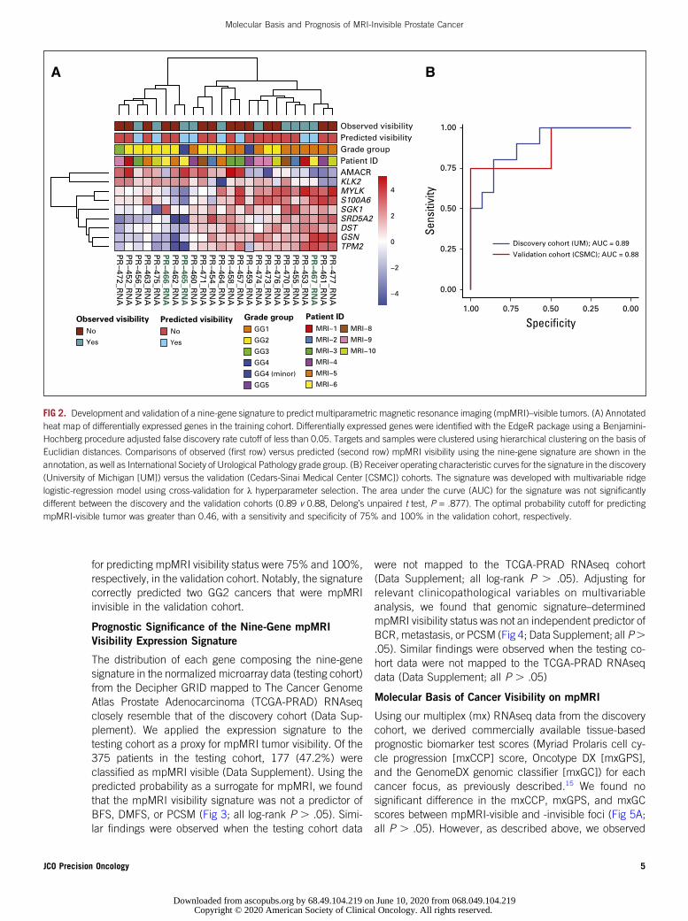
for predicting mpMRI visibility status were 75% and 100%,respectively, in the validation cohort. Notably, the signaturecorrectly predicted two GG2 cancers that were mpMRIinvisible in the validation cohort.
Prognostic Significance of the Nine-Gene mpMRI
Visibility Expression Signature
The distribution of each gene composing the nine-genesignature in the normalized microarray data (testing cohort)from the Decipher GRID mapped to The Cancer GenomeAtlas Prostate Adenocarcinoma (TCGA-PRAD) RNAseqclosely resemble that of the discovery cohort (Data Sup-plement). We applied the expression signature to thetesting cohort as a proxy for mpMRI tumor visibility. Of the375 patients in the testing cohort, 177 (47.2%) wereclassified as mpMRI visible (Data Supplement). Using thepredicted probability as a surrogate for mpMRI, we foundthat the mpMRI visibility signature was not a predictor ofBFS, DMFS, or PCSM (Fig 3; all log-rank P . .05). Simi-lar findings were observed when the testing cohort data
were not mapped to the TCGA-PRAD RNAseq cohort(Data Supplement; all log-rank P . .05). Adjusting forrelevant clinicopathological variables on multivariableanalysis, we found that genomic signature–determinedmpMRI visibility status was not an independent predictor ofBCR, metastasis, or PCSM (Fig 4; Data Supplement; all P..05). Similar findings were observed when the testing co-hort data were not mapped to the TCGA-PRAD RNAseqdata (Data Supplement; all P . .05)
Molecular Basis of Cancer Visibility on mpMRI
Using our multiplex (mx) RNAseq data from the discoverycohort, we derived commercially available tissue-basedprognostic biomarker test scores (Myriad Prolaris cell cy-cle progression [mxCCP] score, Oncotype DX [mxGPS],and the GenomeDX genomic classifier [mxGC]) for eachcancer focus, as previously described.15 We found nosignificant difference in the mxCCP, mxGPS, and mxGCscores between mpMRI-visible and -invisible foci (Fig 5A;all P . .05). However, as described above, we observed
Observed visibility
No
Yes
Predicted visibility
No
Yes
Grade group
GG1
GG2
GG3
GG4
GG4 (minor)
GG5
Patient ID
MRI−1
MRI−2
MRI−3
MRI−4
MRI−5
MRI−6
MRI−8
MRI−9
MRI−10
A
−4
−2
0
2
4
PR
−472_RN
AP
R−452_R
NA
PR
−456_RN
AP
R−463_R
NA
PR
−475_RN
AP
R−
46
6_R
NA
PR
−462_RN
AP
R−
46
5_R
NA
PR
−460_RN
AP
R−471_R
NA
PR
−454_RN
AP
R−464_R
NA
PR
−458_RN
AP
R−457_R
NA
PR
−459_RN
AP
R−474_R
NA
PR
−473_RN
AP
R−476_R
NA
PR
−470_RN
AP
R−455_R
NA
PR
−453_RN
AP
R−
46
7_R
NA
PR
−461_RN
AP
R−477_R
NA
TPM2GSNDSTSRD5A2SGK1S100A6MYLKKLK2AMACR
Observed visibilityPredicted visibilityGrade groupPatient ID
B
0.00
0.25
0.50
0.75
1.00
0.000.250.500.751.00
Specificity
Sens
itivi
ty
Discovery cohort (UM); AUC = 0.89
Validation cohort (CSMC); AUC = 0.88
FIG 2. Development and validation of a nine-gene signature to predict multiparametric magnetic resonance imaging (mpMRI)–visible tumors. (A) Annotatedheat map of differentially expressed genes in the training cohort. Differentially expressed genes were identified with the EdgeR package using a Benjamini-Hochberg procedure adjusted false discovery rate cutoff of less than 0.05. Targets and samples were clustered using hierarchical clustering on the basis ofEuclidian distances. Comparisons of observed (first row) versus predicted (second row) mpMRI visibility using the nine-gene signature are shown in theannotation, as well as International Society of Urological Pathology grade group. (B) Receiver operating characteristic curves for the signature in the discovery(University of Michigan [UM]) versus the validation (Cedars-Sinai Medical Center [CSMC]) cohorts. The signature was developed with multivariable ridgelogistic-regression model using cross-validation for λ hyperparameter selection. The area under the curve (AUC) for the signature was not significantlydifferent between the discovery and the validation cohorts (0.89 v 0.88, Delong’s unpaired t test, P = .877). The optimal probability cutoff for predictingmpMRI-visible tumor was greater than 0.46, with a sensitivity and specificity of 75% and 100% in the validation cohort, respectively.
Molecular Basis and Prognosis of MRI-Invisible Prostate Cancer
JCO Precision Oncology 5
Downloaded from ascopubs.org by 68.49.104.219 on June 10, 2020 from 068.049.104.219Copyright © 2020 American Society of Clinical Oncology. All rights reserved.

underexpression of seven of the nine genes in mpMRI-visible tumors, the majority of which were stromal, cellularorganization, and structure genes (Fig 2A; Data Supple-ment). We then computed three subcomponents of theOncotypeDx GPS, as previously described,15,27 and com-pared these between mpMRI-visible and -invisible tumors.There were no significant differences in the expression ofOncotypeDx GPS androgen signaling and stromal responsesubmodules between mpMRI-visible and -invisible tumors(Fig 5B; both P. .05). However, we found underexpressionof the cellular organization submodule of the OncotypeDxGPS panel in mpMRI-visible tumors consistent with theresults of the nine-gene signature (Fig 5B; all P = .014).
Similarly, using data from the testing cohort, we foundunderexpression of the OncotypeDx GPS cellular organiza-tion module in predicted mpMRI-visible compared with-invisible foci (Data Supplement; all P , .05). Taken to-gether, these findings suggest that loss of cellular organi-zation and structure contributes to PCa visibility on mpMRI.
DISCUSSION
To better understand the molecular alterations associatedwith mpMRI visibility and prognostic significance ofmpMRI-invisible disease, we performed a comprehensivemolecular characterization of primary multifocal PCa in-clusive of both mpMRI-visible and -invisible tumor foci
A
Time (months)
Prob
ablit
y of
BFS
0 60 120 180 240
0.2
0.4
0.6
0.8
1.0
198 128 37 11Low
177 108 36 8High
LowHigh
Low (events = 72)High (events = 64) Log-rank P = .91
B
Time (months)
Prob
ablit
y of
DM
FS
0 60 120 180 240
0.2
0.4
0.6
0.8
1.0
198 163 53 13
177 135 52 10
Low
High
Low (events = 29)High (events = 26) Log-rank P = .97
Low
High
C
Time (months)
Prob
ablit
y of
PCS
M
0 60 120 180 240
0.2
0.4
0.6
0.8
1.0
198 171 59 14
177 145 55 12
LowHigh
Low (events = 16)High (events = 12) Log-rank P = .66
Low
High
No. at risk No. at risk
No. at risk
FIG 3. Prognostic significance of predicted multiparametric magnetic resonance imaging (mpMRI) visibility status.Patients (n = 375) in the testing cohort were pooled from two independent case-cohort studies (Johns HopkinsMedical Institute [n = 260] and Mayo Clinic [n = 235]) to test the capacity of predicted mpMRI visibility status topredict (A) biochemical recurrence-free survival (BFS), (B) distant metastasis-free survival (DMFS), and (C) prostatecancer–specific mortality (PCSM). The expression data in this cohort were generated using Affymetrix human exon1.0 ST array (Santa Clara, CA). Normalization was performed to match the distribution of the genomic data from thiscohort to The Cancer Genome Atlas Prostate Adenocarcinoma RNAseq data, as described in Methods, to facilitatetesting of the RNASeq-based nine-gene signature to predict mpMRI-visible tumors (Data Supplement). mpMRIvisibility status was computed using the signature: high score denotesmpMRI-visible and low score denotesmpMRI-invisible tumor. Kaplan-Meier survival curves were plotted and compared between predicted mpMRI-visible and-invisible tumor using log-rank test. There were no significant differences in BFS, DMFS, and PCSM betweenpredicted mpMRI-visible and -invisible tumors (all P . .05). Similar results were obtained using the Affymetrixmicroarray data that were not matched to the distribution of the The Cancer Genome Atlas Prostate AdenocarcinomaRNAseq data (Data Supplement).
Salami et al
6 © 2019 by American Society of Clinical Oncology
Downloaded from ascopubs.org by 68.49.104.219 on June 10, 2020 from 068.049.104.219Copyright © 2020 American Society of Clinical Oncology. All rights reserved.

using a targeted multiplex NGS approach. We observedthat mpMRI-invisible cancer may possess mutations inknown cancer-associated genes, with close to 15%harboring PTEN one copy number loss. Using robustbiostatistic methods, we developed and validated a novelnine-gene signature to predict PCa mpMRI visibility status.Interrogation of this signature in a distinct cohort with long-term follow-up revealed no significant association with BFS,DMFS, or PCSM. Intriguingly, additional analyses revealedthat underexpression of genes associated with cellularorganization and structure may underpin the molecular
basis of PCa visibility on mpMRI. Taken together, thesefindings indicate that mpMRI-invisible cancer foci harbormany of the same aggressive molecular features as mpMRI-visible foci and may also be clinically significant.
The molecular basis of PCa visibility on mpMRI is poorlyunderstood. Although tumor size and grade contribute tocancer visibility on mpMRI, the architecture of the glandsmay play an important role.28-31 For example, tumorsharboring cribriform Gleason pattern 4 were less likely to bedetected by mpMRI compared with poorly formed or fusedglands, suggesting that tumor size and grade alone do not
A
C
B
MRI Signature
LNI
GG
SM
EPE
SVI
PSA
Low
High
0
1
GG1−2
GG3
GG4−5
0
1
0
1
0
1
< 10
10 to 20
> 20
196
175
324
47
169
69
133
216
155
179
192
282
89
205
116
50
Reference
Reference
Reference
Reference
Reference
Reference
0.98 (0.69 to 1.39)
Reference
1.60 (1.02 to 2.52)
1.54 (0.93 to 2.55)
1.68 (1.12 to 2.52)
2.02 (1.42 to 2.87)
1.27 (0.86 to 1.88)
1.82 (1.23 to 2.71)
1.02 (0.69 to 1.51)
1.47 (0.90 to 2.40)
.916
.041
.090
.012
<.001
.232
.003
.928
.120
Variable No. HR (95% CI)
HR (95% CI)
HR (95% CI)P
MRI Signature
LNI
GG
SM
EPE
SVI
PSA
Low
High
0
1
GG1−2
GG3
GG4−5
0
1
0
1
0
1
< 10
10 to 20
> 20
196
175
324
47
169
69
133
216
155
179
192
282
89
205
116
50
0.98 (0.45 to 2.17)
2.22 (0.93 to 5.28)
1.76 (0.48 to 6.46)
3.31 (1.24 to 8.83)
1.65 (0.76 to 3.58)
0.91 (0.35 to 2.35)
2.70 (1.16 to 6.30)
0.84 (0.34 to 2.08)
1.77 (0.66 to 4.71)
.97
.07
.39
.02
.21
.84
.02
.71
.26
Variable No. P
0.5 1 2 5 10
MRI Signature
LNI
GG
SM
EPE
SVI
PSA
Low
High
0
1
GG1−2
GG3
GG4−5
0
1
0
1
0
1
< 10
10 to 20
> 20
196
175
324
47
169
69
133
216
155
179
192
282
89
205
116
50
1.07 (0.61 to 1.88)
2.03 (1.08 to 3.82)
2.91 (1.10 to 7.67)
5.08 (2.28 to 11.32)
2.11 (1.20 to 3.71)
1.76 (0.85 to 3.64)
1.99 (1.09 to 3.66)
0.77 (0.40 to 1.46)
1.05 (0.48 to 2.26)
.818
.028
.031
<.001
.009
.128
.026
.415
.909
Variable No. P
0.5 1 2 5 100.5 1 2 5 10
Reference
Reference
Reference
Reference
Reference
Reference
Reference
Reference
Reference
Reference
Reference
Reference
Reference
Reference
FIG 4. Multivariable analysis to assess the prognostic significance of predicted multiparametric magnetic resonance imaging (mpMRI) visibility status.Using data from the testing cohort described in Figure 3 (Affymetrix microarray data matched to the distribution of the The Cancer Genome Atlas ProstateAdenocarcinoma RNAseq data; n = 375), multivariable Cox proportional hazardmodels were developed to assess the capacity of predictedmpMRI visibilitystatus to predict: (A) biochemical recurrence-free survival (BFS), (B) distant metastasis–free survival (DMFS), and (C) prostate cancer–specific mortality(PCSM), adjusting for relevant clinicopathological variables. mpMRI visibility status was not an independent predictor of BFS, DMFS, and PCSM (alladjusted P . .05). Similar results were obtained when the Affymetrix microarray data were not matched to the distribution of The Cancer Genome AtlasProstate Adenocarcinoma RNAseq data (Data Supplement). EPE, extraprostatic extension; GG, grade group; HR, hazard ratio; LNI, lymph node invasion;PSA, prostate-specific antigen; SM, surgical margins; SVI, seminal vesical invasion.
Molecular Basis and Prognosis of MRI-Invisible Prostate Cancer
JCO Precision Oncology 7
Downloaded from ascopubs.org by 68.49.104.219 on June 10, 2020 from 068.049.104.219Copyright © 2020 American Society of Clinical Oncology. All rights reserved.
explain cancer visibility on mpMRI.28 In a recent report, Liet al19 observed significant fold changes of differentiallyexpressed genes on the basis of mpMRI visibility regardlessof Gleason score or tumor size, including genes involved incytoskeleton organization. Similarly, the majority of genes
composing our novel mpMRI visibility signature are in-volved in cytoskeletal organization and structure. Othersmaller-scale studies have reported the possible role ofCHD1 deletion32 and PTEN loss33,34 in PCa mpMRIvisibility. In the current study, we found that 25% of
A
No Yes Yes Yes
MRI Visibility
mxC
CP S
core
Grade group
GG1GG2GG3GG4 (minor)
P = .844−2.0
−1.5
−1.0
−0.5
0.0
mxG
PS S
core
−8
−7
−6
−5
−4
−3
P = .558 P = .548
mxG
C Sc
ore
No
MRI VisibilityNo
MRI Visibility
0
25
50
75
100
B
No Yes No Yes No Yes
Grade groupGG1GG2GG3GG4 (minor)
22.5
25.0
27.5
30.0
Andr
ogen
Sub
mod
ule
Scor
e
MRI Visibility
P = .38220
25
30
35
40
45
MRI Visibility MRI Visibility
Cellu
lar O
rgan
izatio
n Su
bmod
ule
Scor
e
P = .014
15.0
17.5
20.0
22.5
25.0
Stro
mal
Sub
mod
ule
Scor
e
P = .197
FIG 5. Derivation and comparison of expression-based prognostic scores between multiparametric magneticresonance imaging (mpMRI)–visible and -invisible lesions. (A) Box plots of derived Prolaris cell cycle progression(mxCCP) score, Oncotype DX genomic prostate score (mxGPS), and Decipher genomic classifier (mxGC) stratifiedby mpMRI visibility status in the discovery cohort (n = 10 patients; 26 cancer foci). Points represent individualcancer focus colored according to International Society of Urological Pathology grade group (GG). Unpaired t testswere used to test for significant differences in mean score. There was no statistically significant difference betweenthe derived prognostic scores of mpMRI-visible and -invisible lesions (P. .05). (B) Comparisons of derived mxGPSsubmodules stratified by mpMRI visibility status. Box plots of derived mxGPS androgen, cellular organization, andstromal submodules stratified by mpMRI visibility status are shown, with each point representing an individualcancer focus colored according to GG. Unpaired t tests were used to comparemean subcomponent scores. Only thecellular organization submodule had a significant difference in mean expression (P = .014), suggesting that at theRNA expression level mpMRI visibility is related to underlying cellular organization of the tumor.
Salami et al
8 © 2019 by American Society of Clinical Oncology
Downloaded from ascopubs.org by 68.49.104.219 on June 10, 2020 from 068.049.104.219Copyright © 2020 American Society of Clinical Oncology. All rights reserved.

mpMRI-visible foci in the discovery cohort demonstratedPTEN one copy number loss compared with 14% inmpMRI-invisible foci. In aggregate, our work and that ofothers suggests that cellular (dis)organization contributessignificantly to the underlying basis of PCa visibility onmpMRI. Additional studies are needed to further charac-terize the fundamental basis of PCa visibility on mpMRI.
The prognostic significance of mpMRI-invisible PCa foci isunknown. Although PCa is multifocal andmpMRI may missup to 35% of intermediate- to high-grade PCa, the absenceof visible lesions on mpMRI has been proposed as a reasonto defer confirmatory biopsy when considering activesurveillance.1,3,16,17 In addition, mpMRI is increasinglybeing used to identify the index or dominant cancer foci forfocal therapy. To be sure, although size and grade arebelieved to be important, how best to define the biologicallydominant cancer in multifocal disease is not known.35 Todate, no study has demonstrated the clinical trajectory ofmpMRI-visualized lesions. Such a study would be a chal-lenge to perform, given the long duration of follow-up re-quired and the multifocal nature of PCa, with frequentcoexistence of mpMRI-visible and -invisible cancers withinthe same gland.31
Salmasi et al36 reported that the PI-RADS (a grading systemfor mpMRI lesion visibility) was not a significant predictor ofadverse pathology at the time of RP. Similarly, Parry et al18
found that 50% of mpMRI-invisible cancers harbored one ormore genetic alterations commonly observed in metastaticcastrate-resistant PCa, suggesting that mpMRI-invisible tu-mors may be as important as visible ones. In the study by Liet al,19 a four-gene signature comprising genes differentiallyexpressed between mpMRI-visible and -invisible PCa wasshown to predict BFS in two external data sets. However, thissignature was not developed as a predictor of or a surrogatefor mpMRI cancer visibility, but rather it was selected on thebasis of their common association with mpMRI visibility andmetastasis. By contrast, in this first study of its kind to ourknowledge, using a validated novel mpMRI-based RNAseqsignature as a surrogate instrument for mpMRI visibilitystatus, we have demonstrated that predicted mpMRI visi-bility status was not associated with BFS, DMFS, or PCSMduring long-term follow-up. Put another way, mpMRI-invisible PCa does not seem to represent purely indolentdisease; mpMRI-invisible lesions may be just as clinicallyrelevant as mpMRI-visible disease. Future studies aimed atbetter defining the biologically dominant nodule and prog-nostic significance of mpMRI are warranted.
Our findings have significant clinical implications in themanagement of PCa. First, in the diagnostic setting, thesedata corroborate findings from several institutions in-dicating that a negative mpMRI does not rule out thepresence of clinically significant PCa3,17,31 and shouldtherefore not preclude a prostate biopsy without consid-eration of clinical risk.37,38 Second, in the setting of active
surveillance, our findings underscore the potential formpMRI-invisible cancer foci to harbor similar biologictrajectories as mpMRI-visible disease. Although additionalstudies are needed to delineate the utility of mpMRI inreducing the frequency of surveillance biopsies, the currentliterature supports systematic in addition to targeted bi-opsies in men undergoing active surveillance.3,6 Last, formen considering focal therapy, our data demonstrate thatmpMRI alone is not sufficient to rule out the presence ofa potentially lethal, nondominant cancer focus.
Our study has several limitations. First, we used a targetedNGS approach; thus it is conceivable that other potentialalterations implicated in mpMRI cancer visibility may havebeen missed. Notwithstanding, our novel RNAseq signa-ture developed from a targeted NGS approach demon-strated high fidelity for predicting mpMRI visibility in thevalidation cohort, where tumors underwent whole-transcriptome profiling. Second, we did not use the com-mercially available platforms for Oncotype Dx, Prolaris, andDecipher assays in the discovery cohort. The validity andconsistency of deriving these scores from RNAseq data hasbeen previously reported.15 Third, there were no GG4 and 5lesions in our cohort. However, our novel RNAseq signaturedemonstrated high accuracy for predicting mpMRI visibilityin the validation cohort, including 19% GG5 lesions.Moreover, GG4 and 5 lesions are generally mpMRI visible,and such patients routinely undergo whole-gland therapy.Fourth, the discovery cohort was made up of a relativelysmall sample, with low proportion of ERG-positive tumors.Nonetheless, we similarly observed high test performancein the validation cohort with ERG overexpression in 31% ofsamples. Fifth, what constitutes mpMRI-visible or -invisiblelesions is not purely objective. To facilitate reproducibility,all lesions in the current study were scored according to thevalidated PI-RADS v2 system. PI-RADS 1 and 2 lesionswere classified as mpMRI invisible, and PI-RADS 3 to 5were classified as mpMRI visible. Finally, the prognosticsignificance of mpMRI-invisible cancer was evaluated inthe testing cohort using a surrogate molecular marker formpMRI visibility status. Thus, additional studies are neededto delineate the prognostic significance of mpMRI-invisiblePCa in a prospective clinical cohort.
Discerning aggressive from indolent disease remains asignificant clinical challenge in the evaluation and man-agement of men with primary PCa. Our findings indicatethat mpMRI-invisible cancers were no less likely to harborlethal biologic potential than visible tumors, highlighting thelimitation of using mpMRI alone to guide patient man-agement or delineate specific index cancer foci for ablativetherapy. Our results also highlight the continued need forbiopsy strategies that detect mpMRI-invisible tumors. Fu-ture PCa molecular studies are needed to further char-acterize the molecular basis of cancer visibility on mpMRIand determine its prognostic significance.
Molecular Basis and Prognosis of MRI-Invisible Prostate Cancer
JCO Precision Oncology 9
Downloaded from ascopubs.org by 68.49.104.219 on June 10, 2020 from 068.049.104.219Copyright © 2020 American Society of Clinical Oncology. All rights reserved.

AFFILIATIONS1Michigan Medicine, Ann Arbor, MI2University of Michigan Rogel Cancer Center, Ann Arbor, MI3GenomeDx Biosciences, San Diego, CA4Cedars-Sinai Medical Center, Los Angeles, CA5Northwestern University Feinberg School of Medicine, Chicago, IL6Mayo Clinic, Rochester, MN7Johns Hopkins Medical Institute, Baltimore, MD8Medical University of Vienna, Vienna, Austria
CORRESPONDING AUTHORSimpa S. Salami, MD, MPH, Department of Urology, The University ofMichigan, 1500 E Medical Center Dr, 7306 RCC/SPC 5948, Ann Arbor,MI 48109-5948; e-mail: [email protected].
EQUAL CONTRIBUTIONJ.B.K., S.N., and M.T. contributed equally to this work; D.E.S., A.M.U.,and G.S.P. share co-senior authorship.
PRIOR PRESENTATIONPresented in part at the ASCO Genitourinary Cancer Symposium, SanFrancisco, CA, February 14-16, 2019; the European Association ofUrology Annual Meeting, Barcelona, Spain, March 15-19, 2019; and theAmerican Urological Association Annual Meeting, Chicago, IL, May 3-6, 2019.
SUPPORTSupported in part by the Prostate Cancer Foundation (S.S.S., T.M.M.,S.A.T., D.E.S., and G.S.P.); the National Institutes of Health Grant No.R01 CA183857 (S.A.T.); the University of Michigan Prostate SpecializedProgram of Research Excellence Grant No. P50 CA186786-05; the Menof Michigan Prostate Cancer Research Fund, the University of MichiganComprehensive Cancer Center core Grant No. 2-P30-CA-046592-24; theA. Alfred Taubman Biomedical Research Institute (T.M.M. and S.A.T.);and the Department of Defense (S.S.S., T.M.M., and D.E.S.).
AUTHOR CONTRIBUTIONSConception and design: Simpa S. Salami, Edward M. Schaeffer, MatthewS. Davenport, Scott A. Tomlins, Daniel E. Spratt, Aaron M. Udager,Ganesh S. PalapattuFinancial support: Scott A. Tomlins, Ganesh S. Palapattu
Administrative support: Ganesh S. PalapattuProvision of study material or patients: R. Jeffrey Karnes, Arvin K. George,Daniel E. SprattCollection and assembly of data: Simpa S. Salami, Jeremy B. Kaplan,Matthew Lee, Komal R. Plouffe, R. Jeffrey Karnes, Tamara L. Lotan, ArvinK. George, Nicole E. Curci, Daniel E. Spratt, Aaron M. Udager
Data analysis and interpretation: Simpa S. Salami, Jeremy B. Kaplan,Srinivas Nallandhighal, Mandeep Takhar, Jeffrey J. Tosoian, MatthewLee, Junhee Yoon, Daniel H. Hovelson, Samuel D. Kaffenberger, EdwardM. Schaeffer, R. Jeffrey Karnes, ToddM.Morgan, Arvin K. George, JeffreyS. Montgomery, Matthew S. Davenport, Sungyong You, Hyung L. Kim,Daniel E. Spratt, Aaron M. Udager
Manuscript writing: All authorsFinal approval of manuscript: All authors
AUTHORS’ DISCLOSURES OF POTENTIAL CONFLICTS OF INTEREST
The following represents disclosure information provided by authors ofthis manuscript. All relationships are considered compensated.Relationships are self-held unless noted. I = Immediate Family Member,Inst =My Institution. Relationshipsmay not relate to the subject matter ofthis manuscript. For more information about ASCO’s conflict of interest
policy, please refer to www.asco.org/rwc or ascopubs.org/po/author-center.
Jeremy B. KaplanEmployment: Tempus Labs
Mandeep TakharEmployment: GenomeDx
Daniel H. HovelsonEmployment: Strata Oncology
Consulting or Advisory Role: TerumoTravel, Accommodations, Expenses: Thermo Fisher Scientific
Samuel D. KaffenbergerConsulting or Advisory Role: MDxHealth, Clovis OncologyTravel, Accommodations, Expenses: Bristol-Myers Squibb
Edward M. SchaefferConsulting or Advisory Role: OPKO Diagnostics, AbbVie
R. Jeffrey KarnesResearch Funding: GenomeDxPatents, Royalties, Other Intellectual Property: GenomeDx
Travel, Accommodations, Expenses: GenomeDx
Tamara L. LotanConsulting or Advisory Role: Janssen OncologyResearch Funding: Ventana Medical Systems
Todd M. MorganConsulting or Advisory Role: Myriad Genetics, Terumo BCT
Research Funding: Myriad Genetics (Inst), MDxHealth (Inst), GenomeDx(Inst)
Arvin K. GeorgeConsulting or Advisory Role: Trod MedicalResearch Funding: Nanospectra Biosciences (Inst)Travel, Accommodations, Expenses: Profound Medical, Trod Medical
Matthew S. DavenportPatents, Royalties, Other Intellectual Property: Royalties from WoltersKluwer, royalties from uptodate.com
Scott A. TomlinsEmployment: Strata OncologyLeadership: Strata OncologyStock and Other Ownership Interests: Strata OncologyConsulting or Advisory Role: AbbVie, Janssen, Astellas Medivation, StrataOncology, Sanofi, Almac Diagnostics
Research Funding: Astellas Medivation (Inst), GenomeDx (Inst)Patents, Royalties, Other Intellectual Property: I am a coauthor on a patentissued to the University of Michigan on ETS gene fusions in prostatecancer. The diagnostic field of use has been licensed to Hologic/Gen-Probe, who has sublicensed some rights to Ventana Medical Systems/Roche.Travel, Accommodations, Expenses: Strata Oncology
Hyung L. KimResearch Funding: GenomeDx
Patents, Royalties, Other Intellectual Property: I am an inventor ofa prognostic biomarker signature and immunotherapies.
Daniel E. SprattConsulting or Advisory Role: Blue Earth Diagnostics, Janssen Oncology
Aaron M. UdagerResearch Funding: Ventana Medical Systems
Salami et al
10 © 2019 by American Society of Clinical Oncology
Downloaded from ascopubs.org by 68.49.104.219 on June 10, 2020 from 068.049.104.219Copyright © 2020 American Society of Clinical Oncology. All rights reserved.

Ganesh S. PalapattuStock and Other Ownership Interests: NantKwestConsulting or Advisory Role: Janssen Scientific Affairs
Research Funding: Minomic
Patents, Royalties, Other Intellectual Property: Implantable nanotechnologyfor long-term testosterone delivery functionalized fiducial marker for drugdelivery
No other potential conflicts of interest were reported.
REFERENCES1. Kasivisvanathan V, Rannikko AS, Borghi M, et al: MRI-targeted or standard biopsy for prostate-cancer diagnosis. N Engl J Med 378:1767-1777, 2018
2. Ahmed HU, El-Shater Bosaily A, Brown LC, et al: Diagnostic accuracy of multi-parametric MRI and TRUS biopsy in prostate cancer (PROMIS): A pairedvalidating confirmatory study. Lancet 389:815-822, 2017
3. Filson CP, Natarajan S, Margolis DJA, et al: Prostate cancer detection with magnetic resonance-ultrasound fusion biopsy: The role of systematic and targetedbiopsies. Cancer 122:884-892, 2016
4. Salami SS, Ben-Levi E, Yaskiv O, et al: In patients with a previous negative prostate biopsy and a suspicious lesion on magnetic resonance imaging, is a 12-corebiopsy still necessary in addition to a targeted biopsy? BJU Int 115:562-570, 2015
5. Salami SS, Vira MA, Turkbey B, et al: Multiparametric magnetic resonance imaging outperforms the Prostate Cancer Prevention Trial risk calculator inpredicting clinically significant prostate cancer. Cancer 120:2876-2882, 2014
6. Siddiqui MM, Rais-Bahrami S, Turkbey B, et al: Comparison of MR/ultrasound fusion-guided biopsy with ultrasound-guided biopsy for the diagnosis of prostatecancer. JAMA 313:390-397, 2015
7. Ahmed HU, Hindley RG, Dickinson L, et al: Focal therapy for localised unifocal and multifocal prostate cancer: A prospective development study. Lancet Oncol13:622-632, 2012
8. Natarajan S, Raman S, Priester AM, et al: Focal laser ablation of prostate cancer: Phase I clinical trial. J Urol 196:68-75, 2016
9. Ahmed HU, Dickinson L, Charman S, et al: Focal ablation targeted to the index lesion in multifocal localised prostate cancer: A prospective development study.Eur Urol 68:927-936, 2015
10. Guillaumier S, Peters M, Arya M, et al: A multicentre study of 5-year outcomes following focal therapy in treating clinically significant nonmetastatic prostatecancer. Eur Urol 74:422-429, 2018
11. Boutros PC, Fraser M, Harding NJ, et al: Spatial genomic heterogeneity within localized, multifocal prostate cancer. Nat Genet 47:736-745, 2015
12. Cooper CS, Eeles R, Wedge DC, et al: Analysis of the genetic phylogeny of multifocal prostate cancer identifies multiple independent clonal expansions inneoplastic and morphologically normal prostate tissue. Nat Genet 47:367-372, 2015 [Erratum: Nat Genet 47:689, 2015]
13. Cancer Genome Atlas Research Network: The molecular taxonomy of primary prostate cancer. Cell 163:1011-1025, 2015
14. Kumar A, Coleman I, Morrissey C, et al: Substantial interindividual and limited intraindividual genomic diversity among tumors from men with metastaticprostate cancer. Nat Med 22:369-378, 2016
15. Salami SS, Hovelson DH, Kaplan JB, et al: Transcriptomic heterogeneity in multifocal prostate cancer. JCI Insight 3:123468, 2018
16. Radtke JP, Kuru TH, Boxler S, et al. Comparative analysis of transperineal template saturation prostate biopsy versus magnetic resonance imaging targetedbiopsy with magnetic resonance imaging-ultrasound fusion guidance. J Urol 193:87-94, 2015
17. Johnson DC, Raman SS, Mirak SA, et al: Detection of individual prostate cancer foci via multiparametric magnetic resonance imaging. Eur Urol 75:712-720,2019
18. Parry MA, Srivastava S, Ali A, et al. Genomic evaluation of multiparametric magnetic resonance imaging-visible and -nonvisible lesions in clinically localisedprostate cancer. Eur Urol Oncol 2:1-11, 2019
19. Li P, You S, Nguyen C, et al: Genes involved in prostate cancer progression determine MRI visibility. Theranostics 8:1752-1765, 2018
20. Ross AE, Johnson MH, Yousefi K, et al: Tissue-based genomics augments post-prostatectomy risk stratification in a natural history cohort of intermediate- andhigh-risk men. Eur Urol 69:157-165, 2016
21. Karnes RJ, Bergstralh EJ, Davicioni E, et al: Validation of a genomic classifier that predicts metastasis following radical prostatectomy in an at risk patientpopulation. J Urol 190:2047-2053, 2013
22. Weinreb JC, Barentsz JO, Choyke PL, et al: PI-RADS Prostate Imaging - Reporting and Data System: 2015, Version 2. Eur Urol 69:16-40, 2016
23. Hovelson DH, McDaniel AS, Cani AK, et al: Development and validation of a scalable next-generation sequencing system for assessing relevant somatic variantsin solid tumors. Neoplasia 17:385-399, 2015
24. Erho N, Crisan A, Vergara IA, et al: Discovery and validation of a prostate cancer genomic classifier that predicts early metastasis following radical prostatectomy.PLoS One 8:e66855, 2013
25. Warrick JI, Hovelson DH, Amin A, et al: Tumor evolution and progression in multifocal and paired non-invasive/invasive urothelial carcinoma. Virchows Arch466:297-311, 2015
26. Palapattu GS, Salami SS, Cani AK, et al: Molecular profiling to determine clonality of serial magnetic resonance imaging/ultrasound fusion biopsies frommen onactive surveillance for low-risk prostate cancer. Clin Cancer Res 23:985-991, 2017
27. Klein EA, Cooperberg MR, Magi-Galluzzi C, et al: A 17-gene assay to predict prostate cancer aggressiveness in the context of Gleason grade heterogeneity,tumor multifocality, and biopsy undersampling. Eur Urol 66:550-560, 2014
28. Truong M, Hollenberg G, Weinberg E, et al: Impact of Gleason subtype on prostate cancer detection using multiparametric magnetic resonance imaging:Correlation with final histopathology. J Urol 198:316-321, 2017
29. Vargas HA, Akin O, Shukla-Dave A, et al: Performance characteristics of MR imaging in the evaluation of clinically low-risk prostate cancer: A prospective study.Radiology 265:478-487, 2012
30. Hurrell SL, McGarry SD, Kaczmarowski A, et al: Optimized b-value selection for the discrimination of prostate cancer grades, including the cribriform pattern,using diffusion weighted imaging. J Med Imaging (Bellingham) 5:011004, 2018
31. Le JD, Tan N, Shkolyar E, et al: Multifocality and prostate cancer detection by multiparametric magnetic resonance imaging: Correlation with whole-mounthistopathology. Eur Urol 67:569-576, 2015
32. Lee D, Fontugne J, Gumpeni N, et al: Molecular alterations in prostate cancer and association with MRI features. Prostate Cancer Prostatic Dis 20:430-435,2017
Molecular Basis and Prognosis of MRI-Invisible Prostate Cancer
JCO Precision Oncology 11
Downloaded from ascopubs.org by 68.49.104.219 on June 10, 2020 from 068.049.104.219Copyright © 2020 American Society of Clinical Oncology. All rights reserved.

33. McCann SM, Fan X, Wang J, et al: Quantitative multiparametric MRI features and PTEN expression of peripheral zone prostate cancer: A pilot study. 206:559-565, 2016
34. Zundel W, Schindler C, Haas-Kogan D, et al: Loss of PTEN facilitates HIF-1-mediated gene expression. Genes Dev 14:391-396, 2000
35. Haffner MC, Mosbruger T, Esopi DM, et al: Tracking the clonal origin of lethal prostate cancer. J Clin Invest 123:4918-4922, 2013
36. Salmasi A, Khoshnoodi P, Felker ER, et al: A 17-gene genomic prostate score assay provides independent information on adverse pathology in the setting ofcombined multiparametric magnetic resonance imaging fusion targeted and systematic prostate biopsy. J Urol 200:564-572, 2018
37. Nassiri N, Natarajan S, Margolis DJ, et al: Targeted prostate biopsy: Lessons learned midst the evolution of a disruptive technology. Urology 86:432-438, 2015
38. Panebianco V, Barchetti G, Simone G, et al: Negative multiparametric magnetic resonance imaging for prostate cancer: What’s next? Eur Urol 74:48-54, 2018
n n n
Salami et al
12 © 2019 by American Society of Clinical Oncology
Downloaded from ascopubs.org by 68.49.104.219 on June 10, 2020 from 068.049.104.219Copyright © 2020 American Society of Clinical Oncology. All rights reserved.

12
3
4
5 Q2
6
7
8
9
10 Q311
12
13
14
EUO 435 1–10
Development of a Whole-urine, Multiplexed, Next-generationRNA-sequencing Assay for Early Detection of Aggressive ProstateCancer
Andi K. Cani a,b,c,d, Kevin Hua,e, Chia-Jen Liu a,c,d, Javed Siddiqui a,c, Yingye Zheng f, Sumin Han a,c,Srinivas Nallandhighal g, Daniel H. Hovelson a,c,e, Lanbo Xiao a,c,d, Trinh Phamg,Nicholas W. Eyrich g, Heng Zheng a,c,d, Randy Vince Jrg, Jeffrey J. Tosoian g,Ganesh S. Palapattu d,g, Todd M. Morgan d,g, John T. Wei a,g, Aaron M. Udager a,c,d,Arul M. Chinnaiyan a,b,c,d,g, Scott A. Tomlins a,b,c,d,g,**, Simpa S. Salami a,d,g,*
aMichigan Center for Translational Pathology, University of Michigan Medical School, Ann Arbor, MI, USA ; bMolecular and Cellular Pathology Graduate
Program, University of Michigan Medical School, Ann Arbor, MI, USA; cDepartment of Pathology, University of Michigan Medical School, Ann Arbor, MI, USA;dRogel Cancer Center, University of Michigan, Ann Arbor, MI, USA; eDepartment of Computational Medicine and Bioinformatics, University of Michigan
Medical School, Ann Arbor, MI, USA; f Public Health Sciences Division, Fred Hutchinson Cancer Center, Seattle, WA, USA; gDepartment of Urology, University
of Michigan Medical School, Ann Arbor, MI, USA
E U R O P E A N U R O L O G Y O N C O L O G Y X X X ( 2 0 2 1 ) X X X – X X X
ava i lable at www.sc iencedirect .com
journa l homepage: euoncology.europeanurology .com
Article info
Article history:
Received 27 November 2020Received in revised form22 February 2021Accepted March 8, 2021
Associate Editor:Gianluca Giannarini
Keywords:
Prostate cancerEarly detectionUrine detectionBiomarkersNext-generation sequencingRNATranscriptomeMutationsMachine learning
Abstract
Background: Despite biomarker development advances, early detection of aggres-sive prostate cancer (PCa) remains challenging. We previously developed a clinical-grade urine test (Michigan Prostate Score [MiPS]) for individualized aggressive PCarisk prediction. MiPS combines serum prostate-specific antigen (PSA), theTMPRSS2:ERG (T2:ERG) gene fusion, and PCA3 lncRNA in whole urine after digitalrectal examination (DRE).Objective: To improve on MiPS with a novel next-generation sequencing (NGS)multibiomarker urine assay for early detection of aggressive PCa.Design, setting, and participants: Preclinical development and validation of apost-DRE urine RNA NGS assay (Urine Prostate Seq [UPSeq]) assessing 84 PCatranscriptomic biomarkers, including T2:ERG, PCA3, additional PCa fusions/iso-forms, mRNAs, lncRNAs, and expressed mutations. Our UPSeq model was trainedon 73 patients and validated on a held-out set of 36 patients representing thespectrum of disease (benign to grade group [GG] 5 PCa).Outcome measurements and statistical analysis: The area under the receiveroperating characteristic curve (AUC) of UPSeq was compared with PSA, MiPS,and other existing models/biomarkers for predicting high-grade (GG �3) PCa.Results and limitations: UPSeq demonstrated high analytical accuracy and con-cordance with MiPS, and was able to detect expressed germline HOXB13 and
tio
Algorithm somatic SPOP muta* Corresponding author. 1500 E. Medical Center Dr. Rm 7306, Ann Arbor, MI 48109, USA.Tel.: +1 (734) 615-6662; Fax: +1 (734) 647-9840.E-mail addresses: [email protected] (S.S. Salami).** Corresponding author. 1500 E. Medical Center Dr. Rm 7420, Ann Arbor, MI 48109, USA.Tel.: +1 (734)764-1549.
[email protected] (S.A. Tomlins).
E-mail address: tomlinsPlease cite this article in press as: Cani AK, et al. Development of aAssay for Early Detection of Aggressive Prostate Cancer. Eur Urol
https://doi.org/10.1016/j.euo.2021.03.0022588-9311/© 2021 Published by Elsevier B.V. on behalf of European Associa
ns. In an extreme design cohort (n = 109; benign/GG 1 vs
Whole-urine, Multiplexed, Next-generation RNA-sequencing Oncol (2021), https://doi.org/10.1016/j.euo.2021.03.002
tion of Urology.

15
16 Q4
17
18
19
20
21
22
23
24
25
26
27
28
29
30
31
32
33
34
35
36
37
38
39
40
41
42
43
44
45
46
47
48
49
50
51
52
53
54
55
56
57
58
59
60
61
62
63
64
65
66
67
68
69
70
71
72
73
74
75
76
77
78
79
80
81
82
83
84
85
86
87
88
89
90
91
GG �3 PCa, stratified to exclude GG 2 cancer in order to capture signal differencebetween extreme ends of disease), UPSeq showed differential expression for T2:ERG.T1E4 (1.2 vs 78.8 median normalized reads, p < 0.00001) and PCA3 (1024 vs2521, p = 0.02), additional T2:ERG splice isoforms, and other candidate biomark-ers. Using machine learning, we developed a 15-transcript model on the trainingset (n = 73) that outperformed serum PSA and sequencing-derived MiPS inpredicting GG �3 PCa in the held-out validation set (n = 36; AUC 0.82 vs0.69 and 0.69, respectively).Conclusions: These results support the potential utility of our novel urine-basedRNA NGS assay to supplement PSA for improved early detection of aggressivePCa.Patient summary: We have developed a new urine-based test for the detectionof aggressive prostate cancer, which promises improvement upon currentbiomarker tests.
© 2021 Published by Elsevier B.V. on behalf of European Association of Urology.
E U R O P E A N U R O L O G Y O N C O L O G Y X X X ( 2 0 2 1 ) X X X – X XX2
EUO 435 1–10
1. Introduction
Despite rec ent biomarker and imaging advances, noninva-sive early detection of aggressive prostate cancer (PCa)remains clinically challenging. Serum prostate-specificantigen (PSA) performance is limited by poor specificity[1], diminishing its utility in early detection of aggressivePCa or active surveillance (AS) of low-risk disease [2,3]. Re-cently, multiparametric magnetic resonance imaging(mpMRI) has improved the detection of aggressive disease[4]; however, mpMRI misses up to 35% of aggressive PCa fociin multifocal disease [5–7]. Additionally, biopsy remains acostly, complication-prone procedure [8–10]. The currentparadigm has led to delayed high-grade PCa detection,unnecessary biopsies, overdetection and overtreatment oflikely indolent, low-grade cancer, and unnecessary health-care expenditure [11]. Hence, accurate noninvasive bio-markers for aggressive PCa are urgently needed.
A urine-based approach has the theoretical potential toovercome PCa multifocality, and inter- and intrafocalheterogeneity [12–14]. Urine provides an opportunity tosample the entire prostate, including lesions missed orundersampled by mpMRI or biopsies. The only Food andDrug Administration–approved PCa urine biomarker test,the PROGENSA PCA3 assay, measures the PCa-associatedlncRNA PCA3 [15]. Other available urine-based laboratory-developed tests (LDTs) include SelectMDx (DLX1+HOXC6)[16], the Prostate IntelliScore (PCA3+ERG) [17], andMichigan Prostate Score (MiPS; serum PSA + urinaryPCA3 + TMPRSS2-ERG [18,19] developed by our group).These tests improve upon PSA alone in predicting thepresence of high-grade PCa (grade group [GG] >1).However, the wealth of genomic/transcriptomic informa-tion generated in the past decade from tissue-based next-generation sequencing (NGS) studies remains largelyunderdeveloped as early detection biomarkers [20–23]. None of the currently available urine (or tissue)assays targets gene fusions (beyond TMPRSS2-ERG.T1E4,the predominant fusion transcript isoform joining the firstTMPRSS2 exon to the fourth ERG exon) or recurrentgermline/somatic mutations, or employs NGS.
Please cite this article in press as: Cani AK, et al. Development of aAssay for Early Detection of Aggressive Prostate Cancer. Eur Urol
Here, we leverage data from PCa genomic and tran-scriptomic profiling (including our previous development ofa 306-gene formalin-fixed paraffin-embedded [FFPE] tis-sue-based RNA NGS assay [24]) to develop a robust post–digital rectal examination (DRE) whole-urine RNA NGSassay for early detection of aggressive PCa—Urine ProstateSeq (UPSeq). We show high analytical performance ofUPSeq, detect germline and somatic PCa driver mutations,and use a machine learning approach to train a preliminaryUPSeq model for aggressive PCa detection, which out-performed serum PSA and current models.
2. Patients and methods
2.1. Patient selection, DRE, and urine collection
All samples were collected under institutional review board–approvedprotocols with informed consent. We retrospectively identified126 patients to represent the spectrum of disease, benign to GG 5 PCa(n = 109 eligible for analyses; Supplementary Table 1 and Supplementarymaterial). Histopathology was assessed based on prostate biopsy (n = 76)and radical prostatectomy (n = 33). To assess MiPS versus UPSeqconcordance, we selected patients with a range of clinical MiPS scores.For UPSeq model development and validation, we used an extremedesign strategy, that is, compared benign/GG 1 (n = 65) versus GG �3(n = 44) PCa. Urine collection was performed after an “attentive” DRE asper the MiPS protocol (see the Supplementary material) [19].
2.2. RNA isolation, UPSeq panel selection, and model
development
RNA isolation for UPSeq was performed using the ZR Viral RNA Kit(Supplementary material). Briefly, �5 ml of urine/GenProbe urinetransport media 1:1 mixture was mixed with 3� volume Viral RNAbuffer, passed through spin-column filters, washed, and eluted.
A schematic of our computational/experimental strategy is shown inSupplementary Figure 1. A starting pool of 306 target transcripts fromour previously developed and validated amplicon-based multiplexedPCa tissue RNAseq panel [24] was filtered down to 84 urine-relevantamplicons, and a custom targeted RNA UPSeq panel was generated by theThermoFisher “white glove” team for the Ion Torrent AmpliSeq platform.
TargetedNGS was performed as previously described [24–30]. Briefly,10–15ng DNase-treated RNA was subjected to random priming reverse
Whole-urine, Multiplexed, Next-generation RNA-sequencing Oncol (2021), https://doi.org/10.1016/j.euo.2021.03.002

92
93
94
95
96
97
98
99
100
101
102
103
104
105
106
107
108
109
110
111
112
113
114
115
116
117
118
119
120
121
122
123
124
Q1
E U R O P E A N U R O L O G Y O N C O L O G Y X X X ( 2 0 2 1 ) X X X – X X X 3
EUO 435 1–10
transcription (RT). Target amplification on the cDNA using the UPSeqpanel with 23 polymerase chain reaction (PCR) amplification cycles(adjusting up for low-RNA samples) and sequencing was performed onthe Ion Torrent platform. Data were analyzed using Torrent Suite5.0.4. Target end-to-end read numbers were normalized to sample-specific KLK3 reads. Traditional MiPS was performed as per the protocol[19]. Expressed mutations were analyzed using aligned read pileups inIGV.
Modeling and statistical analysis were performed in R version 3.2.3(R Foundation for Statistical Computing). We divided the primaryextreme design cohort patients (representing the spectrum of diseasepathology, excluding GG 2) in a 2:1 ratio for model training andvalidation, respectively. In the training dataset, we performed variableselection using a random forest-based method and then logisticregression to develop an UPSeq model for predicting GG � 3 disease,maximizing the area under the receiver operating characteristic (AUC)curve. The performance of the final 15-transcript model was evaluatedin the extreme design validation set by AUC with 95% confidenceinterval (CI).
Fig. 1 – Workflow for the development of urine RNA NGS assay (UPSeq) for earFFPE tissue-based PCa prognostic RNA NGS assay [24] served as the starting pothree commercial tissue-based prognostic assays, relevant transcriptional signalncRNAs, expressed somatic/germline variants, etc. Heatmap shows tissue exprspectrum. We filtered these 306 targets to select 84 transcripts relevant in urinby tissue type in GTEX are shown), (2) transcripts differentially expressed in PCvs prostate cancer are shown [24]), and (3) transcripts differentially expressed
benign vs PCa are shown [24]). For our urine assay, �30 ml of first-catch urineis mixed with RNA-preserving GenProbe urine transport media in a 1:1 ratio. Ftranscript panel described above is performed on the Ion Torrent sequencing pspecific KLK3 read counts. A machine learning approach was used to train a mCRPC = castration-resistant prostate cancer; FFPE = formalin-fixed paraffin embUPSeq = Urine Prostate Seq.
Please cite this article in press as: Cani AK, et al. Development of aAssay for Early Detection of Aggressive Prostate Cancer. Eur Urol
3. Results
3.1. UPSeq assay design
Starting with the 306 transcripts from our previouslyvalidated PCa tissue-based RNA NGS assay (Fig. 1) [24], wegenerated a custom targeted RNA NGS panel (UPSeq)comprising a target set of 84 transcripts (SupplementaryTable 2) for urine-based RNA sequencing using thefollowing three criteria: (1) prostate specificity (to avoidconfounding urine RNA contributions from other tissues);(2) differential expression in PCa versus normal prostatetissue; and (3) differential expression in aggressive/high-grade PCa versus indolent/low-grade PCa (Fig. 1, violin, boxplots). Across a total of 233 samples, we isolated a median of62.5 ng whole-urine RNA (interquartile range 34.2–109.2 ng) from �2.5 ml urine with lysis, enough to perform
ly detection of aggressive prostate cancer (PCa). Our previously validatedol of gene targets and amplicons. Its 306 amplicons include those in thetures, vast majority of PCa gene fusions, known and novel PCa-relatedession data with expected patterns of expression across the diseasee, by prioritizing (1) prostate-specific targets (FOLH1 expression levelsa versus normal prostate tissue (HPN tissue expression levels for benignin aggressive PCa (grade group >1; SChLAP1 tissue expression levels for
obtained immediately after a digital rectal examination of the prostateive milliliters of this mix are used for RNA isolation. NGS with the 84-latform, and target transcript read counts are normalized to sample-odel for predicting the presence of PCa and aggressive PCa.edded; GG = grade group; NGS = next-generation sequencing;
Whole-urine, Multiplexed, Next-generation RNA-sequencing Oncol (2021), https://doi.org/10.1016/j.euo.2021.03.002
125
126
127
128
129
130
131
132
133
134
135
136
137
138
139
140
141
142
143
144
145
146
147
148
149
150
151
152
153
154
155
156
157
158
159
160
161
162
163
164
165
166
167
168
169
170
171
172
173
174
175
176
177
178
179
180
181
182
183
184
185
186
187
188
189
190
191
192
193
194
195
196
197
198
199
200
201
202
203
204
205
206
207
208
209
210
211
212
213
214
215
216
217
218
219
220
221
222
223
224
225
226
227
228
229
230
231
232
233
234
235
236
237
238
E U R O P E A N U R O L O G Y O N C O L O G Y X X X ( 2 0 2 1 ) X X X – X XX4
EUO 435 1–10
UPSeq four times (Supplementary Table 3 and Supplemen-tary material).
3.2. UPSeq analytical performance and robustness testing
Detailed UPSeq assay analytical performance (Fig. 2 andSupplementary Fig. 2), robustness testing (SupplementaryFig. 3), and compatibility with high-throughput, low-volume urine RNA isolation (Supplementary Fig. 4) arepresented in the Supplementary materials.
We performed UPSeq on two replicates each of 10 urinesamples performed �6 mo apart, to mimic real-worldconditions and test whether storage affects reproducibility(Fig. 2A). Target transcript expression shown with unsuper-vised hierarchical clustering yielded highly correlatedreplicates (median Pearson’s R = 0.97, range 0.91–0.99) witha wide dynamic range (representative sample and targetsshown in Fig. 2B and 2C). Target correlation matricesyielded expected clusters of related targets (SupplementaryFig. 2). UPSeq also demonstrated high robustness in varioussuboptimal conditions (Supplementary Fig. 3) and in a high-throughput automated RNA isolation method using ten-foldlower urine input volume (Supplementary Fig. 4). Impor-tantly, we demonstrate the ability of UPSeq to successfullydetect a PCa-predisposing HOXB13 p.G84E variant [31,32] inurine RNA of one patient (Supplementary Fig. 5) at 42%variant allele frequency (VAF), potentially a heterozygousgermline SNP that was validated in normal matched DNA.UPSeq also successfully detected two SPOP hotspot somaticmutations [20], p.F102C and p.F125I, in two PCa patients(Supplementary Fig. 6 and 7, respectively) at low (but wellabove background) VAFs (1.62% and 0.42%, respectively),likely due in part to high SPOP expression in normal prostateand urothelium (Supplementary Fig. 8) [33]. Negligiblelevels of TMPRSS2-ERG reads were present in these samples,consistent with mutual exclusivity between SPOP mutationsand ETS gene fusions [20,21]. Taken together, these datasupport UPSeq’s ability to detect expressed PCa predispos-ing germline variants and somatic mutations in urine, anovel approach in the PCa urinary biomarker field.
We also assessed the performance of UPSeq using wholeurine not preceded by DRE (pre-DRE urine), which hasimplications for direct-to-patient PCa screening. Wedetected robust pre-DRE levels of highly expressed tran-scripts (KLK2 and KLK3), concordant with their matchedpost-DRE samples (R = 0.96 and 0.72, respectively; Supple-mentary Fig. 9). More modestly expressed transcripts PCA3and TMPRSS2-ERG.T1E4 were adequately detected, but withgenerally reduced pre-DRE levels (R = 0.38 and 0.62,respectively). Thus, detection of PCa RNA biomarkers inpre-DRE urine is entirely feasible, even for low-expressiongenes with some additional optimization.
Finally, to assess assay accuracy against orthogonalmethods, we performed reverse transcription quantitativepolymerase chain reaction (RT-qPCR) on patient urine RNAsamples for PCA3 and the two main TMPRSS2-ERG splicingisoforms (T1E4 and T2E4; Fig. 2D). UPSeq and RT-qPCRexpression levels were highly correlated (R = 0.92, 0.95, and0.97, respectively). Additionally, we assessed the perfor-
Please cite this article in press as: Cani AK, et al. Development of aAssay for Early Detection of Aggressive Prostate Cancer. Eur Urol
mance of UPSeq against the transcription-mediatedamplification–based MiPS LDT test clinically offered byour group [19]. Standard clinical MiPS scores were highlyconcordant with UPSeq-derived scores for the two MiPStranscripts (R = 0.75 and 0.96; Fig. 3A and 3B). MiPS alsocombines these scores with serum PSA into two logisticregression models that predict the risk of PCa or high-grade(GG >1) PCa on pathology [19]. Clinical MiPS versus NGS-derived MiPS model risk predictions were also highlyconcordant (R = 0.86 vs 0.82) for each model (Fig. 3C and 3D,and Supplementary Table 3). Taken together, these datasupport UPSeq as a highly reproducible, robust, andaccurate assay that recapitulates clinically validated MiPSfor quantifying PCa biomarkers in post-DRE urine.
3.3. UPSeq model training and validation
Of the 126 patients analyzed by UPSeq in our extremedesign cohort, data from 109 (86.5%) patients met thestringent sequencing quality criteria to constitute theanalytic cohort. It was stratified as 65 men with benign/GG 1 versus 44 men with GG �3 cancer, thus excluding GG2 patients in order to identify any transcriptomic differencesbetween extremes of the disease spectrum, while groupingcommonly indolent GG 1 cancer with benign cases.Clinicopathological data showed expected marked differ-ences between the two groups (Supplementary Fig. 10).Conversely, 50/65 (77%) of benign/GG 1 patients had aserum PSA level greater than the commonly used thresholdof 4.0 ng/ml, consistent with PSA’s modest specificity in thissetting.
A number of UPSeq targets showed differential expres-sion between the two groups. Specifically, biomarkerscurrently (or previously) used in existing clinical urineassays were more highly expressed in urine of higher-GGpatients, validating our assay (PCA3 [2521 vs 1024 mediannormalized reads, p = 0.02], TMPRSS2-ERG.T1E4 [78.8 vs 1.2,p = 0.000003], ERG [955 vs 294, p = 0.0006], TDRD1 [549 vs128, p = 0.0003], and HOXC6 [0.0 vs 0.0, p = 0.03]), and sowere the aggressive PCa-associated lncRNA SChLAP1(1820 vs 1024, p = 0.005) [34] and a number of additionalTMPRSS2-ERG splicing isoforms (Fig. 4A and SupplementaryFig. 11A). T1E4 was the most commonly expressed isoform,followed by T2E4, mirroring PCa tissue (Fig. 4B). Severalisoforms were coexpressed in any individual urine sample,with less common ones having lower expression levels, alsoas previously observed in tissue [24]. Some benign biopsycases contained low-level urine TMPRSS2-ERG RNA (of themore common isoforms), consistent with the presence ofclinically occult cancer foci missed by random systematic oreven MRI-guided biopsy [6,24]. Importantly, higher-gradecancers expressed rarer isoforms such as T1EIIIc_4 andT2EIIIc_4, which were at least not detectably present inbenign/low-grade cases (Fig. 4B). Taken together, thissuggests potential utility of combining multiple TMPRSS2-ERG splicing isoforms as highly specific urine biomarkers forthe detection of high-grade PCa.
We next used a machine learning approach to develop aprebiopsy risk predictor in a training portion of the extreme
Whole-urine, Multiplexed, Next-generation RNA-sequencing Oncol (2021), https://doi.org/10.1016/j.euo.2021.03.002

Fig. 2 – UPSeq shows high technical reproducibility and accuracy. UPSeq showed high performance in reproducibility and accuracy testing. (A) Twentyreplicate NGS libraries (UR_A vs UR_B in sample ID) for ten randomly selected PCa patients’ urine RNA samples were constructed and sequenced onseparate days. Paired replicates are denoted with the same color in header above heatmap. Pearson’s pair-wise correlation R values for all KLK3-normalized targets are shown inside colored boxes and demonstrated high reproducibility (median R = 0.97, range 0.91–0.99). Heatmap showsexpression levels (gene median centered, log2[normalized reads + 1]), where red and blue indicate over- and underexpression, respectively. Gene fusiontargets are restricted to those having >32 normalized sequencing reads in at least one sample (read levels below that threshold are zeroed asbackground noise). Unsupervised hierarchical clustering (uncentered correlation similarity metric and centroid linkage clustering method) assignedpaired replicates adjacent to each other and in separate branches for all ten samples (sample dendrogram). (B) Expression levels (Log2[normalizedreads + 1]) over all 84 transcripts for replicate A versus B are plotted for one representative sample (UR222) showing high concordance over the entirerange of expression. (C) Expression levels (Log2[normalized reads + 1]) over all ten samples for replicate A versus B are plotted for PCA3 and TMPRSS2-ERG isoform T1E4 (targets that are part of the MiPS test) showing high concordance. (D) Assay accuracy against an orthogonal method was assessed bycomparing UPSeq versus RT-qPCR in eight samples for PCA3 and the two main TMPRSS2-ERG splice isoforms T1E4 and T2E4. Bar graphs show KLK3-normalized reads for UPSeq and 2DCtT c for RT-qPCR (where the constant c = 30 000, 100 000, and 500 000 for PCA3, TMPRSS2-ERG.T1E4, and TMPRSS2-ERG.T2E4, respectively, for ease of visualization). Samples are sorted left to right by the RT-qPCR value. Pearson’s R values are shown, demonstratinghigh concordance between the two orthogonal methods for all three transcripts. MiPS = Michigan Prostate Score; NGS = next-generation sequencing;PCa = prostate cancer; RT-qPCR = reverse transcription quantitative polymerase chain reaction; UPSeq = Urine Prostate Seq.
E U R O P E A N U R O L O G Y O N C O L O G Y X X X ( 2 0 2 1 ) X X X – X X X 5
EUO 435 1–10
Please cite this article in press as: Cani AK, et al. Development of a Whole-urine, Multiplexed, Next-generation RNA-sequencingAssay for Early Detection of Aggressive Prostate Cancer. Eur Urol Oncol (2021), https://doi.org/10.1016/j.euo.2021.03.002

239
240
241
242
243
244
245
246
247
248
Fig. 3 – UPSeq shows high accuracy compared with the clinical MiPS laboratory developed test. We performed UPSeq on RNA isolated from storedaliquots of same urine void from PCa patients who had undergone our laboratory developed transcription-mediated amplification (TMA)-based MiPStest (measuring PCA3 and TMPRSS2-ERG.T1E4). (A) For MiPS scores, clinical TMA-based PCA3 number of copies is normalized to sample-specific KLK3number of copies and multiplied by a coefficient of 1000. Similarly, for UPSeq-derived scores, PCA3 sequencing reads were normalized to sample-specific KLK3 reads and multiplied by 1000. Expression level (Log2[normalized number of copies or sequencing reads + 1]) are plotted for 48 sampleswith available clinical MiPS (or PROGENSA) PCA3 data. PCA3 scores were highly concordant between the two methods (Spearman’s rho = 0.75, linear fitR2 = 0.63). (B) TMPRSS2-ERG.T1E4 scores for the two methods were calculated and plotted as for PCA3 in Fig. 3A (with the exception that a coefficient of100 000 is used as opposed to 1000) for 32 samples with available clinical MiPS TMPRSS2-ERG.T1E4 data. TMPRSS2-ERG.T1E4 scores were highlyconcordant between the two methods (Spearman’s rho = 0.96, linear fit R2= 0.91). Concordance plots of urine-based risk probabilities for the presenceof (C) PCa and (D) high-grade PCa (grade group >1) on biopsy. The clinical MiPS algorithm [19], which combines serum PSA with urine PCA3 andTMPRSS2-ERG.T1E4 into a model validated to predict biopsy pathology, was used to calculate risk probabilities for clinical (TMA) and NGS-derived MiPSscores for the two transcripts. Log2(% risk) is plotted for the 32 samples having clinical MiPS data. Risk predictions were highly concordant betweenthe two methods (Spearman’s rho = 0.86 and linear fit R2= 0.71 for all-grade PCa; Spearman’s rho = 0.82 and linear fit R2 = 0.74 for high-grade PCa).MiPS = Michigan Prostate Score; NGS = next-generation sequencing; PCa = prostate cancer; PSA = prostate-specific antigen; UPSeq = Urine Prostate Seq.
E U R O P E A N U R O L O G Y O N C O L O G Y X X X ( 2 0 2 1 ) X X X – X XX6
EUO 435 1–10
design cohort (n = 73). Specifically, a random forest feature-reduction process followed by logistic regression reducedthe 84 UPSeq targets to 15, yielding a model that includedseveral TMPRSS2-ERG splicing isoforms, additional mRNAs,lncRNAs, and other current clinical biomarkers (Supple-
Please cite this article in press as: Cani AK, et al. Development of aAssay for Early Detection of Aggressive Prostate Cancer. Eur Urol
mentary Fig. 11A and 11B). ROC curves (Fig. 4C, left) wereplotted for (1) serum PSA, (2) the high-grade MiPS model(NGS derived) [19], (3) a retrained NGS-derived MiPS model(limited to the three MiPS variables, but retrained on ourtraining set), and (4) the new 15-transcript UPSeq model.
Whole-urine, Multiplexed, Next-generation RNA-sequencing Oncol (2021), https://doi.org/10.1016/j.euo.2021.03.002

0.00.20.40.60.81.0
0.2
0.0
Sens
itivi
ty
0.00.20.40.60.81.0
0.2
0.0
0.00.20.40.60.81.0
0.2
0.00.20.40.60.81.0
0.2
0.2
0.4
0.6
0.8
1.0
Validation Set (n=36)
0.00.20.40.60.81.0
0.2
0.0
0.00.20.40.60.81.0
0.2
0.0
0.00.20.40.60.81.0
0.2
0.0
0.00.20.40.60.81.0
0.2
0.0
0.2
0.4
0.6
0.8
1.0
B
C
A
Log2
(nor
mal
ized
read
s+1)
0
5
10
Benign/GG1 GG3−5
TMPRSS2-ERG.T1E4p = 2.9 x 10⁻⁶
0
5
10
15
Benign/GG1 GG3−5
p = 3.4 x 10⁻²
TMPRSS2-ERG.T2E4
0
25
50
75
100
125
Benign/GG1 GG3−5
p = 7.8 x 10⁻⁴
TMPRSS2-ERG. All
6
9
12
15
18
Benign/GG1 GG3−5
p = 2.2 x 10⁻²
PCA3
0
5
10
Benign/GG1 GG3−5
p = 3.0 x 10⁻⁴
TDRD1
0.0
2.5
5.0
7.5
10.0
Benign/GG1 GG3−5
p = 2.7 x 10⁻²HOXC612.5
5
10
Benign/GG1 GG3−5
SChLAP1p = 5.4 x 10⁻³
Log2
(nor
mal
ized
read
s+1)
Log2
(nor
mal
ized
read
s+1)
0
5
10
15
Benign/GG1 GG3−5
p = 6.1 x 10⁻⁴
ERG
0
5
10
15
Benign/GG1 GG3−5
TMPRSS2-ERG.EF194202
p = 5.4 x 10⁻⁵
Grade group
Type
Serum PSA
UPSeqMiPS
UPSeqSPiM
T1E4T2E4T3E4
EF194202T1E5T1E2T2E5T2E2T5E4
T1EIII c_4T4E4
T2EIII c_4T5E5T1E3T4E5T1E6T2E3T3E2
UPSeq Model Predic�on
T2ERG
PCA3
Biopsy/ RRP result
T2ERG
splice
iso-
-forms
Score
Score
Grade group 2 - 5
UR33
1UR
274
UR24
2UR
282
UR32
0UR
251
UR22
8UR
308
UR23
3UR
235
UR32
7UR
312
UR32
1UR
276
UR34
1UR
314
UR34
0UR
332
UR33
5UR
347
UR29
5UR
326
UR33
9UR
284
UR31
5UR
329
UR31
6UR
325
UR33
0UR
322
UR29
3UR
323
UR24
5UR
345
UR31
8UR
328
UR23
0UR
338
UR30
6UR
346
UR20
9UR
289
UR30
7UR
336
UR34
2UR
305
UR32
4UR
333
UR33
7UR
310
UR33
4UR
309
UR34
4
Grade group 1
UR24
0UR
254
UR28
8UR
227
UR26
5UR
213
UR29
9UR
296
UR25
3UR
256
UR24
1UR
210
UR27
2UR
267
UR28
5UR
262
UR31
3UR
271
UR25
9UR
311
UR34
3
Benign biopsy
UR21
9UR
250
UR28
1UR
294
UR26
6UR
270
UR20
8UR
252
UR22
5UR
243
UR28
0UR
292
UR29
7UR
249
UR29
0UR
246
UR22
2UR
234
UR21
7UR
214
UR22
9UR
264
UR27
9UR
298
UR22
4UR
268
UR26
3UR
248
UR22
6UR
260
UR28
6UR
261
UR24
4UR
206
UR23
9UR
247
UR25
5UR
258
UR26
9UR
273
UR27
5UR
283
UR30
0UR
302
Grade Type PSA Score ScoreUPSeq Model
Predic�on1 2 3 4 5 RRP PBX 001040 0210 K5K10 00510 NA K10 051 2.5K25 NA 57.0 5. 1.25 NANA > > >
groupSerum PCA3 T2ERG T2ERG
ExpressionK20 031 16K8 >
Training Set (n=73)
Hi−grade MiPS (AUC = 0.72) (95% CI 0.60–0.84)Serum PSA (AUC = 0.65) (95% CI 0.51–0.78)
(95% CI 0.74–0.87)
UPSeq (AUC = 0.90) (95% CI 0.83–0.97)Retrained MiPS (AUC = 0.80)
UPSeq (AUC = 0.82) (95% CI 0.65–0.98)
Retrained MiPS (AUC = 0.74)(95% CI 0.55–0.92)
Hi−grade MiPS (AUC = 0.69) (95% CI 0.50–0.87)Serum PSA (AUC = 0.69) (95%CI 0.51–0.87)
Fig. 4 – UPSeq-trained model outperforms serum PSA and derived clinical MiPS models in predicting biopsy results. Urine from a cohort of109 patients representing those with benign or grade group (GG) 1 versus those with GG �3 prostate cancer (PCa) on biopsy (extreme design cohort)was subjected to the UPSeq assay. Sequencing reads for the 84 targets were normalized to sample-specific KLK3 reads and multiplied by 100 000. (A)Boxplots for nine selected targets with differential expression between the two groups are shown (median and interquartile range in log2 scale).Mann-Whitney test p values (or Student t test for normally distributed values) are shown. Boxplots for the rest of the transcripts selected in thetrained UPSeq model (see below) are shown in Supplementary Figure 11. (B) Heatmap shows expression levels for all 18 targeted TMPRSS-2ERG spliceisoforms (gene-median-centered, log2[normalized reads + 1]). Red and blue indicate over- and under-expression, respectively. Headers show tissuepathology results, type of tissue (RRP and PBX), serum PSA (ng/ml), clinical (TMA) and NGS-derived MiPS TMPRSS2-ERG.T1E4 and PCA3 scores(calculated as in Fig. 3), and the UPSeq model (see below) prediction score from 0 to 1 for having GG �3 PCa on biopsy. (C) UPSeq data for all84 targets from 73/109 extreme design cohort patients (training set), randomly selected to have a group-wise ratio proportional to that of the entirecohort, underwent a random forest target reduction method to select the minimal number of the most informative targets (29 transcripts). Aregularized logistic regression model was built on the training set, which showed a higher area under the receiver operating characteristic curve(AUC = 0.9) than serum PSA or the clinical high-grade (Hi-grade), and a retrained MiPS model using derived UPSeq PCA3 and TMPRSS2-ERG.T1E4 scores(left panel). The UPSeq model also outperformed these three models in the held out set of 36 samples (validation set; AUC = 0.82; right panel). AUC 95%CIs are shown. CI = confidence interval; MiPS = Michigan Prostate Score; NGS = next-generation sequencing; PBX = prostate biopsy; PCa = prostate cancer;PSA = prostate-specific antigen; RRP = radical retropubic prostatectomy; TMA = transcription-mediated amplification; UPSeq = Urine Prostate Seq.
E U R O P E A N U R O L O G Y O N C O L O G Y X X X ( 2 0 2 1 ) X X X – X X X 7
EUO 435 1–10
Please cite this article in press as: Cani AK, et al. Development of a Whole-urine, Multiplexed, Next-generation RNA-sequencingAssay for Early Detection of Aggressive Prostate Cancer. Eur Urol Oncol (2021), https://doi.org/10.1016/j.euo.2021.03.002

249
250
251
252
253
254
255
256
257
258
259
260
261
262
263
264
265
266
267
268
269
270
271
272
273
274
275
276
277
278
279
280
281
282
283
284
285
286
287
288
289
290
291
292
293
294
295
296
297
298
299
300
301
302
303
304
305
306
307
308
309
310
311
312
313
314
315
316
317
318
319
320
321
322
323
324
325
326
327
328
329
330
331
332
333
334
335
336
337
338
339
340
341
342
343
344
345
346
347
348
349
350
351
352
353
354
355
356
357
358
359
360
E U R O P E A N U R O L O G Y O N C O L O G Y X X X ( 2 0 2 1 ) X X X – X XX8
EUO 435 1–10
Expectedly, serum PSA showed the poorest discriminatoryability (AUC [95% CI] = 0.65 [0.51–0.78]), followed by high-grade and retrained MiPS models (0.72 [0.60–0.84] and0.80 [0.74–0.87], respectively), confirming MiPS biomarkerand model superiority to serum PSA alone [19]. Our newUPSeq model had the highest training-set AUC of 0.90(0.83–0.97). Importantly, similar results were observed inthe validation set (n = 36) where serum PSA, high grade, andretrained MiPS had AUCs of 0.69 (0.51–0.87), 0.69 (0.50–0.87), and 0.74 (0.55–0.92), respectively. The UPSeq modelhad the strongest performance (AUC = 0.82 [0.65–0.98]) inthe validation set, supporting the potential value ofadditional urine biomarkers beyond PCA3 and TMPRSS2-ERG.T1E4 in early detection of PCa (Fig. 4C, right).
In exploratory analyses (see the Supplementary materialfor details), we retrained NGS-derived HOXC6+DLX1(SelectMDx) [16] and PCA3+ERG (ExoDx Prostate Intelli-Score) [17] models. UPSeq model AUCs were higher thanthose of the HOXC6+DLX1 and PCA3+ERG models in bothtraining and validation sets (training: 0.90 [0.83–0.97] vs0.69 [0.57–0.81] and 0.69 [0.57–0.81], respectively; valida-tion: 0.82 [0.65–0.98] vs 0.50 [0.50–0.50] and 0.66 [0.47–0.85], respectively; Supplementary Fig. 12).
Taken together, our results demonstrate improvedperformance of a multiplex biomarker approach comparedwith a single biomarker or a combination of a fewbiomarkers for detecting aggressive PCa.
4. Discussion
Here, we report the preclinical development and validationof a novel post-DRE urine-based NGS assay for the detectionof aggressive PCa. Using machine learning to interrogate theexpression of 84 mRNA transcripts in urine collected frommen with PCa representing the ends of the diseasespectrum, we developed a novel 15-transcript model thatoutperformed PSA, MiPS (serum PSA + urine T2:ERG + PCA3),and derived PCA3 + ERG and HOXC6 + DLX1 scores forpredicting the presence of aggressive disease. These datasuggest that a multiplex model can improve uponcommercially available tests. Critically, to the best of ourknowledge, we report for the first time the ability to detectexpressed germline and somatic PCa mutations in urineRNA. Our results support the continued development andprospective clinical validation of this assay for the evalua-tion of men at risk of PCa.
A urine-based multiplex biomarker approach has, atleast in theory, the potential to overcome tumor heteroge-neity and multifocality to improve the detection ofaggressive PCa. Serum PSA, a widely used PCa earlydetection biomarker, has led to unnecessary biopsies andoverdetection of low-grade likely indolent disease(AUC = 0.59–0.64) [35]. To improve upon PSA performance,several clinically available serum- or urine-based testscomprising one to four biomarkers have been developed foraggressive PCa detection (eg, 4K [AUC = 0.78], PCA3 [AUC =0.75], MiPS [AUC = 0.77], SelectMDx [AUC = 0.86], andIntelliScore [AUC = 0.77]) [15–17,19,36]. In the current study,our novel 15-transcript UPSeq model outperformed serum
Please cite this article in press as: Cani AK, et al. Development of aAssay for Early Detection of Aggressive Prostate Cancer. Eur Urol
PSA, MiPS, and the derived HOXC6 + DLX1 (SelectMDx) andPCA3 + ERG (ExoDx Prostate IntelliScore) models in detect-ing aggressive PCa. Although for the latter two we used NGSas opposed to these assays’ true method (RT-qPCR) andwhole urine as opposed to IntelliScore’s intended substrate(urinary exosomes), our data support the superiority of ourbiomarker set over currently used ones.
Our novel assay has several potential clinical applica-tions. First, it may be useful for early detection of aggressivePCa in men undergoing initial prostate biopsy. Second, giventhe possible capacity to overcome tumor multifocality andheterogeneity, it has the potential to rule out high-gradedisease in men considering AS or for identifying patients forsurveillance biopsies. However, a formal assessment ofUPSeq’s predictive ability for AS upgrading and its robust-ness to multifocality warrants further investigation. Third, itmay help identify negative MRI or benign biopsy patients,but with ongoing clinical suspicion for PCa, to undergoalternative biopsy strategies such as saturation biopsy.Lastly, the assay’s ability to detect expressed germline andsomatic mutations in urine presents unique opportunitiesfor noninvasively identifying PCa familial predisposition orprecision medicine approaches. Overall, our novel UPSeqassay has several potential clinical applications, and furthervalidation studies can delineate our assay’s application inthose clinical scenarios.
Our study has several limitations. First, an attentive DREis necessary prior to urine collection. Post-DRE whole urine,however, is superior to urinary sediments or exosomes forthe detection of prostate/PCa-derived transcripts [37] and isthe basis for the PROGENSA PCA3 [15,38], MiPS [19], andSelectMDx [16] tests. Second, our cohorts were selected in abiased manner to demonstrate the feasibility of detectingaggressive PCa transcripts in urine. Validation in largerprospective cohorts is necessary to demonstrate clinicalutility. Third, our patients classified to have benign PCa hadundergone prostate biopsy only and may conceivablyharbor small undetected PCa foci, a known limitation ofcurrent biopsy strategies.
5. Conclusions
A urine-based liquid biopsy may help overcome PCaheterogeneity and multifocality. We developed a multiplexurine-based 15-transcript UPSeq model with improvedperformance for the detection of aggressive PCa. Our datasupport the continued development and prospectivevalidation of this assay in larger patient cohorts for potentialclinical applications in the evaluation of men at risk of PCaor aggressive PCa.
Author contributions: Simpa S. Salami had full access to all the data inthe study and takes responsibility for the integrity of the data and theaccuracy of the data analysis.
Study concept and design: Tomlins, Cani, Chinnaiyan, Udager, Salami, Wei.Acquisition of data: Cani, Han, Xiao, H. Zheng, Liu, Pham.Analysis and interpretation of data: Cani, Tomlins, Salami, Udager, Hu,Hovelson, Liu, Nallandhighal, Y. Zheng.
Whole-urine, Multiplexed, Next-generation RNA-sequencing Oncol (2021), https://doi.org/10.1016/j.euo.2021.03.002

361
362
363
364
365
366
367
368
369
370
371
372
373
374
375
376
377
378
379
380
381
382
383
384
385
386
387
388
389
390
391
392
393
394
395
396
397
398
399
400
401
402
403
404
405
406
407 Q5
408
409
410
411
412
413
414
415
416
417
418
419
420
421
422
423
424
425
426
427
428
429
430
431
432
433
434
435
436
437
438
439
440
441
442
443
444
445
446
447
448
449
450
451
452
453
454
455
456
457
458
459
460
E U R O P E A N U R O L O G Y O N C O L O G Y X X X ( 2 0 2 1 ) X X X – X X X 9
EUO 435 1–10
Drafting of the manuscript: Cani, Salami.Critical revision of the manuscript for important intellectual content: Cani,Tomlins, Salami, Udager, Chinnaiyan, Wei, Morgan, Palapattu, Tosoian, H.Zheng, Pham, Xiao, Hovelson, Nallandhighal, Liu, Han, Y. Zheng, Siddiqui,Hu, Vince.Statistical analysis: Cani, Hu, Nallandhighal, Y. Zheng.Obtaining funding: Cani, Tomlins.Administrative, technical, or material support: Tomlins, Salami.Supervision: Tomlins, Salami.Other: None.
Financial disclosures: Simpa S. Salami certifies that all conflicts ofinterest, including specific financial interests and relationships andaffiliations relevant to the subject matter or materials discussed in themanuscript (eg, employment/affiliation, grants or funding, consultan-cies, honoraria, stock ownership or options, expert testimony, royalties,or patents filed, received, or pending), are the following: Scott A.Tomlins has received travel support from and had a sponsored researchagreement with Compendia Bioscience/Life Technologies/ThermoFisher Scientific, which provided access to a DNA-sequencing panelused herein. The University of Michigan and Brigham and Women’sHospital have been issued patents (10,041,123; 9,745,635; 9,719,143;9,303,291; 9,284,609; 8,969,527; 8,580,509; 8,211,645; and 7,718,369)on ETS gene fusions in PCa, of which Arul M. Chinnaiyan, MAR, andScott A. Tomlins are coinventors. The diagnostic field of use waslicensed to Lynx Dx which Arul M. Chinnaiyan serves as a founder. ArulM. Chinnaiyan serves on the SAB of Tempus and Lynx Dx, which areinvolved in cancer diagnostics or precision oncology. Scott A. Tomlinshas served as a consultant for and received honoraria from Janssen,AbbVie, Sanofi, Almac Diagnostics, and Astellas/Medivation; hassponsored research agreements with Astellas/Medivation and Geno-meDX; and is a cofounder and consultant for Strata Oncology. Todd M.Morgan has received research funding from MDxHealth, MyriadGenetics, and GenomeDX; and has served as a consultant for MyriadGenetics.
Funding/Support and role of the sponsor: Andi K. Cani was supported bythe NIH Training Program in Translational Research (T32-GM113900)and the University of Michigan Precision Health 2018 Scholars Awards. This work was supported in part by the UM Rogel Cancer Center NCICancer Center Support grant (NCI CCSG P30CA046592).
Acknowledgments: The authors would like to acknowledge thecontributions of Komal Kunder, Lei Lucy Wang, Dr. Moloy Goswamy,Dr. Kelly Vandenberg, Dr. Kei Omata, Ashwin Iyer, Emily Dolce, MatthewPrifti, and Nolan Bick for their invaluable feedback and/or technicalassistance. In addition, the contributions of Dr. Daniel F. Hayes, Dr. ZanetaNikolovska-Coleska, Dr. Rajesh C. Rao, and Dr. Gary D. Hammer insteering this work as thesis committee members for Andi K. Cani havebeen instrumental. The authors would like to thank the Thermo FisherIon Torrent Ampliseq white glove team for the production of the UPSeqcustom sequencing panel.
Appendix A. Supplementary data
Supplementary material related to this article can befound, in the online version, at doi:https://doi.org/10.1016/j.euo.2021.03.002.
References
[1] Stamey TA, et al. Prostate-specific antigen as a serum marker foradenocarcinoma of the prostate. N Engl J Med 1987;317:909–16.
Please cite this article in press as: Cani AK, et al. Development of aAssay for Early Detection of Aggressive Prostate Cancer. Eur Urol
[2] Fenton JJ, et al. Prostate-specific antigen-based screening for pros-tate cancer: evidence report and systematic review for the USPreventive Services Task Force. JAMA 2018;319(18):1914–31.
[3] Eggener SE, et al. A multi-institutional evaluation of active surveil-lance for low risk prostate cancer. J Urol 2013;189(1 Suppl):S19–25,discussion S25.
[4] Siddiqui MM, et al. Comparison of MR/ultrasound fusion-guidedbiopsy with ultrasound-guided biopsy for the diagnosis of prostatecancer. JAMA 2015;313:390–7.
[5] Filson CP, et al. Prostate cancer detection with magnetic resonance-ultrasound fusion biopsy: the role of systematic and targetedbiopsies. Cancer 2016;122:884–92.
[6] Salami SS, et al. Biologic significance of magnetic resonance imaginginvisibility in localized prostate cancer. JCO Precis Oncol 2019;3:1–12.
[7] Johnson DC, et al. Detection of individual prostate cancer foci viamultiparametric magnetic resonance imaging. Eur Urol 2019;75:712–20.
[8] Raaijmakers R, et al. Complication rates and risk factors of5802 transrectal ultrasound-guided sextant biopsies of the prostatewithin a population-based screening program. Urology 2002;60:826–30.
[9] Sieweke MH, Bissell MJ. The tumor-promoting effect of wounding: apossible role for TGF-beta-induced stromal alterations. Crit RevOncog 1994;5:297–311.
[10] Stuelten CH, et al. Acute wounds accelerate tumorigenesis by a Tcell-dependent mechanism. Cancer Res 2008;68:7278–82.
[11] Loeb S, et al. Overdiagnosis and overtreatment of prostate cancer.Eur Urol 2014;65:1046–55.
[12] Mehra R, et al. Heterogeneity of TMPRSS2 gene rearrangements inmultifocal prostate adenocarcinoma: molecular evidence for anindependent group of diseases. Cancer Res 2007;67:7991–5.
[13] Boutros PC, et al. Spatial genomic heterogeneity within localized,multifocal prostate cancer. Nat Genet 2015;47:736–45.
[14] Palapattu GS, et al. Molecular profiling to determine clonality ofserial magnetic resonance imaging/ultrasound fusion biopsies frommen on active surveillance for low-risk prostate cancer. Clin CancerRes 2017;23:985–91.
[15] Groskopf J, et al. APTIMA PCA3 molecular urine test: developmentof a method to aid in the diagnosis of prostate cancer. Clin Chem2006;52:1089–95.
[16] Van Neste L, et al. Detection of high-grade prostate cancer using aurinary molecular biomarker-based risk score. Eur Urol 2016;70:740–8.
[17] McKiernan J, et al. A novel urine exosome gene expression assay topredict high-grade prostate cancer at initial biopsy. JAMA Oncol2016;2:882–9.
[18] Tomlins SA, et al. Recurrent fusion of TMPRSS2 and ETS transcrip-tion factor genes in prostate cancer. Science 2005;310:644–8.
[19] Tomlins SA, et al. Urine TMPRSS2:ERG plus PCA3 for individualizedprostate cancer risk assessment. Eur Urol 2016;70:45–53.
[20] Cancer Genome Atlas Research Network. The molecular taxonomyof primary prostate cancer. Cell 2015;163:1011–25.
[21] Grasso CS, et al. The mutational landscape of lethal castration-resistant prostate cancer. Nature 2012;487:239–43.
[22] Roychowdhury S, Chinnaiyan AM. Translating cancer genomes andtranscriptomes for precision oncology. CA Cancer J Clin 2016;66:75–88.
[23] Bostrom PJ, et al. Genomic predictors of outcome in prostate cancer.Eur Urol 2015;68:1033–44.
[24] Salami SS, et al. Transcriptomic heterogeneity in multifocal prostatecancer. JCI Insight 2018;3:e123468.
[25] Hovelson DH, et al. Targeted DNA and RNA sequencing of pairedurothelial and squamous bladder cancers reveals discordant geno-
Whole-urine, Multiplexed, Next-generation RNA-sequencing Oncol (2021), https://doi.org/10.1016/j.euo.2021.03.002

461
462
463
464
465
466
467
468
469
470
471
472
473
474
475
476
477
478
479
480
481
482
483
484
485
486
487
488
489
490
491
492
493
494
495
496
497
E U R O P E A N U R O L O G Y O N C O L O G Y X X X ( 2 0 2 1 ) X X X – X XX10
EUO 435 1–10
mic and transcriptomic events and unique therapeutic implica-tions. Eur Urol 2018;74:741–53.
[26] Hovelson DH, et al. Development and validation of a scalable next-generation sequencing system for assessing relevant somatic var-iants in solid tumors. Neoplasia 2015;17:385–99.
[27] Cani AK, et al. Next-gen sequencing exposes frequent MED12mutations and actionable therapeutic targets in phyllodes tumors.Mol Cancer Res 2015;13:613–9.
[28] Cani AK, et al. Comprehensive genomic profiling of orbital andocular adnexal lymphomas identifies frequent alterations inMYD88 and chromatin modifiers: new routes to targeted therapies.Mod Pathol 2016;29:685–97.
[29] Cani AK, et al. Next generation sequencing of vitreoretinal lympho-mas from small-volume intraocular liquid biopsies: new routes totargeted therapies. Oncotarget 2017;8:7989–98.
[30] Paoletti C, et al. Comprehensive mutation and copy number profilingin archived circulating breast cancer tumor cells documents hetero-geneous resistance mechanisms. Cancer Res 2018;78:1110–22.
[31] Ewing CM, et al. Germline mutations in HOXB13 and prostate-cancer risk. N Engl J Med 2012;366:141–9.
Please cite this article in press as: Cani AK, et al. Development of aAssay for Early Detection of Aggressive Prostate Cancer. Eur Urol
[32] Smith SC, et al. HOXB13 G84E-related familial prostate cancers: aclinical, histologic, and molecular survey. Am J Surg Pathol2014;38:615–26.
[33] Niknafs YS, et al. MiPanda: a resource for analyzing and visualizingnext-generation sequencing transcriptomics data. Neoplasia2018;20:1144–9.
[34] Prensner JR, et al. The long noncoding RNA SChLAP1 promotesaggressive prostate cancer and antagonizes the SWI/SNF complex.Nat Genet 2013;45:1392–8.
[35] Martin RM, et al. Effect of a low-intensity PSA-based screeningintervention on prostate cancer mortality: the CAP randomizedclinical trial. JAMA 2018;319:883–95.
[36] Vickers A, et al. Reducing unnecessary biopsy during prostatecancer screening using a four-kallikrein panel: an independentreplication. J Clin Oncol 2010;28:2493–8.
[37] Hendriks RJ, et al. Comparative analysis of prostate cancer specificbiomarkers PCA3 and ERG in whole urine, urinary sediments andexosomes. Clin Chem Lab Med 2016;54:483–92.
[38] Auprich M, et al. Contemporary role of prostate cancer antigen 3 inthe management of prostate cancer. Eur Urol 2011;60:1045–54.
Whole-urine, Multiplexed, Next-generation RNA-sequencing Oncol (2021), https://doi.org/10.1016/j.euo.2021.03.002
Copyright © 2022 FDOKUMEN