Green Tea Catechins for Prostate Cancer Prevention - Preprints
21
Review Green Tea Catechins for Prostate Cancer Prevention: Present Achievements and Future Challenges Valeria Naponelli 1,2,3,4 , Ileana Ramazzina 1,2,3 , Chiara Lenzi 1 , Saverio Bettuzzi 1,2,3, *, Federica Rizzi 1,2,3, * 1 Department of Medicine and Surgery, University of Parma, Via Gramsci 14, 43126 Parma, Italy; [email protected] (V.N.), [email protected] (I.R.), [email protected] (C.L.), [email protected] (S.B.), [email protected] (F.R.) 2 Centre for Molecular and Translational Oncology (COMT), University of Parma, Parco Area delle Scienze 11/a, 43124 Parma, Italy 3 National Institute of Biostructure and Biosystems (INBB), Viale Medaglie d’Oro 305, 00136 Rome, Italy 4 Fondazione Umberto Veronesi, Piazza Velasca 5, Milan 20122, Italy Abstract: Green Tea Catechins (GTCs) are a family of chemically related compounds usually classified as antioxidant molecules. Epidemiological evidences, supported by interventional studies, highlighted a more than promising role for GTCs in human Prostate Cancer (PCa) chemoprevention.In the last decades many efforts have been made to gain new insights into the mechanism of action of GTCs. Now it is clear that GTCs anticancer action can no longer be simplistically limited to their direct antioxidant/pro-oxidant properties. Recent contributions to the advancement of knowledge in this field have shown that GTCs specifically interact with cellular targets including, cell surface receptors, lipid rafts and endoplasmic reticulum, modulate gene expression through direct effect on transcription factors or indirect epigenetic mechanisms, interfere with intracellular proteostasis at various levels. Many of the effects observed in vitro are dose and cell context dependent and take place at concentration that cannot be achieved in vivo.Poor intestinal absorption together with an extensive systemic and enteric metabolism influence GTCs bioavailability through still poor understood mechanisms. Recent efforts to develop delivery systems that increase GTCs overall bioavailability, by mean of biopolymeric nanoparticles, represent the main way to translate preclinical results in a real clinical scenario for PCa chemoprevention. Keywords: green tea catechins; prostate cancer; chemoprevention; metabolism; nanoparticles; epigenetic; proteostasis 1. Introduction Prostate cancer (PCa) is the second most common malignancy and a major cause of cancer deaths in men [1,2]. The increasing trend in PCa incidence reported over the last two decades, as well as differences in the geographic distribution of this value among different countries show a good parallelism with differences in the use of the Prostate Specific Antigen (PSA) assay for PCa diagnosis. The highest number of screenings occurs in Western countries, where the highest incidence is found [3]. In contrast, the incidence of PCa is much lower in Asia and Africa where the PSA screening has not been widely adopted until now. Apart from differences caused by a scarce diffusion of the PSA test in some countries, that may lead to a reduced detection of latent PCa, different dietary habits and lifestyles may independently contribute to the highest risk of developing PCa in elder men who live in western countries. In support of this concept, we have to keep in mind that there was already more than a 50-fold difference in international PCa incidence rates across eastern and western countries in 1980, before the PSA test was introduced [4]. To further emphasize the role of life style in PCa development, migratory studies have found that Asian men who relocate to the United States and adopt a western lifestyle, have a significantly higher risk to develop PCa than their native Asian Preprints (www.preprints.org) | NOT PEER-REVIEWED | Posted: 31 March 2017 doi:10.20944/preprints201703.0239.v1 Peer-reviewed version available at Antioxidants 2017, 6, 26; doi:10.3390/antiox6020026
-
Upload
khangminh22 -
Category
Documents
-
view
4 -
download
0
Transcript of Green Tea Catechins for Prostate Cancer Prevention - Preprints
Review
Green Tea Catechins for Prostate Cancer Prevention: Present Achievements and Future Challenges Valeria Naponelli 1,2,3,4, Ileana Ramazzina 1,2,3, Chiara Lenzi 1, Saverio Bettuzzi 1,2,3,*, Federica Rizzi 1,2,3,*
1 Department of Medicine and Surgery, University of Parma, Via Gramsci 14, 43126 Parma, Italy; [email protected] (V.N.), [email protected] (I.R.), [email protected] (C.L.), [email protected] (S.B.), [email protected] (F.R.) 2 Centre for Molecular and Translational Oncology (COMT), University of Parma, Parco Area delle Scienze 11/a, 43124 Parma, Italy 3 National Institute of Biostructure and Biosystems (INBB), Viale Medaglie d’Oro 305, 00136 Rome, Italy 4 Fondazione Umberto Veronesi, Piazza Velasca 5, Milan 20122, Italy
Abstract: Green Tea Catechins (GTCs) are a family of chemically related compounds usually classified as antioxidant molecules. Epidemiological evidences, supported by interventional studies, highlighted a more than promising role for GTCs in human Prostate Cancer (PCa) chemoprevention.In the last decades many efforts have been made to gain new insights into the mechanism of action of GTCs. Now it is clear that GTCs anticancer action can no longer be simplistically limited to their direct antioxidant/pro-oxidant properties. Recent contributions to the advancement of knowledge in this field have shown that GTCs specifically interact with cellular targets including, cell surface receptors, lipid rafts and endoplasmic reticulum, modulate gene expression through direct effect on transcription factors or indirect epigenetic mechanisms, interfere with intracellular proteostasis at various levels. Many of the effects observed in vitro are dose and cell context dependent and take place at concentration that cannot be achieved in vivo.Poor intestinal absorption together with an extensive systemic and enteric metabolism influence GTCs bioavailability through still poor understood mechanisms. Recent efforts to develop delivery systems that increase GTCs overall bioavailability, by mean of biopolymeric nanoparticles, represent the main way to translate preclinical results in a real clinical scenario for PCa chemoprevention.
Keywords: green tea catechins; prostate cancer; chemoprevention; metabolism; nanoparticles; epigenetic; proteostasis
1. Introduction
Prostate cancer (PCa) is the second most common malignancy and a major cause of cancer deaths in men [1,2]. The increasing trend in PCa incidence reported over the last two decades, as well as differences in the geographic distribution of this value among different countries show a good parallelism with differences in the use of the Prostate Specific Antigen (PSA) assay for PCa diagnosis. The highest number of screenings occurs in Western countries, where the highest incidence is found [3]. In contrast, the incidence of PCa is much lower in Asia and Africa where the PSA screening has not been widely adopted until now. Apart from differences caused by a scarce diffusion of the PSA test in some countries, that may lead to a reduced detection of latent PCa, different dietary habits and lifestyles may independently contribute to the highest risk of developing PCa in elder men who live in western countries. In support of this concept, we have to keep in mind that there was already more than a 50-fold difference in international PCa incidence rates across eastern and western countries in 1980, before the PSA test was introduced [4]. To further emphasize the role of life style in PCa development, migratory studies have found that Asian men who relocate to the United States and adopt a western lifestyle, have a significantly higher risk to develop PCa than their native Asian
Preprints (www.preprints.org) | NOT PEER-REVIEWED | Posted: 31 March 2017 doi:10.20944/preprints201703.0239.v1
Peer-reviewed version available at Antioxidants 2017, 6, 26; doi:10.3390/antiox6020026

2 of 21
counterparts [5,6]. PCa is an age related disease, highly heterogeneous and largely incurable at metastatic stages. The vast majority of prostate adenocarcinomas are characterized by a long latency period, generally clinically silent. Accordingly, only few patients that receive a diagnosis of PCa will experience invasive and eventually metastatic cancer and will die for the cancer. The prognosis of high grade PCa is often unfavourable, and the surgical management of the localized disease is related to adverse events that negatively impact on the patients’ quality of life. For these reasons prevention may be the best approach to reduce PCa morbidity and mortality at present time [7].
Among many dietary agents investigated for chemopreventive properties against PCa, green tea derived polyphenols (GTPs) have received much attention [8]. In many European countries the consumption of tea is occasional, whereas in Asian countries tea is extensively consumed as a beverage and has been associated with a reduced risk of advanced PCa [9].
Green tea is sourced from Camellia sinensis (L.) O. Kuntze (Theaceae), the same plant from which black and oolong teas are derived. The various tea types are classified on the basis of their manufacturing processing that influence taste, colour and polyphenolic content composition. During green tea production the endogenous oxidase enzymes in tea leaves are heating inactivated so that GTPs are preserved [10]. Flavonoids are the major active polyphenolic components of dried green tea extracts, in which flavan-3-ols account for more than 10% by weight [11]. Healthy properties of green tea have been associated to a family of flavan-3-ols, called catechins. In decreasing order of abundance, the main green tea catechins (GTCs) are epigallocatechin gallate (EGCG), epigallocatechin (EGC), epicatechingallate (ECG), epicatechin (EC), gallocatechin (GC) and catechin (C) (Figure 1).
Figure 1. Chemical structure of the four main GTCs: (-)-epicatechin (EC), (-)-epigallocatechin (EGC), (-)-epicatechin gallate (ECG) and (-)-epigallocatechin gallate (EGCG).
EGCG accounts for about 40% of the total catechin content in green tea leaves and is the focus of much of the research on the health beneficial properties of green tea and green tea derived
Preprints (www.preprints.org) | NOT PEER-REVIEWED | Posted: 31 March 2017 doi:10.20944/preprints201703.0239.v1
Peer-reviewed version available at Antioxidants 2017, 6, 26; doi:10.3390/antiox6020026
3 of 21
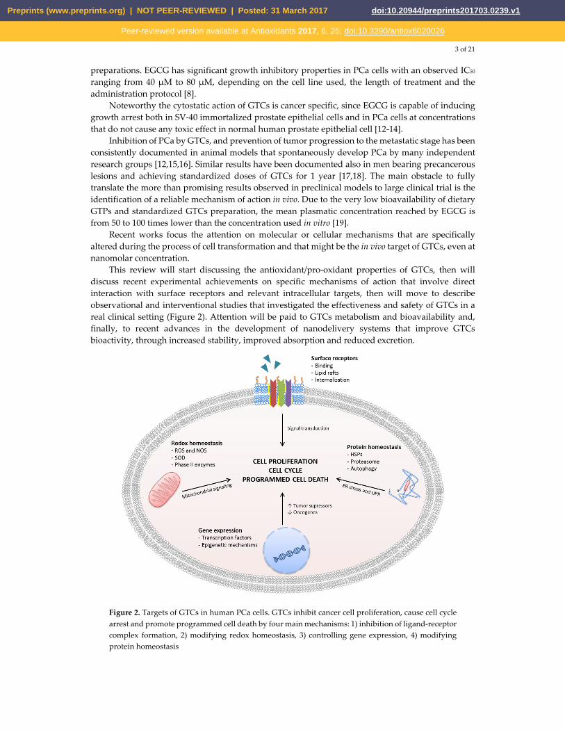
preparations. EGCG has significant growth inhibitory properties in PCa cells with an observed IC50 ranging from 40 μM to 80 μM, depending on the cell line used, the length of treatment and the administration protocol [8].
Noteworthy the cytostatic action of GTCs is cancer specific, since EGCG is capable of inducing growth arrest both in SV-40 immortalized prostate epithelial cells and in PCa cells at concentrations that do not cause any toxic effect in normal human prostate epithelial cell [12-14].
Inhibition of PCa by GTCs, and prevention of tumor progression to the metastatic stage has been consistently documented in animal models that spontaneously develop PCa by many independent research groups [12,15,16]. Similar results have been documented also in men bearing precancerous lesions and achieving standardized doses of GTCs for 1 year [17,18]. The main obstacle to fully translate the more than promising results observed in preclinical models to large clinical trial is the identification of a reliable mechanism of action in vivo. Due to the very low bioavailability of dietary GTPs and standardized GTCs preparation, the mean plasmatic concentration reached by EGCG is from 50 to 100 times lower than the concentration used in vitro [19].
Recent works focus the attention on molecular or cellular mechanisms that are specifically altered during the process of cell transformation and that might be the in vivo target of GTCs, even at nanomolar concentration.
This review will start discussing the antioxidant/pro-oxidant properties of GTCs, then will discuss recent experimental achievements on specific mechanisms of action that involve direct interaction with surface receptors and relevant intracellular targets, then will move to describe observational and interventional studies that investigated the effectiveness and safety of GTCs in a real clinical setting (Figure 2). Attention will be paid to GTCs metabolism and bioavailability and, finally, to recent advances in the development of nanodelivery systems that improve GTCs bioactivity, through increased stability, improved absorption and reduced excretion.
Figure 2. Targets of GTCs in human PCa cells. GTCs inhibit cancer cell proliferation, cause cell cycle arrest and promote programmed cell death by four main mechanisms: 1) inhibition of ligand-receptor complex formation, 2) modifying redox homeostasis, 3) controlling gene expression, 4) modifying protein homeostasis
Preprints (www.preprints.org) | NOT PEER-REVIEWED | Posted: 31 March 2017 doi:10.20944/preprints201703.0239.v1
Peer-reviewed version available at Antioxidants 2017, 6, 26; doi:10.3390/antiox6020026
4 of 21
2. Effects of GTCs on cellular redox homeostasis and antioxidant signaling In both physiological and pathological conditions reactive oxygen and nitrogen species (ROS
and NOS, respectively) are produced. These reactive species are essential for cell survival, and are involved in normal processes such as adaptation to hypoxia, regulation of autophagy, immunity, differentiation and longevity [20]. A moderate ROS increase triggers the hormesis effect, an adaptive paradigmatic response to reactive species that helps cells to cope with oxidative stress through the activation of specific redox-cellular signaling pathways [21-23]. When the production of ROS and NOS overwhelms the cell capacity to scavenge them, oxidative stress occurs, leading to damage to biological molecules, loss of function and eventually cell death.
The redox homeostasis imbalance is considered to be an important mechanism in promoting different diseases, including cancer onset and progression. Chronic exposure to elevated ROS concentration has been associated to PCa development [24]. Different studies have revealed that oxidative stress markers are higher in PCa cells (both in cell lines and human samples) in comparison to the non pathological counterparts [24,25]. The cellular redox imbalance cooperate with other factors associated with PCa etiogenesis as aging, androgen receptor activity, hormonal imbalance and chronic inflammation. Different detoxification enzymes and antioxidant molecules are involved in maintaining the redox balance inside the cell. One of the most important mechanisms involved in superoxide detoxification involves the enzymes superoxide dismutases (SODs), that produce H2O2, which is in turn switched-off by catalase or glutathione peroxidase activity. Along with enzymatic activities, different endogenous compounds like glutathione, plasma protein thiols and iron-binding proteins are needed to maintain the oxidative balance. Dietary components, like vitamins and polyphenols, also contribute in maintaining intracellular redox homeostasis [26,27]. An increasing numbers of studies have demonstrated that GTPs are strong antioxidant compounds [21,28], nonetheless GTPs are also able to exert pro-oxidant effects. The pro-oxidant/antioxidant behavior of GTPs greatly depend on many experimental conditions, such as final concentration of use, temperature, pH, presence of metal ions, and differences in culture media composition [21,29-31]. All these variables make it very difficult to compare results, sometimes contradictory, published by various Authors so far.
2.1 Effects of GTCs on intracellular ROS production
GTCs backbone (Figure 1) is characterized by the presence of two benzene rings (A- and B-ring) with two or more hydroxyl groups attached, and a dihydropyran heterocycle (C-ring). The conjugation of hydroxyl groups present on the C-ring with gallic acid (D-ring) leads to formation of EGCG and ECG. The antioxidant properties of the GTCs are due to the presence of both the phenolic groups and the galloyl moiety, that allow electron delocalization, and free radical quenching ability. Accordingly to their structural features, EGCG and ECG are, among the four most abundant GTCs, the strongest antioxidants [27,32]. However, GTCs may undergo auto-oxidation reactions, leading to the formation of catechin dimers and free radicals that may be responsible of the pro-oxidant action observed in vitro after polyphenols administration. The level of H2O2 generated is positively correlated with exposure time and polyphenols concentration used. Incubation of 100 μM EGCG or EGC in different cell culture media for 1 h determined the generation of H2O2 ranging between 50.7÷71.6 μM and 98.1÷115 μM, respectively. However, at the concentration of 10 μM (a value that is about 10-fold higher than values achievable in human plasma after ingestion of 800 mg of GTCs) the amounts of generated H2O2 is very low [33]. Of note, the administration of EGCG up to 50 μM in different cell culture media in presence of CHO cell exerts mild effects on cell viability, indicating that living cells possess efficient systems of defense against H2O2 produced by GTCs in aqueous solutions [34]. Transition metals are able to initiate the catechins auto-oxidation reaction, also promoting the formation of catechins-metal ions complexes. Depending on the experimental model these mechanisms have been correlated both to an antioxidant or a pro-oxidant behavior of GTCs [21,28]. Zinc is required for normal prostate metabolism due to its ability to inhibit aconitase activity determining the excretion of citrate into seminal fluid. The ability of Zn2+ to accumulate in the prostate gland is lost during PCa progression [35]. Experimental data have shown that in PCa cell lines, a
Preprints (www.preprints.org) | NOT PEER-REVIEWED | Posted: 31 March 2017 doi:10.20944/preprints201703.0239.v1
Peer-reviewed version available at Antioxidants 2017, 6, 26; doi:10.3390/antiox6020026

5 of 21
mixture of Zn2+ and EGCG enhances both the growth inhibitory effect and the free radical scavenging ability with respect to EGCG alone. Moreover, Zn2+ enhances EGCG effects in terms of reduction of mitochondrial membrane potential and caspase-9 activation [36-38]. In PC-3 ML and in IBC-10a primary prostate cell lines, the administration of EGCG (up to 60 μM) determines a reduction of mitochondrial membrane potential and integrity, followed by apoptosis without increase in ROS production [39]. Chung and colleagues have observed that the incubation of DU145 cells with green tea extract (50 μg/mL) or pure green tea compounds (100 μM) for 48 h, induces apoptosis through a peroxide and superoxide anions levels increase and mitochondrial depolarization [40]. Kanwal and colleagues treated LNCaP cells with EGCG (20 μM) or Polyphenon E® (10 μg/mL), a standardized GTCs-enriched mixture, caffeine free, containing 80% to 98% total catechins by weight. The Authors have demonstrated that the 72 h-treatments make cells more resistant to both H2O2-induced oxidative stress and DNA damage [41].
We studied the effects of increasing doses of Polyphenon E® on ROS production and cell viability in PCa cell lines. We reported that concentrations of Polyphenon E® yielding a significant increase in ROS production (5-20 μg/mL and 5-50 μg/mL, for PNT1a and PC3 respectively) displayed marginal effects on cell proliferation. A similar effect on ROS production was obtained treating cells with H2O2 (10 μM and 50 μM for PNT1a and PC3, respectively). At difference, higher concentrations of Polyphenon E® that caused a significant reduction of cell proliferation (25-35 μg/mL and 75-145 μg/mL, for PNT1a and PC3, respectively) produced an amount of ROS below to that of untreated cells or cells given H2O2. There is not good correlation between ROS induction and cell death in PCa cells, following Polyphenon E® treatment. Therefore, very unlikely ROS production plays a direct role in GTCs induced cell death. Nonetheless this process is strictly associated to induction of endoplasmic reticulum stress, a mechanism that impacts on protein homeostasis, finally leading to cell death [42].
2.2 Effect of GTCs on SOD activity
A part from direct effect on ROS production EGCG is also involved in the modulation of SODs activity. Many studies, reviewed in [43] suggest that manganese superoxide dismutase (MnSOD) participates in the maintenance of mitochondrial integrity in cells exposed to oxidative stress. Morrissey and colleagues have shown that EGCG treatment (20 μM for 72 h) of NRP-154, a tumorigenic rat prostate epithelial cell line, determines a reduction of the expression of MnSOD, but not of catalase, concomitantly with apoptosis induction [43]. On the contrary, in DU145 cells, MnSOD expression was not affected by the administration of 3 μM EGCG, while a significant reduction was observed after treatment with ionizing radiation (IR). In the same experimental model, the combination of EGCG and IR significantly reduces IR-induced apoptosis, together with an increase of MnSOD expression. Therefore EGCG might promote radiotherapy resistance inducing MnSOD [44]. In contrast, in Lobund-Wistar rats, the chronic consumption of green tea decreases tumor incidence of genitourinary tissues, in association with an increase of MnSOD expression, without effects on DNA damage, lipid peroxidation and lipofuscin deposition [45]. Treatment of TRAMP (Transgenic Adenocarcinoma of the Mouse Prostate) mice for 13 weeks with EGCG (administered by gavage at a dosage of 200 mg/kg body weight), starting at 7 weeks of age was associated with a significant reduction of severity/focalness of the histopathological lesions observed in the ventral lobe of the murine prostate. These effects are unlikely due to a direct antioxidant effect of EGCG, indeed the Authors observed only a slight reduction in plasma H2O2 concentration and no significant effect on SOD expression [46]. Later on, in the same experimental model, the Authors found that EGCG reduced the DNA oxidative damage marker but had no effects on protein and lipid peroxidation markers [47].
2.3 Effects of GTCs on NOS production
Nitric oxide is a free radical, produced by three different isoforms of nitric oxide synthase (NOS). NO. actions are mediated through cGMP-dependent or cGMP-independent pathways. Moreover, NO. at low concentrations can interact with different cellular compounds like other free radicals,
Preprints (www.preprints.org) | NOT PEER-REVIEWED | Posted: 31 March 2017 doi:10.20944/preprints201703.0239.v1
Peer-reviewed version available at Antioxidants 2017, 6, 26; doi:10.3390/antiox6020026
6 of 21
proteins and DNA. NO. exerts a dual role in cancer cells, which is dose dependent. At low concentrations (less than 100 nM), NO. is associated with tumor cell proliferation and angiogenesis, instead at high concentrations (more than 500 nM) NO. exerts a cytotoxic effect [48]. The anticancer effect of EGCG, through inhibition of NO. production, has been observed in many different tumors [48-51]. In TRAMP mice who received, 0.06% EGCG in tap water ad libitum for 7 weeks starting at weaning, a reduction of the HG-PIN incidence was observed, concomitantly with a significant down-regulation of iNOS [52].
3. Specific molecular mechanisms of action of GTCs
A part from their antioxidant/pro-oxidant activity, GTCs have high affinity to many biomolecules, including phospholipid bilayers, proteins and nucleic acids. It has been shown that GTCs can interact with cell surface receptors or enter the cells and directly interact with specific intracellular molecular targets [53,54]. Once that GTCs have interfered with the ligand-receptor binding, or more generally with receptor activation, the effects are reflected in intracellular signaling pathways that control cell proliferation/cell death through the activation/inhibition of regulatory proteins (kinases/phosphatases) or effector molecules. GTCs can interfere with the transcriptional regulation of gene expression by direct regulation of specific transcription factors and by epigenetic mechanisms that produce modifications of chromatin accessibility to transcription factors. GTCs may regulate gene expression also at post transcriptional level by a mechanism that involves the expression of particular miRNAs. Of particular note, in prostate, an endocrine tissue characterized by a huge protein synthesis activity, GTCs have been shown to interfere with the molecules, cellular compartments or mechanisms deputed to control protein homeostasis, such as molecular chaperons, the endoplasmic reticulum (ER), and the main protein degradation mechanisms.
3.1 Cell surface receptors
GTCs interferes directly or indirectly with the formation of the ligand-receptor complex of several cell surface receptors [55,56]. Three different mechanisms have been proposed to explain the effect of GTCs on the regulation of the activity of cell surface receptors and their signaling pathways. The first mechanism proposes that GTCs physically prevent the interaction of the ligand with its cognate receptor by completely covering the cell surface through a “sealing effect” [57] or binding to the ligand [58,59] or competing with the ligand for the receptor [60].
The second mechanism involves the suppression of the intrinsic activity of lipid rafts-associated signaling proteins by lipid rafts disruption [55,61,62]. The third prevents the interaction between receptor and ligand promoting receptor clearance through physical sequestration inside endosomal vesicles [63].
Receptor tyrosine kinases (RTKs) have been recently identified as targets of GTCs in cancer cell growth inhibition [55,64,65]. The binding of a growth factor or a cytokine to the extracellular domain of RTKs induces the dimerization and autophosphorylation of the RTKs on specific tyrosine residues and activation of downstream intracellular signaling that include Ras/ERK/MAPK and PI3K/Akt. Ras/ERK/MAPK and PI3K/Akt activation triggers a cascade of molecular events involving enzymes, proteins and transcription factors that have been extensively reviewed in [66]. The activation of cell surface RTKs controls many key important processes in normal cells such as cell proliferation, differentiation, survival and migration. These receptor-associated pathways have been found altered in tumors cells, including PCa [55,66-69]. The RTKs family comprises epidermal growth factor receptors (EGFRs), fibroblast growth factor receptors (FGFRs), insulin-like growth factor receptors (IGF-R), platelet-derived growth factor receptors (PDGFRs), vascular endothelial growth factor receptors (VEGFRs), hepatocyte growth factor receptors (HGFRs) also known as Met. The most effective inhibition toward RTKs is exerted by EGCG, followed by ECG and EGC [67].
GTCs administration inhibited EGFR activation in several cancer cell lines, even at concentrations as low as 1 μg/mL [70-73]. Liang et al. demonstrated that EGCG directly prevent the interaction between ligand and EGFR necessary for the tyrosine kinase activity in human epidermoid carcinoma cells (IC50 = 0.5÷1 μg/mL) [59].
Preprints (www.preprints.org) | NOT PEER-REVIEWED | Posted: 31 March 2017 doi:10.20944/preprints201703.0239.v1
Peer-reviewed version available at Antioxidants 2017, 6, 26; doi:10.3390/antiox6020026
7 of 21
Similar effects of GTCs have been observed towards IGF-R1 [74]. IGF-R1 is overexpressed in PCa [67]. Interestingly Li et al. demonstrated that EGCG competitively binds to the ATP binding site of IGF-R1 inhibiting its kinase activity, in vitro (IC50 = 14 μmol/L) and in vivo [60]. The blockage of ligand-receptor binding has been suggested as the mechanism behind the inhibitory effect of GTCs, on PDGFR and HGFR in multiple cell lines, at concentrations of 1÷10 μM [58,75-77]. Tachibana et al. proposed that EGCG is a ligand of the 67-kDa laminin receptor (67-LR) with a dissociation constant (Kd) value of 0.004 μM [78]. 67-LR expression is elevated in cancer cells, where it acts as a cancer-specific cell death receptor. EGCG pro-apoptotic effects in multiple myeloma cells are abrogated by 67-LR silencing, suggesting that EGCG induced cell death is achieved through a 67-LR mediated mechanism [79]. A proof of the direct binding between EGCG and 67-LR has been recently provided by Shukla and colleagues. These Authors have shown that delivery of radioactive gold nanoparticles (NPs) functionalized with EGCG in PCa cells was mediated via the 67-LR. When the 67-LR was blocked, by laminin or by anti-67LR antibody, the uptake of NPs was prevented [80]. Recent data have confirmed that EGCG undergoes oligomer formation by binding to the 67-LR [81]. In particular it has been identified a sensing motif on 67-LR, located within the peptide LR161-170, that is critical for the binding of EGCG [82].
The second mechanism proposed to explain the ubiquitous effect of EGCG on multiple receptors involves the disruption of lipid organization in the plasma membrane [61]. Lipid rafts are high ordered membrane structures rich in cholesterol and sphingolipids that regulate protein-protein interaction, receptor activation and therefore more generally cellular signaling. EGCG alters the lipid organization of the plasma membrane, causes the rearrangement of lipid rafts [83] preventing the activation of cell surface receptors. In multiple myeloma cells EGCG promoted apoptosis through lipid rafts clustering, that in turn mediated the activation of 67-LR and triggered a pro-death signaling [84-87]. GTCs inhibited RTKs activation through lipid rafts clustering. This mechanism has been reported for EGFR, HGFR, IGF-R1 and VEGFR [61,62,88-91].
Finally Adachi et al. found that the EGFR is quickly internalized inside endosomal vesicles in human colon cancer cells treated with EGCG (1 μg/mL for 30). The internalization makes the receptor inaccessible to EGF and abrogates RTKs signaling cascade [63].
3.2 EFFECTS OF GTCs ON REGULATION OF GENE EXPRESSION
Although the mechanisms of the cancer chemopreventive effect of GTCs have not been completely elucidated, there is evidence that EGCG administration results in modulation of gene expression [12,92]. It has been demonstrated that EGCG modulates activity and expression of various transcription factors including Sp1, NF-κB, AP-1, STAT1, STAT3 and nuclear factor erythroid 2-related factor 2 (Nrf2) [27]. Among these Nrf2 and NF-kB are two redox-sensitive transcription factors responding to inflammatory/oxidative stress stimuli that control the expression of many genes involved in PCa onset and progression. It is now accepted that free radicals act as redox cellular messengers, triggering signaling pathway able to influence the redox homeostasis. One of the most important cellular response to cope with oxidative stress is the activation of Nrf2, which in turn activates a plethora of enzymes. Keap1, the Nrf2 inhibitor, is sensitive to redox imbalance. In presence of oxidants or electrophilic compounds, Keap1 dissociates from Nrf2, which then translocates to the nucleus and binds to electrophile-responsive element (EpRE) sequences, driving the transcription of phase II enzymes such as glutathione peroxidase (GPX), glutathione S-transferase (GST), NADPH quinine oxidoreductase 1 (NQO-1), and UDP-glucuronosyltransferase (UGT) [93,94]. Nrf2 also acts through Keap1 independent mechanisms [95,96]. It is important to highlight that GTCs are able to activate multiple pathways, such as MAPKs, PI3K, PKC, NF-kB which may play a crucial role in the Nrf2-mediated response to oxidative stress [93,95]. EGCG, and more generally GTCs, exert their anticancer effect in many different tumor models through the induction of a beneficial moderate oxidative stress, that in turn activate the Nrf2/EpRE response [93,97]. A direct target of Nrf2 is the gene GSTP1, that is downregulated in PCa. Effects of GTCs on GSTP1reexpression are discussed in the next paragraph. NF-kB is constitutively activated in PCa cells, prostate premalignant lesions (intraepithelial tumors, PIN) and PCa tissues [98]. EGCG has been demonstrated to inhibit NF-kB
Preprints (www.preprints.org) | NOT PEER-REVIEWED | Posted: 31 March 2017 doi:10.20944/preprints201703.0239.v1
Peer-reviewed version available at Antioxidants 2017, 6, 26; doi:10.3390/antiox6020026
8 of 21
transcriptional activity in DU145 and LNCaP cells, promoting apoptosis via Bax up-regulation and Bcl2 down-regulation [99,100]. Siddiqui et al. reported that the constitutive activation of NF-kB, observed during PCa progression, was prevented in TRAMP mice receiving 0,1% GTCs in drinking water from weaning, in comparison to water-fed, age matched controls [101]. One of the targets of NF-kB is represented by matrix metalloproteinases (MMPs), a family of proteolytic enzymes that degrade collagene and other extracellular matrix protein contributing to PCa progression. GTCs have been reported to inhibit various MMPs in PCa cells [99,102] and in TRAMP mice [16]. In silico molecular docking analysis have recently shown a strong direct interaction between EGCG and MMP-9, suggesting that EGCG might inhibit MMP-9 also by a NF-kB independent mechanism [103].
3.2.3 Epigenetic mechanisms
Epigenetics generally refers to changes in gene expression and chromatin organization that are independent of alterations in the DNA sequence. Epigenetic phenomena are modifiable by environmental factors that include dietary habits [104]. Cancer development involves a complex multistep process caused and associated to many genetic insults that include genetic mutation as well epigenetic alteration [105]. Many tumor suppressor and receptor genes have been reported to be hypermethylated and transcriptionally silenced during the development of various human cancers, including PCa [106]. The major epigenetic mechanisms that control gene expression are DNA methylation, histone modifications and expression of noncoding regulatory micro RNA (miRNAs). Extensive in vitro experiments reviewed by Henning et al. in [104] have been performed in a variety of cancer cell lines to evaluate the effect of GTCs, or more generally GTPs on DNA methylation. There is evidence that GTCs cancer preventive effect can be ascribed to epigenetic reactivation of silenced gene through inhibition of DNMTs activity. Fang et al. demonstrated that EGCG binds to DNMT and competitively inhibits the enzymatic activity (Ki of 6.89 μM) yielding the reactivation of methylation-silenced genes in PC3 cells [107]. Molecular modeling and docking studies supported the binding of EGCG to DNMT3B in HeLa cells [108]. EGCG (5-20 μM) or Pholyphenon E® (1-10 μg/mL) treatments of different PCa cell lines, have determined a dose- and time-dependent re-expression of GSTP1 enzyme concomitantly with the down-regulation of DNMT1 [41,106]. The re-expression of GSTP1, induced by treatment with EGCG or Polyphenon E® may be, at least in part, responsible for the quenching of free radicals species and reduction of DNA damage associated with oxidative stress [41]. Two studies concluded that GTCs did not produce a significant effect on DNA methylation in PCa cell lines and in TRAMP mice [109,110]. Potential reasons for these discrepancies might be due to different methods of analysis used to evaluate DNA methylation, or different cell lines used, or inefficacy of the treatment protocol. Nonetheless, Henning et al. fed SCID mice implanted with LAPC4 androgen-dependent PCa cells with brewed green tea (containing 0,075% GTPs), or water for 13 weeks. The Authors found that tumor growth and expression of DNMT1 were reduced in xenograft tumors excised from green tea-fed mice in comparison to controls [111]. These results support the concept that GTCs may produce epigenetic changes in vivo following long term administration.
The direct inhibition of EGCG on DNMT1 is enhanced in the presence of catechol-O-methyltransferase (COMT). The mechanism proposed by Lee et al. to explain this addictive effect is due to alteration of the S-adenosyl-methionine (SAM) and S-adenosyl-homocysteine (SAH) intracellular ratio. Indeed, galloylated GTCs are methylated by COMT that catalyzes the transfer of a methyl group from the donor molecule SAM that in turn forms SAH. By this mechanism the SAM/SAH is reduced and inhibits DNMTs activity that depends on the intracellular availability of SAM as methyl donor [112]. Navarro-Perak et al. [113] suggested that EGCG inhibits DHFR and/or folic acid uptake acting as an antifolate compound. A part from SAM, folates are an important source of the 1-carbon unit used to methylate DNA. Low folates in cancer cells induced by EGCG administration prevent DNA hypermethylation of specific genes. The effective concentration of EGCG on human DHFR inhibition is around 33 nM, a value physiologically achievable in human plasma after ingestion of standardized preparation of GTCs [114-117]. One of the most important mechanisms to control gene expression depends on the chromatin condensation status, that is
Preprints (www.preprints.org) | NOT PEER-REVIEWED | Posted: 31 March 2017 doi:10.20944/preprints201703.0239.v1
Peer-reviewed version available at Antioxidants 2017, 6, 26; doi:10.3390/antiox6020026
9 of 21
regulated by different processes, with histone acetylation/deacetylation being one of the most studied and finely regulated [105]. Histone acetylation often correlates with chromatin relaxation. Inhibition of histone deacetylases (HDACs) in cancer cells causes re-expression of genes epigenetically silenced during carcinogenesis [118]. Polyphenon E® administration has been reported to cause inhibition of HDACs activity and expression in PCa cell lines [106,119]. Molecular modeling and docking studies in HeLa cells suggested that EGCG inhibits HDAC1 activity by direct binding of the enzyme [108].
miRNAs are functional small non-coding RNAs that control gene expression by inducing degradation or translational inhibition of their target mRNAs. It has been demonstrated that various miRNAs are often overexpressed in cancer cells, including PCa cells, and their altered expression is associated to cancer progression [120]. miRNA-21 is regulated by androgens and promote disease progression toward a hormone-independent phenotype [121]. Siddiqui et al. demonstrated that EGCG treatment of nude mice subcutaneously implanted with 22Rv1 PCa cells have inhibited tumor growth, by downregulating miRNA-21 and induced upregulation of miRNA-330, a tumor suppressor that induces apoptosis in PCa cells [122].
3.3 Effects of GTCs on protein homeostasis
Tumor cells are continuously exposed to stressful conditions (such as hypoxia, nutrient deprivation, acidosis, chemotherapeutic treatments, etc.) and are characterized by a high translational activity; therefore, they are much more prone to accumulate misfolded and/or unfolded proteins. This stress is even much higher in PCa secretory epithelial cells, that are characterized by high rate of protein synthesis. For the maintenance of protein homeostasis (proteostasis), eukaryotic cells have developed a complex protein quality control system, which comprises molecular chaperons, the ER-associated degradation (ERAD) mechanism, the unfolded protein response (UPR) and the two most important systems of protein degradation, i.e. the proteasome system and the autophagic mechanism. Proteostasis impairment and accumulation of misfolded proteins in the ER lumen induce ER stress, which in turn activates the UPR, a complex network of molecular signaling to adapt and respond to ER stress conditions, thereby promoting cell survival. However under chronic stress when proteostasis cannot be restored, the UPR activates apoptosis signaling that commits cell to death [123,124].
GTCs have been proven to trigger anticancer effects targeting HSPs and proteasome functions and by interfering with the autophagic flux.
3.3.1 Heat shock proteins
Heat shock proteins (HSPs) are a superfamily of molecular chaperones representing the first line of defense against protein misfolding and aggregation. They aid in the folding and refolding of proteins, and target denatured proteins to degradative systems. HSPs function both in physiological and pathological conditions. Different cellular pathways, associated with cancer onset and progression, may be involved in the activation of the heat shock factor-1 (HSF-1), the primary factor associated with HSPs gene activation [125,126]. The expression of different members of HSPs is modulated during PCa progression [127]. It is documented that EGCG inhibits carcinogenesis thanks to down-regulation of both HSP90 and HSP70, and through the dissociation of their complexes with co-chaperones or client proteins [128]. In different cell lines that mimic progressive stages of PCa, Moses and colleagues demonstrated that EGCG inhibited cell proliferation and induced apoptosis by direct binding to HSP90, causing instability and altering the functions of HSP90-client proteins involved in tumor progression [129].
3.3.2 Proteasome inhibition
The ubiquitin-mediated proteasomal degradation is essential for the regulation of several cellular processes. The proteasome is a multicatalytic protein complex present in all eukaryotic cells and it is responsible for the degradation of the major part of the cellular proteins. The chymotrypsin-like activity of the proteasome is associated with the tumor cell survival [130] since most of the
Preprints (www.preprints.org) | NOT PEER-REVIEWED | Posted: 31 March 2017 doi:10.20944/preprints201703.0239.v1
Peer-reviewed version available at Antioxidants 2017, 6, 26; doi:10.3390/antiox6020026
10 of 21
intracellular proteins involved in the onset and progression of tumor are degraded through the ubiquitin-proteasome pathway. GTCs are strong and selective inhibitors of the proteasomal chymotrypsin-like activity in vitro (IC50 = 86-194 nM) and in intact PCa cells (1-10 μM). EGCG, among all the catechins tested, showed the strongest inhibitory activity. [130]. The ester bond contained in galloylated catechins is required for potent inhibition of the proteasomal activity [130,131]. Other structure-activity relationship studies evidenced that the A ring and the gallate ester/amide bond are involved in the mechanism of proteasome inhibition [132]. Similar effects have been described for two synthetic enantiomeric analogs of natural GTCs in PCa cells [133]. The observed accumulation of short-lived proteins, normally addressed to proteosomal degradation such as p21, p27, Bax and ikΒα, in PCa cells after EGCG or GTCs treatment indirectly confirms, that GTCs exert inhibitory effect on the proteasome activity [98,134].
3.3.3 Autophagy
The catabolic process through which long-lived proteins, damaged organelles and other unnecessary intracellular materials are delivered to lysosomes for degradation is called autophagy. Normally autophagy is constitutively active at low levels to guarantee cellular homeostasis, but it can be strongly induced in stressful conditions when this process seems to act primarily as a cellular protective response [135]. The effects of EGCG on autophagy seem to be dependent on concentration used, cell type and stress conditions.
We have observed at morphological level, by electron microscopy, that GTCs administration (0,3% in tap water) in TRAMP mice drastically reduce the condensing and secretory activities of the ER of prostate cells, causing an imbalance of the protein trafficking and an engulfment of ER finally leading to ER stress. No effects were observed in age matched animals water fed. In agreement with morphological data we found that biogenesis of secreted proteins is altered in TRAMP-C2 cells treated with 20 μg/mL [136]. We reported that autophagy was transiently activated in human prostate PNT1a cells, commonly used to mimic the initial stages of PCa, as a survival response to overcome Polyphenon E®-induced ER stress [42]. Treatment of PNT1a with 35 μg/mL of Polyphenon E® for 24 h committed cells to apoptosis, while 145 μg/mL Polyphenon E® committed metastatic prostate cancer PC3 cells to caspase-independent programmed cell death [42].
4. GTCs in human PCa chemoprevention: from epidemiological data to interventional studies.
Three large cohort prospective studies, all conducted in Japanese population, were aimed to examine the association between dietary intake and PCa risk [137-139]. All these observational studies did not find any statistical significant correlation between green tea consumption and PCa risk. A fourth study examined the association between green tea consumption and risk of PCa stratified by disease stage [140] and corrected results for potential confounder factors. The Authors did not find a significant association between green tea consumption and the global risk of being diagnosed with PCa. Nonetheless, a dose-dependent inverse relation for risk to develop advanced PCa for men who consumed 5 cups of green tea/day compared with those who consumed 1 cup/day was observed. Two case-control studies examined the relation between green tea intake and PCa in patients diagnosed with PCa (histologically confirmed) and PCa free matched hospital inpatients as controls [141,142]. Both these studies found an inverse relation between green tea consumption and PCa risk. Nonetheless these results should be interpreted very carefully due to many critical points that include the small sample size, the hospital based case-control design, the simultaneous consumption of green and black teas, differences in tea brewing methods and possible interferences from other dietary factors. Although epidemiological evidences are still not conclusive, the anticancer efficacy of green tea is fully displayed in the TRAMP model when intervention is started early, at weaning, and the malignancy is not fully developed. In this chemopreventive scenario, concentration of standardized green tea extract ranging between 0.1÷0.3% in drinking water are effective to strongly delay or stop PCa growth [12,15,92]. Adhami at al. demonstrated that green tea extracts (GTE) are more effective when feeding (0.1% in drinking water) is started at very early stages of PCa development (presence of “in situ” histological lesions) while the chemopreventive potential
Preprints (www.preprints.org) | NOT PEER-REVIEWED | Posted: 31 March 2017 doi:10.20944/preprints201703.0239.v1
Peer-reviewed version available at Antioxidants 2017, 6, 26; doi:10.3390/antiox6020026
11 of 21
decreases in animals bearing tumors of more advanced stage, finally resulting largely ineffective in animals that display moderately differentiated adenocarcinoma and occasionally metastasis [143]. To reinforce the concept that GTCs are ineffective as chemotherapeutic molecules, very limited or no effects have been observed in patients with advanced PCa [144-146]. At difference, we introduced for first the “Proof of Concept” that secondary chemoprevention of PCa with GTCs is feasible [17]. We enrolled 60 Italian patients (all having a Caucasian genetic background) diagnosed with high grade prostate intraepithelial neoplasia (HG-PIN), a premalignant lesion associated to a 30% increased risk to develop PCa within 1 year [147]. 30 volunteers were randomly assigned to receive, according to a double-blind procedure, 600 mg/day of a standardized GTCs formulation (total catechins 75.7%; EGC, 5.5%; EC, 12.2%; EGCG, 51.9%; ECG) divided in three capsules of 200 mg each, or identical placebo capsules for 1 year. All the patients, during the study, were subjected to regular prostate biopsy (6 and 12 months after the study beginning) to assess differences in PCa rates between the two arms. After 1 year of treatment, only 1 tumor was diagnosed among the 30 GTCs-treated men, while 9 cancers were found among the 30 placebo-treated men. No significant side or adverse effects were documented throughout the whole study. A randomized, placebo-controlled trial evaluated whether 400 mg of Polyphenon E® significantly reduce PCa rates in a cohort of men diagnosed with HG-PIN and/or atypical small acinar proliferation (ASAP) [148]. 74 men (36 GTCs and 38 placebo) completed the 1 year protocol. No significant reduction of PCa incidence was observed between the two arms. Possible explanations of the discrepancies between the results recorded by Kumar et al. and those previously published by us may reflect differences in the study design, that include enrolment criteria (more heterogeneous) and GTCs posology (a lower dosage). Pathologists usually formulate ASAP diagnosis when it is not possible to find alterations in the cells’ morphology to support “without doubts” a PCa diagnosis. For this reason ASAP is often considered an under-diagnosed cancer [149]. Men bearing both ASAP and multifocal HG-PIN are very likely patients in whom, at the moment of the first biopsy, PCa has already developed, even if not clearly supported by histopathological diagnosis [150]. We might hypothesise that the in the study by Kumar et al. were included men for which the narrow window of time for therapeutic intervention with GTCs was already expired at the enrolment. As a consequence of the small number of patients enrolled, unfortunately, the study did not have sufficient statistical power to detect a reduction of PCa incidence in the subgroup of men diagnosed only with HG-PIN receiving GTCs in comparison with HG-PIN patients receiving placebo group [148].
5. GTCs bioavailability and metabolism in humans
Flavanols bioavailability in vivo is strongly limited by their high molecular weight and by a bulky hydration shell that magnify the effective molecular size. Moreover extensive enteroluminal biotransformation and phase II metabolism of flavan-3-ols, which includes methylation, glucuronidation, and O-methylation may affect GTCs bioactivity in vivo, and explain inconsistent results in comparison to effects observed in vitro, using supraphysiological doses of EGCG [151]. Chow et al. investigated the pharmacokinetic of GTCs following single administration of Polyphenon E® at increasing concentration ranging from 200 to 800 mg or equivalent doses of pure EGCG [116,152]. The average EGCG peak plasma concentration (Cmax) was reached after 2-4 h from ingestion and declined rapidly, with very low/undetectable levels at 24 h after dosing. EGCG peak concentration increased with increasing doses in a nonlinear fashion, reaching a value above 1 μM at the highest concentration tested, possibly due to the existence of saturable mechanisms that cause a presystemic elimination of orally administered GTCs. EGC and EC were detected in plasma and urine samples after Polyphenon E® administration, predominantly as glucuronic acid/sulfate conjugates while EGCG was detected prevalently in its unchanged form. The presence of other catechins in the formulation tested does not influence EGCG plasma concentration, at difference the presence of food in the stomach interferes with catechins absorption and reduces by 3-fold the plasmatic concentration of free catechins (EGCG, EGC, EC) [116,152]. The same Authors observed that systemic availability of free EGCG increases by 60% after repeated administration (4 weeks of treatment) of high daily bolus dose (800 mg once daily) and that this concentration is safe and well
Preprints (www.preprints.org) | NOT PEER-REVIEWED | Posted: 31 March 2017 doi:10.20944/preprints201703.0239.v1
Peer-reviewed version available at Antioxidants 2017, 6, 26; doi:10.3390/antiox6020026
12 of 21
tolerated in healthy human subjects [116]. Similar results of catechins serum levels were obtained in patients diagnosed with PCa and receiving 800 mg of Polyphenon E® for 4 to 6 weeks before scheduled prostatectomy surgery [146]. The Authors instead found a very low to undetectable level of catechins in the prostate surgical samples collected from the same patients, possible due to a combination of rapid systemic clearance (less than 24 h) and low bioaccumulation of polyphenols in the tissue [146]. Different GTCs concentrations ranging from 21 to 107 pmol/g tissue were found in the prostate of men who consumed 1.42 L of green tea for 5 days before prostatectomy [153]. In human prostate glands of PCa patients who consumed 6 cups of green tea daily for up to six weeks before prostatectomy, EGCG is detectable mainly in the free form (nonglucuronidated/sulfated) [154]. Approximately half of the EGCG was present in the methylated form as 4″-MeEGCG. Results obtained in LNCaP cells indicated that methylation of EGCG may occurr directly in prostate tissue catalized by cytosolic COMT. None of the nongallated GTCs including EGC and EC or their metabolites were found in the prostate tissue indicating a lower bioavailability for these compounds in comparison with EGCG [154].
Catechins that are not absorbed in the small intestine, as well as conjugated catechins excreted in the bile, reach the large intestine where they may be metabolized by colonic bacteria [151]. (-)-EC, (-)-EGC and (-)-EGCG, are degradated by the human colon microbiota to 5-(3’,4’,5’-trihydroxyphenyl)-γ-valerolactone, 5-(3’,4’-dihydroxyphenyl)-γ-valerolactone and 5-(3’,5’-dihydroxyphenyl)-γ-valerolactone. Some of these colonic metabolites may enter the circulation and undergo further modifications before being excreted with the urine in quantities corresponding to about 40% of total ingested GTCs [155]. Colonic biotransformation represents therefore a significant component of the overall bioavailability equation and significantly influence GTCs bioactivity. To the best of our knowledge no data are available on the bioactivity of these compounds on PCa cells.
6. Nanotechnology strategies to improve GTCs bioactivity
The great potential of use of GTCs or EGCG as safe natural molecules to prevent PCa onset or eventually revert early PCa lesions is limited by the intrinsic chemical instability, low bioavailability and quick in vivo clearance of these products. Drug formulation strategies aimed at improving GTCs/EGCG stability, assist efficient and specific delivery at target cells, limit undesired metabolism and are therefore expected to produce a valuable benefit in terms of improved pharmacodynamic profile. The concept of nanochemoprevention, i.e. the uses of nanotechnology for enhancing the outcome of chemoprevention, was first introduced by Siddiqui et al. [156]. NPs are nanocarriers made of biodegradable/biocompatible materials sized between 1-100 nm, used as vehicles to improve drug delivery. Different types of NPs have been used to improve the pharmacological profile of EGCG, including gold nanoparticles, polymeric nanoparticles, liposomes, carbohydrate and inorganic nanoparticles [157,158].
Siddiqui et al. in their “Proof of Principle” study demonstrated that EGCG, encapsulated in polylactic acid (PLA)–polyethylene glycol (PEG) nanoparticles, has an improved efficacy against human PCa cells both in vitro and in vivo [156]. The inhibition of PC3 proliferation by half of normal values, measured as the IC50, was found to be 10-fold lower when nano-EGCG was used in comparison to non encapsulated EGCG. In vivo, 10-fold lower dose of nano-EGCG was required to achieve a similar extent of xenograft tumors obtained with EGCG.
Polymeric EGCG-encapsulated NPs, functionalised with a mimetic ligand of the prostate specific membrane antigen (PSMA), show an improved selective EGCG delivery in PSMA expressing PCa cells thus leading to a more marked inhibition of cell proliferation [159,160]. The same Authors observed a significant increase of tumor growth inhibition in mouse xenografts treated with PSMA targeted NPs, in comparison to EGCG. This effect may be attributed to the selective binding of NPs to PSMA, which presumably would promote an active targeting through receptor-mediated endocytosis [160]. One disadvantage of using PLA-PEG nanoparticles is their low stability in acidic environment that precludes the oral route of administration. Chitosan NPs encapsulating EGCG (Chit-nanoEGCG) at difference are characterized by slow release of EGCG in simulated gastric juice acidic pH and faster release in simulated intestinal fluid. Treatment with Chit-nanoEGCG resulted in
Preprints (www.preprints.org) | NOT PEER-REVIEWED | Posted: 31 March 2017 doi:10.20944/preprints201703.0239.v1
Peer-reviewed version available at Antioxidants 2017, 6, 26; doi:10.3390/antiox6020026
13 of 21
significant growth inhibition of subcutaneously implanted 22Rν1 PCa xenografts. In tumor tissues of mice treated with Chit-nanoEGCG, compared with animals treated with EGCG, there was significant induction of apoptosis and reduction of cell proliferation [161].
EGCG was efficiently loaded in polysaccharides NPs made of Arabic gum and maltodextrine. Encapsulated EGCG retained its biological activity, indeed it reduced cell viability and induced apoptosis when administered to DU145 PCa cells. Colony formation assay showed that nanoencapsulated EGCG possess enhanced inhibitory effect on cell proliferation at physiological achievable concentrations (1÷2 μM), compared with free EGCG [162].
EGCG encapsulated in solid lipid nanoparticles (EGCG-SLN) increase molecule stability in aqueous solution, improve efficient cell delivery and yield a 4-fold increase of cytotoxicity against DU145 human PCa in comparison to cells that received pure EGCG at the same concentration [163].
EGCG intercalated into Ca/Al-NO3 layered double hydroxide (LDH) NPs, produced higher inhibition of proliferation and colony formation suppression in PC3 cells compared to EGCG alone. Similarly, enhanced apoptosis was observed in PC3 treated with EGCG-LDH in comparison to non-encapsulated EGCG. The enhanced anti-tumor activity of EGCG-loaded LDH-NPs is well correlated to the mechanism of diffusion of the EGCG through the vehicle that allowed a sustained gradual release of the drug in solution [164].
EGCG can also be used as a “Trojan horse” to deliver anticancer therapy specifically in PCa cells. Recently has been demonstrated that EGCG functionalized radioactive biocompatible gold nanoparticles derived from the Au-198 isotope (EGCG-198AuNPs), selectively binds with high affinity to the 67-LR, a receptor that is over expressed in PCa cells. Significant amount of EGCG-AuNPs were internalized, through 67-LR receptor endocytosis in tumor cells. Pharmacokinetic studies in PC-3 xenograft SCID mice showed high retention of EGCG-198AuNPs in tumors and 80% reduction of tumor volumes compared to controls [80]. EGCG and gelatin-doxorubicin conjugate (GLT-DOX)-coated gold nanoparticles (DOX-GLT/EGCG AuNPs) were produced to improve specific uptake of doxorubicin in PC3 cells using EGCG/67-LR receptor-mediated delivery. Apoptosis of DOX-GLT/EGCG AuNPs treated PC3 was improved in comparison with cells treated with DOX [165].
7. Conclusions
Many Authors have suggested that the anticancer effects of GTCs might be achieved through their direct ability to scavenge harmful ROS/NOS, or at the opposite, to their autoxidation that, in turn, produces H2O2, a toxic compound. Both these effects, observed in vitro, have been obtained using supraphysiological concentration of GTCs and are very unlikely to take place in vivo. Recent achievements in this field of research have clarified that GTCs can interact with molecular targets that are specifically deregulated in PCa, such as the 67-LR receptor, at nanomolar concentrations. Others have demonstrated that GTCs modify the expressions of genes relevant for PCa progression, acting on transcription factors and through epigenetic mechanisms, or activating complex adaptive signaling such as the UPR. Despite, GTCs have been proven consistently to effectively inhibit PCa progression in animal models, their chemopreventive action in human PCa is still debated. Lessons learnt from clinical trials so far concluded indicate that the therapeutic window for GTCs intervention is very narrow, and possible corresponding to early signs of prostate tissue transformation, such as HG-PIN. Moreover inadequate dosage and/or short intervention periods may compromise the therapeutic effect. It’s now clear that individual differences in GTCs metabolism and absorption may negatively affect the already low bioavailability, reducing consequently the bioactivity. New opportunities to improve the therapeutic value of GTCs come from the field of the nanotechnologies, that when applied to chemoprevention lead to the development of “nanochemoprevention” strategies. More than encouraging results have shown that biopolymeric NPs helps to selectively deliver GTCs in PCa cancer cell, improving drug stability and increasing in vivo bioactivity. Large, well designed, interventional studies are urgently needed to evaluate the efficacy of GTCs charged NPs in PCa prevention.
Acknowledgements:Valeria Naponelli was supported by a Fondazione Umberto Veronesi fellowship.
Preprints (www.preprints.org) | NOT PEER-REVIEWED | Posted: 31 March 2017 doi:10.20944/preprints201703.0239.v1
Peer-reviewed version available at Antioxidants 2017, 6, 26; doi:10.3390/antiox6020026
14 of 21
Author contribution:All the Authors critically reviewed the literature and wrote the paper.
Conflicts of interest:The Authors declare no conflicts of interest.
References 1. Ferlay J, S.I., Ervik M, Dikshit R, Eser S, Mathers C, Rebelo M, Parkin DM, Forman D, Bray, F. Globocan
2012 v1.0, cancer incidence and mortality worldwide: Iarc cancerbase no. 11. http://globocan.iarc.fr, (01/02/2017),
2. Wong, M.C.; Goggins, W.B.; Wang, H.H.; Fung, F.D.; Leung, C.; Wong, S.Y.; Ng, C.F.; Sung, J.J. Global incidence and mortality for prostate cancer: Analysis of temporal patterns and trends in 36 countries. Eur Urol 2016, 70, 862-874.
3. Chen, S.-L.; Wang, S.-C.; Ho, C.-J.; Kao, Y.-L.; Hsieh, T.-Y.; Chen, W.-J.; Chen, C.-J.; Wu, P.-R.; Ko, J.-L.; Lee, H., et al. Prostate cancer mortality-to-incidence ratios are associated with cancer care disparities in 35 countries. Scientific Reports 2017, 7, 40003.
4. Baade, P.D.; Youlden, D.R.; Krnjacki, L.J. International epidemiology of prostate cancer: Geographical distribution and secular trends. Mol Nutr Food Res 2009, 53, 171-184.
5. Shimizu, H.; Ross, R.K.; Bernstein, L.; Yatani, R.; Henderson, B.E.; Mack, T.M. Cancers of the prostate and breast among japanese and white immigrants in los angeles county. Br J Cancer 1991, 63, 963-966.
6. Lee, J.; Demissie, K.; Lu, S.E.; Rhoads, G.G. Cancer incidence among korean-american immigrants in the united states and native koreans in south korea. Cancer Control 2007, 14, 78-85.
7. Bettuzzi, S.; Rizzi, F.; Belloni, L. Clinical relevance of the inhibitory effect of green tea catechins (gtcs) on prostate cancer progression in combination with molecular profiling of catechin-resistant tumors: An integrated view. Pol J Vet Sci 2007, 10, 57-60.
8. Johnson, J.J.; Bailey, H.H.; Mukhtar, H. Green tea polyphenols for prostate cancer chemoprevention: A translational perspective. Phytomedicine 2010, 17, 3-13.
9. Sawada, N. Risk and preventive factors for prostate cancer in japan: The japan public health center-based prospective (jphc) study. J Epidemiol 2017, 27, 2-7.
10. Henning, S.M.; Wang, P.; Heber, D. Chemopreventive effects of tea in prostate cancer: Green tea versus black tea. Mol Nutr Food Res 2011, 55, 905-920.
11. Takehiko, Y., Mujo, K., 1997. Chemistry and applications of green tea. . CRC Press, New York: 1997; p 1e130. 12. Caporali, A.; Davalli, P.; Astancolle, S.; D'Arca, D.; Brausi, M.; Bettuzzi, S.; Corti, A. The chemopreventive
action of catechins in the tramp mouse model of prostate carcinogenesis is accompanied by clusterin over-expression. Carcinogenesis 2004, 25, 2217-2224.
13. Ahmad, N.; Feyes, D.K.; Nieminen, A.L.; Agarwal, R.; Mukhtar, H. Green tea constituent epigallocatechin-3-gallate and induction of apoptosis and cell cycle arrest in human carcinoma cells. J Natl Cancer Inst 1997, 89, 1881-1886.
14. Albrecht, D.S.; Clubbs, E.A.; Ferruzzi, M.; Bomser, J.A. Epigallocatechin-3-gallate (egcg) inhibits pc-3 prostate cancer cell proliferation via mek-independent erk1/2 activation. Chem Biol Interact 2008, 171, 89-95.
15. Gupta, S.; Hastak, K.; Ahmad, N.; Lewin, J.S.; Mukhtar, H. Inhibition of prostate carcinogenesis in tramp mice by oral infusion of green tea polyphenols. Proc Natl Acad Sci U S A 2001, 98, 10350-10355.
16. Adhami, V.M.; Siddiqui, I.A.; Ahmad, N.; Gupta, S.; Mukhtar, H. Oral consumption of green tea polyphenols inhibits insulin-like growth factor-i-induced signaling in an autochthonous mouse model of prostate cancer. Cancer Res 2004, 64, 8715-8722.
17. Bettuzzi, S.; Brausi, M.; Rizzi, F.; Castagnetti, G.; Peracchia, G.; Corti, A. Chemoprevention of human prostate cancer by oral administration of green tea catechins in volunteers with high-grade prostate intraepithelial neoplasia: A preliminary report from a one-year proof-of-principle study. Cancer Res 2006, 66, 1234-1240.
18. Brausi, M.; Rizzi, F.; Bettuzzi, S. Chemoprevention of human prostate cancer by green tea catechins: Two years later. A follow-up update. European Urology 2008, 54, 472-473.
19. Davalli, P.; Rizzi, F.; Caporali, A.; Pellacani, D.; Davoli, S.; Bettuzzi, S.; Brausi, M.; D'Arca, D. Anticancer activity of green tea polyphenols in prostate gland. Oxid Med Cell Longev 2012, 2012, 984219.
20. Sena, Laura A.; Chandel, Navdeep S. Physiological roles of mitochondrial reactive oxygen species. Molecular Cell 2012, 48, 158-167.
21. Lambert, J.D.; Elias, R.J. The antioxidant and pro-oxidant activities of green tea polyphenols: A role in cancer prevention. Arch Biochem Biophys 2010, 501, 65-72.
Preprints (www.preprints.org) | NOT PEER-REVIEWED | Posted: 31 March 2017 doi:10.20944/preprints201703.0239.v1
Peer-reviewed version available at Antioxidants 2017, 6, 26; doi:10.3390/antiox6020026
15 of 21
22. Gorrini, C.; Harris, I.S.; Mak, T.W. Modulation of oxidative stress as an anticancer strategy. Nat Rev Drug Discov 2013, 12, 931-947.
23. Ludovico, P.; Burhans, W.C. Reactive oxygen species, ageing and the hormesis police. FEMS Yeast Research 2014, 14, 33-39.
24. Oh, B.; Figtree, G.; Costa, D.; Eade, T.; Hruby, G.; Lim, S.; Elfiky, A.; Martine, N.; Rosenthal, D.; Clarke, S., et al. Oxidative stress in prostate cancer patients: A systematic review of case control studies. Prostate Int 2016, 4, 71-87.
25. Khandrika, L.; Kumar, B.; Koul, S.; Maroni, P.; Koul, H.K. Oxidative stress in prostate cancer. Cancer Lett 2009, 282, 125-136.
26. Thapa, D.; Ghosh, R. Antioxidants for prostate cancer chemoprevention: Challenges and opportunities. Biochem Pharmacol 2012, 83, 1319-1330.
27. Singh, B.N.; Shankar, S.; Srivastava, R.K. Green tea catechin, epigallocatechin-3-gallate (egcg): Mechanisms, perspectives and clinical applications. Biochem Pharmacol 2011, 82, 1807-1821.
28. Forester, S.C.; Lambert, J.D. The role of antioxidant versus pro-oxidant effects of green tea polyphenols in cancer prevention. Mol Nutr Food Res 2011, 55, 844-854.
29. Babich, H.; Schuck, A.G.; Weisburg, J.H.; Zuckerbraun, H.L. Research strategies in the study of the pro-oxidant nature of polyphenol nutraceuticals. J Toxicol 2011, 2011, 467305.
30. Elbling, L.; Weiss, R.M.; Teufelhofer, O.; Uhl, M.; Knasmueller, S.; Schulte-Hermann, R.; Berger, W.; Micksche, M. Green tea extract and (-)-epigallocatechin-3-gallate, the major tea catechin, exert oxidant but lack antioxidant activities. FASEB J 2005, 19, 807-809.
31. Kumamoto, M.; Sonda, T.; Nagayama, K.; Tabata, M. Effects of ph and metal ions on antioxidative activities of catechins. Biosci Biotechnol Biochem 2001, 65, 126-132.
32. Nanjo, F.; Mori, M.; Goto, K.; Hara, Y. Radical scavenging activity of tea catechins and their related compounds. Biosci Biotechnol Biochem 1999, 63, 1621-1623.
33. Long, L.H.; Clement, M.V.; Halliwell, B. Artifacts in cell culture: Rapid generation of hydrogen peroxide on addition of (-)-epigallocatechin, (-)-epigallocatechin gallate, (+)-catechin, and quercetin to commonly used cell culture media. Biochem Biophys Res Commun 2000, 273, 50-53.
34. Long, L.H.; Kirkland, D.; Whitwell, J.; Halliwell, B. Different cytotoxic and clastogenic effects of epigallocatechin gallate in various cell-culture media due to variable rates of its oxidation in the culture medium. Mutat Res 2007, 634, 177-183.
35. Twum-Ampofo, J.; Fu, D.X.; Passaniti, A.; Hussain, A.; Siddiqui, M.M. Metabolic targets for potential prostate cancer therapeutics. Curr Opin Oncol 2016, 28, 241-247.
36. Sun, S.L.; He, G.Q.; Yu, H.N.; Yang, J.G.; Borthakur, D.; Zhang, L.C.; Shen, S.R.; Das, U.N. Free zn(2+) enhances inhibitory effects of egcg on the growth of pc-3 cells. Mol Nutr Food Res 2008, 52, 465-471.
37. Yang, J.; Yu, H.; Sun, S.; Zhang, L.; Das, U.N.; Ruan, H.; He, G.; Shen, S. Mechanism of free zn(2+) enhancing inhibitory effects of egcg on the growth of pc-3 cells: Interactions with mitochondria. Biol Trace Elem Res 2009, 131, 298-310.
38. Yang, J.G.; Yu, H.N.; Sun, S.L.; Zhang, L.C.; He, G.Q.; Das, U.N.; Ruan, H.; Shen, S.R. Epigallocatechin-3-gallate affects the growth of lncap cells via membrane fluidity and distribution of cellular zinc. J Zhejiang Univ Sci B 2009, 10, 411-421.
39. Stearns, M.E.; Amatangelo, M.D.; Varma, D.; Sell, C.; Goodyear, S.M. Combination therapy with epigallocatechin-3-gallate and doxorubicin in human prostate tumor modeling studies: Inhibition of metastatic tumor growth in severe combined immunodeficiency mice. Am J Pathol 2010, 177, 3169-3179.
40. Chung, L.Y.; Cheung, T.C.; Kong, S.K.; Fung, K.P.; Choy, Y.M.; Chan, Z.Y.; Kwok, T.T. Induction of apoptosis by green tea catechins in human prostate cancer du145 cells. Life Sci 2001, 68, 1207-1214.
41. Kanwal, R.; Pandey, M.; Bhaskaran, N.; Maclennan, G.T.; Fu, P.; Ponsky, L.E.; Gupta, S. Protection against oxidative DNA damage and stress in human prostate by glutathione s-transferase p1. Mol Carcinog 2014, 53, 8-18.
42. Rizzi, F.; Naponelli, V.; Silva, A.; Modernelli, A.; Ramazzina, I.; Bonacini, M.; Tardito, S.; Gatti, R.; Uggeri, J.; Bettuzzi, S. Polyphenon e(r), a standardized green tea extract, induces endoplasmic reticulum stress, leading to death of immortalized pnt1a cells by anoikis and tumorigenic pc3 by necroptosis. Carcinogenesis 2014, 35, 828-839.
43. Candas, D.; Li, J.J. Mnsod in oxidative stress response-potential regulation via mitochondrial protein influx. Antioxid Redox Signal 2014, 20, 1599-1617.
Preprints (www.preprints.org) | NOT PEER-REVIEWED | Posted: 31 March 2017 doi:10.20944/preprints201703.0239.v1
Peer-reviewed version available at Antioxidants 2017, 6, 26; doi:10.3390/antiox6020026
16 of 21
44. Thomas, F.; Holly, J.M.; Persad, R.; Bahl, A.; Perks, C.M. Green tea extract (epigallocatechin-3-gallate) reduces efficacy of radiotherapy on prostate cancer cells. Urology 2011, 78, 475 e415-421.
45. O'Sullivan, J.; Sheridan, J.; Mulcahy, H.; Tenniswood, M.; Morrissey, C. The effect of green tea on oxidative damage and tumour formation in lobund-wistar rats. Eur J Cancer Prev 2008, 17, 489-501.
46. Nyska, A.; Suttie, A.; Bakshi, S.; Lomnitski, L.; Grossman, S.; Bergman, M.; Ben-Shaul, V.; Crocket, P.; Haseman, J.K.; Moser, G., et al. Slowing tumorigenic progression in tramp mice and prostatic carcinoma cell lines using natural anti-oxidant from spinach, nao--a comparative study of three anti-oxidants. Toxicol Pathol 2003, 31, 39-51.
47. Tam, N.N.; Nyska, A.; Maronpot, R.R.; Kissling, G.; Lomnitski, L.; Suttie, A.; Bakshi, S.; Bergman, M.; Grossman, S.; Ho, S.M. Differential attenuation of oxidative/nitrosative injuries in early prostatic neoplastic lesions in tramp mice by dietary antioxidants. Prostate 2006, 66, 57-69.
48. Vahora, H.; Khan, M.A.; Alalami, U.; Hussain, A. The potential role of nitric oxide in halting cancer progression through chemoprevention. J Cancer Prev 2016, 21, 1-12.
49. Surh, Y.J.; Chun, K.S.; Cha, H.H.; Han, S.S.; Keum, Y.S.; Park, K.K.; Lee, S.S. Molecular mechanisms underlying chemopreventive activities of anti-inflammatory phytochemicals: Down-regulation of cox-2 and inos through suppression of nf-kappa b activation. Mutat Res 2001, 480-481, 243-268.
50. Hayakawa, S.; Saito, K.; Miyoshi, N.; Ohishi, T.; Oishi, Y.; Miyoshi, M.; Nakamura, Y. Anti-cancer effects of green tea by either anti- or pro- oxidative mechanisms. Asian Pac J Cancer Prev 2016, 17, 1649-1654.
51. Dhakshinamoorthy, S.; Porter, A.G. Nitric oxide-induced transcriptional up-regulation of protective genes by nrf2 via the antioxidant response element counteracts apoptosis of neuroblastoma cells. J Biol Chem 2004, 279, 20096-20107.
52. Harper, C.E.; Patel, B.B.; Wang, J.; Eltoum, I.A.; Lamartiniere, C.A. Epigallocatechin-3-gallate suppresses early stage, but not late stage prostate cancer in tramp mice: Mechanisms of action. Prostate 2007, 67, 1576-1589.
53. Lorenz, M. Cellular targets for the beneficial actions of tea polyphenols. Am J Clin Nutr 2013, 98, 1642S-1650S.
54. Hong, J.; Lu, H.; Meng, X.; Ryu, J.H.; Hara, Y.; Yang, C.S. Stability, cellular uptake, biotransformation, and efflux of tea polyphenol (-)-epigallocatechin-3-gallate in ht-29 human colon adenocarcinoma cells. Cancer Res 2002, 62, 7241-7246.
55. Shimizu, M.; Adachi, S.; Masuda, M.; Kozawa, O.; Moriwaki, H. Cancer chemoprevention with green tea catechins by targeting receptor tyrosine kinases. Mol Nutr Food Res 2011, 55, 832-843.
56. Kim, H.S.; Quon, M.J.; Kim, J.A. New insights into the mechanisms of polyphenols beyond antioxidant properties; lessons from the green tea polyphenol, epigallocatechin 3-gallate. Redox Biol 2014, 2, 187-195.
57. Kitano, K.; Nam, K.Y.; Kimura, S.; Fujiki, H.; Imanishi, Y. Sealing effects of (-)-epigallocatechin gallate on protein kinase c and protein phosphatase 2a. Biophys Chem 1997, 65, 157-164.
58. Kondo, T.; Ohta, T.; Igura, K.; Hara, Y.; Kaji, K. Tea catechins inhibit angiogenesis in vitro, measured by human endothelial cell growth, migration and tube formation, through inhibition of vegf receptor binding. Cancer Lett 2002, 180, 139-144.
59. Liang, Y.C.; Lin-shiau, S.Y.; Chen, C.F.; Lin, J.K. Suppression of extracellular signals and cell proliferation through egf receptor binding by (-)-epigallocatechin gallate in human a431 epidermoid carcinoma cells. J Cell Biochem 1997, 67, 55-65.
60. Li, M.; He, Z.; Ermakova, S.; Zheng, D.; Tang, F.; Cho, Y.Y.; Zhu, F.; Ma, W.Y.; Sham, Y.; Rogozin, E.A., et al. Direct inhibition of insulin-like growth factor-i receptor kinase activity by (-)-epigallocatechin-3-gallate regulates cell transformation. Cancer Epidemiol Biomarkers Prev 2007, 16, 598-605.
61. Adachi, S.; Nagao, T.; Ingolfsson, H.I.; Maxfield, F.R.; Andersen, O.S.; Kopelovich, L.; Weinstein, I.B. The inhibitory effect of (-)-epigallocatechin gallate on activation of the epidermal growth factor receptor is associated with altered lipid order in ht29 colon cancer cells. Cancer Res 2007, 67, 6493-6501.
62. Duhon, D.; Bigelow, R.L.; Coleman, D.T.; Steffan, J.J.; Yu, C.; Langston, W.; Kevil, C.G.; Cardelli, J.A. The polyphenol epigallocatechin-3-gallate affects lipid rafts to block activation of the c-met receptor in prostate cancer cells. Mol Carcinog 2010, 49, 739-749.
63. Adachi, S.; Nagao, T.; To, S.; Joe, A.K.; Shimizu, M.; Matsushima-Nishiwaki, R.; Kozawa, O.; Moriwaki, H.; Maxfield, F.R.; Weinstein, I.B. (-)-epigallocatechin gallate causes internalization of the epidermal growth factor receptor in human colon cancer cells. Carcinogenesis 2008, 29, 1986-1993.
Preprints (www.preprints.org) | NOT PEER-REVIEWED | Posted: 31 March 2017 doi:10.20944/preprints201703.0239.v1
Peer-reviewed version available at Antioxidants 2017, 6, 26; doi:10.3390/antiox6020026
17 of 21
64. Khan, N.; Afaq, F.; Saleem, M.; Ahmad, N.; Mukhtar, H. Targeting multiple signaling pathways by green tea polyphenol (-)-epigallocatechin-3-gallate. Cancer Res 2006, 66, 2500-2505.
65. Shimizu, M.; Weinstein, I.B. Modulation of signal transduction by tea catechins and related phytochemicals. Mutat Res 2005, 591, 147-160.
66. Regad, T. Targeting rtk signaling pathways in cancer. Cancers (Basel) 2015, 7, 1758-1784. 67. Larsen, C.A.; Dashwood, R.H.; Bisson, W.H. Tea catechins as inhibitors of receptor tyrosine kinases:
Mechanistic insights and human relevance. Pharmacol Res 2010, 62, 457-464. 68. Yu, H.; Rohan, T. Role of the insulin-like growth factor family in cancer development and progression. J
Natl Cancer Inst 2000, 92, 1472-1489. 69. Rosenzweig, S.A.; Atreya, H.S. Defining the pathway to insulin-like growth factor system targeting in
cancer. Biochem Pharmacol 2010, 80, 1115-1124. 70. Pianetti, S.; Guo, S.; Kavanagh, K.T.; Sonenshein, G.E. Green tea polyphenol epigallocatechin-3 gallate
inhibits her-2/neu signaling, proliferation, and transformed phenotype of breast cancer cells. Cancer Res 2002, 62, 652-655.
71. Masuda, M.; Suzui, M.; Lim, J.T.; Weinstein, I.B. Epigallocatechin-3-gallate inhibits activation of her-2/neu and downstream signaling pathways in human head and neck and breast carcinoma cells. Clin Cancer Res 2003, 9, 3486-3491.
72. Shimizu, M.; Deguchi, A.; Lim, J.T.E.; Moriwaki, H.; Kopelovich, L.; Weinstein, I.B. (-)-epigallocatechin gallate and polyphenon e inhibit growth and activation of the epidermal growth factor receptor and human epidermal growth factor receptor-2 signaling pathways in human colon cancer cells. Clinical Cancer Research 2005, 11, 2735-2746.
73. Shimizu, M.; Deguchi, A.; Joe, A.K.; McKoy, J.F.; Moriwaki, H.; Weinstein, I.B. Egcg inhibits activation of her3 and expression of cyclooxygenase-2 in human colon cancer cells. J Exp Ther Oncol 2005, 5, 69-78.
74. Shimizu, M.; Shirakami, Y.; Sakai, H.; Tatebe, H.; Nakagawa, T.; Hara, Y.; Weinstein, I.B.; Moriwaki, H. Egcg inhibits activation of the insulin-like growth factor (igf)/igf-1 receptor axis in human hepatocellular carcinoma cells. Cancer Lett 2008, 262, 10-18.
75. Sakata, R.; Ueno, T.; Nakamura, T.; Sakamoto, M.; Torimura, T.; Sata, M. Green tea polyphenol epigallocatechin-3-gallate inhibits platelet-derived growth factor-induced proliferation of human hepatic stellate cell line li90. J Hepatol 2004, 40, 52-59.
76. Larsen, C.A.; Dashwood, R.H. Suppression of met activation in human colon cancer cells treated with (-)-epigallocatechin-3-gallate: Minor role of hydrogen peroxide. Biochem Biophys Res Commun 2009, 389, 527-530.
77. Larsen, C.A.; Bisson, W.H.; Dashwood, R.H. Tea catechins inhibit hepatocyte growth factor receptor (met kinase) activity in human colon cancer cells: Kinetic and molecular docking studies. J Med Chem 2009, 52, 6543-6545.
78. Tachibana, H.; Koga, K.; Fujimura, Y.; Yamada, K. A receptor for green tea polyphenol egcg. Nat Struct Mol Biol 2004, 11, 380-381.
79. Umeda, D.; Yano, S.; Yamada, K.; Tachibana, H. Green tea polyphenol epigallocatechin-3-gallate signaling pathway through 67-kda laminin receptor. J Biol Chem 2008, 283, 3050-3058.
80. Shukla, R.; Chanda, N.; Zambre, A.; Upendran, A.; Katti, K.; Kulkarni, R.R.; Nune, S.K.; Casteel, S.W.; Smith, C.J.; Vimal, J., et al. Laminin receptor specific therapeutic gold nanoparticles (198aunp-egcg) show efficacy in treating prostate cancer. Proc Natl Acad Sci U S A 2012, 109, 12426-12431.
81. Huang, Y.; Sumida, M.; Kumazoe, M.; Sugihara, K.; Suemasu, Y.; Yamada, S.; Yamashita, S.; Miyakawa, J.; Takahashi, T.; Tanaka, H., et al. Oligomer formation of a tea polyphenol, egcg, on its sensing molecule 67 kda laminin receptor. Chem. Commun. 2017, 53, 1941-1944.
82. Antopolsky, M.; Fujimura, Y.; Sumida, M.; Sugihara, K.; Tsukamoto, S.; Yamada, K.; Tachibana, H. Green tea polyphenol egcg sensing motif on the 67-kda laminin receptor. PLoS One 2012, 7, e37942.
83. Mocanu, M.M.; Ganea, C.; Georgescu, L.; Varadi, T.; Shrestha, D.; Baran, I.; Katona, E.; Nagy, P.; Szollosi, J. Epigallocatechin 3-o-gallate induces 67 kda laminin receptor-mediated cell death accompanied by downregulation of erbb proteins and altered lipid raft clustering in mammary and epidermoid carcinoma cells. J Nat Prod 2014, 77, 250-257.
84. Tsukamoto, S.; Hirotsu, K.; Kumazoe, M.; Goto, Y.; Sugihara, K.; Suda, T.; Tsurudome, Y.; Suzuki, T.; Yamashita, S.; Kim, Y., et al. Green tea polyphenol egcg induces lipid-raft clustering and apoptotic cell
Preprints (www.preprints.org) | NOT PEER-REVIEWED | Posted: 31 March 2017 doi:10.20944/preprints201703.0239.v1
Peer-reviewed version available at Antioxidants 2017, 6, 26; doi:10.3390/antiox6020026
18 of 21
death by activating protein kinase cdelta and acid sphingomyelinase through a 67 kda laminin receptor in multiple myeloma cells. Biochem J 2012, 443, 525-534.
85. Fujimura, Y.; Umeda, D.; Kiyohara, Y.; Sunada, Y.; Yamada, K.; Tachibana, H. The involvement of the 67 kda laminin receptor-mediated modulation of cytoskeleton in the degranulation inhibition induced by epigallocatechin-3-o-gallate. Biochem Biophys Res Commun 2006, 348, 524-531.
86. Fujimura, Y.; Yamada, K.; Tachibana, H. A lipid raft-associated 67kda laminin receptor mediates suppressive effect of epigallocatechin-3-o-gallate on fcepsilonri expression. Biochem Biophys Res Commun 2005, 336, 674-681.
87. Tachibana, H.; Fujimura, Y.; Yamada, K. Tea polyphenol epigallocatechin-3-gallate associates with plasma membrane lipid rafts: Lipid rafts mediate anti-allergic action of the catechin. Biofactors 2004, 21, 383-385.
88. Patra, S.K. Dissecting lipid raft facilitated cell signaling pathways in cancer. Biochim Biophys Acta 2008, 1785, 182-206.
89. Pike, L.J.; Han, X.; Gross, R.W. Epidermal growth factor receptors are localized to lipid rafts that contain a balance of inner and outer leaflet lipids: A shotgun lipidomics study. J Biol Chem 2005, 280, 26796-26804.
90. Remacle-Bonnet, M.; Garrouste, F.; Baillat, G.; Andre, F.; Marvaldi, J.; Pommier, G. Membrane rafts segregate pro- from anti-apoptotic insulin-like growth factor-i receptor signaling in colon carcinoma cells stimulated by members of the tumor necrosis factor superfamily. Am J Pathol 2005, 167, 761-773.
91. Oshikawa, J.; Urao, N.; Kim, H.W.; Kaplan, N.; Razvi, M.; McKinney, R.; Poole, L.B.; Fukai, T.; Ushio-Fukai, M. Extracellular sod-derived h2o2 promotes vegf signaling in caveolae/lipid rafts and post-ischemic angiogenesis in mice. PLoS One 2010, 5, e10189.
92. Scaltriti, M.; Belloni, L.; Caporali, A.; Davalli, P.; Remondini, D.; Rizzi, F.; Astancolle, S.; Corti, A.; Bettuzzi, S. Molecular classification of green tea catechin-sensitive and green tea catechin-resistant prostate cancer in the tramp mice model by quantitative real-time pcr gene profiling. Carcinogenesis 2006, 27, 1047-1053.
93. Hun Lee, J.; Shu, L.; Fuentes, F.; Su, Z.Y.; Tony Kong, A.N. Cancer chemoprevention by traditional chinese herbal medicine and dietary phytochemicals: Targeting nrf2-mediated oxidative stress/anti-inflammatory responses, epigenetics, and cancer stem cells. J Tradit Complement Med 2013, 3, 69-79.
94. Kaspar, J.W.; Niture, S.K.; Jaiswal, A.K. Nrf2:Inrf2 (keap1) signaling in oxidative stress. Free Radical Biology and Medicine 2009, 47, 1304-1309.
95. Li, W.; Kong, A.N. Molecular mechanisms of nrf2-mediated antioxidant response. Mol Carcinog 2009, 48, 91-104.
96. Bryan, H.K.; Olayanju, A.; Goldring, C.E.; Park, B.K. The nrf2 cell defence pathway: Keap1-dependent and -independent mechanisms of regulation. Biochemical Pharmacology 2013, 85, 705-717.
97. Shanmugam, M.K.; Lee, J.H.; Chai, E.Z.; Kanchi, M.M.; Kar, S.; Arfuso, F.; Dharmarajan, A.; Kumar, A.P.; Ramar, P.S.; Looi, C.Y., et al. Cancer prevention and therapy through the modulation of transcription factors by bioactive natural compounds. Semin Cancer Biol 2016, 40-41, 35-47.
98. Connors, S.K.; Chornokur, G.; Kumar, N.B. New insights into the mechanisms of green tea catechins in the chemoprevention of prostate cancer. Nutr Cancer 2012, 64, 4-22.
99. Vayalil, P.K.; Katiyar, S.K. Treatment of epigallocatechin-3-gallate inhibits matrix metalloproteinases-2 and -9 via inhibition of activation of mitogen-activated protein kinases, c-jun and nf-kappab in human prostate carcinoma du-145 cells. Prostate 2004, 59, 33-42.
100. Hastak, K.; Gupta, S.; Ahmad, N.; Agarwal, M.K.; Agarwal, M.L.; Mukhtar, H. Role of p53 and nf-kappab in epigallocatechin-3-gallate-induced apoptosis of lncap cells. Oncogene 2003, 22, 4851-4859.
101. Siddiqui, I.A.; Shukla, Y.; Adhami, V.M.; Sarfaraz, S.; Asim, M.; Hafeez, B.B.; Mukhtar, H. Suppression of nfkappab and its regulated gene products by oral administration of green tea polyphenols in an autochthonous mouse prostate cancer model. Pharm Res 2008, 25, 2135-2142.
102. Sartor, L.; Pezzato, E.; Dona, M.; Dell'Aica, I.; Calabrese, F.; Morini, M.; Albini, A.; Garbisa, S. Prostate carcinoma and green tea: (-)epigallocatechin-3-gallate inhibits inflammation-triggered mmp-2 activation and invasion in murine tramp model. Int J Cancer 2004, 112, 823-829.
103. Sarkar, J.; Nandy, S.K.; Chowdhury, A.; Chakraborti, T.; Chakraborti, S. Inhibition of mmp-9 by green tea catechins and prediction of their interaction by molecular docking analysis. Biomedicine & Pharmacotherapy 2016, 84, 340-347.
104. Henning, S.M.; Wang, P.; Carpenter, C.L.; Heber, D. Epigenetic effects of green tea polyphenols in cancer. Epigenomics 2013, 5, 729-741.
105. Hanahan, D.; Weinberg, R.A. Hallmarks of cancer: The next generation. Cell 2011, 144, 646-674.
Preprints (www.preprints.org) | NOT PEER-REVIEWED | Posted: 31 March 2017 doi:10.20944/preprints201703.0239.v1
Peer-reviewed version available at Antioxidants 2017, 6, 26; doi:10.3390/antiox6020026
19 of 21
106. Pandey, M.; Shukla, S.; Gupta, S. Promoter demethylation and chromatin remodeling by green tea polyphenols leads to re-expression of gstp1 in human prostate cancer cells. Int J Cancer 2010, 126, 2520-2533.
107. Fang, M.Z.; Wang, Y.; Ai, N.; Hou, Z.; Sun, Y.; Lu, H.; Welsh, W.; Yang, C.S. Tea polyphenol (-)-epigallocatechin-3-gallate inhibits DNA methyltransferase and reactivates methylation-silenced genes in cancer cell lines. Cancer Res 2003, 63, 7563-7570.
108. Khan, M.A.; Hussain, A.; Sundaram, M.K.; Alalami, U.; Gunasekera, D.; Ramesh, L.; Hamza, A.; Quraishi, U. (-)-epigallocatechin-3-gallate reverses the expression of various tumor-suppressor genes by inhibiting DNA methyltransferases and histone deacetylases in human cervical cancer cells. Oncol Rep 2015, 33, 1976-1984.
109. Chuang, J.C.; Yoo, C.B.; Kwan, J.M.; Li, T.W.; Liang, G.; Yang, A.S.; Jones, P.A. Comparison of biological effects of non-nucleoside DNA methylation inhibitors versus 5-aza-2'-deoxycytidine. Mol Cancer Ther 2005, 4, 1515-1520.
110. Morey Kinney, S.R.; Zhang, W.; Pascual, M.; Greally, J.M.; Gillard, B.M.; Karasik, E.; Foster, B.A.; Karpf, A.R. Lack of evidence for green tea polyphenols as DNA methylation inhibitors in murine prostate. Cancer Prevention Research 2009, 2, 1065-1075.
111. Henning, S.M.; Wang, P.; Said, J.; Magyar, C.; Castor, B.; Doan, N.; Tosity, C.; Moro, A.; Gao, K.; Li, L., et al. Polyphenols in brewed green tea inhibit prostate tumor xenograft growth by localizing to the tumor and decreasing oxidative stress and angiogenesis. The Journal of Nutritional Biochemistry 2012, 23, 1537-1542.
112. Lee, W.J.; Shim, J.Y.; Zhu, B.T. Mechanisms for the inhibition of DNA methyltransferases by tea catechins and bioflavonoids. Mol Pharmacol 2005, 68, 1018-1030.
113. Navarro-Peran, E.; Cabezas-Herrera, J.; Campo, L.S.; Rodriguez-Lopez, J.N. Effects of folate cycle disruption by the green tea polyphenol epigallocatechin-3-gallate. Int J Biochem Cell Biol 2007, 39, 2215-2225.
114. Schantz, M.; Erk, T.; Richling, E. Metabolism of green tea catechins by the human small intestine. Biotechnology Journal 2010, 5, 1050-1059.
115. Yang, C.S.; Chen, L.; Lee, M.J.; Balentine, D.; Kuo, M.C.; Schantz, S.P. Blood and urine levels of tea catechins after ingestion of different amounts of green tea by human volunteers. Cancer Epidemiol Biomarkers Prev 1998, 7, 351-354.
116. Chow, H.H.; Cai, Y.; Hakim, I.A.; Crowell, J.A.; Shahi, F.; Brooks, C.A.; Dorr, R.T.; Hara, Y.; Alberts, D.S. Pharmacokinetics and safety of green tea polyphenols after multiple-dose administration of epigallocatechin gallate and polyphenon e in healthy individuals. Clin Cancer Res 2003, 9, 3312-3319.
117. Chow, H.H.; Hakim, I.A.; Vining, D.R.; Crowell, J.A.; Ranger-Moore, J.; Chew, W.M.; Celaya, C.A.; Rodney, S.R.; Hara, Y.; Alberts, D.S. Effects of dosing condition on the oral bioavailability of green tea catechins after single-dose administration of polyphenon e in healthy individuals. Clin Cancer Res 2005, 11, 4627-4633.
118. Zhang, H.; Shang, Y.-P.; Chen, H.-y.; Li, J. Histone deacetylases function as novel potential therapeutic targets for cancer. Hepatology Research 2017, 47, 149-159.
119. Thakur, V.S.; Gupta, K.; Gupta, S. Green tea polyphenols increase p53 transcriptional activity and acetylation by suppressing class i histone deacetylases. Int J Oncol 2012, 41, 353-361.
120. Rivera-Barahona, A.; Pérez, B.; Richard, E.; Desviat, L.R. Role of mirnas in human disease and inborn errors of metabolism. Journal of Inherited Metabolic Disease 2017.
121. Ribas, J.; Ni, X.; Haffner, M.; Wentzel, E.A.; Salmasi, A.H.; Chowdhury, W.H.; Kudrolli, T.A.; Yegnasubramanian, S.; Luo, J.; Rodriguez, R., et al. Mir-21: An androgen receptor-regulated microrna that promotes hormone-dependent and hormone-independent prostate cancer growth. Cancer Res 2009, 69, 7165-7169.
122. Siddiqui, I.A.; Asim, M.; Hafeez, B.B.; Adhami, V.M.; Tarapore, R.S.; Mukhtar, H. Green tea polyphenol egcg blunts androgen receptor function in prostate cancer. The FASEB Journal 2010, 25, 1198-1207.
123. Schonthal, A.H. Targeting endoplasmic reticulum stress for cancer therapy. Front Biosci (Schol Ed) 2012, 4, 412-431.
124. Bruning, A.; Juckstock, J. Misfolded proteins: From little villains to little helpers in the fight against cancer. Front Oncol 2015, 5, 47.
125. Ciocca, D.R.; Arrigo, A.P.; Calderwood, S.K. Heat shock proteins and heat shock factor 1 in carcinogenesis and tumor development: An update. Arch Toxicol 2013, 87, 19-48.
126. Niforou, K.; Cheimonidou, C.; Trougakos, I.P. Molecular chaperones and proteostasis regulation during redox imbalance. Redox Biol 2014, 2, 323-332.
Preprints (www.preprints.org) | NOT PEER-REVIEWED | Posted: 31 March 2017 doi:10.20944/preprints201703.0239.v1
Peer-reviewed version available at Antioxidants 2017, 6, 26; doi:10.3390/antiox6020026
20 of 21
127. Ciocca, D.R.; Fanelli, M.A.; Cuello-Carrion, F.D.; Castro, G.N. Heat shock proteins in prostate cancer: From tumorigenesis to the clinic. Int J Hyperthermia 2010, 26, 737-747.
128. Li, Y.; Zhang, D.; Xu, J.; Shi, J.; Jiang, L.; Yao, N.; Ye, W. Discovery and development of natural heat shock protein 90 inhibitors in cancer treatment. Acta Pharmaceutica Sinica B 2012, 2, 238-245.
129. Moses, M.A.; Henry, E.C.; Ricke, W.A.; Gasiewicz, T.A. The heat shock protein 90 inhibitor, (-)-epigallocatechin gallate, has anticancer activity in a novel human prostate cancer progression model. Cancer Prev Res (Phila) 2015, 8, 249-257.
130. Nam, S.; Smith, D.M.; Dou, Q.P. Ester bond-containing tea polyphenols potently inhibit proteasome activity in vitro and in vivo. J Biol Chem 2001, 276, 13322-13330.
131. Yang, H.; Landis-Piwowar, K.; Chan, T.H.; Dou, Q.P. Green tea polyphenols as proteasome inhibitors: Implication in chemoprevention. Curr Cancer Drug Targets 2011, 11, 296-306.
132. Kazi, A.; Wang, Z.; Kumar, N.; Falsetti, S.C.; Chan, T.H.; Dou, Q.P. Structure-activity relationships of synthetic analogs of (-)-epigallocatechin-3-gallate as proteasome inhibitors. Anticancer Res 2004, 24, 943-954.
133. Smith, D.M.; Wang, Z.; Kazi, A.; Li, L.H.; Chan, T.H.; Dou, Q.P. Synthetic analogs of green tea polyphenols as proteasome inhibitors. Mol Med 2002, 8, 382-392.
134. Modernelli, A.; Naponelli, V.; Giovanna Troglio, M.; Bonacini, M.; Ramazzina, I.; Bettuzzi, S.; Rizzi, F. Egcg antagonizes bortezomib cytotoxicity in prostate cancer cells by an autophagic mechanism. Sci Rep 2015, 5, 15270.
135. Naponelli, V.; Modernelli, A.; Bettuzzi, S.; Rizzi, F. Roles of autophagy induced by natural compounds in prostate cancer. Biomed Res Int 2015, 2015, 121826.
136. Davalli, P.; Rizzi, F.; Caldara, G.F.; Davoli, S.; Corti, A.; Silva, A.; Astancolle, S.; Vitale, M.; Bettuzzi, S.; Arcari, M., et al. Chronic administration of green tea extract to tramp mice induces the collapse of golgi apparatus in prostate secretory cells and results in alterations of protein post-translational processing. Int J Oncol 2011, 39, 1521-1527.
137. Kikuchi, N.; Ohmori, K.; Shimazu, T.; Nakaya, N.; Kuriyama, S.; Nishino, Y.; Tsubono, Y.; Tsuji, I. No association between green tea and prostate cancer risk in japanese men: The ohsaki cohort study. Br J Cancer 2006, 95, 371-373.
138. Allen, N.E.; Sauvaget, C.; Roddam, A.W.; Appleby, P.; Nagano, J.; Suzuki, G.; Key, T.J.; Koyama, K. A prospective study of diet and prostate cancer in japanese men. Cancer Causes and Control 2004, 15, 911-920.
139. Severson, R.K.; Nomura, A.M.; Grove, J.S.; Stemmermann, G.N. A prospective study of demographics, diet, and prostate cancer among men of japanese ancestry in hawaii. Cancer Res 1989, 49, 1857-1860.
140. Kurahashi, N.; Sasazuki, S.; Iwasaki, M.; Inoue, M. Green tea consumption and prostate cancer risk in japanese men: A prospective study. American Journal of Epidemiology 2007, 167, 71-77.
141. Jian, L.; Xie, L.P.; Lee, A.H.; Binns, C.W. Protective effect of green tea against prostate cancer: A case-control study in southeast china. Int J Cancer 2004, 108, 130-135.
142. Sonoda, T.; Nagata, Y.; Mori, M.; Miyanaga, N.; Takashima, N.; Okumura, K.; Goto, K.; Naito, S.; Fujimoto, K.; Hirao, Y., et al. A case-control study of diet and prostate cancer in japan: Possible protective effect of traditional japanese diet. Cancer Sci 2004, 95, 238-242.
143. Adhami, V.M.; Siddiqui, I.A.; Sarfaraz, S.; Khwaja, S.I.; Hafeez, B.B.; Ahmad, N.; Mukhtar, H. Effective prostate cancer chemopreventive intervention with green tea polyphenols in the tramp model depends on the stage of the disease. Clinical Cancer Research 2009, 15, 1947-1953.
144. Jatoi, A.; Ellison, N.; Burch, P.A.; Sloan, J.A.; Dakhil, S.R.; Novotny, P.; Tan, W.; Fitch, T.R.; Rowland, K.M.; Young, C.Y., et al. A phase ii trial of green tea in the treatment of patients with androgen independent metastatic prostate carcinoma. Cancer 2003, 97, 1442-1446.
145. McLarty, J.; Bigelow, R.L.; Smith, M.; Elmajian, D.; Ankem, M.; Cardelli, J.A. Tea polyphenols decrease serum levels of prostate-specific antigen, hepatocyte growth factor, and vascular endothelial growth factor in prostate cancer patients and inhibit production of hepatocyte growth factor and vascular endothelial growth factor in vitro. Cancer Prev Res (Phila) 2009, 2, 673-682.
146. Nguyen, M.M.; Ahmann, F.R.; Nagle, R.B.; Hsu, C.H.; Tangrea, J.A.; Parnes, H.L.; Sokoloff, M.H.; Gretzer, M.B.; Chow, H.H. Randomized, double-blind, placebo-controlled trial of polyphenon e in prostate cancer patients before prostatectomy: Evaluation of potential chemopreventive activities. Cancer Prev Res (Phila) 2012, 5, 290-298.
147. O'Dowd G, J.; Miller, M.C.; Orozco, R.; Veltri, R.W. Analysis of repeated biopsy results within 1 year after a noncancer diagnosis. Urology 2000, 55, 553-559.
Preprints (www.preprints.org) | NOT PEER-REVIEWED | Posted: 31 March 2017 doi:10.20944/preprints201703.0239.v1
Peer-reviewed version available at Antioxidants 2017, 6, 26; doi:10.3390/antiox6020026
21 of 21
148. Kumar, N.B.; Pow-Sang, J.; Spiess, P.E.; Park, J.; Salup, R.; Williams, C.R.; Parnes, H.; Schell, M.J. Randomized, placebo-controlled trial evaluating the safety of one-year administration of green tea catechins. Oncotarget 2016.
149. Venigalla, S.; Zhao, C.; Miyamoto, H. Histopathologic features of atypical glands on prostate biopsy: Nucleolar size is a predictor of subsequent detection of prostatic adenocarcinoma. Prostate 2013, 73, 376-381.
150. Adamczyk, P.; Wolski, Z.; Butkiewicz, R.; Nussbeutel, J.; Drewa, T. Significance of atypical small acinar proliferation and extensive high-grade prostatic intraepithelial neoplasm in clinical practice. Cent European J Urol 2014, 67, 136-141.
151. Higdon, J.V.; Frei, B. Tea catechins and polyphenols: Health effects, metabolism, and antioxidant functions. Critical Reviews in Food Science and Nutrition 2003, 43, 89-143.
152. Chow, H.H.; Cai, Y.; Alberts, D.S.; Hakim, I.; Dorr, R.; Shahi, F.; Crowell, J.A.; Yang, C.S.; Hara, Y. Phase i pharmacokinetic study of tea polyphenols following single-dose administration of epigallocatechin gallate and polyphenon e. Cancer Epidemiol Biomarkers Prev 2001, 10, 53-58.
153. Henning, S.M.; Aronson, W.; Niu, Y.; Conde, F.; Lee, N.H.; Seeram, N.P.; Lee, R.P.; Lu, J.; Harris, D.M.; Moro, A., et al. Tea polyphenols and theaflavins are present in prostate tissue of humans and mice after green and black tea consumption. J Nutr 2006, 136, 1839-1843.
154. Wang, P.; Aronson, W.J.; Huang, M.; Zhang, Y.; Lee, R.P.; Heber, D.; Henning, S.M. Green tea polyphenols and metabolites in prostatectomy tissue: Implications for cancer prevention. Cancer Prevention Research 2010, 3, 985-993.
155. Roowi, S.; Stalmach, A.; Mullen, W.; Lean, M.E.J.; Edwards, C.A.; Crozier, A. Green tea flavan-3-ols: Colonic degradation and urinary excretion of catabolites by humans. Journal of Agricultural and Food Chemistry 2010, 58, 1296-1304.
156. Siddiqui, I.A.; Adhami, V.M.; Bharali, D.J.; Hafeez, B.B.; Asim, M.; Khwaja, S.I.; Ahmad, N.; Cui, H.; Mousa, S.A.; Mukhtar, H. Introducing nanochemoprevention as a novel approach for cancer control: Proof of principle with green tea polyphenol epigallocatechin-3-gallate. Cancer Research 2009, 69, 1712-1716.
157. Granja, A.; Pinheiro, M.; Reis, S. Epigallocatechin gallate nanodelivery systems for cancer therapy. Nutrients 2016, 8, 307.
158. Tyagi, N.; De, R.; Begun, J.; Popat, A. Cancer therapeutics with epigallocatechin-3-gallate encapsulated in biopolymeric nanoparticles. International Journal of Pharmaceutics 2017, 518, 220-227.
159. Sanna, V.; Pintus, G.; Roggio, A.M.; Punzoni, S.; Posadino, A.M.; Arca, A.; Marceddu, S.; Bandiera, P.; Uzzau, S.; Sechi, M. Targeted biocompatible nanoparticles for the delivery of (−)-epigallocatechin 3-gallate to prostate cancer cells. Journal of Medicinal Chemistry 2011, 54, 1321-1332.
160. Sanna, V.; Singh, C.K.; Jashari, R.; Adhami, V.M.; Chamcheu, J.C.; Rady, I.; Sechi, M.; Mukhtar, H.; Siddiqui, I.A. Targeted nanoparticles encapsulating (−)-epigallocatechin-3-gallate for prostate cancer prevention and therapy. Scientific Reports 2017, 7, 41573.
161. Khan, N.; Bharali, D.J.; Adhami, V.M.; Siddiqui, I.A.; Cui, H.; Shabana, S.M.; Mousa, S.A.; Mukhtar, H. Oral administration of naturally occurring chitosan-based nanoformulated green tea polyphenol egcg effectively inhibits prostate cancer cell growth in a xenograft model. Carcinogenesis 2013, 35, 415-423.
162. Rocha, S.; Generalov, R.; Pereira, M.d.C.; Peres, I.; Juzenas, P.; Coelho, M.A.N. Epigallocatechin gallate-loaded polysaccharide nanoparticles for prostate cancer chemoprevention. Nanomedicine 2011, 6, 79-87.
163. Radhakrishnan, R.; Kulhari, H.; Pooja, D.; Gudem, S.; Bhargava, S.; Shukla, R.; Sistla, R. Encapsulation of biophenolic phytochemical egcg within lipid nanoparticles enhances its stability and cytotoxicity against cancer. Chemistry and Physics of Lipids 2016, 198, 51-60.
164. Amin, A.R.M.R.; Shafiei, S.S.; Solati-Hashjin, M.; Samadikuchaksaraei, A.; Kalantarinejad, R.; Asadi-Eydivand, M.; Abu Osman, N.A. Epigallocatechin gallate/layered double hydroxide nanohybrids: Preparation, characterization, and in vitro anti-tumor study. PLoS One 2015, 10, e0136530.
165. Tsai, L.-C.; Hsieh, H.-Y.; Lu, K.-Y.; Wang, S.-Y.; Mi, F.-L. Egcg/gelatin-doxorubicin gold nanoparticles enhance therapeutic efficacy of doxorubicin for prostate cancer treatment. Nanomedicine 2016, 11, 9-30.
© 2017 by the authors. Licensee Preprints, Basel, Switzerland. This article is an open access article distributed under the terms and conditions of the Creative Commons by Attribution (CC-BY) license (http://creativecommons.org/licenses/by/4.0/).
Preprints (www.preprints.org) | NOT PEER-REVIEWED | Posted: 31 March 2017 doi:10.20944/preprints201703.0239.v1
Peer-reviewed version available at Antioxidants 2017, 6, 26; doi:10.3390/antiox6020026