
The functional significance of microRNA145 in prostate cancer
9
The functional significance of microRNA-145 in prostate cancer MS Zaman 1 , Y Chen 1 , G Deng 1 , V Shahryari 1 , SO Suh 1 , S Saini 1 , S Majid 1 , J Liu 1 , G Khatri 1 , Y Tanaka 1 and R Dahiya * ,1 1 Department of Urology, San Francisco Veterans Affairs Medical Center and University of California at San Francisco, San Francisco, CA, USA BACKGROUND: MicroRNAs (miRNAs) are small noncoding RNAs that have important roles in numerous cellular processes. Recent studies have shown aberrant expression of miRNAs in prostate cancer tissues and cell lines. On the basis of miRNA microarray data, we found that miR-145 is significantly downregulated in prostate cancer. METHODS AND RESULTS: We investigated the expression and functional significance of miR-145 in prostate cancer. The expression of miR-145 was low in all the prostate cell lines tested (PC3, LNCaP and DU145) compared with the normal cell line, PWR-1E, and in cancerous regions of human prostate tissue when compared with the matched adjacent normal. Overexpression of miR-145 in PC3-transfected cells resulted in increased apoptosis and an increase in cells in the G2/M phase, as detected by flow cytometry. Investigation of the mechanisms of inactivation of miR-145 through epigenetic pathways revealed significant DNA methylation of the miR-145 promoter region in prostate cancer cell lines. Microarray analyses of miR-145-overexpressing PC3 cells showed upregulation of the pro-apoptotic gene TNFSF10, which was confirmed by real-time PCR and western analysis. CONCLUSION: One of the genes significantly upregulated by miR-145 overexpression is the proapoptotic gene TNFSF10. Therefore, modulation of miR-145 may be an important therapeutic approach for the management of prostate cancer. British Journal of Cancer (2010) 103, 256 – 264. doi:10.1038/sj.bjc.6605742 www.bjcancer.com Published online 29 June 2010 & 2010 Cancer Research UK Keywords: miR145; prostate; cell growth; apoptosis; TNFSF10 Micro RNAs (miRNAs) are small noncoding RNAs that regulate the expression of protein-coding genes (He and Hannon, 2004; Lim et al, 2005; Volinia et al, 2006). MicroRNAs have important roles in numerous cellular processes, including development, proliferation and apoptosis (Carrington and Ambros, 2003). Characteristic miRNA signatures for several epithelial cancers, including breast, lung, pancreatic and gastric cancers, have been reported (Iorio et al, 2005; Yanaihara et al, 2006; Bloomston et al, 2007; Petrocca et al, 2008). Micro RNAs control gene expression by binding to complementary sites in the 3 0 untranslated regions (3 0 UTRs) of target mRNAs, triggering either translational inhibition or mRNA degradation (Baek et al, 2008). However, miRNAs can also function to positively regulate gene expression by binding to complementary or partial complementary sequences in the promoter regions of genes (Place et al, 2008). Recent studies have shown the aberrant expression of miRNAs in prostate cancer tissues and cell lines (Galardi et al, 2007; Porkka et al, 2007; Ozen et al, 2008). One of the miRNAs reported to be consistently downregulated in prostate tumours is miR-145, and it has been shown to act as a tumour suppressor in breast and colon cancers (Schepeler et al, 2008; Spizzo et al, 2009). In this study we analysed the functional significance of miR-145 in prostate cancer. We examined the expression level of miR-145 in 27 matched human prostate cancer laser microdissected tissue samples, and found a significant difference between adjacent normal and cancerous regions. We also investigated whether the mechanism of inactivation of miR-145 in prostate cancer cell lines is through methylation of its promoter. After combination treatments with low doses of genistein (G), 5-aza-2 0 -deoxycytidine (5-aza) and trichostatin A (TSA), there was an increase in the expression of miR-145, suggesting that silencing of miR-145 is through DNA methylation. To identify potential miR-145 targets, we performed microarray analyses in miR-145-overexpressing prostate cancer cells and found upregulation of the pro-apoptotic gene TNFSF10. We also observed that overexpression of miR-145 in PC3 cells led to cell cycle arrest and increased apoptosis. This is the first report on the mechanisms of inactivation of miR-145 through DNA methylation in prostate cancer. MATERIALS AND METHODS Cell lines and cell culture Human prostate carcinoma cell lines, PC3, LNCaP and Du145, and the normal epithelial prostate cell line, PWR-1E, were obtained from the American Type Culture Collection (ATCC, Manassas, VA, USA). The prostate cancer cell lines were cultured as monolayers in RPMI-1640 (UCSF Cell Culture Facility, San Francisco, CA, USA) supplemented with 10% foetal bovine serum (Hyclone, Logan, UT, USA), 50 mg ml –1 penicillin and 50 mg ml –1 strepto- mycin (Invitrogen, Carlsbad, CA, USA), and maintained in an incubator with a humidified atmosphere of 95% air and 5% CO 2 at 37 1C. The PWR-1E cells were cultured in keratinocyte growth medium supplemented with 5 ng ml –1 human recombinant epidermal growth factor and 0.05 mg ml –1 bovine pituitary extract (Life Technologies/Invitrogen, Carlsbad, CA, USA) and maintained in an incubator under the conditions described above. Revised 22 April 2010; accepted 20 May 2010; published online 29 June 2010 *Correspondence: Dr R Dahiya; E-mail: [email protected] British Journal of Cancer (2010) 103, 256 – 264 & 2010 Cancer Research UK All rights reserved 0007 – 0920/10 www.bjcancer.com Molecular Diagnostics
Transcript of The functional significance of microRNA145 in prostate cancer

The functional significance of microRNA-145 in prostate cancer
MS Zaman1, Y Chen1, G Deng1, V Shahryari1, SO Suh1, S Saini1, S Majid1, J Liu1, G Khatri1, Y Tanaka1 andR Dahiya*,1
1Department of Urology, San Francisco Veterans Affairs Medical Center and University of California at San Francisco, San Francisco, CA, USA
BACKGROUND: MicroRNAs (miRNAs) are small noncoding RNAs that have important roles in numerous cellular processes. Recentstudies have shown aberrant expression of miRNAs in prostate cancer tissues and cell lines. On the basis of miRNA microarray data,we found that miR-145 is significantly downregulated in prostate cancer.METHODS AND RESULTS: We investigated the expression and functional significance of miR-145 in prostate cancer. The expression ofmiR-145 was low in all the prostate cell lines tested (PC3, LNCaP and DU145) compared with the normal cell line, PWR-1E, and incancerous regions of human prostate tissue when compared with the matched adjacent normal. Overexpression of miR-145 inPC3-transfected cells resulted in increased apoptosis and an increase in cells in the G2/M phase, as detected by flow cytometry.Investigation of the mechanisms of inactivation of miR-145 through epigenetic pathways revealed significant DNA methylation of themiR-145 promoter region in prostate cancer cell lines. Microarray analyses of miR-145-overexpressing PC3 cells showed upregulationof the pro-apoptotic gene TNFSF10, which was confirmed by real-time PCR and western analysis.CONCLUSION: One of the genes significantly upregulated by miR-145 overexpression is the proapoptotic gene TNFSF10. Therefore,modulation of miR-145 may be an important therapeutic approach for the management of prostate cancer.British Journal of Cancer (2010) 103, 256–264. doi:10.1038/sj.bjc.6605742 www.bjcancer.comPublished online 29 June 2010& 2010 Cancer Research UK
Keywords: miR145; prostate; cell growth; apoptosis; TNFSF10
������������������������������������������������
Micro RNAs (miRNAs) are small noncoding RNAs that regulatethe expression of protein-coding genes (He and Hannon, 2004; Limet al, 2005; Volinia et al, 2006). MicroRNAs have important roles innumerous cellular processes, including development, proliferationand apoptosis (Carrington and Ambros, 2003). CharacteristicmiRNA signatures for several epithelial cancers, including breast,lung, pancreatic and gastric cancers, have been reported (Iorioet al, 2005; Yanaihara et al, 2006; Bloomston et al, 2007; Petroccaet al, 2008). Micro RNAs control gene expression by binding tocomplementary sites in the 30 untranslated regions (30 UTRs) oftarget mRNAs, triggering either translational inhibition or mRNAdegradation (Baek et al, 2008). However, miRNAs can alsofunction to positively regulate gene expression by binding tocomplementary or partial complementary sequences in thepromoter regions of genes (Place et al, 2008). Recent studies haveshown the aberrant expression of miRNAs in prostate cancertissues and cell lines (Galardi et al, 2007; Porkka et al, 2007; Ozenet al, 2008). One of the miRNAs reported to be consistentlydownregulated in prostate tumours is miR-145, and it has beenshown to act as a tumour suppressor in breast and colon cancers(Schepeler et al, 2008; Spizzo et al, 2009).
In this study we analysed the functional significance of miR-145in prostate cancer. We examined the expression level of miR-145in 27 matched human prostate cancer laser microdissected tissuesamples, and found a significant difference between adjacentnormal and cancerous regions. We also investigated whether themechanism of inactivation of miR-145 in prostate cancer cell lines
is through methylation of its promoter. After combinationtreatments with low doses of genistein (G), 5-aza-20-deoxycytidine(5-aza) and trichostatin A (TSA), there was an increase in theexpression of miR-145, suggesting that silencing of miR-145 isthrough DNA methylation. To identify potential miR-145 targets,we performed microarray analyses in miR-145-overexpressingprostate cancer cells and found upregulation of the pro-apoptoticgene TNFSF10. We also observed that overexpression of miR-145in PC3 cells led to cell cycle arrest and increased apoptosis. This isthe first report on the mechanisms of inactivation of miR-145through DNA methylation in prostate cancer.
MATERIALS AND METHODS
Cell lines and cell culture
Human prostate carcinoma cell lines, PC3, LNCaP and Du145, andthe normal epithelial prostate cell line, PWR-1E, were obtainedfrom the American Type Culture Collection (ATCC, Manassas, VA,USA). The prostate cancer cell lines were cultured as monolayersin RPMI-1640 (UCSF Cell Culture Facility, San Francisco, CA,USA) supplemented with 10% foetal bovine serum (Hyclone,Logan, UT, USA), 50 mg ml – 1 penicillin and 50 mg ml – 1 strepto-mycin (Invitrogen, Carlsbad, CA, USA), and maintained in anincubator with a humidified atmosphere of 95% air and 5% CO2 at37 1C. The PWR-1E cells were cultured in keratinocyte growthmedium supplemented with 5 ng ml – 1 human recombinantepidermal growth factor and 0.05 mg ml – 1 bovine pituitary extract(Life Technologies/Invitrogen, Carlsbad, CA, USA) and maintainedin an incubator under the conditions described above.
Revised 22 April 2010; accepted 20 May 2010; published online 29 June2010
*Correspondence: Dr R Dahiya; E-mail: [email protected]
British Journal of Cancer (2010) 103, 256 – 264
& 2010 Cancer Research UK All rights reserved 0007 – 0920/10
www.bjcancer.com
Mo
lecu
lar
Dia
gn
ostic
s

Subconfluent cells (60–70% confluent) were treated with varyingconcentrations of genistein (25mM), 5-aza (5 mM) and TSA(100 ngml – 1). Genistein (Sigma-Aldrich Corp., St Louis, MO,USA) was dissolved in DMSO, and cells treated only with vehicle(DMSO) served as control. Fresh genistein and 5-aza (Sigma) wereadministered everyday along with a change of medium, and thecells were grown for 96 h. Trichostatin A (Upstate Biotech, LakePlacid, NY, USA) was administered for the last 24 h of treatment.
Quantitative real-time PCR
First-strand cDNA was prepared from total RNA (1 mg) using thereverse transcription system (Promega, Madison, WI, USA). TotalRNA was extracted using the RNeasy mini kit from Qiagen(Valencia, CA, USA). In the real-time polymerase chain reaction(PCR) step, complementary DNA was amplified with InventoriedGene Assay Products containing two gene-specific primers andone TaqMan MGB probe (6-FAM dye-labeled) using the TaqManUniversal Fast PCR Master Mix in a 7500 Fast Real-Time PCRSystem (Applied Biosystems, Foster City, CA, USA). Thermalcycling conditions included 95 1C for 20 s, 40 cycles of 95 1C for 3 sand 60 1C for 30 s according to the TaqMan Fast Universal PCRprotocol. GAPDH was used as an endogenous control. For miRNAreal-time experiments the cDNA strand was synthesised usingApplied Biosystems Taqman MicroRNA Reverse Transcription kit,using a total of 200 ng of total extracted miRNA. RNU48 was usedas an endogenous control. In real-time experiments, in whichmiRNAs were isolated from formalin-fixed paraffin-embedded(FFPE) prostate samples, let7-f was used as an endogenous control.Total miRNA was extracted from the FFPE samples using themiRNA FFPE kit from Qiagen.
Laser capture microdissection (LCM) and RNA extractionfrom FFPE human prostate tumour samples
Adjacent normal and cancerous prostate tissues were obtainedfrom 27 representative FFPE tissue blocks of radical prostatectomyspecimens from the Pathology Department of the Veterans AffairsMedical Center of San Francisco. For the identification of prostaticadenocarcinoma as well as normal and hyperplastic glandularepithelium and stroma, the slides were reviewed by a certifiedpathologist. Microdissection of cancerous and adjacent normalregions was performed using the Arcturus Autopix system fromMolecular Devices (Sunnyvale, CA, USA). In short, 8mm sectionswere placed on glass slides, deparaffinised, stained with haema-toxylin, dehydrated and placed in the Arcturus instrument formicrodissection. Tissue samples of the different cancerous andadjacent normal regions were captured with infrared laser pulsesonto CapSure Macro LCM caps and RNA was extracted followingthe instructions in the miRNA FFPE kit from Qiagen. The levels ofmiR-145 were assessed by the Taqman miR assay as describedabove. The relative miR-145 expression levels in cancerous regionswere normalised to their adjacent normals (value 1).
Sodium bisulphite modification and sequencing
Bisulphite modification of DNA was performed using the Epi-TectBisulphite kit (Qiagen) following the manufacturer’s directions.The basic principle of bisulphite modification of DNA is that in thebisulphite reaction, all unmethylated cytosines are deaminated andsulphonated, converting them to thymines, whereas methylatedcytonies (5-methylcytosines) remain unaltered. Thus, thesequence of the treated DNA will differ depending on whetherthe DNA is originally methylated or unmethylated. Primers forbisulphite genomic sequencing PCR were designed using theonline program MethPrimer (Li and Dahiya, 2002). All reactionsfor tissue samples were subjected to two rounds of amplificationsusing a nested primer approach. Bisulphite-modified DNA (1 ml)
was amplified using a primer pair in a total volume of 20 ml.Aliquots (2 ml) of the first PCR reactions were subjected to second-round amplifications using a pair of nested primers in a totalvolume of 30 ml. The amplification products were confirmed byelectrophoresis on a 2% agarose gel and sequenced directly by anoutside vendor (McLab, South San Francisco, CA, USA).
Cell transfection
PC3 cells were transiently transfected with either precursors ofmiRNA-145 or negative control 1 (both from Ambion, Austin, TX,USA), using Lipofectamine reagent (Invitrogen), according to themanufacturer’s recommendations. In brief, cells were seeded in100 mm dishes (Nunc, Roskilde, Denmark) 24 h before transfectionand were transiently transfected at a confluency of 40– 50%. Mocktransfection, which only had the transfection reagent, was alsoused as a control. The transfection mixture was dissolved inOpti-MEM serum-free media (Invitrogen) and at the time oftransfection cells were seeded in RPMI-1640 media (UCSF Cellculture facility), with 10% FBS (Atlanta Biologicals, Lawrenceville,GA, USA) and no antibiotics. On the subsequent day the media waschanged and RPMI media containing both FBS and 1% antibiotic(penicillin– streptomycin, 100� , UCSF Cell Culture Facility) wasadded to the cells. Cells were pelleted after 72 h of transfection forflow cytometry, RNA and protein extraction.
Cell proliferation assay
For cell proliferation assay, PC3 cells were seeded in 96-wellmicroplates at a density of 5� 103 cells per well 24 h beforetransfection. After transfection, cell viability was determined at 24,48 and 72 h using the CellTiter 96 Aqueous One Solution CellProliferation Assay kit (Promega) according to the manufacturer’sprotocol. Absorbance at 490 nm was measured with a kineticmicroplate reader (Spectra MAX 190; Molecular Devices) and wasused as a measure of cell number. Experiments were performed inquadruplicate and repeated three times.
Cell cycle analysis
Cell cycle analysis was performed 72 h after transfection. The cellswere harvested, washed with cold PBS, UCSF Cell Culture Facility,and resuspended in the nuclear stain 40,6-diamidino-2-phenylin-dole (Beckman Coulter, Brea, CA, USA). Stained cells wereimmediately analysed with a flow cytometer (Cell Lab QuantaSC; Beckman Coulter).
Apoptosis assay
For apoptosis, transfected cells at 72 h after transfection were dualstained with the viability dye 7-amino-actinomycin D and AnnexinV-FITC using Annexin V-FITC/7-amino-actinomycin D kit(Beckman Coulter) according to the manufacturer’s protocol.Stained cells were immediately analysed by flow cytometry (CellLab Quanta SC; Beckman Coulter).
Western analysis
Whole-cell extracts were prepared in radioimmunoprecipitationassay buffer (RIPA; Thermo Scientific, Rockford, IL, USA;50 mmol l – 1 Tris (pH 8.0), 150 mmol l – 1 NaCl, 0.5% deoxycholate,0.1% SDS and 1.0% NP-40) containing 1� protease inhibitorcocktail (Roche, Basel, Switzerland). Protein estimations wereperformed using a BCA Protein assay kit (Pierce/ThermoScientific, Rockford, IL, USA) according to the manufacturer’sinstructions. Total protein (40mg) was electrophoresed in 15%SDS– PAGE gels, and western blotting was carried out usingstandard protocols and detected by chemiluminescence.
miR-145 in prostate cancer
MS Zaman et al
257
British Journal of Cancer (2010) 103(2), 256 – 264& 2010 Cancer Research UK
Mo
lecu
lar
Dia
gn
ost
ics

All antibodies, which included TNFSF10 (cat. no. 3219), cdk6 (cat.no. 3136) and GAPDH (cat. no. 2118), were purchased from CellSignaling (Danvers, MA, USA).
Statistical analysis
Statistical analysis was performed using StatView version 5.0 forWindows as needed. Student’s t-test was used to compare thedifferent groups. The P-values of o0.05 were regarded asstatistically significant.
RESULTS
Expression of miR-145 in prostate cell lines andhuman tissues
We first checked the expression levels of miR-145 in prostate celllines and human tissue samples. The cell lines used were PC3,LNCaP, Du145 and PWR-1E, in which PC3, LNCaP and Du145were prostate cancer cell lines, whereas PWR-1E was a normalprostatic epithelial cell line. The expression of miR-145 was foundto be low in all the prostate cancer cell lines tested (untreated PC3,LNCaP and Du145) when compared with the normal PWR-1E cells(Figure 1A). Treatment with 5-aza resulted in a significantincrease in the expression of miR-145 in all cell lines, whichsuggests epigenetic silencing of this miRNA in the prostatecancer cell lines (Figure 1B). Subsequently, we examined 27
matched prostate cancer laser microdissected human tissuesamples. The level of miR-145 expression in adjacent normalregions was found to be significantly higher when compared withmatched cancerous regions in 80% of the samples tested(Figure 1C).
miR-145 promoter CpG island methylation and increasedexpression with 5-aza treatment
To elucidate the low expression of miR-145 in prostate cells andtumours, we analysed the methylation status of the 50 upstreamregion of the miR-145 gene. In prostate cancer cell lines, PC3,LNCaP and Du145, there are 10 CpG sites in the 50 upstream regionof miR-145. In the first 100 bases proximal to the start site, all theCpG sites were found to be completely methylated in these celllines (Figure 2). In PC3 cells the CpG sites in the second 100 baseswere also completely methylated, whereas in the other two thepercentage of methylation of these CpG sites decreased to 40% inLNCaP cells. We also checked the methylation levels in prostatecarcinoma tissue samples. Out of the 10 samples checked, all hadhigh levels of methylation (470–80%, data not shown). To seewhether the low level of miR-145 in prostate cancer cell lines isbecause of methylation, we treated PC3, LNCaP and Du145 cellswith the demethylating agent 5-aza deoxycytidine. After extractingthe total miRNA from these treated cell lines, real-time PCRshowed an increase in expression of miR-145 levels ofapproximately 148, 18- and 64-fold in Du145, LNCaP and PC3
Rel
ativ
e m
iRN
A-1
45 e
xpre
ssio
n
Rel
ativ
e m
iRN
A-1
45 e
xpre
ssio
n
LNCaP PC3Du145 PWR-1E
1.8
1.6
1.4
1.2
1
0.8
0.6
0.4
0.2
0
Rel
ativ
e m
iR-1
45 e
xpre
ssio
n
1 2 3 4 5 6 7 8 9 10 11 12 13 14 15 16 17 18 19 20 21 22 23 24 25 26 27Patient sample number
160
140
120
100
80
60
40
20
0 149.
10
1.00
miR
-145
117.
88
4.36
64.0
0
1.18
70.00
60.00
50.00
40.00
30.00
20.00
10.00
0.00
miR
-145
6.13
3
1.00
0 3.72
7
60.3
91
5azaDu145
5azaLNCaP
5azaPC3
UntreatedLNCaP
UntreatedDu145
UntreatedPC3
Figure 1 miR-145 expression is silenced in prostate cancer cell lines and tumour samples. (A) Level of miR-145 in untreated PC3, LNCaP, Du145 andPWR-1E cell lines. The expression of miR-145 was found to be low in all the prostate cancer cell lines tested (untreated PC3, LNCaP and Du145) whencompared with the normal PWR-1E cells. (B) 5-aza treatment of prostate cancer cell lines resulted in a significant increase in the expression of miR-145.(C) Relative miR-145 levels in cancerous regions of prostate tissue, as normalised to adjacent normal (value 1). The level of miR-145 expression in adjacentnormal prostate tissue regions was found to be significantly higher when compared with the matched cancerous regions in 80% of the samples tested.
miR-145 in prostate cancer
MS Zaman et al
258
British Journal of Cancer (2010) 103(2), 256 – 264 & 2010 Cancer Research UK
Mo
lecu
lar
Dia
gn
ostic
s

cells, respectively, when compared with untreated cells (Figure 1B).This suggests that methylation of the 50 upstream regions of themiR-145 gene is one of the factors responsible for its lowexpression in prostate cancer cells.
Effect of genistein, 5-aza and TSA on miR-145 expression
Owing to the toxicity of the individual treatment agents (genistein,5-aza and TSA) at high concentrations, all the compounds wereused at a comparatively low dose. PC3 cells were treated withgenistein (25 mM), 5-aza (5mM) and TSA (100 ng ml – 1), individuallyand in combination for 96 h. Genistein and 5-aza were adminis-tered for the total time period, whereas TSA was administered forthe last 24 h. Subsequently, total miRNA was extracted and thelevel of miR-145 was analysed by real-time PCR. The expressionlevel of miR-145 increased significantly in 5-aza-treated cells(Figure 3) and the highest levels were observed in combinationwith genistein, 5-aza and TSA (Figure 3).
Effect of overexpression of miR-145 in PC3 cells
To see the effect of overexpression of miR-145 on prostate celllines, pre-miR-145, a precursor of miR-145, was transfected into
Rel
ativ
e m
iRN
A-1
45 e
xpre
ssio
n
G+TSA Genistein TSA5-aza G+5-aza G+5-aza+TSA Untreated
32
28
24
20
16
12
8
4
0 13.3
3
16.1
4
25.9
2
miR
-145
0.44
0.30 1.
00
1.12
Figure 3 Effect of genistein, 5-aza and TSA on miR-145 expression in PC3 cells. Genistein in combination with 5-aza and TSA significantly increasedmiR-145 levels.
Per
cent
met
hyla
tion
100
80
60
40
20
0
Number of bases
PC3
PC3
LNCaP
Du145
–189
–175
–148
–112
–109
–106
–32
–29
–20
–6 14
142
224
242
Figure 2 Methylation status of the 50 upstream region of the miR-145transcription start site in prostate cancer cell lines. The x axis denotes thenumber of bases. The þ ve and �ve values attached to the basesrepresent downstream (þ ve) and upstream (�ve) of the putativetranscriptional start site of the precursor of miR-145. The y axis indicatesthe percentage of methylation in the CpG sites.
Rel
ativ
e m
iRN
A-1
45 e
xpre
ssio
n
miR-145MockNegative control
Cel
l via
bilit
y (%
)
100
120
80
40
60
0
20
Day1 Day2 Day3
1400
1200
1000
800
600
400
200
0 1.00
0.19
miR
-145
1165
.48
Negative controlMockmiR-145
Figure 4 Overexpression of miR-145 in PC3 cells. (A) Increase in thelevel of miR-145 after transfection in PC3 cells. (B) Decrease in cell viabilityin a time-dependent manner in miR-145-transfected cells compared withthe negative control and mock samples.
miR-145 in prostate cancer
MS Zaman et al
259
British Journal of Cancer (2010) 103(2), 256 – 264& 2010 Cancer Research UK
Mo
lecu
lar
Dia
gn
ost
ics
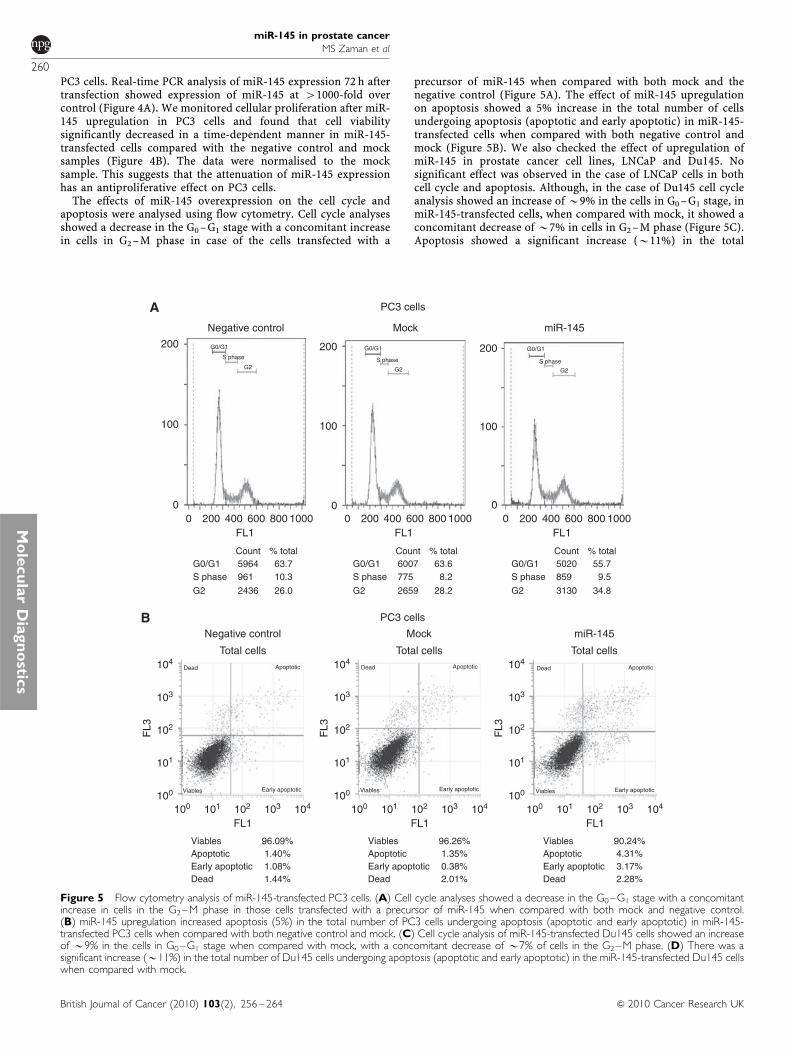
PC3 cells. Real-time PCR analysis of miR-145 expression 72 h aftertransfection showed expression of miR-145 at 41000-fold overcontrol (Figure 4A). We monitored cellular proliferation after miR-145 upregulation in PC3 cells and found that cell viabilitysignificantly decreased in a time-dependent manner in miR-145-transfected cells compared with the negative control and mocksamples (Figure 4B). The data were normalised to the mocksample. This suggests that the attenuation of miR-145 expressionhas an antiproliferative effect on PC3 cells.
The effects of miR-145 overexpression on the cell cycle andapoptosis were analysed using flow cytometry. Cell cycle analysesshowed a decrease in the G0 –G1 stage with a concomitant increasein cells in G2 –M phase in case of the cells transfected with a
precursor of miR-145 when compared with both mock and thenegative control (Figure 5A). The effect of miR-145 upregulationon apoptosis showed a 5% increase in the total number of cellsundergoing apoptosis (apoptotic and early apoptotic) in miR-145-transfected cells when compared with both negative control andmock (Figure 5B). We also checked the effect of upregulation ofmiR-145 in prostate cancer cell lines, LNCaP and Du145. Nosignificant effect was observed in the case of LNCaP cells in bothcell cycle and apoptosis. Although, in the case of Du145 cell cycleanalysis showed an increase of B9% in the cells in G0 –G1 stage, inmiR-145-transfected cells, when compared with mock, it showed aconcomitant decrease of B7% in cells in G2 –M phase (Figure 5C).Apoptosis showed a significant increase (B11%) in the total
Negative control Mock
Mock
PC3 cells
miR-145
miR-145Negative control
Total cells Total cells Total cells
PC3 cells
104
103
102
101
100
FL1
FL3
104
103
102
101
100
FL3
104
103
102
101
100
FL3
200
100
0
200
100
0
200
100
0
CountG0/G1
G2
S phase
G0/G1
G2
S phase
G0/G1
G2
S phase
G0/G1
G2
S phase
961
2436 26.0
10.35964 63.7
% total CountG0/G1
G2
S phase 775
2659 28.2
8.26007 63.6
% total CountG0/G1
G2
S phase 859
3130 34.8
9.55020 55.7
% total
Viables
100 101 102 103 104
FL1100 101 102 103 104
FL1100 101 102 103 104
96.09%1.40%1.08%1.44%Dead
Apoptotic
Viables
Dead
Early apoptotic
Apoptotic
Viables
Dead
Early apoptotic
Apoptotic
Viables
Dead
Early apoptotic
Apoptotic
Viables 96.26%1.35%0.38%2.01%Dead
Early apoptoticEarly apoptoticApoptotic
Viables 90.24%4.31%3.17%2.28%Dead
Early apoptoticApoptotic
FL10 200 400 600 800 1000
FL10 200 400 600 800 1000
FL10 200 400 600 800 1000
Figure 5 Flow cytometry analysis of miR-145-transfected PC3 cells. (A) Cell cycle analyses showed a decrease in the G0–G1 stage with a concomitantincrease in cells in the G2–M phase in those cells transfected with a precursor of miR-145 when compared with both mock and negative control.(B) miR-145 upregulation increased apoptosis (5%) in the total number of PC3 cells undergoing apoptosis (apoptotic and early apoptotic) in miR-145-transfected PC3 cells when compared with both negative control and mock. (C) Cell cycle analysis of miR-145-transfected Du145 cells showed an increaseof B9% in the cells in G0–G1 stage when compared with mock, with a concomitant decrease of B7% of cells in the G2–M phase. (D) There was asignificant increase (B11%) in the total number of Du145 cells undergoing apoptosis (apoptotic and early apoptotic) in the miR-145-transfected Du145 cellswhen compared with mock.
miR-145 in prostate cancer
MS Zaman et al
260
British Journal of Cancer (2010) 103(2), 256 – 264 & 2010 Cancer Research UK
Mo
lecu
lar
Dia
gn
ostic
s

number of cells undergoing apoptosis (apoptotic and earlyapoptotic) in the miR-145-transfected cells when compared withmock (Figure 5D).
mRNA microarray of miR-145-overexpressing PC3 cells
Micro RNAs are known to have numerous cellular targets.Therefore, we analysed the changes occurring as a result ofmiR-145 overexpression in PC3 cells using mRNA microarray(data not shown). These results showed the upregulation of anumber of pro-apoptotic genes such as TNFSF10 and IL-24(Pitti et al, 1996; Sauane et al, 2004), and cyclin binding ubiquitinligases such as HERC5 (Hochrainer et al, 2008). There was alsodownregulation of antiapoptotic genes such as BIRC5 (Tamm et al,1998), also known as survivin and of the prometastatic gene FSCN1(Hashimoto et al, 2009).
TNFSF10 is increased after miR-145 transfection in PC3
The results of the microarray were validated by real-time PCR. ThemRNA expression level of TNFSF10, HERC5 and IL-24 was foundto be upregulated by approximately 132-, 16- and 3.5-fold,respectively, by the transfection of miR-145 in PC3 cells (Figure6A– C). TNFSF10, a member of the family of TRAIL (TNF-related
apoptosis-inducing ligands) molecules (Pitti et al, 1996) had themaximum increase in mRNA levels by real-time PCR. Itsexpression was further substantiated by checking the level ofprotein expression of TNFSF10 by western blotting in thetransfected PC3 cells. Protein expression of TNFSF10 was foundto be substantially upregulated for the miR-145 sample whencompared with the negative control (Figure 6D). Moreover, we alsochecked the protein expression of different genes that are involvedin regulating apoptosis and cell cycle. Among these were survivin,Bcl-2, cdc7, cdk6 and PUMA. Out of these, only cdk6 showed asignificant decrease in its expression in the miR-145-transfectedsample when compared with the negative control (Figure 6D).
DISCUSSION
Prostate cancer accounts for approximately 29% of cancerincidence and is second only to lung cancer in male cancer deathsin the United States (Jemal et al, 2007). Treatments available forthis disease include hormone therapy, radical prostatectomy orirradiation. Generally, a subset of prostate cancer patients is proneto disease relapse and metastasis, which frequently develops evenafter surgery and may cause death. Therefore, a number ofalternative ways for controlling prostate cancer have emerged,
Negative control Mock
Du145 cells
Du145 cells
miR-145
Negative control Mock miR-145
104
103
102
101
100
FL3
104
103
102
101
100
FL3
104
103
102
101
100
FL3
400
200
0
200
100
0
200
100
0
CountG0/G1
G2
S phase
G0/G1
G2S phase
G0/G1
G2S phase
G0/G1
G2S phase
1026
2031 23.2
11.75711 65.1
% total CountG0/G1
G2
S phase 1548
1862 21.1
17.65409 61.3
% total CountG0/G1
G2
S phase 1233
1120 14.0
15.45640 70.6
% total
FL1100 101 102 103 104
FL1100 101 102 103 104
FL1100 101 102 103 104
Viables
Dead
(Total cells)
Early apoptotic
Apoptotic
Viables
Dead
(Total cells)
Early apoptotic
Apoptotic
Viables
Dead
(Total cells)
Early apoptotic
Apoptotic
FL10 200 400 600 8001000
FL10 200 400 600 8001000
FL10 200 400 600 8001000
Viables 68.98%18.50%6.37%6.15%Dead
Early apoptoticApoptotic
Viables
DeadEarly apoptoticApoptotic
Viables
DeadEarly apoptoticApoptotic
73.08%20.25%3.28%3.39%
59.99%26.91%8.33%4.77%
Figure 5 (continued)
miR-145 in prostate cancer
MS Zaman et al
261
British Journal of Cancer (2010) 103(2), 256 – 264& 2010 Cancer Research UK
Mo
lecu
lar
Dia
gn
ost
ics

including chemoprevention and delaying or reversing carcino-genesis by pharmacologic intervention with naturally occurring orsynthetic agents (Sporn and Suh, 2002; Tsao et al, 2004).Tumourigenesis is a multistep process that results from theaccumulation and interplay of genetic and epigenetic changes.Increased DNA methylation of CpG islands in the promoter region
of genes is well established as a common epigenetic mechanism forthe silencing of tumour suppressor genes (TSGs) in cancer cells(Jones and Laird, 1999). Epigenetic silencing of a gene can bereversed by drugs such as 5-aza-20-deoxycytidine (5Aza-C) thatforms a covalent complex with the active site of methyltransferaseresulting in generalised demethylation. Unfortunately, the applic-ability of this commonly used drug is hampered by its high toxicityand instability in physiological solutions (Bender et al, 1998).Histone deacetylases (HDACs) are frequently overexpressed in abroad range of cancer types, in which they alter cellular epigeneticprogramming to promote cell proliferation and survival (Markset al, 2001). HDAC inhibitors are presently approved for treatmentof advanced primary cutaneous T-cell lymphoma (Mann et al,2007), and have been shown to be effective in animal models ofprostate cancer (Wedel et al, 2008). In a previous study, it hasbeen shown that treatment of prostate cancer cells with theHDAC inhibitor TSA induces cell cycle arrest but not apoptosis(Roy et al, 2005).
Epidemiologic evidence suggests that intake of a soy-rich dietmay have a protective effect against prostate cancer (Hebert et al,1998; Jacobsen et al, 1998). Genistein, one of the principal soyisoflavones, shows a wide array of chemopreventive actions. Theanticancer effects of genistein have been ascribed to severalsignalling pathways and mechanisms that lead to cell cycle arrest,apoptosis, invasion, metastasis and angiogenesis, the attributesthat could potentially prevent tumour initiation and progression(Magee and Rowland, 2004; Bektic et al, 2005). Genistein (40,5,7-trihydroxyflavone) is a naturally occurring isoflavenoid that isabundant in soy products and has been identified as an inhibitor ofprotein tyrosine kinases and thus it has a key role in cell growthand apoptosis (Hunter, 1987; Ullrich and Schlessinger, 1990).It has been reported to have estrogenic properties and anti-neoplastic activity in multiple tumour types (Zava and Duwe,1997). It was also found to have epigenetic effects in the mouseprostate (Day et al, 2002). The above findings prompted us toexamine its effect on the expression of miRNAs. Furthermore, therole of 5-aza and TSA in the reversal of epigenetic silencingof genes prompted us to use them in combination with genistein.
Micro RNAs have important roles in many biological processes,including cell growth, apoptosis, haematopoietic lineage differ-entiation and gene regulation. They are also involved in a widevariety of human diseases such as cancer, vascular disease,immune disease and infections. More than 50% of human miRNAgenes are thought to be located in cancer-associated regions or atfragile sites of chromosomes, which are hot spots for gene deletion,amplification and mutations. Many genomic profiling studies haveindicated a general downregulation of miRNAs in varioustumours, including pancreatic cancer, breast cancer, prostatecancer, liver cancer, colon cancer and ovarian cancer, suggesting amore negative regulation of cancer growth by miRNAs (Graman-tieri et al, 2007; Hurteau et al, 2007; Lee et al, 2007; Shi et al, 2007;Barbarotto et al, 2008; Felicetti et al, 2008; Mitomo et al, 2008;Schetter et al, 2008; Yang et al, 2008).
The consistency with which miR-145 was found to be down-regulated in both prostate cell lines and carcinoma samplesprompted us to examine the role it has in prostate cancer. It isevident from our results that loss of miR-145 might be importantin the development and progression of prostate cancer, as it’soverexpression resulted in an increase in the total number of cellsundergoing apoptosis. This led us to hypothesise that miR-145might be affecting apoptosis. mRNA microarray of the over-expressed miR-145 in PC3 cells confirmed our hypothesis when weobserved the upregulation of pro-apoptotic genes such as TNFSF10and IL-24. Real-time PCR of TNFSF10 mRNA showed upregulationof its expression (132-fold), which was confirmed by westernblotting. TNFSF10, also known as TRAIL or Apo-2 ligand (Pittiet al, 1996), is a member of the TNF superfamily, and since itsdiscovery it has been used as an antitumour protein because of its
Rel
ativ
e T
NF
SF
10m
RN
A e
xpre
ssio
nR
elat
ive
IL24
mR
NA
exp
ress
ion
Rel
ativ
e H
ER
C5
mR
NA
exp
ress
ion
HERC5MockNegative control
TNFSF10MockNegative control
MockIL24Negative control
160
140
120
100
80
60
40
20
0
18
16
14
12
10
8
6
4
2
0
3.60
3.20
2.80
2.40
2
1.60
1.20
0.80
0.40 1.00
3.22
0.71
IL24
1.00
TN
FS
F10
HE
RC
5
132.
54
1.00
1.07
15.3
8
0.59
Negative control miR-145
TNFSF10
CDK6
GAPDH
Figure 6 Effect of miR-145 overexpression on apoptosis/cell cycle genesin PC3 cells. Increased expression of (A) TNFSF10, (B) HERC5 and (C)IL24 in miR-145-transfected PC3 cells when compared with mock and thenegative control. (D) Western analysis of upregulated and downregulatedgenes in miR-145-transfected PC3 cells. Upregulation of TNFSF10 proteinexpression occurred concomitantly with downregulation of cdk6 in PC3cells transfected with miR-145 precursor.
miR-145 in prostate cancer
MS Zaman et al
262
British Journal of Cancer (2010) 103(2), 256 – 264 & 2010 Cancer Research UK
Mo
lecu
lar
Dia
gn
ostic
s

ability to induce apoptosis in a variety of human cancer cell lineswhile leaving normal cells unaffected (Walczak et al, 1999).Moreover, studies performed on TRAIL knockout mice show thatTNFSF10 has an important role in normal tumour immuno-surveillance as TNFSF10 knockout mice support tumour growth ata higher rate when compared with normal mice and are moresusceptible to tumour metastasis (Cretney et al, 2002). Cellsundergoing TRAIL-induced death show many of the hallmarks ofapoptosis, including DNA fragmentation, expression of pro-phagocytic signals on the cell membrane and cleavage of multipleintracellular proteins by caspases (Griffith et al, 1998).
In summary, our results suggest methylation of the promoterregion of miR-145 in prostate cancer cell lines and prostate cancertissue samples as a major reason for its low expression. This is the
first report on the mechanism of inactivation of miR-145 throughDNA methylation as well as the functional significance of miR-145in prostate cancer. As one of the genes significantly upregulated bymiR-145 overexpression is the pro-apoptotic TNFSF10 gene,modulation of miR-145 may be an important therapeutic approachfor the management of prostate cancer.
ACKNOWLEDGEMENTS
We thank Dr Roger Erickson for support and assistance with thepreparation of the article. This study was supported by the NIHGrant RO1CA111470 (to the principal investigator, Dr R Dahiya).
REFERENCES
Baek D, Villen J, Shin C, Camargo FD, Gygi SP, Bartel DP (2008) The impactof microRNAs on protein output. Nature 455: 64 – 71
Barbarotto E, Schmittgen TD, Calin GA (2008) MicroRNAs and cancer:profile, profile, profile. Int J Cancer 122: 969 – 977
Bektic J, Guggenberger R, Eder IE, Pelzer AE, Berger AP, Bartsch G,Klocker H (2005) Molecular effects of the isoflavonoid genistein inprostate cancer. Clin Prostate Cancer 4: 124 – 129
Bender CM, Pao MM, Jones PA (1998) Inhibition of DNA methylation by5-aza-20-deoxycytidine suppresses the growth of human tumor cell lines.Cancer Res 58: 95 – 101
Bloomston M, Frankel WL, Petrocca F, Volinia S, Alder H, Hagan JP,Liu CG, Bhatt D, Taccioli C, Croce CM (2007) MicroRNA expressionpatterns to differentiate pancreatic adenocarcinoma from normalpancreas and chronic pancreatitis. JAMA 297: 1901 – 1908
Carrington JC, Ambros V (2003) Role of microRNAs in plant and animaldevelopment. Science 301: 336 – 338
Cretney E, Takeda K, Yagita H, Glaccum M, Peschon JJ, Smyth MJ (2002)Increased susceptibility to tumor initiation and metastasis inTNF-related apoptosis-inducing ligand-deficient mice. J Immunol 168:1356 – 1361
Day JK, Bauer AM, DesBordes C, Zhuang Y, Kim BE, Newton LG, Nehra V,Forsee KM, MacDonald RS, Besch-Williford C, Huang TH, Lubahn DB(2002) Genistein alters methylation patterns in mice. J Nutr 132:2419S – 2423S
Felicetti F, Errico MC, Bottero L, Segnalini P, Stoppacciaro A, Biffoni M,Felli N, Mattia G, Petrini M, Colombo MP, Peschle C, Care A (2008) Thepromyelocytic leukemia zinc finger-microRNA-221/-222 pathway con-trols melanoma progression through multiple oncogenic mechanisms.Cancer Res 68: 2745 – 2754
Galardi S, Mercatelli N, Giorda E, Massalini S, Frajese GV, Ciafre SA,Farace MG (2007) miR-221 and miR-222 expression affects theproliferation potential of human prostate carcinoma cell lines bytargeting p27Kip1. J Biol Chem 282: 23716 – 23724
Gramantieri L, Ferracin M, Fornari F, Veronese A, Sabbioni S, Liu CG,Calin GA, Giovannini C, Ferrazzi E, Grazi GL, Croce CM, Bolondi L,Negrini M (2007) Cyclin G1 is a target of miR-122a, a microRNAfrequently down-regulated in human hepatocellular carcinoma. CancerRes 67: 6092 – 6099
Griffith TS, Chin WA, Jackson GC, Lynch DH, Kubin MZ (1998)Intracellular regulation of TRAIL-induced apoptosis in human melano-ma cells. J Immunol 161: 2833 – 2840
Hashimoto Y, Loftis DW, Adams JC (2009) Fascin-1 promoter activity isregulated by CREB and the aryl hydrocarbon receptor in humancarcinoma cells. PLoS One 4: e5130
He L, Hannon GJ (2004) MicroRNAs: small RNAs with a big role in generegulation. Nat Rev Genet 5: 522 – 531
Hebert JR, Hurley TG, Olendzki BC, Teas J, Ma Y, Hampl JS (1998)Nutritional and socioeconomic factors in relation to prostate cancermortality: a cross-national study. J Natl Cancer Inst 90: 1637 – 1647
Hochrainer K, Kroismayr R, Baranyi U, Binder BR, Lipp J (2008)Highly homologous HERC proteins localize to endosomes andexhibit specific interactions with hPLIC and Nm23B. Cell Mol Life Sci65: 2105 – 2117
Hunter T (1987) A thousand and one protein kinases. Cell 50: 823 – 829
Hurteau GJ, Carlson JA, Spivack SD, Brock GJ (2007) Overexpression of themicroRNA hsa-miR-200c leads to reduced expression of transcriptionfactor 8 and increased expression of E-cadherin. Cancer Res 67:7972 – 7976
Iorio MV, Ferracin M, Liu CG, Veronese A, Spizzo R, Sabbioni S, Magri E,Pedriali M, Fabbri M, Campiglio M, Menard S, Palazzo JP, Rosenberg A,Musiani P, Volinia S, Nenci I, Calin GA, Querzoli P, Negrini M, Croce CM(2005) MicroRNA gene expression deregulation in human breast cancer.Cancer Res 65: 7065 – 7070
Jacobsen BK, Knutsen SF, Fraser GE (1998) Does high soy milk intakereduce prostate cancer incidence? The Adventist Health Study (UnitedStates). Cancer Causes Control 9: 553 – 557
Jemal A, Siegel R, Ward E, Murray T, Xu J, Thun MJ (2007) Cancerstatistics, 2007. CA Cancer J Clin 57, 43 – 66
Jones PA, Laird PW (1999) Cancer epigenetics comes of age. Nat Genet 21:163 – 167
Lee EJ, Gusev Y, Jiang J, Nuovo GJ, Lerner MR, Frankel WL, Morgan DL,Postier RG, Brackett DJ, Schmittgen TD (2007) Expression profiling identifiesmicroRNA signature in pancreatic cancer. Int J Cancer 120: 1046 – 1054
Li LC, Dahiya R (2002) MethPrimer: designing primers for methylationPCRs. Bioinformatics 18: 1427 – 1431
Lim LP, Lau NC, Garrett-Engele P, Grimson A, Schelter JM, Castle J,Bartel DP, Linsley PS, Johnson JM (2005) Microarray analysis shows thatsome microRNAs downregulate large numbers of target mRNAs. Nature433: 769 – 773
Magee PJ, Rowland IR (2004) Phyto-oestrogens, their mechanism of action:current evidence for a role in breast and prostate cancer. Br J Nutr 91:513 – 531
Mann BS, Johnson JR, Cohen MH, Justice R, Pazdur R (2007) FDA approvalsummary: vorinostat for treatment of advanced primary cutaneous T-celllymphoma. Oncologist 12: 1247 – 1252
Marks P, Rifkind RA, Richon VM, Breslow R, Miller T, Kelly WK (2001)Histone deacetylases and cancer: causes and therapies. Nat Rev Cancer 1:194 – 202
Mitomo S, Maesawa C, Ogasawara S, Iwaya T, Shibazaki M, Yashima-Abo A,Kotani K, Oikawa H, Sakurai E, Izutsu N, Kato K, Komatsu H, Ikeda K,Wakabayashi G, Masuda T (2008) Downregulation of miR-138 is associatedwith overexpression of human telomerase reverse transcriptase protein inhuman anaplastic thyroid carcinoma cell lines. Cancer Sci 99: 280 – 286
Ozen M, Creighton CJ, Ozdemir M, Ittmann M (2008) Widespreadderegulation of microRNA expression in human prostate cancer.Oncogene 27: 1788 – 1793
Petrocca F, Visone R, Onelli MR, Shah MH, Nicoloso MS, de Martino I,Iliopoulos D, Pilozzi E, Liu CG, Negrini M, Cavazzini L, Volinia S,Alder H, Ruco LP, Baldassarre G, Croce CM, Vecchione A (2008) E2F1-regulated microRNAs impair TGFbeta-dependent cell-cycle arrest andapoptosis in gastric cancer. Cancer Cell 13: 272 – 286
Pitti RM, Marsters SA, Ruppert S, Donahue CJ, Moore A, Ashkenazi A(1996) Induction of apoptosis by Apo-2 ligand, a new memberof the tumor necrosis factor cytokine family. J Biol Chem 271:12687 – 12690
Place RF, Li LC, Pookot D, Noonan EJ, Dahiya R (2008) MicroRNA-373induces expression of genes with complementary promoter sequences.Proc Natl Acad Sci USA 105: 1608 – 1613
miR-145 in prostate cancer
MS Zaman et al
263
British Journal of Cancer (2010) 103(2), 256 – 264& 2010 Cancer Research UK
Mo
lecu
lar
Dia
gn
ost
ics

Porkka KP, Pfeiffer MJ, Waltering KK, Vessella RL, Tammela TL, Visakorpi T(2007) MicroRNA expression profiling in prostate cancer. Cancer Res 67:6130 – 6135
Roy S, Packman K, Jeffrey R, Tenniswood M (2005) Histonedeacetylase inhibitors differentially stabilize acetylated p53 and inducecell cycle arrest or apoptosis in prostate cancer cells. Cell Death Differ 12:482 – 491
Sauane M, Lebedeva IV, Su ZZ, Choo HT, Randolph A, Valerie K, Dent P,Gopalkrishnan RV, Fisher PB (2004) Melanoma differentiation asso-ciated gene-7/interleukin-24 promotes tumor cell-specific apoptosisthrough both secretory and nonsecretory pathways. Cancer Res 64:2988 – 2993
Schepeler T, Reinert JT, Ostenfeld MS, Christensen LL, Silahtaroglu AN,Dyrskjot L, Wiuf C, Sorensen FJ, Kruhoffer M, Laurberg S, Kauppinen S,Orntoft TF, Andersen CL (2008) Diagnostic and prognostic microRNAsin stage II colon cancer. Cancer Res 68: 6416 – 6424
Schetter AJ, Leung SY, Sohn JJ, Zanetti KA, Bowman ED, Yanaihara N,Yuen ST, Chan TL, Kwong DL, Au GK, Liu CG, Calin GA, Croce CM,Harris CC (2008) MicroRNA expression profiles associated withprognosis and therapeutic outcome in colon adenocarcinoma. JAMA299: 425 – 436
Shi XB, Xue L, Yang J, Ma AH, Zhao J, Xu M, Tepper CG, Evans CP,Kung HJ, deVere White RW (2007) An androgen-regulated miRNAsuppresses Bak1 expression and induces androgen-independent growthof prostate cancer cells. Proc Natl Acad Sci USA 104: 19983 – 19988
Spizzo R, Nicoloso MS, Lupini L, Lu Y, Fogarty J, Rossi S, Zagatti B, FabbriM, Veronese A, Liu X, Davuluri R, Croce CM, Mills G, Negrini M, CalinGA (2009) miR-145 participates with TP53 in a death-promotingregulatory loop and targets estrogen receptor-alpha in human breastcancer cells. Cell Death Differ 17: 246 – 254
Sporn MB, Suh N (2002) Chemoprevention: an essential approach tocontrolling cancer. Nat Rev Cancer 2: 537 – 543
Tamm I, Wang Y, Sausville E, Scudiero DA, Vigna N, Oltersdorf T, Reed JC(1998) IAP-family protein survivin inhibits caspase activity andapoptosis induced by Fas (CD95), Bax, caspases, and anticancer drugs.Cancer Res 58: 5315 – 5320
Tsao AS, Kim ES, Hong WK (2004) Chemoprevention of cancer. CA CancerJ Clin 54: 150 – 180
Ullrich A, Schlessinger J (1990) Signal transduction by receptors withtyrosine kinase activity. Cell 61: 203 – 212
Volinia S, Calin GA, Liu CG, Ambs S, Cimmino A, Petrocca F, Visone R,Iorio M, Roldo C, Ferracin M, Prueitt RL, Yanaihara N, Lanza G, ScarpaA, Vecchione A, Negrini M, Harris CC, Croce CM (2006) A microRNAexpression signature of human solid tumors defines cancer gene targets.Proc Natl Acad Sci USA 103: 2257 – 2261
Walczak H, Miller RE, Ariail K, Gliniak B, Griffith TS, Kubin M, Chin W,Jones J, Woodward A, Le T, Smith C, Smolak P, Goodwin RG, Rauch CT,Schuh JC, Lynch DH (1999) Tumoricidal activity of tumor necrosisfactor-related apoptosis-inducing ligand in vivo. Nat Med 5: 157 – 163
Wedel SA, Sparatore A, Soldato PD, Al-Batran SE, Atmaca A, Juengel E,Hudak L, Jonas D, Blaheta RA (2008) New histone deacetylase inhibitorsas potential therapeutic tools for advanced prostate carcinoma. J Cell MolMed 12: 2457 – 2466
Yanaihara N, Caplen N, Bowman E, Seike M, Kumamoto K, Yi M, StephensRM, Okamoto A, Yokota J, Tanaka T, Calin GA, Liu CG, Croce CM,Harris CC (2006) Unique microRNA molecular profiles in lung cancerdiagnosis and prognosis. Cancer Cell 9: 189 – 198
Yang H, Kong W, He L, Zhao JJ, O’Donnell JD, Wang J, Wenham RM,Coppola D, Kruk PA, Nicosia SV, Cheng JQ (2008) MicroRNA expressionprofiling in human ovarian cancer: miR-214 induces cell survival andcisplatin resistance by targeting PTEN. Cancer Res 68: 425 – 433
Zava DT, Duwe G (1997) Estrogenic and antiproliferative properties ofgenistein and other flavonoids in human breast cancer cells in vitro. NutrCancer 27: 31 – 40
miR-145 in prostate cancer
MS Zaman et al
264
British Journal of Cancer (2010) 103(2), 256 – 264 & 2010 Cancer Research UK
Mo
lecu
lar
Dia
gn
ostic
s