Prognostic Significance of Matrix Metalloproteinase 2 and Tissue Inhibitor of Metalloproteinase 2...
8
Prognostic Significance of Matrix Metalloproteinase 2 and Tissue Inhibitor of Metalloproteinase 2 Expression in Prostate Cancer Jeffrey S. Ross, M.D., Prabhjot Kaur, M.D., Christine E. Sheehan, B.S, Hugh A.G . Fisher, M.D., Ronald A. Kaufman, Jr, M.D., Bhaskar V. S. Kallakury, M.D. Departments of Pathology and Laboratory Medicine (JSR, PK, CES, BVSK) and Surgery (Urology; HAGF, RAK Jr), Albany Medical College, Albany, New York Matrix metalloproteinases (MMPs) are proteolytic enzymes capable of degrading the structural sup- port network for normal and malignant cells, pro- moting neoplastic cell invasion and metastasis. Tis- sue inhibitors of metalloproteinases (TIMPs) maintain connective tissue integrity by modulating MMP activity. Formalin-fixed paraffin-embedded tissue sections from 138 prostatic adenocarcinomas (PACs) were immunostained by a combined auto- mated/manual method using monoclonal antibod- ies against MMP2 and TIMP2. Immunoreactivity was semiquantitatively scored based on stain inten- sity and distribution, and results were correlated with Gleason grade, pathologic stage, ploidy status, and disease recurrence. One hundred five of 138 (76%) and 113/138 (82%) PACs expressed MMP2 and TIMP2, respectively. Co-expression was ob- served in 94/138 (68%) of PACs (P .01), correlated with advanced tumor stage (P .05), and tended to be associated with disease recurrent cases (P .07). TIMP2 expression individually correlated with ad- vanced tumor stage (P .04) and reached near significance with disease recurrence (P .06). MMP2 expression was also more frequent in recur- rent PACs, although this value did not reach statis- tical significance (P .07). However, on multivari- ate analysis, only pathologic stage (P .009) and ploidy status (P .03) independently predicted dis- ease recurrence. In conclusion, MMP2 and TIMP2 are co-expressed in a majority of PACs and correlate with prognostic variables. Interestingly, contrary to the previously documented anti-tumor effects of TIMPs, TIMP2 expression appears to have a tumor- promoting role in PACs and warrants further investigation. KEY WORDS: Immunohistochemistry, MMP, Prog- nosis, Prostate cancer, TIMP. Mod Pathol 2003;16(3):198 –205 Matrix metalloproteinases (MMPs), a family of zinc-dependent endopeptidases, degrade the base- ment membrane and extracellular matrix, facilitat- ing cell migration, tumor invasion, and metastasis (1–5). There are at least 20 human MMPs, divided into the collagenases, gelatinases, stromelysins, and membrane-type MMPs (MT-MMPs; 1–5). Tissue in- hibitors of metalloproteinases (TIMPs) are the ma- jor endogenous regulators of MMPs and consist of four homologous members (TIMP1– 4; 6 – 8). TIMPs are multifunctional proteins that inhibit cell inva- sion in vitro and tumorigenesis and metastasis in vivo (6). Although each TIMP appears capable of inhibiting several MMPs, these proteins exhibit preferential inhibitory capacity; for example, TIMPs1 and 2 selectively inhibit MMP9 and 2, re- spectively (9). Increased expression of MMPs has been associ- ated with poor prognosis and shortened patient survival in a variety of malignancies including car- cinomas of the esophagus (10), stomach (11), colon (12), breast (13), pancreas (14), lung (15), kidney (16), and ovary (17). TIMP expression has been associated with both tumor suppressor or anti- metastatic effects and tumor-promoting effects in selected cancers (18 –20). MMP and TIMP expres- sion in prostate cancer has been recently reviewed (21). Both MMPs and TIMPs have been character- ized in prostate cancer cell lines (22–25) and clinical samples from prostate cancer patients (26 –33), with conflicting results. Similarly, serum levels of circulating MMPs and TIMPs have shown variable Copyright © 2003 by The United States and Canadian Academy of Pathology, Inc. VOL. 16, NO. 3, P. 198, 2003 Printed in the U.S.A. Date of acceptance: January 6, 2002. Presented in part at the 90th annual meeting of the United States and Canadian Academy of Pathology, March 2001, Atlanta, Georgia. Address reprint requests to: Jeffrey S. Ross, M.D., Department of Pathol- ogy and Laboratory Medicine, Albany Medical College, Mail Code 81, 47 New Scotland Avenue, Albany, NY 12208; fax: (518) 262-3663; e-mail: [email protected]. DOI: 10.1097/01.MP.0000056984.62360.6C 198
-
Upload
independent -
Category
Documents
-
view
1 -
download
0
Transcript of Prognostic Significance of Matrix Metalloproteinase 2 and Tissue Inhibitor of Metalloproteinase 2...
Prognostic Significance of Matrix Metalloproteinase 2and Tissue Inhibitor of Metalloproteinase 2 Expressionin Prostate CancerJeffrey S. Ross, M.D., Prabhjot Kaur, M.D., Christine E. Sheehan, B.S, Hugh A.G . Fisher, M.D.,Ronald A. Kaufman, Jr, M.D., Bhaskar V. S. Kallakury, M.D.
Departments of Pathology and Laboratory Medicine (JSR, PK, CES, BVSK) and Surgery (Urology; HAGF,RAK Jr), Albany Medical College, Albany, New York
Matrix metalloproteinases (MMPs) are proteolyticenzymes capable of degrading the structural sup-port network for normal and malignant cells, pro-moting neoplastic cell invasion and metastasis. Tis-sue inhibitors of metalloproteinases (TIMPs)maintain connective tissue integrity by modulatingMMP activity. Formalin-fixed paraffin-embeddedtissue sections from 138 prostatic adenocarcinomas(PACs) were immunostained by a combined auto-mated/manual method using monoclonal antibod-ies against MMP2 and TIMP2. Immunoreactivitywas semiquantitatively scored based on stain inten-sity and distribution, and results were correlatedwith Gleason grade, pathologic stage, ploidy status,and disease recurrence. One hundred five of 138(76%) and 113/138 (82%) PACs expressed MMP2and TIMP2, respectively. Co-expression was ob-served in 94/138 (68%) of PACs (P � .01), correlatedwith advanced tumor stage (P � .05), and tended tobe associated with disease recurrent cases (P � .07).TIMP2 expression individually correlated with ad-vanced tumor stage (P � .04) and reached nearsignificance with disease recurrence (P � .06).MMP2 expression was also more frequent in recur-rent PACs, although this value did not reach statis-tical significance (P � .07). However, on multivari-ate analysis, only pathologic stage (P � .009) andploidy status (P � .03) independently predicted dis-ease recurrence. In conclusion, MMP2 and TIMP2are co-expressed in a majority of PACs and correlatewith prognostic variables. Interestingly, contrary to
the previously documented anti-tumor effects ofTIMPs, TIMP2 expression appears to have a tumor-promoting role in PACs and warrants furtherinvestigation.
KEY WORDS: Immunohistochemistry, MMP, Prog-nosis, Prostate cancer, TIMP.
Mod Pathol 2003;16(3):198–205
Matrix metalloproteinases (MMPs), a family ofzinc-dependent endopeptidases, degrade the base-ment membrane and extracellular matrix, facilitat-ing cell migration, tumor invasion, and metastasis(1–5). There are at least 20 human MMPs, dividedinto the collagenases, gelatinases, stromelysins, andmembrane-type MMPs (MT-MMPs; 1–5). Tissue in-hibitors of metalloproteinases (TIMPs) are the ma-jor endogenous regulators of MMPs and consist offour homologous members (TIMP1– 4; 6 – 8). TIMPsare multifunctional proteins that inhibit cell inva-sion in vitro and tumorigenesis and metastasis invivo (6). Although each TIMP appears capable ofinhibiting several MMPs, these proteins exhibitpreferential inhibitory capacity; for example,TIMPs1 and 2 selectively inhibit MMP9 and 2, re-spectively (9).
Increased expression of MMPs has been associ-ated with poor prognosis and shortened patientsurvival in a variety of malignancies including car-cinomas of the esophagus (10), stomach (11), colon(12), breast (13), pancreas (14), lung (15), kidney(16), and ovary (17). TIMP expression has beenassociated with both tumor suppressor or anti-metastatic effects and tumor-promoting effects inselected cancers (18 –20). MMP and TIMP expres-sion in prostate cancer has been recently reviewed(21). Both MMPs and TIMPs have been character-ized in prostate cancer cell lines (22–25) and clinicalsamples from prostate cancer patients (26 –33),with conflicting results. Similarly, serum levels ofcirculating MMPs and TIMPs have shown variable
Copyright © 2003 by The United States and Canadian Academy ofPathology, Inc.VOL. 16, NO. 3, P. 198, 2003 Printed in the U.S.A.Date of acceptance: January 6, 2002.Presented in part at the 90th annual meeting of the United States andCanadian Academy of Pathology, March 2001, Atlanta, Georgia.Address reprint requests to: Jeffrey S. Ross, M.D., Department of Pathol-ogy and Laboratory Medicine, Albany Medical College, Mail Code 81, 47New Scotland Avenue, Albany, NY 12208; fax: (518) 262-3663; e-mail:[email protected].
DOI: 10.1097/01.MP.0000056984.62360.6C
198
capability of predicting disease progression (34 –37). The aim of the current study was to evaluatethe immunohistochemical expression of MMP2and TIMP2 in prostate cancer and determinewhether the expression of these markers correlateswith prognostic variables, including patientsurvival.
MATERIALS AND METHODS
Patients and SpecimensOne hundred thirty-eight randomly selected
prostatic adenocarcinomas (PACs) treated by radi-cal retropubic prostatectomy obtained from thefiles of the Albany Medical Center Hospital between1987 and 1997 were included in this study. All he-matoxylin and eosin–stained slides from each casewere reviewed, and tumors were graded accordingto the Gleason system (38) and staged according toTNM criteria (39). Multiple blocks were identifiedbased on the presence of adequate tumor and therepresentative nature of the overall grade. For sta-tistical evaluations, tumors with Gleason scores of 6or lower were considered as low grade, and tumorswith Gleason scores of 7 or higher were consideredas high grade. Statistical analysis was also per-formed using a three-tier scheme isolating tumorswith Gleason score of 7. Serum PSA levels as mea-sured by the Hybritech Tandem method (Hy-britech) were obtained from review of the patients’medical records. Postoperative PSA of �0.4 ng/mLon two consecutive occasions after prostatectomywas considered as biochemical evidence of diseaserecurrence.
ImmunohistochemistryTo analyze for the expression of MMP2 and of
TIMP2 proteins, contiguous 4-�m sections were cutfrom a single block of formalin-fixed, paraffin-embedded tissue randomly chosen from those ini-tially identified; sections were placed on chargedslides. After deparaffinization, primary antibody in-cubation was performed by an automated system(Ventana Medical Systems, Tucson, AZ) for MMP2and manually for TIMP2. Pertinent details regard-ing antibodies and staining procedure are summa-rized in Table 1. The remainder of the stainingprocedure included incubation with a biotinylatedanti-mouse secondary antibody, diaminobenzidinesubstrate, and hematoxylin counterstain and was
performed on the Ventana ES automated immuno-histochemistry system. Negative-control slideswere incubated with isotype-matched immuno-globulin in parallel with each batch of staining toconfirm the specificity of the antibodies.
Staining InterpretationStaining results were interpreted without prior
knowledge of clinical and pathologic parameters bytwo observers using a consensus method. For allmarkers, both the intensity of staining and approx-imate percentage of positive tumor cells were con-sidered in the semiquantitative assessment, as pre-viously published (40, 41). Briefly, the distributionof positive staining in the tumors was graded asfocal (�10%), regional (11–50%), and diffuse(�50%). The staining intensity was subjectivelyscored as weak, moderate, or intense. Staining pat-terns of moderate diffuse, moderate regional, in-tense regional, and intense diffuse were consideredas increased expression of each protein.
Quantitative DNA AnalysisQuantitative analysis of DNA content was deter-
mined for each case using 5-�m tissue sectionsstained by the Feulgen reaction and evaluated bythe CAS 200 image analyzer (Tripath Corp., Burl-ington, NC), as previously described (42, 43).
Statistical AnalysisStatistical comparisons were performed using
Stata software (Stata Corp, College Station, TX).Correlation between protein expression and patho-logic variables was performed using the �2 univar-iate analysis. Survival curves for all univariate anal-yses were assessed using the Kaplan-Meier method.Overall survival was defined as the interval betweensurgery and postsurgical biochemical disease recur-rence. Multivariate analyses of clinicopathologicparameters, including survival, were performed us-ing the Cox proportional hazards model. The levelof significance was set at .05.
RESULTS
Of the 138 PACs, there were 75 (54%) low-gradeand 63 (46%) high-grade tumors. At prostatectomy,there were 78 (57%) organ-confined tumors (StagesI and II) and 60 (43%) advanced-stage (Stages III
TABLE 1. Antibodies and Immunohistochemical Procedure
Antibody Manufacturer CloneCitrate AntigenRetrieval (min)
AntibodyDilution
Primary AntibodyIncubation
Positive Controls
MMP2 Novocastra 4D3 60 1:10 32 min at 41°C Colon carcinomaTIMP2 Neomarkers 2TMP04 60 1:10 Overnight at 4°C Breast carcinoma
MMP2 and TIMP2 in Prostate Cancer (J.S. Ross et al.) 199
and IV) cancers. Of the 77 cases tested for total DNAcontent, 52 (68%) were diploid, and 25 (32%) werenondiploid. A total of 131/138 (95%) had sequentialserum PSA follow-up information available. Ofthese 131 patients, 50(38%) had biochemical post-surgical disease recurrence.
Immunohistochemistry and Statistical AnalysisImmunostaining pattern for both proteins was
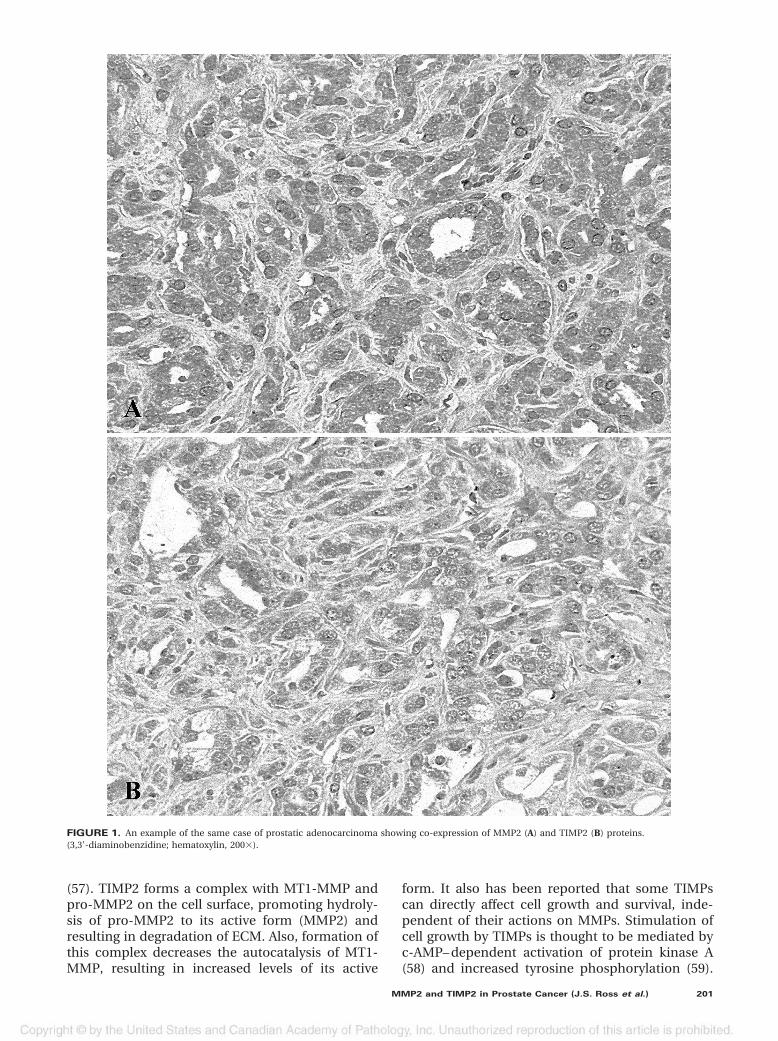
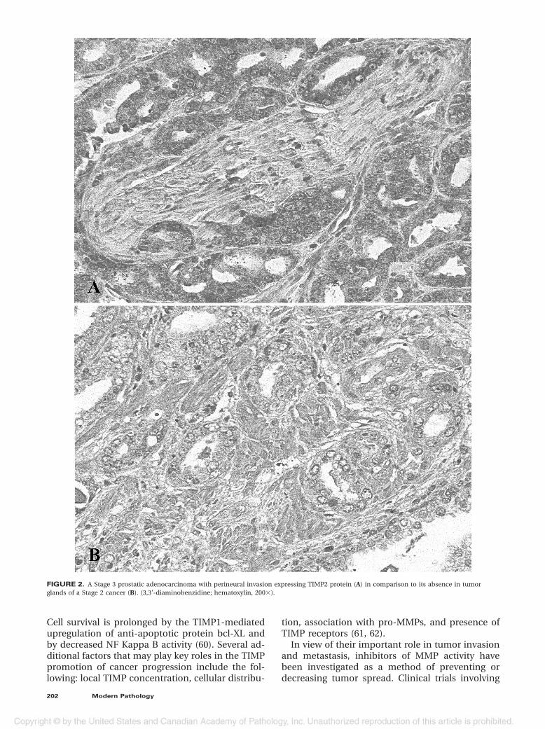
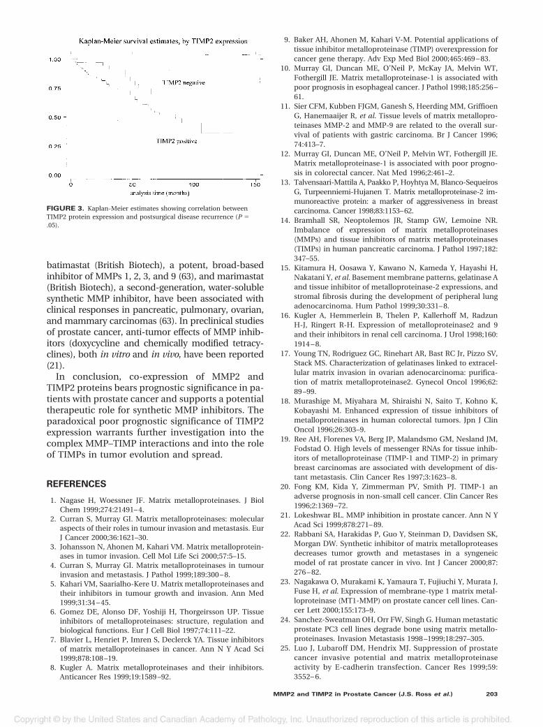
cytoplasmic, with tumor cells showing moderate tointense positivity, as opposed to relatively weakerexpression in the benign elements, which served asinternal control in each case. One hundred five of138 (76%) PACs expressed MMP2, and 113/138(82%) expressed TIMP2. There was an overall sig-nificant coexpression of MMP2 and TIMP2 in 94/138 (68%) PACs (P � .01; Fig. 1). The co-expressionof MMP2 and TIMP2 correlated with advanced tu-mor stage (P � .05) and reached near-significanceas a univariate predictor of disease recurrence (P �.07). TIMP2 expression individually correlated withadvanced tumor stage (P � .04; Fig. 2) and reachednear significance with disease recurrence (P � .06;Fig. 3). MMP2 expression was also more frequent inthe PACs that recurred, although this value did notreach statistical significance (P � .07). On univari-ate analysis, neither MMP2 nor TIMP2 expressioncorrelated with tumor grade (using either the two-or three-tier scheme) or DNA ploidy.
On multivariate analysis, only tumor stage (P �.009) and DNA ploidy status (P � .03) indepen-dently predicted disease recurrence.
DISCUSSION
MMP expression has been reported to be low orundetectable in most benign elements but is sub-stantially increased in a majority of human malig-nancies (10 –21). Analysis of both primary and met-astatic tumors has shown increased relative MMPexpression at the metastatic site, supporting a rolein tumor migration and spread (44). Additionally,increased MMP levels have been reported in theplasma and urine of patients with a variety of ad-vanced malignancies (45). Cancer outcome studieshave also shown that increased expression of MMPsis associated with shortened patient survival (10, 12,46). Aberrant expression of MMPs in prostate can-cer was first described using in situ hybridization in1991 (31). MMP7 expression has been linked toprostate cancer pathologic stage and incidence ofmetastasis (33). Using both Northern analysis andin situ hybridization, Still and co-workers (27)linked increased MMP2 and TIMP2 to high tumorgrade and advanced tumor stage of the disease.Increased MMP2 expression has also been associ-ated with high tumor Gleason score (28). Finally,
increased MMP expression also has been impli-cated in the development of prostate cancer, asevidenced by increased levels found in carcinomasversus benign prostatic hypertrophy and prostaticintraepithelial neoplasia (47, 48).
Serum measurements of MMPs in prostate can-cer have yielded conflicting correlations with dis-ease outcome. Several studies have found a corre-lation between circulating MMPs (MMP1,2,3) andcirculating TIMPs (TIMP1,3) and advanced or pro-gressive disease (34, 36); others have failed to con-firm this association (35).
In the present study, increased immunohisto-chemical co-expression of MMP2 and TIMP2 wasassociated with advanced tumor stage and reachednear-significance as a predictor of disease recur-rence. TIMP2 expression correlated with tumorgrade and predicted disease recurrence on univar-iate analysis but was not an independent predictorwhen tumor stage and DNA ploidy status were in-cluded in the multivariate analysis model.
At the time of their discovery, TIMPs were con-sidered to be tumor suppressor proteins. Recombi-nant TIMP2 was shown to inhibit invasion of HT1080 fibrosarcoma cells in vitro (49). IncreasedTIMP expression has been associated with de-creased tumor growth, invasiveness, and metastasisin a variety of prostate cancer and non–prostatecancer cell lines (22–25, 49 –52). However, the re-sults of the current study, demonstrating a poorprognostic significance in prostate cancer for in-creased TIMP2 expression, are contrary to the orig-inal tumor suppressor role hypothesized for TIMPsand more in line with recent evidence documentinga multifunctional complex role for TIMPs. Nemithet al. (53) described the growth-promoting abilitiesof TIMP2 in several human cell types, includingfibroblasts, keratocytes, lymphocytes, and stemcells. Increased TIMP1 and TIMP2 mRNA levelshave been correlated with tumor stage, lymph nodemetastasis, and shortened survival in patients withcarcinomas of colon (18), breast (19), and bladder(54). Our findings of the poor prognostic role ofincreased TIMP2 expression in prostate cancer alsoconcur with the data of Kugler et al. (16), whichdemonstrated a correlation of increased TIMP2 lev-els with aggressive phenotype in renal cellcarcinoma.
Although the paradoxical positive effect of TIMPin tumor progression is not completely understood,the tumor-promoting activity may be due either toproteolytic degradation of ECM or direct influenceon cell survival and growth. TIMP2 is reported toregulate matrix degradation, acting through amembrane type MMP (MT1-MMP; 55, 56). MT1-MMP is a key enzyme in tumor angiogenesis andmetastasis, hydrolyzes a variety of ECM compo-nents, and is a physiologic activator of pro-MMP2
200 Modern Pathology
(57). TIMP2 forms a complex with MT1-MMP andpro-MMP2 on the cell surface, promoting hydroly-sis of pro-MMP2 to its active form (MMP2) andresulting in degradation of ECM. Also, formation ofthis complex decreases the autocatalysis of MT1-MMP, resulting in increased levels of its active
form. It also has been reported that some TIMPscan directly affect cell growth and survival, inde-pendent of their actions on MMPs. Stimulation ofcell growth by TIMPs is thought to be mediated byc-AMP– dependent activation of protein kinase A(58) and increased tyrosine phosphorylation (59).
FIGURE 1. An example of the same case of prostatic adenocarcinoma showing co-expression of MMP2 (A) and TIMP2 (B) proteins.(3,3'-diaminobenzidine; hematoxylin, 200�).
MMP2 and TIMP2 in Prostate Cancer (J.S. Ross et al.) 201
Cell survival is prolonged by the TIMP1-mediatedupregulation of anti-apoptotic protein bcl-XL andby decreased NF Kappa B activity (60). Several ad-ditional factors that may play key roles in the TIMPpromotion of cancer progression include the fol-lowing: local TIMP concentration, cellular distribu-
tion, association with pro-MMPs, and presence ofTIMP receptors (61, 62).
In view of their important role in tumor invasionand metastasis, inhibitors of MMP activity havebeen investigated as a method of preventing ordecreasing tumor spread. Clinical trials involving
FIGURE 2. A Stage 3 prostatic adenocarcinoma with perineural invasion expressing TIMP2 protein (A) in comparison to its absence in tumorglands of a Stage 2 cancer (B). (3,3'-diaminobenzidine; hematoxylin, 200�).
202 Modern Pathology
batimastat (British Biotech), a potent, broad-basedinhibitor of MMPs 1, 2, 3, and 9 (63), and marimastat(British Biotech), a second-generation, water-solublesynthetic MMP inhibitor, have been associated withclinical responses in pancreatic, pulmonary, ovarian,and mammary carcinomas (63). In preclinical studiesof prostate cancer, anti-tumor effects of MMP inhib-itors (doxycycline and chemically modified tetracy-clines), both in vitro and in vivo, have been reported(21).
In conclusion, co-expression of MMP2 andTIMP2 proteins bears prognostic significance in pa-tients with prostate cancer and supports a potentialtherapeutic role for synthetic MMP inhibitors. Theparadoxical poor prognostic significance of TIMP2expression warrants further investigation into thecomplex MMP–TIMP interactions and into the roleof TIMPs in tumor evolution and spread.
REFERENCES
1. Nagase H, Woessner JF. Matrix metalloproteinases. J BiolChem 1999;274:21491– 4.
2. Curran S, Murray GI. Matrix metalloproteinases: molecularaspects of their roles in tumour invasion and metastasis. EurJ Cancer 2000;36:1621–30.
3. Johansson N, Ahonen M, Kahari VM. Matrix metalloprotein-ases in tumor invasion. Cell Mol Life Sci 2000;57:5–15.
4. Curran S, Murray GI. Matrix metalloproteinases in tumourinvasion and metastasis. J Pathol 1999;189:300 – 8.
5. Kahari VM, Saarialho-Kere U. Matrix metalloproteinases andtheir inhibitors in tumour growth and invasion. Ann Med1999;31:34 – 45.
6. Gomez DE, Alonso DF, Yoshiji H, Thorgeirsson UP. Tissueinhibitors of metalloproteinases: structure, regulation andbiological functions. Eur J Cell Biol 1997;74:111–22.
7. Blavier L, Henriet P, Imren S, Declerck YA. Tissue inhibitorsof matrix metalloproteinases in cancer. Ann N Y Acad Sci1999;878:108 –19.
8. Kugler A. Matrix metalloproteinases and their inhibitors.Anticancer Res 1999;19:1589 –92.
9. Baker AH, Ahonen M, Kahari V-M. Potential applications oftissue inhibitor metalloproteinase (TIMP) overexpression forcancer gene therapy. Adv Exp Med Biol 2000;465:469 – 83.
10. Murray GI, Duncan ME, O’Neil P, McKay JA, Melvin WT,Fothergill JE. Matrix metalloproteinase-1 is associated withpoor prognosis in esophageal cancer. J Pathol 1998;185:256 –61.
11. Sier CFM, Kubben FJGM, Ganesh S, Heerding MM, GriffioenG, Hanemaaijer R, et al. Tissue levels of matrix metallopro-teinases MMP-2 and MMP-9 are related to the overall sur-vival of patients with gastric carcinoma. Br J Cancer 1996;74:413–7.
12. Murray GI, Duncan ME, O’Neil P, Melvin WT, Fothergill JE.Matrix metalloproteinase-1 is associated with poor progno-sis in colorectal cancer. Nat Med 1996;2:461–2.
13. Talvensaari-Mattila A, Paakko P, Hoyhtya M, Blanco-SequeirosG, Turpeenniemi-Hujanen T. Matrix metalloproteinase-2 im-munoreactive protein: a marker of aggressiveness in breastcarcinoma. Cancer 1998;83:1153–62.
14. Bramhall SR, Neoptolemos JR, Stamp GW, Lemoine NR.Imbalance of expression of matrix metalloproteinases(MMPs) and tissue inhibitors of matrix metalloproteinases(TIMPs) in human pancreatic carcinoma. J Pathol 1997;182:347–55.
15. Kitamura H, Oosawa Y, Kawano N, Kameda Y, Hayashi H,Nakatani Y, et al. Basement membrane patterns, gelatinase Aand tissue inhibitor of metalloproteinase-2 expressions, andstromal fibrosis during the development of peripheral lungadenocarcinoma. Hum Pathol 1999;30:331– 8.
16. Kugler A, Hemmerlein B, Thelen P, Kallerhoff M, RadzunH-J, Ringert R-H. Expression of metalloproteinase2 and 9and their inhibitors in renal cell carcinoma. J Urol 1998;160:1914 – 8.
17. Young TN, Rodriguez GC, Rinehart AR, Bast RC Jr, Pizzo SV,Stack MS. Characterization of gelatinases linked to extracel-lular matrix invasion in ovarian adenocarcinoma: purifica-tion of matrix metalloproteinase2. Gynecol Oncol 1996;62:89 –99.
18. Murashige M, Miyahara M, Shiraishi N, Saito T, Kohno K,Kobayashi M. Enhanced expression of tissue inhibitors ofmetalloproteinases in human colorectal tumors. Jpn J ClinOncol 1996;26:303–9.
19. Ree AH, Florenes VA, Berg JP, Malandsmo GM, Nesland JM,Fodstad O. High levels of messenger RNAs for tissue inhib-itors of metalloproteinase (TIMP-1 and TIMP-2) in primarybreast carcinomas are associated with development of dis-tant metastasis. Clin Cancer Res 1997;3:1623– 8.
20. Fong KM, Kida Y, Zimmerman PV, Smith PJ. TIMP-1 anadverse prognosis in non-small cell cancer. Clin Cancer Res1996;2:1369 –72.
21. Lokeshwar BL. MMP inhibition in prostate cancer. Ann N YAcad Sci 1999;878:271– 89.
22. Rabbani SA, Harakidas P, Guo Y, Steinman D, Davidsen SK,Morgan DW. Synthetic inhibitor of matrix metalloproteasesdecreases tumor growth and metastases in a syngeneicmodel of rat prostate cancer in vivo. Int J Cancer 2000;87:276 – 82.
23. Nagakawa O, Murakami K, Yamaura T, Fujiuchi Y, Murata J,Fuse H, et al. Expression of membrane-type 1 matrix metal-loproteinase (MT1-MMP) on prostate cancer cell lines. Can-cer Lett 2000;155:173–9.
24. Sanchez-Sweatman OH, Orr FW, Singh G. Human metastaticprostate PC3 cell lines degrade bone using matrix metallo-proteinases. Invasion Metastasis 1998 –1999;18:297–305.
25. Luo J, Lubaroff DM, Hendrix MJ. Suppression of prostatecancer invasive potential and matrix metalloproteinaseactivity by E-cadherin transfection. Cancer Res 1999;59:3552– 6.
FIGURE 3. Kaplan-Meier estimates showing correlation betweenTIMP2 protein expression and postsurgical disease recurrence (P �.05).
MMP2 and TIMP2 in Prostate Cancer (J.S. Ross et al.) 203
26. Upadhyay J, Shekarriz B, Nemeth JA, Dong Z, CummingsGD, Fridman R, et al. Membrane type 1-matrix metallopro-teinase (MT1-MMP) and MMP-2 immunolocalization in hu-man prostate: change in cellular localization associated withhigh-grade prostatic intraepithelial neoplasia. Clin CancerRes 1999;5:4105–10.
27. Still K, Robson CN, Autzen P, Robinson MC, Hamdy FC.Localization and quantification of mRNA for matrixmetalloproteinase-2 (MMP-2) and tissue inhibitor of matrixmetalloproteinase-2 (TIMP-2) in human benign and malig-nant prostatic tissue. Prostate 2000;42:18 –25.
28. Stearns M, Stearns ME. Evidence for increased activated met-alloproteinase 2 (MMP-2a) expression associated with humanprostate cancer progression. Oncol Res 1996;8:69–75.
29. Stearns ME, Stearns M. Immunohistochemical studies ofactivated matrix metalloproteinase-2 (MMP-2a) expressionin human prostate cancer. Oncol Res 1996;8:63–7.
30. Hamdy FC, Fadlon EJ, Cottam D, Lawry J, Thurrell W, Sil-cocks PB, et al. Matrix metalloproteinase 9 expression inprimary human prostatic adenocarcinoma and benign pros-tatic hyperplasia. Br J Cancer 1994;69:177– 82.
31. Pajouh MS, Nagle RB, Breathnach R, Finch JS, Brawer MK,Bowden GT. Expression of metalloproteinase genes in humanprostate cancer. J Cancer Res Clin Oncol 1991;117:144–50.
32. Sang QA, Schwartz MA, Li H, Chung LW, Zhau HE. Targetingmatrix metalloproteinases in human prostate cancer. Ann NY Acad Sci 1999;878:538 – 40.
33. Hashimoto K, Kihira Y, Matuo Y, Usui T. Expression of matrixmetalloproteinase-7 and tissue inhibitor of metalloproteinase-1 inhuman prostate. J Urol 1998;160:1872–6.
34. Lein M, Nowak L, Jung K, Laube C, Ulbricht N, Schnorr D, etal. Metalloproteinases and tissue inhibitors of matrix-metalloproteinases in plasma of patients with prostate can-cer and in prostate cancer tissue. Ann N Y Acad Sci 1999;878:544 – 6.
35. Jung K, Laube C, Lein M, Turk I, Lichtinghagen R, Rudolph B,et al. Matrix metalloproteinase-2 in blood does not indicate theprogression of prostate cancer. Int J Cancer 1998;78:392–3.
36. Gohji K, Fujimoto N, Hara I, Fujii A, Gotoh A, Okada H, et al.Serum matrix metalloproteinase-2 and its density in menwith prostate cancer as a new predictor of disease extension.Int J Cancer 1998;79:96 –101.
37. Jung K, Nowak L, Lein M, Priem F, Schnorr D, Loening SA.Matrix metalloproteinases 1 and 3, tissue inhibitor ofmetalloproteinase-1 and the complex of metalloproteinase-1/tissue inhibitor in plasma of patients with prostate cancer.Int J Cancer 1997;74:220 –3.
38. Gleason DF. Histologic grading of prostate cancer: a per-spective. Hum Pathol 1992;23:273–9.
39. Ohori M, Wheeler TM, Scardino PT. The new American JointCommittee on Cancer and International Union against Cancer.TNM classification of prostate cancer. Cancer 1994;74:104–14.
40. Kallakury BVS, Yang F, Figge J, Smith KE, Kausik SJ, Tacy NJ,et al. Decreased levels of CD44 protein and mRNA in pros-tate cancer. Correlation of tumor grade and ploidy. Cancer1996;78:1461–9.
41. Kallakury BVS, Sheehan CE, Ambros RA, Fisher HAG, Kauf-man RP Jr, Muraca PJ, et al. Correlation of p34cdc2 cyclin-dependent kinase overexpression, CD44s downregulation,and HER-2/neu oncogene amplification with recurrence andprostatic adenocarcinomas. J Clin Oncol 1998;16:1302–9.
42. Ross JS, Figge H, Bui HX, del Rosario AD, Jennings TA, RifkinMD, et al. Prediction of pathologic stage and post-prostatectomy disease recurrence by DNA ploidy analysis ofinitial needle biopsy specimens of prostate cancer. Cancer1994;74:2811– 8.
43. Ross JS, Sheehan CE, Ambros RA, Nazeer T, Jennings TA, Kauf-man RP, et al. Needle biopsy DNA ploidy status predicts gradeshifting in prostate cancer. Am J Surg Pathol 1999;23:296–301.
44. Sutinen M, Kainulainen T, Hurskainen T, Vesterlund E, Al-exander JP, Overall CM, et al. Expression of matrix metallo-proteinases (MMP-1 and -2) and their inhibitors (TIMP-1, -2,and -3) in oral lichen planus, dysplasia, squamous cell car-cinoma and lymph node metastasis. Br J Cancer 1998;77:2239 – 45.
45. Zucker S, Hymowitz M, Conner C, Zarribi HM, Hurewitz AN,Matrisian L, et al. Measurement of matrix metalloprotein-ases and tissue inhibitors of metalloproteinases in blood andtissues. Clinical and experimental applications. Ann N YAcad Sci 1999;878:212–27.
46. Yamamoto H, Adachi Y, Itoh F, Iku S, Matsuno K, Kusano M,et al. Association of matrilysin expression with recurrenceand poor prognosis in human esophageal squamous cellcarcinoma. Cancer Res 1999;59:3313– 6.
47. Boag AH, Young ID. Immunohistochemical analysis of typeIV collagenase expression in prostatic hyperplasia and ade-nocarcinoma. Mod Pathol 1993;6:65– 8.
48. Hamdy FC, Fadlon EJ, Cottam D, Lawry J, Thurrell W, Sil-cocks PB, et al. Matrix metalloproteinase 9 expression inprimary human prostatic adenocarcinoma and benign pros-tatic hyperplasia. Br J Cancer 1994;69:177– 82.
49. Albini A, Melchiori A, Santi L, Liotta LA, Brown PD, Stetler-Stevenson WG. Tumor cell invasion inhibited by TIMP-2.J Natl Cancer Inst 1991;83:775–9.
50. Watanabe M, Takahashi Y, Ohta T, Mai M, Sasaki T, Seiki M.Inhibition of metastasis in human gastric cancer cells trans-fected with tissue inhibitor metalloproteinase1 gene in nudemice. Cancer 1996;77:1676 – 80.
51. Bramhall SR, Stamp GW, Dunn J, Lemoine NR, NeoptolemosJP. Expression of collagenous (MMP2), stromelysin (MMP3)and tissue inhibitor of the metalloproteinases (TIMP1) in pan-creatic and ampullary disease. Br J Cancer 1996;73:972–8.
52. Alonso DF, Skilton G, DeLorenzo MS, Scursoni AM, YoshijiH, Gomez DE. Histopathologic findings in a highly invasivemouse mammary carcinoma transfected with human tissueinhibitor of metalloproteinases-1. Oncol Rep 1998;5:1083–7.
53. Nemeth JA, Rafe A, Steiner M, Goolsby CL. TIMP-2 growthstimulatory activity: a concentration and cell-type specificresponse in the presence of insulin. Exp Cell Res 1996;224:110 –5.
54. Grignon DJ, Sakr W, Toth M, Ravery V, Angulo J, Shamsa F,et al. High levels of tissue inhibitors metalloproteinase-2(TIMP-2) expression are associated with poor outcome ininvasive bladder cancer. Cancer Res 1996;56:1654 –9.
55. Shofuda K, Moriyama K, Nishihashi A, Higashi S, MizushimaH, Yasumitsu H, et al. Role of tissue inhibitor ofmetalloproteinase-2 (TIMP-2) in regulation of pro-gelatinaseA activation catalyzed by membrane-type matrixmetalloproteinase-1 (MT1-MMP) in human cancer cells.J Biochem 1998;124:462–70.
56. Butler GS, Butler MJ, Atkinson SJ, Will H, Tamura T, VanWe-strum SS, et al. The TIMP2 membrane type 1 metallopro-teinase “receptor” regulates the concentration and efficientactivation of pro-gelatinase A—a kinetic study. J Biol Chem1998;273:871– 80.
57. Toth M, Bernardo MM, Gervasi DC, Soloway PG, Wang Z,Bigg HF, et al. Tissue inhibitor of metalloproteinase(TIMP)-2 acts synergistically with synthetic matrix metallo-proteinase (MMP) inhibitors but not with TIMP-4 to en-hance the (membrane type 1)-MMP-dependent activation ofpro-MMP-2. J Biol Chem 2000;275:41415–23.
58. Corcoran ML, Stetler-Stevenson WG. Tissue inhibitor ofmetalloproteinase-2 stimulates fibroblast proliferation viac-AMP-dependent mechanism. J Biol Chem 1995;270:13458–9.
59. Yamashita K, Suzuki M, Iwata H, Koike T, Hamaguchi M,Shinagawa A, et al. Tyrosine phosphorylation is crucial forgrowth signaling by tissue inhibitors of metalloproteinases(TIMP-1 and TIMP-2). FEBS Lett 1996;396:103–7.
204 Modern Pathology
60. Guedez L, Stetler-Stevenson WG, Wolff L, Wang J, Fuku-shima P, Mansoor A, et al. In vitro suppression of pro-grammed cell death of B cells by tissue inhibitormetalloproteinases-1. J Clin Invest 1998;102:2002–10.
61. Hayakawa T, Yamashita K, Ohuchi E, Shinagawa A. Cell growthpromoting activity of tissue inhibitor metalloproteinases-2(TIMP-2). J Cell Sci 1994;107:2373–9.
62. DeClerck YA, Yean TD, Chan D, Shimada H, Langley KE.Inhibition of tumor invasion of smooth muscle cell layers byrecombinant human metalloproteinase inhibitor. CancerRes 1991;51:2151–7.
63. Brown PD, Giavazzi R. Matrix metalloproteinase inhibi-tion: a review of anti-tumour activity. Ann Oncol 1995;6:967–74.
Book Review
Miettinen MM: Diagnostic Soft Tissue Pathol-ogy, 1st Edition, 800 pp, London, ChurchillLivingstone, 2002 ($199.00).
With his book on soft tissue pathology, Dr.Markku Miettinen continues the tradition of theChairs of the Department of Soft Tissue Pathol-ogy in publishing superb textbooks in this area ofdiagnostic pathology. This time it is a 800-page,superbly illustrated (all microphotographs are infull color) review of traditional microscopic andup-to-date immunohistochemical and moleculargenetic characteristics of lesions of the organsystem commonly referred to as “soft tissues.”Reading this outstanding book, one may wonderwhat constitutes soft tissues. The book goes intoa great detail describing gastrointestinal stromaltumors, melanomas, and distribution of cytoker-atins’ expression; all this could be considered abit of an unorthodox approach in writing of “softtissues” textbook. However, these chapters andparagraphs are excellently incorporated into atextbook that provides the most recent views onthe diagnosis and pathogenesis of entities foundunder the umbrella of soft tissues.
There are 21 chapters. A separate chapter onimmunohistochemistry of soft tissue tumors is a
marvel and a reflection of the author’s extensivepersonal involvement in research and diagnosticapplications of this technique. This is followedby a comprehensive chapter on the genetics ofsoft tissue tumors by Dr. Jerzy Lasota, again re-flecting the great personal research experience ofthe author. Each following chapter deals with atraditional histogenetic group of tumors andconsistently lists the most important clinical fea-tures, followed by the review of the most relevanthistologic features, immunophenotype, and ge-netic alterations. The references are as recent as2002, which is highly impressive.
This new book, although approximately one-third the size of the now classic Enzinger andWeiss’s Soft Tissue Tumors, manages to cover thesame territory without compromising the qual-ity. Future research on molecular mechanismswill undoubtedly shed more light on under-standing of the diseases of soft tissues, which willhopefully lead to new editions of this thoroughlymodern book.
Zoran GatalicaCreighton University Medical CenterOmaha, Nebraska
MMP2 and TIMP2 in Prostate Cancer (J.S. Ross et al.) 205