Prognostic Impacts of Angiopoietins in NSCLC Tumor Cells and Stroma: VEGF-A Impact Is Strongly...
9
Prognostic Impacts of Angiopoietins in NSCLC Tumor Cells and Stroma: VEGF-A Impact Is Strongly Associated with Ang-2 Sigve Andersen 1,2 *, Tom Donnem 1,2 , Khalid Al-Shibli 3,4 , Samer Al-Saad 3,5 , Helge Stenvold 1,2 , Lill-Tove Busund 3,5 , Roy M. Bremnes 1,2 1 Institute of Clinical Medicine, University of Tromso, Tromso, Norway, 2 Department of Oncology, University Hospital of North Norway, Tromso, Norway, 3 Institute of Medical Biology, University of Tromso, Tromso, Norway, 4 Department of Pathology, Nordland Central Hospital, Bodo, Norway, 5 Department of Pathology, University Hospital of North Norway, Tromso, Norway Abstract Introduction: Angiopoietins and their receptor Tie-2 are, in concert with VEGF-A, key mediators in angiogenesis. This study evaluates the prognostic impact of all known human angiopoietins (Ang-1, Ang-2 and Ang-4) and their receptor Tie-2, as well as their relation to the prognostic expression of VEGF-A. Methods: 335 unselected stage I-IIIA NSCLC-patients were included and tissue samples of respective tumor cells and stroma were collected in tissue microarrays (TMAs). Immunohistochemistry (IHC) was used to semiquantitatively evaluate the expression of markers in duplicate tumor and stroma cores. Principal Findings: In univariate analyses, low tumor cell expression of Ang-4 (P = 0.046) and low stromal expressions of Ang-4 (P = 0.009) and Ang-2 (P = 0.017) were individually associated with a poor survival. In the multivariate analysis, low stromal Ang-2 (HR 1.88; CI 95% 1.15-3.08) and Ang-4 (HR 1.47, CI 95% 1.02–2.11, P = 0.04) expressions were independently associated with a poor prognosis. In patients with high tumor cell expression of Ang-2, a concomitantly high tumor VEGF-A expression mediated a dramatic survival reduction (P,0.001). In the multivariate analysis of patients with high Ang-2 expression, high tumor VEGF-A expression appeared an independent poor prognosticator (HR 6.43; CI 95% 2.46–16.8; P,0.001). Conclusions: In tumor cells, only Ang-4 expression has prognostic impact in NSCLC. In tumor stroma, Ang-4 and Ang-2 are independently associated with survival. The prognostic impact of tumor cell VEGF-A in NSCLC appears strongly associated with a concomitantly high tumor cell expression of Ang-2. Citation: Andersen S, Donnem T, Al-Shibli K, Al-Saad S, Stenvold H, et al. (2011) Prognostic Impacts of Angiopoietins in NSCLC Tumor Cells and Stroma: VEGF-A Impact Is Strongly Associated with Ang-2. PLoS ONE 6(5): e19773. doi:10.1371/journal.pone.0019773 Editor: Marianne Koritzinsky, University Health Network, Canada Received December 31, 2010; Accepted April 4, 2011; Published May 16, 2011 Copyright: ß 2011 Andersen et al. This is an open-access article distributed under the terms of the Creative Commons Attribution License, which permits unrestricted use, distribution, and reproduction in any medium, provided the original author and source are credited. Funding: The study was solely funded by the Northern Norway Regional Health Authority (Helse Nord RHF), which is responsible for the public hospitals in northern Norway. The funders had no role in study design, data collection and analysis, decision to publish, or preparation of the manuscript. Competing Interests: The authors have declared that no competing interests exist. * E-mail: [email protected] Introduction Although lung cancer mortality in the western world is now declining, lung cancer still holds the position as the number one killer among cancers [1]. Effective novel therapies are warranted and angiogenesis is an attractive target [2,3]. In the complex and dynamic process of angiogenesis, Angio- poietin (Ang)/Tie-2 receptor signaling has been shown to play a critical role in concert with vascular endothelial growth factor (VEGF) [4,5]. Withdrawal of VEGF-A causes endothelial cells (ECs) lacking support of pericytes to undergo rapid apoptosis while ECs with associated mural cells expressing molecules involved in vascular remodeling (including Angs) survive [4,6–8]. The three known human ligands for Tie-2 are Ang-1, Ang-2 and Ang-4. Ang-1 stimulates the kinase activity of Tie-2 upon binding. Ang-2 has been shown to act as a context-dependent antagonist or agonist for Tie-2 with the antagonism as the best described effect [9,10]. Ang-4 is a ligand which seems to have the same agonistic effect on Tie-2 as Ang-1, but is less studied [11,12]. Tie-2 downstream signaling most importantly mediates cell survival which in the vascular compartment maintains vascular quiescence, but also exerts anti-inflammatory effects. In NSCLC, the collaborating activities of Ang-2 and VEGF pathways have been suggested to promote tumor angiogenesis [13]. There are, however, conflicting reports on the role of the ang/Tie- 2 axis. Under stimuli of Ang-1, Tie-2 signaling appears to mediate localization-specific effects as ECs with endothelial connections form Tie-2 bridges and reduces permeability and angiogenesis, while the same signaling in migrating ECs enhances motility and proliferation [14]. In addition, systemic treatment with an Ang-1 agonist in mice, has been shown to support tumor progression by increasing vascular entry of tumor cells leading to lung metastases [15]. As agents targeting the Ang/Tie-signaling pathway, alone or in combination with VEGF inhibition, are currently in phase I and PLoS ONE | www.plosone.org 1 May 2011 | Volume 6 | Issue 5 | e19773
-
Upload
independent -
Category
Documents
-
view
3 -
download
0
Transcript of Prognostic Impacts of Angiopoietins in NSCLC Tumor Cells and Stroma: VEGF-A Impact Is Strongly...

Prognostic Impacts of Angiopoietins in NSCLC TumorCells and Stroma: VEGF-A Impact Is Strongly Associatedwith Ang-2Sigve Andersen1,2*, Tom Donnem1,2, Khalid Al-Shibli3,4, Samer Al-Saad3,5, Helge Stenvold1,2,
Lill-Tove Busund3,5, Roy M. Bremnes1,2
1 Institute of Clinical Medicine, University of Tromso, Tromso, Norway, 2 Department of Oncology, University Hospital of North Norway, Tromso, Norway, 3 Institute of
Medical Biology, University of Tromso, Tromso, Norway, 4 Department of Pathology, Nordland Central Hospital, Bodo, Norway, 5 Department of Pathology, University
Hospital of North Norway, Tromso, Norway
Abstract
Introduction: Angiopoietins and their receptor Tie-2 are, in concert with VEGF-A, key mediators in angiogenesis. This studyevaluates the prognostic impact of all known human angiopoietins (Ang-1, Ang-2 and Ang-4) and their receptor Tie-2, aswell as their relation to the prognostic expression of VEGF-A.
Methods: 335 unselected stage I-IIIA NSCLC-patients were included and tissue samples of respective tumor cells and stromawere collected in tissue microarrays (TMAs). Immunohistochemistry (IHC) was used to semiquantitatively evaluate theexpression of markers in duplicate tumor and stroma cores.
Principal Findings: In univariate analyses, low tumor cell expression of Ang-4 (P = 0.046) and low stromal expressions ofAng-4 (P = 0.009) and Ang-2 (P = 0.017) were individually associated with a poor survival. In the multivariate analysis, lowstromal Ang-2 (HR 1.88; CI 95% 1.15-3.08) and Ang-4 (HR 1.47, CI 95% 1.02–2.11, P = 0.04) expressions were independentlyassociated with a poor prognosis. In patients with high tumor cell expression of Ang-2, a concomitantly high tumor VEGF-Aexpression mediated a dramatic survival reduction (P,0.001). In the multivariate analysis of patients with high Ang-2expression, high tumor VEGF-A expression appeared an independent poor prognosticator (HR 6.43; CI 95% 2.46–16.8;P,0.001).
Conclusions: In tumor cells, only Ang-4 expression has prognostic impact in NSCLC. In tumor stroma, Ang-4 and Ang-2 areindependently associated with survival. The prognostic impact of tumor cell VEGF-A in NSCLC appears strongly associatedwith a concomitantly high tumor cell expression of Ang-2.
Citation: Andersen S, Donnem T, Al-Shibli K, Al-Saad S, Stenvold H, et al. (2011) Prognostic Impacts of Angiopoietins in NSCLC Tumor Cells and Stroma: VEGF-AImpact Is Strongly Associated with Ang-2. PLoS ONE 6(5): e19773. doi:10.1371/journal.pone.0019773
Editor: Marianne Koritzinsky, University Health Network, Canada
Received December 31, 2010; Accepted April 4, 2011; Published May 16, 2011
Copyright: � 2011 Andersen et al. This is an open-access article distributed under the terms of the Creative Commons Attribution License, which permitsunrestricted use, distribution, and reproduction in any medium, provided the original author and source are credited.
Funding: The study was solely funded by the Northern Norway Regional Health Authority (Helse Nord RHF), which is responsible for the public hospitals innorthern Norway. The funders had no role in study design, data collection and analysis, decision to publish, or preparation of the manuscript.
Competing Interests: The authors have declared that no competing interests exist.
* E-mail: [email protected]
Introduction
Although lung cancer mortality in the western world is now
declining, lung cancer still holds the position as the number one
killer among cancers [1]. Effective novel therapies are warranted
and angiogenesis is an attractive target [2,3].
In the complex and dynamic process of angiogenesis, Angio-
poietin (Ang)/Tie-2 receptor signaling has been shown to play a
critical role in concert with vascular endothelial growth factor
(VEGF) [4,5]. Withdrawal of VEGF-A causes endothelial cells
(ECs) lacking support of pericytes to undergo rapid apoptosis while
ECs with associated mural cells expressing molecules involved in
vascular remodeling (including Angs) survive [4,6–8].
The three known human ligands for Tie-2 are Ang-1, Ang-2
and Ang-4. Ang-1 stimulates the kinase activity of Tie-2 upon
binding. Ang-2 has been shown to act as a context-dependent
antagonist or agonist for Tie-2 with the antagonism as the best
described effect [9,10]. Ang-4 is a ligand which seems to have the
same agonistic effect on Tie-2 as Ang-1, but is less studied [11,12].
Tie-2 downstream signaling most importantly mediates cell
survival which in the vascular compartment maintains vascular
quiescence, but also exerts anti-inflammatory effects. In NSCLC,
the collaborating activities of Ang-2 and VEGF pathways have
been suggested to promote tumor angiogenesis [13].
There are, however, conflicting reports on the role of the ang/Tie-
2 axis. Under stimuli of Ang-1, Tie-2 signaling appears to mediate
localization-specific effects as ECs with endothelial connections form
Tie-2 bridges and reduces permeability and angiogenesis, while the
same signaling in migrating ECs enhances motility and proliferation
[14]. In addition, systemic treatment with an Ang-1 agonist in mice,
has been shown to support tumor progression by increasing vascular
entry of tumor cells leading to lung metastases [15].
As agents targeting the Ang/Tie-signaling pathway, alone or in
combination with VEGF inhibition, are currently in phase I and
PLoS ONE | www.plosone.org 1 May 2011 | Volume 6 | Issue 5 | e19773

phase II trials (www.clinicaltrials.gov), we aimed to evaluate the
prognostic relevance of all angiopoietins, and their receptor Tie-2
in both tumor and stromal cells in a large unselected cohort of
NSCLC patients. Based on the proposed interplay between
VEGF-A and angiopoietins, it was also examined if angiopoietins
influenced the prognostic impact of VEGF-A expression.
Results
Patient characteristicsThe patients’ demographic, clinical and histopathological data
are presented in Table 1. The median follow-up time of survivors
was 86 months (range 48–216). The median patient age was 67
(range 28–85) and 76% were male, 95% were in performance
status 0–1 and 95% were present or previous smokers. The
NSCLC tumors comprised 191 squamous cell carcinomas (SCC),
113 adenocarcinomas (AC) including 18 bronchioalveolar carci-
nomas (BAC) and 31 large-cell carcinomas (LCC).
Expression of angiopoietins and correlationsExpressions of all the markers were mainly cytoplasmic. There
was rare nuclear and membranous staining, except for Ang-4
which exclusively showed cytoplasmic staining. The staining was
homogenous within cores except for Ang-2 and Tie-2 which had a
small degree of heterogeneity.
When testing correlations between molecular markers and
clinicopathological variables, we found high tumor cell expression
of Ang-4 to correlate to histology (r = 0.19, P = 0.003), as it was
expressed at a higher level in squamous cells. Between different
molecular markers we found high tumor Ang-4 and Ang-1
expression to be moderately correlated (r = 0.18, P = 0.001).
Further, high tumor cell Ang-2 expression correlated to high
tumor cell VEGF-A expression (r = 0.15, P = 0.007).
Univariate analysesResults regarding the clinicopathological variables are presented
in Table 1. WHO performance status (P = 0.013), differentiation
(P,0.001), surgical procedure (P = 0.004), pathological stage
(P,0.001), T-status (P,0.001), N-status (P,0.001) and vascular
infiltration (P,0.001) were significant prognostic factors (Table 1).
Data on the association between molecular markers and disease-
specific survival (DSS), are given in Table 2 and Figure 1. High
tumor cell expression of Ang-4 (P = 0.046) as well as high stromal
cell expression of Ang-4 (P = 0.009) and Ang-2 (P = 0.017) were
associated with a favorable DSS. For tumor cell Ang-2 expression
alone there was no impact on survival (Figure 1b). The favorable
impact of high tumor cell Ang-4 expression was most prominent for
subgroups of patients below 65 years (P = 0.002), males (P = 0.027),
squamous cell histology (P = 0.038), nodal status 1 (P = 0.007) and
those without vascular infiltration (P = 0.015). For both Tie-2 and
Ang-1 expression there was no association with DSS.
There was a profound survival impact of high tumor cell VEGF-
A expression, but only in patients with concomitantly high Ang-2
tumor cell expression (Table 2, P,0.001, Figure 2). At low Ang-2
expression, tumor cell expression of VEGF-A had an insignificant
prognostic impact (P = 0.078). Detailed results regarding VEGF-A
expression data have been published earlier [16].
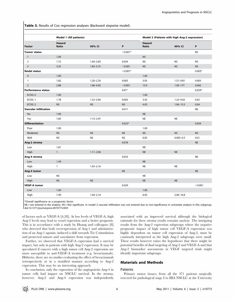
Multivariate analysesResults of the multivariate analysis are presented in Table 3. In
model 1, where all patients were assessed, high stromal Ang-4
(HR = 1.47, CI 95% 1.02–2.11, P = 0.04), stromal Ang-2 expres-
sion (HR = 1.88, CI 95% 1.15–3.08, P = 0.012) and high tumor
cell expression of VEGF-A (HR = 1.49, CI 1.04–2.14, P = 0.029)
were significant independent prognosticators for DSS in addition
to several clinicopathological variables (tumor status, P,0.001:
nodal status, P,0.001; performance status, P = 0.013; vascular
infiltration; P = 0.011; differentiation, P = 0.033). High tumor cell
expression of Ang-4 did not, however, reach statistical significance
(P = 0.15).
In model 2, only patients with high tumor cell expression of
Ang-2 were assessed (N = 88, Table 3). In this subgroup, high
tumor cell expression of VEGF-A mediated an independent
negative prognostic effect (HR = 6.43, CI 95% 2.46–16.79,
P,0.001), in addition to positive nodal status (P = 0.003), reduced
performance status (P = 0.024) and poor differentiation (P = 0.034).
Discussion
We present a large-scale study in an unselected population of
surgically resected NSCLC patients using high-throughput TMA.
In tumor cells, Ang-4 was the only angiopoietin to be associated
with survival, although not in an independent fashion. We found
that low stromal cell expressions of Ang-4 and Ang-2 were
independent poor prognostic factors for survival. VEGF-A was a
powerful poor prognosticator in patients with high tumor cell Ang-
2 expression, but not in those with low expression.
To our knowledge, this is the first prognostic evaluation of Ang-
4 expression in any human cancer. An improved prognosis
following high tumor cell expression of Ang-4 is in accordance
with an earlier functional in vitro study. Olsen and co-workers
found [12] Ang-4 to inhibit angiogenesis and reduce the elevated
interstitial pressure induced by basic fibroblast growth factor
(bFGF) and VEGF in small cell lung cancer tumor cells (GLC19).
However, recently Brunckhorst and colleagues found Ang-4 to
promote glioblastoma progression in vitro by enhancing tumor cell
viability and angiogenesis [17].
In the present study, there was no association between tumor
cell expression of Ang-2, Tie-2 and survival. Earlier IHC-studies in
NSCLC have shown conflicting results. In the largest study of 236
NSCLC cases, Tanaka et al. found that high tumor cell Ang-2
expression was significantly associated with poor overall survival in
the multivariate analysis [18]. They found that high tumor cell
Ang-2 expression was significantly associated with poor overall
survival even in the multivariate analysis. Corroborating our
results, they observed that the co-expression of Ang-2 and VEGF
resulted in a particularly poor survival. Due to their heterogeneous
staining intensity and low expression in endothelial cells, they were
not able, however, to semiquantitatively evaluate the degree of
Ang-1 or Ang-2 expression or to distinguish positive or negative
expression in endothelial cells. This is in contrast to the experience
by us and others [19,20]. In a smaller NSCLC study, Reinmuth et
al. found that high tumor cell Ang-1 expression, combined
intensity and percent of positive tumor cells, was statistically
associated to poor survival. Takanami investigated mRNA
expression of Ang-2 in 77 operable NSCLC patients and found
that a high expression was independently associated with a poor
survival [21]. High Ang-2 expression has also been associated with
poor prognosis in oral [22], colorectal [20] and bladder cancer
[23] whereas Sie et al. found the Ang-1/Ang-2 balance to be
associated to a poor survival in glioblastoma multiforme [24].
These conflicting results may be due to variations in methods, end-
points, patient selection and/or cut-off levels.
In stroma, on the other hand, there are no previous studies
examining the prognostic impact of these markers. This is
surprising since angiogenesis definitely also involves endothelial
cells and surrounding stromal cells. Attention should be paid to the
different tumor compartments. To illustrate this, the expression of
Angiopoietins and Prognosis in NSCLC
PLoS ONE | www.plosone.org 2 May 2011 | Volume 6 | Issue 5 | e19773
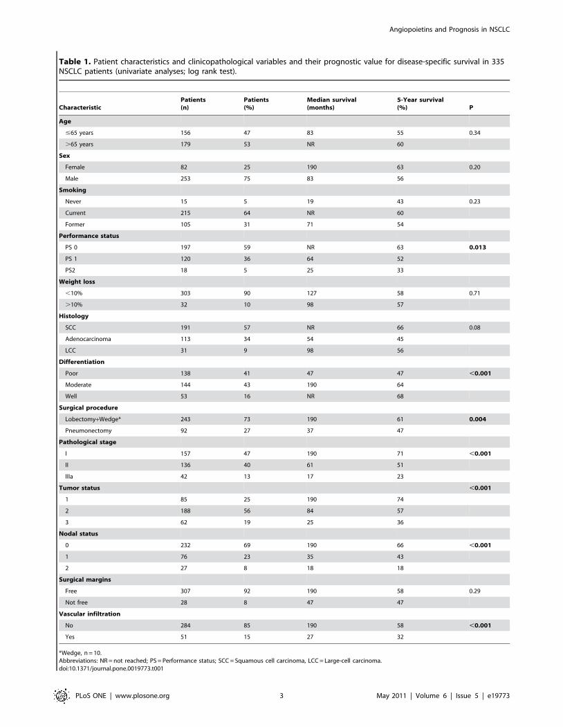
Table 1. Patient characteristics and clinicopathological variables and their prognostic value for disease-specific survival in 335NSCLC patients (univariate analyses; log rank test).
CharacteristicPatients(n)
Patients(%)
Median survival(months)
5-Year survival(%) P
Age
#65 years 156 47 83 55 0.34
.65 years 179 53 NR 60
Sex
Female 82 25 190 63 0.20
Male 253 75 83 56
Smoking
Never 15 5 19 43 0.23
Current 215 64 NR 60
Former 105 31 71 54
Performance status
PS 0 197 59 NR 63 0.013
PS 1 120 36 64 52
PS2 18 5 25 33
Weight loss
,10% 303 90 127 58 0.71
.10% 32 10 98 57
Histology
SCC 191 57 NR 66 0.08
Adenocarcinoma 113 34 54 45
LCC 31 9 98 56
Differentiation
Poor 138 41 47 47 ,0.001
Moderate 144 43 190 64
Well 53 16 NR 68
Surgical procedure
Lobectomy+Wedge* 243 73 190 61 0.004
Pneumonectomy 92 27 37 47
Pathological stage
I 157 47 190 71 ,0.001
II 136 40 61 51
IIIa 42 13 17 23
Tumor status ,0.001
1 85 25 190 74
2 188 56 84 57
3 62 19 25 36
Nodal status
0 232 69 190 66 ,0.001
1 76 23 35 43
2 27 8 18 18
Surgical margins
Free 307 92 190 58 0.29
Not free 28 8 47 47
Vascular infiltration
No 284 85 190 58 ,0.001
Yes 51 15 27 32
*Wedge, n = 10.Abbreviations: NR = not reached; PS = Performance status; SCC = Squamous cell carcinoma, LCC = Large-cell carcinoma.doi:10.1371/journal.pone.0019773.t001
Angiopoietins and Prognosis in NSCLC
PLoS ONE | www.plosone.org 3 May 2011 | Volume 6 | Issue 5 | e19773

Table 2. Tumor cell and stromal markers as prognostic factors for disease-specific survival in 335 NSCLC patients (univariateanalyses; log-rank test).
Characteristics Patients (n) Patients (%)Median survival(months) 5-year survival (%) P
Ang-1
Tumor 0.150
High 183 55 190 63
Low 141 42 84 52
Missing 11 3
Stroma
High 237 71 190 62 0.096
Low 92 27 58 50
Missing 6 2
Ang-2
Tumor 0.238
High 77 23 NR 63
Low 247 74 127 56
Missing 11 3
Stroma 0.017
High 295 88 190 60
Low 33 11 30 41
Missing 7 2
Ang-4
Tumor 0.046
High 149 44 190 64
Low 177 53 71 53
Missing 9 3
Stroma 0.009
High 219 65 190 62
Low 110 33 58 49
Missing 6 2
Tie2 0.267
Tumor 182 54 98 56
High 140 42 NR 60
Low 13 4
Missing
Stroma 0.116
High 58 17 NR 69
Low 269 80 98 56
Missing 8 3
VEGFA and Ang-2*
Low Ang-2 247 0.078
High VEGF-A 117 35 64 50
Low VEGF-A 130 39 190 61
High Ang-2 77 ,0.001
High VEGF-A 23 7 30 32
Low VEGF-A 54 16 NR 78
Missing 11 3
*Tumor data.doi:10.1371/journal.pone.0019773.t002
Angiopoietins and Prognosis in NSCLC
PLoS ONE | www.plosone.org 4 May 2011 | Volume 6 | Issue 5 | e19773

immunological markers in NSCLC impacts survival differentially
in tumor and stroma [25–27]. Herein, systematic studies of tumor
vessels could not be performed due to the fact that we had only
two stromal cores of 0.6 mm available from each patient.
Nevertheless, the expression of Ang/Tie-2 markers has been
investigated in the stromal compartments where the cross-talk
between endothelial cells, fibroblasts, immunological cells and
tumor cells are vital for angiogenesis [28]. Consistently, both Ang-
4 and Ang-2 expression in stroma proved to be independently
associated with an improved survival. Since these markers are
known to exert opposite effects upon binding to Tie-2, the similar
beneficial prognostic effect remains to be elucidated.
Since the angiogenic effect of the angiopoietin system is strongly
linked to VEGF we examined the prognostic effects of co-
expressions of angiopoietins and VEGF-A co-expressions in
tumor. High tumor cell Ang-2 expression alone had no prognostic
survival impact, but when co-expressed with a high rather than
low VEGF-A level, this combination led to a significantly worse 5-
year survival (32%) with an HR at 6.43. Besides, patients with a
concomitantly low VEGF-A expression and high Ang-2 expression
in tumor cells tended to a better survival compared with those with
a low tumor cell Ang-2 expression. These results can be explained
by the functional role of Ang-2, as it is known to destabilize the
endothelium, and the plastic state triggered by Ang-2 can lead to
new vessel growth or vessel regression, depending on the presence
Figure 1. Disease-specific survival according to factor expression. Disease-specific Kaplan-Meier survival curves according to: A) Ang-4expression in tumor cells, B) Ang-2 in tumor cells, C) Ang-4 in stromal cells and D) Ang-2 in stromal cells in resected NSCLC patients. The P-value isaccording to the log-rank test.doi:10.1371/journal.pone.0019773.g001
Figure 2. Disease-specific survival according to co-expressionof Ang-2 and VEGF-A. Disease-specific Kaplan-Meier survival curvesaccording to the co-expression of VEGF-A and Ang-2 in resected NSCLCpatients. The P-value is according to the log-rank test.doi:10.1371/journal.pone.0019773.g002
Angiopoietins and Prognosis in NSCLC
PLoS ONE | www.plosone.org 5 May 2011 | Volume 6 | Issue 5 | e19773
of factors such as VEGF-A [4,28]. At low levels of VEGF-A, high
Ang-2 levels may lead to vessel regression and a better prognosis.
This is in accordance with a study by Huang and colleagues [8]
who detected that both overexpression of Ang-1 and administra-
tion of an Ang-1 agonist, induced a shift towards Tie-2 stimulation
and protected tumors and vasculature from regression.
Further, we observed that VEGF-A expression had a survival
impact, but only in patients with high Ang-2 expression. It may be
speculated if cancers with a high tumor cell Ang-2 expression are
more susceptible to anti-VEGF-A treatment (e.g. bevacizumab).
Hitherto, there are no studies evaluating the effect of bevacizumab
retrospectively or in a stratified manner according to Ang-2
expression. This may be an interesting approach.
In conclusion, only the expression of the angiopoietin Ang-4 in
tumor cells had impact on NSCLC survival. In the stroma,
however, Ang-2 and Ang-4 expression was independently
associated with an improved survival although the biological
rationale for these stroma results remains unclear. The intriguing
results from the Ang-2 expression subgroups where the negative
prognostic impact of high tumor cell VEGF-A expression was
highly dependent on tumor cell expression of Ang-2, must be
cautiously interpreted as the high Ang-2 subgroups were small.
These results however raises the hypotheses that there might be
potential benefits of dual targeting of Ang-2 and VEGF-A and that
Ang-2 biomarker assessments in VEGF targeted trials might
identify important subgroups.
Materials and Methods
PatientsPrimary tumor tissues, from all the 371 patients surgically
resected for pathological stage I to IIIA NSCLC at the University
Table 3. Results of Cox regression analyses (Backward stepwise model).
Model 1 (All patients) Model 2 (Patients with high Ang-2 expression)
FactorHazardRatio 95% CI P
HazardRatio 95% CI P
Tumor status ,0.001* NS
1 1.00 NS
2 1.72 1.04–2.85 0.034 NS NS NS
3 3.25 1.83–5.75 ,0.001 NS NS NS
Nodal status ,0.001* 0.003*
0 1.00 1.00
1 1.82 1.20–2.76 0.005 3.93 1.57–9.81 0.003
2 2.88 1.68–4.92 ,0.001 13.9 1.09–177 0.042
Performance status 0.01* 0.024*
ECOG 0 1.00 1.00
ECOG 1 1.78 1.22–2.60 0.003 3.42 1.22–9.62 0.02
ECOG 2 NS NS NS 4.03 1.06–15.5 0.04
Vascular infiltration 0.011 NE
No 1.00 NE
Yes 1.85 1.15–2.97 NE NE
Differentiation 0.023* 0.034
Poor 1.00 1.00
Moderate NS NS NS NS NS NS
Well NS NS NS 0.05 0.005–0.5 0.01
Ang-2 stroma 0.018 NE
Low 1.81 NE
High 1 1.11–2.96 NE NE
Ang-4 stroma 0.033 NE
Low 1.49 NE
High 1 1.03–2.16 NE NE
Ang-4 tumor NS NE
Low NS NE
High NS NS NE NE
VEGF-A tumor 0.029 ,0.001
Low 1.00 1.00
High 1.49 1.04–2.14 6.43 2.46–16.8
*Overall significance as a prognostic factor.NE = not entered in the analysis. NS = Not significant. In model 2 vascular infiltration was not entered due to non-significance in univariate analysis in this subgroup.doi:10.1371/journal.pone.0019773.t003
Angiopoietins and Prognosis in NSCLC
PLoS ONE | www.plosone.org 6 May 2011 | Volume 6 | Issue 5 | e19773

Hospital of North Norway and Nordland Central Hospital from
1990 to 2004, were retrospectively identified in the archives of the
two pathological departments. A total of 36 patients were excluded
from the study due to inadequate paraffin-embedded fixed tissue
blocks (n = 13), other malignancy within the 5 years prior to
diagnosis (n = 13) or having received radiotherapy or chemother-
apy prior to surgery (n = 10). Preoperative chemotherapy was not
considered standard treatment in Norway during the actual
period. Complete demographic and clinicopathological data for
these 335 eligible patients were obtained retrospectively. The
pathological data were revised according to the 7th edition of
UICC TNM classification of lung cancer [29,30]. The last DSS
update was done in November 2008.
Tissue Microarray constructionDuplicate 0.6 mm core biopsies from the most representative
areas of tumor cells (neoplastic epithelial cells) and tumor stroma
were collected from each surgical specimen using a tissue-arraying
instrument (Beecher Instruments, Silver Springs, MD). Normal lung
tissue localized distant from the primary tumor as well as lung tissue
samples from 20 normal lungs were used as controls. Eight tissue
microarray blocks (TMAs) were constructed to include all the cores.
The detailed methodology has been reported previously [16].
ImmunohistochemistryThe applied commercial antibodies had been subjected to in-
house validation by the manufacturer for IHC on paraffin-
embedded material (IHC-P). One exception was Ang-1 which was
selected due to others’ published success with this antibody
[18,20,22,31,32] and failure to achieve satisfying quality with
other available IHC-P tested antibodies. The 4 mm TMA sections
were deparaffinised with xylene and rehydrated with ethanol. The
sections containing tissue cores were subjected to the following
antibodies: Ang-1 (goat polyclonal, sc-6319, Santa Cruz biotech-
nology, Inc., 2 mg/ml), Ang-2 (rabbit, polyclonal, ab65835,
Abcam, 17 mg/ml), Ang-4 (goat polyclonal, AF964, R&D systems,
4 mg/ml), Tie-2 (rabbit polyclonal, sc-9026, Santa Cruz biotech-
nology, Inc., 4 mg/ml) and VEGF-A (rabbit polyclonal; RB-1678;
Neomarkers; 100 mg/ml).
Antigen retrieval was done manually for all antibodies except
Ang-2, by placing the specimens in 0.01 M citrate buffer at pH 6.0
and exposed to microwave heating of 20 minutes at 450 W. All
antibodies were incubated overnight at 4uC except for VEGF-A
where the primary antibody was incubated for 30 min in room
temperature. For Ang-2 we used the Ventana Benchmark XTH(Ventana Medical Systems Inc.), procedure ultraview DAB v3 with
automatic antigen retrieval with CC1 mild (30 min). Negative
Figure 3. Immunohistochemical staining. Immunohistochemical analyses of NSCLC representing high and low intensities for tumor cell andstromal expression of Ang-1, Ang-2, Ang-4 and Tie-2.doi:10.1371/journal.pone.0019773.g003
Angiopoietins and Prognosis in NSCLC
PLoS ONE | www.plosone.org 7 May 2011 | Volume 6 | Issue 5 | e19773

controls were simultaneously performed for all antibodies by
omitting the primary antibody and an appropriate isotype-control
was done for all antibodies on one of the TMA slides. Capillary
vessels in stromal cores with high expression were used for internal
positive controls and skin haemangiomas as external positive
controls
Finally, all slides were counterstained with hematoxylin to
visualize the nuclei.
Scoring of immunohistochemistryEach anonymized core was scored independently and semi-
quantitatively by two pathologists (S.A.S and K.A.S) by light
microscopy. Only viable parts were scored and even though some
cores comprised of both tumor and stroma, only one entity was
scored at a time. In stroma, we scored all non-neoplastic cells. The
dominant staining intensity in both tumor and stroma cores was
scored as: 0 = negative, 1 = weak, 2 = intermediate and 3 = strong
(Figure 3). Only cytoplasmic staining was evaluated. In stroma
cores the cytoplasmic staining was scored with regards to density
as well: 1 = low, 2 = intermediate and 3 = high.
In case of disagreement the slides were re-examined and
consensus was reached by the observers. Interindividual variability
with respect to IHC-scoring in both tumor cells and stromal cells
was evaluated on the current material in a previous paper
(r = 0.95, range 0.93–0.98) [16].
A mean score was calculated separately for the two tumor cell
cores and the two stromal cells cores in each individual. In tumor,
high expression was defined as $2.0 for Ang-1, Ang-4 and Tie-2,
and $2.5 for Ang-2. In stroma, the sum of mean density (1–3) and
intensity score (0–3) was calculated. High expression in stroma was
defined as $2.5 for Ang-1, Ang-2 and Ang-4, and $5.0 for Tie2.
Details regarding VEGF-A scoring has been reported earlier [16].
Similar scoring methods have been used in our previous IHC-
scoring studies [16,27,33] and by others [34].
Statistical methodsThe statistical analyses were done using the SPSS 16.0 package
(Chicago, IL). The x2 test and Fishers exact tests were used to
examine the associations between molecular marker expressions
and the clinicopathological markers. Plots of the DSS, according
to marker expressions, were drawn using the Kaplan-Meier
method, and statistical significance between survival curves was
assessed by the log rank test. The survival curves were terminated
at 146 months, due to less than 10% of patients at risk after this
point. The chosen endpoint, DSS, was calculated from the time of
surgery to the time of lung cancer death. All significant variables
from the univariate analyses were entered into the multivariate
analyses in a backward stepwise Cox regression analysis with a
probability for stepwise entry and removal at 0.05 and 0.10,
respectively. A P,0.05 was considered statistically significant for
all analyses.
EthicsThe Norwegian Data Inspection Board and The Regional
Committee for research ethics have approved the study.
Information and subsequent written consent from patients was
considered, but as this was a retrospective study with more than
half of patients deceased, the rest of the patients having to
reminded about the death rate of the disease and the possible
raising of unrealistic hope for the individual, they specifically
waived the need for consent.
Author Contributions
Conceived and designed the experiments: L-TB RB SA TD. Performed the
experiments: SA SA-S KA-S. Analyzed the data: SA SA-S KA-S TD HS.
Contributed reagents/materials/analysis tools: L-TB SA-S KA-S. Wrote
the paper: SA.
References
1. Jemal A, Siegel R, Ward E, Hao Y, Xu J, et al. (2009) Cancer statistics, 2009.
CA Cancer J Clin 59: 225–249.
2. Mantovani A (2009) Cancer: Inflaming metastasis. Nature 457: 36–37.
3. Hanahan D, Weinberg RA (2000) The hallmarks of cancer. Cell 100: 57–70.
4. Saharinen P, Bry M, Alitalo K (2010) How do angiopoietins Tie in with vascular
endothelial growth factors? Curr Opin Hematol 17: 198–205.
5. Fukuhara S, Sako K, Noda K, Zhang J, Minami M, et al. (2010) Angiopoietin-
1/Tie2 receptor signaling in vascular quiescence and angiogenesis. HistolHistopathol 25: 387–396.
6. Benjamin LE, Golijanin D, Itin A, Pode D, Keshet E (1999) Selective ablation ofimmature blood vessels in established human tumors follows vascular endothelial
growth factor withdrawal. J Clin Invest 103: 159–165.
7. Puri MC, Rossant J, Alitalo K, Bernstein A, Partanen J (1995) The receptortyrosine kinase TIE is required for integrity and survival of vascular endothelial
cells. EMBO J 14: 5884–5891.
8. Huang J, Bae JO, Tsai JP, Kadenhe-Chiweshe A, Papa J, et al. (2009)
Angiopoietin-1/Tie-2 activation contributes to vascular survival and tumorgrowth during VEGF blockade. Int J Oncol 34: 79–87.
9. Augustin HG, Koh GY, Thurston G, Alitalo K (2009) Control of vascularmorphogenesis and homeostasis through the angiopoietin-Tie system. Nat Rev
Mol Cell Biol 10: 165–177.
10. Sato TN, Tozawa Y, Deutsch U, Wolburg-Buchholz K, Fujiwara Y, et al. (1995)
Distinct roles of the receptor tyrosine kinases Tie-1 and Tie-2 in blood vesselformation. Nature 376: 70–74.
11. Lee HJ, Cho CH, Hwang SJ, Choi HH, Kim KT, et al. (2004) Biological
characterization of angiopoietin-3 and angiopoietin-4. FASEB J 18: 1200–1208.
12. Olsen MW, Ley CD, Junker N, Hansen AJ, Lund EL, et al. (2006) Angiopoietin-
4 inhibits angiogenesis and reduces interstitial fluid pressure. Neoplasia 8:364–372.
13. Wong MP, Chan SY, Fu KH, Leung SY, Cheung N, et al. (2000) The
angiopoietins, tie2 and vascular endothelial growth factor are differentially
expressed in the transformation of normal lung to non-small cell lungcarcinomas. Lung Cancer 29: 11–22.
14. Saharinen P, Eklund L, Miettinen J, Wirkkala R, Anisimov A, et al. (2008)
Angiopoietins assemble distinct Tie2 signalling complexes in endothelial cell-cell
and cell-matrix contacts. Nat Cell Biol 10: 527–537.
15. Holopainen T, Huang H, Chen C, Kim KE, Zhang L, et al. (2009)
Angiopoietin-1 overexpression modulates vascular endothelium to facilitate
tumor cell dissemination and metastasis establishment. Cancer Res 69:4656–4664.
16. Donnem T, Al-Saad S, Al-Shibli K, Delghandi MP, Persson M, et al. (2007)Inverse prognostic impact of angiogenic marker expression in tumor cells versus
stromal cells in non small cell lung cancer. Clin Cancer Res 13: 6649–6657.
17. Brunckhorst MK, Wang H, Lu R, Yu Q (2010) Angiopoietin-4 promotesglioblastoma progression by enhancing tumor cell viability and angiogenesis.
Cancer Res 70: 7283–7293.
18. Tanaka F, Ishikawa S, Yanagihara K, Miyahara R, Kawano Y, et al. (2002)
Expression of angiopoietins and its clinical significance in non-small cell lung
cancer. Cancer Res 62: 7124–7129.
19. Reinmuth N, Piegelbrock E, Raedel M, Fehrmann N, Hintelmann H, et al.
(2007) Prognostic significance of vessel architecture and vascular stability in non-small cell lung cancer. Lung Cancer 55: 53–60.
20. Chung YC, Hou YC, Chang CN, Hseu TH (2006) Expression and prognostic
significance of angiopoietin in colorectal carcinoma. J Surg Oncol 94: 631–638.
21. Takanami I (2004) Overexpression of Ang-2 mRNA in non-small cell lung
cancer: association with angiogenesis and poor prognosis. Oncol Rep 12:849–853.
22. Chien CY, Su CY, Chuang HC, Fang FM, Huang HY, et al. (2008)
Angiopoietin-1 and -2 expression in recurrent squamous cell carcinoma of theoral cavity. J Surg Oncol 97: 273–277.
23. Szarvas T, Jager T, Totsch M, vom DF, Kempkensteffen C, et al. (2008)Angiogenic switch of angiopietins-Tie2 system and its prognostic value in
bladder cancer. Clin Cancer Res 14: 8253–8262.
24. Sie M, Wagemakers M, Molema G, Mooij JJ, de Bont ES, et al. (2009) Theangiopoietin 1/angiopoietin 2 balance as a prognostic marker in primary
glioblastoma multiforme. J Neurosurg 110: 147–155.
25. Welsh TJ, Green RH, Richardson D, Waller DA, O’Byrne KJ, et al. (2005)Macrophage and mast-cell invasion of tumor cell islets confers a marked survival
advantage in non-small-cell lung cancer. J Clin Oncol 23: 8959–8967.JCO.2005.01.4910 [pii];10.1200/JCO.2005.01.4910 [doi].
26. Wakabayashi O, Yamazaki K, Oizumi S, Hommura F, Kinoshita I, et al. (2003)CD4+ T cells in cancer stroma, not CD8+ T cells in cancer cell nests, are
Angiopoietins and Prognosis in NSCLC
PLoS ONE | www.plosone.org 8 May 2011 | Volume 6 | Issue 5 | e19773

associated with favorable prognosis in human non-small cell lung cancers.
Cancer Sci 94: 1003–1009.27. Al-Shibli KI, Donnem T, Al-Saad S, Persson M, Bremnes RM, et al. (2008)
Prognostic effect of epithelial and stromal lymphocyte infiltration in non-small
cell lung cancer. Clin Cancer Res 14: 5220–5227.28. Huang H, Bhat A, Woodnutt G, Lappe R (2010) Targeting the ANGPT-TIE2
pathway in malignancy. Nat Rev Cancer 10: 575–585.29. Goldstraw P (2009) The 7th Edition of TNM in Lung Cancer: what now?
J Thorac Oncol 4: 671–673.
30. World Health Organization (1999) Histological Typing of Lung and PleuralTumours. Geneva, Switzerland: Springer-Verlag.
31. Nakayama T, Yoshizaki A, Kawahara N, Ohtsuru A, Wen CY, et al. (2004)Expression of Tie-1 and 2 receptors, and angiopoietin-1, 2 and 4 in gastric
carcinoma; immunohistochemical analyses and correlation with clinicopatho-
logical factors. Histopathology 44: 232–239.32. Nakayama T, Inaba M, Naito S, Mihara Y, Miura S, et al. (2007) Expression of
angiopoietin-1, 2 and 4 and Tie-1 and 2 in gastrointestinal stromal tumor,
leiomyoma and schwannoma. World J Gastroenterol 13: 4473–4479.33. Al-Saad S, Al-Shibli K, Donnem T, Persson M, Bremnes RM, et al. (2008) The
prognostic impact of NF-kappaB p105, vimentin, E-cadherin and Par6expression in epithelial and stromal compartment in non-small-cell lung cancer.
Br J Cancer 99: 1476–1483.
34. Soltermann A, Tischler V, Arbogast S, Braun J, Probst-Hensch N, et al. (2008)Prognostic significance of epithelial-mesenchymal and mesenchymal-epithelial
transition protein expression in non-small cell lung cancer. Clin Cancer Res 14:7430–7437.
Angiopoietins and Prognosis in NSCLC
PLoS ONE | www.plosone.org 9 May 2011 | Volume 6 | Issue 5 | e19773