MMP-9 and MMP-2 gelatinases and TIMP-1 and TIMP-2 inhibitors in breast cancer: correlations with...
12
J. Cell. Mol. Med. Vol 10, No 2, 2006 pp. 499-510 The matrix metalloproteinases (MMPs) are a fami- ly of structurally and functionally related proteinas- es, initially characterized by their ability to degrade the extracellular matrix (ECM) [1]. Nowadays, at least 20 enzymes that share considerable homology within their major domains (signal peptide, propep- tide, catalytic, hinge and hemopexin-like domains) were included in MMPs family [2]. Most of MMPs are synthesised and secreted as partially activated latent forms, requiring, for full activation, removal of the entire propeptide domain by proteinases including other MMPs [3]. The expression of MMP-9 and MMP-2 gelatinases and TIMP-1 and TIMP-2 inhibitors in breast cancer: correlations with prognostic factors D. C. Jinga b , A. Blidaru b , Ileana Condrea b , Carmen Ardeleanu c , Cristina Dragomir a , Geza Szegli a , Maria Stefanescu a , Cristiana Matache a * a Department of Immunology, "Cantacuzino" National Institute for Microbiology and Immunology, Bucharest, Romania b "Trestioreanu" National Institute of Oncology, Bucharest, Romania c "Victor Babeº" National Institute of Pathology and Biomedical Science, Bucharest, Romania Received: December 22, 2005; Accepted: April 20, 2006 Abstract The goal of our study was to analyse the prognostic values for some matrix metalloproteinases (MMPs) and tissue inhibitors of matrix metalloproteinases (TIMPs) in breast cancer. We evaluated the activity and the expression levels of MMP-9, MMP-2, TIMP-1 and TIMP-2 in malignant versus benign fresh breast tumor extracts. For this purpose, gelat- inzymography, immunoblotting and ELISA were used to analyse the activity and expression of MMPs and TIMPs. We found that MMP-9 expression level and activity are increased in malignant tumors. In addition, MMP-9/TIMP-1 and MMP-2/TIMP-2 ratio values obtained by us were significantly different in malignant tumors compared to benign tumors. We suggest that the abnormal MMP-9/TIMP-1 balance plays a role in the configuration of breast invasive carcinoma of no special type and also in tumor growth, while altered MMP-2/TIMP-2 ratio value could be associated with lymph node invasion and used as a prognostic marker in correlation with Nottingham Prognostic Index. Finally, we showed that in malignant tumors high expression of estrogen receptors is associated with enhanced activity of MMP-2 and increased bcl- 2 levels, while high expression of progesterone receptors is correlated with low TIMP-1 protein levels. Keywords: breast cancer • MMP-9 • MMP-2 • TIMP-1 • TIMP-2 • clinicopathological markers * Correspondence to: Dr. Cristiana MATACHE Department of Immunology, "Cantacuzino" National Institute for Microbiology and Immunology, Splaiul Independentei 103, Bucharest 050096, Romania. Tel.: ++40(21)3184410 (252); Fax: ++40(21)3184414 E-mail: [email protected] Introduction Available online at www.jcmm.ro www.jcmm.org Published by: the CMM Foundation Reprinted from: Journal of Cellular and Molecular Medicine JCMM JCMM
-
Upload
independent -
Category
Documents
-
view
4 -
download
0
Transcript of MMP-9 and MMP-2 gelatinases and TIMP-1 and TIMP-2 inhibitors in breast cancer: correlations with...

J. Cell. Mol. Med. Vol 10, No 2, 2006 pp. 499-510
The matrix metalloproteinases (MMPs) are a fami-ly of structurally and functionally related proteinas-es, initially characterized by their ability to degrade
the extracellular matrix (ECM) [1]. Nowadays, atleast 20 enzymes that share considerable homologywithin their major domains (signal peptide, propep-tide, catalytic, hinge and hemopexin-like domains)were included in MMPs family [2]. Most of MMPsare synthesised and secreted as partially activatedlatent forms, requiring, for full activation, removalof the entire propeptide domain by proteinasesincluding other MMPs [3]. The expression of
MMP-9 and MMP-2 gelatinases and TIMP-1 and TIMP-2 inhibitors in breast cancer:
correlations with prognostic factors
D. C. Jinga b, A. Blidaru b, Ileana Condrea b, Carmen Ardeleanu c, Cristina Dragomir a, Geza Szegli a, Maria Stefanescu a, Cristiana Matache a *
a Department of Immunology, "Cantacuzino" National Institute for Microbiology and Immunology,Bucharest, Romania
b "Trestioreanu" National Institute of Oncology, Bucharest, Romaniac "Victor Babeº" National Institute of Pathology and Biomedical Science, Bucharest, Romania
Received: December 22, 2005; Accepted: April 20, 2006
Abstract
The goal of our study was to analyse the prognostic values for some matrix metalloproteinases (MMPs) and tissueinhibitors of matrix metalloproteinases (TIMPs) in breast cancer. We evaluated the activity and the expression levels ofMMP-9, MMP-2, TIMP-1 and TIMP-2 in malignant versus benign fresh breast tumor extracts. For this purpose, gelat-inzymography, immunoblotting and ELISA were used to analyse the activity and expression of MMPs and TIMPs. Wefound that MMP-9 expression level and activity are increased in malignant tumors. In addition, MMP-9/TIMP-1 andMMP-2/TIMP-2 ratio values obtained by us were significantly different in malignant tumors compared to benign tumors.We suggest that the abnormal MMP-9/TIMP-1 balance plays a role in the configuration of breast invasive carcinoma ofno special type and also in tumor growth, while altered MMP-2/TIMP-2 ratio value could be associated with lymph nodeinvasion and used as a prognostic marker in correlation with Nottingham Prognostic Index. Finally, we showed that inmalignant tumors high expression of estrogen receptors is associated with enhanced activity of MMP-2 and increased bcl-2 levels, while high expression of progesterone receptors is correlated with low TIMP-1 protein levels.
Keywords: breast cancer • MMP-9 • MMP-2 • TIMP-1 • TIMP-2 • clinicopathological markers
* Correspondence to: Dr. Cristiana MATACHEDepartment of Immunology, "Cantacuzino" National Institutefor Microbiology and Immunology, Splaiul Independentei 103,Bucharest 050096, Romania.Tel.: ++40(21)3184410 (252); Fax: ++40(21)3184414E-mail: [email protected]
Introduction
Available online atwww.jcmm.ro www.jcmm.org
Published by:the CMM Foundation
Reprinted from: Journal of Cellular and Molecular Medicine
JCMMJCMM

MMPs is tightly regulated at different levels: tran-scription, secretion and activation [1, 4]. Factorsincluding inflammatory cytokines, hormones,growth factors and oncogene products induceMMPs expression [1, 5, 6]. In addition, cell-matrixand cell-cell interactions affect MMPs gene tran-scription [7]. The activity of MMPs is controlled byinteraction with the tissue inhibitors of metallopro-teinases (TIMPs) [8]. Currently, four TIMPs havebeen identified, most of them being capable to bindand inhibit the activity of all members of MMPsfamily. The members of TIMP family are known tohave other different roles, like growth factor prop-erties, stimulation of cell migration, and stimulationor inhibition of apoptosis [9–11].
Based on their substrate specificity and domainstructure, the MMPs were divided into four mainsubgroups. One of these subgroups is represented bygelatinases, also known as type IV collagenases,which degrade gelatine and types IV, V, VII, IX andX collagen. This subgroup of MMPs includes twodistinct members, known as gelatinase A (MMP-2)and gelatinase B (MMP-9). Due to their specificityfor the substrate (collagen IV, particularly abundantin basal membrane), MMP-2 and MMP-9 were themost involved in tumor initiation, growth and metas-tasis, especially in breast cancer [12]. A large numberof publications presented various studies about therole of gelatinases (MMP-2/MMP-9) and their natu-ral inhibitors (TIMP-1/TIMP-2) in breast cancer,some of the results being contradictory [13–20].Therefore, the aim of this study was also to corrobo-rate our results with previously reported data, inorder to introduce new valuable prognostic markersin breast cancer.
This study used freshly isolated tissues speci-men from the malignant and benign breast tumorsanalysing the activity and the expression level ofgelatinases and their natural inhibitors, in correla-tion with prognostic factors for breast cancer.
Material and methods
Patients and tissue specimens
The study was performed on freshly isolated tumor sam-ples from thirty-two consecutive patients with primaryinvasive breast carcinomas (IC) (13 of special type and
19 of no special type, based on Miller and Sainsburyclassification [21, 22]) and sixteen consecutive patientswith benign breast tumors (eight fibroadenomas, sixintraductal papillomas, one fibroadenoma and intraduc-tal papilloma and one sclerocystic mastose). All thepatients were hospitalized and had undergone surgery at“Prof. Al. Trestioreanu” Institute of Oncology,Bucharest, between 2002 and 2003. The patients withmalignant tumors were aged from 37 to 78 years (meanage 53 years) and those with benign tumors were agedfrom 16 to 62 years (mean age 31 years). Table 1 showsthe prognostic factors of the patients with IC and thearbitrary scores assigned to each of the analysed marker,based on increased aggressiveness.
Freshly isolated breast tumor extracts frompatients who underwent surgery were mechanicallydisaggregated, washed with 150 mM NaCl andhomogenized for 30 minutes on ice in Triton lysisbuffer (50 mM HEPES buffer pH 7.5, 1% Triton-X100, 250 mM NaCl, 10% glycerol, 1 mM PMSF and10 μg/ml leupeptin). The homogenates were cen-trifuged at 1200 rpm for 10 minutes at +4°C and thesupernatants were recentrifuged at 15000 rpm for 15minutes at +4°C. Finally, the resulted supernatantswere analyzed for total protein content, using a com-mercial BCA Protein Assay Reagent (Pierce) and thealiquotes were frozen at –80°C until processing.
Expression levels of estrogen andprogesterone receptors and bcl-2
Estrogen receptors (ER), progesterone receptors (PR), andbcl-2 expression levels were analyzed as described byBussolati and Gugliotta [23]. Briefly, the tissue sectionswere incubated with primary antibodies anti-ER, 1D5clone (1:100), anti-PR, 1A6 clone (1:100) or anti-bcl-2,124 clone (1:40) (DAKO). The detection was performedusing avidin-biotin peroxidase complex and diaminoben-zidine as a chromogen. The tissue sections counterstainedwith Meyer’s haematoxylin were mounted.
The immunoreactivity for ER and PR was evaluatedby Histochemistry Score System (HSCORE), calculatedbased on algorithm HSCORE = Σ [(I + 1) x PC], whereI signifies the staining intensity and PC signifies the per-centage of cells that stain at each intensity, respectively.Staining intensity was classified based on an arbitraryscale from 0 to 3 as follows: 0 for negative staining, 1for mild reactivity, 2 for moderate reactivity and 3 forstrong reactivity.
500
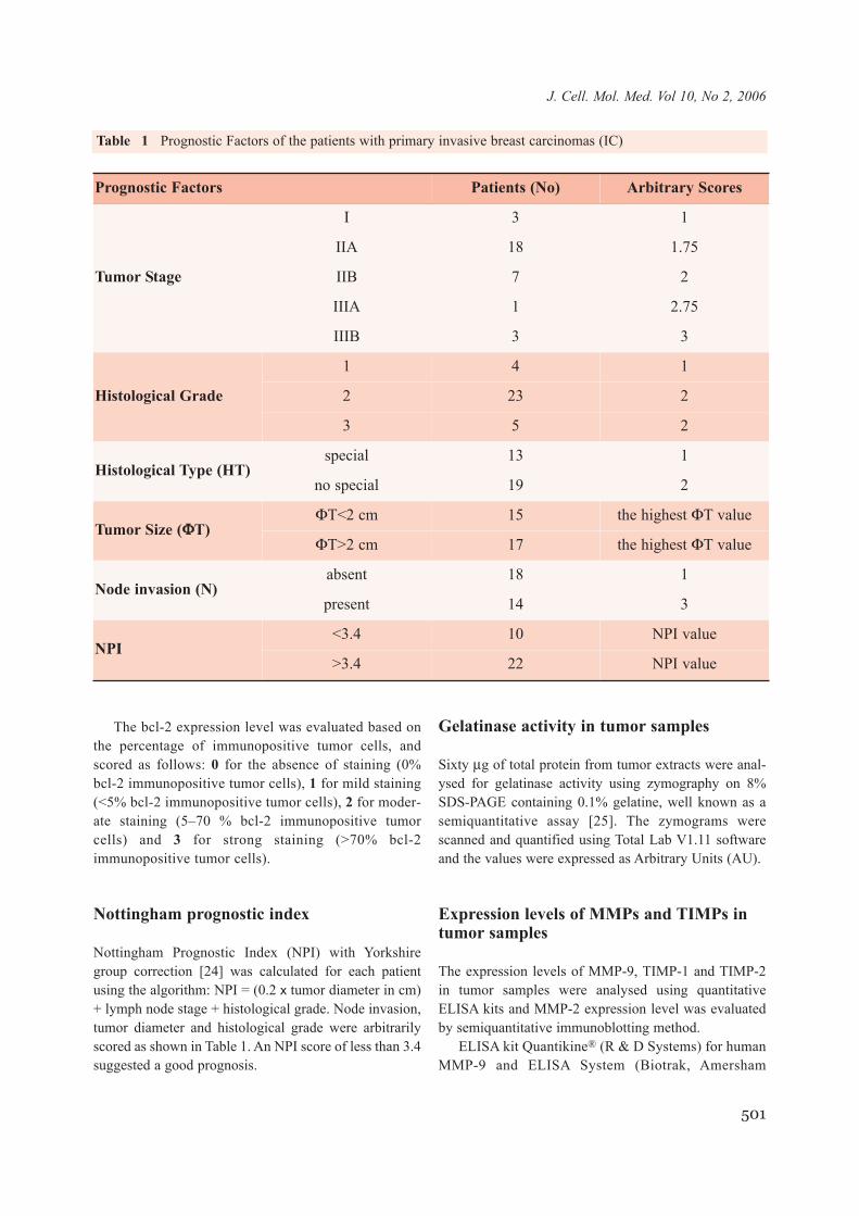
The bcl-2 expression level was evaluated based onthe percentage of immunopositive tumor cells, andscored as follows: 0 for the absence of staining (0%bcl-2 immunopositive tumor cells), 1 for mild staining(<5% bcl-2 immunopositive tumor cells), 2 for moder-ate staining (5–70 % bcl-2 immunopositive tumorcells) and 3 for strong staining (>70% bcl-2immunopositive tumor cells).
Nottingham prognostic index
Nottingham Prognostic Index (NPI) with Yorkshiregroup correction [24] was calculated for each patientusing the algorithm: NPI = (0.2 x tumor diameter in cm)+ lymph node stage + histological grade. Node invasion,tumor diameter and histological grade were arbitrarilyscored as shown in Table 1. An NPI score of less than 3.4suggested a good prognosis.
Gelatinase activity in tumor samples
Sixty μg of total protein from tumor extracts were anal-ysed for gelatinase activity using zymography on 8%SDS-PAGE containing 0.1% gelatine, well known as asemiquantitative assay [25]. The zymograms werescanned and quantified using Total Lab V1.11 softwareand the values were expressed as Arbitrary Units (AU).
Expression levels of MMPs and TIMPs intumor samples
The expression levels of MMP-9, TIMP-1 and TIMP-2in tumor samples were analysed using quantitativeELISA kits and MMP-2 expression level was evaluatedby semiquantitative immunoblotting method.
ELISA kit Quantikine® (R & D Systems) for humanMMP-9 and ELISA System (Biotrak, Amersham
501
J. Cell. Mol. Med. Vol 10, No 2, 2006
Prognostic Factors Patients (No) Arbitrary Scores
Tumor Stage
I 3 1
IIA 18 1.75
IIB 7 2
IIIA 1 2.75
IIIB 3 3
Histological Grade
1 4 1
2 23 2
3 5 2
Histological Type (HT)special 13 1
no special 19 2
Tumor Size (ΦΦT)ΦT<2 cm 15 the highest ΦT value
ΦT>2 cm 17 the highest ΦT value
Node invasion (N)absent 18 1
present 14 3
NPI<3.4 10 NPI value
>3.4 22 NPI value
Table 1 Prognostic Factors of the patients with primary invasive breast carcinomas (IC)

Biosciences) for TIMP-1 and TIMP-2 were used.Tumor samples were diluted 1:25 for MMP-9 and 1:40for TIMP-1 and TIMP-2 and analysed using the manu-facturer’s protocol.
Sixty μg of total protein from each tumor extractwere subjected to 8% SDS-PAGE gel electrophoresisunder reducing conditions and analysed by immunoblot-ting with 1 μg/ml monoclonal antibody specific topro/active forms of human MMP-2 (R & D Systems) fol-lowed by anti-mouse IgG peroxidase conjugate (1:20000dilution) and Enhancement Chemiluminescence (ECL)detection (Amersham Pharmacia Biotech). The imageswere scanned and quantified using Total Lab V1.11 soft-ware and the levels of MMP-2 were expressed asArbitrary Units (AU).
Statistical analysis
The significance of the differences between malignantand benign tumors was examined using Student’s t-testand Student’s t-test with Bonferroni correction. The sig-nificance level of 0.05 by Student’s t-test (p< 0.05) wasadjusted to 0.01 after Bonferroni correction (Pc< 0.01).Spearman’s coefficient was calculated in order to estab-lish the correlation of both the activity and expressionlevel of gelatinases, and the expression level of TIMP-1
and TIMP-2 with prognostic factors for breast cancer.The correlation coefficient |r|>0.5, with a probability P <0.01, was considered as significant.
Results
Activities and levels of MMP-9, MMP-2,TIMP-1 and TIMP-2 in malignant versusbenign breast tumors
Tumor extracts were analysed for gelatinase activi-ty by gelatine zymography. The lytic activity waspresent at different apparent molecular weights (92,86, 72 and 66 kDa respectively), corresponding tolatent MMP-9 (lMMP-9), fully activated MMP-9(aMMP-9), latent MMP-2 (lMMP-2) and fully acti-vated MMP-2 (aMMP-2) forms. A representativeexample of gelatine zymography performed on fourmalignant and four benign tumor tissue extracts isillustrated in Fig. 1.
Densitometry analysis of all zymograms revealedthat the activity of lMMP-9 form was almost compa-rable between malignant and benign tumor extracts,while the activity of aMMP-9 form was moreincreased in malignant than in benign tumor extracts
502
Fig. 1 Gelatine zymography of breast tumours extracts. Breast tumor extracts were resolved in SDS-PAGE 0.1 %gelatine zymography. Subsequently, the gel was incubated in a 2.5 % Triton X-100 solution and then in substratebuffer containing 5 mM CaCl2. The lytic gelatinase activity was identified after Coomassie Brilliant Blue R stain-ing as a clear band on a blue background. Figure presents gelatinase activities in benign (lines 1, 3, 4, 5) and maligntumour extracts (lines 2, 6, 7, 8), and a positive control for the activities of lMMP-9 and lMMP-2 (line 9).

(Pc = 0.0001) (Fig. 2A). Further analyses showed anenhanced activity of lMMP-2 form in benign speci-mens compared to malignant tumor samples (p =0.048), while the gelatinolytic activity of aMMP-2form was higher in malignant specimens comparedto benign tumor samples (p = 0.031) (Fig. 2A).
When the expression levels of MMP-9, -2 andTIMP-1, -2 were analysed, some interesting resultswere obtained. The MMP-9 expression level wassignificantly higher in malignant than in benigntumor samples (Pc = 0.0002). Although the malig-nant tumors showed an increased level of MMP-2compared to benign tumors, the result of statisticalanalysis was under the significance level (p =0.039). In contrast, the levels of TIMP-1 and TIMP-2 were comparable between malignant and benigntumor extracts (Fig. 2B).
To evaluate the relative inhibition of the MMPsactivity, we calculated MMP-9/TIMP-1 andMMP-2/TIMP-2 ratio value for each tumor sam-ple. Our analysis showed that both ratio valueswere increased in malignant tumor samples, clear-ly differentiating between the malignant andbenign tumors (Pc = 0.002 and Pc = 0.007, respec-tively) (Fig. 2C).
Correlation between gelatinases and theirtissue inhibitors and breast cancerprognostic factors
Afterwards, we correlated the activity and the expres-sion of gelatinases and their natural inhibitors withaccepted and used prognostic factors for breast cancer(tumor stage, histological type, histological grade,tumor size, nodal status and NPI) [21, 24]. A positivecorrelation was found between the score asigned forthe histological type and each of the following: theactivity of aMMP-9 form, the expression of MMP-9and the MMP-9/TIMP-1 ratio value. When IC tumorswere divided in special and no special types, based onMiller and Sainsbury classification [21, 22], the activ-ities of MMP-9 forms, the expression level of MMP-9 and the MMP-9/TIMP-1 ratio values were marked-ly different between these tumor types (Table 2).
Unfortunately, due to the small number of cases with-in some groups (see Table 1), we could not use statisticalanalysis to identify the correlation between MMPs andTIMPs and tumor stage or histological grade.
A weak positive correlation between MMP-9/TIMP-1 ratio value and tumor size was found(Table 3). In the large tumors, the MMP-9/TIMP-1
503
J. Cell. Mol. Med. Vol 10, No 2, 2006
Fig. 2A MMP-9 and MMP-2 activity in freshly isolatedbreast tumor extracts.Histograms show the densito-metry results on zymogramsobtained for all tumor extractsamples. Data represent themean values ± SD for eachanalysed parameter and foreach tumor type. *Pc < 0.01reveals the most importantdifferences between themalign and benign tumors.

ratio values were higher than in small tumors;therefore, the MMP-9/TIMP-1 ratio values statisti-cally differentiated between these two types oftumors (Pc = 0.013).
A relevant positive correlation between MMP-2/TIMP-2 ratio and node invasion was identified(Table 4). The patients with node invasion presentedan increased level of MMP-2 and a reduced TIMP-2
504
Fig. 2B Expression level ofMMP-9, MMP-2, TIMP-1 andTIMP-2 proteins in freshly iso-lated breast tumor extractsanalysed by immunoblottingand ELISA. Data represent themean values ± SD for eachanalysed parameter and foreach tumor type. *Pc < 0.01reveals the most important dif-ferences between the malignand benign tumors.
Fig. 2C Ratio MMP-9/TIMP-1 and MMP-2/TIMP-2 values calcu-lated to evaluate the rela-tive inhibition of theMMPs activity. Data rep-resent the mean values ±SD for each ratio and foreach tumor type.*Pc <0.01 reveals the mostimportant differencesbetween the malign andbenign tumors.

level. Therefore, the mean value of MMP-2/TIMP-2ratios was three times higher in patients with than in
patients without node invasion, the ratios statistical-ly differentiating between these groups of patients.
505
J. Cell. Mol. Med. Vol 10, No 2, 2006
Group lMMP-9 activity(AU/100)
aMMP-9 activi-ty (AU/100)
MMP-9 Expression(ng/mg protein)
TIMP-1 expression(ng/mg protein) MMP-9/TIMP-1
all IC r = 0.490 §P = 0.005
r = 0.663P = 0.0002
r = 0.563P = 0.001 no correlation r = 0.662
P < 0.0001
G1* 352 ± 250‡ 26 ± 32 1.90 ± 1.05 126.9 ± 88.2 0.02 ± 0.02
G2** 669 ± 283 209 ± 138 11.53 ± 9.68 63.5 ± 52.3 0.28 ± 0.31
G1 vs. G2 0.003*** 0.00003 0.001 0.032 0.002
Table 2 Correlations and differences when Histological Type (HT) of tumor was used as an evaluation parameter
Table 3 Correlations and differences when Tumor Size (ΦT) was used as an evaluation parameter
Table 4 Correlations and differences when Node invasion (N) status was used as an evaluation parameter
Group TIMP-2 expression (ng/mg protein) MMP-2/TIMP-2
all IC r = - 0.513§P = 0.002
r = 0.543P = 0.001
G7* 63.62 ± 25.86‡ 0.18 ± 0.13
G8* 40.84 ± 21.31 0.43 ± 0.38
G7 vs. G8 0.038*** 0.016
*ΦT<2 cm, **ΦT>2 cm, §correlation with ΦT (r; P), ‡average ± SD, ***t-test (Pc)
* special type, ** no special type, § correlation with HT (r; P), ‡average ± SD, *** t-test (Pc)
*N absent, **N present, § correlation with N (r; P), ‡average ± SD, ***t-test (Pc)
Group MMP-2 expression (AU) TIMP-2 expression (ng/mg total protein) MMP-2/TIMP-2
all IC no correlation § no correlation r = 0.532P = 0.001
G5* 10.2 ± 7.36‡ 56.16 ± 26.35 0.20 ± 0.14
G6** 18.39 ± 13.12 39.09 ± 22.62 0.60 ± 0.43
G5 vs. G6 no difference*** no difference 0.011

The expression and activity of gelatinases andtheir tissue inhibitors in malignant tumor sampleswere next correlated with NPI (Table 5). The TIMP-2 level was negatively correlated with NPI, where-as MMP-2/TIMP-2 ratio values and NPI were pos-itively correlated. The tumors with favorable prog-nosis expressed higher levels of TIMP-2 and lowerMMP-2/TIMP-2 ratio values compared to tumorswith poor prognosis.
The relationship between the expression levelsof hormone receptors and bcl-2 and theactivity/expression of MMPs and TIMPs in malig-nant samples was also investigated. Based on themean value of HSCORE, the tumors were dividedin groups with high (HSCORE > 200) and low(HSCORE < 200) expression of ER and PR.Statistical analysis demonstrated that the activity ofaMMP-2 tends to a positive correlation with ER
506
Group TIMP-2 expression (ng/mg protein) MMP-2/TIMP-2
all IC r = - 0.513 §
P = 0.002r = 0.543P = 0.001
G7* 63.62 ± 25.86‡ 0.18 ± 0.13
G8* 40.84 ± 21.31 0.43 ± 0.38
G7 vs. G8 0.038*** 0.016
Table 5 Correlations and differences when Nottingham Prognostic Index (NPI) was used as an evaluation parameter
*NPI<3.4, **NPI>3.4, § correlation with NPI (r; P), ‡average ± SD, ***t-test (Pc)
Fig. 3 bcl-2 expressionand aMMP-2 activity ininvasive breast carcinomaswith low (group 1) andhigh (group 2) expressionof ER. The expression ofestrogen receptors and bcl-2 protein in breast carcino-mas were analysed byimmunohistochemistryand MMP-2 activity wasevaluated by SDS-PAGE0.1% gelatine zymogra-phy. Data represent themean values ± SD for eachanalysed parameter and foreach group.

expression (r = 0.431, P = 0.010). Indeed, in tumorsexpressing high levels of ER, the activity of aMMP-2 form (64238 ± 31326 AU) was higher than intumors expressing low levels of ER (35798 ± 20004AU) (Pc = 0.018). When TIMP-1 levels were corre-lated with PR expression, a clear negative correla-tion was obtained (r = -0.543, P = 0.003). Using theexpression level of PR as an evaluation parameter,our data showed that the tumors with high expres-sion of PR had a more reduced level of TIMP-1 (50± 15 ng/mg total protein) than the tumors with lowexpression of PR (116 ± 88 ng/mg total protein) (Pc= 0.006). This observation suggested that theexpression level of PR could be involved in theexpression of TIMP-1 in malignant tumors.
When the tumors were grouped based on bcl-2expression, the activity of aMMP-2 was significant-ly higher in tumors with bcl-2 expression than intumors without bcl-2 expression (56956 ± 31891AU, 29213 ± 8816 AU respectively) (Pc = 0.002).
Corroborating ER expression, bcl-2 expression andthe activity of aMMP-2 form in breast cancer, anotherimportant aspect was revealed. Thus, the tumors withhigh ER expression level presented increased bcl-2expression and aMMP-2 activity (Fig. 3).
Discussion
Our results showed that MMP-9 and MMP-2 wereexpressed in both malignant and benign tumors, butto different extent. Thus, the expression and activi-ty of MMP-9 and MMP-2 tended to be greater inmalignant than in benign tumors. However, only theexpression of total MMP-9 protein and the activityof aMMP-9 form significantly differentiated thetwo types of breast tumors, suggesting that these areparticularities associated with malignancy.Generally, our results are in line with the previousreports, showing that the levels of gelatinases inhuman breast malignant tumors are increased com-pared to normal breast tissues [26–29], fibroadeno-mas [30] or hyperplasia [31].
Contrary to MMPs, the levels of TIMP-1 andTIMP-2 determined in our investigation showedan even distribution regardless of the tumor type.TIMP-1 and TIMP-2 expression constitutes a sub-ject of contradictions. Nakopoulou et al. [32] andJones et al. [33] found a comparable immunore-
activity for TIMP-2 in cancer cells and fibrob-lasts, while Garbett et al. [34] reported anenhanced expression of TIMP-2 in tumor cellscompared to fibroblasts and inflammatory cells.The level of TIMP-1 was found either significant-ly lower [35] or significantly higher [36] in carci-nomas versus fibroadenomas.
It has been well demonstrated that in physiolog-ical conditions the balance between MMPs andTIMPs is tightly regulated. Because the balance ofthese proteins is critical to matrix destruction, theratio of MMP-9/TIMP-1 and MMP-2/TIMP-2 mayadequately reflect the proteolytic potential.Giannelli et al. showed that the imbalance betweenMMPs and TIMPs is responsible for cancer metas-tasis [18]. Therefore, the ratios MMP-9/TIMP-1and MMP-2/TIMP-2 were evaluated for all thestudied tumor extracts, and we found that bothratios were increased, more in malignant than inbenign tumor tissues. This imbalance betweenMMPs and TIMPs suggested a strong proteolyticpotential of gelatinases in malignant tumors.
Despite a large number of studies, the conflict-ing relationship between MMPs and TIMPs andthe prognostic factors for breast cancer stillstands. For the first time we identified an associ-ation between MMPs and TIMPs and the subtypesof invasive breast carcinomas. We found thatinvasive carcinomas of no special type, with themost increased aggressiveness, display the high-est levels of MMP-9 expression and activity, thelowest level of TIMP-1 and the highest ratio val-ues for MMP-9/TIMP-1. These results suggestedthat MMP-9 and TIMP-1 imbalance could beinvolved in the configuration of invasive carcino-ma of no special type. We have noticed in ourstudy a relationship between MMP-9/TIMP-1imbalance and the size of the tumor. This associ-ation was also reported by other groups ofresearchers, showing positive correlations ofMMP-9 and TIMP-1 with the tumor size [36–38].
MMP-2 and TIMP-2 could play an importantrole in lymph node invasion. Onisto et al. [39]showed a positive correlation between MMP-2/TIMP-2 mRNA balance and node invasion, sug-gesting that the evaluation of this ratio has a higherprognostic value than the evaluation of MMP-2 andTIMP-2 expression alone. We also found a signifi-cant positive correlation between lymph nodemetastasis and MMP-2/TIMP-2 ratio value, attribut-
507
J. Cell. Mol. Med. Vol 10, No 2, 2006

ed to increased MMP-2 and decreased TIMP-2 lev-els. Recently, it was demonstrated that lMMP-2,aMMP-2, lMMP-9 and aMMP-9 levels are signifi-cantly higher in tumor tissues than in adjacent nor-mal tissue of breast cancer patients [26]. In addition,it was established that MMP-2, specifically, con-tributes to cancer cell migration by a mechanisminvolving MMP-2 interaction with collagen. Toexclude potential overlapping effects of MMP-9,additional experiments of authors showed thatMMP-2 also contributed to migration of MMP-9-/-
cells. The results provide evidence that MMP-2 is animportant determinant of cancer cell behavior [40].
We found an evident negative correlation betweenTIMP-2 and NPI score, therefore the MMP-2/TIMP-2 ratio positively correlated with NPI. Our observa-tion is supported by data from a previous report show-ing that TIMP-2 expression is associated with a betteroverall survival of the patients [32]. While our datashowed a negative correlation between TIMP-2 andNPI, Baker et al. demonstrated a negative correlationbetween TIMP-1 and NPI [41].
The prognostic value of hormone receptors inbreast cancer was largely discussed [42–46]. Basedon previous reports, we investigated the relation-ship between MMPs or TIMPs and hormone recep-tors. We found a significant negative correlationbetween TIMP-1 and PR, TIMP-1 level being a dis-criminating factor between tumors with high andlow PR expression. The negative correlationbetween TIMP-1 and the expression of PR has notyet been reported, although McCarthy et al. [36]have shown a negative correlation of TIMP-1 withthe expression of ER.
Another tendency of positive correlationbetween the activity of aMMP-2 and ER expressionlevel was found, the activity of aMMP-2 form beingdifferent in tumors with high ER levels versus lowER levels. Regarding this relationship betweenMMP-2 and ER, Razandi et al. [47] have demon-strated that estradiol, via estrogen receptors, rapid-ly stimulates the signal transduction cascade fromplasma membrane to a G protein, finally leading toMMP-2 and MMP-9 activation.
The role of bcl-2 in breast cancer progression, asanti-apoptotic molecule and mediator of MMPs andTIMPs, was also previously acknowledged[48–50]. We also found a significantly higher activ-ity of aMMP-2 form in malignant tumors express-ing high levels of bcl-2 than in malignant tumors
with low bcl-2 expression. Recently, it was demon-strated the anti-apoptotic role of TIMP-1 in breastcancer [51]. In our study, we did not found a corre-lation between bcl-2 expression and TIMP-1 level.
Our general conclusion is that tumors with highexpression of ER presented the highest activity ofaMMP-2 form and the highest expression of bcl-2. Inline with our results, another group also observed asignificant correlation between the expressions ofbcl-2 and estrogen receptors in fresh breast cancerspecimens [52]. Therefore, we suggest that the levelof hormone receptors might modulate the expressionof bcl-2, MMPs and TIMPs in breast cancer cells.
Taken together, our results are showing thatgelatinases and their tissue inhibitors could be con-sidered as valuable markers for diagnosis and prog-nosis of breast cancers. It is our hope that, in linewith other studies performed by different authors,these markers could be also included in one of theprognostic factors group from the College ofAmerican Pathologist Consensus Statement [53].
Acknowledgements
We would like to thank Georgeta Butur, PhD (VictorBabes Institute, Bucharest, Romania), Ms. DoinaPreoteasa, and Ms. Daniela Florescu (CantacuzinoInstitute, Bucharest, Romania) for technical support. Inaddition, we thank all patients for their agreement andco-operation in this study. The Romanian Ministry ofEducation and Research Grant 069 VIASAN/2002 -2003 supported this work.
References
1. Nagase H, Woessner JF. Matrix metalloproteinases. JBiol Chem. 1999; 274: 21491–4.
2. Matrisian LM. Metalloproteinases and their inhibitors inmatrix remodelling. Trends Genet. 1990; 6: 121–5.
3. Nagase H. Activation mechanisms of matrix metallopro-teinases. Biol Chem. 1997; 378: 151–60.
4. Massova I, Kotra LP, Fridman R, Mobashery S. Matrixmetalloproteinases: structures, evolution, and diversifica-tion. FASEB J. 1998; 12: 1075–95.
5. Benz CC. Transcriptional factors and breast cancer. EndocRel Cancer 1998; 5: 271–82.
6. Giunciuglio D, Culty M, Fassina G, Masiello L,Melchiori A, Paglialunga G, Arand G, Ciardiello F,
508
Basolo F, Thomson EW. Invasive phenotype of MCF10Acells overexpressing c-Ha-ras and c-erbB-2 oncogenes. IntJ Cancer 1995, 63: 815–22.
7. George SJ. Therapeutic potential of matrix metallopro-teinase inhibitors in atherosclerosis. Exp Opin InvestDrugs 2000, 9: 993–1007.
8. Willenbrock F, Murphy G. Structure-function relation-ship in the tissue inhibitors of metalloproteinases. Am JRespir Crit Care Med. 1994; 150: S165–70.
9. Baker AH, Zaltsman AB, George SJ, Newby AC.Divergent effects of tissue inhibitor of metallopro-teinase-1,-2,-3 overexpression on rat vascular smoothmuscle cell invasion, proliferation and death in vitro.TIMP-3 promotes apoptosis. J Clin Invest. 1998, 101:1478–87.
10. Hayakawa T, Yamashita K, Tanzawa K, Uchijima E,Iwata K. Growth-promoting activity of tissue inhibitor ofmetalloproteinases-1 (TIMP-1) for a wide range of cells. Apossible new growth factor in serum. FEBS Lett. 1992;298: 29–32.
11. Stetler-Stevenson WG, Bersch N, Golde DW. Tissueinhibitor of metalloproteinase-2 (TIMP-2) has erythroid-potentiating activity. FEBS Lett. 1992; 296: 231–4.
12. Duffy MJ, Maguire TM, Hill A, McDermott E,O'Higgins N. Metalloproteinases: role in breast carcino-genesis, invasion and metastasis. Brest Cancer Res.2000; 2: 252–7.
13. Matrisian LM. Extracellular proteinases in malignancy.Current Biol. 1999; 9: R776–8.
14. McCawely LJ, Matrisian LM. Matrix metalloproteinas-es: multifunctional contributors to tumour progression. MoMed Today 2000, 6: 149–56.
15. McCawely LJ, Matrisian LM. Matrix metalloproteinase:they’re not just for matrix anymore! Curr Opin Cell Biol.2001; 13: 534–40.
16. Foda HD, Zucker S. Matrix metalloproteinase in cancerinvasion, metastasis and angiogenesis. Drug DiscoveryToday 2001; 6: 478–82.
17. Fuberg AH. and Ambrosone CB. Molecular epidemiolo-gy, biomarkers and cancer prevention. Trends in MolecMed. 2001; 7: 517–521.
18. Giannelli G, Erriquez R, Fransvea E, Daniele A,Trerotoli P, Schittulli F, Grano M, Quaranta M,Antonaci S. Proteolytic imbalance is reversed after thera-peutic surgery in breast cancer patients. Int J Cancer 2004;109: 782–5.
19. Würtz SØ, Schrohl A-S, Sørensen NM, Lademann U,Christensen IJ, Mouridsen H and Brünner N. Tissueinhibitor of metalloproteinases-1 in breast cancer.Endocrine-Related Cancer 2005; 12: 125–227.
20. Kuvaja P, Talvensaari-Mattila A, Paakko P,Turpeenniemi-Hujanen T. The absence of immunoreac-tivity for tissue inhibitor of metalloproteinase-1 (TIMP-1),but not for TIMP-2, protein is associated with the favor-able prognossis in aggressive breast carcinoma. Oncology2005; 68: 196–203.
21. Miller WR, Ellis IO, Sainsbury JRC, Dixon JM. ABC ofbreast diseases: prognostic factors. BMJ. 1994; 309: 1573–6.
22. Sainsbury JRC, Anderson TJ, Morgan DAL. ABC ofbreast diseases. BMJ 2000; 321: 745–50.
23. Bussolati G, Gugliotta P. Nonspecific staining of mastcells by avidin-biotin-peroxidase complexes (ABC). JHistochem Cytochem. 1983; 31: 1419–21.
24. Elston CW, Ellis IO, Pinder SE. Pathological prognosticfactors in breast cancer. Crit Rev Oncol Hematol. 1999;31: 209–23.
25. Matache C, Stefanescu M, Dragomir C, Tanaseanu S,Onu A, Ofiteru A, Szegli G. Matrix Metalloproteinase-9and its natural inhibitor TIMP-1 expressed or secreted byperipheral blood mononuclear cells from patients with sys-temic lupus erythematosus. Journal of Autoimmunity2003; 20: 323–31.
26. Scorilas A, Karameris A, Arnogiannaki N, ArdavanisA, Bassilopoulos P, Trangas T, Talieri M.Overexpression of matrix-metalloproteinase-9 in humanbreast cancer: a potent favourable indicator in node-nega-tive patients. Br J Cancer 2001; 84: 1488–96.
27. Liu SC, Yang SF, Yeh KT, Yeh CM, Chiou HL, Lee CY,Chou MC, Hsieh YS. Relationships between the level ofmatrix metalloproteinase-2 and tumor size of breast can-cer. 2006; 366: 243–8.
28. Remacle AG, Noel A, Duggan C, McDermott E,O'Higgins N, Foidart JM, Duffy MJ. Assay of matrixmetalloproteinases types 1, 2, 3, and 9 in breast cancer.Brit J Cancer 1998; 77: 926–31.
29. Nakopoulou L, Tsirmpa I, Alexandrou P, Louvrou A,Ampela C, Markaki S, Davaris PS. MMP-2 protein ininvasive breast cancer and the impact of MMP-2/TIMP-2phenotype on overall survival. Breast Cancer Res Treat.2003; 77: 145–55.
30. Hanemaaijer R, Verheijen JH, Maguire TM, Visser H,Toet K, McDermott E, O'Higgins N, Duffy MJ. IncreasedGelatinase-A and Gelatinase-B activities in malignant vsbenign breast tumors. Int J Cancer 2000; 86: 204–7.
31. Wang HY, Zhang XB, Wang M. Expression of matrixmetalloproteinase 9 (MMP-9) and laminin-receptor inbreast carcinoma and their correlation with tumor metasta-sis and prognosis. Ai Zheng. 2003; 22: 529–32.
32. Nakopoulou L, Katsarou S, Giannopoulou I,Alexandrou P, Tsirmpa I, Panayotopoulou E,Mavrommatis J, Keramopoulos A. Correlation of tissueinhibitor of Metalloproteinase-2 with proliferative activityand patients’ survival in breast cancer. Mod Pathol. 2002;15: 26–34.
33. Jones JL, Glynn P, Walker RA. Expression of MMP-2and MMP-9, their inhibitors, and the activator MT1-MMPin primary breast carcinomas. J Pathol. 1999; 189: 161–8.
34. Garbett EA, Reed MWR, Stephenson TJ, Brown NJ.Proteolysis in human breast cancer. J Clin Pathol: MolPathol. 2000; 53: 99–106
35. Iwata H, Kobayashi S, Iwase H, Masaoka A, FujimotoN, Okada Y. Production of matrix metalloproteinases andtissue inhibitors of metalloproteinases in human breastcarcinomas. Jpn J Cancer Res. 1996; 87: 602–11.
36. McCarthy K, Maguire T, McGreal G, McDermott E,O'Higgins N, Duffy MJ. High levels of tissue inhibitor ofmetalloproteinase-1 predict poor outcome in patients withbreast cancer. Int J Cancer 1999; 84: 44–8.
37. Fan SQ, Wei QY, Li MR, Zhang LQ, Liang QC.Expression and clinical significance of MMP-2, MMP-9,
509
J. Cell. Mol. Med. Vol 10, No 2, 2006
TIMP-1 and TIMP-2 in breast carcinoma. Ai Zheng 2003;22: 968–73.
38. Rha SY, Yang WI, Kim JH, Roh JK, Min JS, Lee KS,Kim BS, Chung HC. Different expression patterns of MMP-2 and MMP-9 in breast cancer. Oncol Rep. 1998; 5: 875–9.
39. Onisto M, Riccio MP, Scannapieco P, Caenazzo C,Griggio L, Spina M, Stetler-Stevenson WG, Garbisa S.Gelatinase A/TIMP-2 imbalance in lymph-node-positivebreast carcinomas, as measured by RT-PCR. Int J Cancer1995; 63: 621–6.
40. Xu X, Wang Y, Chen Z, Sternlicht MD, Hidalgo M,Steffensen B. Matrix metalloproteinase-2 contributes to can-cer cell migration on collagen. Cancer Res. 2005; 65: 130–6.
41. Baker EA, Stephenson TJ, Reed MWR, Brown NJ.Expression of proteinases and inhibitors in human breastcancer progression and survival. J Clin Pathol: MolPathol. 2002; 55: 300–4.
42. Struse K, Audretsch W, Rezai M, Pott G, Bojar H. Theestrogen receptor paradox in breast cancer: Association ofhigh receptor concentrations with reduced overall sur-vival. Breast J. 2000; 6: 115–25.
43. Hou YF, Yuan ST, Li HC, Wu J, Lu JS, Liu G, Lu LJ,Shen ZZ, Ding J, Shao ZM. ERβ exerts multiple stimu-lative effects on human breast carcinoma cells. Oncogene2004; 23: 5799–806.
44. Spencer F, Chi L, Zhu MX. Time-dependent relationshipbetween the estrogen receptors and the matrix metallopro-teinases following deciduoma induction in rats. CompBiochem Physiol C Pharmacol Toxicol Endocrinol. 1998;120: 283–8.
45. Crowe DL, Brown TN. Transcriptional inhibition ofmatrix metalloproteinase 9 (MMP-9) activity by a c-fos/estrogen receptor fusion protein is mediated by theproximal AP-1 site of the MMP-9 promoter and correlateswith reduced tumor cell invasion. Neoplasia 1999; 1:368–72.
46. Potier M, Elliot SJ, Tack I, Lenz O, Striker GE, StrikerLJ, Karl M. Expression and regulation of estrogen recep-
tors in mesangial cells: influence on matrix metallopro-teinase-9. J Am Soc Nephrol. 2001; 12: 241–51.
47. Razandi M, Pedram A, Park ST, Levin ER. Proximalevents in signalling by plasma membrane estrogen recep-tors. J Biol Chem. 2003; 24: 2710–2.
48. Nakopoulou L, Michalopoulou A, Giannopoulou I,Tzonou A, Keramopoulos A, Lazaris AC, DavarisPS. Bcl-2 protein expression is associated with a prog-nostically favourable phenotype in breast cancer irre-spective of p53 immunostaining. Histopathology 1999;34: 310–9.
49 Oliver L, Tremblais K, Guriec N, Martin S, Meflah K,Menanteau J, Vallette FM. Influence of bcl-2-relatedproteins on matrix metalloproteinase expression in a ratglioma cell line. Biochem Biophys Res Commun. 2000;273: 411–6.
50. Ricca A, Biroccio A, Del Bufalo D, Mackay AR, SantoniA, Cippitelli M. bcl-2 over-expression enhances NF-κ Bactivity and induces mmp-9 transcription in human MCF7(ADR) breast-cancer cells. Int J Cancer. 2000; 86:188–96.
51. Liu XW, Taube ME, Jung KK, Dong Z, Lee YJ, RoshyS, Sloane BF, Fridman R, Kim HR. Tissue inhibitor ofmetalloproteinase-1 protects human breast epithelial cellsfrom extrinsic cell death: a potential oncogenic activity oftissue inhibitor of metalloproteinase-1. Cancer Res. 2005;65: 898–906.
52. Martinez-Arribas F, Nunez-Villar MJ, Lucas AR,Sanchez J, Tejerina A, Schneider J.Immunofluorometric study of Bcl-2 and Bax expression inclinical fresh tumor samples from breast cancer patients.Anti Cancer Res. 2003; 23: 565–8.
53. Fitzgibbons PL, Page DL, Weaver D, Thor AD, AllredDC, Clark GM, Ruby SG, O'Malley F, Simpson JF,Connolly JL, Hayes DF, Edge SB, Lichter A, SchnittSJ. Prognostic factors in breast cancer. College ofAmerican Pathologists Consensus Statement 1999. ArchPathol Lab Med. 2000; 124: 966–78.
510