Pentraxin 3 Induces Vascular Endothelial Dysfunction Through a P-selectin/MMP-1 Pathway
57
DOI: 10.1161/CIRCULATIONAHA.114.014822 1 Pentraxin 3 Induces Vascular Endothelial Dysfunction Through a P-selectin/MMP-1 Pathway Running title: Carrizzo et al.; PTX3 induces endothelial dysfunction Albino Carrizzo, MSc 1 ; Paola Lenzi, PhD 2 ; Claudio Procaccini, PhD 3 ; Antonio Damato, BSc 1 ; Francesca Biagioni, PhD 1 ; Ambrosio Mariateresa, BSc 1 ; Giusy Amodio, PhD 4,5 ; Paolo Remondelli, PhD 5 ; Carmine Del Giudice, MSc 6 ; Raffaele Izzo, MD 6 ; Alberto Malovini, PhD 7 ; Luigi Formisano, PhD 8 ; Vincenzo Gigantino, MSc 3,9 ; Michele Madonna, DVM, PhD 1 ; Annibale A. Puca, MD 5,10 ; Bruno Trimarco, MD 6 ; Giuseppe Matarese, MD 5,10 , Francesco Fornai, PhD, MD 1,2 , Carmine Vecchione, MD 1,5 1 IRCCS Neuromed, Pozzilli (IS), Italy; 2 Dept of Human Morphology and Applied Biology, University of Pisa, Pisa, Italy; 3 Laboratorio di Immunologia, Istituto di Endocrinologia e Oncologia Sperimentale, (IEOS-CNR), Consiglio Nazionale delle Ricerche c/o Dipartimento di Medicina Molecolare e Biotecnologie Mediche, Università di Napoli “Federico II”, Napoli, Italy; 4 Dept of Pharmaceutical Sciences,Università degli Studi di Salerno, Fisciano (Salerno), Italy; 5 Medicine and Surgery, Università degli Studi di Salerno, Baronissi (Salerno), Italy; 6 Dept of Clinical Medicine, Cardiovascular and Immunological Sciences, “Federico II” University of Naples, Italy; 7 Dept of Industrial and Information Engineering, University of Pavia, Pavia, Italy; 8 Dept of Science and Technology, University of Sannio, Benevento, Italy; 9 Pathology Unit, “Istituto Nazionale Tumori, IRCCS, Fondazione Pascale”, Naples, Italy; 10 IRCCS Multimedica, Milan, Italy Address for Correspondence: Carmine Vecchione, MD Department of Medicine and Surgery University of Salerno; IRCCS Neuromed Via Allende, Loc. Camerelle Salerno, 86077 Italy Tel: +39 08 6591 5229 Fax: +39 08 6592 7575 E-mail: [email protected] Journal Subject Codes: Vascular biology:[97] Other vascular biology, Hypertension:[14] Other hypertension 1 IRCCS Neuromed, Pozzilli (IS), Italy; 2 Dept of Human Morphol ogy and Ap pp pl plie ie ed d B Bi Biol ol olog og ogy, y, University of Pisa, Pisa, Italy; 3 Laboratorio di Immunologia, Istituto di Endo docr cr crin in inol ol olog og ogia ia ia e e Oncologia Sperimentale, (IEOS-CNR), Consiglio N azionale delle Ricerche c/o Dipartimento di Medicina Molecolare e Biotecnolog ie Mediche, Università di Napo li “Federico II”, Napoli, Italy 4 De De Dept pt pt o o of f f Ph Ph Pharma ma mace c utical Sciences,Università d d deg eg e l l i Studi di Sal ler erno, , Fi Fi Fisc s iano (Salerno), Italy; 5 5 5 M Me Medi d cine ne a a and d S Sur urge ge ry ry , , Un U iv iver ersi sità tà d deg egli li S Stu tud d i i d di di Sal ler erno no, , Ba aro ro ron ni niss ssi i (S (S (Sal a er erno no), ), I Ita taly ly; ; 6 De Dept pt o of C Cl Clinical Med ed dic ic i in ne, e, C Ca ar ardi di diov ov ova as ascu cu ul a ar ar a a an nd d Imm mm mmuno ol olo ogic ic ca al al S S Sci i ien enc c ces s, s, “F “F Fed ed ederic ic co o o II II” ” ” Un Un Univ iv iver r rsi si sity y ty o o of N Na N p pl ples, Ital ly; y; 7 De De Dep pt o of I Indu du ust t tr rial al a and nd I In nforma ma mation n n E E Eng ng gin in ine ee eeri r n n n g, U Un niver r rsi it ity y of f f P Pa vi vi ia, , P Pav avia, I I Ita a aly; 8 De De Dept pt o of f f Sc Sc S ie ienc nc nce e an an nd d d Te Tech ch chno no olo lo logy gy gy, , Un Un Univ iv ive e er si si ity ty o o of f Sa Sa ann nn nio io io, , Be Be Be ne ne eve ve vent nt n o o o, I I Ita taly ly y; 9 9 Pa Pa th th tho o olo og ogy y y Un Un nit t, , “Ist stit itut uto Na Nazi ion onal ale e Tu Tumo mori r , IR IR RC CC CCS, F on ond da dazi zion one e P Pasc scal al le” e”, , Na Naples es, , It Ital aly; 10 10 IR IRCC CC CCS S Mu Mult ltim imed edic ica, a, Mi Mi Mila la an, n, n, I I Ita ta aly ly by guest on April 3, 2016 http://circ.ahajournals.org/ Downloaded from by guest on April 3, 2016 http://circ.ahajournals.org/ Downloaded from by guest on April 3, 2016 http://circ.ahajournals.org/ Downloaded from by guest on April 3, 2016 http://circ.ahajournals.org/ Downloaded from by guest on April 3, 2016 http://circ.ahajournals.org/ Downloaded from by guest on April 3, 2016 http://circ.ahajournals.org/ Downloaded from by guest on April 3, 2016 http://circ.ahajournals.org/ Downloaded from by guest on April 3, 2016 http://circ.ahajournals.org/ Downloaded from by guest on April 3, 2016 http://circ.ahajournals.org/ Downloaded from by guest on April 3, 2016 http://circ.ahajournals.org/ Downloaded from by guest on April 3, 2016 http://circ.ahajournals.org/ Downloaded from by guest on April 3, 2016 http://circ.ahajournals.org/ Downloaded from by guest on April 3, 2016 http://circ.ahajournals.org/ Downloaded from by guest on April 3, 2016 http://circ.ahajournals.org/ Downloaded from by guest on April 3, 2016 http://circ.ahajournals.org/ Downloaded from by guest on April 3, 2016 http://circ.ahajournals.org/ Downloaded from by guest on April 3, 2016 http://circ.ahajournals.org/ Downloaded from by guest on April 3, 2016 http://circ.ahajournals.org/ Downloaded from by guest on April 3, 2016 http://circ.ahajournals.org/ Downloaded from by guest on April 3, 2016 http://circ.ahajournals.org/ Downloaded from by guest on April 3, 2016 http://circ.ahajournals.org/ Downloaded from by guest on April 3, 2016 http://circ.ahajournals.org/ Downloaded from by guest on April 3, 2016 http://circ.ahajournals.org/ Downloaded from by guest on April 3, 2016 http://circ.ahajournals.org/ Downloaded from
-
Upload
independent -
Category
Documents
-
view
3 -
download
0
Transcript of Pentraxin 3 Induces Vascular Endothelial Dysfunction Through a P-selectin/MMP-1 Pathway

DOI: 10.1161/CIRCULATIONAHA.114.014822
1
Pentraxin 3 Induces Vascular Endothelial Dysfunction Through a
P-selectin/MMP-1 Pathway
Running title: Carrizzo et al.; PTX3 induces endothelial dysfunction
Albino Carrizzo, MSc1; Paola Lenzi, PhD2; Claudio Procaccini, PhD3; Antonio Damato, BSc1; Francesca Biagioni, PhD1; Ambrosio Mariateresa, BSc1; Giusy Amodio, PhD4,5; Paolo
Remondelli, PhD5; Carmine Del Giudice, MSc6; Raffaele Izzo, MD6; Alberto Malovini, PhD7; Luigi Formisano, PhD8; Vincenzo Gigantino, MSc3,9; Michele Madonna, DVM, PhD1;
Annibale A. Puca, MD5,10; Bruno Trimarco, MD6; Giuseppe Matarese, MD5,10, Francesco Fornai, PhD, MD1,2, Carmine Vecchione, MD1,5
1IRCCS Neuromed, Pozzilli (IS), Italy; 2Dept of Human Morphology and Applied Biology, University of Pisa, Pisa, Italy; 3Laboratorio di Immunologia, Istituto di Endocrinologia e
Oncologia Sperimentale, (IEOS-CNR), Consiglio Nazionale delle Ricerche c/o Dipartimento di Medicina Molecolare e Biotecnologie Mediche, Università di Napoli “Federico II”, Napoli, Italy;
4Dept of Pharmaceutical Sciences,Università degli Studi di Salerno, Fisciano (Salerno), Italy; 5Medicine and Surgery, Università degli Studi di Salerno, Baronissi (Salerno), Italy; 6Dept of Clinical Medicine, Cardiovascular and Immunological Sciences, “Federico II” University of
Naples, Italy; 7Dept of Industrial and Information Engineering, University of Pavia, Pavia, Italy; 8Dept of Science and Technology, University of Sannio, Benevento, Italy; 9Pathology Unit,
“Istituto Nazionale Tumori, IRCCS, Fondazione Pascale”, Naples, Italy; 10IRCCS Multimedica, Milan, Italy
Address for Correspondence:
Carmine Vecchione, MD
Department of Medicine and Surgery
University of Salerno; IRCCS Neuromed
Via Allende, Loc. Camerelle
Salerno, 86077 Italy
Tel: +39 08 6591 5229
Fax: +39 08 6592 7575
E-mail: [email protected]
Journal Subject Codes: Vascular biology:[97] Other vascular biology, Hypertension:[14] Other hypertension
1IRCCS Neuromed, Pozzilli (IS), Italy; 2Dept of Human Morphology and Apppplplieieedd BBiBiololologogogy,y, University of Pisa, Pisa, Italy; 3Laboratorio di Immunologia, Istituto di Endodocrcrcrinininololologogogiaiaia ee
Oncologia Sperimentale, (IEOS-CNR), Consiglio Nazionale delle Ricerche c/o Dipartimento di Medicina Molecolare e Biotecnologie Mediche, Università di Napoli “Federico II”, Napoli, Italy
4DeDeDeptptpt ooof f f PhPhPharmamamacec utical Sciences,Università dddegege lli Studi di Sallererno,, FiFiFiscs iano (Salerno), Italy; 555MMeMedid cinene aaandd SSururgegeg ryryy,, , UnU iviverersisitàtà ddegeglili SStutuddii ddidi Sallerernono,,, Baarororonninissssii (S(S(Sala erernono),),), IItatalyly;; 6DeDeptptp oof CClClinical Mededdicici inne,e, CCaarardididiovovovaasascucuulaarar aaanndd Immmmmmunoololoogiciccaalal SSSciiienencccess,s, “F“FFedededericiccoo o IIII””” UnUnUniviviverrrsisisityyty ooof
NNaN pplples, Itally;y; 7DeDeDeppt oof IInduduustttrrialal aandnd IInnformamamationnn EEEngnggininineeeeerir nnng, UUnniverrrsiitity y offf PPaviviia,, PPavavia, IIItaaaly;8DeDeDeptptp ooff f ScScS ieiencncncee ananndd d TeTechchchnonoololologygygy, , UnUnUniviviveeersisiitytyt ooof f f SaSaannnnnioioio, , , BeBeBeneneeveveventntn ooo, IIItatalylyy; 99PaPaathththoooloogogyy y UnUnnitt,,
“Iststititututo NaNaziiononalale e TuTumomorir , IRIRRCCCCCS, Fononddadaziziononee PPascscalalle”e”,, NaNapleses, , ItItalaly; 1010IRIRCCCCCCSS MuMultltimimededicica,a,MiMiMilalaan,n,n, IIItataalyly
by guest on April 3, 2016http://circ.ahajournals.org/Downloaded from by guest on April 3, 2016http://circ.ahajournals.org/Downloaded from by guest on April 3, 2016http://circ.ahajournals.org/Downloaded from by guest on April 3, 2016http://circ.ahajournals.org/Downloaded from by guest on April 3, 2016http://circ.ahajournals.org/Downloaded from by guest on April 3, 2016http://circ.ahajournals.org/Downloaded from by guest on April 3, 2016http://circ.ahajournals.org/Downloaded from by guest on April 3, 2016http://circ.ahajournals.org/Downloaded from by guest on April 3, 2016http://circ.ahajournals.org/Downloaded from by guest on April 3, 2016http://circ.ahajournals.org/Downloaded from by guest on April 3, 2016http://circ.ahajournals.org/Downloaded from by guest on April 3, 2016http://circ.ahajournals.org/Downloaded from by guest on April 3, 2016http://circ.ahajournals.org/Downloaded from by guest on April 3, 2016http://circ.ahajournals.org/Downloaded from by guest on April 3, 2016http://circ.ahajournals.org/Downloaded from by guest on April 3, 2016http://circ.ahajournals.org/Downloaded from by guest on April 3, 2016http://circ.ahajournals.org/Downloaded from by guest on April 3, 2016http://circ.ahajournals.org/Downloaded from by guest on April 3, 2016http://circ.ahajournals.org/Downloaded from by guest on April 3, 2016http://circ.ahajournals.org/Downloaded from by guest on April 3, 2016http://circ.ahajournals.org/Downloaded from by guest on April 3, 2016http://circ.ahajournals.org/Downloaded from by guest on April 3, 2016http://circ.ahajournals.org/Downloaded from by guest on April 3, 2016http://circ.ahajournals.org/Downloaded from

DOI: 10.1161/CIRCULATIONAHA.114.014822
2
Abstract
Background—Pentraxin 3 (PTX3), the prototype of long pentraxins, has been described
associated with endothelial dysfunction in different cardiovascular disorders. So far, no study has
evaluated the possible direct effect of PTX3 on vascular function.
Methods and Results—Through in-vitro experiments of vascular reactivity and ultrastructural
analyses, we demonstrate that PTX3 induces, per se, dysfunction and morphological changes of
the endothelial layer through a P-selectin/metalloproteinase-1 (MMP1) pathway. The latter
hampered the detachment of eNOS from Caveolin1, leading to an impairment of nitric oxide
(NO) signaling. In vivo studies showed that the administration of PTX3 to wild-type mice
induced endothelial dysfunction and increased blood pressure, an effect absent in P-selectin-
deficient mice. In isolated endothelial cells (HUVEC), PTX3 significantly blunted NO
production through the MMP1 pathway. Finally, using ELISA, we found that hypertensive
patients (n=31) have higher plasma levels of PTX3 and its mediators P-Selectin and MMP1 than
normotensive subjects (n=21).
Conclusions—Our data show for the first time a direct role of PTX3 on vascular function and
blood pressure homeostasis, identifying the molecular mechanisms involved. The findings in
humans suggest that PTX3, P-selectin and MMP-1 may be novel biomarkers that predict the
onset of vascular dysfunction in hypertensive patients.
Key words: endothelial dysfunction, vascular biology, biomarker
NO) signaling. In vivo studies showed that the administration of PTX3 to wild-t-ttypypype ee mimimicecece
nduced endothelial dysfunction and increased blood pressure, an effect absent in P-selectin-
deefifificicicienenenttt mimimicee. InInn iiisos lated endothelial cells (HUVUVVEECC), PTX3 signnnifi icananntltltly y blunted NO
proddducu tion thrrououughhh ttheheh MMMMPMPMP111 papathththwawaway.y Finnnalllly, usususing g g EELELISISI A,A,A wwweee fofouuunddd ththhatatat hhypypperrrtetet nnnsivivive e
papatititienenntstst (((n=n==31313 )) hhahavveve hhhigggheher plplplasassmamama llevevvelellsss oofof PTPTPTXX3X3 aanndnd iiitststs mmededediaiaatooorsrsrs PPP-SSSelelececectitiinn n anananddd MMMMMMP1P1 thhahan
normotensiveve sssububu jejejectctc s s s (n(n( =2=221)1)1).
by guest on April 3, 2016http://circ.ahajournals.org/Downloaded from

DOI: 10.1161/CIRCULATIONAHA.114.014822
3
Introduction
The pentraxin (PTX) family includes two subgroups of proteins that are structurally divided into
short and long forms and that are encoded by different genes and produced by different cells1-3.
PTX3 is the prototype of the long PTX group, which differs from short PTXs by the presence of
an unrelated long N-terminal domain giving it a different ligand recognition capacity 4. PTX3 is
highly conserved from mouse to humans (82% identical and 92% conserved amino acids) and is
induced by primary inflammatory stimuli in a variety of cell types5, 6, including mononuclear
phagocytes, fibroblasts, adipocytes, dendritic, endothelial and smooth muscle cells5, 7. Marked as
an innate immunity protein, PTX3 regulates not only inflammatory responses but is involved in a
range of important biological mechanisms, including vascular pathology. In fact, blood vessels
produce large amounts of PTX3 during inflammation, and the level of circulating PTX3
increases in several pathological conditions affecting the cardiovascular system8, 9. Moreover, in
advanced atherosclerotic lesions and in patients with vasculitis, the protein is abundantly present
in endothelial cells10, 11. Thus, PTX3 seems to be a rapid marker of primary local innate
immunity and inflammation activation, and a novel diagnostic tool for vascular disorders.
In addition, high plasma PTX3 has been linked with vascular endothelial dysfunction in
several human diseases, including chronic kidney disease and preeclampsia, a multisystemic
disorder associated with hypertension12-14. Endothelial dysfunction is considered to be an early
pathophysiological feature of hypertension, which is strictly associated with increased
inflammatory and adhesion molecules in the vascular wall8. So far, no studies have reported a
possible direct role of inflammatory molecules, such as PTX3, on vascular homeostasis or
possible involvement in modulation of endothelial function.
Here, we demonstrate that: 1) PTX3 induces, per se, vascular dysfunction and
ange of important biological mechanisms, including vascular pathology. In fact,t, bblololoododod vvvesesesseses lsls
produce large amounts of PTX3 during inflammation, and the level of circulating PTX3
nncrcrreaeaeassesess ininin sseeverereraalal pathological conditions affeecctctinnng the cardiovavaascs ululararar ssystem8, 9. Moreover, in
addvvavanced athereroossccllereroooticicc llleesesioioionnsns aaandndnd inn ppatiienenents wwwitthh vvvasasscuculil ttitiss, tthhee pprorooteeeinn iiis s ababununundadadantnttlyyy pprereeseseent
nn eeendndndototo hehelililialalal cceelllsss10,10, 111. ThThususs, ,, PTPTPTX3X3X3 sseeeeeemsmsms tto o bebeb aa rrapappiddd mmamarkrkkerere oofff prprp imimmaarary y lololocacac l l inininnanattte
mmunity andndd iinfnfn lalalammmmmmataa iooon n n acaa tititivavav titiiononon,, anannd d d a a a nononovevevel l didid agaggnononostststicicc tttooooo l l fofofor rr vavavascscsculu ararar dddisisisorororded rs.
by guest on April 3, 2016http://circ.ahajournals.org/Downloaded from

DOI: 10.1161/CIRCULATIONAHA.114.014822
4
morphological changes of the endothelial layer in experimental models via an adhesion molecule
P-selectin/MMP1 pathway that converges on nitric oxide signaling; 2) in vivo PTX3 induces
endothelial dysfunction and increases blood pressure in mice; 3) PTX3 impairs NO production
through the MMP1 pathway in HUVEC; and 4) PTX3 and its mediators of vascular damage, P-
selectin and MMP-1, are elevated in plasma of hypertensive patients:
Methods
For detailed methodology, please see the Online Data Supplement. In brief, we performed
vascular reactivity studies on resistance vessels from C57BL6 mice and from FC R- and P-
selectin-knock-out mice to evaluate PTX3 in-vitro vascular effects. To test the in-vivo effect of
PTX3 on blood pressure in mice, we used a polyethylene catheter inserted into a femoral artery.
After vascular reactivity, proteins were rapidly extracted from vessels to perform
immunoprecipitation and immunoblot studies. Low temperature SDS-PAGE was used to
evaluate ratio of eNOS dimer and monomer. Morphological analyses and ultrastructural
evaluation of CAV-1 or MMP-1 localization in mesenteric arteries were performed using
Transmission Electron Microscopy (TEM). Macroscopic alterations on mesenteric arteries were
evaluated using evans blue dye method. Human Umbilical Vein Endothelial Cells (HUVEC)
were used to measured MMP1 activity through the SensoLyte® Plus 520 MMP-1 Assay Kit
*Fluorimetric and Enhanced Selectivity* (Anaspec); NO production was measured using Sievers
280i NO Analyzer; P-selectin/PTX3 colocalization was evaluated using the immunofluorescence
analysis, and P-selectin expression was evaluated through flow cytometric analysis. Finally,
plasma levels of PTX3, P-selectin and MMP-1 in hypertensive and normotensive subjects were
measured using a high-sensitivity, enzyme-linked immunosorbent assay (ELISA) system for
electin-knock-out mice to evaluate PTX3 in-vitro vascular effects. To test the inn-v- ivivi oo eeeffffffececctt t ofof
PTX3 on blood pressure in mice, we used a polyethylene catheter inserted into a femoral artery.
AfAfteteter r vvavascculululaarar reaeaeacctctivity, proteins were rapidly exexxtrraacted from vesessses lss tttooo pep rform
mmmmumunoprecippititaaatiion n anand dd imimimmummunonooblblblotot stttudieeses.. Loowww tempmpmpererratatururee SSDDS-S PAPAPAGEGEE wwasas uussesed d tooo
evevalalaluauaatetete rrratatatioioio oof ff eNNNOOSOS dddimimerrr annddd mommonnonomememerr.r MMMororrphphholollogogogicicicaalal aaannnalallysysysesese aaanddd uultltltrararastststrururucctctururraal
evaluation oof f f CACACAV-V-V-111 ororor MMMMMMP-P--111 loloocacac liliizazazatitit ononon iin n n mememesesesentttererericicic aaarrrteteteririr esss wwwererereee pepeperfrfororormememed d d usuu ing
by guest on April 3, 2016http://circ.ahajournals.org/Downloaded from

DOI: 10.1161/CIRCULATIONAHA.114.014822
5
human plasma. The study was approved by an institutional review committee and the subjects
gave their informed consent.
Statistical analyses
All statistical analyses were performed with Prism (GraphPad). Two-tailed Student’s t test was
used to calculate statistical significance of western blot experiments. Data from all experiments
are given as means ± standard deviation (SD), except for the vascular reactivity experiments for
which data are given as means ± standard error of mean (SEM). Analysis of Mean arterial
pressure and vascular reactivity curves were performed using the non-parametric test of Mann-
Whitney. P-value <0.05 was considered to be significant. The Shapiro-Wilk test was used to
evaluate potential deviations of the analysed quantitative variables of ELISA values from the
normal distribution (p < 0.05). Quantitative variables following the normal distribution were
described by mean ± standard deviation (SD) or by median (25th – 75th percentiles) otherwise.
The Welch’s t-test was applied to test for statistically significant difference in terms of normally
distributed variables between hypertensive and control subjects. The Wilcoxon rank-sum test
was used to test for statistically significant difference in terms of quantitative variables deviating
from the normal distribution between hypertensive and control subjects. The roc function
implemented in the R package called pROC was used to estimate the Area Under the Receiver
Operating Characteristic curves (AUROC) and corresponding 95% Confidence Interval (95%
CI) for each variable. The response variable of each model was represented by the
hypertensive/control condition, while the predictor variables were: PTX3, SELP or MMP-1 raw
measures. ROC curves were compared by the roc test function implemented in the R package
called “pROC”. In order to identify the most informative threshold for each variable to
discriminate hypertensive subjects from controls, the dataset was randomly split into a training
evaluate potential deviations of the analysed quantitative variables of ELISA valaluuues ss frfrromomom ttthehehe
normal distribution (p < 0.05). Quantitative variables following the normal distribution were
deescsccririribebebeddd bybyby mmeaeaannn ±± standard deviation (SD) or rr bybyb median (25th –– 75ththh pppeercentiles) otherwise.
TThee e WeW lch’s t-t-teteestt wwaasas aaapppppliliiededed ttoo teteests ffooor staaatiiisticcaalllly ssisiggngnififficccananttt ddidiffffererrennncece iiinn n tetermrmmss s ofofof nnnoorormamamallly
didistststririribububutetedd vavavaririababblelees s bbbettwweeenn n hyhyhypepepertrtrtenennsisiiveveve aaandndd ccoonontrtroolol sssubububjjjectctcts.s. TThehehe WWWilllcocoxxoxon nn raraanknknk-ssumumum ttesesst
was used to teteeststst ffororor ssstataatitiisticiccalalallyly sssigigi nininificacacantntnt dddififffefeferereencncnce e e innn tttererermsmsms ooof f f ququuananantitititatatatititivevev vvvararariaiaiablblblese deviatinggg
by guest on April 3, 2016http://circ.ahajournals.org/Downloaded from

DOI: 10.1161/CIRCULATIONAHA.114.014822
6
set (70% of the whole sample) and a test set (30% of the data) with stratification, so that the
proportion of cases and controls observed in the whole sample was maintained in the two sub-
cohorts. Thus, the most informative thresholds were identified among all the possible values
observed on the training set as the ones guaranteeing the highest Mattew’s Correlation
Coefficient (MCC), which is the most appropriate parameter for the analysis of datasets
characterized by unbalanced outcomes. The discriminative performances of the analysed
variables discretized according to the identified thresholds were assessed on the independent
training and test set in terms of MCC, sensitivity (Sens.), specificity (Spec.), positive predictive
value (PPV), negative predictive value (NPV) and F-Measure (F). Statistical tests were
performed by the R software v. 3.1.0 (www.r-project.org/).
Results
PTX3 induces endothelial dysfunction in mouse resistance vessels.
Elevated levels of PTX3 have been associated with endothelial dysfunction in humans 12-15.
Therefore, we evaluated whether the protein was able, per se, to influence endothelial function,
which is impaired in hypertension16. To this end, we tested the effects of increasing doses of
PTX3 (2, 20 and 200 ng/mL) for different preincubation times (15, 30, 45 and 60 minutes) on
acetylcholine-evoked endothelial vasorelaxation of preconstricted mouse mesenteric vessels
(Fig. 1). We found that 2 ng PTX3/mL did not influence endothelial function (Fig. 1a). In
contrast, 20 ng/mL induced a significant reduction in acetylcholine-evoked vasorelaxation after
45 minutes of preincubation; the effect was similar to that observed after 60 minutes
(Fig.1b).The endothelial vasorelaxation observed in the presence of 200ng PTX3/mL (Fig.1c)
was not statistically different from that evoked by 20 ng/mL. Thus, we decided to use 20 ng/mL
performed by the R software v. 3.1.0 (www.r-project.org/).
ReResususultltltsss
PPTXXX3 induceses eeendddotothehehelililialalal dddyysysfufuuncncnctitiononn in mmmousse rressisisststananncee vveesesseselsls.
ElElevevevatatatedede lleveveveleels s oofof PPTXTXX333 hah vevee bbbeeeeeen n n asasssosociciciatatatededd wwwititthh enenndodoothththeeeliaiaial l ddydysfsfsfunununctctiioion n ininin hhumummaaansss 122-2-151 ..
Therefore, wweee evevevalalaluauau teteed dd whwhwhetetetheheer rr thththe e e prprrotototeieie n n n wawawass ababablelel , pepeper r r sesese,, tototo iinfnfflululuenenencecece eeendnddototothehehelilil alala function,
by guest on April 3, 2016http://circ.ahajournals.org/Downloaded from

DOI: 10.1161/CIRCULATIONAHA.114.014822
7
for 45 minutes of preincubation in all subsequent in vitro experiments.
To understand whether PTX3’s vascular action on the endothelium was generalized or
specific for a vascular district, we tested its effect on other vascular beds, such as femoral arteries
and aorta. PTX3 induced endothelial dysfunction of femoral artery (Fig. S1a) but not of
windkessel, such us aorta (Fig. S1b). Moreover, the vascular action of PTX3 was specific for
endothelial function, since nitroglycerine-evoked relaxation of smooth muscle was not
influenced (data not shown).
Fluorescence immunostaining using exogenous biotinylated PTX3 clearly showed that
the protein was endothelially localized, as evidenced by the specific interaction between biotin
and fluorescent streptavidin (Fig. 1d). Of note, it was not observed in the aorta, in which PTX3
failed to exert any vascular effect (Fig. S1c). Taken together, these findings demonstrate that the
target of PTX3’s vascular action is the endothelium and that it induces dysfunction of resistance
vessels.
P-selectin mediates PTX3’s action on the endothelium.
In the immunology field, P-selectin and Fc R have been identified as two receptors of PTX317, 18,
but there is no data on the involvement of such receptor(s) at the vascular level. To clarify this
issue, we performed experiments on mesenteric arteries from P-selectin and FC R-knockout
mice, testing the endothelial vascular effect of PTX3. Vascular reactivity studies demonstrated
that PTX3 blunted acetylcholine-evoked vasorelaxation in FC R knockout vessels (Fig. 2a), but
failed to exert any effect in P-selectin-deficient ones (Fig. 2b). These results indicated P-selectin
as the cognate receptor of PTX3 in endothelial cells. In agreement, P-selectin
immunoprecipitated with PTX3 in wild-type (WT) mesenteric artery lysates. As expected, P-
selectin could not be immunoprecipitated from P-selectin-knockout mice (Fig. 2c).
and fluorescent streptavidin (Fig. 1d). Of note, it was not observed in the aorta, ininn wwwhihiichchch PPPTXTXTX33
failed to exert any vascular effect (Fig. S1c). Taken together, these findings demonstrate that the
aargrggetetet ooofff PTPTPTX3XX ’sss vvvasa cular action is the endothelliiiumumum and that it indndducu eses dddyyysfunction of resistance
vvessssele s.
P-P-seseselelelectctctinin mmmedediaiaatetees s PTPTPTX3X3’sss aaccctititiononon oonnn thththeee ennndodod thththele iuiuium.m.m.
n the immunnololo ogogogyyy fifif elele d,d,d PPP-s-sselee ececectitit n n n anana d d d FcFcFc R R R hahah veveve bbbeeeenn n ididdenenentititifififiedede aaasss twtwtwooo rererececeeptptptorororsss ofofo PTX3171717, 18181
by guest on April 3, 2016http://circ.ahajournals.org/Downloaded from

DOI: 10.1161/CIRCULATIONAHA.114.014822
8
In addition to PTX3, P-Selectin interacts also with Serum Amyloid Protein (SAP) in
immunological19 and vascular processes. We found that SAP-evoked endothelial dysfunction
failed to exert an effect in P-selectin KO mice (Fig. S2a). This action is not common to other
inflammatory factors such as C-reactive protein (CRP) that induce endothelial dysfunction
through interaction with the FCgammaIIB receptor20. In agreement, CRP was still able to
induce its vascular effect in P-selectin KO mice (Fig. S2b).
PTX3-evoked endothelial dysfunction is associated with morphological changes of
endothelial cells.
An interesting observation emerging from vascular reactivity studies was that endothelial
dysfunction was still present after continuous wash-out of PTX3 from the organ bath housing the
vessels (Fig. 3a). This finding lead us to hypothesize that PTX3 induced a morphological
alteration. We therefore studied mesenteric arteries at the ultrastructural level to see whether
treatment with PTX3 could induce phenotypical alterations in vascular cells. WT vessels treated
with vehicle had a normal architecture with typical indented nuclei, well-organized cytoplasm
and intact plasma membrane (Fig. 3b, panel 1). In contrast, PTX3 induced the presence of large
vacuoles, a diluted cytoplasm and a broken plasma membrane (Fig. 3b, panel 2). Of note, no
structural alterations were detected in P-selectin-/- mesenteric endothelial cells after exposure to
vehicle or PTX3 (Fig. 3b, panels 3 and 4); moreover, no structural changes were seen in the
smooth muscle layer after exposure to PTX3 (data not shown). In agreement, Evans Blue Dye
(EBD) staining, which identifies vascular leakage, was acquired only by PTX3-treated vessels
(additional data in the Results section of the Online Data Supplement and Figure S3).
PTX3-induced vascular morphological changes are mediated by a P-
selectin/metalloproteinase-1 pathway
dysfunction was still present after continuous wash-out of PTX3 from the organn bbbatatthh hohohousususinining g tthe
vessels (Fig. 3a). This finding lead us to hypothesize that PTX3 induced a morphological
allteteerarratititioonon. WeWeW theheherrerefore studied mesenteric arterrriieiess at the ultrastrrucucu tuuraraall l llevel to see whether
rreaaatmtment withh PPPTXTXX333 cocoulululddd ininindduducecee pphpheenenootypppicccal aaaltteeraattioioonsns iinn n vavaasccululararr ccelellslss. WTWTWT vveesesseseelsss ttrereeattteed
wiwiiththth vvvehehe iciclelele hhhadad aa nnororrmammal l arrchchchitititececectututurere wwwititith h h tyyypipip cacaal ini deded ntntnteeded nnnucucucleleii,i, wweeelll-o-orgrgganannizizededed cytytytoppplalasmssm
and intact pplalasmsmsma a memem mbmbmbraanenene ((FiFiFig.g.g 333bbb, papapanenenell l 1)1)1 .. InInIn cconono trtrrasasast,t,t, PPPTXTXTX33 ininindududucececed d d ththt eee prprpresesesenenence of large
by guest on April 3, 2016http://circ.ahajournals.org/Downloaded from

DOI: 10.1161/CIRCULATIONAHA.114.014822
9
Based on the results of the ultrastructural analysis of mesenteric arteries, we focused our
attention to the identification of the mediators of the morphological alterations induced by PTX3,
exploring the involvement of important molecules recruited in vascular structural changes,
namely metalloproteinases (MMPs). We first evaluated the effect of PTX3 on endothelial
function in the presence of GM6001, an inhibitor of MMP-1, -2, -3, -8 and -9. In agreement with
our hypothesis, this non-selective MMP inhibitor protected mesenteric arteries from the
endothelial dysfunction promoted by PTX3 (Fig. 4a). We then set out to identify the specific
MMP involved, with the use of siRNA to selectively silence expression of MMP-1 and MMP-9,
which are highly present MMPs in the endothelial layer. We found that silencing of MMP-9 did
not alter PTX3’s vascular effects (Fig. 4b); in contrast, silencing of MMP-1 protected from
PTX3-evoked endothelial dysfunction (Fig. 4c). In agreement, Western blotting revealed that
PTX3 enhanced MMP1 expression in WT mouse mesenteric artery (Fig. 4d), an effect absent in
P-selectin-/- vessels (Fig. 4e), and that PTX3-induced ultrastructural alterations were absent in
vessels transfected with MMP-1 siRNA (Fig. 4f). Taken together, these findings clearly
demonstrate the involvement of a P-selectin/MMP-1 pathway in PTX3-induced damage to the
membrane of endothelial cells.
PTX3 alters Nitric Oxide signaling
Nitric oxide (NO) is the main determinant of the vasorelaxant effects mediated by endothelium21.
Our data showing selective impairment of the endothelial layer after exposure to PTX3 prompted
us to explore its effect on nitric oxide signaling. In particular, we found that PTX3 blunted
phosphorylation of endothelial NO synthase (eNOS) at serine 1177, an activation site of the
enzyme (Fig. 5a), and uncoupled eNOS, as evidenced by the increase of the monomeric form
(Fig. 5b).
not alter PTX3’s vascular effects (Fig. 4b); in contrast, silencing of MMP-1 proteteectcttededd fffroorom m m
PTX3-evoked endothelial dysfunction (Fig. 4c). In agreement, Western blotting revealed that
PTTX3X3X3 eeenhnhnhanana ccced d MMMMMP1 expression in WT mousesese mmmesenteric arteeryryr (FiFiigg.g. 4d), an effect absent in
PP-seeelel ctin-/- veesssselee sss ((FiFiF g.g.. 444eee),),, aaandnd ttthhahat PTPTTX33-iinnduucceeed uuultlttrarasststruuucctutuurraall alallttterratitionononss weweereee aabsbsb eenent t ininin
vevesssssselelelss s trtranannsfsfsfecectteedd d wiwithth MMMPMPMP-1-11 sssiRiRiRNANAA (((FiFiFigg.g 444ff).).). TTTakakkenenn tttogogogetetetheheher,r, tttheheh ssse fffininddidingngngss clclcleeearlrllyfff
demonstrate ththheee ininnvovovolvlvvememe enenent t t ofoff aaa PP-s-selllececectititin/n/n/MMMMMMP-P-P 111 papaathththwawaway y y ininin PPTXTXTX3-3-3 ininndududuceceed d d dadadamamamage to the
by guest on April 3, 2016http://circ.ahajournals.org/Downloaded from

DOI: 10.1161/CIRCULATIONAHA.114.014822
10
At the function level, L-NAME-induced eNOS inhibition significantly blunted the
acetylcholine response in control vessels, indicating the presence of NO in acetylcholine’s
vasorelaxant action (Fig. S4a upper). In agreement with what we observed at the molecular
level, this effect was absent in PTX3-treated vessels, clearly indicating an impairment of NO
signaling (Fig. 5d upper).
P-selectin KO mice were protected from PTX3’s effect on nitric oxide signaling (Fig.
5c).
A comparable effect on eNOS phosphorylation was observed using SAP, which has been
reported to act via the P-Selectin receptor19 (additional data in the Results section of the online-
only Data Supplement and Figure S5).
Nitric oxide plays a crucial role in the modulation of vascular tone, counteracting smooth
muscle vasoconstriction22. Accordingly, we observed that the inhibition of eNOS obtained with
L-NAME enhanced smooth muscle phenylephrine-evoked vasoconstriction in control vessels
(Fig. S4b bottom). To be noted, this effect was impaired in PTX3-treated vessels, which show
per se an enhanced adrenergic vascular response compared to control vessels (Fig. 5d bottom).
These data clearly demonstrate that the greater smooth muscle contraction observed in PTX-
treated vessels is dependent upon an impairment of nitric oxide signaling.
Finally, recruitment of macrophages, which may play a role in vasoconstriction23, was
not detected in vessel walls after PTX3 administration (additional data in the Results section of
the Online Data Supplement and Figure S6).
PTX3 evokes the detachment of Caveolin-1 from plasma membrane
We performed further analysis aimed at understanding how the morphological changes
correlated with impairment of endothelial signaling. Ultrastructural analyses revealed that
only Data Supplement and Figure S5).
Nitric oxide plays a crucial role in the modulation of vascular tone, counteracting smooth
mumuscscsclelele vvasasasococo ononstststrririctc ion22. Accordingly, we obbbseses rrvved that the innhihh bititiiononon of eNOS obtained with
LL-NNANAME enhhanancececed smsmmoooooththth mmmususclcllee phphhennnyleephhhrinne-e-eevokokokeded vvaasasococcoonnststriirictctioion n n iiinn cocoontntntroror l ll veveesssselee sss
FiFiFig.g.g SSS4b4b bobobotttttomomm).. TToo o bebee nnotottededed, thththisisis eefffffecececttt wawawasss imimmpapaairrrededd iiinn n PTPTTXX3X3-t-t-trerereatatteddd vveesesseseselsls,, wwwhicicch h shshowoow
per se an enhahaancncncededed aaadrdrdrenene ererrgigigiccc vavavascscs ululu ararr rreeespspsponononseses ccomomompapaarerered d d totot ccconono trtrrololol vvvesesesseseselsls (((FiFiFig.g.g. 555dd bottom).
by guest on April 3, 2016http://circ.ahajournals.org/Downloaded from

DOI: 10.1161/CIRCULATIONAHA.114.014822
11
endothelial cells from mesenteric arteries of WT mice treated with vehicle or from mice treated
with siRNA targeting MMP-1 had a well-preserved architecture and vesicles (caveolae) under
the plasma membrane (Fig. 5e panel 1). These caveolae presented as uncoated vesicles organized
in clusters forming a complicated network just below the plasma membrane (Fig. 5e panel 1 and
panel 3). Moreover, these vesicles were delimited by a membrane that was continuous with the
plasma membrane (Fig. 5e panel 3). The ultrastructural features of the caveolae were in line with
what was observed by Rajamannan and coworkers in their detailed transmission electron
microscopy study on endothelial cell24 After exposure to PTX3, endothelial cells presented with
disruption of the plasma membrane, and Cav-1 immunogold particles were found relocated to
poorly preserved vesicles within intracellular cytoplasmic regions (Fig. 5e panel 2).
When mesenteric artery endothelial cells from control and siRNA-MMP-1-treated WT
mice were processed for MMP-1 immunocytochemistry, the gold particles were found located in
vesicles under the plasma membrane (Fig. 5e panel 4) and in vesicles continuous with the plasma
membrane (Fig. 5e panel 6). After exposure to PTX3, endothelial cells had a great number of
MMP-1 gold particles located inside the cells (Fig. 5e panel 5 upper); moreover, particles were
located on the plasma membrane and on vesicles under it (Fig. 5e panel 5 bottom).
Finally, immunoprecipitation studies revealed that eNOS remained linked to Cav-1 in
PTX3-treated vessels (Fig. 5f). The silencing of MMP1 via siRNA protected from plasma
membrane disruption and rescued eNOS detachment from Cav-1 (Fig. 5f).
In vivo effects of PTX3 on vascular function and blood pressure.
Endothelial dysfunction is a typical marker of arterial hypertension8. Our in vitro findings
therefore prompted us to explore the effects of PTX3 on blood pressure and vascular function in
vivo. To this end, we injected mice with PTX3 or vehicle and evaluated blood pressure in a time
poorly preserved vesicles within intracellular cytoplasmic regions (Fig. 5e panelel 222).)..
When mesenteric artery endothelial cells from control and siRNA-MMP-1-treated WT
mimicecece wwwerereee prpp oocesessssesed d for MMP-1 immunocytochhhemememistry, the goldd pparrtitiiclclclees were found located in
vvesiiiclc es underr tthehh plalasmsmma a mememembmmbraraannene ((FiFig. 555e pannell 4) anannd d inin vveessicccleless ccocontntininnuuouousus wwwititith hh thhheee plplplasassma
memeembmbmbrarar nene (((FiFiF g.g. 55eee ppaananeelel 66). AAAftftftererer eeexpxpposossururureee tooo PPTXTXTX3,3, endndndoototheheheliliaaal cccelele lslss hhhadad aaa grgrgreaeaatt t nnnumbmbmberer ooof
MMP-1 goldd ppparara titiiclclcleseses llocoo atatatededed iinsnsnsididide e e thhhe ee cececelllllls s s ((FiFiFig.g.. 555ee pppanananelelel 55 uuupppp ererer););; mmmorororeoeoe vevever,r,r, pppararartitit cles were
by guest on April 3, 2016http://circ.ahajournals.org/Downloaded from

DOI: 10.1161/CIRCULATIONAHA.114.014822
12
period previously reported to be sufficient for effects to appear in vivo18. We found that
administration of the protein significantly increased the blood pressure of WT mice (Fig. 6a,
S7a), an effect counteracted by the absence of P-selectin (Fig. 6b, S7b). In addition, when we
excised vessels for vascular reactivity studies and ultrastructural analysis, we observed
endothelial dysfunction (Fig. 6c) and morphological changes (Fig. 6d panel 2) similar to those
encountered in the ex vivo experiments. As expected, administration of PTX3 did not evoke
vascular effects in P-selectin knockout mice (Fig. 6c and Fig. 6d panel 4).
No significant differences were observed between mice with a genetic ablation of PTX3
and their controls (Fig. S7c), suggesting that the absence of PTX3 does not influence blood
pressure levels under basal conditions.
PTX3 impairs Nitric Oxide production in HUVEC
Data obtained in vessels demonstrated that the endothelium is the target of PTX3’s vascular
action. Thus, we focalized our attention on HUVEC: we found that PTX3 impaired nitric oxide
production in HUVEC through an involvement of the MMP-1 signaling pathway (additional data
in the Results section of the Online Data Supplement and Figures S8,S9).
Hypertensive patients have elevated plasma levels of PTX3, P-selectin and MMP-1
The findings from our experimental models indicated that P-selectin and MMP-1 mediated
PTX3’s vascular effects, including the increase in blood pressure. To corroborate this finding
also in humans, we measured the plasma levels of PTX3, P-selectin and MMP-1 in 31
hypertensive patients taking antihypertensive medication but that did not have good blood-
pressure control, as well as in a group of subjects that were not on antihypertensive medication
and had normal systolic and diastolic blood pressures (Table 1). We found that hypertensive
patients had significantly higher plasma concentrations of PTX3, P-selectin and MMP1
pressure levels under basal conditions.
PTX3 impairs Nitric Oxide production in HUVEC
DaDatatata ooobtbtbtaiaiainenen ddd inn vvvesessels demonstrated that the enenendodoothelium is thee ttargegeett t oof PTX3’s vascular
acctiiono . Thus, wewe ffoococalalalizizededed ooururur aattttenenentitioonn on HUHUHUVEEECCC: wewewe ffoouunndnd tthahaat t PPTXTXTX33 imimimpapairirrededed nnititi riiicc c oxoxxididde
prprodododucucuctitiononn iiinn n HUHUUVVEVECCC ththhrorouguggh hh ananan iiinnvnvooolvevevemmmentntnt oofff tthhe MMMMMMPP-P-111 sisiigngngnalalalininng papap thththwwaway y y (((aaaddddiiitiooonanall ddadata
n the Resultsts sssecece tititionono ooof f f thhheee OnOnO lililinenen DDDatatata a a SuSuSuppppppleleememementntnt andndnd FiFiFigugugurereres S8S8S8,S,S,S999 ).).).
by guest on April 3, 2016http://circ.ahajournals.org/Downloaded from

DOI: 10.1161/CIRCULATIONAHA.114.014822
13
compared to normotensive subjects. (Fig. 7a and Table 1). In particular, PTX3 value reaching
the highest accuracy as shown by the Area Under the Receiver Operating Characteristic curve
(AUROC) compared to other variables (Fig. 7b) (additional data in the Results section of the
Online Data Supplement and Tables S1 and S2).
Discussion
We report here for the first time the direct role of PTX3 on vascular endothelial function,
identifying the molecular mechanisms involved. In particular, we demonstrate that PTX3 induces
endothelial dysfunction in resistance vessels, a district involved in the modulation of blood
pressure level, and we show that the protein localizes to the endothelial monolayer, suggesting a
possible interaction with a receptor.
So far, the presence of a PTX3 receptor has been reported for inflammatory cells, where
PTX3 interacts either with the Fc receptor to amplify innate resistance against pathogens18 or
with P-selectin in a negative feedback loop to prevent excessive recruitment of neutrophils19.
Here, we demonstrate that P-selectin is the cognate PTX3 receptor on vascular cells, since
vessels from P-selectin-knockout mice were refractive to PTX3-evoked endothelial dysfunction,
whereas those from FC R knockout mice were not. It is important to emphasize that P-selectin is
both a receptor and an adhesion molecule stored within the -granules of platelets and the
Weibel–Palade bodies of endothelial cells, in which it is located on membranes at a low
concentration also in the basal condition25, 26. We also demonstrate that PTX3 induces an
increase in P-selectin on the membrane of isolated endothelial cells.
An interesting observation emerging from our study is that endothelial dysfunction is still
present after the removal of PTX3. This led us to hypothesize a mechanism that induced changes
pressure level, and we show that the protein localizes to the endothelial monolayyeeer,, susuggggggesesestititingngn a
possible interaction with a receptor.
SoSoSo fffararr, thhhee e prpresence of a PTX3 receptor hhahass been reported d fof r ininnflflflama matory cells, where
PPTXXX3 interactsts eeeittheher r wiwiiththth ttthehehe FFcc rrrececepepptor toto ammppllifyy iinnnnnaatatee reresssissttananccee aaagagaainininstst ppatata hohohoggennsns181818 orrr
wiwiiththth PPP-s-selelececectititinn ininn aaa nneeegaatativive fefef edededbababackck lloooooop p p tto ppprereeveveentnt excxcxceesessisiivevev recececrrruiuiitmtmmenent tt ofofof nneueueuttrtropopphhiilslsrr 19199.
Here, we demmmonononststtrararatete tthahh t t P-P-P sess lelelectctc ininn isss thththe ee cococogngngnatata eee PTPTP X3X3X3 rrreeecececeptptptoror ooon n n vavavascscscululu ararar cccelele lslsls,, sis nce
by guest on April 3, 2016http://circ.ahajournals.org/Downloaded from

DOI: 10.1161/CIRCULATIONAHA.114.014822
14
in vascular structure. In fact, ultrastructural analysis of the endothelial layer of vessels revealed
extensive vacuolization of the cytoplasm and disruption of basal membranes after exposure to
PTX3. These findings are in agreement with a previous study showing the ability of the PTX
family to induce membrane disruption in Gram-negative bacteria27. The morphological
alterations seen in the endothelial cells were completely absent at the muscular level, pointing to
the endothelium as the specific target of PTX3. Absence of P-selectin protected from PTX3-
mediated morphological changes and vascular dysfunction, leading us to hypothesize that the
structural alterations contributed to endothelial dysfunction.
The main mechanism related to endothelial dysfunction in hypertension is represented by
alteration of the nitric oxide pathway28, 29. On this point, our studies revealed that PTX3 induced,
per se, an impairment in NO signaling that evoked endothelial dysfunction and enhanced smooth
muscle vasoconstriction, two typical traits involved in increased vascular resistance in
hypertension22.
We also aimed to understand how the morphological changes observed in our study
correlated with the impairment in endothelial signaling. In endothelial cells, it is known that
eNOS is mostly targeted to caveolae on the plasma membrane through interaction with Cav-130,
the major protein constituent of this membrane microdomain caveolae31. Binding to eNOS
negatively regulates the enzyme’s activity, maintaining eNOS in the monomeric state32. In
vessels treated with PTX3, we found that, following disruption of the plasma membrane, Cav-1
relocated to intracellular cytoplasmic regions and remained linked to eNOS, as evidenced by
immunoprecipitation studies. This condition was associated with the presence of eNOS in its
monomeric state, clearly indicative of inactivated nitric oxide intracellular signaling.
It was previously reported that absence of PTX3 is accompanied by greatly diminished
alteration of the nitric oxide pathway28, 29. On this point, our studies revealed thatatt PPTXTXTX333 ininindududuceced,
per se, an impairment in NO signaling that evoked endothelial dysfunction and enhanced smooth
mumuscscsclelele vvasasasococo ononstststrririctc ion, two typical traits involvlvvededd in increased vavav sculululararar resistance in
hhyhyppepertension2222.
WeWee aaallslsoo aaaimmemeddd totoo uundnddererstststanananddd hhohoww w thththe momom rrprphhhololol gigigiccacal chchchaanangegegess ooobssserervveved dd ininn ooouuur ssstuuudydy
correlated wwititth hh ththt eee imimimpapapairmemementnn iiin n n enenndodoothththeleleliaiaial l l sisiigngngnalala inini g.g.. IIIn n n enenendododoththt eleleliaiaial l l cececelllllls,s,s iit t t isiss kkknononownw that
by guest on April 3, 2016http://circ.ahajournals.org/Downloaded from

DOI: 10.1161/CIRCULATIONAHA.114.014822
15
tissue inflammation and injury and decreased lethality after reperfusion of an ischemic superior
mesenteric artery in mice, demonstrating that PTX3 is relevant to tissue damage33, 34. In
agreement, PTX3-overexpressing mice had greater tissue damage and mortality after reperfusion
of the ischemic vascular bed35. Our finding that PTX3 is harmful for the mesenteric district is
fully in agreement with previous reports suggesting that therapeutic blockade of PTX3’s action
may be useful for injuries associated with severe ischemia and reperfusion syndromes in the this
area35.
We also identify signaling molecules recruited by the PTX3/P-selectin pathway to induce
the vascular changes. Among the possible candidates, metalloproteinases play a prominent role.
This is an expanding family of endopeptidases with the ability to degrade extracellular matrix
components and digest the endothelial basal lamina36. In addition, MMPs are inflammatory
mediators linking inflammation with angiogenesis and vascular remodeling, and are also
implicated in the pathogenesis of vascular diseases, such as atherosclerosis, aortic aneurysms,
plaque rupture and neointimal hyperplasia37. We demonstrate that non-selective inhibition of
MMP with GM6001 protected from PTX3-evoked vascular damage. The major MMP isoforms
expressed in the vasculature include MMP-1, MMP-2, MMP-3 and MMP-9, and among these,
MMP-1 is predominantly secreted by endothelial cells38, 39 and has been reported associated with
organ damage during arterial hypertension40. We demonstrate for the first time that PTX3
increases MMP-1 expression in resistance vessels. The evidence showing that this effect is
absent in P-selectin-/- vessels clearly demonstrates that MMP-1 is a downstream member of the
PTX3/P-selectin pathway. To definitively demonstrate MMP-1’s involvement in PTX3-mediated
vascular changes, we silenced it with siRNA: in this experimental setting, PTX3 failed to
promote endothelial nitric oxide dysfunction and morphological vascular alterations, including
This is an expanding family of endopeptidases with the ability to degrade extracceelelluluulalarr r mamamatrtrtrixixix
components and digest the endothelial basal lamina36. In addition, MMPs are inflammatory
memedididiatatatoororsss lililinknkn inng gg iininflammation with angiogenesisisiss aand vascular rrememe odddelelelinini g, and are also
mmpplplici ated in ththheee pppaththhogoggenenenesesesisisis oof f vavavascscululaar ddiseeaseees, suuchchh aasss aatatheherooscclelerrorosisis,s,, aaaorortiticcc anananeueuuryyysmsmms,,
plplaqaqaqueueue rrupupuptututurrere aandndnd nneoeoininntitimamaal l hyhyhypepeperprpllaasisisiaaa37377. WWeWe ddeememonononstststrararateee ttthahaat t nononon-n--seeelelectctctivivvee ininnhhihibiittioonon oooff
MMP with GGM6M6M6000000111 prprp otoo ececcteteted dd frfrfromomom PPTXTXTX3-3-3 evevevokokokededed vvvaascccululu ararar dddamamamagagage.e.e TTThehehe mmmajajororor MMMMPMPM isoforms
by guest on April 3, 2016http://circ.ahajournals.org/Downloaded from

DOI: 10.1161/CIRCULATIONAHA.114.014822
16
Cav-1 relocation from the plasma membrane.
Our data clearly candidate the endothelium as the target of PTX3’s vascular action. We
focalized our attention on isolated HUVEC to specifically evaluate the effect of PTX3 on nitric
oxide production. We demonstrate for the first time that PTX3 reduces NO production in isolated
endothelial cells, and that this effect is associated with an enhanced expression and activity of
MMP1. The silencing of the latter by siRNA protected from the deleterious action of PTX3 on
NO.
We also characterized the effect of PTX3 in vivo, by injecting the protein
intraperitoneally at a dose already reported to modulate P-selectin-mediated inflammatory
response18. Also in this experimental setting, PTX3 induced endothelial dysfunction, vascular
morphological changes and an increase in MMP-1 expression. Lack of P-selectin protected the
vascular system from the harmful effects of PTX3. Of note, in vivo administration was not
accompanied by reductions in neutrophil accumulation and an inflammatory status, suggesting
that the effect observed did not depend on the inflammatory system18. It is important to
emphasize that our in vivo findings are similar to those obtained in vitro, a setting that cannot be
influenced by systemic inflammatory responses.
The transgenic PTX3-overexpressing mouse is an experimental model characterized by
the presence of higher PTX3 levels than normal. A careful analysis of data revealed that
overexpression of the protein is induced by different stimuli33, 35. So far, there are no available
data on blood pressure in this experimental condition, and future studies will need to pursue this
goal.
Endothelial dysfunction is a typical marker of several cardiovascular disorders, such as
arterial hypertension. It is still debated whether endothelial dysfunction participates in
esponse18. Also in this experimental setting, PTX3 induced endothelial dysfuncctititionnn, vavavascscscululularara
morphological changes and an increase in MMP-1 expression. Lack of P-selectin protected the
vaascscculululaarar sssysysystetetem m frfrfroomom the harmful effects of PTXX3X3. Of note, in viivovov admdmdmiininistration was not
acccooompm anied byby reededucucctiionononsss ininin nneueuutrtrropophihill accucuumulalattionnn aaandndd aan n ininnflflammmmamatotoryryry ssstatatutuus,s, susuggggggeesestititingngg
hhatatat tthehehe eefffffececectt obobbseeervrveeed ddidid nnnotot dededepepependndd ooonn n thththee inininflflflamammmamamatototoryryry sssysysy tttemmm1818. . IIt iiss imimimpopoportrttananant totoo
emphasize thhatatat oooururur iiin n vivivivovo fffininindidiingngngs s araa e e e sisisimimimilalaar r r tototo thththososo ee obobobtatatainininededed innn vivivitrtrtro,o,o, aaa seses ttttttinining g g thththataa cannot beee
by guest on April 3, 2016http://circ.ahajournals.org/Downloaded from

DOI: 10.1161/CIRCULATIONAHA.114.014822
17
hypertension or is evoked by high blood pressure. Our in vivo experiments demonstrate that
PTX3 significantly increases blood pressure in our observation period and that this
hemodynamic effect was mediated by P-selectin. In vitro experiments demonstrated that PTX3
induces endothelial dysfunction in an experimental condition not influenced by high pressure
levels. Thus, taken together, our results suggest that endothelial dysfunction induced by PTX3
participates in the increase in blood pressure observed after in vivo administration of the protein.
In addition to PTX3, other inflammatory factors are able to induce vascular dysfunction.
Interestingly, SAP interacts with P-selectin in immunological processes and induces
endothelial dysfunction via the same receptor at the vascular level. However, P-Selectin does not
represent the common target of all inflammatory factors, since CRP evokes its deleterious
vascular effect through interaction with the FCgammaIIB receptor20. Thus, these inflammatory
molecules recruit different mechanisms for their vascular effects; their characterization could
help to better define their roles in the clinical setting.
Finally, we found that the plasma levels of P-Selectin and MMP1, two mediators of
PTX3’s vascular damaging effects, are higher in hypertensive patients. Our data is supported by
previous studies reporting association of elevated plasma P-selectin with hypertension41, 42, and
with a study in which MMP-1 was associated with organ damage and progression of
hypertension40.
In conclusion, we describe for the first time the direct involvement of the acute phase
protein PTX3 in cardiovascular homeostasis, with the modulation of vascular function and blood
pressure in experimental models. Data obtained in humans suggest that PTX3, P-selectin and
MMP-1 may represent new biomarkers for the prediction of vascular dysfunction onset in
hypertensive patients. To be noted, these biomarkers are closely linked, since P-selectin and
epresent the common target of all inflammatory factors, since CRP evokes its ddeeleletettererioioioususus
vascular effect through interaction with the FCgammaIIB receptor20. Thus, these inflammatory
momolelelecucucullelesss rerer cccruiuit tt ddidifferent mechanisms for theirrr vvvaasscular effects;; ttheirir ccchhharacterization could
hhehelppp to better ddefefefinnne thththeieiirr rororolelelesss inin ttthhehe cccliinnicaaal ssettiiningg.
FiFinananalllllly,y, wwweee fofounuund d tht atatat tthehehe pppllalasmsmma aa lelelevevev lslsls ooff PPP-SSSelellecececttitin nn anana dd d MMMMMMP1P1P1,, twtwwoo memeedididiatooorss s ofof f
PTX3’s vascucuulalalar r dadadamamamagigig ngngg eeeffff ececectstst ,, araa eee hihihighghghererr iiinn hhhypypypere tetetensnsnsivivive e papapatit enenentststs. . OuOuOur r r dadaatataa iiis s susuuppp orted by
by guest on April 3, 2016http://circ.ahajournals.org/Downloaded from

DOI: 10.1161/CIRCULATIONAHA.114.014822
18
MMP1 are down-stream molecules of the signaling pathway initiated by PTX3. Thus, the
monitoring of these proteins could become part of a novel preventative strategy aimed at
blunting the evolution of high blood pressure.
Study limitations
We analyzed only a short time-window of PTX3’s effects on vascular function and blood
pressure homeostasis. This decision was based on a previous study reporting that two hours was
enough to evoke effects induced by PTX3 in vivo19. More studies are therefore warranted to fully
characterize PTX3’s action on cardiovascular homeostasis over a longer period. Nevertheless,
the effects observed in this short time-frame were sufficient for us to define the molecular
mechanisms involved. In addition, the hypertensive patients studied were receiving
antihypertensive treatment, so we cannot exclude that results were influenced by drug therapy.
Thus, further multicenter prospective studies are needed before PTX3, P-selectin and/or MMP-1
could be used routinely as screening biomarkers.
Acknowledgments: We thank Professor Alberto Mantovani and his research group (Istituto
Clinico Humanitas) for their precious support and for the supply of transgenic mice and PTX3
reagents.
Funding Sources: G.M. is supported by grants from Fondazione Italiana Sclerosi Multipla
(FISM) 2012/R/11, the European Union IDEAS Programme European Research Council Starting
Grant “menTORingTregs” 310496, and CNR-Program “Medicina Personalizzata”.
Conflict of Interest Disclosures: None.
he effects observed in this short time-frame were sufficient for us to define the mmmolollececulululararar
mechanisms involved. In addition, the hypertensive patients studied were receiving
anntitiihyhyhyppepertrtrtenenensssivee tttrrereatment, so we cannot excludddee e thhhat results werreee inflfllueueuencn ed by drug therapy.
TThuusus, further mumuulttiicenennteeerrr prprprosososppepecctitit vveve sstuuudieess aaare nnneeedeeedd d bebeefooorere PTPTPTX3X33,, PP--sesellelecctctinin aaandndnd/o/o/or MMMMMMPPP-1
cooululu d dd bebeb uuseseseddd roroutututininelellyy asas scrcrreeeeenininingngng bbioioi mamamarrrkererers.s.
by guest on April 3, 2016http://circ.ahajournals.org/Downloaded from

DOI: 10.1161/CIRCULATIONAHA.114.014822
19
References:
1. Moalli F, Jaillon S, Inforzato A, Sironi M, Bottazzi B, Mantovani A, Garlanda C. Pathogen recognition by the long pentraxin PTX3. J Biomed Biotechnol. 2011;2011:830421. 2. Inforzato A, Bottazzi B, Garlanda C, Valentino S, Mantovani A. Pentraxins in humoral innate immunity. Adv Exp Med Biol. 2012;946:1-20. 3. Mantovani A, Valentino S, Gentile S, Inforzato A, Bottazzi B, Garlanda C. The long pentraxin PTX3: a paradigm for humoral pattern recognition molecules. Ann N Y Acad Sci. 2013;1285:1-14. 4. Bottazzi B, Vouret-Craviari V, Bastone A, De Gioia L, Matteucci C, Peri G, Spreafico F, Pausa M, D'Ettorre C, Gianazza E, Tagliabue A, Salmona M, Tedesco F, Introna M, Mantovani A. Multimer formation and ligand recognition by the long pentraxin PTX3. Similarities and differences with the short pentraxins C-reactive protein and serum amyloid P component. J Biol Chem. 1997;272:32817-32823. 5. Introna M, Alles VV, Castellano M, Picardi G, De Gioia L, Bottazzai B, Peri G, Breviario F, Salmona M, De Gregorio L, Dragani TA, Srinivasan N, Blundell TL, Hamilton TA, Mantovani A. Cloning of mouse ptx3, a new member of the pentraxin gene family expressed at extrahepatic sites. Blood. 1996;87:1862-1872. 6. Garlanda C, Bottazzi B, Bastone A, Mantovani A. Pentraxins at the crossroads between innate immunity, inflammation, matrix deposition, and female fertility. Annu Rev Immunol. 2005;23:337-366. 7. Basile A, Sica A, d'Aniello E, Breviario F, Garrido G, Castellano M, Mantovani A, Introna M. Characterization of the promoter for the human long pentraxin PTX3. Role of NF-kappaB in tumor necrosis factor-alpha and interleukin-1beta regulation. J Biol Chem. 1997;272:8172-8178. 8. Norata GD, Garlanda C, Catapano AL. The long pentraxin PTX3: a modulator of the immunoinflammatory response in atherosclerosis and cardiovascular diseases. TrendsCardiovasc Med. 2010;20:35-40. 9. Parlak A, Aydogan U, Iyisoy A, Dikililer MA, Kut A, Cakir E, Saglam K. Elevated pentraxin-3 levels are related to blood pressure levels in hypertensive patients: an observational study. Anadolu Kardiyol Derg. 2012;12:298-304. 10. Presta M, Camozzi M, Salvatori G, Rusnati M. Role of the soluble pattern recognition receptor PTX3 in vascular biology. J Cell Mol Med. 2007;11:723-738. 11. Booth AD, Jayne DR, Kharbanda RK, McEniery CM, Mackenzie IS, Brown J, Wilkinson IB. Infliximab improves endothelial dysfunction in systemic vasculitis: a model of vascular inflammation. Circulation. 2004;109:1718-1723.
5. Introna M, Alles VV, Castellano M, Picardi G, De Gioia L, Bottazzai B, Peri GG,G, BBBrreeviviviarararioioio FF,,Salmona M, De Gregorio L, Dragani TA, Srinivasan N, Blundell TL, Hamilton TATAA, MaMaMantntntoovovannanii A. Cloning of mouse ptx3, a new member of the pentraxin gene family expressed at extrahepaticites. Blood. 199696;8;87:1862-1872.
6.6. GGGaarlal ndda CCC, BBototottatazzzzziii B,B,B, BBasastototonenene AA,,, MaMantntovovana iii AAA. PPenenntrtrtraxaxa innss s atatat tthehe cccroror sssroroadadadss s bebetwtweeeeeen n inininnanatettmmmmumunity, inflflamammmmamatititiononn, mamamatrtrtriixix dddeeppoposssittiion,, aannd fffemmmallele fferertit llilityty.. AAnAnnunu RRevev IIImmmmmununnololol..
2020005055;23:337-7-363 666.
7. BBasasilile A,A, Sicicaa A,A, dd''Aniniele loo EE, BBreviarrioio FF, GaGarrrido o G,G CCasastetellanano o M,M MManantoovavani AA,, Inntrtronona MMCharacterizaatitiiononon ooof f f ththt eee prprp omomomotoo ererer fffororr thehehe hhhumumumananan llononong g g pepentntntrararaxixix n n n PTPTP X3X3X .. RoRoRolelele oof f f NFNFNF-k-kkapapappaB in nnuumomorr nenecrcrososisis ffacactotorr-alalphphaa aandnd iintntererleleukukinin 1-1bebetata rregegululatatioionn JJ BiBiolol CChehemm 19199797;2;27272:8:8171722-81817878
by guest on April 3, 2016http://circ.ahajournals.org/Downloaded from

DOI: 10.1161/CIRCULATIONAHA.114.014822
20
12. Witasp A, Ryden M, Carrero JJ, Qureshi AR, Nordfors L, Naslund E, Hammarqvist F, Arefin S, Kublickiene K, Stenvinkel P. Elevated circulating levels and tissue expression of pentraxin 3 in uremia: a reflection of endothelial dysfunction. PLoS One. 2013;8:e63493. 13. Hamad RR, Eriksson MJ, Berg E, Larsson A, Bremme K. Impaired endothelial function and elevated levels of pentraxin 3 in early-onset preeclampsia. Acta Obstet Gynecol Scand. 2012;91:50-56. 14. Cozzi V, Garlanda C, Nebuloni M, Maina V, Martinelli A, Calabrese S, Cetin I. PTX3 as a potential endothelial dysfunction biomarker for severity of preeclampsia and IUGR. Placenta. 2012;33:1039-1044. 15. Suliman ME, Yilmaz MI, Carrero JJ, Qureshi AR, Saglam M, Ipcioglu OM, Yenicesu M, Tong M, Heimburger O, Barany P, Alvestrand A, Lindholm B, Stenvinkel P. Novel links between the long pentraxin 3, endothelial dysfunction, and albuminuria in early and advanced chronic kidney disease. Clin J Am Soc Nephrol. 2008;3:976-985. 16. Iiyama K, Nagano M, Yo Y, Nagano N, Kamide K, Higaki J, Mikami H, Ogihara T. Impaired endothelial function with essential hypertension assessed by ultrasonography. AmHeart J. 1996;132:779-782. 17. Moalli F, Doni A, Deban L, Zelante T, Zagarella S, Bottazzi B, Romani L, Mantovani A, Garlanda C. Role of complement and Fc{gamma} receptors in the protective activity of the long pentraxin PTX3 against Aspergillus fumigatus. Blood. 2010;116:5170-5180. 18. Deban L, Russo RC, Sironi M, Moalli F, Scanziani M, Zambelli V, Cuccovillo I, Bastone A, Gobbi M, Valentino S, Doni A, Garlanda C, Danese S, Salvatori G, Sassano M, Evangelista V, Rossi B, Zenaro E, Constantin G, Laudanna C, Bottazzi B, Mantovani A. Regulation of leukocyte recruitment by the long pentraxin PTX3. Nat Immunol. 2010;11:328-334. 19. Ji Z, Ke ZJ, Geng JG. SAP suppresses the development of experimental autoimmune encephalomyelitis in C57BL/6 mice. Immunol Cell Biol. 2012;90:388-395. 20. Sundgren NC, Zhu W, Yuhanna IS, Chambliss KL, Ahmed M, Tanigaki K, Umetani M, Mineo C, Shaul PW. Coupling of Fcgamma receptor I to Fcgamma receptor IIb by SRC kinase mediates C-reactive protein impairment of endothelial function. Circ Res. 2011;109:1132-1140. 21. Ignarro LJ. Nitric oxide as a unique signaling molecule in the vascular system: a historical overview. J Physiol Pharmacol. 2002;53:503-514. 22. Volpe M, Iaccarino G, Vecchione C, Rizzoni D, Russo R, Rubattu S, Condorelli G, Ganten U, Ganten D, Trimarco B, Lindpaintner K. Association and cosegregation of stroke with impaired endothelium-dependent vasorelaxation in stroke prone, spontaneously hypertensive rats. J Clin Invest. 1996;98:256-261. 23. Zaloudikova M, Herget J, Vizek M. The contractile response of isolated small pulmonary
16. Iiyama K, Nagano M, Yo Y, Nagano N, Kamide K, Higaki J, Mikami H, Ogihhararara a a T.T. mpaired endothelial function with essential hypertension assessed by ultrasonoggrgrappphyhyy. AmAmAm
Heart J. 1996;132:779-782. JJ
17. Moalli F, , Doni A, Deban L, Zelante T, Zagaarella S, Bottazzi B, Romani L, Mantovani A, GaGarlrlrlananndadada CCC. RRRolelee ooof f complement and Fc{gammmaa}a} rrreceptors in thheee protottececectit ve activity of the long pepeentnttrraxin PTPTTXX3X3 aaagagag ininststst AAAspspsperergigigillllllususu ffumumigiggatatusus.. BBllooood. 202020101010;1;; 16166:5:5:517170-0-51515 800.
18188. DDeD ban L,L, Ruusussso RRC,C, Sirirononni M,M MMMooaoallll i FF, SSScannnzziiananii i M,M,M, ZZaaambbebellli VV, CCCucuccccooviv llooo II,, BBBaasstonnne A, GoGoobbbbbbiii M,M,, VVValaalenentttinnono SSS, DDoDonii AAA, GaGaGarlrrlananndadaa CCC,, DaDaDaneneessee SSS,, SaSaSalvlvlvatatatororo iii G,G,G SSaaasssaanonoo MMM, , , EvEvEvaaangggelliliststa aa VV,V, Rossssi i B,B ZZeenararo o E,E, CCoonststana tiin n G,G LLaudaannnna a CC, BBottaazzzzi i B,B, MMaantoovavanini A. ReRegug lalatit onn oof eukocyte recrcrruiuiuitmtmmenenent t bybyb tthehehe llonnng g g pepepentnttrararaxixixin n n PTPTPTX3X3X3. NaNaN t t ImImImmumumunononolll. 202020101010;1;1;11:1:1:32323 8-8-8 3333334.4.4
by guest on April 3, 2016http://circ.ahajournals.org/Downloaded from

DOI: 10.1161/CIRCULATIONAHA.114.014822
21
arteries induced by activated macrophages. Physiol Res. 2014;63:267-270. 24. Rajamannan NM, Springett MJ, Pederson LG, Carmichael SW. Localization of caveolin 1 in aortic valve endothelial cells using antigen retrieval. J Histochem Cytochem. 2002;50:617-628. 25. Zerr M, Hechler B, Freund M, Magnenat S, Lanois I, Cazenave JP, Leon C, Gachet C. Major contribution of the P2Y(1)receptor in purinergic regulation of TNFalpha-induced vascular inflammation. Circulation. 2011;123:2404-2413. 26. Ushiyama S, Laue TM, Moore KL, Erickson HP, McEver RP. Structural and functional characterization of monomeric soluble P-selectin and comparison with membrane P-selectin. JBiol Chem. 1993;268:15229-15237. 27. Harrington JM, Chou HT, Gutsmann T, Gelhaus C, Stahlberg H, Leippe M, Armstrong PB. Membrane pore formation by pentraxin proteins from Limulus, the American horseshoe crab. Biochem J. 2008;413:305-313. 28. Forstermann U, Munzel T. Endothelial nitric oxide synthase in vascular disease: from marvel to menace. Circulation. 2006;113:1708-1714. 29. Puca AA, Carrizzo A, Ferrario A, Villa F, Vecchione C. Endothelial nitric oxide synthase, vascular integrity and human exceptional longevity. Immun Ageing. 2012;9:26. 30. Feron O, Belhassen L, Kobzik L, Smith TW, Kelly RA, Michel T. Endothelial nitric oxide synthase targeting to caveolae. Specific interactions with caveolin isoforms in cardiac myocytes and endothelial cells. J Biol Chem. 1996;271:22810-22814. 31. Minshall RD, Sessa WC, Stan RV, Anderson RG, Malik AB. Caveolin regulation of endothelial function. Am J Physiol Lung Cell Mol Physiol. 2003;285:L1179-83. 32. Komers R, Schutzer WE, Reed JF, Lindsley JN, Oyama TT, Buck DC, Mader SL, Anderson S. Altered endothelial nitric oxide synthase targeting and conformation and caveolin-1 expression in the diabetic kidney. Diabetes. 2006;55:1651-1659. 33. Souza DG, Amaral FA, Fagundes CT, Coelho FM, Arantes RM, Sousa LP, Matzuk MM, Garlanda C, Mantovani A, Dias AA, Teixeira MM. The long pentraxin PTX3 is crucial for tissue inflammation after intestinal ischemia and reperfusion in mice. Am J Pathol. 2009;174:1309-1318. 34. Zhu H, Cui D, Liu K, Wang L, Huang L, Li J. Long pentraxin PTX3 attenuates ischemia reperfusion injury in a cardiac transplantation model. Transpl Int. 2014;27:87-95. 35. Souza DG, Soares AC, Pinho V, Torloni H, Reis LF, Teixeira MM, Dias AA. Increased mortality and inflammation in tumor necrosis factor-stimulated gene-14 transgenic mice after ischemia and reperfusion injury. Am J Pathol. 2002;160:1755-1765.
28. Forstermann U, Munzel T. Endothelial nitric oxide synthase in vascular diseasesee::: frfromom mmararvev lro menace. Circulation. 2006;113:1708-1714.
29. Puca AA, Carrizzo A, Ferrario A, Villa F, Vecchione C. Endothelial nitric oxide synthase, vascular integegrityty and human exceptional longevity.y Immun Ageing. 2012;9:26.
30300. FFeFeron O,O, BBBelelhahahassssenenn LLL,, KoKoK bzbzzikikik LL,,, SmSmitithh TWTW, eKeK lly y RARARA, ,, Miichchcheelel TT. EnEnE dooththeleleliaiaial l ninitrtricicc ooxixixideded yyntntthah se targeetitiinnng too cccavveveolololaeaeae. SpSppecececififiic iinteerraaactioonnss wiwiiththh ccavavaveoeolliinn iisosofofof rrmms s ininin ccararrdididiacaca mmyyoyocyccytttes
anannd d ene dotheleliaial ccecellls. JJ BBBiolol CCCheh mm. 19191 96966;2711:22281000-22282828141414..
31. MiMinshahalll RRD,D SSesessas WWC, SStatan n RVR , AnAndedersr onon RG,G MMalalikik AAB.. CCavaveoe liin n reregugulalationon of fendothelial fufuuncncnctitiononon. AmAmAm JJJ PPPhyhyh sisisiololo LLLununng gg CeCeCellllll MMMololo PPPhyhyhysisiiololol.. 20202 030303;2;2; 858585:L:L:L111111797979-8-83.3.3.
by guest on April 3, 2016http://circ.ahajournals.org/Downloaded from

DOI: 10.1161/CIRCULATIONAHA.114.014822
22
36. Pardo A, Selman M. MMP-1: the elder of the family. Int J Biochem Cell Biol. 2005;37:283-288. 37. Galis ZS, Khatri JJ. Matrix metalloproteinases in vascular remodeling and atherogenesis: the good, the bad, and the ugly. Circ Res. 2002;90:251-262. 38. Lee SW, Song KE, Shin DS, Ahn SM, Ha ES, Kim DJ, Nam MS, Lee KW. Alterations in peripheral blood levels of TIMP-1, MMP-2, and MMP-9 in patients with type-2 diabetes. Diabetes Res Clin Pract. 2005;69:175-179. 39. Death AK, Fisher EJ, McGrath KC, Yue DK. High glucose alters matrix metalloproteinase expression in two key vascular cells: potential impact on atherosclerosis in diabetes. Atherosclerosis. 2003;168:263-269.
40. Morillas P, Quiles J, de Andrade H, Castillo J, Tarazon E, Rosello E, Portoles M, Rivera M, Bertomeu-Martinez V. Circulating biomarkers of collagen metabolism in arterial hypertension: relevance of target organ damage. J Hypertens. 2013;31:1611-1617.
41. Blann AD, Nadar SK, Lip GY. The adhesion molecule P-selectin and cardiovascular disease. Eur Heart J. 2003;24:2166-2179. 42. Ridker PM, Buring JE, Rifai N. Soluble P-selectin and the risk of future cardiovascular events. Circulation. 2001;103:491-495. Table 1. Characteristics of hypertensive patients and matched control subjects.
Parameter Hypertensives Controls pAge, years 58.63±12.8 55.81±14.6 NS Sex, M/F 22/9 15/6 NS Smokers, M/F 10/4 8/3 NS Anti-hypertensive medication, n 31 0 Systolic BP, mmHg 147.5±24 116±11 0.0064 Diastolic BP, mmHg 86.8±11 72±8 0.0062 Plasma PTX3, ng/mL 3.97±2.3 1.72±0.66 <0.0001 Plasma P-selectin, ng/mL 64.5±15.6 52.06±10 0.0119 Plasma MMP-1, pg/mL 304.1±134 116.7±63 0.0002 Data are reported as mean±SD. M, male; F, female; BP, blood pressure; NS, not statistically significant.
41. Blann AD, Nadar SK, Lip GY. The adhesion molecule P-selectin and cardiovovvassscucuc lalaar rr dididiseseseasa e.Eur Heart J. 2003;24:2166-2179. JJ
42. Ridker PM, Buring JE, Rifai N. Soluble P-selectin and the risk of future cardiovascular evvenenentststs. CiCiCircrcrcululu attioioionnn. . 2001;103:491-495.
TaTaablblbleee 1.1. ChChChaararacacttterririststiicicss ofof hhhypypy ererertetetennsnsivivve e papapattitienenntstst aanndnd mmmatatatchchcheddd ccoonontrtrtrololo ssuububjejeectctc s.s.s
Parameter HyHyHypepepertrtrtenennsisisivevevesss CoCoContntntrororolslss pAA 5858 6633±1212 88 5555 8181 1±144 66 NSNS
by guest on April 3, 2016http://circ.ahajournals.org/Downloaded from

DOI: 10.1161/CIRCULATIONAHA.114.014822
23
Figure Legends
Figure 1. a–c) Dose–response curves to acetylcholine (Ach) of phenylephrine-precontracted
mesenteric arteries from C57BL/6N mice. Vessels were pre-exposed to pentraxin 3 (PTX3) for
15, 30, 45 or 60 minutes at different dosages (2, 20 or 200 ng/mL). **, p<0.01, ***, p<0.001 vs
vehicle, 15’, 30’; n=7 for each group). d) Immunohistochemical staining, using fluorescent
streptavidin, of mesenteric arteries from C57BL/6N mice treated with vehicle or with
biotinylated PTX3. Scale bar�100 m.
Figure 2. a) Dose–response curves to acetylcholine (Ach) of phenylephrine-precontracted Fc +/+
and Fc -/- mesenteric arteries exposed to vehicle or pentraxin 3 (PTX3) for 45 minutes; *,
p<0.05; **, p<0.01 vs FC -/-+ vehicle and FC +/++ vehicle; n=5 for each group. b) Dose–
response curves to Ach of phenylephrine-precontracted P-selectin+/+ and P-selectin-/- mesenteric
arteries after 45 minutes of exposure to vehicle or PTX3. *, p<0.05; **, p<0.01 vs P-sel-/- +
vehicle, P-sel-/- + PTX3 and P-sel+/+ + vehicle; n=5 for each group. c) Endogenous P-selectin
immunoprecipited with PTX3 in mesenteric artery lysate from wild-type (WT) but not from P-
selectin knockout (KO) mice. Extracts were immunoprecipitated for P-selectin and then probed
with anti-P-selectin or anti-PTX3 antibodies; n=3.
Figure 3. a) Dose–response curves to acetylcholine (Ach) of phenylephrine-precontracted
mesenteric arteries from C57BL/6N mice (WT); vessels were either untreated, exposed to
pentraxin 3 (PTX3) for 45 minutes (WT+PTX3), or exposed to PTX3 with continuous wash-out
(WT+PTX3 wash-out). **, p<0.01 vs WT. ##, p<0.01 vs WT; n=6 for each group. b) Effect of
Figure 2. a) Dose–response curves to acetylcholine (Ach) of phenylephrine-preccoontntntraaactcctededed FFFcc +/+
and Fc -/- mesenteric arteries exposed to vehicle or pentraxin 3 (PTX3) for 45 minutes; *,
p<<00.0.050505;; *****,, pp<<0.0.010101 vvs FC -/-+ vehicle and FC +/+/+/++++ vvehicle; n=5 ffororo eacachh h grg oup. b) Dose–
eespppono se curveves toto AAcchh oof f phphheenenylylepepephrhrininee-prrececcontrrraaccted d d P-P-P-ssselleectctinin+/++/+ aanndnd PP--s-seelelecectitiinnn-/--/--/ mmmessenenttterrric
ararteteeriririeseses aaaftftftererer 44555 mimiminununuttetesss ofofo expxpxpososurururee totoo vevevehihihiclclle e e ororor PPPTXTXTX3.3. ***,, p<p<p<0.0.050505;;; *****,,, pp<p<0.0.0 010101 vvvs s s PP-P-sesesel--/-/- +++
vehicle, P-sselele -/--/-/ +++ PPPTXTXTX33 anannd d d P-PP seseselll+/++/+/ ++ vvvehehehicicclelee; ; n=n=n=555 fofofor eaeaeachchch gggrororoupupu .. c)c)c) EnEnEndododogegenononoususu PPP-s-s- electin
by guest on April 3, 2016http://circ.ahajournals.org/Downloaded from

DOI: 10.1161/CIRCULATIONAHA.114.014822
24
PTX3 on the ultrastructure of endothelial cells. Representative micrographs of endothelial cells
from WT and P-selectin knockout (KO) mice under basal conditions (panels 1 and 3) or after
exposure to PTX3 (panels 2 and 4). WT and P-selectin KO endothelial cells under basal
conditions and P-selectin KO endothelial cells exposed to PTX3 had well-preserved cytoplasm
and nuclei; WT mesenteric artery endothelial cells exposed to PTX3 had a dilution of the
cytoplasm and disruption of the plasma membrane (arrows); n=4 for each group. C=cytoplasm,
N=nucleus, V=vacuole. Scale bars, =1.17(1) and =0.76 m(2).
Figure 4. a) Dose–response curves to acetylcholine (Ach) of phenylephrine-precontracted
mesenteric arteries from C57BL/6N mice: vessels were exposed to vehicle only, to PTX3 for 45
minutes (PTX3), to GM6001 plus PTX3 (GM6001+PTX3), or to GM6001 alone (n=6 for each
group). *, p<0.05 vs vehicle, GM6001+ PTX3, GM6001; **, p<0.01 vs vehicle,
GM6001+PTX3, GM6001. b, c) Dose–response curves to Ach of phenylephrine-precontracted
mesenteric arteries from C57BL/6Nmice: vessels were exposed to vehicle or transfected with
siRNA against MMP-9 (**, p<0.01 vs Vehicle, scr. siRNA; ##, p<0.01 vs Vehicle, scr. siRNA)
or MMP-1 and then exposed to PTX3; scrambled siRNA was used as a control. **, p<0.01 vs
Vehicle, scr. siRNA and siRNA MMP-1 + PTX3; n=8 for each group. d) Representative Western
blots for MMP-1 in wild-type (WT) mesenteric arteries exposed to vehicle, PTX3, siRNA to
silence MMP-1 and/or to a scrambled siRNA (n=3). *, p<0.05; **, p<0.01. e) Representative
Western blots for MMP-1 in mesenteric arteries from WT and P-selectin-/- vessels exposed to
vehicle or PTX3 (n=3). **, p<0.01. f) Effect of silencing MMP-1 on the ultrastructural
alterations induced by PTX3: panel 1) control endothelial cells had well-preserved cytoplasm
and nucleus; panel 2) cells exposed to PTX3 had diluted cytoplasm with numerous vacuoles
mesenteric arteries from C57BL/6N mice: vessels were exposed to vehicle only,, tto PTPTTX3X3X3 fffororo 445
minutes (PTX3), to GM6001 plus PTX3 (GM6001+PTX3), or to GM6001 alone (n=6 for each
grgrououup)p)p). . **,*, ppp<0<0< .00555 vvsvs vehicle, GM6001+ PTX3, GMGMGM6001; **, p<000.0. 1 vsvsvs vvvehicle,
GMGMGM606 01+PTXX3,3,3 GGGMM6M600000011.1. b,b,b, cc)) DoDoDosese–r–rrespoonnnse cucucurrveeses tto o AcAcAch h oofof pphehenynnylelephphphriririnene-p-p-prereecocoontnttrraracctcteedd ff
memeeseses ntntnterericicc aaartrrtereriieiesss frfromomom CC5777BLBLBL/6/6/6NmNmN iicice:e:e: vvvesese seseselsls wwewereree eeexpxpxposossedede tto o vevev hhihiclllee ororor tttrarar nsnsnsfefefectteeded wwititth
iRNA againsnsstt t MMMMMMP-P-P 999 (*( *,,, ppp<000.0.0.011 vsvv VVVehehehiciciclelele,, scscscr.r.r sssiRiRRNANANA;; ######, , p<p<p<0.0.0 010101 vvvsss VeVeV hihihiclclcle,e,e, ssscrc . siRNA)
by guest on April 3, 2016http://circ.ahajournals.org/Downloaded from
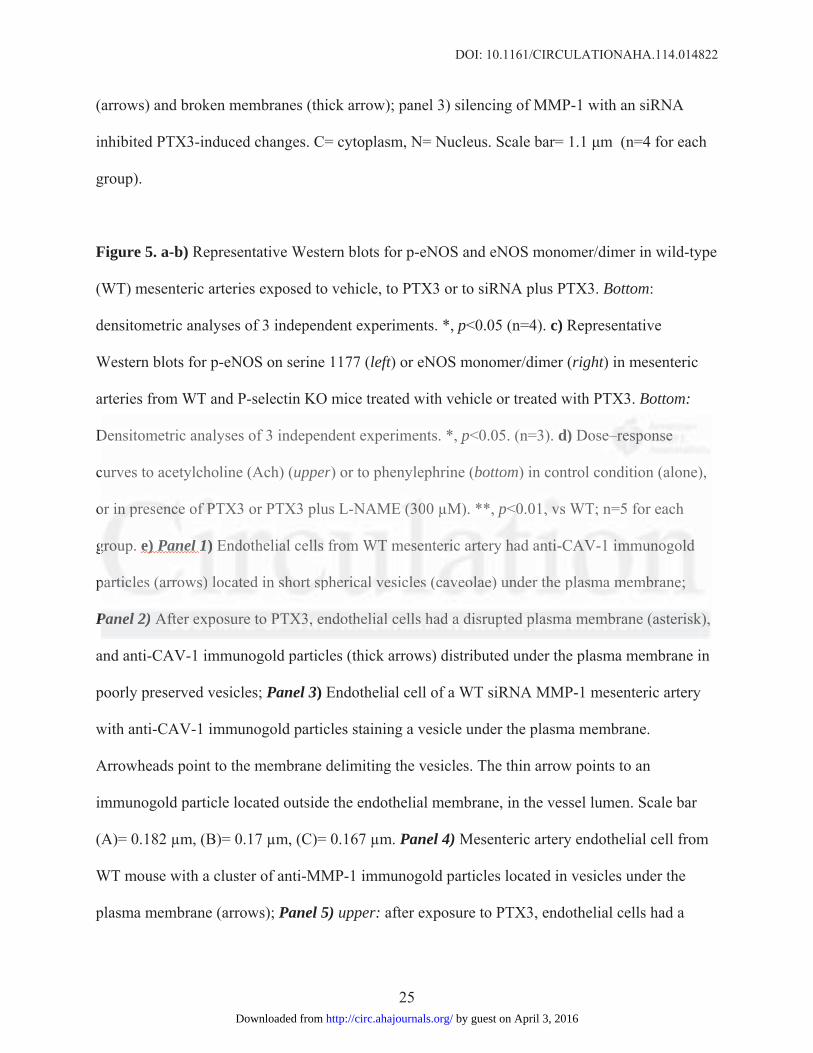
DOI: 10.1161/CIRCULATIONAHA.114.014822
25
(arrows) and broken membranes (thick arrow); panel 3) silencing of MMP-1 with an siRNA
inhibited PTX3-induced changes. C= cytoplasm, N= Nucleus. Scale bar= 1.1 m (n=4 for each
group).
Figure 5. a-b) Representative Western blots for p-eNOS and eNOS monomer/dimer in wild-type
(WT) mesenteric arteries exposed to vehicle, to PTX3 or to siRNA plus PTX3. Bottom:
densitometric analyses of 3 independent experiments. *, p<0.05 (n=4). c) Representative
Western blots for p-eNOS on serine 1177 (left) or eNOS monomer/dimer (right) in mesenteric
arteries from WT and P-selectin KO mice treated with vehicle or treated with PTX3. Bottom:
Densitometric analyses of 3 independent experiments. *, p<0.05. (n=3). d) Dose–response
curves to acetylcholine (Ach) (upper) or to phenylephrine (bottom) in control condition (alone),
or in presence of PTX3 or PTX3 plus L-NAME (300 μM). **, p<0.01, vs WT; n=5 for each
group. e) Panel 1) Endothelial cells from WT mesenteric artery had anti-CAV-1 immunogold
particles (arrows) located in short spherical vesicles (caveolae) under the plasma membrane;
Panel 2) After exposure to PTX3, endothelial cells had a disrupted plasma membrane (asterisk),
and anti-CAV-1 immunogold particles (thick arrows) distributed under the plasma membrane in
poorly preserved vesicles; Panel 3) Endothelial cell of a WT siRNA MMP-1 mesenteric artery
with anti-CAV-1 immunogold particles staining a vesicle under the plasma membrane.
Arrowheads point to the membrane delimiting the vesicles. The thin arrow points to an
immunogold particle located outside the endothelial membrane, in the vessel lumen. Scale bar
(A)= 0.182 μm, (B)= 0.17 μm, (C)= 0.167 μm. Panel 4) Mesenteric artery endothelial cell from
WT mouse with a cluster of anti-MMP-1 immunogold particles located in vesicles under the
plasma membrane (arrows); Panel 5) upper: after exposure to PTX3, endothelial cells had a
Densitometric analyses of 3 independent experiments. *, p<0.05. (n=3). d) Dosee–––resese popoponssnsee e
curves to acetylcholine (Ach) (upper) or to phenylephrine (bottom) in control condition (alone),
orr iiinnn prprpresesenenencecee of f f PPTPTX3 or PTX3 plus L-NAMEE (((303000 μM). **, p<0<0.01,1, vvvss WT; n=5 for each
ggrouuup.p e) Panenelll 111)) EEndndn otototheheheliliialalal ccelelllslsls ffrroommm WWTWT meesseenteeeririicc aarrteeeryry hhaadad aannntii--CACAAVV-V-11 imimimmumumunonoogogooldldd
papartrtrticiccleleles s (a(a(arrrrrrowowss)s) lllococaaateeded iin shshs orororttt spspspheheeririicacacall l veveesisis clcllesss ((cccaveveveoloolaeaee) )) uunundedederr tththee e plpllasasa mamam mmmememembbrbrananee;e;
Panel 2) Afttererer eeexpxpxpososo ururu e ee tooo PPPTXTXTX3,3,3, eeendnn ototothehehelililialalal cccelelllslss hhhaad d a a a didid srsrs upupuptetet d d d plplplasasasmamama mmmememembrbrbrananane e (asterisk),,
by guest on April 3, 2016http://circ.ahajournals.org/Downloaded from

DOI: 10.1161/CIRCULATIONAHA.114.014822
26
greater number of anti-MMP-1 gold particles located inside the cells, on the plasma membrane
and on vesicles under the plasma membrane (bottom) (arrows); Panel 6) endothelial cell from
siRNA MMP-1 mice showing MMP-1 immunogold particles located in vesicles continuous with
the plasma membrane (arrows); n=3 for each experimental condition. f) Endogenous eNOS
immunoprecipitated with CAV-1 in mesenteric artery lysate from C57BL/6N: vessels were
exposed to vehicle alone, to PTX3 alone or were transfected with siRNA MMP-1 and exposed to
PTX3. Extracts were immunoprecipitated for CAV-1 and then probed with anti-eNOS or anti-
CAV-1 antibodies; n=3.
Figure 6. Mean arterial pressure (MAP) of wild-type mice (a) and P-selectin KO mice (b)
treated with vehicle or with PTX3. *, p<0.05 vs WT plus vehicle; n=5 for each treatment. c)
Dose–response curves to acetylcholine (Ach) of phenylephrine-precontracted mesenteric arteries
from WT and P-selectin-/- mice after in vivo injection of PTX3. **, p<0.01 vs WT + vehicle, P-
selectin-/- + vehicle and P-selectin-/- + PTX3; n=5 for each group. d) In vivo effect of PTX3
administration on the ultrastructure of endothelial cells: panels 1 and 3) representative
micrographs of endothelial cells from WT and P-selectin-/- mice under basal conditions; panels 2
and 4) after PTX3 administration. In panel B, note a large cytoplasmic vacuole (V) and the
broken plasma membrane (arrow) of an endothelial cell from a PTX3-treated WT mouse; no
subcellular alterations are encountered in endothelial cells from mice belong to the other
experimental groups. C= cytoplasm, N= nucleus, V= vacuole. Scale bar=0.45 m; n=4 for each
group.
Figure 7. a) Boxplots representing PTX3 (left), P-selectin (middle) and MMP-1 (right)
Figure 6. Mean arterial pressure (MAP) of wild-type mice (a) and P-selectin KOO O mimim ceee (((bbb) ) )
reated with vehicle or with PTX3. *, p<0.05 vs WT plus vehicle; n=5 for each treatment. c)
DoDosesese–r–r–resespopoponsnse cucucurrvrves to acetylcholine (Ach) offf phphp eenylephrine-prprrece onnntrtrtraaacted mesenteric arteriesd
ffrrommm WT and P-P-P seseleectcttininn-/--/--/- mmmiciciceee afaffteteterr r innn vvvivoo innnjeccttioon ofofof PPTXTXTX3.3 ****,*, pp<0<0< .0011 vsvsvs WWTT T +++ veveehihiiclcle,ee, PPP-
eelelelectctctininin-/- +++ vvvehehiciccleee aanndnd PPP-s-selellecece tititinnn-/--/-/- ++ PTPTPTX3X3X3; n=n=n=55 fooor r eeeachchch gggroooupupu .. d)d)d) IInn n viviivovo eeffffffecectt t oofof PPPTXXX33
administrationonn ooon n thththe e ululultrt asasstrtrtrucucu tututurerer ooof enenendododothththeleleliaiaial ll cececelllls:: papapanenenelslsls 111 andndnd 333))) rrrepepepreresesesentntntatatativivive
by guest on April 3, 2016http://circ.ahajournals.org/Downloaded from

DOI: 10.1161/CIRCULATIONAHA.114.014822
27
distributions in hypertensive and control subjects. Each boxplot gives for each distribution, from
bottom to top, the values corresponding to: lower non-outlier bound, 25th percentile, median,
75th percentile and upper non-outliers bound. Dots represent single outlier measures. b) Receiver
Operating Characteristic curves corresponding to PTX3 (in red), SEL-P (in green) and MMP-1
(in blue). The legend gives the AUROC values and 95% Confidence Interval (95% CI) of the
three analyzed variables.
by guest on April 3, 2016http://circ.ahajournals.org/Downloaded from

Figure 1 by guest on April 3, 2016http://circ.ahajournals.org/Downloaded from

Figure 2 by guest on April 3, 2016http://circ.ahajournals.org/Downloaded from

Figure 3 by guest on April 3, 2016http://circ.ahajournals.org/Downloaded from

Figure 4 by guest on April 3, 2016http://circ.ahajournals.org/Downloaded from

Figure 5 by guest on April 3, 2016http://circ.ahajournals.org/Downloaded from

Figure 6 by guest on April 3, 2016http://circ.ahajournals.org/Downloaded from

Figure 7 by guest on April 3, 2016http://circ.ahajournals.org/Downloaded from

Trimarco, Giuseppe Matarese, Francesco Fornai and Carmine VecchioneMalovini, Luigi Formisano, Vincenzo Gigantino, Michele Madonna, Annibale A. Puca, BrunoMariateresa, Giusy Amodio, Paolo Remondelli, Carmine Del Giudice, Raffaele Izzo, Alberto
Albino Carrizzo, Paola Lenzi, Claudio Procaccini, Antonio Damato, Francesca Biagioni, AmbrosioPentraxin 3 Induces Vascular Endothelial Dysfunction Through a P-selectin/MMP-1 Pathway
Print ISSN: 0009-7322. Online ISSN: 1524-4539 Copyright © 2015 American Heart Association, Inc. All rights reserved.
is published by the American Heart Association, 7272 Greenville Avenue, Dallas, TX 75231Circulation published online March 6, 2015;Circulation.
http://circ.ahajournals.org/content/early/2015/03/06/CIRCULATIONAHA.114.014822World Wide Web at:
The online version of this article, along with updated information and services, is located on the
http://circ.ahajournals.org/content/suppl/2015/03/06/CIRCULATIONAHA.114.014822.DC1.htmlData Supplement (unedited) at:
http://circ.ahajournals.org//subscriptions/
is online at: Circulation Information about subscribing to Subscriptions:
http://www.lww.com/reprints Information about reprints can be found online at: Reprints:
document. Permissions and Rights Question and Answer available in the
Permissions in the middle column of the Web page under Services. Further information about this process isOnce the online version of the published article for which permission is being requested is located, click Request
can be obtained via RightsLink, a service of the Copyright Clearance Center, not the Editorial Office.Circulation Requests for permissions to reproduce figures, tables, or portions of articles originally published inPermissions:
by guest on April 3, 2016http://circ.ahajournals.org/Downloaded from

1
Pentraxin 3 induces vascular endothelial dysfunction through a P-selectin/MMP-1 pathway
Albino Carrizzo1, M.Sc, Paola Lenzi2, Ph.D, Claudio Procaccini3, Ph.D, Antonio Damato1, B.Sc,
Francesca Biagioni1, Ph.D, Ambrosio Mariateresa1, B.Sc, Giusy Amodio4,5, Ph.D, Paolo
Remondelli5, Ph.D, Carmine Del Giudice6, M.Sc, Raffaele Izzo6, M.D, Alberto Malovini7, Ph.D,
Luigi Formisano8, Ph.D, Vincenzo Gigantino3,9, M.Sc, Michele Madonna1, DVM, Ph.D, Annibale
A. Puca5,10, M.D, Bruno Trimarco6, M.D, Giuseppe Matarese5,10, M.D, Francesco Fornai1,2, Ph.D,
M.D, Carmine Vecchione1,5, M.D.
1 IRCCS Neuromed, Pozzilli (IS), Italy
2 University of Pisa, Department of Human Morphology and Applied Biology, Pisa, Italy
3 Laboratorio di Immunologia, Istituto di Endocrinologia e Oncologia Sperimentale, (IEOS-CNR),
Consiglio Nazionale delle Ricerche c/o Dipartimento di Medicina Molecolare e Biotecnologie
Mediche, Università di Napoli “Federico II”, Napoli, Italy
4Università degli Studi di Salerno, Department of Pharmaceutical Sciences, Fisciano (Salerno), Italy
5Università degli Studi di Salerno, Medicine and Surgery, Baronissi (Salerno), Italy
6 Department of Clinical Medicine, Cardiovascular and Immunological Sciences, “Federico II”
University of Naples, Italy
7 University of Pavia, Department of Industrial and Information Engineering, Pavia, Italy
8 Department of Science and Technology, University of Sannio, Benevento, Italy
9 Pathology Unit, "Istituto Nazionale Tumori, IRCCS, Fondazione Pascale", Naples, Italy.
10 IRCCS Multimedica, Milan, Italy
Corresponding author:
Prof. Carmine Vecchione, MD

2
Vascular Physiopatology unit, IRCCS Neuromed, 86077 Pozzilli (IS), Italy
Università degli Studi di Salerno, Dipartimento di Medicina, Via S. Allende, 84081 Baronissi (SA),
Italy.
SUPPLEMENTAL MATERIAL
Supplemental Material and Methods
Proteins and reagents. Recombinant human PTX3 and biotinylated PTX3 were obtained as
previously described 1. Recombinant Serum amyloid protein (SAP) and C-reactive protein (CRP)
were obtained from Sigma. MMP-1 and MMP-9 siRNA was obtained from Santa Cruz
Biotechnology, Inc. Control (non-silencing) siRNA was purchased from QIAGEN. Sepharose A
was purchased from Sigma. Horseradish peroxidase (HRP)-labeled anti-rabbit fragment
immunoglobulin, HRP linked streptavidin, and enhanced chemiluminescence for Western blotting
detection reagent were purchased from Amersham Biosciences. OPTI-MEM, Lipofectamine LTX
reagent, and FBS were purchased from Invitrogen.
Animals. Fcγ-R- and P-selectin-deficient mice were obtained from Instituto Humanitas. Procedures
involving animals and their care conformed to institutional guidelines. All effort was made to
minimize the number of animals used and their suffering.
Vascular reactivity study. Second-order branches of the mesenteric arterial tree were removed
from C57BL6 mice and from FCγR- and P-selectin-knock-out mice for vascular studies performed
as described previously 2. Briefly, vessels were placed in a wire or pressure myograph system filled
with Krebs solution maintained at pH 7.4 at 37°C. Adventitial fat was carefully removed and
arteries were cut into rings (̴ 2-mm thickness). The rings were suspended by two tungsten wires (25-
µm in diameter) and mounted with 0.3 g of resting tension in a wire myograph system (5-mL

3
chamber size; DMT Danish Myosystem). Isometric tension was measured using a force transducer
coupled to a data acquisition system. After an equilibration period of 60 minutes, rings were pre-
contracted with phenylephrine (1 ×10⁻⁹ to 10–5M) until a plateau was reached. Vessels were then
washed, and this was repeated at least three times in order to stabilize the tissue. Endothelium-
dependent and -independent relaxations were assessed by measuring the dilatory response of
mesenteric arteries to cumulative concentrations of acetylcholine (from 10-9M to 10-5M) or
nitroglycerine (from 10-9M to 10-5M), respectively, in vessels pre-contracted with phenylephrine at
a dose necessary to obtained a similar level of pre-contraction in each ring (80% of initial KCl-
evoked contraction). Some mesenteric arteries mounted on a pressure myograph were transfected
with 20 μg of MMP-1 siRNA or with scrambled siRNA. In particular , siRNA was diluted into 200
mL Opti-MEM I. An equal volume of PLUS reagent was added. The mixture was incubated for 5
min. Lipofectamine LTX was added, and the complexes were allowed to form by incubation for 25
minutes3. Subsequently, 200 mL of complex were injected into P1-way (into vessel) of pressure
myograph and then vessels were perfused at 100 mmHg for 1 hour and then at 60 mmHg for 5
hours as previously described3.
Blood pressure Measurement
Two days before testing PTX3 on blood pressure, mice were anesthetized with 0.03 mL of a 2:1
mixture of ketamine (100 mg/mL IM, Aveco Co) and xylazine (20 mg/mL IM, Miles Inc) and
placed on an operating surface maintained at 38°C. A polyethylene catheter (PE-10; Becton
Dickinson, Sparks) was inserted into a femoral artery as previously described4.
The surgery was performed in a uniform fashion by a single investigator (A.D.) between 8 AM
and noon. The catheter was filled with heparinized saline solution (100 μU/ml) and exteriorized
subcutaneously at the interscapular area. It was connected to a pressure transducer (WPI, TBM4M)
and arterial blood pressure was monitored and recorded on a computer system (WPI, Trans Bridge
4M). After surgery, mice were individually housed in cages and allowed to recover. Direct

4
intrafemoral arterial pressure was measured in conscious, freely moving mice after an overnight fast
as previously described4.
Intra-arterial blood pressure was evaluated in WT and P-selectin KO mice under basal
conditions and after PTX3 injection (100µg, intraperitoneally). Mice used as controls were treated
in a similar way, but with PTX3 substituted with the vehicle alone. Basal blood pressure was
evaluated also in WT and PTX3 KO mice for one week. At the end of blood pressure
measurements, some vessels were excised for vascular reactivity, western blot or morphological
analyses.
Immunoprecipitation. Mesenteric artery homogenates were prepared in 5 mM Tris-HCl buffer, pH
7.5, containing 1 mM CaCl2, 1 mM MgCl2, and 1 mM NaHCO3. Samples containing 1 mg of total
protein were lysed for 40 min at +4°C with lysis buffer at pH 7.5 containing 50 mM Tris-HCl, 150
mM NaCl, 1% NP-40, 1 mM Na2P2O7, 1 mM NaF, 1 mM EDTA, 2 mM NaVO4, 0.1 mM PMSF,
and complete protease inhibitor cocktail (Roche), and centrifuged for 15 min at 20,000 g at 4°C.
Supernatants were cleared with protein A/G–agarose beads (Santa Cruz Biotechnology, Inc.) for 2
hours at 4° and incubated or with anti P-selectin or with anti-Caveolin-1 antibody, followed by
precipitation with protein A/G–agarose beads for 1 hour at 4°C. The beads were washed three times
with lysis buffer and two times with PBS, and then analyzed by immunoblotting.
Gel electrophoresis and immunoblotting. Protein extraction was performed from four pooled
mesenteric arteries. Proteins were separated on 10% SDS-PAGE and electroblotted onto
nitrocellulose transfer membrane (Amersham) for 1.5h at 250 mA. Immunoblots were incubated
with the appropriate primary antibodies, followed by incubation with HRP-labeled secondary
antibodies, and visualized using ECL prime (Amersham & Co.). Molecular mass markers were
prestained protein standards obtained from Bio-Rad laboratories. For quantitative comparisons of
chemiluminescence between the lanes, the same amounts of total protein or equal amounts of
immunoprecipitate were loaded in each lane. All preparations were performed three times. Values

5
of all experiments were used to calculate mean values and standard deviation. The
chemiluminescence quantification was performed using ImageJ software. Group comparisons were
made by paired t test.
Low temperature SDS-PAGE.
Low-temperature SDS-PAGE (LT-PAGE) was performed for detection of SDS-resistant eNOS
dimer and monomer, as described previously5.
Cell cultures. HUVEC (Lonza) were cultured in EBM-2 media (Lonza). HUVEC were cultured at
37°C in 5% CO2/95% air by standard methodologies in 25-cm2 T tissue culture flasks (50 ml
capacity) (Falcon, Becton Dickinson Labware) in the presence of endothelial cell growth
supplement. In general, HUVEC were used for our experiments after 3–5 passages. For starvation
experiments, serum- and growth factor-free medium was used. For siRNA-mediated gene
knockdown, HUVECs were grown to 60% to 70% confluence and transfected with Lipofectamine
LTX (Invitrogen) according to the protocol of the manufacturer. As control, a scrambled-RNA was
used. 24 h after siRNA transfection, cells were washed with PBS and then left for 2h in serum- and
growth factor-free medium before of each treatment.
Measurement of MMP-1 activity.
HUVEC were cultured at 37°C in 5% CO2/95% air by standard methodologies in 100 mm dishes to
achieve 70–80% of confluence. Treatment with PTX3 (2 and 20 ng/mL) and P-Selectin (50 and 100
ng/mL) was performed in serum- and growth factor-free medium. Cells were treated for 1 hour; at
the end of treatments, medium was collected and concentrated 10X using Ultra-Amicon 10K
(Sigma). MMP-1 activity was measured with a TECAN Infinite 200PRO, using SensoLyte® Plus
520 MMP-1 Assay Kit *Fluorimetric and Enhanced Selectivity* (Anaspec) by monitoring at
excitation/emission wavelengths 490 nm/520 nm at the end-point, following the manufacturer’s
protocol.
Measurement of NO

6
Total NO content in the supernatants of HUVEC cultures was measured in an additional
experiment. Cells were grown in 100mm dishes and supernatants collected after stimulation with
acetylcholine (100 µM for 20 minutes), used to induce NO production. In some experiments, cells
were pretreated with PTX3 (20ng/mL) or with siRNA MMP1 plus PTX3 (20ng/mL). The nitric
oxide concentrations in the supernatant was assayed as nitrite, the stable breakdown product of NO,
by a chemiluminescence detector (Sievers 280i NO Analyzer), as previously described6.
Transmission Electron Microscopy (TEM). HUVEC pellets and mesenteric artery from control
and KO mice (5 mm3 samples) were processed for ultrastructural analysis at transmission electron
microscopy by fixing in 2% paraformaldehyde and 0.1% glutaraldehyde in 0.1 M PBS, pH 7.4, for
90 min; after washing in PBS, samples were post-fixed in 1% OsO4 for 2 hours at 4°C. Then,
samples were dehydrated in ethyl alcohol and embedded in Epon–araldite. Ultrathin sections (50
nm thin) were stained with uranyl acetate and lead citrate and finally examined under an Jeol Jem
100SX transmission electron microscope (Jeol, Tokyo, Japan).
For immunocytochemistry, mesenteric arteries were processed as described for plain TEM.
Post-embedding immunoelectron microscopy was performed. Ultra-thin sections (50 nm) were de-
osmicated with aqueous solution of sodium metaperiodate and incubated for 24 h at 4°C with
primary antibodies against CAV-1 or MMP1 (Sigma-Aldrich) diluted 1:10 in PBS containing 0.2%
saponin (Sigma) and 1% normal goat serum. Then, sections were incubated with gold-conjugated
secondary antibodies (20-nm gold particles) diluted 1:10 in buffer solution for 1 h at 25°C. Finally,
sections were fixed with 1% glutaraldehyde, stained with uranyl acetate and lead citrate, and
examined under a Jeol JEM SX 100 electron microscope. Control sections were obtained by
omitting the primary antibody and incubating with only the secondary antibody.
Evans blue Dye
Vascular structure alterations on mesenteric arteries were assessed also using Evans Blue Dye
(Sigma) according to the method previously described with minor modification7. Briefly, 50 mL of

7
solution containing 2% Evans blue diluted in saline was injected into the tail vein 10 minutes before
euthanasia, followed by fixation with a perfusion of 4% paraformaldehyde for 10 minutes. Images
were captured with an LSM 510 microscope (Carl Zeiss MicroImaging).
Immunofluorescence analysis.
On vessels:
Mesenteric arteries were treated with biotinylated PTX3 (20ng/mL) for 45 minutes and then
cryopreserved in killik medium (Bioptica). Vessels were then processed in a cryostat to obtain 10-
µm sections disposed on polarized slides (Carlo Erba). Sections were then extensively washed and
treated with fluorescence streptavidin (1:500) and incubated for 2 hours at room temperature.
Different sections of mesenteric arteries from WT and P-selectin KO mice were processed with
different primary antibodies (CD31, a-SMA) to evaluate the integrity of endothelial or smooth
muscular layer, and then with respective secondary antibodies. Finally, the sections were visualized
with a Zeiss 2 fluorescence microscope.
Another experimental series of vessels was processed for immunohistochemical staining on
10 μm sections of cryopreserved mouse mesenteric artery in order to evaluate F4-80 expression, a
well-characterized macrophage marker. All slides were stained at same time. Mesenteric arteries
treated with LPS was considered as positive control. The slides were rinsed with TBS and the
endogenous peroxidase inactivated with 3% hydrogen peroxide. After protein block with BSA (5%
in PBS 1), the sections were incubated for one hour with antibody against F4-80 (NBPB2-12506
Rabbit Ab, Novus Biologicals, diluted 1: 20). The sections were rinsed in TBS and incubated for 20
minutes with Novocastra biotinylated secondary antibody (RE7103), a biotin-conjugated secondary
antibody formulation that recognizes mouse and rabbit immunoglobulines. Then, the sections were
rinsed in TBS and incubated for 20 minutes with Novocastra Streptavidin-HRP (RE7104). The
peroxidase reactivity was visualized using a 3,3'-diaminobenzideine (DAB). Finally, the sections
were counterstained with hematoxylin and mounted. Results were interpreted using a light

8
microscope LSM 510 (Carl Zeiss MicroImaging). Immunohistochemistry expression was evaluated
by a pathologist.
On Cells:
HUVEC were seeded on glass coverslips and treated with 20 ng/ml PTX for the indicated time. At
the end of treatments, coverslips were washed in PBS, fixed 10 min in PBS–4% paraformaldehyde
and incubated 30 min in PBS containing 0.5% BSA and 50 mM NH4Cl at room temperature
without any permeabilization. For the staining of P-selectin and PTX3, coverslips were incubated
for 1 hour at room temperature with 10 µg/ml mouse anti-human CD62P (BD Biosciences) and/or
20 µg/ml PTX3-specific rabbit polyclonal antibody (Sigma-Aldrich). Alexa 594- and Alexa 488-
coniugated secondary antibodies (Jackson Immunoresearch) were used. The staining of CD31 was
performed for 1 hour at room temperature with a 1:25 dilution of rat polyclonal CD31 specific
antibody. Biotinylated anti-rat secondary antibody and Alexa 488-labelled streptavidin were used.
Coverslips were mounted with the Prolong AntiFade kit. Images were collected with a laser
scanning confocal microscope (LSM 510; Carl Zeiss MicroImaging) equipped with a plan Apo
63X, NA 1.4 oil immersion objective lens.
Colocalization measurements. Per cent colocalization of the fluorescence signals was quantified on
a minimum of 30 different cells. 8-bit thresholded images were analyzed for Manders colocalizing
coefficients with the LSM 510 4.0 SP2 software. The per cent colocalization is relative to the in-
focus z-plane stack shown in the immunofluorescence panels.
Flow cytometric analysis. HUVEC were treated with PTX3 (20 ng/ml) or vehicle for 2 hours and
then incubated with anti-P-selectin antibody (Santa Cruz) (diluted 1/25 in PBS) for 30 minutes at 4°
C. Cells were then washed with PBS and stained with PE-conjugated goat anti-mouse IgG (Sigma-
Aldrich) (diluted 1/100 in PBS) for 30 minutes at 4°C, and then followed by PBS washes. Flow
cytometry measurements were performed with a FACSCanto (Becton-Dickinson, San Diego, CA)
and analyzed by Flow-Jo software (Tree Star Inc., Ashland, OR).

9
Human samples and ELISA. Thirty-one patients with hypertension (defined as DBP ≥90 mmHg
and/or SBP ≥140 mmHg or on the basis of use of anti-hypertensive medication) and 21 healthy
control subjects were recruited from a specialist center at the University of Naples Federico II, Italy.
The study was approved by an institutional review committee and the subjects gave their informed
consent. Clinical data from each patient were collected. Patients with lung disease, heart disease,
acute or chronic inflammatory disease, recent infection, presence of neoplasia, concomitant
treatment with steroids or nonsteroidal anti-inflammatory drugs, or alcoholism were excluded.
Blood pressure (BP) was measured with the patient seated with the use of a validated automatic
device. Three consecutive readings were recorded, and the mean was used for the analysis.
Characteristics of the patients are given in Table 1.
Venous blood was collected in EDTA vacuum containers and stored at −80°C. PTX3, P-
selectin and MMP-1 were then measured using a high-sensitivity, enzyme-linked immunosorbent
assay (ELISA) system for human plasma. Preparation of plates and sandwich ELISA were
performed according to the manufacturers’ instructions for each specific kit (PTX3 Human ELISA
Kit, Abnova # KA1007; CD62P P-Selectin Human ELISA Kit, abcam # ab100631; MMP1 Human
ELISA Kit, abcam # ab100603).
Supplemental Results
Macroscopic evaluation of PTX3-evoked vascular changes
Our data demonstrate that the target of PTX3’s vascular action is the endothelium and that it
induces dysfunction of resistance vessels. To further support this data, macroscopic images of
changes in vascular structure were assessed using Evans Blue Dye (EBD) staining and
immunohistochemistry using labeled markers for endothelium and smooth muscle (CD31, a-SMA).
We found that only PTX3-treated vessels acquired EBD (Fig. S3a) and showed a significant

10
reduction of CD31, indicating a specific alteration of the endothelial layer, since no differences
were observed in the smooth muscle marker (Fig. S3b).
SAP induces an impairment of eNOS phosphorylation
Considering the endothelial dysfunction induced by SAP at the vascular level, we performed
Western blotting on resistance vessels from WT and P-selectin deficient mice exposed to SAP, to
evaluate the phosphorylation status of eNOS. Interestingly, the impairment of eNOS
phosphorylation evidenced in vessels from WT mice was absent in P-selectin knockout mice (Fig
S5). These data were coherent with the results on vascular reactivity.
PTX3-evoked vascular damage is not associated with macrophage recruitment
We studied the possible recruitment of immune cells in vessels from WT and P-selectin KO mice
treated with PTX3. Our histologic and immunohistochemistry analyses clearly demonstrate that in
both WT and P-selectin KO mice treated with PTX3, there was no positivity for F4-80, an
extensively used marker for macrophage cells, compared to positive control vessels treated with
LPS, in which each sections showed a well-marked cell. We conclude that PTX3-evoked vascular
action is not associated with a recruitment of macrophages to the vessel wall (Fig. S6).
Effect of PTX3 on HUVEC
a) Effect of PTX3 on P-selectin expression
Confocal microscopy revealed that treatment with PTX3 (20ng/mL for 0–60 minutes) induced
expression of P-selectin on cell membranes (Fig. S8a). To confirm this, we performed flow
cytometry experiments, finding that exposure to exogenous PTX3 up-regulated P-selectin
expression in HUVEC, as indicated by a rightward shift in the output curve (increased percentage of
positive cells and higher mean fluoresce intensity) (Fig. S8b).
b) Effect of PTX3 on NO production

11
Western blotting revealed a significant increase of MMP-1 expression in cells treated with PTX3
(Fig. S9a). In addition, spectrofluorimetry detected a significant increase in MMP-1 activity (Fig.
S9b). As observed at the vascular level, PTX3 impaired NO signaling also in isolated endothelial
cells, as evidenced by reduced dimerization of eNOS and phosphorylation on serine 1177 (Fig.
S9c). At the function level, PTX3 markedly decreased nitric oxide production when compared to
vehicle (Fig. S9d). Inhibition of MMP-1 by siRNA MMP1 hampered PTX3’s deleterious effect on
nitric oxide signaling, as revealed by a complete rescue of dimerization and phosphorylation of
eNOS in serine 1177, effects associated with an enhanced production of nitric oxide (Fig. S9d).
To clarify whether P-selectin, per se, was able to modulate MMP-1 signaling, we treated
HUVEC with P-Selectin at different dosages. Although P-selectin did not change MMP-1
expression, it was able to significantly enhance its activity. This effect was lower compared to that
observed in the presence of PTX3 (Fig. S9a, S9b).
Hypertensive patients have elevated plasma levels of PTX3, P-selectin and MMP-1
Univariate tests showed that PTX3 (ng/mL), P-selectin (ng/mL) and MMP-1 (pg/mL) values were
significantly higher in hypertensive subjects with respect to controls (p < 2 x 10⁻⁸) (Table S1). It is
important to underline that PTX3 was the strongest predictor of hypertension, reaching the highest
p-value, as reported in Table S1. The second goal of the analysis was to identify the most
informative threshold for each variable to discriminate hypertensive subjects from controls. To this
aim, the dataset was randomly split into a training set (37 subjects, 70% of the whole sample) and a
test set (15 subjects, the remaining 30% of the data). Random sampling was performed with
stratification, so that the proportion of cases and controls observed in the whole sample (60% vs.
40%) was maintained also in the two sub-cohorts. Thus, the training set was used to identify the
most discriminative threshold for each variable, as described in Methods Section. The test set was
then used to evaluate the discriminative performances corresponding to the variables discretized
according to the identified threshold. Results are reported in Table S2 and show that PTX3

12
discretized according the threshold corresponding to ≥ 2.508 reaches the highest MCC, sensitivity
and F-Measure among the analysed variables.
Supplemental Figures and Figure Legends.
Figure S1. Effects of PTX3 on femoral arteries and aorta. a) Dose–response curves of
phenylephrine-precontracted femoral arteries from C57BL/6N mice to acetylcholine after 45
minutes of PTX3 treatment at different dosage (2, 20, 200 ng/mL). b) Dose–response curves of
phenylephrine-precontracted aorta rings from C57BL/6N mice to acetylcholine after 45 minutes of
PTX3 treatments at different dosage (2, 20, 200 ng/mL). *, p<0.05; **, p<0.001; ***, p<0.0001 vs
control or 2 ng/mL. (n=8 both for femoral arteries and aorta. c) Immunohistochemical staining,
using fluorescent streptavidin, of aorta sections from C57BL/6N mice treated with vehicle or with
biotinylated-PTX3. Scale bar, 100 μm. (n=4)

13
Figures S2. a) Dose–response curves to acetylcholine (Ach) in phenylephrine-precontracted
mesenteric arteries from wild-type (WT) and P-Selectin KO (P-sel-/-
) mice: vessels were exposed to
vehicle alone or to SAP for 45 minutes (200ng/mL); n=4 for each group. b) Dose–response curves
to Ach of phenylephrine-precontracted mesenteric arteries from WT and P-Selectin KO mice:
vessels were exposed to vehicle only or to CRP for 45 minutes (25mg/mL) n=4 for each group. ***,
p<0.0001 vs WT. ###, p<0.0001 vs WT.

14
Figure S3. a) Representative images of Evans blue staining in mesenteric artery from WT and P-
Selectin KO mice treated with vehicle or with PTX3. (n=3 for each group); b)
Immunohistochemical staining of mesenteric arteries from WT and P-Selectin KO mice using a
labeled endothelial marker, CD31 or a labeled smooth muscle marker, α-SMA. DAPI was used to
stain nuclei. Scale bar 100 μm. (n=3 for each group).

15
Figure S4. a) Dose–response curves to acetylcholine (Ach) (upper) or to phenylephrine (bottom) in
mesenteric arteries from wild-type mice in the basal condition and after exposure to L-NAME (300
µM). **, p<0.001 vs WT; n=4 for each group.
Figures S5. a) Representative Western blots for p-eNOS on serine 1177 in mesenteric arteries from
WT and P-selectin KO mice exposed to vehicle or to SAP. Bottom: Densitometric analyses of 3
independent experiments. *, p<0.05. (n=3).

16
Figure S6. a) Histologic and immunohistochemistry analyses of F4-80 expression in mouse
mesenteric arteries; positive F4-80 cytoplasmic expression was found only in LPS-treated vessel
(C+), while WT and P-selectin KO mice vessels, treated with vehicle or with PTX3, were negative
for the macrophage marker (n=3). Scale bar 100 μm.

17
Figure S7. Representative original traces obtained during the measurement of blood pressure with a
catheter injected into mouse femoral arteries. a) Graphs of the effects on blood pressure in wild-
type mice after injection with vehicle (left) or with PTX3 (right). b) Graphs of the effects on blood
pressure in P-selectin knockout mice (P-selectin-/-
) after injection with vehicle (left) or with PTX3
(right). c) Mean arterial pressure (MAP) measured by indwelling femoral catheters in WT and
PTX3-knockout mice; n=5 for each group.

18
Figure S8. a) HUVEC seeded on glass coverslips were either untreated (0 minutes) or treated for
60 minutes with PTX3 (20ng/mL). Colocalization is shown in yellow in the merged panels, and
quantified as Manders colocalizing coefficients in the histogram below. **p˂0.05. Cells were
processed for immunofluorescence with P-selectin (P-sel) and CD-31 (a membrane-specific)
antibodies; n=4. b) Cytofluorimetric analysis of HUVEC treated for 1 hour with 20ng /mL PTX3.
The plot gives the increment as a percentage of P-selectin on the membrane of the human
endothelial cells; n=4. MFI, mean fluorescence intensity.

19
Figure S9. a) Upper: representative Western blots for MMP-1 expression in HUVEC without
treatment (untreated), treated for 1 hour with PTX3 (2 or 20ng/mL) or with P-selectin (50 or
100ng/mL). Bottom: Densitometric analyses of 3 independent experiments. *, p<0.05. b) MMP1
activity measured in HUVEC without treatment (untreated), treated for 1 hour with PTX3 (2 or
20ng/mL) or with P-selectin (50 or 100ng/mL). Data of 3 independent experiments. *, p<0.05; **,
p<0.001; ***, p<0.0001. c) representative Western blots for eNOS monomers/dimers (left) and for

20
p-eNOS on serine 1177 (right) in HUVEC treated with vehicle, with PTX3, or with PTX3 after
siRNA MMP1 transfection. All cells were stimulated with acetylcholine (100µM for 20 minutes).
Bottom: Densitometric analyses of 3 independent experiments. *, p<0.05. d) NO measured in
supernatants of HUVEC treated with vehicle, with PTX3, or with PTX3 after siRNA MMP1
transfection. All cells were stimulated with acetylcholine (100µM). Data are mean±SEM of 3
independent experiments. **, p<0.01).
Supplemental Tables.
Table S1. Discriminative performances of the analyzed variables.
Variable Hypertensive Control p-value AUROC (95% CI)
PTX3 (ng/mL) 3.36 (2.74-4.69) 1.44 (1.11-2.03) 4.01 x 10⁻⁹ 0.985 (0.963-1)
SEL-P (ng/mL) 69.68 ± 11.47 48.84 ± 9.85 8.08 x 10⁻⁹ 0.919 (0.850-0.989)
MMP-1 (pg/mL) 301.7 (239.9-369.86) 96.25 (78.22-123.66) 1.59 x 10⁻⁸ 0.966 (0.927-1)
Variable, analyzed variable; Hypertensive, mean ± SD or median (25th-75th percentiles) of each
variable’s distribution in hypertensive subjects (n = 31); Control, mean ± SD or median (25th-75th
percentiles) of each variable’s distribution in control subjects (n = 21); p-value, probability value
deriving from the Welch’s t-test or Wilcoxon rank-sum test as appropriate; AUROC, Area Under
the Receiver Operating Characteristic curve and corresponding 95% Confidence Interval (CI).

21
Table S2. Discriminative performances of the analyzed variables discretized according to the
identified thresholds.
Variable Threshold Set MCC Sens. Spec. PPV NPV F
PTX3 ≥ 2.508 ng/mL Training 0.850 0.864 1.000 1.000 1.000 0.927
Test 1.000 1.000 1.000 1.000 1.000 1.000
SEL-P ≥ 56.333 ng/mL Training 0.660 0.909 0.733 0.833 0.833 0.869
Test 0.720 0.889 0.833 0.889 0.889 0.889
MMP-1 ≥ 192.028 pg/mL Training 0.850 0.864 1.000 1.000 1.000 0.927
Test 0.870 0.889 1.000 1.000 1.000 0.941
Variable, analysed variable; Threshold, identified threshold; set, training (22 hypertensive subjects
vs. 15 controls) or test (9 hypertensive subjects vs. 6 controls) sets; MCC, Mattew’s Correlation
Coefficient; Sens., sensitivity; Spec., specificity; PPV, Positive Predictive Value; NPV, Negative
Predictive Value; F, F-Measure.
References.
1. Bottazzi B, Vouret-Craviari V, Bastone A, De Gioia L, Matteucci C, Peri G, Spreafico F,
Pausa M, D'Ettorre C, Gianazza E, Tagliabue A, Salmona M, Tedesco F, Introna M and Mantovani
A. Multimer formation and ligand recognition by the long pentraxin PTX3. Similarities and
differences with the short pentraxins C-reactive protein and serum amyloid P component. J Biol
Chem. 1997;272:32817-23.
2. Zacchigna L, Vecchione C, Notte A, Cordenonsi M, Dupont S, Maretto S, Cifelli G, Ferrari
A, Maffei A, Fabbro C, Braghetta P, Marino G, Selvetella G, Aretini A, Colonnese C, Bettarini U,
Russo G, Soligo S, Adorno M, Bonaldo P, Volpin D, Piccolo S, Lembo G and Bressan GM.
Emilin1 links TGF-beta maturation to blood pressure homeostasis. Cell. 2006;124:929-42.
3. Vecchione C, Carnevale D, Di Pardo A, Gentile MT, Damato A, Cocozza G, Antenucci G,
Mascio G, Bettarini U, Landolfi A, Iorio L, Maffei A and Lembo G. Pressure-induced vascular
oxidative stress is mediated through activation of integrin-linked kinase 1/betaPIX/Rac-1 pathway.
Hypertension. 2009;54:1028-34.
4. Lembo G, Vecchione C, Fratta L, Marino G, Trimarco V, d'Amati G and Trimarco B. Leptin
induces direct vasodilation through distinct endothelial mechanisms. Diabetes. 2000;49:293-7.

22
5. Carrizzo A, Puca A, Damato A, Marino M, Franco E, Pompeo F, Traficante A, Civitillo F,
Santini L, Trimarco V and Vecchione C. Resveratrol improves vascular function in patients with
hypertension and dyslipidemia by modulating NO metabolism. Hypertension. 2013;62:359-66.
6. Cudmore MJ, Hewett PW, Ahmad S, Wang KQ, Cai M, Al-Ani B, Fujisawa T, Ma B,
Sissaoui S, Ramma W, Miller MR, Newby DE, Gu Y, Barleon B, Weich H and Ahmed A. The role
of heterodimerization between VEGFR-1 and VEGFR-2 in the regulation of endothelial cell
homeostasis. Nat Commun. 2012;3:972.
7. Lindner V, Fingerle J and Reidy MA. Mouse model of arterial injury. Circ Res.
1993;73:792-6.