Critical role of endothelial P-selectin glycoprotein ligand 1 in chronic murine ileitis
11
The Journal of Experimental Medicine ARTICLE JEM © The Rockefeller University Press $8.00 Vol. 203, No. 4, April 17, 2006 907–917 www.jem.org/cgi/doi/10.1084/jem.20052530 907 The inflammatory bowel diseases (ulcerative colitis [UC] and Crohn’s disease [CD]) are chronic immune-mediated conditions that af- fect more than one million patients in North America and lead to more than $3.6 billion in productivity losses each year (1, 2). The in- flammatory process is characterized by heavy leukocytic infiltration of the intestinal lamina propria (LP), leading to fibrosis and loss of function (3, 4). Although the etiology of the disease remains mostly unknown, modulation of the immune response by immunomodu- lators (e.g., azathioprine, 6-mercaptopurine), corticosteroids, and mAbs to TNF-α improve disease symptoms and outcomes (3–6). Al- though UC affects strictly the large intestine, CD primarily involves the small intestine, pre- dominantly the terminal ileum (chronic ileitis) (3, 4). Thus, the recirculating lymphocyte pool likely possesses a defined repertoire of adhesion molecules and chemokines (“address code”) (7) that allows them to distinguish between the small and large intestine. However, the specific combination of molecules engaged by patho- genic cells to localize specifically to the small intestine has not been identified. The recent characterization of mouse mod- els that spontaneously develops chronic ileitis (i.e., the SAMP1/Yit and SAMP1/YitFc mod- els) enabled us to study the trafficking pathways into the chronically inflamed small intestine (8– 10). SAMP1/YitFc mice develop discontinu- ous, transmural chronic inflammation localized to the small intestine (8, 9) that closely reca- pitulates the human disease (10, 11). The LP is intensely infiltrated by effector T cells, which display an activated phenotype and adoptively transfer disease to SCID mice (9). However, unlike the CD45RB high transfer model (12), lymphocytes from SAMP1/YitFc mice pre- dominantly induce ileitis and not colitis, suggesting an inherent capacity to recirculate Critical role of endothelial P-selectin glycoprotein ligand 1 in chronic murine ileitis Jesús Rivera-Nieves, 1,4 Tracy L. Burcin, 2,3 Timothy S. Olson, 4 Margaret A. Morris, 2,3 Marcia McDuffie, 4,5 Fabio Cominelli, 1,4 and Klaus Ley 2,3,6 1 Digestive Health Center of Excellence, 2 Robert M. Berne Cardiovascular Research Center, 3 Department of Biomedical Engineering, 4 Internal Medicine, 5 Microbiology and 6 Molecular Physiology and Biological Physics, University of Virginia Health Sciences Center, Charlottesville, VA 22908 L-selectin ligands might be relevant for inflammatory cell trafficking into the small intes- tine in a spontaneous model of chronic ileitis (i.e., SAMP1/YitFc mice). Immunoblockade of peripheral node addressin or mucosal addressin cell adhesion molecule 1 failed to ame- liorate ileitis, whereas P-selectin glycoprotein ligand 1 (PSGL-1) neutralization attenuated both the adoptively transferred and spontaneous disease. PSGL-1 was detected in venules of mesenteric lymph node and small intestine by immunohistochemistry and confirmed by real-time reverse transcription polymerase chain reaction and flow cytometry. In addition, reconstitution of wild-type mice with PSGL-1 −/− bone marrow demonstrated that PSGL-1 messenger RNA and PSGL-1 protein expression remained on endothelium, localized within mesenteric lymph node and small intestine. Endothelial PSGL-1 bound P-selectin–IgG and its blockade or genetic deletion altered the recruitment of lymphocytes to the small in- testine, as revealed by intravital microscopy and homing studies. Endothelial expression of PSGL-1 adds a new dimension to the various cellular interactions involved in small intesti- nal recruitment. Thus, the multiple roles of PSGL-1 may explain why targeting this single adhesion molecule results in attenuation of chronic murine ileitis, a disease previously resistant to antiadhesion molecule strategies. CORRESPONDENCE Jesús Rivera-Nieves: [email protected] Abbreviations used: CD, Crohn’s disease; FSC, forward light scat- ter; HEV, high endothelial venule; HRP, horseradish peroxidase; LP, lamina propria; MAdCAM-1, mucosal addressin cell adhesion molecule; MLN, mesenteric lymph node; PNAd, peripheral node addressin; PSGL-1, P-selectin glycoprotein ligand 1; SSC, side light scatter; UC, ulcerative colitis. The online version of this article contains supplemental material.
-
Upload
independent -
Category
Documents
-
view
0 -
download
0
Transcript of Critical role of endothelial P-selectin glycoprotein ligand 1 in chronic murine ileitis

The
Journ
al o
f Exp
erim
enta
l M
edic
ine
ARTICLE
JEM © The Rockefeller University Press $8.00
Vol. 203, No. 4, April 17, 2006 907–917 www.jem.org/cgi/doi/10.1084/jem.20052530
907
The infl ammatory bowel diseases (ulcerative colitis [UC] and Crohn’s disease [CD]) are chronic immune-mediated conditions that af-fect more than one million patients in North America and lead to more than $3.6 billion in productivity losses each year (1, 2). The in-fl ammatory process is characterized by heavy leukocytic infi ltration of the intestinal lamina propria (LP), leading to fi brosis and loss of function (3, 4). Although the etiology of the disease remains mostly unknown, modulation of the immune response by immunomodu-lators (e.g., azathioprine, 6-mercaptopurine), corticosteroids, and mAbs to TNF-α improve disease symptoms and outcomes (3–6). Al-though UC aff ects strictly the large intestine, CD primarily involves the small intestine, pre-dominantly the terminal ileum (chronic ileitis) (3, 4). Thus, the recirculating lymphocyte pool likely possesses a defi ned repertoire of adhesion
molecules and chemokines (“address code”) (7) that allows them to distinguish between the small and large intestine. However, the specifi c combination of molecules engaged by patho-genic cells to localize specifi cally to the small intestine has not been identifi ed.
The recent characterization of mouse mod-els that spontaneously develops chronic ileitis (i.e., the SAMP1/Yit and SAMP1/YitFc mod-els) enabled us to study the traffi cking pathways into the chronically infl amed small intestine (8–10). SAMP1/YitFc mice develop discontinu-ous, transmural chronic infl ammation localized to the small intestine (8, 9) that closely reca-pitulates the human disease (10, 11). The LP is intensely infi ltrated by eff ector T cells, which display an activated phenotype and adoptively transfer disease to SCID mice (9). However, unlike the CD45RBhigh transfer model (12), lymphocytes from SAMP1/YitFc mice pre-dominantly induce ileitis and not colitis, suggesting an inherent capacity to recirculate
Critical role of endothelial P-selectin glycoprotein ligand 1 in chronic murine ileitis
Jesús Rivera-Nieves,1,4 Tracy L. Burcin,2,3 Timothy S. Olson,4 Margaret A. Morris,2,3 Marcia McDuffi e,4,5 Fabio Cominelli,1,4 and Klaus Ley2,3,6
1Digestive Health Center of Excellence, 2Robert M. Berne Cardiovascular Research Center, 3Department of Biomedical
Engineering, 4Internal Medicine, 5Microbiology and 6Molecular Physiology and Biological Physics, University of Virginia Health
Sciences Center, Charlottesville, VA 22908
L-selectin ligands might be relevant for infl ammatory cell traffi cking into the small intes-
tine in a spontaneous model of chronic ileitis (i.e., SAMP1/YitFc mice). Immunoblockade
of peripheral node addressin or mucosal addressin cell adhesion molecule 1 failed to ame-
liorate ileitis, whereas P-selectin glycoprotein ligand 1 (PSGL-1) neutralization attenuated
both the adoptively transferred and spontaneous disease. PSGL-1 was detected in venules
of mesenteric lymph node and small intestine by immunohistochemistry and confi rmed by
real-time reverse transcription polymerase chain reaction and fl ow cytometry. In addition,
reconstitution of wild-type mice with PSGL-1−/− bone marrow demonstrated that PSGL-1
messenger RNA and PSGL-1 protein expression remained on endothelium, localized within
mesenteric lymph node and small intestine. Endothelial PSGL-1 bound P-selectin–IgG and
its blockade or genetic deletion altered the recruitment of lymphocytes to the small in-
testine, as revealed by intravital microscopy and homing studies. Endothelial expression of
PSGL-1 adds a new dimension to the various cellular interactions involved in small intesti-
nal recruitment. Thus, the multiple roles of PSGL-1 may explain why targeting this single
adhesion molecule results in attenuation of chronic murine ileitis, a disease previously
resistant to antiadhesion molecule strategies.
CORRESPONDENCE
Jesús Rivera-Nieves:
Abbreviations used: CD, Crohn’s
disease; FSC, forward light scat-
ter; HEV, high endothelial
venule; HRP, horseradish
peroxidase; LP, lamina propria;
MAdCAM-1, mucosal addressin
cell adhesion molecule; MLN,
mesenteric lymph node; PNAd,
peripheral node addressin;
PSGL-1, P-selectin glycoprotein
ligand 1; SSC, side light scatter;
UC, ulcerative colitis.
The online version of this article contains supplemental material.
908 ROLE OF PSGL-1 IN CHRONIC MURINE ILEITIS | Rivera-Nieves et al.
preferentially to the small intestine (9). The resulting ileitis is also resistant to modulation by regulatory cells as cotransfer of CD45Rblow or CD25+ T cells, which abolishes colitis and has no eff ect on ileitis (13). In addition, although colitis re-sponds to single adhesion molecule blockade (14–17), ileitis requires interference with two or more adhesion pathways (i.e., ICAM-1/VCAM-1, L-selectin/mucosal addressin cell adhesion molecule-1 [MAdCAM-1], α4 integrins) (18, 19). This is in keeping with clinical trials in which patients with CD receiving natalizumab (a mAb that targets the shared α4 moiety of both α4β7 and α4β1 integrins) achieved a clinical response, whereas specifi c blockade of the gut-homing integ-rin α4β7 was not more effi cacious than the placebo (20–22).
Gut-homing CD4+ T cells from SAMP1/YitFc mice co-express not only α4β7 and α4β1 integrins, but also L-selectin (19). This L-selectinhi population produces copious amounts of TNF-α and plays an important role in disease induction in SCID mice (19). L-selectin is involved in traffi cking of naive and subpopulations of memory CD4+ T cells to intestinal sites (19, 23, 24), but the small intestinal endothelial L-se-lectin ligands are not known. Identifi cation of endothelial ligands that may be preferentially used in infl ammatory traf-fi cking may prove valuable for the development of therapeu-tic agents, as L-selectin is ubiquitously expressed.
Specialized endothelial cells express glycoproteins that serve as L-selectin ligands in secondary lymphoid organs, including a heterogeneous group of glycans that share the peripheral node addressin (PNAd) epitope (25, 26). MAd-CAM-1 (the endothelial ligand for α4β7 integrin) is present in intestinal- and gastrointestinal-associated lymphoid tissues (27) and can also serve as an L-selectin ligand when appro-priately glycosylated (28). P-selectin glycoprotein ligand 1 (PSGL-1) (29) can also bind L-selectin (expressed on most leukocytes), P-selectin (expressed on infl amed endothelial cells and activated platelets), and E-selectin (expressed on in-fl amed endothelial cells) (30, 31).
The potential role of L-selectin in traffi cking to the in-fl amed small intestine prompted us to search for its associ-ated intestinal endothelial ligands. A monoclonal antibody against PSGL-1, but not any other single antiadhesion mol-ecule strategy, attenuated both the CD4+-induced and the spontaneous ileitis. Unexpectedly, not only the infi ltrating leukocytes, but also endothelial cells from small intestine and mesenteric lymph node (MLN) expressed functional PSGL-1.Our results, therefore, suggest that PSGL-1 is an integral part of the address code for small intestinal homing and that it is critically involved in recruitment of leukocytes, includ-ing eff ector CD4+ T cells, into the LP of the chronically infl amed small intestine.
RESULTS
PSGL-1 plays a critical role in chronic murine ileitis
We have recently shown that L-selectin plays an important role in adoptively transferred chronic ileitis (19), a disease in-duced by pathogenic CD4+ T cells (9). To identify relevant L-selectin ligands within the microcirculation of the terminal
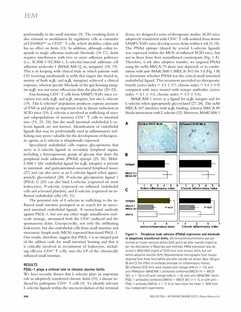
ileum, we designed a series of therapeutic studies. SCID mice adoptively transferred with CD4+ T cells isolated from donor SAMP1/YitFc mice develop severe ileitis within 6 wk (9, 18).The PNAd epitope (shared by several L-selectin ligands) was expressed within the MLN of infl amed SCID mice, but not in those from their noninfl amed counterparts (Fig. 1 A). Therefore, 6 wk after adoptive transfer, we targeted PNAd using the mAb MECA-79 alone (not depicted) or in combi-nation with anti–MAdCAM-1 (MECA-367) for 3 d (Fig. 1 B) to determine whether PNAd was the critical small intestinal endothelial ligand. This treatment provided no therapeutic benefi t (active index = 4.5 ± 0.7, chronic index = 4.4 ± 0.9) compared with mice treated with isotype antibodies (activeindex = 4.1 ± 0.6, chronic index = 4.9 ± 0.6).
MAdCAM-1 serves as a ligand for α4β7 integrin and for L-selectin when appropriately glycosylated (27, 28). The mAb MECA-367 interferes with α4β7 binding, whereas MECA-89 blocks interactions with L-selectin (32). However, MAdCAM-1
Figure 1. Peripheral node adressin (PNAd) expression and blockade
in adoptively transferred ileitis. (A) Immunohistochemistry was per-
formed on frozen sections before (left) and 6 wk after transfer (right) as
per the description in Materials and methods. PNAd expression was de-
tected in MLN HEVs (right) of SCID mice with chronic ileitis, but not
before adoptive transfer (left). Representative micrographs from tissues
obtained from three mice before and after transfer are shown. Bars, 100 μm.
(B and C) The effect of antibody blockade on infl ammatory indices.
(B) Infl amed SCID mice were treated with isotype mAb (n = 12), with
anti-PNAd/anti–MAdCAM-1 antibodies combined (MECA-79 + MECA
367, n = 10) or (C) with isotype mAb (n = 8), with anti–MAdCAM-1/anti–
PSGL-1 antibodies combined (4RA10 + MECA 367, n = 7), or with anti–
PSGL-1 antibody (4RA10, n = 7). Error bars show the mean ± SEM from
four independent experiments.
JEM VOL. 203, April 17, 2006 909
ARTICLE
does not appear to be the critical small intestinal endothelial ligand, as there was no therapeutic benefi t after MECA-89 anti-body treatment alone or when combined with MECA-367 (active index = 4.8 ± 0.7, chronic index = 5.7 ± 0.7) com-pared with isotype-treated controls (active index = 5.3 ± 0.8, chronic index = 5.2 ± 0.9) (unpublished data).
We tested whether PSGL-1, a known pan-selectin li-gand, could be the endothelial counterpart responsible for the therapeutic eff ect of L-selectin blockade. Using the function-blocking mAb 4RA10 (33), we targeted PSGL-1 alone or combined with anti–MAdCAM-1 mAb MECA-367 in in-fl amed SCID mice. Animals were administered three doses of mAb/s every other day, 6 wk after adoptive transfer, as the ileitis reached maximum severity. 4RA10 reduced both ac-tive and chronic infl ammatory indices (active index = 3.4 ±0.4, chronic index = 3.8 ± 0.5) compared with isotype-treated controls (active index = 5.6 ± 0.3, chronic index =5.5 ± 0.4, P < 0.01) (Fig. 1 C). Additional MAdCAM-1 blockade did not signifi cantly improve the eff ect (4RA10/MECA 367; active index = 2.8 ± 0.4, chronic index = 2.7 ±0.6; the p-value vs. 4RA10 was not signifi cant).
PSGL-1 blockade attenuated spontaneous chronic ileitis
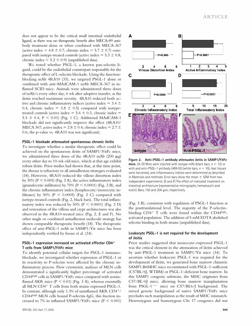
To investigate whether a similar therapeutic eff ect could be achieved on the spontaneous ileitis of SAMP1/YitFc mice, we administered three doses of the 4RA10 mAb (200 μg) every other day to 10-wk-old mice, which at that age exhibit robust ileitis. Prior studies had shown that, at this time point, the disease is refractory to all antiadhesion strategies evaluated (18). However, 4RA10 reduced the villous distortion index by 30% (P < 0.005) (Fig. 2 A), the active infl ammatory index (granulocytic infi ltrates) by 70% (P < 0.0001) (Fig. 2 B), and the chronic infl ammatory index (lymphocytic/monocytic in-fi ltrates) by 50% (P < 0.0005) (Fig. 2 C) compared with isotype-treated controls (Fig. 2, black bars). The total infl am-matory index was reduced by 50% (P < 0.0001) (Fig. 2 D) and restoration of the villous and crypt architectures was also observed in the 4RA10-treated mice (Fig. 2, E and F). No other single or combined antiadhesion molecule strategy has shown comparable therapeutic benefi t (18). The therapeutic eff ect of anti–PSGL-1 mAb in SAMP1/Yit mice has been independently verifi ed by Inoue et al. (34).
PSGL-1 expression increased on activated effector CD4+
T cells from SAMP1/YitFc mice
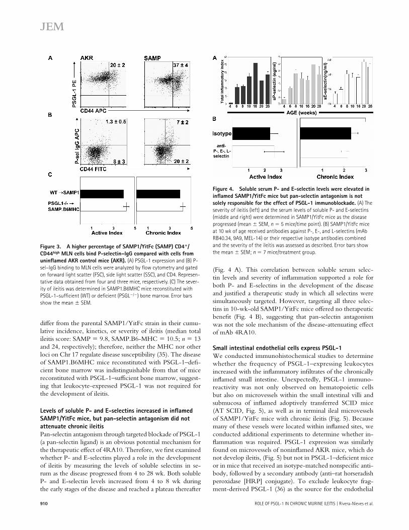
To identify potential cellular targets for PSGL-1 immuno-blockade, we investigated whether expression of PSGL-1 or its reactivity to P-selectin were aff ected by the chronic in-fl ammatory process. Flow cytometric analyses of MLN cells demonstrated a signifi cantly higher percentage of activated CD44high cells in SAMP1/YitFc mice compared with nonin-fl amed AKR mice (P < 0.01) (Fig. 3 A), whereas essentially all MLN CD4+ T cells from both strains expressed PSGL-1. In contrast, although only 1.3% of uninfl amed AKR CD4+/CD44high MLN cells bound P-selectin–IgG, this fraction in-creased to 7% in infl amed SAMP1/YitFc mice (P < 0.001)
(Fig. 3 B), consistent with regulation of PSGL-1 function at the posttranslational level. The majority of the P-selectin–binding CD4+ T cells were found within the CD44high-activated population. The addition of 5 mM EDTA abolished selectin binding in both strains (unpublished data).
Leukocyte PSGL-1 is not required for the development
of ileitis
Prior studies suggested that monocyte-expressed PSGL-1 was the critical element in the attenuation of ileitis achieved by anti–PSGL-1 treatment in SAMP1/Yit mice (34). To ascertain whether leukocyte PSGL-1 was required for the development of ileitis, we generated bone marrow chimeric SAMP1.B6MHC mice reconstituted with PSGL-1–suffi cient (C57BL/6J, WTBM) or PSGL-1–defi cient bone marrow. In this SAMP1 congenic substrain, the MHC originates from C57/BL/6J mice, allowing bone marrow transplantation from PSGL-1−/− mice on C57/BL6/J background. The mixed genetic background of native SAMP1/YitFc mice precludes such manipulation as the result of MHC mismatch. Heterozygous and homozygous Chr 17 congenics did not
Figure 2. Anti–PSGL-1 antibody attenuates ileitis in SAMP1/YitFc
mice. (A–D) Mice were injected with isotype mAb (black bars, n = 12) or
with and anti–PSGL-1 antibody (4RA10) (white bars, n = 10), ileal tissues
were harvested, and infl ammatory indices were determined as described
in Materials and methods. Error bars show the mean ± SEM from two
independent experiments. (E and F) The effect of indicated treatment on
intestinal architecture (representative micrographs, hematoxylin and
eosin). Bars, 150 and 200 μm, respectively.
910 ROLE OF PSGL-1 IN CHRONIC MURINE ILEITIS | Rivera-Nieves et al.
diff er from the parental SAMP1/YitFc strain in their cumu-lative incidence, kinetics, or severity of ileitis (median total ileitis score: SAMP = 9.8, SAMP.B6-MHC = 10.5; n = 13 and 24, respectively); therefore, neither the MHC nor other loci on Chr 17 regulate disease susceptibility (35). The disease of SAMP1.B6MHC mice reconstituted with PSGL-1–defi -cient bone marrow was indistinguishable from that of mice reconstituted with PSGL-1–suffi cient bone marrow, suggest-ing that leukocyte-expressed PSGL-1 was not required for the development of ileitis.
Levels of soluble P- and E-selectins increased in infl amed
SAMP1/YitFc mice, but pan-selectin antagonism did not
attenuate chronic ileitis
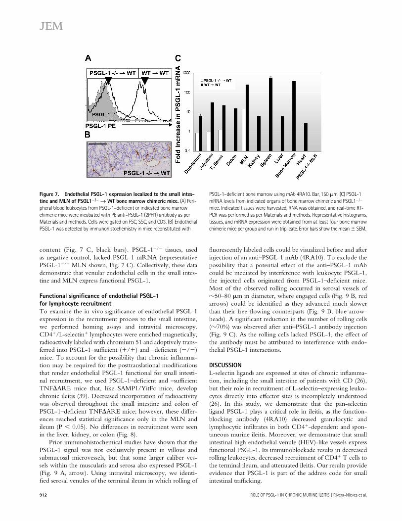
Pan-selectin antagonism through targeted blockade of PSGL-1 (a pan-selectin ligand) is an obvious potential mechanism for the therapeutic eff ect of 4RA10. Therefore, we fi rst examined whether P- and E-selectins played a role in the development of ileitis by measuring the levels of soluble selectins in se-rum as the disease progressed from 4 to 28 wk. Both soluble P- and E-selectin levels increased from 4 to 8 wk during the early stages of the disease and reached a plateau thereafter
(Fig. 4 A). This correlation between soluble serum selec-tin levels and severity of infl ammation supported a role for both P- and E-selectins in the development of the disease and justifi ed a therapeutic study in which all selectins were simultaneously targeted. However, targeting all three selec-tins in 10-wk-old SAMP1/YitFc mice off ered no therapeutic benefi t (Fig. 4 B), suggesting that pan-selectin antagonism was not the sole mechanism of the disease-attenuating eff ect of mAb 4RA10.
Small intestinal endothelial cells express PSGL-1
We conducted immunohistochemical studies to determine whether the frequency of PSGL-1–expressing leukocytes increased with the infl ammatory infi ltrates of the chronically infl amed small intestine. Unexpectedly, PSGL-1 immuno-reactivity was not only observed on hematopoietic cells but also on microvessels within the small intestinal villi and submucosa of infl amed adoptively transferred SCID mice (AT SCID, Fig. 5), as well as in terminal ileal microvessels of SAMP1/YitFc mice with chronic ileitis (Fig. 5). Because many of these vessels were located within infl amed sites, we conducted additional experiments to determine whether in-fl ammation was required. PSGL-1 expression was similarly found on microvessels of noninfl amed AKR mice, which do not develop ileitis, (Fig. 5) but not in PSGL-1–defi cient mice or in mice that received an isotype-matched nonspecifi c anti-body, followed by a secondary antibody (anti–rat horseradish peroxidase [HRP] conjugate). To exclude leukocyte frag-ment-derived PSGL-1 (36) as the source for the endothelial
Figure 3. A higher percentage of SAMP1/YitFc (SAMP) CD4+/
CD44high MLN cells bind P-selectin–IgG compared with cells from
uninfl amed AKR control mice (AKR). (A) PSGL-1 expression and (B) P-
sel–IgG binding to MLN cells were analyzed by fl ow cytometry and gated
on forward light scatter (FSC), side light scatter (SSC), and CD4. Represen-
tative data obtained from four and three mice, respectively. (C) The sever-
ity of ileitis was determined in SAMP1.B6MHC mice reconstituted with
PSGL-1–suffi cient (WT) or defi cient (PSGL−/−) bone marrow. Error bars
show the mean ± SEM.
Figure 4. Soluble serum P- and E-selectin levels were elevated in
infl amed SAMP1/YitFc mice but pan-selectin antagonism is not
solely responsible for the effect of PSGL-1 immunoblockade. (A) The
severity of ileitis (left) and the serum levels of soluble P- and E-selectins
(middle and right) were determined in SAMP1/YitFc mice as the disease
progressed (mean ± SEM, n = 5 mice/time point). (B) SAMP1/YitFc mice
at 10 wk of age received antibodies against P-, E-, and L-selectins (mAb
RB40.34, 9A9, MEL-14) or their respective isotype antibodies combined
and the severity of the ileitis was assessed as described. Error bars show
the mean ± SEM; n = 7 mice/treatment group.

JEM VOL. 203, April 17, 2006 911
ARTICLE
signal, we tested mice that lack P-, E-, and L-selectins (37). Leukocytes from these mice are expected to show little or no leukocyte rolling, limiting leukocyte-derived PSGL-1 depo-sition. However, P-, E-, L-selectin–defi cient mice similarly showed robust endothelial PSGL-1 expression (Fig. 5).
Endothelial PSGL-1 binds P-selectin
To confi rm endothelial PSGL-1 expression and function at the single cell level, we obtained ileal LP mononuclear cells by collagenase digestion, followed by a 2-h incubation at 37°C and staining of the adherent population for CD31, a pan- endothelial cell marker. Leukocytes were further depleted magnetically, removing cells that expressed CD45, a pan-leuko-cyte marker (Fig. 6 A) (38). 34% of the CD45neg/CD31pos cells expressed PSGL-1 (Fig. 6 B), consistent with PSGL-1 expres-sion in venules, which account for approximately one third of all microvascular endothelial cells. The same percentage of ileal endothelial cells was able to bind P-selectin–IgG (Fig. 6 C), but not a control human IgG chimera (not depicted), demonstrating that the endothelium-expressed PSGL-1 was functional. To directly demonstrate the expression of PSGL-1message by endothelial cells, mRNA was extracted from a cellular fraction magnetically depleted of CD45+ cells (98% CD45neg), followed by further purifi cation CD45neg/CD31pos small intestinal endothelial cells using FACS sorting. The presence of PSGL-1 mRNA was subsequently confi rmed by real-time RT-PCR (unpublished data).
PSGL-1 mRNA expression localized to the small intestine
and MLN of PSGL-1−/−→WT bone marrow chimeric mice
To quantitatively measure PSGL-1 mRNA in situ and to determine whether PSGL-1 expression localized to specifi c
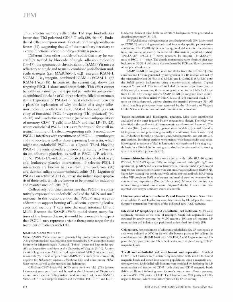
tissues, we generated bone marrow chimeric mice through reconstitution of lethally irradiated wild-type C57BL/6J mice with bone marrow progenitors from PSGL-1−/− mice (31). PSGL-1−/−→WT chimeric mice lacked leukocyte-derived PSGL-1(Fig. 7 A), but continued to express PSGL-1 within small intestinal endothelium (Fig. 7 B). Real-time RT-PCR of PSGL-1−/−→WT chimeric mice tissues localized PSGL-1 mRNA within the duodenum, jejunum, ileum, and MLN but not within the kidney, spleen, liver, bone marrow, or heart. Peripheral blood leukocytes from control mice recon-stituted with wild-type bone marrow (WT→WT) expressed PSGL-1 on leukocytes (Fig. 7 A), as well as PSGL-1 mRNA in all organs surveyed, commensurate with their leukocyte
Figure 5. Small intestinal endothelial cells express PSGL-1. Immuno-
histochemistry was performed on frozen terminal ileal sections from the
indicated mouse strains as per Materials and methods. PSGL-1 expression
was detected on leukocytes and in venules within the LP and submucosa
of adoptively transferred SCID mice (AT SCID), SAMP1/YitFc, and AKR
mice. Minimal or no expression was observed in PSGL-1–defi cient (−/−)
mice or in AKR ileum reacted with anti–rat HRP after isotype antibody
injection (Anti–rat HRP), whereas robust expression was seen in P-, E-,
L-selectin triple-defi cient (−/−) mice, which lack rolling leukocytes. Bar,
200 μm. Inset, high power of SAMP1/YitFc submucosal venules. Bar, 20 μm.
Figure 6. Primary intestinal endothelial cell PSGL-1 expression
and reactivity with P-sel-IgG chimera. (A). LP mononuclear cells were
obtained from terminal ilea of SAMP1/YitFc mice and plated for 2 h at
37°C. Adherent cells were recovered and stained for three color fl ow
cytometry as per Materials and methods. Cells were gated on FSC, SSC,
CD45neg, CD31+. (B) PSGL-1 expression was determined within the CD-
45neg, CD31+ gated cells (unshaded histogram) and compared with cells
stained with isotype mAb control (shaded histogram). (C) Reactivity with
P-sel–IgG chimera was determined in CD45neg, CD31+ gated cells. Repre-
sentative data from four (A and B) and two (C) independent experiments,
respectively, are shown.
912 ROLE OF PSGL-1 IN CHRONIC MURINE ILEITIS | Rivera-Nieves et al.
content (Fig. 7 C, black bars). PSGL-1−/− tissues, used as negative control, lacked PSGL-1 mRNA (representative PSGL-1−/− MLN shown, Fig. 7 C). Collectively, these data demonstrate that venular endothelial cells in the small intes-tine and MLN express functional PSGL-1.
Functional signifi cance of endothelial PSGL-1
for lymphocyte recruitment
To examine the in vivo signifi cance of endothelial PSGL-1expression in the recruitment process to the small intestine, we performed homing assays and intravital microscopy. CD4+/L-selectin+ lymphocytes were enriched magnetically, radioactively labeled with chromium 51 and adoptively trans-ferred into PSGL-1–suffi cient (+/+) and –defi cient (−/−) mice. To account for the possibility that chronic infl amma-tion may be required for the posttranslational modifi cations that render endothelial PSGL-1 functional for small intesti-nal recruitment, we used PSGL-1–defi cient and –suffi cient TNF∆ARE mice that, like SAMP1/YitFc mice, develop chronic ileitis (39). Decreased incorporation of radioactivity was observed throughout the small intestine and colon of PSGL-1–defi cient TNF∆ARE mice; however, these diff er-ences reached statistical signifi cance only in the MLN and ileum (P < 0.05). No diff erences in recruitment were seen in the liver, kidney, or colon (Fig. 8).
Prior immunohistochemical studies have shown that the PSGL-1 signal was not exclusively present in villous and submucosal microvessels, but that some larger caliber ves-sels within the muscularis and serosa also expressed PSGL-1 (Fig. 9 A, arrow). Using intravital microscopy, we identi-fi ed serosal venules of the terminal ileum in which rolling of
fl uorescently labeled cells could be visualized before and after injection of an anti–PSGL-1 mAb (4RA10). To exclude the possibility that a potential eff ect of the anti–PSGL-1 mAb could be mediated by interference with leukocyte PSGL-1, the injected cells originated from PSGL-1–defi cient mice. Most of the observed rolling occurred in serosal vessels of 50–80 μm in diameter, where engaged cells (Fig. 9 B, red arrows) could be identifi ed as they advanced much slower than their free-fl owing counterparts (Fig. 9 B, blue arrow-heads). A signifi cant reduction in the number of rolling cells ( 70%) was observed after anti–PSGL-1 antibody injection (Fig. 9 C). As the rolling cells lacked PSGL-1, the eff ect ofthe antibody must be attributed to interference with endo-thelial PSGL-1 interactions.
D I S C U S S I O N
L-selectin ligands are expressed at sites of chronic infl amma-tion, including the small intestine of patients with CD (26), but their role in recruitment of L-selectin–expressing leuko-cytes directly into eff ector sites is incompletely understood (26). In this study, we demonstrate that the pan-selectin ligand PSGL-1 plays a critical role in ileitis, as the function-blocking antibody (4RA10) decreased granulocytic and lymphocytic infi ltrates in both CD4+-dependent and spon-taneous murine ileitis. Moreover, we demonstrate that small intestinal high endothelial venule (HEV)-like vessels express functional PSGL-1. Its immunoblockade results in decreased rolling leukocytes, decreased recruitment of CD4+ T cells to the terminal ileum, and attenuated ileitis. Our results provide evidence that PSGL-1 is part of the address code for small intestinal traffi cking.
Figure 7. Endothelial PSGL-1 expression localized to the small intes-
tine and MLN of PSGL1−/− → WT bone marrow chimeric mice. (A) Peri-
pheral blood leukocytes from PSGL-1–defi cient or indicated bone marrow
chimeric mice were incubated with PE anti–PSGL-1 (2PH1) antibody as per
Materials and methods. Cells were gated on FSC, SSC, and CD3. (B) Endothelial
PSGL-1 was detected by immunohistochemistry in mice reconstituted with
PSGL-1–defi cient bone marrow using mAb 4RA10. Bar, 150 μm. (C) PSGL-1
mRNA levels from indicated organs of bone marrow chimeric and PSGL1−/−
mice. Indicated tissues were harvested, RNA was obtained, and real-time RT-
PCR was performed as per Materials and methods. Representative histograms,
tissues, and mRNA expression were obtained from at least four bone marrow
chimeric mice per group and run in triplicate. Error bars show the mean ± SEM.

JEM VOL. 203, April 17, 2006 913
ARTICLE
The mAb MECA-79 recognizes several sialomucins (PNAd) that, when properly glycosylated, serve as L-selectin ligands. Functionally, MECA-79 blocks lymphocyte interac-tions with HEVs in vitro and short-term lymphocyte homing to lymph nodes in vivo (25). Its role in infl ammatory traf-fi cking has been shown in the infl amed pancreas (40, 41) and lacrimal glands (42). MECA-79 binds to HEV-like vessels in the organized lymphoid aggregates present in chronically infl amed tissues (26). Although we demonstrate PNAd ex-pression in SCID mice with ileitis, no benefi cial therapeutic eff ect was seen by MECA-79 administration.
MAdCAM-1, the α4β7 integrin endothelial ligand ex-pressed in intestine and gastrointestinal-associated lymphoid tissues (27), serves as an L-selectin ligand when appropriately glycosylated (7, 27, 28). MAdCAM-1 is constitutively ex-pressed in postcapillary venules of intestinal LP, MLN, and Peyer’s patches HEVs (27, 43), and is aberrantly expressed in the infl amed pancreas (39). Furthermore, MAdCAM-1 is up-regulated in the chronically infl amed small and large intestine of patients with UC and CD, where it is likely induced by TNF-α (43, 44). The mAb MECA-367 binds to the fi rst Ig domain of MAdCAM-1 and interferes with α4β7 binding, whereas MECA-89 binds to the second Ig domain, which in-terferes with L-selectin interactions as a result of its proximity to the mucinous portion of MAdCAM-1 (32). If MAdCAM-1mediated critical L-selectin interactions in the gut, we would expect that combined MECA-367/MECA-89 treatment would provide the same benefi t as combined MECA-367/MEL-14 (19). However, administering MECA-89 together with MECA-367 provided no added therapeutic benefi t.
PSGL-1 binds to L-selectin (29), E-selectin, and P-selec-tin (30, 31). The most comprehensive survey of PSGL-1 ex-pression to date identifi ed bone marrow–derived cells as the primary site of expression, whereas expression by endothelial cells was limited to small venules and capillaries of pathologi-cal tissues (i.e., benign prostatic hypertrophy and fi brocystic disease of the breast) (45). PSGL-1 binding to selectins is reg-ulated by fucosyltransferases, core 2-glucosaminyltransferases, sialyltransferases, and sulfotransferases, which are constitu-tively expressed in myeloid cells but regulated in lymphocytes.
Figure 8. Homing of CD4+ T cells to the terminal ileum is impaired
in PSGL-1–defi cient mice. Chromium 51–labeled L-selectin+ CD4 T cells
were injected into PSGL-1–suffi cient (+/+) and –defi cient (−/−)
TNF∆ARE mice as per Materials and methods. Radioactivity was deter-
mined in the aforementioned organs 72 h after cell transfer. Error bars
show the mean ± SEM from three mice/experimental group. *, P < 0.05.
Figure 9. PSGL-1 blockade decreased splenocyte–endothelial
interactions in the small intestinal vasculature. (A) Endothelial PSGL-1
signal was observed in small (arrowhead) and large vessels (yellow arrow)
within the small intestinal muscularis and serosa. The larger serosal ves-
sels were monitored via intravital microscopy. Bar, 200 μm. (B) Rolling
CFSE-labeled PSGL-1–deficient cells (red arrows) proceed much
slower than cells not interacting with wild-type vessels (blue arrowhead)
before (B) PSGL-1 blockade. (C) A signifi cant decrease in the number of
rolling cells was observed after injection of 4RA10 (percent of normalized
total rolling cells [error bars show the mean ± SD] before and after
4RA10 injection from four experiments in which one to three vessels/
mouse were monitored; *, P < 0.05).
914 ROLE OF PSGL-1 IN CHRONIC MURINE ILEITIS | Rivera-Nieves et al.
Thus, eff ector memory cells of the Th1 type bind selectins better than Th2 polarized CD4+ T cells (30, 46–48). Endo-thelial cells also express most, if not all, of these glycosyltrans-ferases (49), suggesting that all of the machinery necessary to express functional selectin binding activity is present.
Diff erent from other models of colitis that may be suc-cessfully treated by blockade of single adhesion molecules (14–17), the spontaneous chronic ileitis of SAMP/Yit mice is refractory to single and to many combined antiadhesion mol-ecule strategies (i.e., MAdCAM-1, α4β7 integrin, ICAM-1, VCAM-1, α4 integrin, combined ICAM-1/VCAM-1, and ICAM-1/α4) (18). In contrast, the current data shows that targeting PSGL-1 alone ameliorates ileitis. This eff ect cannot be solely explained by the expected pan-selectin antagonism as combined blockade of all three selectins failed to attenuate ileitis. Expression of PSGL-1 on ileal endothelium provides a plausible explanation of why blockade of a single adhe-sion molecule is eff ective. First, PSGL-1 blockade prevents entry of functional PSGL-1–expressing (Th1-polarized) (30, 46–48) and L-selectin–expressing (naive and subpopulations of memory CD4+ T cells) into MLN and ileal LP (19, 23), where endothelial PSGL-1 acts as an “addressin” for small in-testinal homing of L-selectin–expressing cells. Second, anti–PSGL-1 interferes with recruitment of PSGL-1+ granulocytes and monocytes, as well as those expressing L-selectin, which might use endothelial PSGL-1 as a ligand. Third, blocking PSGL-1 prevents secondary leukocyte tethering to P-selec-tin on adherent platelets, as well as PSGL-1/P-selectin- and/or PSGL-1/L-selectin–mediated leukocyte–leukocyte and leukocyte–platelet interactions. P-selectin–PSGL-1 interactions are known to exacerbate atherosclerosis (50) and dextran sulfate sodium–induced colitis (51). Ligation of PSGL-1 on activated Th1 cells may also induce rapid apopto-sis of these cells, which are known to be pivotal for induction and maintenance of ileitis (52).
Collectively, our data demonstrate that PSGL-1 is consti-tutively expressed on endothelial cells of the MLN and small intestine. In this location, endothelial PSGL-1 may act as an addressin to support homing of L-selectin–expressing leuko-cytes and memory T cells into the small intestinal LP and MLN. Because the SAMP1/YitFc model shares many fea-tures of the human disease, it would be reasonable to expect that PSGL-1 may represent a viable therapeutic target for the treatment of patients with CD.
MATERIALS AND METHODSMice. SAMP1/YitFc mice were generated by brother–sister matings for
>30 generations from two breeding pairs provided by S. Matsumoto (Yakult
Institute for Microbiological Research, Tokyo, Japan) and kept under spe-
cifi c pathogen-free conditions at the University of Virginia (9, 10). As most
identifi able genes were AKR derived, age-matched AKR/J mice were used
as controls (35). Fecal samples from SAMP1/YitFc mice were consistently
negative for Helicobacter hepaticus, Helicobacter bilis, and other mouse Helico-
bacter species, as well as for protozoa and helminthes.
C3SnSmn.CB17-Prkdcscid/J SCID mice (6–8 wk old) (The Jackson
Laboratory) were purchased and housed at the University of Virginia vi-
varium under specifi c pathogen-free conditions for 1 wk before SAMP1/
YitFc CD4+ T cell adoptive transfer and thereafter. PSGL-1−/− and E-, P-,
L-selectin–defi cient mice, both on C57BL/6 background were generated as
described previously (31, 37).
TNF∆ARE mice were generated as described previously (39), backcrossed
to C57BL/6J mice (18 generations), and kept under specifi c pathogen-free
conditions. The C57BL/6J genetic background did not alter the localiza-
tion, time course, or severity the intestinal infl ammation (unpublished data).
TNF∆ARE+/−/PSGL-1−/− were generated by crossing TNF∆ARE+/−
mice to PSGL-1−/− mice. The double mutant mice were obtained after two
backcrosses. PSGL-1 defi ciency was confi rmed by PCR and fl ow cytometry
of peripheral leukocytes.
SAMP.B6-MHC congenic mice for alleles from the C57BL/6J (B6)
chromosome 17 were generated by introgression of a B6 interval defi ned by
the microsatellite loci D17Mit16 (31.3 Mb) and D17Mit221 (87.9 Mb) onto
the SAMP genetic background using a marker-assisted selection (“speed
congenic”) protocol. This interval included the entire major histocompat-
ibility complex, converting the new congenic strain to the H-2b haplotype
from H-2k. This change renders SAMP.B6-MHC congenic mice as suit-
able recipients for bone marrow from C57BL/6J (B6) mice and PSGL-1−/−
mice on this background, without altering the intestinal phenotype (35). All
animal handling procedures were approved by the University of Virginia
Health Sciences Center institutional committee for animal use.
Tissue collection and histological analyses. Mice were anesthetized
and killed at the times required by the experimental design. The MLN was
identifi ed at the confl uence of the mesenteric vasculature and harvested. The
distal ilea (10 cm) were resected, opened, rinsed of debris, oriented from dis-
tal to proximal, and pinned longitudinally in corkboard. Tissues were fi xed
in 10% buff ered formalin or Bouin’s, embedded in paraffi n, and cut into 3–5
μm sections. Resulting sections were stained with hematoxylin and eosin.
Histological assessment of ileal infl ammation was performed by a single pa-
thologist in a blinded fashion using a standardized semi-quantitative scoring
system as described previously (18).
Immunohistochemistry. Mice were injected with mAbs 4RA-10 against
PSGL-1, MECA-79 against PNAd or isotype control mAb (IgG1, IgM, re-
spectively) i.p. MLN and ilea were harvested 30 min after mAb injection and
snap frozen, and sections (5 μm) were cut on a cryostat (Microm HM505N).
Secondary staining was conducted with rabbit anti–rat antibody HRP using
either VIP purple or DAB as substrates and methyl green or hematoxylin as
counterstains, respectively (Vector Laboratories). Nonspecifi c binding was
reduced using normal mouse serum (Sigma-Aldrich). Tissues from mice
injected with isotype antibody served as controls.
Determination of serum soluble P- and E-selectin levels. Serum lev-
els of soluble P- and E-selectins were determined by ELISA per the manu-
facturer’s instructions from mice of the indicated ages (R&D Systems).
Intestinal LP lymphocyte and endothelial cell isolation. MLN were
aseptically removed at the time of necropsy. Single cell suspensions were
obtained by gently pressing the MLN against a 100-μm cell strainer. LP
mononuclear cell isolation was performed as described previously (19).
Cell culture. For enrichment of adherent endothelial cells, LP mononuclear
cells were cultured at 37°C in six-well fl at-bottom plates at 106 cells/ml in
complete medium (RPMI 1640 with 10% FBS, 2 mM L-glutamine and 1%
penicillin/streptomycin) for 2 h or leukocytes were depleted using CD45+
magnetic beads.
T cell and endothelial cell enrichment and separation. Enriched
CD4+ T cell fractions were obtained by incubation with anti-CD4–bound
magnetic beads and sorted into discrete populations, using a magnetic cell–
sorting system. Endothelial cell fractions were enriched by depleting the LP
mononuclear cell fraction of CD45+ cells using anti-CD45 magnetic beads
(Miltenyi Biotec) following manufacturer’s instructions. Flow cytometry
confi rmed 95–97% purity of CD4+ T cell fractions and 98% purity of CD45
negative fraction, which was further purifi ed by FACS sorting.
JEM VOL. 203, April 17, 2006 915
ARTICLE
CD4+ T cell adoptive transfer. SAMP1/YitFc (30–40 wk old) mice
MLN were harvested and rendered into a single cell suspension, followed by
positive selection for CD4 using magnetic beads. CD4+ lymphocytes (105/
mouse) were injected i.p. or i.v. into 6–8-wk-old SCID mice (The Jackson
Laboratory). Neither the selection protocols (positive or negative) nor the
route of injection (i.p. or i.v.) altered the severity or time course of disease.
Mice were housed in a barrier facility and fed irradiated, standard chow. After
5 wk, the adoptively transferred mice showed ileitis with moderate to severe
LP leukocyte infi ltration and architectural changes (villous and crypt distor-
tion, goblet cell hyperplasia, and hypertrophy of the muscularis propia).
In vivo homing. The in vivo migration of lymphocytes was analyzed as
described previously (53). In brief, magnetically enriched CD4+/CD62L+
lymphocytes were labeled with 20 μCi chromium 51/ml. A total of 50 ×
106 lymphocytes suspended in 0.5 ml were injected i.p. into 8–12-wk-old
C57BL/6J (n = 3) or PSGL-1−/− mice (n = 3). Mice were killed after 72 h
and the distribution of radioactivity in diff erent organs was measured. Each
sample was counted to 3% statistical error. The mean values and their stan-
dard error were determined from three animals per strain.
Flow cytometry and cell sorting. Fluorescently tagged monoclonal an-
tibodies reactive with PSGL-1 (PE-labeled 2PH1; BD Biosciences) or P-sel
human Ig-G chimera (2 μg/106 cells; BD Biosciences) or control CD4-
human IgG chimera (2 μg/106 cells, a gift from J.B. Lowe, University of
Michigan, Ann Arbor, MI) preincubated with allophycocyanin-labeled
anti–human IgG Fab fragments (Zenon Human IgG labeling kit; Invitrogen)
were subsequently incubated with cells in suspension for 20 min, includ-
ing FITC-labeled anti-CD4 (GK1.5) for gating of MLN cells or PERCP
anti-CD45 (30-F11) and FITC or allophycocyanin–anti-CD31 (390) (BD
Biosciences) for gating of intestinal endothelial cells. Cells were fi xed with
1% paraformaldehyde and three-to-four color analyses were performed using
the FACS Calibur system (Becton Dickinson Immunocytometry Systems).
Further analyses were performed using FLOWJo software (Tree Star Inc.).
Endothelial cells were sorted using a FACSVantage sorter.
Real-time RT PCR. RT-PCR primers and Taqman probes were designed
using Beacon Designer V3.0 software (Premier Biosoft). The primers were
designed to amplify WT PSGL-1 mRNA but not mRNA from PSGL-1−/−
mice (truncated protein). Total RNA was isolated using RNAeasy Mini-
Kit (QIAGEN) with DNase treatment (Rnase-Free Dnase Kit; QIAGEN).
Reverse transcription was performed using 500 ng of RNA with an Om-
niscript RT Kit (QIAGEN) and oligo-dT primers. The primers were tested
for specifi city by standard PCR using cDNA made from WT mice and
PSGL-1−/− mice. Reaction conditions were optimized and 2 μL of sample
cDNA was multiplexed with GAPDH and PSGL-1 primers on an iCycler
iQ Real-Time Detection System (QIAGEN). Values were determined using
the iCycler iQ Real-Time Detection System Software v3.0a (QIAGEN).
The corresponding values were normalized to GAPDH. The PSGL-1 prim-
ers used were: forward 5′-C T T C C T T G T G C T G C T G A C C A T -3′, reverse
5′-T C A G G G T C C T C A A A A T C G T C A T C -3′ and probe 5′-C C A A C C A C-
C T G C C T C C G T T C C C G -3′.
Therapeutic interventions. mAbs (200 μg each) against the following:
PNAd (MECA-79, IgM), PSGL-1 (4RA-10, IgG1), MAdCAM-1 (MECA-
367 or MECA 89, rat IgG 2a), P-selectin (RB40-34, IgG1), E-selectin
(10E9.6, IgG2A), L-selectin (MEL-14, IgG2a), or irrelevant corresponding
isotype control mAb were injected i.p. every other day for 3 d, 5 wk after
adoptive transfer of SCID mice or at 10 wk of age for SAMP1/YitFc mice.
Mice were killed 16–18 h after the last dose. MECA-367, MECA 79, and
MECA-89 were obtained from the American Type Culture Collection.
4RA10 was obtained from D. Vestweber (University of Munster, Munster,
Germany). Antibodies were produced and purifi ed by HPLC from hybrid-
oma supernatants at the University of Virginia Biomolecular Core Facility.
Generation of bone marrow chimeric mice. C57BL/6J bone marrow
transplant recipients were irradiated twice with 600 rad before and 4–6 h after
injection of donor cells. Bone marrow cells were harvested in RPMI 1640
containing 10% fetal calf serum. 7.5 × 10⁶ cells/500 μl were injected into the
tail veins of recipient mice. Mice were killed 6–10 wk after transplantation.
Intravital microscopy. C57BL/6J mice were prepared for intravital mi-
croscopy as per previously described methods (54). A 1.5-cm incision was
made along the linea alba and the cecum and terminal ileum were exposed.
The intestinal serosa was covered with plastic wrap and superfused with
bicarbonate-buff ered saline containing 100 nM isoproterenol. CFSE-la-
beled cells (10–20 × 106/200 μl PBS) from PSGL-1−/− mice were injected
through a jugular vein catheter and the serosal vessels in which rolling cells
could be visualized by stroboscopic epifl uorescence microscopy were se-
lected for monitoring. All monitored vessels were 30–80 μm in diameter.
Two to three vessels per mouse were selected and rolling cells were counted
before and after antibody injection. The number of rolling cells before
4RA10 was normalized to 100% and the percent reduction after 4RA10
treatment was calculated.
Statistics. Statistical analyses for fl ow cytometry and infl ammatory indices
were performed using the two-tailed unpaired Student’s t test. Data were
expressed as mean and standard error of the mean. Statistical signifi cance was
set at P < 0.05.
Online supplemental material. Supplemental Videos 1 and 2 depict the
interactions between CFSE-labeled PSGL-1−/− cells and intestinal endothe-
lium before and after injection of mAb 4RA10; they are available at http://
www.jem.org/cgi/content/full/jem.20052530/DC1.
We thank the Morphology and Immunology cores of the UVA Silvio Conte Digestive
Diseases Research Center (i.e., Dr. R. Mize for evaluation of histological sections,
M. Solga, and J. Lannigan for valuable discussions and assistance with fl ow cytometry,
S. Hoang for expert assistance with immunohistochemistry); T. Deem for assistance
with intravital microscopy; A. Bruce, J. Ho, and O. Castañón-Cervantes for technical
assistance; and Dr. G. Kollias for providing the original TNF∆ARE breeding pairs.
This work was supported by grants from the National Institutes of Health
(nos. DK-57880 and DK-067254) and by a Harold Amos grant from the Robert Wood
Johnson Foundation.
The authors have no confl icting fi nancial interests.
Submitted: 21 December 2005
Accepted: 22 February 2006
R E F E R E N C E S 1. Davidson, A., and B. Diamond. 2001. Autoimmune diseases. N. Engl.
J. Med. 345:340–350. 2. Longobardi, T., P. Jacobs, and C.N. Bernstein. 2003. Work losses re-
lated to infl ammatory bowel disease in the United States: results from the National Health Interview Survey. Am. J. Gastroenterol. 98:1064–1072.
3. Podolsky, D.K. 1991. Infl ammatory bowel disease (1). N. Engl. J. Med. 325:928–937.
4. Fiocchi, C. 1998. Infl ammatory bowel disease: etiology and pathogen-esis. Gastroenterology. 115:182–205.
5. Targan, S.R., S.B. Hanauer, S.J. van Deventer, L. Mayer, D.H. Present, T. Braakman, K.L. DeWoody, T.F. Schaible, and P.J. Rutgeerts. 1997. A short-term study of chimeric monoclonal antibody cA2 to tumor ne-crosis factor α for Crohn’s disease. Crohn’s Disease cA2 Study Group. N. Engl. J. Med. 337:1029–1035.
6. Sandborn, W.J., and S.R. Targan. 2002. Biologic therapy of infl amma-tory bowel disease. Gastroenterology. 122:1592–1608.
7. Springer, T.A. 1995. Traffi c signals on endothelium for lymphocyte recirculation and leukocyte emigration. Annu. Rev. Physiol. 57:827–872.
8. Matsumoto, S., Y. Okabe, H. Setoyama, K. Takayama, J. Ohtsuka, H. Funahashi, A. Imaoka, Y. Okada, and Y. Umesaki. 1998. Infl ammatory bowel disease-like enteritis and caecitis in a senescence accelerated mouse P1/Yit strain. Gut. 43:71–78.
9. Kosiewicz, M.M., C.C. Nast, A. Krishnan, J. Rivera-Nieves, C.A. Moskaluk, S. Matsumoto, K. Kozaiwa, and F. Cominelli. 2001.
916 ROLE OF PSGL-1 IN CHRONIC MURINE ILEITIS | Rivera-Nieves et al.
Th1-type responses mediate spontaneous ileitis in a novel murine model of Crohn’s disease. J. Clin. Invest. 107:695–702.
10. Rivera-Nieves, J., G. Bamias, A. Vidrich, M. Marini, T.T. Pizarro, M.J. McDuffi e, C.A. Moskaluk, S.M. Cohn, and F. Cominelli. 2003. Emergence of perianal fi stulizing disease in the SAMP1/YitFc mouse, a spontaneous model of chronic ileitis. Gastroenterology. 124:972–982.
11. Strober, W., K. Nakamura, and A. Kitani. 2001. The SAMP1/Yit mouse: another step closer to modeling human infl ammatory bowel disease. J. Clin. Invest. 107:667–670.
12. Powrie, F. 1995. T cells in infl ammatory bowel disease: protective and pathogenic roles. Immunity. 3:171–174.
13. Olson, T.S., G. Bamias, M. Naganuma, J. Rivera-Nieves, T.L. Burcin, W. Ross, M.A. Morris, T.T. Pizarro, P.B. Ernst, F. Cominelli, and K. Ley. 2004. Expanded B cell population blocks regulatory T cells and exacerbates ileitis in a murine model of Crohn disease. J. Clin. Invest. 114:389–398.
14. Kato, S., R. Hokari, K. Matsuzaki, A. Iwai, A. Kawaguchi, S. Nagao, T. Miyahara, K. Itoh, H. Ishii, and S. Miura. 2000. Amelioration of mu-rine experimental colitis by inhibition of mucosal addressin cell adhesion molecule-1. J. Pharmacol. Exp. Ther. 295:183–189.
15. Ludviksson, B.R., W. Strober, R. Nishikomori, S.K. Hasan, and R.O. Ehrhardt. 1999. Administration of mAb against α E β 7 prevents and ameliorates immunization-induced colitis in IL-2−/− mice. J. Immunol. 162:4975–4982.
16. Picarella, D., P. Hurlbut, J. Rottman, X. Shi, E. Butcher, and D.J. Ringler. 1997. Monoclonal antibodies specifi c for β 7 integrin and mucosal addressin cell adhesion molecule-1 (MAdCAM-1) reduce in-fl ammation in the colon of scid mice reconstituted with CD45RBhigh CD4+ T cells. J. Immunol. 158:2099–2106.
17. Podolsky, D.K., R. Lobb, N. King, C.D. Benjamin, B. Pepinsky, P. Sehgal, and M. deBeaumont. 1993. Attenuation of colitis in the cotton-top tamarin by anti-α 4 integrin monoclonal antibody. J. Clin. Invest. 92:372–380.
18. Burns, R.C., J. Rivera-Nieves, C.A. Moskaluk, S. Matsumoto, F. Cominelli, and K. Ley. 2001. Antibody blockade of ICAM-1 and VCAM-1 ameliorates infl ammation in the SAMP-1/Yit adoptive trans-fer model of Crohn’s disease in mice. Gastroenterology. 121:1428–1436.
19. Rivera-Nieves, J., T. Olson, G. Bamias, A. Bruce, M. Solga, R.F. Knight, S. Hoang, F. Cominelli, and K. Ley. 2005. L-selectin, α4 β1, and α4 β7 integrins participate in CD4+ T cell recruitment to chroni-cally infl amed small intestine. J. Immunol. 174:2343–2352.
20. Ghosh, S., E. Goldin, F.H. Gordon, H.A. Malchow, J. Rask-Madsen, P. Rutgeerts, P. Vyhnalek, Z. Zadorova, T. Palmer, and S. Donoghue. 2003. Natalizumab for active Crohn’s disease. N. Engl. J. Med. 348:24–32.
21. von Andrian, U.H., and B. Engelhardt. 2003. α4 integrins as therapeu-tic targets in autoimmune disease. N. Engl. J. Med. 348:68–72.
22. Feagan, B.G., G.R. Greenberg, G. Wild, R.N. Fedorak, P. Pare, J.W. McDonald, R. Dube, A. Cohen, A.H. Steinhart, S. Landau, et al. 2005. Treatment of ulcerative colitis with a humanized antibody to the α4β7 integrin. N. Engl. J. Med. 352:2499–2507.
23. Sallusto, F., D. Lenig, R. Forster, M. Lipp, and A. Lanzavecchia. 1999. Two subsets of memory T lymphocytes with distinct homing potentials and eff ector functions. Nature. 401:708–712.
24. von Andrian, U.H., and C.R. Mackay. 2000. T-cell function and mi-gration. Two sides of the same coin. N. Engl. J. Med. 343:1020–1034.
25. Hemmerich, S., E.C. Butcher, and S.D. Rosen. 1994. Sulfation-depen-dent recognition of high endothelial venules (HEV)-ligands by L-selec-tin and MECA 79, and adhesion-blocking monoclonal antibody. J. Exp. Med. 180:2219–2226.
26. Rosen, S.D. 1999. Endothelial ligands for L-selectin: from lymphocyte recirculation to allograft rejection. Am. J. Pathol. 155:1013–1020.
27. Butcher, E.C., M. Williams, K. Youngman, L. Rott, and M. Briskin. 1999. Lymphocyte traffi cking and regional immunity. Adv. Immunol. 72:209–253.
28. Berg, E.L., L.M. McEvoy, C. Berlin, R.F. Bargatze, and E.C. Butcher. 1993. L-selectin-mediated lymphocyte rolling on MAdCAM-1. Nature. 366:695–698.
29. McEver, R.P., and R.D. Cummings. 1997. Role of PSGL-1 binding to selectins in leukocyte recruitment. J. Clin. Invest. 100:S97–S103.
30. Hirata, T., G. Merrill-Skoloff , M. Aab, J. Yang, B.C. Furie, and B. Furie. 2000. P-Selectin glycoprotein ligand 1 (PSGL-1) is a physiologi-cal ligand for E-selectin in mediating T helper 1 lymphocyte migration. J. Exp. Med. 192:1669–1676.
31. Xia, L., M. Sperandio, T. Yago, J.M. McDaniel, R.D. Cummings, S. Pearson-White, K. Ley, and R.P. McEver. 2002. P-selectin glycopro-tein ligand-1-defi cient mice have impaired leukocyte tethering to E-selectin under fl ow. J. Clin. Invest. 109:939–950.
32. Bargatze, R.F., M.A. Jutila, and E.C. Butcher. 1995. Distinct roles of L-selectin and integrins α4 β7 and LFA-1 in lymphocyte homing to Peyer’s patch-HEV in situ: the multistep model confi rmed and refi ned. Immunity. 3:99–108.
33. Thatte, A., S. Ficarro, K.R. Snapp, M.K. Wild, D. Vestweber, D.F. Hunt, and K.F. Ley. 2002. Binding of function-blocking mAbs to mouse and human P-selectin glycoprotein ligand-1 peptides with and without tyrosine sulfation. J. Leukoc. Biol. 72:470–477.
34. Inoue, T., Y. Tsuzuki, K. Matsuzaki, H. Matsunaga, J. Miyazaki, R. Hokari, Y. Okada, A. Kawaguchi, S. Nagao, K. Itoh, et al. 2005. Blockade of PSGL-1 attenuates CD14+ monocytic cell recruitment in intestinal mucosa and ameliorates ileitis in SAMP1/Yit mice. J. Leukoc. Biol. 77:287–295.
35. Kozaiwa, K., K. Sugawara, M.F. Smith Jr., V. Carl, V. Yamschikov, B. Belyea, S.B. McEwen, C.A. Moskaluk, T.T. Pizarro, F. Cominelli, and M. McDuffi e. 2003. Identifi cation of a quantitative trait locus for ileitis in a spontaneous mouse model of Crohn’s disease: SAMP1/YitFc. Gastroenterology. 125:477–490.
36. Sperandio, M., M.L. Smith, S.B. Forlow, T.S. Olson, L. Xia, R.P. McEver, and K. Ley. 2003. P-selectin glycoprotein ligand-1 medi-ates L-selectin–dependent leukocyte rolling in venules. J. Exp. Med. 197:1355–1363.
37. Collins, R.G., U. Jung, M. Ramirez, D.C. Bullard, M.J. Hicks, C.W. Smith, K. Ley, and A.L. Beaudet. 2001. Dermal and pulmonary infl am-matory disease in E-selectin and P-selectin double-null mice is reduced in triple-selectin-null mice. Blood. 98:727–735.
38. M’Rini, C., G. Cheng, C. Schweitzer, L.L. Cavanagh, R.T. Palframan, T.R. Mempel, R.A. Warnock, J.B. Lowe, E.J. Quackenbush, and U.H. von Andrian. 2003. A novel endothelial L-selectin ligand activity in lymph node medulla that is regulated by α(1,3)-fucosyltransferase-IV. J. Exp. Med. 198:1301–1312.
39. Kontoyiannis, D., M. Pasparakis, T.T. Pizarro, F. Cominelli, and G. Kollias. 1999. Impaired on/off regulation of TNF biosynthesis in mice lacking TNF AU-rich elements: implications for joint and gut-associ-ated immunopathologies. Immunity. 10:387–398.
40. Fabien, N., I. Bergerot, J. Orgiazzi, and C. Thivolet. 1996. Lymphocyte function associated antigen-1, integrin α4, and L-selectin mediate T-cell homing to the pancreas in the model of adoptive transfer of diabetes in NOD mice. Diabetes. 45:1181–1186.
41. Hanninen, A., C. Taylor, P.R. Streeter, L.S. Stark, J.M. Sarte, J.A. Shizuru, O. Simell, and S.A. Michie. 1993. Vascular addressins are induced on islet vessels during insulitis in nonobese diabetic mice and are involved in lym-phoid cell binding to islet endothelium. J. Clin. Invest. 92:2509–2515.
42. Mikulowska-Mennis, A., B. Xu, J.M. Berberian, and S.A. Michie. 2001. Lymphocyte migration to infl amed lacrimal glands is mediated by vascular cell adhesion molecule-1/ α(4)β(1) integrin, peripheral node addressin/l-selectin, and lymphocyte function-associated antigen-1 ad-hesion pathways. Am. J. Pathol. 159:671–681.
43. Sikorski, E.E., R. Hallmann, E.L. Berg, and E.C. Butcher. 1993. The Peyer’s patch high endothelial receptor for lymphocytes, the mucosal vascular addressin, is induced on a murine endothelial cell line by tumor necrosis factor-α and IL- 1. J. Immunol. 151:5239–5250.
44. Briskin, M., D. Winsor-Hines, A. Shyjan, N. Cochran, S. Bloom, J. Wilson, L.M. McEvoy, E.C. Butcher, N. Kassam, C.R. Mackay, et al. 1997. Human mucosal addressin cell adhesion molecule-1 is preferen-tially expressed in intestinal tract and associated lymphoid tissue. Am. J. Pathol. 151:97–110.
45. Laszik, Z., P.J. Jansen, R.D. Cummings, T.F. Tedder, R.P. McEver, and K.L. Moore. 1996. P-selectin glycoprotein ligand-1 is broadly ex-pressed in cells of myeloid, lymphoid, and dendritic lineage and in some nonhematopoietic cells. Blood. 88:3010–3021.

JEM VOL. 203, April 17, 2006 917
ARTICLE
46. Austrup, F., D. Vestweber, E. Borges, M. Lohning, R. Brauer, U. Herz, H. Renz, R. Hallmann, A. Scheff old, A. Radbruch, and A. Hamann. 1997. P- and E-selectin mediate recruitment of T-helper-1 but not T- helper-2 cells into infl ammed tissues. Nature. 385:81–83.
47. Borges, E., W. Tietz, M. Steegmaier, T. Moll, R. Hallmann, A. Hamann, and D. Vestweber. 1997. P-selectin glycoprotein ligand-1 (PSGL-1) on T helper 1 but not on T helper 2 cells binds to P-selectin and supports migration into infl amed skin. J. Exp. Med. 185:573–578.
48. Ley, K., and G.S. Kansas. 2004. Selectins in T-cell recruitment to non-lym-phoid tissues and sites of infl ammation. Nat. Rev. Immunol. 4:325–335.
49. Majuri, M.L., M. Pinola, R. Niemela, S. Tiisala, J. Natunen, O. Renkonen, and R. Renkonen. 1994. α2,3-sialyl and α1,3-fucosyltrans-ferase-dependent synthesis of sialyl Lewis x, an essential oligosaccharide present on L-selectin counterreceptors, in cultured endothelial cells. Eur. J. Immunol. 24:3205–3210.
50. Huo, Y., A. Schober, S.B. Forlow, D.F. Smith, M.C. Hyman, S. Jung, D.R. Littman, C. Weber, and K. Ley. 2003. Circulating activated plate-
lets exacerbate atherosclerosis in mice defi cient in apolipoprotein E. Nat. Med. 9:61–67.
51. Mori, M., J.W. Salter, T. Vowinkel, C.F. Krieglstein, K.Y. Stokes, and D.N. Granger. 2005. Molecular determinants of the prothrombo-genic phenotype assumed by infl amed colonic venules. Am. J. Physiol. Gastrointest. Liver Physiol. 288:G920–G926.
52. Chen, S.C., C.C. Huang, C.L. Chien, C.J. Jeng, H.T. Su, E. Chiang, M.R. Liu, C.H. Wu, C.N. Chang, and R.H. Lin. 2004. Cross-linking of P-selectin glycoprotein ligand-1 induces death of activated T cells. Blood. 104:3233–3242.
53. Hamann, A., D.P. Andrew, D. Jablonski-Westrich, B. Holzmann, and E.C. Butcher. 1994. Role of α4-integrins in lymphocyte homing to mucosal tissues in vivo. J. Immunol. 152:3282–3293.
54. Kunkel, E.J., C.L. Ramos, D.A. Steeber, W. Muller, N. Wagner, T.F. Tedder, and K. Ley. 1998. The roles of L-selectin, β7 integrins, and P-selectin in leukocyte rolling and adhesion in high endothelial venules of Peyer’s patches. J. Immunol. 161:2449–2456.