
Intracellular transport of a variant surface glycoprotein in Trypanosoma brucei
Upload
independentCategory
view
1download
0
A New Variant of Glanzmann's Thrombasthenia (Strasbourg I)Platelets with Functionally Defective Glycoprotein llb-IllaComplexes and a Glycoprotein Ilia 214Arg - 214Trp Mutation
Franpois Lanza, Anita Stierle, Dominique Foumier,* Martine Morales, Gabriel Andre,Alan T. Nurden,* and Jean-Pierre CazenaveInstitut National de la Sante et de la Recherche Medicale Unite 311, Centre Regional de Transfusion Sanguine, 67085 Strasbourg,France; and *Unite de Recherche Associee 1464 Centre National de la Recherche Scientifique, Universite de Bordeaux II, H6pitalCardiologique, 33604 Pessac, France.
Abstract
We describe a new variant of Glanzmann's thrombasthenia(variant Strasbourg I). The patient (M.S.) showed an absenceof platelet aggregation to ADP, thrombin, and collagen, and adecreased clot retraction. Platelet fibrinogen was - 20% ofnormal levels. ADP-stimulated platelets bound markedly re-duced amounts of soluble fibrinogen and platelet adhesion tosurface-bound fibrinogen was defective. Normal to subnormalamounts of glycoprotein (GP) Ilb-Illa (am,83) complexes, theplatelet fibrinogen receptor, were revealed by SDS-PAGE,crossed immunoelectrophoresis, and antibody binding. How-ever, the complexes were unusually sensitive to dissociationwith EDTA at room temperature. Furthermore, flow cytometryshowed that the platelets failed to bind the activation-depen-dent monoclonal antibody, PAC-1, after stimulation. In con-trast, an RGDS-containing peptide induced significant bindingof the anti-ligand-induced binding site antibody, D3GP3, sug-gesting the presence ofa functional RGD binding domain on thepatient's GPIIb-IIIa complex. Sequence analysis was per-formed after polymerase chain reaction amplification of se-lected patient's GPIIIa exons, and of the patient's plateletGPIIb and GPIIIa mRNAs. A point mutation (C to T) waslocalized in exon D (iv) of GPIIIa that resulted in an 214Arg to214Trp amino acid substitution. The defect has been inheritedfrom the parents who are heterozygous for the same mutation.This substitution points to an essential amino acid in a region ofGPIIIa involved in the binding of fibrinogen and influencing theCa2+-dependent stability of the GPIIb-IIIa complex. (J. Clin.Invest. 1992. 89:1995-2004.) Key words: bleeding disorder.calcium * fibrinogen * integrin * polymerase chain reaction
Introduction
Glanzmann's thrombasthenia is a rare autosomal recessive dis-order characterized clinically by mucocutaneous bleeding dueto a defective hemostatic plug formation (1). Platelets of thesepatients fail to aggregate in response to physiologic agonistssuch as ADP, thrombin, or collagen, and have a defective fi-
Address reprint requests to Dr. Lanza, INSERM U 311, CentreRegional de Transfusion Sanguine de Strasbourg, 10 rue Spielmann,67085 Strasbourg, France.
Receivedfor publication 24 October 1991 and in revisedform 30January 1992.
brinogen binding. Early studies demonstrated that Glanz-mann's thrombasthenia is due to an absence or dysfunction ofthe platelet glycoprotein (GP)' IIb-IIIa complex (2, 3). Variousthrombasthenic phenotypes have been described, and havebeen tentatively classified in three categories: (a) type I patientswith a severe GPIIb-IIIa deficiency (< 5% of normal), (b) typeII patients with a moderate GPIIb-IIIa deficiency (5-20% ofnormal), and (c) variants with half-normal to normal amountsof dysfunctional GPI1b-IIIa complexes (4, 5). However, it isnot known whether this classification corresponds to the molec-ular defects being assigned to different patients.
The platelet GPIIb-IIIa complex, also referred to as a1i03,belongs to a large family ofadhesive receptors named integrins(6). Other integrins have sequence homology with GPIIb-IIIa,are also formed by the noncovalent association ofan a and a #subunit, and are involved in cell-cell and ligand-cell interac-tions (7, 8). Recent molecular biology studies have made avail-able both the primary amino acid and the genomic sequencesof GPIIb (9, 10) and GPIIIa (11-15), thus allowing geneticstudies on Glanzmann's thrombasthenia patients. Knowledgeofthe molecular defects in this disorder may identify elementsinvolved in the expression and assembly of GPIIb-IIIa. In thecase of variants this can also lead to the identification of func-tional domains within GPIIb-IIIa. These may include the deter-minants responsible for ligand binding, for the Ca2+-dependentstability ofthe GPIIb-IIIa heterodimer, and for the interactionof the complex with the platelet cytoskeleton.
Two genetic defects have been described for GPIIb (16, 17),and three defects have been attributed to GPIIIa (17-19). Onlyone molecular defect giving rise to a Glanzmann's variant hasbeen described so far. Loftus et al. (19) performed studies on aGuam family, patients whose platelets have a normal GPIIb-lIIa content but failed to bind fibrinogen or synthetic RGD-containing peptides. In addition, these platelets have a consti-tutive expression of an epitope on GPIIb-IIIa, the PMI- 1 epi-tope, that is only expressed in normal platelets exposed to low(< 0.1 ,uM) concentrations ofCa2" (20). Polymerase chain reac-tion (PCR) analysis of their platelet mRNA revealed a single"19Asp "̀9Tyr substitution on the GPIIIa molecule. This mu-tation resides in a putative RGD recognition domain ofGPIIIa(21, 22), and was reproduced by site-directed mutagenesis (19),resulting in the synthesis of a defective GPIIb-IIIa complexincapable ofbinding fibrinogen and RGD-containing peptides.
This study describes a second type ofGlanzmann's throm-basthenia variant, named variant Strasbourg I, characterizedby an absence of platelet aggregation to all agonists and defec-
1. Abbreviations used in thispaper: CIE, crossed immunoelectrophore-sis; GP, glycoprotein; LIBS, ligand-induced binding site; PCR, polymer-ase chain reaction.
New Glycoprotein IIIa Mutation in Glanzmann's Thrombasthenia 1995
J. Clin. Invest.© The American Society for Clinical Investigation, Inc.0021-9738/92/06/1995/10 $2.00Volume 89, June 1992, 1995-2004

tive fibrinogen binding. This patient (M.S.)2 has subnormalamounts of platelet GPIIb-IIIa complexes with an increasedsensitivity to dissociation by EDTA. Three patients with moreeasily dissociable GPIIb-IIIa have been described so far (23-25), but the genetic defects associated with these variants havenot been reported. In order to identify the molecular abnormal-ities of the Strasbourg I variant, we sought to determine thesequence ofthe patient's GPIIb and GPIIIa after PCR amplifi-cation of the platelet mRNA and genomic DNA. We report anovel mutation in GPIIIa which provides evidence for an ac-tive site in GPIIIa that may be involved in fibrinogen bindingand influence the Ca2+-dependent stabilisation of the GPIIb-Illa complex.
Methods
Case history. The patient (M.S.) is a caucasian female now 19 yr oldwho was initially diagnosed as a typical case of Glanzmann's throm-basthenia. She has been examined on four occasions over a 7-yr period.Bleeding episodes started at birth and mainly consisted ofunprovokedbruising. She suffered a traumatic intracerebral hematoma when 6 yrold. Hematologic examination revealed a prolonged bleeding time(> 10 min), and an absence of platelet aggregation in response to allagonists tested: i.e., ADP, collagen, platelet-activating factor-acether,thrombin, and arachidonic acid. However, platelet shape change andthe release reaction were both normal. Ristocetin-induced platelet ag-glutination was normal. Clot retraction occurred but was subnormal incomparison with that ofcontrol donors. Routine tests showed no otherhematological or coagulation factor abnormalities. The parents, whoare direct cousins have been studied in parallel. Paternity was estab-lished by human leukocyte antigen typing. There is no family history ofbleeding. Preliminary results concerning this case have been reviewedin George et al. (5) under patient number 49.
Polyclonal and monoclonal antibodies (MAbs). Polyclonal antiseraagainst purified GPIIb and GPIIIa were provided by Dr. DominiquePidard (INSERM U 150, Paris, France) (26). The following MAbs wereused: AP-2, specific for the GPIIb-IIIa complex (27), was provided byDr. Thomas Kunicki (Blood Center of Southeastern Wisconsin, Mil-waukee, WI); Tab, directed against an epitope on GPIIb (28), was pro-vided by Dr. Roger McEver (Department of Medicine, University ofOklahoma Health Sciences Center, Oklahoma City, OK); PAC-1, di-rected against an activation-dependent epitope on the GPIIb-IIIa com-plex (29), was provided by Dr. Sanford Shattil (University of Pennsyl-vania, Philadelphia, PA); CS14 and D12A, specific for the GPIIb-IIIacomplex, were provided by Dr. Gerard Marguerie (INSERM U 217,Commissariat a l'Energie Atomique, Grenoble, France); and D3GP3,directed against a ligand-induced binding site (LIBS) epitope on theGPIIb-IIIa complex (30), was provided by Dr. Lisa Jennings (Univer-sity of Tennessee, Memphis, TN). Other MAbs, including those spe-cific for GPIIb-IIIa (P2), GPIb (SZ2), GMP- 140 (CLB-Thromb-6), andCD63 (GRAN 12), were purchased from Immunotech SA, Luminy,France.
Platelet preparation. Platelets were isolated from acid-citrate-dex-trose (ACD) anticoagulated blood by differential centrifugation, andwashed at 37°C according to a modification (31) of the method de-scribed by Kinlough-Rathbone et al. (32). Unless otherwise stated,platelets were washed in Tyrode's buffer, pH 7.3, 295 mosmol/kg, con-taining 5 mM Hepes, 0.35% ('Wt/vol) purified human albumin (CentreR6gional de Transfusion Sanguine, Strasbourg, France), (Tyrode-Hepes-albumin), containing 1 ,uM prostacyclin (PGI2). Platelets werefinally suspended in Tyrode-Hepes-albumin buffer containing 2 IMapyrase, and adjusted to 3 X 10⁸ platelets/ml.
2. Early biochemical studies on this patient were performed while Drs.Fournier and Nurden were members of Unit6 150 INSERM, HopitalLariboisiere, Paris, France.
Radiolabeling ofplatelet membrane proteins. Platelets washed ac-cording to the standard procedure were finally suspended at 109/ml in10 mM Tris-HCl, 0.15 M NaCl, 5 mM glucose, pH 7.3. Lactoperoxi-dase-catalyzed 125I-labeling of the platelet surface proteins was thenperformed as previously described (33, 34).
Single-dimension SDS-PAGE. Unlabeled or '25I-labeled plateletswere resuspended at 2 X 10⁹/ml in 50mM Tris-HCl, 2% (wt/vol) SDS,2.5% (vol/vol) glycerol, pH 6.8, and for nonreducing conditions 2 mMN-ethylmaleimide. The samples were solubilized by heating at 100ICfor 10 min with (reduced) or without (nonreduced) 5% (vol/vol) 2-mer-captoethanol. Solubilized proteins (40 Mg) were electrophoresed on a5-10% gradient acrylamide slab gel (33, 34). '25I-labeled proteins ondried gels were located by autoradiography.
Western blot analysis ofplatelet GPIIb-IIIa. Platelet proteins (50Ag) were electrophoresed on a nonreduced 7% SDS-PAGE gel and elec-trotransferred on Immobilon membranes (Millipore Corp., Molsheim,France). The blots were incubated with rabbit polyclonal antisera spe-cific for GPIIb or GPIIIa (26), and the immunoreactive bands wererevealed with '25I-protein A (Amersham Corp., Les Ulis, France).
Crossed immunoelectrophoresis (CIE). Washed unlabeled or '25I-labeled platelets were resuspended at 5 X 10⁹/ml in 38 mM Tris-HCl,0.1 M glycine, pH 8.7, and solubilized at 4°C for 30 min after theaddition of 1/10 vol of 10% (vol/vol) Triton X-100. CIE of the solubi-lized proteins was carried out as described previously (34) with separa-tion by a first-dimension electrophoresis in agarose followed by a sec-ond-dimension electrophoresis across an intermediate gel into an up-per gel containing a rabbit antibody to human platelets. In someexperiments, '25I-labeled MAbs (106 cpm/cm2) were incorporated inthe intermediate gel (27, 34). Immunoprecipitates were located by CB-R250 (Bio-Rad SA, Ivry sur Seine, France) staining and by autoradiog-raphy when '25I-labeled platelets or '25I-labeled antibodies were used.
Binding of 25I-labeledMAbs to intact platelets. AP-2 and Tab IgGswere radiolabeled using the chloramine-T method (27) and the labeledIgGs were purified on a Sephadex G25 column (Pharmacia, Uppsala,Sweden). Specific activities ranged from 150 to 300 cpm/ng protein.Platelets isolated according to the standard procedure were resus-pended in Tyrode-Hepes-albumin buffer or in the same buffer wheredivalent cations were omitted and replaced by 2 mM EDTA, pH 7.3.Aliquots (0.3 ml) ofplatelets (1.5 X 10⁸/ml) were incubated at 22°C for30 min. Binding was performed in the presence of 0.5-5 ,g/ml 1251-la-beled antibody. After incubation for 30 min at room temperature, du-plicate samples (0.16 ml) of each reaction were layered over 0.5 ml of20% (wt/vol) sucrose and centrifuged at 12,000 g for 2 min. Totalcounts per minute, and counts per minute in the pellet and supernatantfractions, were measured and Scatchard analysis was performed (27).
Flow cytometry analysis. Aliquots (0.1 ml) of washed platelets (3X 10⁸/ml) were incubated at 37°C for 3 min, with or without 1 U/mlthrombin, or at 37°C for 15 min with or without 1 mM GRGDSP(Appligene, Strasbourg, France). Samples were then incubated with 2%(wt/vol) paraformaldehyde, pH 7.3, for 1 h at 37°C. The fixed plateletswere centrifuged at 6,000 g for 1 min, resuspended in 50 Ml ofTyrode'sbuffer, and incubated with 2 Mg of the different purified MAbs. Excessunbound antibodies were removed by washing in Tyrode's buffer.Bound antibodies were revealed by incubation with a 1/5 dilution of aFITC-conjugated goat anti-mouse antibody (Becton, Dickinson, &Co., Pont de Claix, France). Cell sorting was performed on a FACScananalyzer (Becton, Dickinson, & Co., Mountain View, CA). Cells weregated at 372 forward light scatter and 309 side light scatter, and fluores-cence was monitored at 488 nm. 5,000 platelets were analyzed and theresults were expressed on a logarithmic scale. MARK-1, a MAb againstrat K chain (Immunotech SA) was used in parallel to monitor the levelof nonspecific fluorescence.
Fibrinogen binding assay. Human fibrinogen purified according toKekwick et al. (35), was labeled with l25I to a specific activity of75,000-200,000 cpm/Mg using the Iodogen procedure (Pierce Chemical Co.,Rockford, IL). The binding of purified human '25I-fibrinogen to ADP-stimulated platelets was performed according to the procedure de-scribed by Mustard et al. (36). The binding was measured in triplicate
1996 Lanza et al.
samples 15 s after the addition of 5 uM ADP, with a 0.3 uM finalconcentration of fibrinogen, and a platelet concentration of 3 x 10⁸/ml. Under these nonequilibrium conditions normal human plateletsbind - 1.2 pmol fibrinogen per 108 platelets which represent 7,200binding sites per platelet. When platelets are stimulated by a strongagonist such as thrombin, binding at 3 min represents - 35,000 sitesper platelet.
Platelet fibrinogen content. Washed platelets were pelleted and re-suspended in 38 mM Tris-HCl, 0.1 M glycine, pH 8.7, to a final con-centration of 5 X 10⁹/ml. One volume of 10% (vol/vol) Triton X-100was added, and the mixture was agitated for 30 min at 4°C. Solubilizedproteins were subjected to rocket immunoelectrophoresis according tothe method of Laurell (37) using a polyclonal antibody to purifiedhuman fibrinogen (Centre Regional de Transfusion Sanguine, Stras-bourg). The fibrinogen content was evaluated by comparison with aninternal standard curve obtained using known amounts of purifiedhuman fibrinogen.
Platelet adhesion assay. Washed platelets (2 X 10⁸/ml) in Tyrode-Hepes-albumin buffer were allowed to adhere for 30 min at room tem-perature in microtiter 96-well plates coated with BSA (Sigma ChemicalCo.) or purified human fibrinogen, according to the procedure de-scribed by Cheresh et al. (38). Adherent platelets were fixed with 2%paraformaldehyde, and stained using the May-Grtinwald Giemsamethod. The number of adherent platelets was measured by micros-copy using a combination of visual and image analysis counting (Bio-com, Les Ulis, France). Six areas totaling 0.3 mm2 were analyzed foreach well and the number ofadherent platelets per well was calculated.Specific adhesion of platelets was that obtained after substraction ofadhesion on BSA.
PCR amplification ofgenomic DNA and platelet cDNA. DNA wasextracted from peripheral blood lymphocytes as previously described(39). In brief, the mononuclear cells from 5 ml of EDTA-anticoagu-lated blood were isolated using Lymphoprep (Nycomed, Oslo, Nor-way). The cells were lysed for 1 h in 3.5 ml of 6 M guanidium chlorideand 0.25 ml of7.5 M ammonium acetate. After addition of0.15 mg/mlof proteinase K and I% (wt/vol) sarkosyl, the mixture was incubatedfor I h at 60°C, and the DNA was ethanol precipitated. Platelets corre-sponding to 50 ml ofACD anticoagulated blood were washed accord-ing to the standard procedure. The final platelet pellet was dissolved inI ml of a 4 M guanidium isothiocyanate, 3 M Na acetate, and 0.8%(vol/vol) 2-mercaptoethanol mixture, layered onto a 0.8-ml 5.7 M
CsCl, 25 mM Na acetate, pH 6 cushion, and centrifuged for 3 h at201,000 g in a TL-100 tabletop ultracentrifuge (Beckman Instruments,Inc., Palo Alto, CA). The RNA pellet was dissolved in 0.1 ml of0.3 MNa acetate and ethanol precipitated. Platelet cDNA was synthesizedusing a commercially available kit (Boehringer, Mannheim, FRG).First-strand synthesis was performed at 37°C for 30 min using oligodTpriming, and 400 U of MuMLV reverse transcriptase (Gibco BRL,Cergy-Pontoise, France). Sequences of the GPIIIa gene correspondingto exons B (it), C (iii), and D (iv), and sequences covering the entirecoding sequence of GPIIb and GPIIIa cDNAs were selected for PCRamplification. The list of the oligonucleotides used in these experi-ments is provided in Table I. The target sequences were amplified in a0.1-ml reaction volume containing 500 ng of chromosomal DNA or100 ng of platelet cDNA; 20 pmol of each oligonucleotide primer; 0.2mM each dNTP; lx reaction buffer (50mM KCI, 10 mM Tris-HCl, 3mM MgCl2, 0.00 1% (wt/vol) gelatin, pH 10), and 2 U/ml Taq polymer-ase (Perkin Elmer-Cetus, St. Quentin, France). 30 cycles ofPCR ampli-fication were performed using a programmable thermal cycler (PerkinElmer-Cetus). Each cycle consisted of 1 min of denaturation at 94°C,annealing for 1 min at 55°C (genomic DNA) or 45°C (platelet cDNA),and extension for 2 min at 72°C. After the PCR, the amplified sampleswere analyzed on a 2-3% agarose gel.
Subcloning and sequencing of the amplified fragments. The PCRamplified fragments were digested with Eco RI and Sal I restrictionendonucleases (Xba I and Sal I for primers G and K), purified on a 2%low-melt agarose gel, and electroeluted from the gel. The purified frag-ments were subcloned in theM 13 vector in both orientations, and weresequenced using the Sequenase kit procedure (United States Biochemi-cal Corp., Cleveland, OH).
Results
Plateletfunction testing. The Glanzmann's thrombasthenia pa-tient (M.S.) possessed platelets that failed to aggregate with arange ofphysiologic agonists whereas ristocetin-induced agglu-tination was normal (see case history). This platelet functiondefect was accompanied by a much reduced binding of '251-la-beled fibrinogen to platelets stimulated with ADP (Table II).The patient had a subnormal clot retraction, and a decreasedbut detectable platelet fibrinogen content (36 ng/108 platelets
Table L Sequences and Locations ofNucleotide Primers Used in the PCR Amplification ofGPIIIa and GPIIb DNA
Primers Sequence, (+) strand Sequence, (-) strand Location
GPIIIa geneA 5'GAGGTAGAGAGTCGCCATAGT3' 5TCTCCCCATGGCAAAGAGTCC3' Exon BB 5'CCAAATCTGCTTATTCAATCT3' 5'GAACCAGGACTTGGACCTTCC3' Exon CC 5'CATGCTGCCF1TrCCATGAAG3' 5'GCCATTTTGATCTATGCCAGC3' Exon D
GPIIIa cDNAD 5'GGGAGGCGGACGAGATGC3' 5'CCTGCCGCACTTGGATGG3' 1-417E 5'CGGCTCCGGCCAGATGAT3' 5TGGGATGAGCTCACTATA3' 369-1076F 5'GTAGTCAATCTCTATCAG3' 5'CCCACAGCTGCACTGGCC3' 1038-1734G 5'AAGGGGGAGATGTGCTCA3' 5TGAGGATGACTGCTTATC3' 1692-2406
GPIIb cDNAH 5'GATGGCCAGAGCTTTGTG3' 5'GCCCACGGCCACCGAGTA3' 1-820I 5'CCAGAGTACTTCGACGGC3' 5'CAGCAGCAGCACCCGCCG3' 776-1665J 5AGCTGGACCGGCAGAAGC3' 5'AGCTCATAGGTGTGCTCC3' 1620-2451K 5'CAGAACAGCTTGGACAGC3' 5TAGAATAGTGTAGGCTGC3' 2403-3143
The oligonucleotide primers for the GPIIIa gene exons corresponding to flanking intronic sequence are taken from Lanza et al. (14). Thenucleotide locations for primers corresponding to GPIIb and GPIIIa cDNAs are numbered according to Poncz et al. (9) and Fitzgerald et al. (11),respectively. To facilitate further subcloning EcoR I sites were added to the (+) strand primers (with the exception of primers G and K wherean Xba I site was added), and Sal I sites were added to the (-) strand primers.
New Glycoprotein IIIa Mutation in Glanzmann's Thrombasthenia 1997
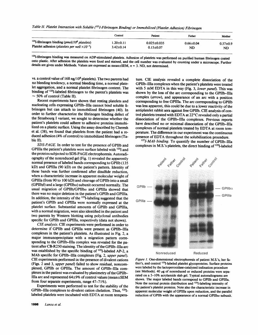
Table II. Platelet Interaction with Soluble ('25I-Fibrinogen Binding) or Immobilized (Platelet Adhesion) Fibrinogen
Control Patient Father Mother
'251-fibrinogen binding (pmol/lO' platelets) 1.20±0.11 0.025±0.035 0.66±0.04 0.57±0.9Platelet adhesion (platelets per well X105) 3.42±0.14 0.15±0.07 ND ND
'25I-fibrinogen binding was measured on ADP-stimulated platelets. Adhesion of platelets was performed on purified human fibrinogen coatedonto plastic. After adhesion the platelets were fixed and stained, and the cell number was evaluated by counting under a microscope. Furtherdetails are given under Methods. Values are expressed as mean±SEM, n = 3. ND, not determined.
vs. a control value of 168 ng/108 platelets). The two parents hadno bleeding tendency, a normal bleeding time, a normal plate-let aggregation, and a normal platelet fibrinogen content. Thebinding of "25I-labeled fibrinogen to the parent's platelets was
50% of control (Table II).Recent experiments have shown that resting platelets and
nucleating cells expressing GPIIb-IIIa cannot bind soluble fi-brinogen but can attach to immobilized fibrinogen (40). Inorder to further characterize the fibrinogen binding defect ofthe Strasbourg I variant, we sought to determine whether thepatient's platelets could adhere to adhesive proteins immobi-lized on a plastic surface. Using the assay described by Chereshet al. (38), we found that platelets from the patient had a re-duced adhesion (4% ofcontrol) to immobilized fibrinogen (Ta-ble II).
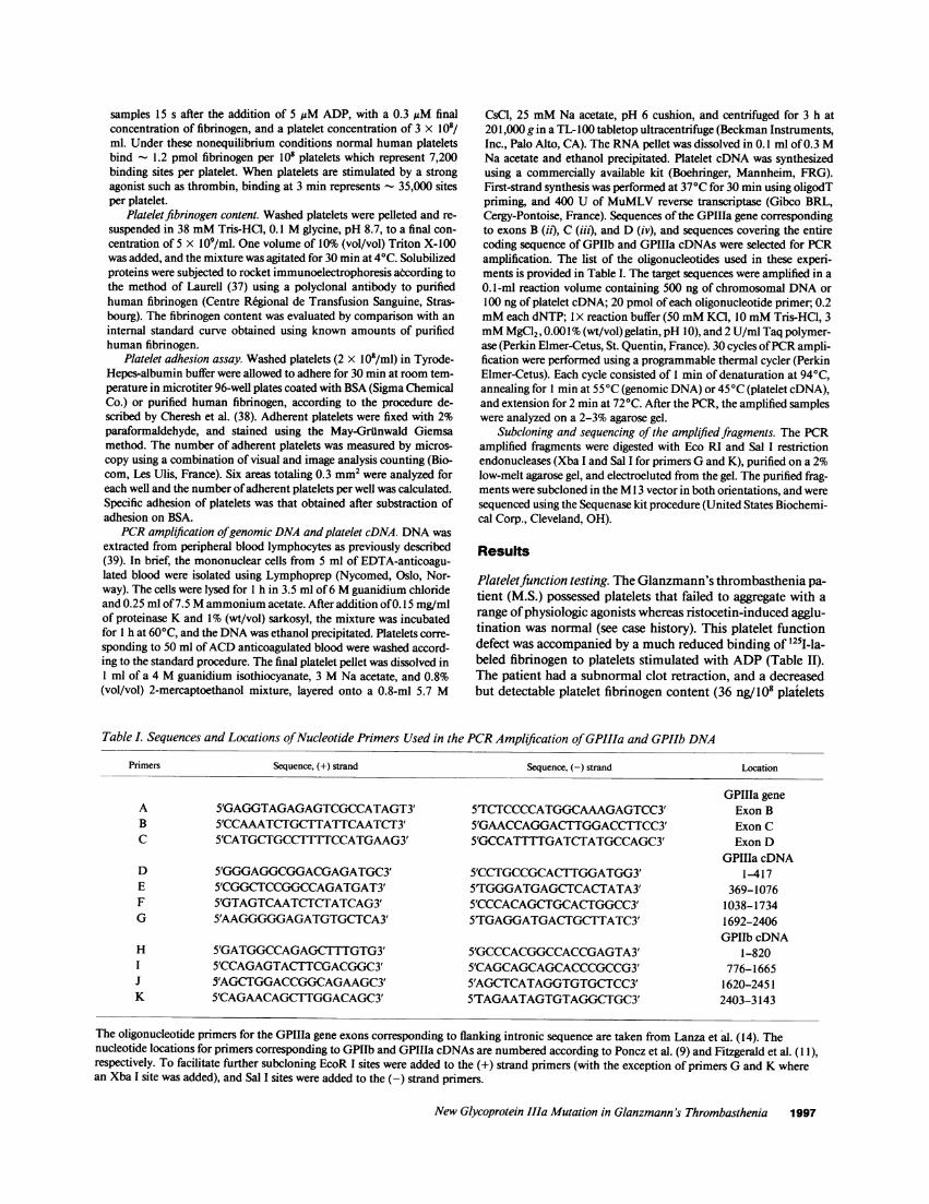
SDS-PAGE. In order to test for the presence of GPIIb andGPTIIa the patient's platelets were surface labeled with 125I andthe proteins subjected to SDS-PAGE electrophoresis. Autoradi-ography of the nonreduced gel (Fig. 1) revealed the apparentlynormal presence oflabeled bands corresponding to GPIlb (135kD) and GPIIIa (90 kD) on the patient's pattern. Identity ofthese bands was further confirmed after disulfide reduction,when a characteristic increase in apparent molecular weight ofGPIIla (from 90 to 100 kD) and cleavage ofGPIIb into a small(GPIIbf3) and a large (GPIIba) subunit occurred normally. Theusual migration of GPIIb/GPIIba and GPIIIa showed thatthere was no major deletion in the patient's GPIIb and GPIIIa.In addition, the intensity of the "25I-labeling suggested that thepatient's GPIIb and GPIIIa were normally expressed at theplatelet surface. Substantial amounts of GPIIb and GPIIIa,with a normal migration, were also identified in the patient andtwo parents by Western blotting using polyclonal antibodiesspecific for GPIIb and GPIIIa, respectively (data not shown).
CIE analysis. CIE experiments were performed in order todetermine if GPIIb and GPIIIa were present as GPIIb-IIIacomplexes in the patient's platelets. As illustrated in Fig. 2, amajor immunoprecipitate with a migration pattern corre-sponding to the GPIIb-IIIa complex was revealed for the pa-tient after CB-R250 staining. The identity ofthe GPIIb-IIIa arcwas established by the specific binding of '25I-labeled AP-2, aMAb specific for GPIIb-IIIa complexes (Fig. 2, upper panel).CIE experiments performed in the presence ofdivalent cations(Figs. 2 and 3, upper panel) failed to show residual, noncom-plexed, GPIIb or GPIIIa. The amount of GPIIb-IIIa com-plexes in the patient was evaluated by planimetry ofthe GPIIb-IIa arc and represented 64±9% ofcontrol values (mean±SEMfrom four separate experiments, range 47-71%).
Experiments were performed to test for the stability of theGPIIb-IIIa complexes to divalent cation chelation. Thus, 125I-labeled platelets were incubated with EDTA at room tempera-
ture. CIE analysis revealed a complete dissociation of theGPIIb-II1a complexes when the patient's platelets were treatedwith 5 mM EDTA in this way (Fig. 3, lower panel). This wasshown by the loss of the arc corresponding to the GPIIb-IT1acomplex (arrow), and appearance of an arc with a positioncorresponding to free GPIIIa. The arc corresponding to GPIIbwas less apparent, this could be due to a lower reactivity oftheantiplatelet rabbit sera against free GPIIb. CIE analysis ofcon-trol platelets treated with EDTA at 22°C revealed only a partialdissociation of the GPIIb-IITa complexes. Previous reportshave described no or minimal dissociation of the GPIIb-IIIacomplexes of normal platelets treated by EDTA at room tem-perature. The difference in our experiment was the continuouspresence ofEDTA throughout the solubilization procedure.
'25I-MAb binding. To quantify the number of GPIIb-IIIacomplexes in M.S.'s platelets, the direct binding of "25I-labeled
g$ I \$
(( C IQ e' CP~j
GPIlb
GPIIla
Nonreduced
- GPllbo.- GPIIla
;..... Ask
...._I
Reduced
Figure 1. One-dimensional electrophoresis of patient M.S.'s, her fa-ther's, and control 125I-labeled platelet glycoproteins. Surface proteinswere labeled by the lactoperoxidase-catalyzed iodination procedure(see Methods). 40 1Ag of nonreduced or reduced proteins were sepa-rated on a 5-10% acrylamide slab gel. Typical autoradiograms areshown. The major labeled bands correspond to GPIIb and GPIIIa.Note the normal protein distribution and '251-labeling intensity ofthe patient's platelet proteins. Note also the characteristic increase inapparent molecular weight ofGPIIIa upon reduction, and the parallelreduction of GPIIb with the appearance of a normal GPIIba subunit.
1998 Lanza et al.

patient's platelets with 2 mM EDTA at 220C resulted in thedissociation of most of the patient's GPIIb-IIIa complexes asseen by a negative shift in the fluorescence (Fig. 4 B). In conclu-sion, these experiments firmly established an increased sensitiv-ity for EDTA dissociation of the Strasbourg I variant GPIIb-i11a.
Recently, antibodies have been described that preferen-tially react with GPIIb-IIIa after it binds RGD containing pep-tides. These have been referred to as LIBS antibodies (41). Thebinding of such a LIBS antibody, D3GP3, was evaluated byFACS analysis after incubation ofcontrol or patient's plateletswith 1 mM GRGDSP for 30 min. A positive shift in fluores-
Ptient cence was seen in both the control (Fig. 4 C) and patient'splatelet populations (Fig. 4 D) when compared to untreatedplatelets or platelets incubated with the control GRGESP pep-tide (data not shown). These results imply that the patient's
ca2+
Figure 2. GPIIb-IIIa complexes in patient M.S.'s platelets as revealedby CIE. Washed, unlabeled platelets were solubilized with TritonX-100 and the soluble proteins were separated by electrophoresis inagarose. Second-dimension electrophoresis was performed across an
intermediate gel containing trace amounts of '25I-labeled AP-2, andinto an upper gel containing a rabbit anti-human platelet antibody.A series of immunoprecipitate arcs were revealed by CB-R250 stain-ing (lower panel). An immunoprecipitate in the position correspond-ing to GPIIb-IIIa can be visualized on the patient's gel. This arc was
confirmed as being given by GPIIb-IIIa after autoradiography re-
vealed the presence of the GPIIb-IIIa complex-specific '251-labeledAP-2 antibody (upperpane). Trace levels of platelet fibrinogen (FBG)can be visualized on the patient's CB-R250-stained gel. A controlarc corresponding to GPIb is also indicated.
AP-2 was measured using washed platelets. Scatchard analysisrevealed the presence of 19,400 AP-2 binding sites on the pa-
tient's platelets (Table III) compared with 36,130 binding sitesfor the control. Treatment ofthe patient's platelets with EDTAat 220C resulted in a 98% decrease in '251I-AP-2 binding sitesfrom 19,400 to 300 binding sites per platelet. In comparisonEDTA-treated control platelets bound the same amount of'251-AP-2 as in the absence of divalent cation chelation. Interest-ingly, the parents' platelets had a 38-49% decrease in '251-AP-2binding upon EDTA treatment.
Flow cytometry analysis. A panel of GPIIb-IIIa complex-specific MAbs, including AP-2, P2, CS 14, and DI2A, was
shown to bind to the patient's platelets by FACS analysis. Fig. 4illustrates results obtained for P2. Binding to control plateletsincubated with EDTA at room temperature was identical tothat observed in the presence of Ca2+ (Fig. 4 A). However,binding to normal platelets fell to background levels whenGPIIb-IIIa complexes were dissociated with 5 mM EDTA at370C, conditions where GPIIb-IIIa complexes ofnormal plate-lets do dissociate (see Discussion). In contrast, treatment ofthe
lb ,:
A.4:,.
lb
Ilb-Illa ..
A:
Cont rol
EDTA 220C
Patient
lb
lb
i,Or. ..
U b :Il
1 1 a ..~~~~~~~~~~~~~~~~~k
/' '".C'n''';
Patient
Figure 3. Increased sensitivity of patient M.S. platelet GPIIb-IIIa toEDTA dissociation as revealed by CIE. '25I-labeled platelets were in-cubated for 30 min at 220C in the presence of divalent cations (upperpanel) or in the presence of 5 mM EDTA in a buffer where Ca2+ andMg2" were omitted (lower panel). Platelets were then processed forCIE as described in Fig. 2 with the exception that no '25l-labeled an-
tibody was present in the intermediate gel. Immunoprecipitatescorresponding to surface-labeled proteins were revealed after autora-diography ofthe dried gel. In the presence ofCa2", a GPIIb-IIIa im-munoprecipitate is present in the control and patient's CIE (upperpanel). After treatment of the platelets with EDTA, there is a partialreduction ofthe GPIIb-IIIa immunoprecipitate in the control (lowerleft panel) with the appearance of a new immunoprecipitate corre-
sponding to dissociated GPIIIa. Similar treatment with EDTA of thepatient's platelets (lower right panel) induces the complete disappear-ance of the GPIIb-IIIa immunoprecipitate (arrow), which is fully dis-sociated into GPIIIa (arrow) and GPIIb (not visible on this gel).
New Glycoprotein IIIa Mutation in Glanzmann's Thrombasthenia 1999
1251_AP-2
lib-Illa
I. Nb
Control
CS-Rlb
lb-Illa 1
.. A I
lb
IIIA:-sr`
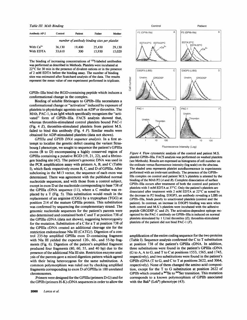
Table III. MAb Binding
Antibody AP-2 Control Patient Father Mother
number ofantibody binding sites per platelet
With Ca2+ 36,130 19,400 25,430 29,130With EDTA 33,610 300 15,930 15,020
The binding of increasing concentrations of 1251-labeled antibodieswas performed as described in Methods. Platelets were incubated at220C for 30 min in the presence of divalent cations or in the presenceof 2 mM EDTA before the binding assay. The number of bindingsites was estimated after Scatchard analysis of the data. The resultsrepresent the mean value of one experiment performed in triplicate.
GPIIb-IIIa bind the RGD-containing peptide which induces aconformational change in the complex.
Binding of soluble fibrinogen to GPIIb-IIIa necessitates aconformational change or "activation" induced by exposure ofplatelets to physiologic agonists such as ADP or thrombin. TheMAb, PAC-l, is an IgM which specifically recognizes the "acti-vated" form of GPIIb-IIIa. FACS analysis showed that,whereas thrombin-stimulated control platelets bound PAC-1(Fig. 4 E), thrombin-stimulated platelets from patient M.S.failed to bind this antibody (Fig. 4 F). Similar results wereobtained for ADP-stimulated platelets (data not shown).
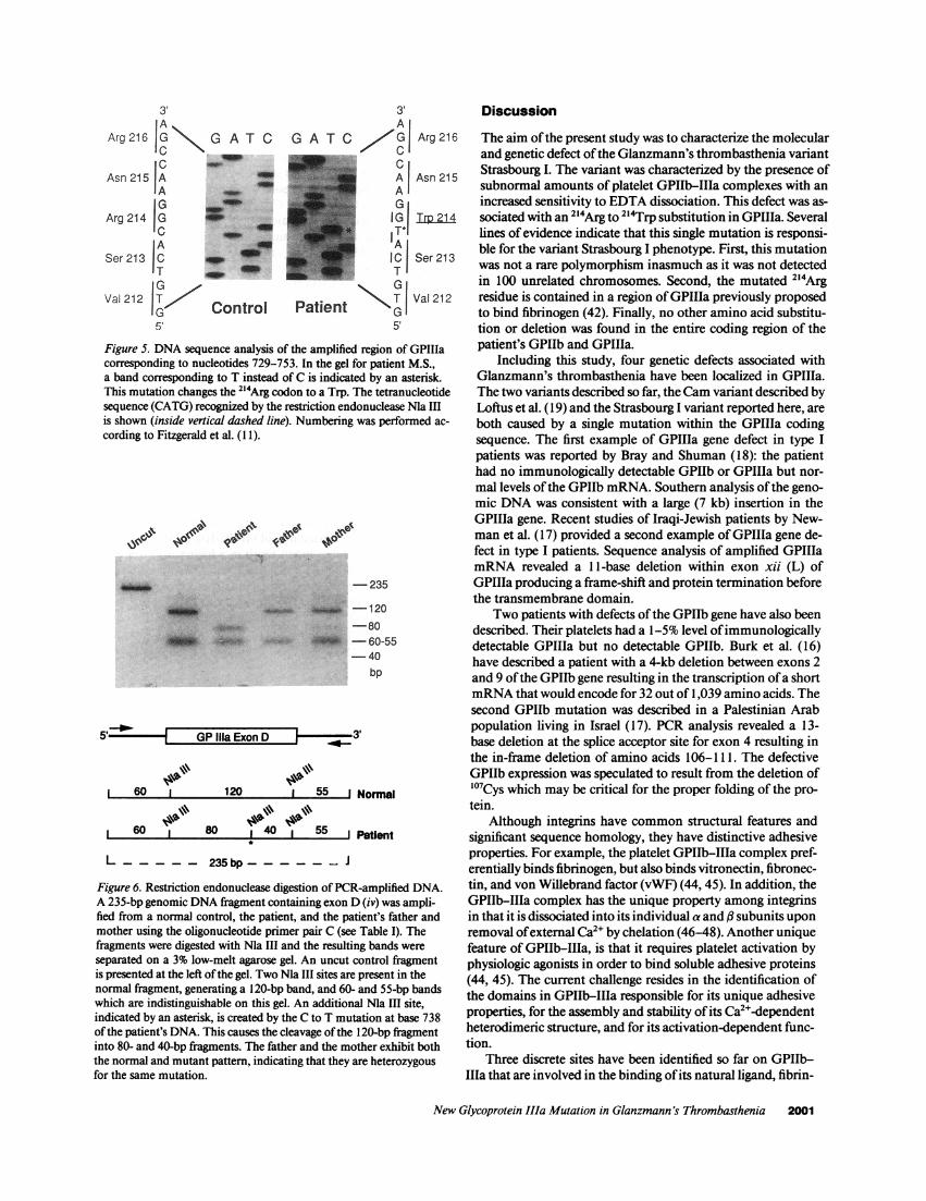
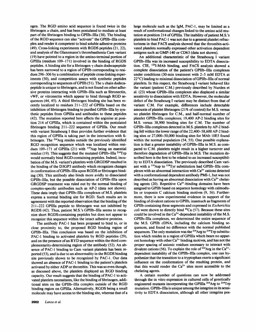
GPIIIa and GPIIb DNA sequence analysis. In a first at-tempt to localize the genetic defect causing the variant Stras-bourg I phenotype, we sought to sequence the patient's GPIIIaexons (B to D) encompassing a highly conserved region ofGPIIIa containing a putative RGD (19, 21, 22), and a fibrino-gen binding site (42). The patient's genomic DNA was used inthe PCR amplification assay with primers A, B, and C (TableI), which flank respectively exons B, C, and D of GPIIIa. Aftersubcloning in the M 13 vector, the sequence of each exon wasdetermined. There was agreement with the published normalnucleotide sequences, and with those from a control subject,except in exon D at the nucleotide corresponding to base 738 ofthe GPIIIa cDNA sequence (1 1), where a C residue was re-placed by a T (Fig. 5). This substitution would result in thereplacement of an arginine (CGG) by a tryptophan (TGG) atposition 214 of the mature GPIIIa protein. This substitutionwas confirmed by sequencing the complementary strand. Thegenomic nucleotide sequences for the patient's parents werealso determined and contained both C and T at position 738 ofthe GPIIIa cDNA (data not shown), suggesting heterozygosityfor the mutation. Substitution ofa C for a T at position 738 ofthe GPIIIa cDNA created an additional cleavage site for therestriction endonuclease Ma III (CATG/). Digestion of a con-trol 235-bp amplified GPIIIa exon D-containing fragmentwith Ma III yielded the expected 120-, 60-, and 55-bp frag-ments (Fig. 6). Digestion of the patient's amplified fragmentproduced four fragments (80, 60, 55, and 40 bp) due to thepresence ofthe additional Ma III site. Restriction enzyme anal-ysis ofthe parents gave a mixed digestion pattern which agreedwith their being heterozygous for the same substitution. Acommon polymorphism was ruled out by checking amplifiedfragments corresponding to exon D ofGPIIIa in 100 unrelatedchromosomes.
-Primers were designed for the GPIIIa (primers D-G) and forthe GPIIb (primers H-K) cDNA sequences in order to allow the
.0
E
C:
0
c
Control
P2 (GPIIb-IlIla) A
EDTA37°C EDTA22°C
D/
I,
I,-a~~~~~~~N
Patient
P2 (GPIIb-l11a) B
EDTA37°CEDTA22'C
/\ C
-%
'~
Fluorescence Intensity (Log)
Figure 4. Flow cytometric analysis of the control and patient M.S.platelet GPIIb-IIIa. FACS analysis was performed on washed platelets(see Methods). Results are expressed as histograms of cell number on
the ordinate versus fluorescence intensity (log scale) on the abscissa.The shaded area represents platelet autofluorescence in experimentsperformed with an irrelevant antibody. The presence of the GPIIb-Ila complex on control and patient M.S.'s platelets is attested by thebinding ofthe MAb P2 (A and B). Complete dissociation ofsurfaceGPIIb-IIIa occurs after treatment of both the control and patient'splatelets with 5 mM EDTA at 37°C. Only the patient's platelets are
dissociated after treatment with 2 mM EDTA at 22°C as noted bythe decrease in P2 binding. D3GP3, an antibody revealing a LIBS on
GPIIb-IIIa, binds poorly to unactivated platelets (control and thepatient). In contrast, an increase in D3GP3 binding was seen whenboth control and M.S.'s platelets were incubated with the adhesivepeptide GRGDSP (C and D). The activation-dependent epitope rec-
ognized by the PAC- I antibody on GPIIb-IIIa is induced on normalplatelets stimulated by 1 U/ml thrombin (E); thrombin-stimulatedplatelets of the patient did not bind PAC- (F).
amplification ofthe entire coding sequence for the two proteins(Table I). Sequence analysis confirmed the C to T substitutionat position 738 of the patient's GPIIIa cDNA. In addition,three substitutions were found in the patient's GPIIIa cDNA(G to A, A to G, and T to C at positions 1553, 1565, and 1745,respectively), and two substitutions were found in the patient'sGPIIb cDNA (T to G, and C to T at positions 2622, and 3064,respectively). None of them changed the amino acid composi-tion, except for the T to G substitution at position 2622 ofGPIIb which created a M3lie to 843Ser transition. This mutationcorresponds to a known polymorphism of GPIIb associatedwith the Bakb (Lekb) phenotype (43).
2000 Lanza et al.
D3GP3 (LIBS) C
Unstim. ,GRGDSP
IAI.1 ;:..
D3GP3 (LIBS) D
Unstim. GRGDSP
''
PAC-1 E
Unstim. Thrombin
1 10 100
PAC-1 F
Unstim.
Thrombin
1 10 100
3,
Arg 216 G G A T C G A TC16IC ECl
Asn 215 A A Asn215
A _ _ AG Gl
Arg 214IGC To
A4 A
Ser 213 C IC Ser 213T ___ TVa 212 T Control Patient VGI
5' 5'
Figure 5. DNA sequence analysis of the amplified region of GPIIIacorresponding to nucleotides 729-753. In the gel for patient M.S.,a band corresponding to T instead of C is indicated by an asterisk.This mutation changes the 214Arg codon to a Trp. The tetranucleotidesequence (CATG) recognized by the restriction endonuclease Ma IIIis shown (inside vertical dashed line). Numbering was performed ac-
cording to Fitzgerald et al. (I 1).
\ag Gov ho
amm
*_ '
-235
_ - 120-80
41W - 60-55-40
bp
GP Ilia Exon D
1 60 1
60
120 1 55 I Normal
80 1 40 1 55 J Patient
L 235 bp .-. J
Figure 6. Restriction endonuclease digestion of PCR-amplified DNA.A 235-bp genomic DNA fragment containing exon D (iv) was ampli-fied from a normal control, the patient, and the patient's father andmother using the oligonucleotide primer pair C (see Table I). Thefragments were digested with Ma III and the resulting bands wereseparated on a 3% low-melt agarose gel. An uncut control fragmentis presented at the left ofthe gel. Two Ma III sites are present in thenormal fragment, generating a 120-bp band, and 60- and 55-bp bandswhich are indistinguishable on this gel. An additional Ma III site,indicated by an asterisk, is created by the C to T mutation at base 738of the patient's DNA. This causes the cleavage ofthe 120-bp fragmentinto 80- and 40-bp fiagments. The father and the mother exhibit boththe normal and mutant pattern, indicating that they are heterozygousfor the same mutation.
Discussion
The aim of the present study was to characterize the molecularand genetic defect ofthe Glanzmann's thrombasthenia variantStrasbourg I. The variant was characterized by the presence ofsubnormal amounts of platelet GPIIb-IIIa complexes with anincreased sensitivity to EDTA dissociation. This defect was as-sociated with an 2"4Arg to 214Trp substitution in GPIIIa. Severallines of evidence indicate that this single mutation is responsi-ble for the variant Strasbourg I phenotype. First, this mutationwas not a rare polymorphism inasmuch as it was not detectedin 100 unrelated chromosomes. Second, the mutated 2`4Argresidue is contained in a region ofGPIIIa previously proposedto bind fibrinogen (42). Finally, no other amino acid substitu-tion or deletion was found in the entire coding region of thepatient's GPIIb and GPIIIa.
Including this study, four genetic defects associated withGlanzmann's thrombasthenia have been localized in GPIIIa.The two variants described so far, the Cam variant described byLoftus et al. (19) and the Strasbourg I variant reported here, areboth caused by a single mutation within the GPIIIa codingsequence. The first example of GPIIIa gene defect in type Ipatients was reported by Bray and Shuman (18): the patienthad no immunologically detectable GPIIb or GPIIIa but nor-mal levels of the GPIIb mRNA. Southern analysis of the geno-mic DNA was consistent with a large (7 kb) insertion in theGPIIIa gene. Recent studies of Iraqi-Jewish patients by New-man et al. (17) provided a second example ofGPIIIa gene de-fect in type I patients. Sequence analysis of amplified GPIIIamRNA revealed a 11-base deletion within exon xii (L) ofGPIIIa producing a frame-shift and protein termination beforethe transmembrane domain.
Two patients with defects ofthe GPIIb gene have also beendescribed. Their platelets had a 1-5% level ofimmunologicallydetectable GPIIIa but no detectable GPIIb. Burk et al. (16)have described a patient with a 4-kb deletion between exons 2and 9 ofthe GPIIb gene resulting in the transcription ofa shortmRNA that would encode for 32 out of 1,039 amino acids. Thesecond GPIIb mutation was described in a Palestinian Arabpopulation living in Israel (17). PCR analysis revealed a 13-base deletion at the splice acceptor site for exon 4 resulting inthe in-frame deletion of amino acids 106-1 1 1. The defectiveGPIIb expression was speculated to result from the deletion of107Cys which may be critical for the proper folding of the pro-tein.
Although integrins have common structural features andsignificant sequence homology, they have distinctive adhesiveproperties. For example, the platelet GPIIb-IIIa complex pref-erentially binds fibrinogen, but also binds vitronectin, fibronec-tin, and von Willebrand factor (vWF) (44, 45). In addition, theGPIIb-IIIa complex has the unique property among integrinsin that it is dissociated into its individual a and ,B subunits uponremoval ofexternal Ca2" by chelation (46-48). Another uniquefeature of GPIIb-IIIa, is that it requires platelet activation byphysiologic agonists in order to bind soluble adhesive proteins(44, 45). The current challenge resides in the identification ofthe domains in GPIIb-IIIa responsible for its unique adhesiveproperties, for the assembly and stability of its Ca2-dependentheterodimeric structure, and for its activation-dependent func-tion.
Three discrete sites have been identified so far on GPIIb-MIIa that are involved in the binding ofits natural ligand, fibrin-
New Glycoprotein IIIa Mutation in Glanzmann's Thrombasthenia 2001
ogen. The RGD amino acid sequence is found twice in thefibrinogen a chain, and has been postulated to mediate at leastpart of the fibrinogen binding to GPIIb-IIIa (38). The bindingof the RGD sequence can also "activate" the GPIIb-IIIa com-plex and render it competent to bind soluble adhesive proteins(49). Cross-linking experiments with RGDS peptides (21, 22),and analysis ofthe Glanzmann's thrombasthenia Cam variant(19) have pointed to a region in the amino-terminal portion ofGPIIIa (residues 109-171) involved in the binding of RGDSpeptides. A binding site for a fibrinogen y chain dodecapeptidehas been narrowed to a region ofGPIIb corresponding to resi-dues 296-306 by a combination ofpeptide cross-linking exper-iments (50), and competition assays with synthetic peptidescorresponding to sequences ofGPIIb (51). They chain dodeca-peptide is unique to fibrinogen, and is not found on other adhe-sive proteins interacting with GPIIb-IIIa such as fibronectin,vWF, or vitronectin which appear to bind through RGD se-quences (44, 45). A third fibrinogen binding site has been re-cently localized to residues 211-222 of GPIIIa based on theinhibition of fibrinogen binding to purified GPIIb-IIIa by syn-thetic peptides from GPIIIa and antibodies to these peptides(42). The mutation reported here affects the arginine at posi-tion 214 of GPIIIa, which is contained within the postulatedfibrinogen binding 211-222 sequence of GPIIIa. Our resultwith variant Strasbourg I thus provides further evidence thatthis region of GPIIIa is taking part in the interaction with fi-brinogen. The 2"4Arg resides outside the previously identifiedRGD recognition sequence which was localized within resi-dues 109-171 of GPIIIa (21) with "9Asp being an essentialresidue (19). This suggests that the mutated GPIIb-IIIa 2`4Trpwould normally bind RGD-containing peptides. Indeed, incu-bation ofthe M.S. variant's platelets withGRGDSP resulted inthe binding of the D3GP3 antibody which recognizes changesin conformation ofGPIIb-IIIa upon RGDS or fibrinogen bind-ing (30). This antibody also binds more avidly to dissociatedGPIIb-IIIa, but the possible dissociation of GPIIb-IIIa uponGRGDSP treatment was ruled out by the normal binding ofcomplex-specific antibodies such as AP-2 (data not shown).These data imply that GPIIb-IIIa complexes of M.S. plateletsexpress a normal binding site for RGD. Such results are inagreement with the reported observation that the binding ofthe211-222 GPIIIa peptide to fibrinogen was not inhibited byRGDS (42). Thus, patient M.S.'s GPIIb-IIIa seems to recog-nize short RGDS-containing peptides but does not appear torecognize this sequence within the intact adhesive proteins.
The antibody PAC-1 has been proposed to bind to, or inclose proximity to, the proposed RGD binding region ofGPIIb-IIIa. This conclusion was based on the inhibition ofPAC-1 binding to activated platelets by RGD peptides (29),and on the presence ofan RYD sequence within the third com-plementarity-determining region of the antibody (52). An ab-sence of PAC- I binding to Cam variant platelets has been re-ported (53), and is due to an abnormality in the RGDS bindingsite previously shown to be recognized by PAC-1. Our datashowed an absence of PAC- 1 binding to the patient's plateletsactivated by eitherADP or thrombin. This was so even though,as discussed above, the platelets displayed an RGD bindingcapacity. Our result suggests that the binding ofPAC-1 to acti-vated platelets necessitates, like the binding offibrinogen, addi-tional sites on the GPIIb-IIIa complex outside of the RGDbinding region on GPIIIa. Alternatively, RGDS being a smallmolecule may have access to the binding site, whereas that ofa
large molecule such as the IgM, PAC-1, may be limited as aresult of conformational changes linked to the amino acid mu-tation at position 214 of GPIIIa. The inability of patient M.S.'splatelets to bind PAC-l was not due to a general platelet refrac-toriness in that FACS analysis showed that the thrombin-acti-vated platelets normally expressed other activation dependentantigens such as GMP-140 or CD63 (data not shown).
An additional characteristic of the Strasbourg I variantGPIIb-IIIa was its increased susceptibility to EDTA dissocia-tion. CIE, '23I-MAb binding, and FACS analysis showed acomplete dissociation of the patient's GPIIb-IIIa complexesunder conditions (30-min treatment with 2-5 mM EDTA at220C) leading to minimal dissociation ofGPIIb-IIIa ofnormalplatelets. In this respect, the Strasbourg I variant behaved likethe variant (patient C.M.) previously described by Nurden etal. (23) whose GPIIb-IIIa complexes also displayed a similarsensitivity to dissociation with EDTA. However, the moleculardefect of the Strasbourg I variant may be distinct from that ofvariant C.M. For example, differences include detectableamounts of platelet fibrinogen (21% of control) for M.S. versusno platelet fibrinogen for C.M., and half-normal number ofplatelet GPIIb-IIIa complexes; 19,400 AP-2 binding sites forM.S. versus 38,900 binding sites for C.M. The number ofGPIIb-IIIa complexes detected in M.S. platelets by AP-2 bind-ing fell within the lower range ofthe 22,400-58,600 AP-2 bind-ing sites or 27,000-50,000 binding sites for MAb 10E5 foundwithin the normal population (54, 55). One possible explana-tion is that a greater instability of GPIIb-IIIa in M.S. as com-pared to C.M. platelets might result in a higher turnover andtherefore degradation of GPIIb-IIIa in M.S. The mutation de-scribed here is the first to be related to an increased susceptibil-ity to EDTA dissociation. The previously described Cam vari-ant with a "19Asp to `9Tyr substitution had GPIIb-IIIa com-plexes with an abnormal interaction with Ca2" cations detectedwith a conformational dependent antibody PMI- 1, but was notdescribed as having an abnormal dissociation with Ca2" chelat-ing agents (20). Repetitive Ca2"-binding domains have beenassigned to GPIIb based on sequence homology with calmodu-lin or troponin C calcium binding motives (9, 56). Further-more, there is now experimental evidence in support of thebinding ofdivalent cations to GPIIb, inasmuch as fragments ofGPIIb containing these segments and expressed in Escherichiacoli were shown to directly bind 45Ca (57). Because these sitescould be involved in the Ca2+-dependent instability ofthe M.S.GPIIb-IIIa complexes, we determined the entire sequence ofthe M.S. GPIIb cDNA, including the calcium binding se-quences, and found no difference with the normal publishedsequences. The only mutation was the 214Arg to 214Trp substitu-tion which resides in a region ofGPIIIa which bears no appar-ent homology with other Ca2+ binding motives, and has not theproper spacing of anionic residues necessary to interact withdivalent cations (56). To explain the role of214Arg in the Ca2`-dependent instability of the GPIIb-IIIa complex, one can hy-pothesize that the transition to a tryptophan exerts a significantinfluence on the conformation of the resulting protein, andthat this would render the Ca2' sites more accessible to thechelating agents.
A certain number of questions can now be addressedthrough the in vitro expression in cultured cells of geneticallyengineered mutants incorporating the GPIIIa 214Arg to 214Trpmutation. GPIIb-IIIa is unique among the integrins in its sensi-tivity to EDTA dissociation, although all other integrins pos-
2002 Lanza et al.
sess homologous Ca2" binding motives in their a subunits. Thea,,/3 vitronectin receptor is very closely related to GPIIb-IIIa(58, 59), contains a common 3 subunit (GPIIIa), and has simi-lar ligand binding specifities, but is not susceptible to EDTAdissociation (60). We have not been able to study the patient'saf33 in cells who highly express this receptor, such as the vascu-lar endothelial cells. However, it will now be possible to exam-ine by oligonucleotide-directed mutagenesis if a a!V33 214Trpvitronectin receptor would lose its ligand binding capacity andalso be rendered sensitive to EDTA dissociation. The 214Arg to214Trp mutation resides in a region ofGPIIIa, which has a highdegree ofsequence identity (- 75%), when compared to that ofother integrin /3 subunits, i.e., /31, /2, /4, -35 (61-66). The high-est degree of homology is found between /3 (GPIIIa) and f5,and in the region corresponding to residues 211-222 ofGPIIIathere is 100% sequence identity. Like /3 (GPIIIa), /5 associateswith av to form a receptor for vitronectin and fibronectin.Thus, introduction ofthe Arg to Trp mutation could be appliedto av#5 and help to further define the role ofthe region around214Arg in ligand and subunit-subunit interactions.
The two parents of M.S. were studied in parallel and dis-played an heterozygous phenotype in terms offibrinogen bind-ing to ADP-stimulated platelets (- 50% of control), and intheir sensitivity to EDTA dissociation, i.e., treatment of theirplatelets at 220C with EDTA decreased by half their plateletGPIIb-IIIa complex content. The heterozygous phenotype wasextended at the molecular level by the finding that both parentswere heterozygous for the same C to T mutation at base 738 ofthe GPIIIa cDNA. The inheritance of this mutation has notbeen explored in other members ofthe family but could now beeasily studied by restriction enzyme analysis ofamplified geno-mic DNA corresponding to the GPIIIa gene exon D. This mu-tation might have originated from a de novo mutation. The Cto T sequence change reported here is a transition within a CGdinucleotide. The cytidine residue in CG dinucleotides is fre-quently methylated and may undergo deamination to yield athymidine (67). Such regions are mutational hotspots in manygenes; for example frequent mutations within hotspots havebeen described in the factor VIII (68), factor IX (69), and vWFgenes (70, 71).
In conclusion, the 214Arg to 214Trp mutation of GPIIIafound in the Glanzmann's variant Strasbourg I points to animportant domain of GPIIIa, distinct from the RGD bindingregion, involved in the binding of fibrinogen, and influencingthe stability of the GPIIb-IIIa complex. This study shows thatthe molecular characterization of Glanzmann's variants is avery useful approach in order to identify functional domains inthe GPIIb-IIIa complex, and should be of further aid in map-ping sites involved in ligand and subunit-subunit interactionwithin the integrin family of adhesive receptors.
AcknowledgmentsWe thank Dominique Pidard for his help in preparing the CIE experi-ments; Helene Pfitzinger from Codgene, Strasbourg, France who per-formed the HLA typing experiments; Corinne De la Salle and Jean-Louis Mandel for helpful discussions; Agnes Schwartz, Danila Go-baille, and Marie-Jeanne Baas for technical assistance; and ClaudineHelbourg for secretarial assistance.NoteAdded in proof After the manuscript for this article was acceptedfor publication, a thrombasthenic variant with properties similar to theStrasbourg I variant and a 214Arg to Gln mutation of GPIIIa was re-ported by Bajt et al. (72).
References
1. Caen, J. P., P. A. Castaldi, J. C. Leclerc, S. Inceman, M. J. Larrieu, M.Probst, and J. Bernard. 1966. Congenital bleeding disorders with long bleedingtime and normal platelet count. Am. J. Med. 41:4-26.
2. Nurden, A. T., and J. P. Caen. 1974. An abnormal platelet glycoproteinpattern in three cases ofGlanzmann's thrombasthenia. Br. J. Haematol. 28:253-260.
3. Phillips, D. R., and P. Poh Agin. 1977. Platelet membrane defects in Glanz-mann's thrombasthenia: evidence for decreased amounts oftwo major glycopro-teins. J. Clin. Invest. 60:535-545.
4. George, J. N., A. T. Nurden, and D. R. Philipps. 1984. Molecular defects ofthe interaction with platelets and the vessel wall. N. Engl. J. Med. 311:1084-1098.
5. George, J. N., J. P. Caen, and A. T. Nurden. 1990. Glanzmann thrombas-thenia: the spectrum of clinical disease. Blood. 75:1383-1395.
6. Hynes, R. 0. 1987. Integrins: a family of cell surface receptors. Cell.48:549-554.
7. Phillips, D. R., I. F. Charo, L. V. Parise, and L. A. Fitzgerald. 1988. Theplatelet membrane glycoprotein Ilb-Illa complex. Blood. 71:831-843.
8. Ginsberg, M. H., J. C. Loftus, and E. F. Plow. 1988. Cytoadhesins, inte-grins, and platelets. Thromb. Haemostasis. 59:1-6.
9. Poncz, M., R. Eisman, R. Heidenreich, S. M. Silver, G. Vilaire, S. Surrey, E.Schwartz, and J. S. Bennett. 1987. Structure ofthe platelet membrane glycopro-tein Ilb: homology to the a subunit ofthe vitronectin and fibronectin receptors. J.Biol. Chem. 262:8476-8482.
10. Heidenreich, R., R. Eisman, K. Delgrosso, S. Surrey, J. S. Bennett, E.Schwartz, and M. Poncz. 1990. The organization ofthe gene for platelet glycopro-tein Ilb. Biochemistry. 29:1232-1244.
11. Fitzgerald, L. A., B. Steiner, S. C. Rall, S.-S. Lo, and D. R. Phillips. 1987.Protein sequence of endothelial glycoprotein Ila derived from a cDNA clone:identity with platelet glycoprotein Illa and similarity to integrin. J. Biol. Chem.262:3936-3939.
12. Rosa, J.-P., P. F. Bray, 0. Gayet, G. I. Johnston, R. G. Cook, M. A.Shuman, and R. P. McEver. 1988. Cloning of glycoprotein Uila cDNA fromhuman erythroleukemia cells and localization of the gene to chromosome 17.Blood. 72:593-600.
13. Zimrin, A. B., R. Eisman, G. Vilaire, E. Schwartz, J. S. Bennett, and M.Poncz. 1988. Structure of platelet glycoprotein IIa: a common subunit for twodifferent membrane receptors. J. Clin. Invest. 81:1470-1475.
14. Lanza, F., N. Kieffer, D. R. Phillips, and L. A. Fitzgerald. 1990. Character-ization of the human platelet glycoprotein Ila gene: comparison with the fibro-nectin receptor ,B-subunit gene. J. Biol. Chem. 265:18098-18103.
15. Zimrin, A. B., S. Gidwitz, S. Lord, E. Schwartz, J. S. Bennett, G. C. WhiteII, and M. Poncz. 1990. The genomic organization ofplatelet glycoprotein II1a. J.Biol. Chem. 265:8590-8595.
16. Burk, C. D., P. J. Newman, S. Lyman, J. Gill, B. S. Coller, and M. Poncz.1991. A deletion in the gene for glycoprotein Ilb associated with Glanzmann'sthrombasthenia. J. Clin. Invest. 87:270-276.
17. Newman, P. J., U. Seligsohn, S. Lyman, and B. S. Coller. 1991. Themolecular genetic basis of Glanzmann thrombasthenia in the Iraqi-Jewish andArab population living in Israel. Proc. Natl. Acad. Sci. USA. 88:3160-3164.
18. Bray, P. F., and M. A. Shuman. 1990. Identification ofan abnormal genefor the GPIIIa subunit of the platelet fibrinogen receptor resulting in Glanz-mann's thrombasthenia. Blood. 75:881-888.
19. Loftus, J. C., T. E. O'Toole, E. F. Plow, A. Glass, A. L. Frelinger III, andM. H. Ginsberg. 1990. A 3 integrin mutation abolishes ligand binding and altersdivalent cation-dependent conformation. Science (Wash. DC). 249:915-918.
20. Ginsberg, M. H., A. Lightsey, T. J. Kunicki, A. Kaufmann, G. Marguerie,and E. F. Plow. 1986. Divalent cation regulation of the surface orientation ofplatelet membrane glycoprotein I.b. J Clin. Invest. 78:1103-1 111.
21. D'Souza, S. E., M. H. Ginsberg, T. A. Burke, S. C. T. Lam, and E. F. Plow.1988. Localization of an Arg-Gly-Asp recognition site within an integrin adhe-sion receptor. Science (Wash. DC). 242:91-94.
22. Smith, J. W., and D. A. Cheresh. 1988. The Arg-Gly-Asp binding domainof the vitronectin receptor. J. Biol. Chem. 263:18726-18731.
23. Nurden, A. T., J.-P. Rosa, D. Fournier, C. Legrand, D. Didry, A. Parquet,and D. Pidard. 1987. A variant ofGlanzmann's thrombasthenia with abnormalglycoprotein lb-Illa complexes in the platelet membrane. J. Clin. Invest. 79:962-969.
24. Legrand, C., V. Dubernard, A. T. Nurden. 1989. Studies on the mecha-nism of expression of secreted fibrinogen on the surface of activated humanplatelets. Blood. 73:1226-1234.
25. Fournier, D. J., A. Kabral, P. A. Castaldi, and M. C. Berndt. 1989. Avariant ofGlanzmann's thrombasthenia characterized by abnormal glycoproteinIlb/Illa complex formation. Thromb. Haemostasis. 62:977-983.
26. Pidard, D., A. L. Frelinger III, C. Bouillot, and A. T. Nurden. 1991.Activation of the fibrinogen receptor on human platelets exposed to alpha-chy-motrypsin: relation with a major proteolytic cleavage at the carboxyterminus ofthe membrane glycoprotein-Ilb heavy chain. Eur. J. Biochem. 200:437-448.
27. Pidard, D., R. R. Montgomery, J. S. Bennett, and T. J. Kunicki. 1983.
New Glycoprotein IIIa Mutation in Glanzmann's Thrombasthenia 2003
Interaction ofAP-2, a monoclonal antibody specific for the human platelet glyco-protein Ilb-I11a complex, with intact platelets. J. Biol. Chem. 258:12582-12586.
28. McEver, R. P., E. M. Bennett, and M. N. Martin. 1983. Identification oftwo structurally and functionally distinct sites on human platelet membraneglycoprotein I1b-IIIa using monoclonal antibodies. J. Biol. Chem. 258:5269-5275.
29. Shattil, S. J., M. Cunningham, and J. A. Hoxie. 1987. Detection of acti-vated platelets in whole blood using activation-dependent monoclonal antibodiesand flow cytometry. Blood. 70:307-315.
30. Kouns, W. C., C. D. Wall, M. M. White, C. F. Fox, and L. K. Jennings.1990. A conformation-dependent epitope ofhuman platelet glycoprotein IIIa. J.Biol. Chem. 265:20594-20601.
31. Cazenave, J.-P., S. Hemmendinger, A. Beretz, A. Sutter-Bay, and J.Launay. 1983. L'agr6gation plaquettaire: outil d'investigation clinique et d'6tudepharmacologique. Mhthodologie. Ann. Biol. Clin. 41:167-179.
32. Kinlough-Rathbone, R. L., J. F. Mustard, M. A. Packham, D. W. Perry,H.-J. Reimers, and J.-P. Cazenave. 1977. Properties of washed human platelets.Thromb. Haemostasis. 37:291-308.
33. Nurden, A. T., D. Dupuis, T. J. Kunicki, andJ. P. Caen. 1981. Analysis ofthe glycoprotein and protein composition of Bernard-Soulier platelets by singleand two-dimensional sodium dodecyl sulfate-polyacrylamide gel electrophoresis.J. Clin. Invest. 67:431-440.
34. Kunicki, T. J., D. Pidard, J.-P. Rosa, and A. T. Nurden. 1981. The forma-tion of Ca'+-dependent complexes of platelet membrane glycoproteins Ilb andIIla in solution as determined by crossed immunoelectrophoresis. Blood. 58:268-278.
35. Kekwick, R. A., M. E. McKay, M. H. Nance, and B. R. Record. 1955. Thepurification of human fibrinogen. Biochem. J. 60:671-683.
36. Mustard, J. F., R. L. Kinlough-Rathbone, M. A. Packham, D. W. Perry,E. J. Harfenist, and K. R. M. Pai. 1979. Comparison of fibrinogen associationwith normal and thrombasthenic platelets on exposure to ADP or chymotrypsin.Blood. 54:987-993.
37. Laurell, C. B. 1966. Quantitative estimation ofproteins by electrophoresisin agarose gel containing antibodies. Anal. Biochem. 15:45-52.
38. Cheresh, D. A., S. A. Berliner, V. Vicente, and Z. M. Ruggeri. 1989.Recognition of distinct adhesive sites on fibrinogen by related integrins on plate-lets and endothelial cells. Cell. 58:945-953.
39. De la Salle, C., M. J. Baas, L. Grunebaum, M. L. Wiesel, A. Bianco, R.Gialeraki, T. Mandalaki, and J.-P. Cazenave. 1989. Carrier detection in hemo-philia using pedigree analysis coagulation tests and DNA probes. Nouv. Rev. Fr.Hematol. 31:193-202.
40. Kieffer, N., L. A. Fitzgerald, D. Wolf, D. A. Cheresh, and D. R. Phillips.1991. Adhesive properties of the /3 integrins: comparison ofGP IIb-IIIa and thevitronectin receptor individually expressed in human melanoma cells. J. CellBiol. 113:451-461.
41. Frelinger III, A. L., S. C.-T. Lam, E. F. Plow, M. A. Smith, J. C. Loftus,and M. H. Ginsberg. 1988. Occupancy ofan adhesive glycoprotein receptor mod-ulates expression of an antigenic site involved in cell adhesion. J. Biol. Chem.263:12397-12402.
42. Charo, I. F., L. Nannizzi, D. R. Phillips, M. A. Hsu, and R. M. Scarbo-rough. 1991. Inhibition offibrinogen binding to GP IIb-IIIa by aGP Illa peptide.J. Biol. Chem. 266:1415-1421.
43. Lyman, S., R. H. Aster, G. P. Visentin, and P. J. Newman. 1990. Polymor-phism ofhuman platelet glycoprotein Ilb associated with the Bak/Bakb alloanti-gen system. Blood. 75:2343-2348.
44. Plow, E. F., M. H. Ginsberg, and G. A. Marguerie. 1986. Expression andfunction ofadhesive proteins on the platelet surface. In Biochemistry ofPlatelets.D. R. Phillips and M. A. Shuman, editors. Academic Press, Inc., New York.225-256.
45. Kieffer, N., and D. R. Phillips. 1990. Platelet membrane glycoproteins:functions in cellular interactions. Annu. Rev. Cell Biol. 6:329-357.
46. Pidard, D., D. Didry, T. J. Kunicki, and A. T. Nurden. 1986. Tempera-ture-dependent effects ofEDTA on the membrane glycoprotein IIb-IIIa complexand platelet aggregability. Blood. 67:604-611.
47. Fitzgerald, L. A., and D. R. Phillips. 1985. Calcium regulation of theplatelet membrane glycoprotein Ilb-Illa complex. J. Biol. Chem. 260:11366-11374.
48. Brass, L. F., S. J. Shattil, T. J. Kunicki, and J. S. Bennett. 1985. Effect ofcalcium on the stability of the platelet membrane glycoprotein IIb-IIIa complex.J. Biol. Chem. 260:7875-7881.
49. Du, X., E. F. Plow, A. L. Frelinger III, T. E. O'Toole, J. C. Loftus, andM. H. Ginsberg. 1991. Ligands "activate" integrin a,/3 (platelet GP UIb-IIIa).Cell. 65:409-416.
50. D'Souza, S. E., M. H. Ginsberg, T. A. Burke, and E. F. Plow. 1990. Theligand binding site of the platelet integrin receptor GPIIb-IIIa is proximal to thesecond calcium binding domain of its a subunit. J. Biol. Chem. 265:3440-3446.
51. D'Souza, S. E., M. H. Ginsberg, G. R. Matsueda, and E. F. Plow. 1991. A
discrete sequence in a platelet integrin is involved in ligand recognition. Nature(Lond.). 350:66-68.
52. Taub, R., R. J. Gould, V. M. Garsky, T. M. Ciccarone, J. Hoxie, P. A.Friedman, and S. J. Shattil. 1989. A monoclonal antibody against the plateletfibrinogen receptor contains a sequence that mimics a receptor recognition do-main in fibrinogen. J. Biol. Chem. 264:259-265.
53. Ginsberg, M. H., A. L. Frelinger, S. C.-T. Lam, J. Forsyth, R. McMillan,E. F. Plow, and S. J. Shattil. 1990. Analysis ofplatelet aggregation disorders basedon flow cytometric analysis of membrane glycoprotein Ilb-IIIa with conforma-tion-specific monoclonal antibodies. Blood. 76:2017-2023.
54. Nurden, A. T., D. Fournier, and D. Pidard. 1988. Pathological aspects ofplatelet aggregation: a consideration ofthe heterogeneity in Glanzmann's throm-basthenia. In Biochemistry and Physiopathology of Platelet Membrane. G. Mar-guerie and R. F. A. Zwall, editors. Colloq. INSERM (Inst. Natl. Sante Rech.Med.). 158:95-105.
55. Coller, B. S., U. Seligsohn, A. Zivelin, E. Zwang, A. Lusky, and M. Mo-dan. 1986. Immunologic and biochemical characterization of homozygous andheterozygous Glanzmann thrombasthenia in the Iraqi-Jewish and Arab popula-tions of Israel: comparison of techniques for carrier detection. Br. J. Hematol.62:723-735.
56. Strydnaka, N. C. J., and M. N. G. James. 1989. Crystal structures of thehelix-loop-helix calcium-binding proteins. Annu. Rev. Biochem. 58:951-998.
57. Gulino, D., C. Boudignon, L. Zhang, E. Concord, and G. Marguerie.1991. Analysis ofthe plateletGP Ilb metal binding domain. Thromb. Haemosta-sis. 65:697. (Abstr.)
58. Fitzgerald, L. A., M. Poncz, B. Steiner, S. C. Rall, J. S. Bennett, and D. R.Phillips. 1987. Comparison of cDNA-derived protein sequences of the humanfibronectin and vitronectin receptor a-subunits and platelet glycoprotein Ilb.Biochemistry. 26:8158-8165.
59. Suzuki, S., W. S. Argraves, H. Arai, L. R. Languino, M. D. Pierschbacher,and E. Ruoslahti. 1987. Amino acid sequence of the vitronectin receptor a sub-unit and comparative expression of adhesion receptor mRNAs. J. Biol. Chem.262:14080-14085.
60. Cheresh, D. A., and R. C. Spiro. 1987. Biosynthetic and functional proper-ties of an Arg-Gly-Asp-directed receptor involved in human melanoma cell at-tachment to vitronectin, fibrinogen and von Willebrand factor. J. Biol. Chem.262:17703-17711.
61. Argraves, W. S., S. Suzuki, H. Arai, K. Thompson, M. D. Pierschbacher,and E. Ruoslahti. 1987. Amino acid sequence ofthe human fibronectin receptor.J. Cell Biol. 105:1183-1 190.
62. Kishimoto, T. K., K. O'Connor, A. Lee, T. M. Roberts, and T. A.Springer. 1987. Cloning of the ,B subunit of the leukocyte adhesion proteins:homology to an extracellular matrix receptor defines a novel supergene family.Cell. 48:681-690.
63. Hogervorst, F., I. Kuikman, A. E. G. Kr. von dem Borne, and A. Sonnen-berg. 1990. Cloning and sequence analysis of beta-4 cDNA: an integrin subunitthat contains a unique 118 kd cytoplasmic domain. EMBO (Eur. Mol. Biol.Organ.) J. 9:765-770.
64. Suzuki, S., and Y. Naitoh. 1990. Amino acid sequence ofa novel integrin/4 subunit and primary expression ofthe mRNA in epitheial cells. EMBO (Eur.Mol. Biol. Organ.) J. 9:757-763.
65. Ramaswnamy, R., and M. E. Hemler. 1990. Cloning, primary structureand properties of a novel human integrin P1 subunit. EMBO (Eur. Mol. Biol.Organ.) J. 9:1561-1568.
66. McLean, J. W., D. J. Vestal, D. A. Cheresh, andS. C. Bodary. 1990. cDNAsequence of the human integrin /1s subunit. J. Biol. Chem. 265:17126-17131.
67. Bird, A. P. 1980. DNA methylation and the frequency ofCpG in animalDNA. Nucleic Acids Res. 8:1499-1504.
68. Youssoufian, H., H. H. Kazazian, Jr., D. G. Phillips, S. Aronis, G. Tsiftis,V. A. Brown, and S. E. Antonarakis. 1986. Recurrent mutations in haemophiliaA give evidence for CpG mutation hotspots. Nature (Lond.). 324:380-382.
69. Giannelli, F., P. M. Green, K. A. High, J. N. Lozier, D. P. Lillicrap, M.Ludwig, K. Olek, P. H. Reitsma, M. Goossens, A. Yoshioka, et al. 1990. Haemo-philia B: database of point mutations and short additions and deletions. NucleicAcids Res. 18:4053-4059.
70. Randi, A. M., I. Rabinowitz, D. J. Mancuso, P. M. Mannucci, and J. E.Sadler. 1991. Molecular basis ofvon Willebrand disase type IIB: candidate muta-tions cluster in one disulfide loop between proposed platelet glycoprotein lb bind-ing sequences. J. Clin. Invest. 87:1220-1226.
71. Cooney, K. A., W. C. Nichols, M. E. Vruck, W. F. Bahou, A. D. Shapiro,E. J. W. Bowie, H. R. Gralnick, and D. Ginsburg. 1991. The molecular defect intype Ilb von Willebrand disease: identification of four potential missense muta-tions within the putative GP lb binding domain. J. Clin. Invest. 87:1227-1233.
72. Bajt, M. L., M. H. Ginsberg, A. L. Frelinger, M. C. Berndt, and J. C.Loftus. 1992. A spontaneous mutation of integrin ab,183 (a platelet glycoproteinlIb-Illa) helps define a ligand binding site. J. Biol. Chem. 267:3789-3794.
2004 Lanza et al.
Copyright © 2022 FDOKUMEN