Isolated P-selectin Glycoprotein Ligand1 Dynamic Adhesion to P- and E-selectin
11
The Rockefeller University Press, 0021-9525/97/04/509/11 $2.00 The Journal of Cell Biology, Volume 137, Number 2, April 21, 1997 509–519 509 Isolated P-selectin Glycoprotein Ligand-1 Dynamic Adhesion to P- and E-selectin Douglas J. Goetz,* Daniel M. Greif,* Han Ding,* Raymond T. Camphausen, ‡ Steven Howes, ‡ Kenneth M. Comess, ‡ Karen R. Snapp, § Geoffrey S. Kansas, § and Francis W. Luscinskas* *Vascular Research Division, Department of Pathology, Brigham and Women’s Hospital, Harvard Medical School, Boston, Massachusetts 02115; ‡ Small Molecule Drug Discovery Group, Genetics Institute, Cambridge, Massachusetts 02140; and § Department of Microbiology-Immunology, Northwestern University Medical School, Chicago, Illinois 60611 Abstract. Leukocyte adhesion to vascular endothelium under flow involves an adhesion cascade consisting of multiple receptor pairs that may function in an overlap- ping fashion. P-selectin glycoprotein ligand-1 (PSGL-1) and L-selectin have been implicated in neutrophil ad- hesion to P- and E-selectin under flow conditions. To study, in isolation, the interaction of PSGL-1 with P- and E-selectin under flow, we developed an in vitro model in which various recombinant regions of extracellular PSGL-1 were coupled to 10-mm-diameter micro- spheres. In a parallel plate chamber with well defined flow conditions, live time video microscopy analyses re- vealed that microspheres coated with PSGL-1 attached and rolled on 4-h tumor necrosis factor-a–activated en- dothelial cell monolayers, which express high levels of E-selectin, and CHO monolayers stably expressing E- or P-selectin. Further studies using CHO-E and -P monolayers demonstrate that the first 19 amino acids of PSGL-1 are sufficient for attachment and rolling on both E- and P-selectin and suggest that a sialyl Lewis x–containing glycan at Threonine-16 is critical for this sequence of amino acids to mediate attachment to E- and P-selectin. The data also demonstrate that a sul- fated, anionic polypeptide segment within the amino terminus of PSGL-1 is necessary for PSGL-1–mediated attachment to P- but not to E-selectin. In addition, the results suggest that PSGL-1 has more than one binding site for E-selectin: one site located within the first 19 amino acids of PSGL-1 and one or more sites located between amino acids 19 through 148. A critical step in the recruitment of leukocytes to a site of inflammation is the adhesion of the leukocytes to the vascular endothelium in the fluid dynamic environment of the microcirculation. This adhesion pro- cess involves a cascade of events including initial attach- ment, rolling, spreading, and ultimately transendothelial migration (11, 20, 28, 50). In vivo and in vitro studies have shown that the inducible endothelial cell adhesion mole- cules P-selectin (CD62P) and E-selectin (CD62E) are in- volved in leukocyte initial attachment and rolling on acti- vated vascular endothelium (1, 20–22, 29–31). The third known member of the selectin family, L-selectin (CD62L), is expressed only on leukocytes and is also involved in leu- kocyte recruitment (4, 16, 22–24, 46, 50). The expression of E-selectin requires de novo protein synthesis and can be elicited by a 4-h treatment of cultured endothelium with certain Gram-negative endotoxins and cytokines such as IL-1 and tumor necrosis factor-a (TNF-a) 1 (7). P-selectin is stored in the Weibel-Palade bodies of endothelial cells and is rapidly mobilized to the cell surface by secreta- gogues such as thrombin and histamine (32). The cytokine activation pathway may also elicit limited P-selectin ex- pression on cultured human umbilical vein endothelium (29, 30). A notable feature of the selectins is their NH 2 -ter- minal, lectin-like domain that binds carbohydrate moieties in a Ca 21 -dependent manner (6). Thus, several carbohy- drate ligands for P- and E-selectin have been proposed in- cluding the sialyl Lewis x (SLe x ) tetrasaccharide and re- lated glycans (15, 41, 53). Recent studies have focused on identifying the underly- Address correspondence to Francis W. Luscinskas, Ph.D., Brigham and Women’s Hospital, Department of Pathology, 221 Longwood Ave., Bos- ton, MA 02115. Tel.: (617) 732-6004. Fax: (617) 732-5933. Douglas J. Goetz’s present address is The University of Memphis, De- partment of Biomedical Engineering, Engineering Technology Building Room 330, Memphis, TN 38152. Tel.: (901) 678-8675. Fax: (901) 678-5281. E-mail: [email protected] Douglas J. Goetz and Daniel M. Greif contributed equally to this work. 1. Abbreviations used in this paper: CHO-E, and -P, CHO cell monolayers stably expressing E- or P-selectin; DPBS, Dulbecco’s phosphate buffered saline; Fuc-TVII, a(1,3)fucosyltransferase-VII; HUVEC, human umbili- cal vein endothelial cell(s); OSGE, O-sialoglycoprotein endopeptidase; PSGL-1, P-selectin glycoprotein ligand-1; RT, room temperature; SLe x , sialyl Lewis x; TNF-a, tumor necrosis factor-a. on September 29, 2014 jcb.rupress.org Downloaded from Published April 21, 1997
Transcript of Isolated P-selectin Glycoprotein Ligand1 Dynamic Adhesion to P- and E-selectin

The Rockefeller University Press, 0021-9525/97/04/509/11 $2.00The Journal of Cell Biology, Volume 137, Number 2, April 21, 1997 509–519 509
Isolated P-selectin Glycoprotein Ligand-1Dynamic Adhesion to P- and E-selectin
Douglas J. Goetz,* Daniel M. Greif,* Han Ding,* Raymond T. Camphausen,
‡
Steven Howes,
‡
Kenneth M. Comess,
‡
Karen R. Snapp,
§
Geoffrey S. Kansas,
§
and Francis W. Luscinskas*
*Vascular Research Division, Department of Pathology, Brigham and Women’s Hospital, Harvard Medical School, Boston,
Massachusetts 02115;
‡
Small Molecule Drug Discovery Group, Genetics Institute, Cambridge, Massachusetts 02140; and
§
Department of Microbiology-Immunology, Northwestern University Medical School, Chicago, Illinois 60611
Abstract.
Leukocyte adhesion to vascular endothelium under flow involves an adhesion cascade consisting of multiple receptor pairs that may function in an overlap-ping fashion. P-selectin glycoprotein ligand-1 (PSGL-1) and L-selectin have been implicated in neutrophil ad-hesion to P- and E-selectin under flow conditions. To study, in isolation, the interaction of PSGL-1 with P- and E-selectin under flow, we developed an in vitro model in which various recombinant regions of extracellular
PSGL-1 were coupled to 10-
m
m-diameter micro-spheres. In a parallel plate chamber with well defined flow conditions, live time video microscopy analyses re-vealed that microspheres coated with PSGL-1 attached and rolled on 4-h tumor necrosis factor-
a
–activated en-dothelial cell monolayers, which express high levels of
E-selectin, and CHO monolayers stably expressing E- or P-selectin. Further studies using CHO-E and -P monolayers demonstrate that the first 19 amino acids of PSGL-1 are sufficient for attachment and rolling on both E- and P-selectin and suggest that a sialyl Lewis x–containing glycan at Threonine-16 is critical for this sequence of amino acids to mediate attachment to E- and P-selectin. The data also demonstrate that a sul-fated, anionic polypeptide segment within the amino terminus of PSGL-1 is necessary for PSGL-1–mediated attachment to P- but not to E-selectin. In addition, the results suggest that PSGL-1 has more than one binding site for E-selectin: one site located within the first 19 amino acids of PSGL-1 and one or more sites located between amino acids 19 through 148.
A
critical step in the recruitment of leukocytes to a siteof inflammation is the adhesion of the leukocytesto the vascular endothelium in the fluid dynamic
environment of the microcirculation. This adhesion pro-cess involves a cascade of events including initial attach-ment, rolling, spreading, and ultimately transendothelialmigration (11, 20, 28, 50). In vivo and in vitro studies haveshown that the inducible endothelial cell adhesion mole-cules P-selectin (CD62P) and E-selectin (CD62E) are in-volved in leukocyte initial attachment and rolling on acti-vated vascular endothelium (1, 20–22, 29–31). The thirdknown member of the selectin family, L-selectin (CD62L),is expressed only on leukocytes and is also involved in leu-kocyte recruitment (4, 16, 22–24, 46, 50). The expression of
E-selectin requires de novo protein synthesis and can beelicited by a 4-h treatment of cultured endothelium withcertain Gram-negative endotoxins and cytokines such asIL-1 and tumor necrosis factor-
a
(TNF-
a
)
1
(7). P-selectinis stored in the Weibel-Palade bodies of endothelial cellsand is rapidly mobilized to the cell surface by secreta-gogues such as thrombin and histamine (32). The cytokineactivation pathway may also elicit limited P-selectin ex-pression on cultured human umbilical vein endothelium(29, 30). A notable feature of the selectins is their NH
2
-ter-minal, lectin-like domain that binds carbohydrate moietiesin a Ca
2
1
-dependent manner (6). Thus, several carbohy-drate ligands for P- and E-selectin have been proposed in-cluding the sialyl Lewis x (SLe
x
) tetrasaccharide and re-lated glycans (15, 41, 53).
Recent studies have focused on identifying the underly-
Address correspondence to Francis W. Luscinskas, Ph.D., Brigham andWomen’s Hospital, Department of Pathology, 221 Longwood Ave., Bos-ton, MA 02115. Tel.: (617) 732-6004. Fax: (617) 732-5933.
Douglas J. Goetz’s present address is The University of Memphis, De-partment of Biomedical Engineering, Engineering Technology BuildingRoom 330, Memphis, TN 38152. Tel.: (901) 678-8675. Fax: (901) 678-5281.E-mail: [email protected]
Douglas J. Goetz and Daniel M. Greif contributed equally to thiswork.
1.
Abbreviations used in this paper
: CHO-E, and -P, CHO cell monolayersstably expressing E- or P-selectin; DPBS, Dulbecco’s phosphate bufferedsaline; Fuc-TVII,
a
(1,3)fucosyltransferase-VII; HUVEC, human umbili-cal vein endothelial cell(s); OSGE,
O
-sialoglycoprotein endopeptidase;PSGL-1, P-selectin glycoprotein ligand-1; RT, room temperature; SLe
x
,sialyl Lewis x; TNF-
a
, tumor necrosis factor-
a
.
on Septem
ber 29, 2014jcb.rupress.org
Dow
nloaded from
Published April 21, 1997
The Journal of Cell Biology, Volume 137, 1997 510
ing leukocyte proteins which present carbohydrate ligandsfor binding to E- and P-selectin. Perhaps the best charac-terized of these is P-selectin glycoprotein ligand-1 (PSGL-1),which was first isolated from HL-60 cells (33) and subse-quently cloned from an HL-60 cell cDNA library (43).PSGL-1, a homodimer of disulfide-linked subunits with anapparent molecular mass of 120 kD each (33), is presenton a variety of leukocytes including neutrophils, mono-cytes, and lymphocytes (35). PSGL-1 is extensively glyco-sylated with N-linked glycans and closely spaced O-linkedglycans, a portion of which are modified by SLe
x
(34, 37,54). L-selectin expressed on myeloid leukocytes also hasbeen proposed as a ligand for E- and P-selectin since, simi-larly to PSGL-1, it displays SLe
x
-type structures and is lo-calized on the tips of microvilli (22, 35, 39).
In vitro flow adhesion assays have investigated PSGL-1–and L-selectin–mediated leukocyte adhesion to E- andP-selectin under fluid flow. Patel et al. (38) measured neu-trophil accumulation on CHO cell monolayers stably ex-pressing P-selectin (CHO-P) or E-selectin (CHO-E). Basedon mAb blocking studies, the authors inferred that PSGL-1is necessary for attachment of neutrophils to P-selectinand for optimal attachment to E-selectin. In contrast, theyalso reported that neutrophils pretreated (in the presenceof cytochalasin D to prevent shape change) with fMLP, achemoattractant known to induce endoproteolytic cleav-age of L-selectin, did not accumulate on CHO-P or -Emonolayers but did retain functional PSGL-1–dependentbinding sites for P-selectin (38). A separate study (22) re-ported that the accumulation of neutrophils on solubleE-selectin under flow was inhibited (
z
90%) by mAbs toL-selectin. The authors inferred that neutrophil attach-ment to E-selectin under flow requires L-selectin, imply-ing that PSGL-1 is not sufficient to mediate attachment toE-selectin under flow. mAb approaches have suggestedthe involvement of PSGL-1 in neutrophil rolling, subse-quent to attachment, on E- and P-selectin (35, 38). How-ever, since attachment is a prerequisite for rolling, if theattachment step is blocked by an mAb to PSGL-1 (35, 38),it is difficult to determine if PSGL-1 can mediate adhesiveevents subsequent to attachment such as rolling. There-fore, while it appears that PSGL-1 is involved in neutro-phil attachment to E- and P-selectin, it is unclear if PSGL-1is sufficient to mediate attachment and rolling.
Previous studies have investigated PSGL-1 regions nec-essary for recognition of P- and E-selectin (42, 44). Thesestudies have revealed that an anionic polypeptide segmentcontaining three tyrosine residues, at least one of which issulfated, is located within the amino terminus of maturePSGL-1 (42, 44). This region is necessary for fluid phasePSGL-1 recognition of cell surface–expressed P-selectinbut is unnecessary for fluid phase PSGL-1 recognition ofcell surface–expressed E-selectin (44). However, the struc-ture–function relationship for PSGL-1 and E- and P-selec-tin has not been investigated in the more relevant (49) invitro flow assay.
To examine isolated PSGL-1 attachment and rolling onE- and P-selectin, we have studied the adhesion of 10-
m
m-diam nondeformable microspheres, coated with variousextracellular regions of PSGL-1, to TNF-
a
–activated cul-tured human endothelium and to CHO-E and -P monolay-ers under defined flow conditions. The results demonstrate
that: (
a
) PSGL-1 is sufficient to mediate microsphere at-tachment and rolling on TNF-
a
–activated endothelialmonolayers and on CHO-E and -P monolayers; (
b
) the first19 amino acids of PSGL-1 are sufficient for attachmentand rolling on both E- and P-selectin; (
c
) an SLe
x
contain-ing O-glycan at Threonine-16 is critical for the first 19amino acids of PSGL-1 to mediate attachment to E- andP-selectin; (
d
) the anionic polypeptide segment within theamino terminus of PSGL-1 is necessary for PSGL-1–medi-ated attachment to P- but not E-selectin; and (
e
) PSGL-1has more than one binding site for E-selectin: one site lo-cated within the first 19 amino acids of PSGL-1 and one ormore sites located between amino acids 19 through 148.
Materials and Methods
Materials
RPMI-1640 containing 1 mM
l
-glutamine and 20 mM Hepes, Alpha me-dia, DPBS with (DPBS
1
) or without Ca
2
1
and Mg
2
1
, were obtained fromBiowhittaker (Walkersville, MD). FBS and dialyzed FBS were obtained fromHyclone Labs (Urem, UT).
O
-sialoglycoprotein endopeptidase (OSGE)was obtained from Accurate Chemicals (Westbury, NY). Neuraminidasefrom
Vibrio cholerae
was obtained from Boehringer Mannheim Corp. (In-dianapolis, IN). BSA was obtained from Sigma Chemical Co. (St. Louis,MO). All other chemicals were of the highest grade available from J.T.Baker, Inc. (Phillipsburg, NJ).
Antibodies
Function blocking murine mAb to E-selectin 7A9 (IgG
1
) was obtainedfrom American Type Culture Collection (Rockville, MD) (clone HB 10135)and used as F(ab
9
)
2
(10
m
g/ml). Nonblocking murine mAb to E-selectinH4/18 (IgG
1
) was used as F(ab
9
)
2
(10
m
g/ml). Leukocyte function blockingmurine anti–P-selectin mAb, HPDG2/3 (IgG
1
) (43), was used as F(ab
9
)
2
(10
m
g/ml), and nonblocking murine anti–P-selectin mAb, HPDG2/1 (IgG
1
)(43), was used as purified IgG
1
(10
m
g/ml). The following antibodies rec-ognize human PSGL-1 and have been described previously: murine mAbPSL-275 (IgG
1
; purified IgG, 20
m
g/ml) (43, 48) and polyclonal rabbit sera,Rb3026 (1:20 dilution) (43, 48). Normal rabbit sera (1:20 dilution) wasused as a control for Rb3026. mAbs KPL1 and KPL2 (Snapp, K.R., F.W.Luscinskas, R. Warnke, and G.S. Kansas. 1997.
J. Allergy Clin. Immunol.
99:s459) recognize PSGL-1 and were used as ascites (1:200 dilution for ad-hesion assays and 1:500 for flow cytometric analysis) and purified IgG.Murine antihuman IgG
1
Fc specific (CALTAG Laboratories, San Fran-cisco, CA) was used as a control for the purified KPL1. Murine anti–ICAM-1 mAb Hu5/3 (IgG
1
) was used as purified IgG (20
m
g/ml). Murineantibody to human Class I, W6/32, (IgG
2a
) was used as F(ab
9
)
2
(10
m
g/ml).FITC-labeled goat F(ab
9
)
2
secondary antibodies (each at 1:50 dilution) torabbit IgG, mouse IgG, and human Fc were obtained from CALTAGLaboratories. FITC-labeled goat F(ab
9
)
2
secondary antibody (1:50 dilu-tion) to mouse Fc and unlabeled goat F(ab
9
)
2
were obtained from JacksonImmunoResearch Laboratories (West Grove, PA).
Cell Culture
CHO cells stably expressing human E-selectin (CHO-E) or human P-selectin(CHO-P) were prepared and cultured in Alpha media containing 10% dia-lyzed FBS as previously detailed (43). Human umbilical vein endothelialcells (HUVEC) were isolated and cultured as described previously (28).For induction of adhesion molecule expression, HUVEC monolayerswere activated by a 4-h treatment with 25 ng/ml TNF-
a
(29).
Soluble Forms of PSGL-1
The PSGL-1 molecules used in these studies are chimeras consisting ofvarious truncated extracellular regions of mature PSGL-1 fused to theheavy chain CH2-CH3 (Fc) region of human IgG
1
. Their construction, ex-pression, and selectin-binding properties have been described previously(44). In the present studies, plasmids encoding two of these chimeras,
on Septem
ber 29, 2014jcb.rupress.org
Dow
nloaded from
Published April 21, 1997

Goetz et al.
PSGL-1-mediated Attachment of Microspheres
511
148.Fc and
D
Y148.Fc (abbreviated to
D
Y.Fc here), were transfected intoCOS cells as described except for the substitution of a plasmid encodingan
a
(1,3)fucosyltransferase-VII (Fuc-TVII) enzyme (36, 45) for the Fuc-TIII plasmid used in earlier cotransfections (44). The resulting condi-tioned media were supplemented with 0.002% NaN
3
and 10
m
M PMSF andthen clarified by centrifugation. Fc chimeras were purified by proteinA–Sepharose (Pharmacia LKB Biotechnology, Inc., Piscataway, NJ) chro-matography per the manufacturer’s instructions and quantified by an anti-Fcenzyme-linked immunosorbent assay using human IgG
1
(Sigma ChemicalCo.) as a standard. The 19.Fc form of PSGL-1 described earlier (44) wasmutated to include an enterokinase cleavage site (19) located between thePSGL-1 and IgG Fc regions and is termed 19.ek.Fc. A plasmid encodingthis construct was stably transfected into CHO cells that were engineeredto also express Fuc-TVII and
b
(1,6)-
N
-acetylglucosaminyltransferase(“core2” [9]) activities (17, 25). 19.ek.Fc was purified from conditionedmedia and quantified as above. Similar to COS produced 19.Fc, 19.ek.Fcis tyrosine sulfated and glycosylated with SLe
x
-type containing
O
-glycansand, in E- and P-selectin binding experiments (44), is functionally indistin-guishable (data not shown). The nomenclature for microspheres coatedwith various extracellular regions of PSGL-1 is as follows: the 148.Fc con-struct, 148.Fc microspheres; the 19.ek.Fc construct, 19.ek.Fc microspheres;the
D
Y.Fc construct,
D
Y.Fc microspheres (see Fig. 6
a
).
Preparation, Flow Cytometric Analysis, and Enzymatic Treatment of PSGL-1 Microspheres
Preparation.
10-
m
m nondeformable polystyrene microspheres (Poly-sciences Inc., Warrington, PA) were washed twice in 0.1 M NaHCO
3
, pH9.2, and incubated (2.5
3
10
7
microspheres/ml) overnight at room temper-ature (RT) with 300
m
g/ml protein A (Zymed Labs, South San Francisco,CA) in 0.1 M NaHCO
3
, pH 9.2, with end-to-end rotation. Microsphereswere washed twice with DPBS and blocked for 30 min at RT with DPBScontaining 1% BSA. After the blocking step, the microspheres (2
3
10
8
microspheres/ml) were incubated with a PSGL-1.Fc extracellular con-struct or human IgG
1
diluted in DPBS for 1 h at RT with agitation. Themicrospheres were washed with DPBS
1
, 1% BSA and blocked withDPBS
1
, 1% BSA containing 200
m
g/ml human IgG
1
kappa or lambda(Sigma Chemical Co.). Microspheres were held in this buffer at 1
3
10
8
microspheres/ml at RT for 1 h before use in the assays.
Indirect Immunofluorescence and Flow Cytometric Analysis of PSGL-1Microspheres.
After the human IgG
1
blocking step, replicate aliquots (
z
5
3
10
5
) of microspheres were washed in DPBS
1
containing 1% BSA andincubated for 20 min at RT with 20
m
g/ml primary mAb in DPBS
1
, 1%BSA. After incubation, microspheres were washed and blocked with unla-beled goat F(ab
9
)
2
fragments (100
m
g/ml), and the primary mAb was de-tected with FITC-labeled F(ab
9
)
2
fragments of goat anti–mouse Fc-spe-cific secondary antibody. The microspheres were incubated for 20 min atRT, washed twice in DPBS
1
, 1% BSA, once in DPBS
1
, and fixed with2% formaldehyde in DPBS
1
. FITC fluorescence was determined on aFACScan
®
flow cytometer (Becton-Dickinson Immunocytometry Sys.,Mountain View, CA) by counting the fluorescence of 5,000 or 10,000 mi-crospheres. The data are presented as single parameter histograms on afour-decade scale.
OSGE Treatment of PSGL-1 Microspheres.
Microspheres were preparedas described above up to blocking with human IgG
1
. A suspension ofPSGL-1–coated microspheres (1
3
10
8
/ml) was incubated in DPBS
1
,0.2% FBS, 0.05% NaN
3
, 25 mM Hepes, pH 7.4, with or without 160
m
g/mlOSGE for 30 min at 37
8
C. The microspheres were then washed withDPBS
1
, 1% BSA and blocked with DPBS
1
, 1% BSA containing 200
m
g/mlhuman IgG1 at 1 3 108 microspheres/ml. The microspheres used in theflow cytometric analysis to detect human Fc were incubated with 0.2% nor-mal rabbit serum instead of human IgG1, blocked with 100 mg/ml unla-beled goat (Fab9)2 fragments, and then stained with goat FITC-labeled(Fab9)2 fragments of antibodies to either human Fc or mouse IgG.
Sialidase Treatment of PSGL-1 Microspheres. Microspheres were pre-pared with the 19.ek.Fc construct as described above. 19.ek.Fc micro-spheres were then washed twice in RPMI-1640, resuspended to 1 3 108 inRPMI-1640 with or without 0.1 U/ml neuraminidase, and incubated for 30min at 378C. Microspheres were then washed twice in DPBS1, 1% BSAand blocked (1 3 108 microspheres/ml) with DPBS1, 1% BSA containing200 mg/ml human IgG1.
Parallel Plate Flow Chamber AnalysisThe parallel plate flow chamber apparatus used in these studies has been
described in detail (28). Temperature was maintained at 378C by a heatingplate. The flow apparatus was mounted on an inverted microscope (modelDiaphot; Nikon Inc., Melville, NY), and the entire perfusion period wasrecorded on videotape by a video camera and VCR. Microspheres weredrawn through the chamber at 5 3 105 microspheres/ml in DPBS1, 0.5%BSA, and microsphere attachment rates were quantified by observing a103 field of view for z1 min and 15 s. The number of microspheres thatattached throughout this time period was determined and divided by thetime of observation and the area of the field of view to yield the rate of at-tachment per unit area. For a given coverslip, an attachment rate was de-termined at two different fields of view and the two values were averagedto give an n 5 1. Microsphere accumulation was determined by countingthe number of microspheres interacting with the substrate in a particularfield of view (103 magnification). This value was determined for three tofive different fields of view between 3.5 to 4 min after initiation of micro-sphere perfusion. These numbers were averaged and normalized to thearea of the field of view. Rolling velocities were determined manually bymeasuring the distance a microsphere traveled in a given amount of time.
Antibody Blocking ExperimentsAll control or blocking antibodies to molecules on the cell monolayerswere diluted (10 mg/ml) in the appropriate media and added to the mono-layers 20 min before the adhesion assays described above. For mAb block-ing of the PSGL-1 microspheres, 10–15 ml of a 1 3 108 microsphere/mlsuspension was mixed 1:1 with DPBS1, 1% BSA containing a 1:100 dilu-tion of ascites mAb KPL1 or KPL2. This suspension was allowed to incu-bate for 15 min at RT, diluted to 5 3 105 microspheres/ml in DPBS1,0.5% BSA, and used immediately in the flow chamber assay.
StatisticsAll differences were evaluated by a two-tailed Student’s t test. P valuesrepresent the results of these t tests and values <0.05 were considered sig-nificant. All error bars represent standard deviations.
Results
A PSGL-1.Fc Chimera Can Be Coupled to Polystyrene Microspheres Precoated with Protein A
Previous investigators have developed methods to couplebiotinylated SLex (10) or various mAbs (18, 40) to micro-spheres and used the resulting microspheres as model cellsin adhesion assays. We adapted these techniques to studythe interaction of PSGL-1, in isolation, with E- and P-selectinunder fluid flow. Sako et al. (44) have generated and de-scribed recombinant PSGL-1.Fc constructs that consist ofvarious portions of the extracellular region of PSGL-1fused to the Fc region of human IgG1. The presence of theFc region led to the idea of coupling the PSGL-1.Fc con-structs to microspheres precoated with protein A. Such anapproach allows for the “correct” orientation of PSGL-1.Fcchimera on the microsphere, i.e., the PSGL-1 portion of thePSGL-1.Fc chimera oriented away from the microsphereand available for binding to other molecules and the Fcportion of the chimera coupled to the protein A adsorbedto the surface of the microsphere. Although recombinantforms of PSGL-1 may differ slightly from native PSGL-1,other investigators have used recombinant forms of PSGL-1to gain significant insights into the biochemistry of PSGL-1–E- and –P-selectin interactions (25, 42, 44) . In addition,Sako et al. (44) have documented that recombinant andnative PSGL-1 exhibit similar function in a variety of bio-chemical assays.
In initial studies, 148.Fc, a truncated PSGL-1 constructconsisting of the first 148 amino acids of PSGL-1 linked tothe Fc region of human IgG1 (44), was coupled to 10-mm-
on Septem
ber 29, 2014jcb.rupress.org
Dow
nloaded from
Published April 21, 1997
The Journal of Cell Biology, Volume 137, 1997 512
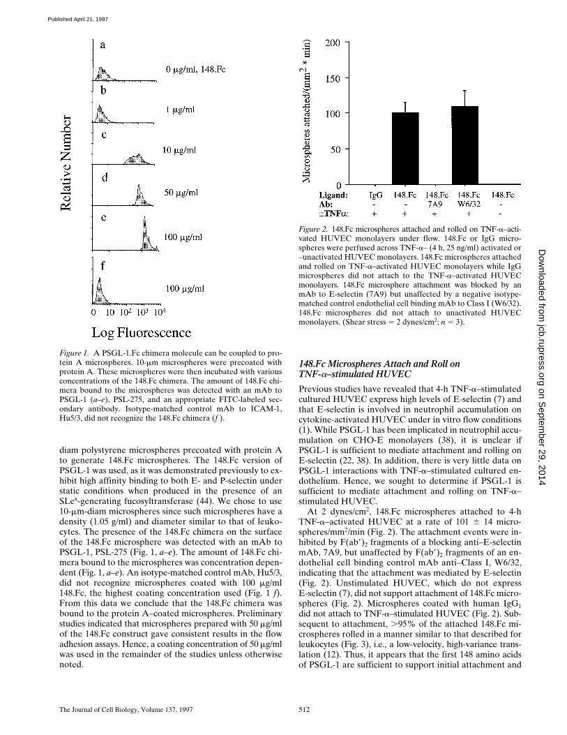
diam polystyrene microspheres precoated with protein Ato generate 148.Fc microspheres. The 148.Fc version ofPSGL-1 was used, as it was demonstrated previously to ex-hibit high affinity binding to both E- and P-selectin understatic conditions when produced in the presence of anSLex-generating fucosyltransferase (44). We chose to use10-mm-diam microspheres since such microspheres have adensity (1.05 g/ml) and diameter similar to that of leuko-cytes. The presence of the 148.Fc chimera on the surfaceof the 148.Fc microsphere was detected with an mAb toPSGL-1, PSL-275 (Fig. 1, a–e). The amount of 148.Fc chi-mera bound to the microspheres was concentration depen-dent (Fig. 1, a–e). An isotype-matched control mAb, Hu5/3,did not recognize microspheres coated with 100 mg/ml148.Fc, the highest coating concentration used (Fig. 1 f).From this data we conclude that the 148.Fc chimera wasbound to the protein A–coated microspheres. Preliminarystudies indicated that microspheres prepared with 50 mg/mlof the 148.Fc construct gave consistent results in the flowadhesion assays. Hence, a coating concentration of 50 mg/mlwas used in the remainder of the studies unless otherwisenoted.
148.Fc Microspheres Attach and Roll onTNF-a–stimulated HUVEC
Previous studies have revealed that 4-h TNF-a–stimulatedcultured HUVEC express high levels of E-selectin (7) andthat E-selectin is involved in neutrophil accumulation oncytokine-activated HUVEC under in vitro flow conditions(1). While PSGL-1 has been implicated in neutrophil accu-mulation on CHO-E monolayers (38), it is unclear ifPSGL-1 is sufficient to mediate attachment and rolling onE-selectin (22, 38). In addition, there is very little data onPSGL-1 interactions with TNF-a–stimulated cultured en-dothelium. Hence, we sought to determine if PSGL-1 issufficient to mediate attachment and rolling on TNF-a–stimulated HUVEC.
At 2 dynes/cm2, 148.Fc microspheres attached to 4-hTNF-a–activated HUVEC at a rate of 101 6 14 micro-spheres/mm2/min (Fig. 2). The attachment events were in-hibited by F(ab9)2 fragments of a blocking anti–E-selectinmAb, 7A9, but unaffected by F(ab9)2 fragments of an en-dothelial cell binding control mAb anti–Class I, W6/32,indicating that the attachment was mediated by E-selectin(Fig. 2). Unstimulated HUVEC, which do not expressE-selectin (7), did not support attachment of 148.Fc micro-spheres (Fig. 2). Microspheres coated with human IgG1did not attach to TNF-a–stimulated HUVEC (Fig. 2). Sub-sequent to attachment, .95% of the attached 148.Fc mi-crospheres rolled in a manner similar to that described forleukocytes (Fig. 3), i.e., a low-velocity, high-variance trans-lation (12). Thus, it appears that the first 148 amino acidsof PSGL-1 are sufficient to support initial attachment and
Figure 1. A PSGL-1.Fc chimera molecule can be coupled to pro-tein A microspheres. 10-mm microspheres were precoated withprotein A. These microspheres were then incubated with variousconcentrations of the 148.Fc chimera. The amount of 148.Fc chi-mera bound to the microspheres was detected with an mAb toPSGL-1 (a–e), PSL-275, and an appropriate FITC-labeled sec-ondary antibody. Isotype-matched control mAb to ICAM-1,Hu5/3, did not recognize the 148.Fc chimera (f ).
Figure 2. 148.Fc microspheres attached and rolled on TNF-a–acti-vated HUVEC monolayers under flow. 148.Fc or IgG micro-spheres were perfused across TNF-a– (4 h, 25 ng/ml) activated or–unactivated HUVEC monolayers. 148.Fc microspheres attachedand rolled on TNF-a–activated HUVEC monolayers while IgGmicrospheres did not attach to the TNF-a–activated HUVECmonolayers. 148.Fc microsphere attachment was blocked by anmAb to E-selectin (7A9) but unaffected by a negative isotype-matched control endothelial cell binding mAb to Class I (W6/32).148.Fc microspheres did not attach to unactivated HUVECmonolayers. (Shear stress 5 2 dynes/cm2; n 5 3).
on Septem
ber 29, 2014jcb.rupress.org
Dow
nloaded from
Published April 21, 1997

Goetz et al. PSGL-1-mediated Attachment of Microspheres 513
subsequent rolling on TNF-a–activated endothelium andthat the attachment step is mediated by E-selectin.
148.Fc Microspheres Attach and Roll on CHO-Pand -E Monolayers
While it appears that PSGL-1 is necessary for accumula-tion of neutrophils on CHO-P monolayers, it is unclear ifPSGL-1 is sufficient to mediate attachment and rolling(38). To determine if PSGL-1 is sufficient to mediate at-tachment and rolling on P-selectin and to further test the
hypothesis that PSGL-1 is sufficient to mediate attach-ment and rolling on E-selectin, we studied the interactionof 148.Fc microspheres with CHO-P and -E monolayers.
At 2 dynes/cm2, 148.Fc microspheres attached to CHO-Eand -P monolayers at a rate of 122 6 50 microspheres/mm2/min and 119 6 43 microspheres/mm2/min, respec-tively (Fig. 4). Subsequent to attachment, .95% of the at-tached 148.Fc microspheres rolled on either CHO-E or -Pmonolayers. Function blocking F(ab9)2 mAbs to E- (7A9)and P-selectin (HPDG2/3) completely inhibited the attach-ment of 148.Fc microspheres to CHO-E and -P monolayers,respectively (Fig. 4). In contrast, control isotype-matchedF(ab9)2 preparation of mAb W6/32 and non–function block-ing mAbs to E- (H4/18) and P-selectin (HPDG2/1) (Fig. 4and data not shown) had no effect on the rate of attach-ment of the 148.Fc microspheres to CHO-E and -P mono-layers, respectively. Under identical conditions, the 148.Fcmicrospheres did not attach to the parental CHO cell line(n 5 2, data not shown), nor did human IgG1 microspheresattach to CHO-P or -E monolayers (Fig. 4). Thus, it ap-pears that the first 148 amino acids of PSGL-1 are suffi-cient to support attachment and subsequent rolling onCHO cell monolayers expressing E- or P-selectin.
Attachment of 148.Fc Microspheres to CHO-Eand -P Cell Monolayers Is Eliminated by Pretreatment of 148.Fc Microspheres with OSGE
Previous studies have demonstrated that PSGL-1 is cleavedby the metalloprotease OSGE (37). To investigate the speci-ficity of the 148.Fc microsphere attachment events, wetreated 148.Fc microspheres with OSGE before use in ourflow assay. Treatment of the 148.Fc microspheres withOSGE significantly reduced (.95%) the presence of theepitope recognized by the PSGL-1 mAb PSL-275 (Fig. 5 A).However, the level of human IgG Fc present on the micro-sphere was unchanged indicating that the PSGL-1 peptideof the 148.Fc chimera was specifically cleaved by OSGE(Fig. 5 A). Treatment of 148.Fc microspheres with OSGEbefore use in the flow assay eliminated attachment to bothCHO-P and -E monolayers (Fig. 5 B). From these data, weconclude that 148.Fc microsphere attachment to CHO-E
Figure 3. 148.Fc microspheres rolled on TNF-a–stimulatedHUVEC. The image shows two 148.Fc microspheres (whitespheres) rolling over a TNF-a–activated HUVEC monolayer(gray background). Images were captured, every 0.6 s, from a vid-eotape of the experiment and layered together to give the com-posite image shown. Note that the 148.Fc microspheres translatedin the direction of the flow with a nonconstant velocity. The aver-age velocity of 10 different 148.Fc microspheres was determinedand found to be 14 mm/sec, which is ,3% of the hydrodynamicvelocity of a noninteracting hard sphere translating 50 nm fromthe surface (13). The length of the image shown is 100 mm. Shearstress 5 2 dynes/cm2.
Figure 4. 148.Fc microspheresattached and rolled on CHO-Pand -E monolayers. 148.Fc mi-crospheres attached and rolledon CHO-E and -P monolayerswhile IgG microspheres did notattach to either CHO mono-layer. 148.Fc microsphere at-tachment to CHO-E and -Pmonolayers was blocked by anmAb to E-selectin (7A9) and anmAb to P-selectin (HPDG2/3),respectively, but was unaffectedby an isotype-matched controlmAb (W6/32). 148.Fc micro-spheres did not attach to the pa-rental CHO cell line (data notshown). (Shear stress 5 2 dynes/cm2; n 5 3).
on Septem
ber 29, 2014jcb.rupress.org
Dow
nloaded from
Published April 21, 1997
The Journal of Cell Biology, Volume 137, 1997 514
and -P monolayers is specifically mediated by the PSGL-1peptide of the 148.Fc chimera.
PSGL-1 Regions Necessary and/orSufficient for Attachment and Rolling on CHO-Eand -P Monolayers
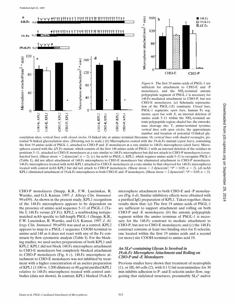
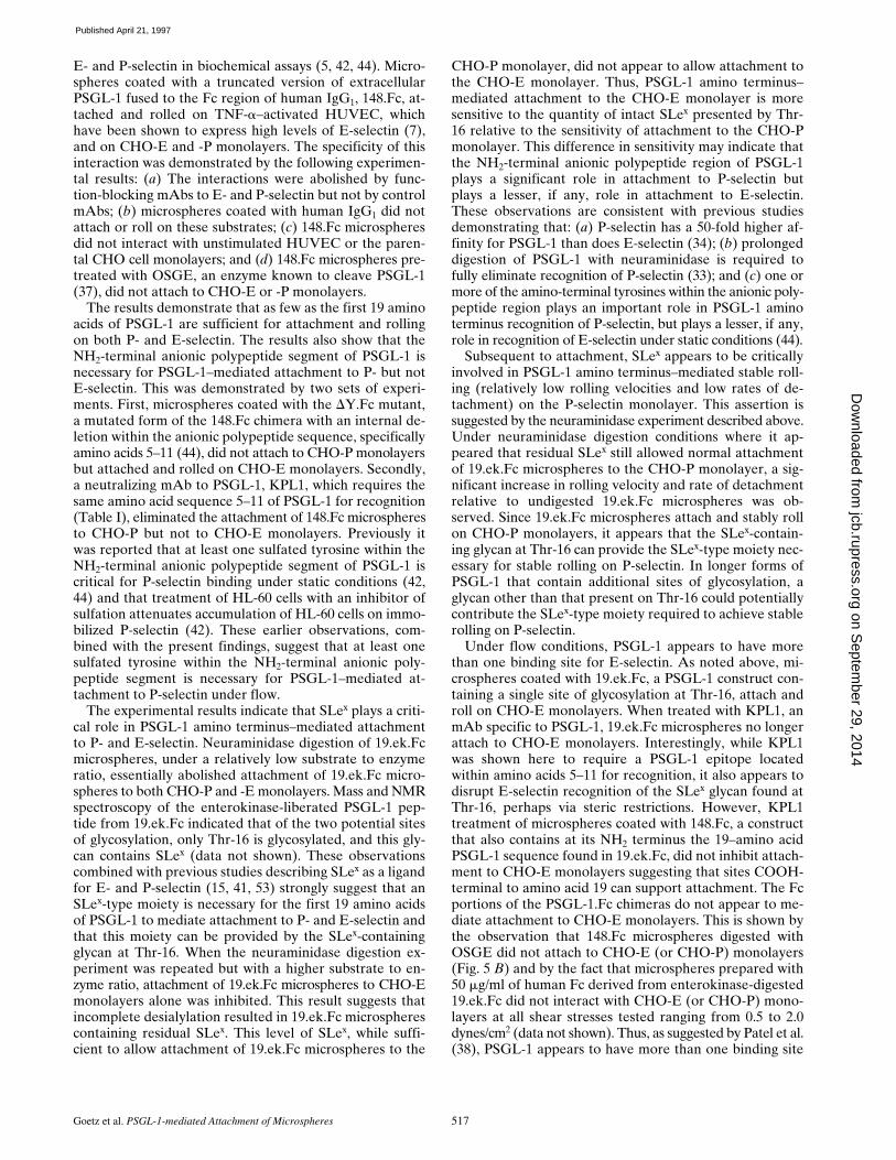
By using Fc constructs consisting of various extracellularportions of PSGL-1, Sako et al. (44) identified an NH2-ter-minal anionic polypeptide region within PSGL-1 neces-sary for recognition of P- but not E-selectin under staticconditions. This sequence is rich in acidic amino acids andcontains one or more tyrosine sulfate residues (42, 44). Todetermine whether this region of PSGL-1 is necessary and/orsufficient for attachment and rolling on P- and E-selectin,we used two additional Fc constructs cited in the earlier study(44) (Fig. 6 a). One of these constructs, DY.Fc (44), is amutated form of the 148.Fc chimera with an internal dele-tion within the anionic polypeptide region, specificallyamino acids 5–11. The second construct, 19.ek.Fc, is amodified version of the 19.Fc construct used in the earlierstudy (44). 19.ek.Fc contains only the first 19 amino acidsof PSGL-1, which includes the anionic, sulfated tyrosineregion and a critical O-linked glycosylation site at Threo-nine-16, Thr-16, also present in the 148.Fc. Unique to the19.ek.Fc construct is a cleavable polypeptide sequence in-troduced between the PSGL-1 and Fc sequences. This ad-ditional sequence contains no potential sites for glycosyla-tion. The DY.Fc construct has a similar molecular weightas the 148.Fc (44) and, because of material limitations, wascoupled to microspheres at a concentration of 40 mg/ml.For these experiments, 148.Fc microspheres were also pre-pared at this concentration. Since the molecular weight ofthe 19.ek.Fc is a little over 1/3 that of the 148.Fc, we pre-pared the 19.ek.Fc microspheres with 15 mg/ml of the
19.ek.Fc. As shown with a polyclonal antibody to PSGL-1,Rb3026, both mutants were coupled to the microspheres(Table I).
19.ek.Fc microspheres attached to CHO-E and -P mono-layers at a rate similar to that observed for 148.Fc micro-spheres (Fig. 6 b). Subsequent to attachment, .95% of the19.ek.Fc microspheres were observed to roll on both theCHO-E and -P monolayers. DY.Fc microspheres attachedto CHO-E monolayers at a rate similar to that observedfor 148.Fc microspheres (Fig. 6 b). Subsequent to attach-ment, .95% of the DY.Fc microspheres were observed toroll. In contrast, DY.Fc microspheres did not attach toCHO-P monolayers (Fig. 6 b). Neither the 19.ek.Fc northe DY.Fc microspheres attached to the parental CHO cellline (n 5 2; data not shown). We next attempted to block148.Fc and 19.ek.Fc microsphere attachment to CHO-Eand -P monolayers with an mAb, KPL1, specific for PSGL-1.KPL1 blocks neutrophil and memory T cell adhesion to
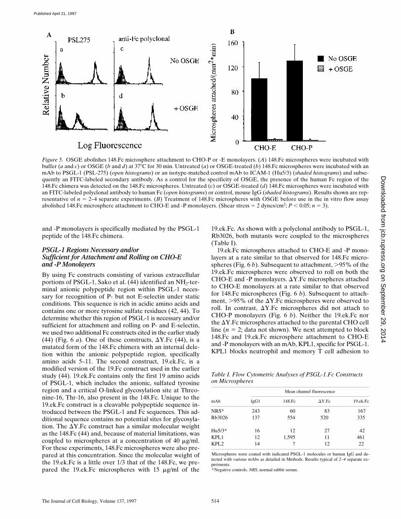
Figure 5. OSGE abolishes 148.Fc microsphere attachment to CHO-P or -E monolayers. (A) 148.Fc microspheres were incubated withbuffer (a and c) or OSGE (b and d) at 378C for 30 min. Untreated (a) or OSGE-treated (b) 148.Fc microspheres were incubated with anmAb to PSGL-1 (PSL-275) (open histograms) or an isotype-matched control mAb to ICAM-1 (Hu5/3) (shaded histograms) and subse-quently an FITC-labeled secondary antibody. As a control for the specificity of OSGE, the presence of the human Fc region of the148.Fc chimera was detected on the 148.Fc microspheres. Untreated (c) or OSGE-treated (d) 148.Fc microspheres were incubated withan FITC-labeled polyclonal antibody to human Fc (open histograms) or control, mouse IgG (shaded histograms). Results shown are rep-resentative of n 5 2–4 separate experiments. (B) Treatment of 148.Fc microspheres with OSGE before use in the in vitro flow assayabolished 148.Fc microsphere attachment to CHO-E and -P monolayers. (Shear stress 5 2 dynes/cm2; P , 0.05; n 5 3).
Table I. Flow Cytometric Analyses of PSGL-1.Fc Constructs on Microspheres
Mean channel fluorescence
mAb IgG1 148.Fc DY.Fc 19.ek.Fc
NRS* 243 60 83 167Rb3026 137 554 520 335
Hu5/3* 16 12 27 42KPL1 12 1,595 11 461KPL2 14 7 12 22
Microspheres were coated with indicated PSGL-1 molecules or human IgG and de-tected with various mAbs as detailed in Methods. Results typical of 2–4 separate ex-periments.*Negative controls. NRS, normal rabbit serum.
on Septem
ber 29, 2014jcb.rupress.org
Dow
nloaded from
Published April 21, 1997
Goetz et al. PSGL-1-mediated Attachment of Microspheres 515
Figure 6. The first 19 amino acids of PSGL-1 aresufficient for attachment to CHO-E and -Pmonolayers, and the NH2-terminal anionicpolypeptide segment of PSGL-1 is necessary for148.Fc-mediated attachment to CHO-P, but notCHO-E monolayers. (a) Schematic representa-tion of the PSGL-1.Fc constructs. Closed bars,PSGL-1 segments; open bars, human Fc seg-ments; open bar with X, an internal deletion ofamino acids 5–11 within the NH2-terminal an-ionic polypeptide region; shaded bar, the enteroki-nase cleavage site; Y, amino-terminal tyrosine;vertical lines with open circles, the approximatenumber and location of potential O-linked gly-
cosylation sites; vertical lines with closed circles, O-linked site at amino terminal threonine 16; vertical lines with shaded rectangles, po-tential N-linked glycosylation sites. (Drawing not to scale.) (b) Microspheres coated with the 19.ek.Fc mutant (open bars), containingthe first 19 amino acids of PSGL-1, attached to CHO-P and -E monolayers at a rate similar to 148.Fc microspheres (dark bars). Micro-spheres coated with the DY.Fc mutant, which consists of the first 148 amino acids of PSGL-1 with an internal deletion of the residues inpositions 5–11, attached to CHO-E monolayers at a rate similar to 148.Fc microspheres but did not attach to CHO-P monolayers (cross-hatched bars). (Shear stress 5 2 dynes/cm2; n 5 2). (c) An mAb to PSGL-1, KPL1, which requires amino acids 5–11 to recognize PSGL-1(Table I), did not affect attachment of 148.Fc microspheres to CHO-E monolayers but eliminated attachment to CHO-P monolayers.148.Fc microspheres treated with mAb KPL1 attached to CHO-E monolayers at a rate similar to that observed for 148.Fc microspherestreated with control mAb KPL2 but did not attach to CHO-P monolayers. (Shear stress 5 2 dynes/cm2; *P , 0.05; n 5 2). (d) mAbKPL1 eliminated attachment of 19.ek.Fc microspheres to both CHO-E and -P monolayers. (Shear stress 5 2 dynes/cm2; *P , 0.05; n 5 2).
CHO-P monolayers (Snapp, K.R., F.W. Luscinskas, R.Warnke, and G.S. Kansas. 1997. J. Allergy Clin. Immunol.99:s459). As shown in the present study, KPL1 recognitionof the 148.Fc microspheres appears to be dependent onthe presence of amino acid sequence 5–11 of PSGL-1 (Ta-ble I; 148.Fc versus DY.Fc). KPL2, a nonblocking isotype-matched mAb specific to full-length PSGL-1 (Snapp, K.R.,F.W. Luscinskas, R. Warnke, and G.S. Kansas. 1997. J. Al-lergy Clin. Immunol. 99:s459) was used as a control. KPL2appears to map to a PSGL-1 sequence COOH-terminal toamino acid 148 as it does not react with any of the Fc con-structs by flow cytometric analysis (Table I). For the block-ing studies, we used ascites preparations of both KPL1 andKPL2. KPL1 did not block 148.Fc microsphere attachmentto CHO-E monolayers but completely blocked attachmentto CHO-P monolayers (Fig. 6 c). 148.Fc microsphere at-tachment to CHO-E monolayers was not inhibited by treat-ment with a higher concentration of an ascites preparationof KPL1 (1:100 vs. 1:200 dilution) or 80 mg/ml purified KPL1relative to 148.Fc microspheres treated with control anti-bodies (data not shown). In contrast, KPL1 blocked 19.ek.Fc
microsphere attachment to both CHO-E and -P monolay-ers (Fig. 6 d). Similar inhibitory effects were obtained witha purified IgG preparation of KPL1. Taken together, theseresults show that: (a) The first 19 amino acids of PSGL-1are sufficient to support attachment and rolling on bothCHO-P and -E monolayers; (b) the anionic polypeptidesegment within the amino terminus of PSGL-1 is neces-sary for the 148.Fc construct to mediate attachment toCHO-P, but not to CHO-E monolayers; and (c) the 148.Fcconstruct contains at least two binding sites for E-selectin,one located within the first 19 amino acids and a second(or more) site COOH-terminal to amino acid 19.
An SLex-containing Glycan Is Involved in19.ek.Fc Microsphere Attachment and Rolling on CHO-P and -E Monolayers
Previous studies have shown that treatment of neutrophils(1), or HL-60 cells (2), with 0.1 U/ml neuraminidase for 30min inhibits adhesion to P- and E-selectin under flow, sug-gesting that sialylated structures, presumably SLex and/or
on Septem
ber 29, 2014jcb.rupress.org
Dow
nloaded from
Published April 21, 1997
The Journal of Cell Biology, Volume 137, 1997 516
related glycans, are involved. However, since both neu-trophils and HL-60 cells express multiple surface glycopro-teins and glycolipids that may contain SLex-modified glycans,it is difficult to ascribe the role of PSGL-1 glycosylation inattachment and rolling on P- and E-selectin. We attemptedto address this issue in studies using 19.ek.Fc-coated mi-crospheres in light of the data presented in the previoussection demonstrating that this highly defined construct issufficient for attachment and rolling on E- and P-selectin.The PSGL-1 peptide region of the 19.ek.Fc construct con-tains two potential O-linked glycosylation sites, Thr-3 andThr-16, but no N-linked glycosylation sites. Mass and NMRspectroscopy of the enterokinase-liberated PSGL-1 pep-tide from 19.ek.Fc indicated that of the two threonines,only Thr-16 is glycosylated, and this glycan contains SLex
(data not shown). Preliminary studies with microspherescoated with 15 mg/ml of the 19.ek.Fc construct indicatedthat a sialylated glycan, presumably SLex, is critically in-volved in the attachment of 19.ek.Fc microspheres toCHO-E and -P monolayers since pretreatment of 19.ek.Fcmicrospheres with 0.1 U/ml neuraminidase at 378C for 30min diminished the rate of attachment to CHO-E mono-layers by .95% and to CHO-P monolayers by .85% (datanot shown).
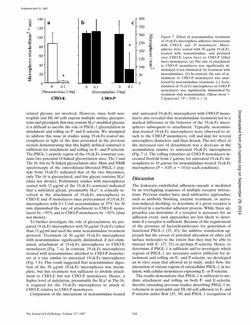
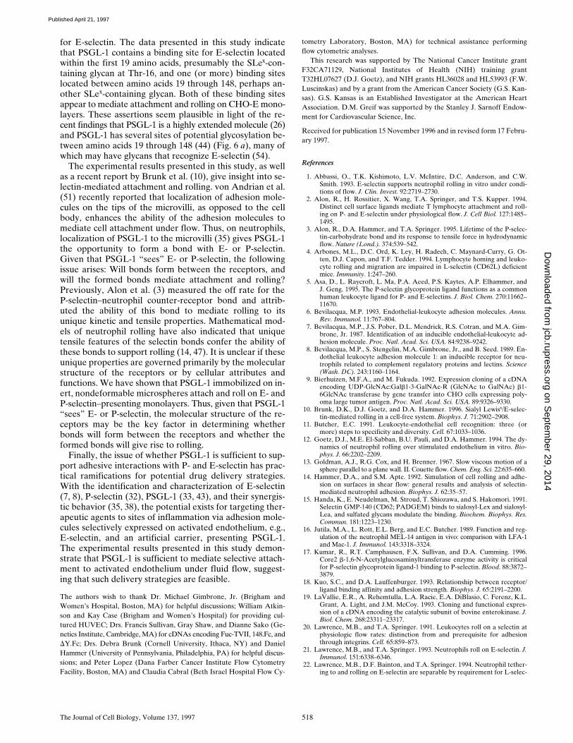
To further investigate the role of glycosylation, we pre-pared 19.ek.Fc microspheres with 50 mg/ml 19.ek.Fc ratherthan 15 mg/ml and used the same neuraminidase treatmentprotocol. Treatment of 50 mg/ml 19.ek.Fc microsphereswith neuraminidase significantly diminished, if not elimi-nated, attachment of 19.ek.Fc microspheres to CHO-Emonolayers (Fig. 7 a). In contrast, 19.ek.Fc microspherestreated with neuraminidase attached to CHO-P monolay-ers at a rate similar to untreated 19.ek.Fc microspheres(Fig. 7 b). This result suggested that neuraminidase diges-tion of the 50 mg/ml 19.ek.Fc microspheres was incom-plete, but this treatment was sufficient to abolish attach-ment to CHO-E but not CHO-P monolayers. Hence, ahigher level of sialylation, presumably the SLex at Thr-16,is required for the 19.ek.Fc microspheres to attach toCHO-E relative to CHO-P monolayers.
Comparison of the interactions of neuraminidase-treated
and -untreated 19.ek.Fc microspheres with CHO-P mono-layers also revealed that neuraminidase treatment led to amarked difference in the behavior of the 19.ek.Fc micro-spheres subsequent to attachment. Typically, neuramini-dase-treated 19.ek.Fc microspheres were observed to at-tach to the CHO-P monolayers, roll and skip for severalmicrosphere diameters, and then detach. The net effect ofthe increased rate of detachment was a decrease in theaccumulation relative to untreated 19.ek.Fc microspheres(Fig. 7 c). The rolling velocities on CHO-P monolayers in-creased fivefold from 2 mm/sec for untreated 19.ek.Fc mi-crospheres to 10 mm/sec for neuraminidase-treated 19.ek.Fcmicrospheres (P , 0.05; n 5 10 for each condition).
DiscussionThe leukocyte–endothelial adhesion cascade is mediatedby an overlapping sequence of multiple receptor interac-tions. Previous studies have used subtractive approaches,such as antibody blocking, enzyme treatment, or activa-tion-induced shedding, to determine if a given receptor isinvolved in a particular adhesion process. While these ap-proaches can determine if a receptor is necessary for anadhesion event, such approaches are less likely to deter-mine if a receptor is sufficient. Because of the requirementof the presence of fucosyltransferases for generation offunctional PSGL-1 (25, 43), the additive transfection ap-proach has the caveat of potential alteration of other cellsurface molecules to the extent that they may be able tointeract with E- (27, 52) or perhaps P-selectin. Hence, todetermine if PSGL-1 is sufficient and to investigate whichregions of PSGL-1 are necessary and/or sufficient for at-tachment and rolling on E- and P-selectin, we developedan in vitro assay that allowed us to study, under flow, theinteraction of various regions of extracellular PSGL-1, in iso-lation, with cellular monolayers expressing E- or P-selectin.
The results demonstrate that PSGL-1 is sufficient to me-diate attachment and rolling on both P- and E-selectin,thereby extending previous studies describing PSGL-1 in-volvement in neutrophil and HL-60 cell adhesion to E- andP-selectin under flow (35, 38) and PSGL-1 recognition of
Figure 7. Effect of neuraminidase treatmentof 19.ek.Fc microsphere adhesive interactionswith CHO-E and -P monolayers. Micro-spheres were coated with 50 mg/ml 19.ek.Fc,treated with neuraminidase, and perfusedover CHO-E (open bars) or CHO-P (filledbars) monolayers. (a) The rate of attachmentto CHO-E monolayers was significantly di-minished, if not eliminated, by treatment withneuraminidase. (b) In contrast, the rate of at-tachment to CHO-P monolayers was unaf-fected by neuraminidase treatment. (c) Accu-mulation of 19.ek.Fc microspheres on CHO-Pmonolayers was significantly diminished bytreatment with neuraminidase. (Shear stress 52 dynes/cm2; *P , 0.05; n 5 3).
on Septem
ber 29, 2014jcb.rupress.org
Dow
nloaded from
Published April 21, 1997
Goetz et al. PSGL-1-mediated Attachment of Microspheres 517
E- and P-selectin in biochemical assays (5, 42, 44). Micro-spheres coated with a truncated version of extracellularPSGL-1 fused to the Fc region of human IgG1, 148.Fc, at-tached and rolled on TNF-a–activated HUVEC, whichhave been shown to express high levels of E-selectin (7),and on CHO-E and -P monolayers. The specificity of thisinteraction was demonstrated by the following experimen-tal results: (a) The interactions were abolished by func-tion-blocking mAbs to E- and P-selectin but not by controlmAbs; (b) microspheres coated with human IgG1 did notattach or roll on these substrates; (c) 148.Fc microspheresdid not interact with unstimulated HUVEC or the paren-tal CHO cell monolayers; and (d) 148.Fc microspheres pre-treated with OSGE, an enzyme known to cleave PSGL-1(37), did not attach to CHO-E or -P monolayers.
The results demonstrate that as few as the first 19 aminoacids of PSGL-1 are sufficient for attachment and rollingon both P- and E-selectin. The results also show that theNH2-terminal anionic polypeptide segment of PSGL-1 isnecessary for PSGL-1–mediated attachment to P- but notE-selectin. This was demonstrated by two sets of experi-ments. First, microspheres coated with the DY.Fc mutant,a mutated form of the 148.Fc chimera with an internal de-letion within the anionic polypeptide sequence, specificallyamino acids 5–11 (44), did not attach to CHO-P monolayersbut attached and rolled on CHO-E monolayers. Secondly,a neutralizing mAb to PSGL-1, KPL1, which requires thesame amino acid sequence 5–11 of PSGL-1 for recognition(Table I), eliminated the attachment of 148.Fc microspheresto CHO-P but not to CHO-E monolayers. Previously itwas reported that at least one sulfated tyrosine within theNH2-terminal anionic polypeptide segment of PSGL-1 iscritical for P-selectin binding under static conditions (42,44) and that treatment of HL-60 cells with an inhibitor ofsulfation attenuates accumulation of HL-60 cells on immo-bilized P-selectin (42). These earlier observations, com-bined with the present findings, suggest that at least onesulfated tyrosine within the NH2-terminal anionic poly-peptide segment is necessary for PSGL-1–mediated at-tachment to P-selectin under flow.
The experimental results indicate that SLex plays a criti-cal role in PSGL-1 amino terminus–mediated attachmentto P- and E-selectin. Neuraminidase digestion of 19.ek.Fcmicrospheres, under a relatively low substrate to enzymeratio, essentially abolished attachment of 19.ek.Fc micro-spheres to both CHO-P and -E monolayers. Mass and NMRspectroscopy of the enterokinase-liberated PSGL-1 pep-tide from 19.ek.Fc indicated that of the two potential sitesof glycosylation, only Thr-16 is glycosylated, and this gly-can contains SLex (data not shown). These observationscombined with previous studies describing SLex as a ligandfor E- and P-selectin (15, 41, 53) strongly suggest that anSLex-type moiety is necessary for the first 19 amino acidsof PSGL-1 to mediate attachment to P- and E-selectin andthat this moiety can be provided by the SLex-containingglycan at Thr-16. When the neuraminidase digestion ex-periment was repeated but with a higher substrate to en-zyme ratio, attachment of 19.ek.Fc microspheres to CHO-Emonolayers alone was inhibited. This result suggests thatincomplete desialylation resulted in 19.ek.Fc microspherescontaining residual SLex. This level of SLex, while suffi-cient to allow attachment of 19.ek.Fc microspheres to the
CHO-P monolayer, did not appear to allow attachment tothe CHO-E monolayer. Thus, PSGL-1 amino terminus–mediated attachment to the CHO-E monolayer is moresensitive to the quantity of intact SLex presented by Thr-16 relative to the sensitivity of attachment to the CHO-Pmonolayer. This difference in sensitivity may indicate thatthe NH2-terminal anionic polypeptide region of PSGL-1plays a significant role in attachment to P-selectin butplays a lesser, if any, role in attachment to E-selectin.These observations are consistent with previous studiesdemonstrating that: (a) P-selectin has a 50-fold higher af-finity for PSGL-1 than does E-selectin (34); (b) prolongeddigestion of PSGL-1 with neuraminidase is required tofully eliminate recognition of P-selectin (33); and (c) one ormore of the amino-terminal tyrosines within the anionic poly-peptide region plays an important role in PSGL-1 aminoterminus recognition of P-selectin, but plays a lesser, if any,role in recognition of E-selectin under static conditions (44).
Subsequent to attachment, SLex appears to be criticallyinvolved in PSGL-1 amino terminus–mediated stable roll-ing (relatively low rolling velocities and low rates of de-tachment) on the P-selectin monolayer. This assertion issuggested by the neuraminidase experiment described above.Under neuraminidase digestion conditions where it ap-peared that residual SLex still allowed normal attachmentof 19.ek.Fc microspheres to the CHO-P monolayer, a sig-nificant increase in rolling velocity and rate of detachmentrelative to undigested 19.ek.Fc microspheres was ob-served. Since 19.ek.Fc microspheres attach and stably rollon CHO-P monolayers, it appears that the SLex-contain-ing glycan at Thr-16 can provide the SLex-type moiety nec-essary for stable rolling on P-selectin. In longer forms ofPSGL-1 that contain additional sites of glycosylation, aglycan other than that present on Thr-16 could potentiallycontribute the SLex-type moiety required to achieve stablerolling on P-selectin.
Under flow conditions, PSGL-1 appears to have morethan one binding site for E-selectin. As noted above, mi-crospheres coated with 19.ek.Fc, a PSGL-1 construct con-taining a single site of glycosylation at Thr-16, attach androll on CHO-E monolayers. When treated with KPL1, anmAb specific to PSGL-1, 19.ek.Fc microspheres no longerattach to CHO-E monolayers. Interestingly, while KPL1was shown here to require a PSGL-1 epitope locatedwithin amino acids 5–11 for recognition, it also appears todisrupt E-selectin recognition of the SLex glycan found atThr-16, perhaps via steric restrictions. However, KPL1treatment of microspheres coated with 148.Fc, a constructthat also contains at its NH2 terminus the 19–amino acidPSGL-1 sequence found in 19.ek.Fc, did not inhibit attach-ment to CHO-E monolayers suggesting that sites COOH-terminal to amino acid 19 can support attachment. The Fcportions of the PSGL-1.Fc chimeras do not appear to me-diate attachment to CHO-E monolayers. This is shown bythe observation that 148.Fc microspheres digested withOSGE did not attach to CHO-E (or CHO-P) monolayers(Fig. 5 B) and by the fact that microspheres prepared with50 mg/ml of human Fc derived from enterokinase-digested19.ek.Fc did not interact with CHO-E (or CHO-P) mono-layers at all shear stresses tested ranging from 0.5 to 2.0dynes/cm2 (data not shown). Thus, as suggested by Patel et al.(38), PSGL-1 appears to have more than one binding site
on Septem
ber 29, 2014jcb.rupress.org
Dow
nloaded from
Published April 21, 1997
The Journal of Cell Biology, Volume 137, 1997 518
for E-selectin. The data presented in this study indicatethat PSGL-1 contains a binding site for E-selectin locatedwithin the first 19 amino acids, presumably the SLex-con-taining glycan at Thr-16, and one (or more) binding siteslocated between amino acids 19 through 148, perhaps an-other SLex-containing glycan. Both of these binding sitesappear to mediate attachment and rolling on CHO-E mono-layers. These assertions seem plausible in light of the re-cent findings that PSGL-1 is a highly extended molecule (26)and PSGL-1 has several sites of potential glycosylation be-tween amino acids 19 through 148 (44) (Fig. 6 a), many ofwhich may have glycans that recognize E-selectin (54).
The experimental results presented in this study, as wellas a recent report by Brunk et al. (10), give insight into se-lectin-mediated attachment and rolling. von Andrian et al.(51) recently reported that localization of adhesion mole-cules on the tips of the microvilli, as opposed to the cellbody, enhances the ability of the adhesion molecules tomediate cell attachment under flow. Thus, on neutrophils,localization of PSGL-1 to the microvilli (35) gives PSGL-1the opportunity to form a bond with E- or P-selectin.Given that PSGL-1 “sees” E- or P-selectin, the followingissue arises: Will bonds form between the receptors, andwill the formed bonds mediate attachment and rolling?Previously, Alon et al. (3) measured the off rate for theP-selectin–neutrophil counter-receptor bond and attrib-uted the ability of this bond to mediate rolling to itsunique kinetic and tensile properties. Mathematical mod-els of neutrophil rolling have also indicated that uniquetensile features of the selectin bonds confer the ability ofthese bonds to support rolling (14, 47). It is unclear if theseunique properties are governed primarily by the molecularstructure of the receptors or by cellular attributes andfunctions. We have shown that PSGL-1 immobilized on in-ert, nondeformable microspheres attach and roll on E- andP-selectin–presenting monolayers. Thus, given that PSGL-1“sees” E- or P-selectin, the molecular structure of the re-ceptors may be the key factor in determining whetherbonds will form between the receptors and whether theformed bonds will give rise to rolling.
Finally, the issue of whether PSGL-1 is sufficient to sup-port adhesive interactions with P- and E-selectin has prac-tical ramifications for potential drug delivery strategies.With the identification and characterization of E-selectin(7, 8), P-selectin (32), PSGL-1 (33, 43), and their synergis-tic behavior (35, 38), the potential exists for targeting ther-apeutic agents to sites of inflammation via adhesion mole-cules selectively expressed on activated endothelium, e.g.,E-selectin, and an artificial carrier, presenting PSGL-1.The experimental results presented in this study demon-strate that PSGL-1 is sufficient to mediate selective attach-ment to activated endothelium under fluid flow, suggest-ing that such delivery strategies are feasible.
The authors wish to thank Dr. Michael Gimbrone, Jr. (Brigham andWomen’s Hospital, Boston, MA) for helpful discussions; William Atkin-son and Kay Case (Brigham and Women’s Hospital) for providing cul-tured HUVEC; Drs. Francis Sullivan, Gray Shaw, and Dianne Sako (Ge-netics Institute, Cambridge, MA) for cDNAs encoding Fuc-TVII, 148.Fc, andDY.Fc; Drs. Debra Brunk (Cornell University, Ithaca, NY) and DanielHammer (University of Pennsylvania, Philadelphia, PA) for helpful discus-sions; and Peter Lopez (Dana Farber Cancer Institute Flow CytometryFacility, Boston, MA) and Claudia Cabral (Beth Israel Hospital Flow Cy-
tometry Laboratory, Boston, MA) for technical assistance performingflow cytometric analyses.
This research was supported by The National Cancer Institute grantF32CA71129, National Institutes of Health (NIH) training grantT32HL07627 (D.J. Goetz), and NIH grants HL36028 and HL53993 (F.W.Luscinskas) and by a grant from the American Cancer Society (G.S. Kan-sas). G.S. Kansas is an Established Investigator at the American HeartAssociation. D.M. Greif was supported by the Stanley J. Sarnoff Endow-ment for Cardiovascular Science, Inc.
Received for publication 15 November 1996 and in revised form 17 Febru-ary 1997.
References
1. Abbassi, O., T.K. Kishimoto, L.V. McIntire, D.C. Anderson, and C.W.Smith. 1993. E-selectin supports neutrophil rolling in vitro under condi-tions of flow. J. Clin. Invest. 92:2719–2730.
2. Alon, R., H. Rossitier, X. Wang, T.A. Springer, and T.S. Kupper. 1994.Distinct cell surface ligands mediate T lymphocyte attachment and roll-ing on P- and E-selectin under physiological flow. J. Cell Biol. 127:1485–1495.
3. Alon, R., D.A. Hammer, and T.A. Springer. 1995. Lifetime of the P-selec-tin-carbohydrate bond and its response to tensile force in hydrodynamicflow. Nature (Lond.). 374:539–542.
4. Arbones, M.L., D.C. Ord, K. Ley, H. Radech, C. Maynard-Curry, G. Ot-ten, D.J. Capon, and T.F. Tedder. 1994. Lymphocyte homing and leuko-cyte rolling and migration are impaired in L-selectin (CD62L) deficientmice. Immunity. 1:247–260.
5. Asa, D., L. Raycroft, L. Ma, P.A. Aeed, P.S. Kaytes, A.P. Elhammer, andJ. Geng. 1995. The P-selectin glycoprotein ligand functions as a commonhuman leukocyte ligand for P- and E-selectins. J. Biol. Chem. 270:11662–11670.
6. Bevilacqua, M.P. 1993. Endothelial-leukocyte adhesion molecules. Annu.Rev. Immunol. 11:767–804.
7. Bevilacqua, M.P., J.S. Pober, D.L. Mendrick, R.S. Cotran, and M.A. Gim-brone, Jr. 1987. Identification of an inducible endothelial-leukocyte ad-hesion molecule. Proc. Natl. Acad. Sci. USA. 84:9238–9242.
8. Bevilacqua, M.P., S. Stengelin, M.A. Gimbrone, Jr., and B. Seed. 1989. En-dothelial leukocyte adhesion molecule 1: an inducible receptor for neu-trophils related to complement regulatory proteins and lectins. Science(Wash. DC). 243:1160–1164.
9. Bierhuizen, M.F.A., and M. Fukuda. 1992. Expression cloning of a cDNAencoding UDP-GlcNAc:Galb1-3-GalNAc-R (GlcNAc to GalNAc) b1-6GlcNAc transferase by gene transfer into CHO cells expressing poly-oma large tumor antigen. Proc. Natl. Acad. Sci. USA. 89:9326–9330.
10. Brunk, D.K., D.J. Goetz, and D.A. Hammer. 1996. Sialyl Lewisx/E-selec-tin-mediated rolling in a cell-free system. Biophys. J. 71:2902–2908.
11. Butcher, E.C. 1991. Leukocyte-endothelial cell recognition: three (ormore) steps to specificity and diversity. Cell. 67:1033–1036.
12. Goetz, D.J., M.E. El-Sabban, B.U. Pauli, and D.A. Hammer. 1994. The dy-namics of neutrophil rolling over stimulated endothelium in vitro. Bio-phys. J. 66:2202–2209.
13. Goldman, A.J., R.G. Cox, and H. Brenner. 1967. Slow viscous motion of asphere parallel to a plane wall. II. Couette flow. Chem. Eng. Sci. 22:635–660.
14. Hammer, D.A., and S.M. Apte. 1992. Simulation of cell rolling and adhe-sion on surfaces in shear flow: general results and analysis of selectin-mediated neutrophil adhesion. Biophys. J. 62:35–57.
15. Handa, K., E. Neudelman, M. Stroud, T. Shiozawa, and S. Hakomori. 1991.Selectin GMP-140 (CD62; PADGEM) binds to sialosyl-Lex and sialosyl-Lea, and sulfated glycans modulate the binding. Biochem. Biophys. Res.Commun. 181:1223–1230.
16. Jutila, M.A., L. Rott, E.L. Berg, and E.C. Butcher. 1989. Function and reg-ulation of the neutrophil MEL-14 antigen in vivo: comparison with LFA-1and Mac-1. J. Immunol. 143:3318–3324.
17. Kumar, R., R.T. Camphausen, F.X. Sullivan, and D.A. Cumming. 1996.Core2 b-1,6-N-Acetylglucosaminyltransferase enzyme activity is criticalfor P-selectin glycoprotein ligand-1 binding to P-selectin. Blood. 88:3872–3879.
18. Kuo, S.C., and D.A. Lauffenburger. 1993. Relationship between receptor/ligand binding affinity and adhesion strength. Biophys. J. 65:2191–2200.
19. LaVallie, E.R., A. Rehemtulla, L.A. Racie, E.A. DiBlasio, C. Ferenz, K.L.Grant, A. Light, and J.M. McCoy. 1993. Cloning and functional expres-sion of a cDNA encoding the catalytic subunit of bovine enterokinase. J.Biol. Chem. 268:23311–23317.
20. Lawrence, M.B., and T.A. Springer. 1991. Leukocytes roll on a selectin atphysiologic flow rates: distinction from and prerequisite for adhesionthrough integrins. Cell. 65:859–873.
21. Lawrence, M.B., and T.A. Springer. 1993. Neutrophils roll on E-selectin. J.Immunol. 151:6338–6346.
22. Lawrence, M.B., D.F. Bainton, and T.A. Springer. 1994. Neutrophil tether-ing to and rolling on E-selectin are separable by requirement for L-selec-
on Septem
ber 29, 2014jcb.rupress.org
Dow
nloaded from
Published April 21, 1997

Goetz et al. PSGL-1-mediated Attachment of Microspheres 519
tin. Immunity. 1:137–145.23. Lewinsohn, D.M., R.F. Bargatze, and E.C. Butcher. 1986. Leukocyte-
endothelial cell recognition: evidence of a common molecular mechanismshared by neutrophils, lymphocytes, and other leukocytes. J. Immunol.138:4313–4321.
24. Ley, K., P. Gaehtgens, C. Fennie, M.S. Singer, L.A. Lasky, and S.D. Rosen.1991. Lectin-like cell adhesion molecule 1 mediates leukocyte rolling inmesenteric venules in vivo. Blood. 77:2553–2555.
25. Li, F., P.P. Wilkins, S. Crawley, J. Weinstein, R.D. Cummings, and R.P.McEver. 1996. Post-translational modifications of recombinant P-selectinglycoprotein ligand-1 required for binding to P- and E-selectin. J. Biol.Chem. 271:3255–3264.
26. Li, F., H.P. Erickson, J.A. James, K.L. Moore, R.D. Cummings, and R.P.McEver. 1996. Visualization of P-selectin glycoprotein ligand-1 as a highlyextended molecule and mapping of protein epitopes for monoclonal anti-bodies. J. Biol. Chem. 271:6342–6348.
27. Lowe, J.B., L.M. Stoolman, R.P. Nair, R.D. Larsen, T.L. Berhend, andR.M. Marks. 1990. ELAM-1-dependent cell adhesion to vascular endo-thelium determined by a transfected human fucosyltransferase cDNA. J.Biol. Chem. 63:475–484.
28. Luscinskas, F.W., G.S. Kansas, H. Ding, P. Pizcueta, B.E. Schleiffenbaum,T.F. Tedder, and M.A. Gimbrone, Jr. 1994. Monocyte rolling, arrest, andspreading on IL-4-activated vascular endothelium under flow is mediatedvia sequential action of L-selectin, b1-integrins, and b2-integrins. J. CellBiol. 125:1417–1427.
29. Luscinskas, F.W., H. Ding, and A.H. Lichtman. 1995. P-selectin andVCAM-1 mediate rolling and arrest of CD41 T-lymphocytes on TNF-a-activated vascular endothelium under flow. J. Exp. Med. 181:1179–1186.
30. Luscinskas, F.W., H. Ding, P. Tan, D. Cumming, T.F. Tedder, and M.E.Gerritsen. 1996. L- and P-selectins, but not CD49d (VLA-4) integrins,mediate monocyte initial attachment to TNF-a-activated vascular endo-thelium under flow in vitro. J. Immunol. 156:326–335.
31. Mayadas, T.N., R.C. Johnson, H. Rayburn, R.O. Hynes, and D.D. Wagner.1993. Leukocyte rolling and extravasation are severely compromised inP-selectin deficient mice. Cell. 74:541–554.
32. McEver, R.P., J.H. Beckstead, K.L. Moore, L. Marshall-Carlson, and D.F.Bainton. 1989. GMP-140, a platelet a-granule membrane protein, is alsosynthesized by vascular endothelial cells and is localized in Weibel-Palade bodies. J. Clin. Invest. 84:92–99.
33. Moore, K.L., N.L. Stults, S. Diaz, D.F. Smith, R.D. Cummings, A. Varki,and R.P. McEver. 1992. Identification of a specific glycoprotein ligandfor P-selectin (CD62) on myeloid cells. J. Cell Biol. 118:445–456.
34. Moore, K.L., S.F. Eaton, D.E. Lyons, H.S. Lichenstein, R.D. Cummings,and R.P. McEver. 1994. The P-selectin glycoprotein ligand from humanneutrophils displays sialylated, fucosylated, O-linked poly-N-acetyllac-tosamine. J. Biol. Chem. 269:23318–23327.
35. Moore, K.L., K.D. Patel, R.E. Bruehl, L. Fugang, D.A. Johnson, H.S. Lich-enstein, R.D. Cummings, D.F. Bainton, and R.P. McEver. 1995. P-selectinglycoprotein ligand-1 mediates rolling of human neutrophils on P-selec-tin. J. Cell Biol. 128:661–671.
36. Natsuka, S., K.M. Gersten, K. Zentia, R. Kannagi, and J.B. Lowe. 1994.Molecular cloning of a cDNA encoding a novel human leukocyte a-1,3-fucosyltransferase capable of synthesizing the sialyl Lewis X determi-nant. J. Biol. Chem. 269:16789–16794.
37. Norgard, K.E., K.L. Moore, S. Diaz, N.L. Stults, S. Ushiyama, R.P.McEver, R.D. Cummings, and A. Varki. 1993. Characterization of a spe-cific ligand for P-selectin on myeloid cells. J. Biol. Chem. 268:12764–12774.
38. Patel, K.D., K.L. Moore, M.U. Nollert, and R.P. McEver. 1995. Neutro-phils use both shared and distinct mechanisms to adhere to selectins un-der static and flow conditions. J. Clin. Invest. 96:1887–1896.
39. Picker, L.J., R.A. Warnock, A.R. Burns, C.M. Doerschuk, E.L. Berg, andE.C. Butcher. 1991. The neutrophil selectin LECAM-1 presents carbohy-drate ligands to the vascular selectin ELAM-1 and GMP-140. Cell. 66:921–933.
40. Pierres, A., O. Tissot, B. Malissen, and P. Bongrand. 1994. Dynamic adhe-sion of CD8-positive cells to antibody-coated surfaces: the initial step isindependent of microfilaments and intracellular domains of cell bindingmolecules. J. Cell Biol. 125:945–953.
41. Polley, M.J., M.L. Phillips, E. Wayner, E. Nudelman, A.K. Singhal, S. Hako-mori, and J.C. Paulson. 1991. CD62 and endothelial cell-leukocyte adhe-sion molecule 1 (ELAM-1) recognize the same carbohydrate ligand, sia-lyl-Lewis x. Proc. Natl. Acad. Sci. USA. 88:6224–6228.
42. Pouyani, T., and B. Seed. 1995. PSGL-1 recognition of P-selectin is con-trolled by a tyrosine sulfation consensus at the PSGL-1 amino terminus.Cell. 83:333–343.
43. Sako, D., X.J. Chang, K.M. Barone, G. Vachino, H.M. White, G. Shaw,G.M. Veldman, K.M. Bean, T.J. Ahern, B. Furie, et al. 1993. Expressioncloning of a functional glycoprotein ligand for P-selectin. Cell. 75:1179–1186.
44. Sako, D., K.M. Comess, K.M. Barone, R.T. Camphausen, D.A. Cumming,and G.D. Shaw. 1995. A sulfated peptide segment at the amino terminusof PSGL-1 is critical for P-selectin binding. Cell. 83:323–331.
45. Sasaki, K., K. Kurata, K. Funayama, M. Nagata, M. Watanabe, S. Ohta, N.Hanai, and T. Nishi. 1994. Expression cloning of a novel a1,3-fucosyl-transferase that is involved in biosynthesis of the sialyl Lewis X carbohy-drate determinants in leukocytes. J. Biol. Chem. 269:14730–14737.
46. Spertinni, O., F.W. Luscinskas, G.S. Kansas, J.D. Griffin, M.A. Gimbrone,Jr., and T.F. Tedder. 1991. Leukocyte adhesion molecule (LAM-1, L-selec-tin) interacts with an inducible endothelial cell ligand to support leuko-cyte adhesion. J. Immunol. 147:2565–2573.
47. Tozeren, A., and K. Ley. 1992. How do selectins mediate leukocyte rollingin venules? Biophys. J. 63:700–709.
48. Vachino, G., X.-J. Chang, G.M. Veldman, R. Kumar, L.A. Fouser, M.C.Berndt, and D.A. Cumming. 1995. P-selectin glycoprotein ligand-1 is themajor counter-receptor for P-selectin on stimulated T cells and is widelydistributed in non-functional form on many lymphocytic cells. J. Biol.Chem. 270:21966–21974.
49. Varki, A. 1994. Selectin ligands. Proc. Natl. Acad. Sci. USA. 91:7390–7397.50. von Andrian, U.H., J.D. Chambers, L.M. McEvoy, R.F. Bargatze, K.E. Ar-
fors, and E.C. Butcher. 1991. Two-step model of leukocyte-endothelialcell interactions in inflammation: distinct roles for LECAM-1 and theleukocyte b2 integrins in vivo. Proc. Natl. Acad. Sci. USA. 88:7538–7542.
51. von Andrian, U.H., S.R. Hasslen, R.D. Nelson, S.L. Erlandsen, and E.C.Butcher. 1995. A central role for microvillous receptor presentation inleukocyte adhesion under flow. Cell. 82:989–999.
52. Wagers, A.J., J.B. Lowe, and G.S. Kansas. 1996. An important role for thea1,3 fucosyltransferase, FucT-VII, in leukocyte adhesion to E-selectin.Blood. 88:2125–2132.
53. Walz, G., A. Aruffo, W. Kolanus, M.P. Bevilacqua, and B. Seed. 1990. Rec-ognition by ELAM-1 of the sialyl-Lex determinant on myeloid and tu-mor cells. Science (Wash. DC). 250:1132–1135.
54. Wilkins, P.P., R.P. McEver, and R.D. Cummings. 1996. Structures of theO-glycans on P-selectin glycoprotein ligand-1 from HL-60 cells. J. Biol.Chem. 271:18732–18742.
on Septem
ber 29, 2014jcb.rupress.org
Dow
nloaded from
Published April 21, 1997