P-glycoprotein inserted in planar lipid bilayers formed by liposomes opened on amorphous carbon and...
11
P-glycoprotein inserted in planar lipid bilayers formed by liposomes opened on amorphous carbon and Langmuir^Blodgett monolayer Marco Diociaiuti a ; *, Agnese Molinari a , Irene Ruspantini b , Maria Cristina Gaudiano c , Rodolfo Ippoliti d , Eugenio Lendaro e , Federico Bordi f ;g , Pietro Chistolini b , Giuseppe Arancia a a Laboratorio di Ultrastrutture, Istituto Superiore di Sanita ', Viale Regina Elena, 299, 00161 Rome, Italy b Laboratorio di Ingegneria Biomedica, Istituto Superiore di Sanita ', Viale Regina Elena, 299, 00161 Rome, Italy c Laboratorio di Chimica del Farmaco, Istituto Superiore di Sanita ', Viale Regina Elena, 299, 00161 Rome, Italy d Dipartimento di Biologia di Base ed Applicata, Universita ' degli Studi dell’Aquila, Via Vetoio, 67100 L’Aquila, Italy e Dipartimento di Scienze Biochimiche, Universita ' ‘La Sapienza’ di Roma, Piazzale Aldo Moro, 5, 00185 Rome, Italy f Dipartimento di Medicina Interna, Universita ' ‘Tor Vergata’ di Roma, Via di Tor Vergata, 00100 Rome, Italy g INFM Unita ' di Roma I, Rome, Italy Received 25 July 2001; received in revised form 25 September 2001; accepted 26 September 2001 Abstract The insertion of proteins into planar lipid layers is of outstanding interest as the resulting films are suitable for the investigation of protein structure and aggregation in a lipid environment and/or the development of biotechnological applications as biosensors. In this study, purified P-glycoprotein (P-gp), a membrane drug pump, was incorporated in model membranes deposited on solid supports according to the method by Puu and Gustafson, Biochim. Biophys. Acta 1327 (1997) 149-161. The models were formed by a double lipid layer obtained by opening P-gp-containing liposomes onto two hydrophobic supports : amorphous carbon films and Langmuir^Blodgett (L^B) lipid monolayers, which were then observed by transmission electron microscopy and atomic force microscopy, respectively. Before the opening of liposomes, the P-gp structure and functionality were verified by circular dichroism spectroscopy and enzymatic assay. Our micrographs showed that liposomes containing P-gp fuse to the substrates more easily than plain liposomes, which keep their rounded shape. This suggests that the protein plays an essential role in the fusion of liposomes. To localize P-gp, the immunogold labeling of two externally exposed protein epitopes was carried out. Both imaging techniques confirmed that P-gp was successfully incorporated in the model membranes and that the two epitopes preserved the reactivity with specific mAbs, after sample preparation. Model membranes obtained on L^B monolayer incorporated few molecules with respect to those incorporated in the model membrane deposited onto amorphous carbon, probably because of the different mechanism of proteoliposome opening. Finally, all particles appeared as isolated units, suggesting that P-gp molecules were present as monomers. ß 2002 Elsevier Science B.V. All rights reserved. Keywords : P-glycoprotein ; Lipid bilayer ; Circular dichroism ; Atomic force microscopy ; Transmission electron microscopy 1. Introduction P-glycoprotein (P-gp), a protein encoded by the human MDR1 gene, belongs to the superfamily of 0005-2736 / 02 / $ ^ see front matter ß 2002 Elsevier Science B.V. All rights reserved. PII:S0005-2736(01)00425-4 * Corresponding author. Fax : +39-6-4938 7140. E-mail address : [email protected] (M. Diociaiuti). Biochimica et Biophysica Acta 1559 (2002) 21^31 www.bba-direct.com
Transcript of P-glycoprotein inserted in planar lipid bilayers formed by liposomes opened on amorphous carbon and...

P-glycoprotein inserted in planar lipid bilayers formed by liposomesopened on amorphous carbon and Langmuir^Blodgett monolayer
Marco Diociaiuti a;*, Agnese Molinari a, Irene Ruspantini b, Maria Cristina Gaudiano c,Rodolfo Ippoliti d, Eugenio Lendaro e, Federico Bordi f ;g, Pietro Chistolini b,
Giuseppe Arancia a
a Laboratorio di Ultrastrutture, Istituto Superiore di Sanita©, Viale Regina Elena, 299, 00161 Rome, Italyb Laboratorio di Ingegneria Biomedica, Istituto Superiore di Sanita©, Viale Regina Elena, 299, 00161 Rome, Italyc Laboratorio di Chimica del Farmaco, Istituto Superiore di Sanita©, Viale Regina Elena, 299, 00161 Rome, Italy
d Dipartimento di Biologia di Base ed Applicata, Universita© degli Studi dell'Aquila, Via Vetoio, 67100 L'Aquila, Italye Dipartimento di Scienze Biochimiche, Universita© `La Sapienza' di Roma, Piazzale Aldo Moro, 5, 00185 Rome, Italy
f Dipartimento di Medicina Interna, Universita© `Tor Vergata' di Roma, Via di Tor Vergata, 00100 Rome, Italyg INFM Unita© di Roma I, Rome, Italy
Received 25 July 2001; received in revised form 25 September 2001; accepted 26 September 2001
Abstract
The insertion of proteins into planar lipid layers is of outstanding interest as the resulting films are suitable for theinvestigation of protein structure and aggregation in a lipid environment and/or the development of biotechnologicalapplications as biosensors. In this study, purified P-glycoprotein (P-gp), a membrane drug pump, was incorporated in modelmembranes deposited on solid supports according to the method by Puu and Gustafson, Biochim. Biophys. Acta 1327 (1997)149-161. The models were formed by a double lipid layer obtained by opening P-gp-containing liposomes onto twohydrophobic supports : amorphous carbon films and Langmuir^Blodgett (L^B) lipid monolayers, which were then observedby transmission electron microscopy and atomic force microscopy, respectively. Before the opening of liposomes, the P-gpstructure and functionality were verified by circular dichroism spectroscopy and enzymatic assay. Our micrographs showedthat liposomes containing P-gp fuse to the substrates more easily than plain liposomes, which keep their rounded shape. Thissuggests that the protein plays an essential role in the fusion of liposomes. To localize P-gp, the immunogold labeling of twoexternally exposed protein epitopes was carried out. Both imaging techniques confirmed that P-gp was successfullyincorporated in the model membranes and that the two epitopes preserved the reactivity with specific mAbs, after samplepreparation. Model membranes obtained on L^B monolayer incorporated few molecules with respect to those incorporatedin the model membrane deposited onto amorphous carbon, probably because of the different mechanism of proteoliposomeopening. Finally, all particles appeared as isolated units, suggesting that P-gp molecules were present as monomers. ß 2002Elsevier Science B.V. All rights reserved.
Keywords: P-glycoprotein; Lipid bilayer; Circular dichroism; Atomic force microscopy; Transmission electron microscopy
1. Introduction
P-glycoprotein (P-gp), a protein encoded by thehuman MDR1 gene, belongs to the superfamily of
0005-2736 / 02 / $ ^ see front matter ß 2002 Elsevier Science B.V. All rights reserved.PII: S 0 0 0 5 - 2 7 3 6 ( 0 1 ) 0 0 4 2 5 - 4
* Corresponding author. Fax: +39-6-4938 7140.E-mail address: [email protected] (M. Diociaiuti).
BBAMEM 78186 24-1-02
Biochimica et Biophysica Acta 1559 (2002) 21^31www.bba-direct.com

ABC (ATP-binding cassette) transporters. This pro-tein is embedded in the lipid bilayers and acts as ananoscale pump, utilizing the energy of ATP hydro-lysis to translocate molecules across cellular mem-branes. P-gp evolved to protect cells from xenobioticmolecules and it is normally found in cells of thedigestive tract, the kidney, the liver and in endothe-lial cells lining capillaries in the brain [1]. In tumorcells P-gp provides cross-resistance to a variety ofchemotherapeutic drugs (multidrug resistance,MDR) and its overexpression is responsible for thefailure of the chemotherapeutic treatment of severaltumors [2].
P-gp structure and function are far from beingcompletely elucidated. P-gp is generally describedas constituted by K-helices and L-structures [3]. How-ever, the actual folding of P-gp in a lipid bilayer isstill a matter of debate. Experimental data have sofar generally supported a model describing the pro-tein as a toroidal ring of 12 K-helices, deployed intwo arcs of six helices each (6+6 helix model), form-ing a single aqueous pore in the bilayer [4,5]. How-ever, Jones and George [6] have recently proposed adi¡erent interpretation of the bulk of experimentaldata: an alternative model in which the proteinforms two transmembrane pores constituted by L-barrels.
To elucidate the structure^function link, it is ofcrucial importance to understand the physiologicaland functional aggregation state of the protein. ForP-gp, data suggest that the minimum functional unitshould be a monomer, but do not exclude the possi-bility that these monomers might form dimers orlarger oligomers under certain physiological condi-tions, and that oligomerization might alter the struc-tural and functional properties of the constitutingmonomers [5,7].
Immunogold labeling, if performed with well-char-acterized monoclonal antibodies recognizing de¢nedprotein epitopes, would detect the single monomericunit in the cellular plasma membrane. Transmissionelectron microscopy (TEM) localizes, with high res-olution, gold particles up to 5 nm diameter, but ad-mits only thin samples, and the information that itfurnishes is limited to a bi-dimensional projection. Instudies performed by scanning electron microscopy(SEM) on cells expressing P-gp, previously labeledwith immunogold complexes, the protein epitopes
were detected on the cellular membrane surface [8].However, conventional SEM has a low spatial reso-lution limit (about 5^10 nm) and does not yield a ¢nedetailed three-dimensional (3D) analysis by whichthe presence of P-gp dimers or oligomers can be as-sessed. In addition, sample preparation for SEMcould introduce artifacts that modify the nativestructure of P-gp.
In order to overcome these limitations we usedatomic force microscopy (AFM) to study the struc-ture of P-gp in a model phospholipid double-layermembrane. AFM is a very attractive tool for biolog-ical investigations because it is suitable to observe the3D shape of a whole cell as well as its substructures,e.g. proteins and DNA, without having to resort toimpairing treatments like ¢xation, which can com-pletely alter the original structure. Moreover, lateralhigh-resolution images can be attained (up to fewnanometers) and useful 3D topological maps of thesample can be reconstructed. However, if molecularresolution has to be attained, AFM needs very £atand homogeneous samples where only molecular dis-continuities occur.
Thus, given the intrinsic complexity of biomem-branes, it seemed more appropriate to approach thestudy by using simpli¢ed model membranes such aslipid bilayers, where all experimental parameters canbe accurately monitored. The visualization of P-gpmonomers and of dimers and oligomers, if any,could be improved by incorporating the protein ina lipid bilayer. Puu and Gustafson [9] developed amethod by which stable lipid bilayers can be pre-pared by transferring liposomes to a planar support,constituted by a Langmuir^Blodgett (L^B) lipidmonolayer [10] or amorphous carbon. They per-formed a systematic investigation to determine fac-tors of importance involved in the kinetics and stabil-ity of lipid bilayer formation. In particular, theystudied the in£uence of lipid composition of lipo-somes and monolayers, and the in£uence of Ca2�
concentration on the fusion process. They evaluatedbilayer stability after storage and transfer betweendi¡erent media by crossing the air^water interface.By their method, proteins with retained activitiescan be successfully introduced in the bilayer. Thesame authors refer that lipid bilayers can also becreated by directly opening liposomes onto hydro-phobic supports such as amorphous carbon, suggest-
BBAMEM 78186 24-1-02
M. Diociaiuti et al. / Biochimica et Biophysica Acta 1559 (2002) 21^3122
ing that two di¡erent mechanisms of the planar bi-layer formation operate.
In this paper we compare results obtained by TEMon model membrane with P-gp prepared by directdeposition of proteoliposomes onto amorphous car-bon support, with those achieved by AFM on amodel membrane prepared by opening proteolipo-somes onto an L^B lipid monolayer deposited ontomica.
The aims of this study were to look inside thebiochemical and structural features of P-gp reconsti-tuted in liposomes, to obtain information about themechanisms of bilayer formation on two di¡erenthydrophobic surfaces, in the presence and absenceof P-gp, to ¢nd out whether the insertion of P-gpin the model membrane a¡ects its conformationalfeatures, at least in the surroundings of two externalepitopes and, ¢nally, to elucidate P-gp aggregationstate.
2. Materials and methods
2.1. Cell culture
The MDR variant of human leukemic lymphoblas-tic cell line (CEM VBL100) was grown in suspensionin RPMI 1640 medium supplemented with 10% heat-inactivated fetal calf serum, 1% L-glutamine and 1%penicillin and streptomycin at 37.0³C in a 5% CO2
humidi¢ed atmosphere in air. CEM VBL100 cellswere grown in complete medium containing 10 ng/ml vinblastine sulfate (Velbe, Lilly, Fegersheim,France).
2.2. mAbs reagents
mAbs MM4.17 [11] and MRK-16 (Kamiya, Thou-sand Oaks, CA, USA), that recognize two distincthuman-speci¢c epitopes of extracellular domains ofMDR-1-P-gp, were used in this study. MM4.17 is anIgG2ak monoclonal immunoglobulin reacting with acontinuous-linear epitope on the apical part of thefourth loop of P-gp. MRK-16 is an IgG2ak monoclo-nal immunoglobulin reacting with a conformationalepitope distributed on the ¢rst and fourth loop of P-gp [12].
2.3. P-gp puri¢cation, liposome preparation andfunctionality tests
P-gp was extracted from a plasma membrane frac-tion separated by means of a sucrose density gradientas described by Dong et al. [13]. Brie£y, the mem-branes were dissolved with 2% sodium dodecyl sul-fate (SDS) and ultracentrifuged for 30 min at100 000Ug at 20.0³C. The supernatant was dilutedto 1% SDS with an equal volume of 10 mM phos-phate bu¡er, pH 7.0, containing 1 mM dithiothreitol(DTT). P-gp was puri¢ed by hydrophobic interactionchromatography on a hydroxyapatite high-perform-ance liquid chromatography (HPLC) column equili-brated with 50 mM phosphate bu¡er (pH 7.0), 1%SDS and 1 mM DTT. After binding of the P-gpextract and extensive washing, the protein was elutedwith a linear gradient of phosphate (0C0.5 M) at28.0³C. The chromatographic fractions from theTSK column were analyzed by electrophoresis andimmunoblot. SDS^polyacrylamide gel electrophore-sis (PAGE) was performed on slab gels by the meth-od of Laemmli [14] using 10% acrylamide. After elec-trophoresis, proteins separated by SDS^PAGE weretransferred to nylon membranes (Immobilon P fromMillipore), and stained with a mouse monoclonalanti-P-gp C219 (Signet Laboratories Inc., workingdilution 1:100) at 37³C for 60 min. Bound antibodieswere detected with alkaline phosphatase-conjugatedsecondary anti-mouse immunoglobulin (Sigma,working dilution 1:1000). The puri¢cation grade ofP-gp was determined by HPLC analysis on reverse-phase C8 (Vydac) columns.
Reconstitution of puri¢ed P-gp into liposomes wascarried out following the method by Rigaud et al.[15]. 25 mg of L-K-phosphatidylcholine (PC) and2.5 mg of L-K-phosphatidic acid (AP) were dissolvedin chloroform and dried under a stream of nitrogen.The lipid ¢lm was dissolved in 20 mM Tris^HCl, pH7.4, 75 mM NaCl, 1 mM DTT and 0.5 mM EDTAbu¡er and sonicated on ice until the mixture becametransparent. A 100 Wl aliquot of liposome prepara-tion was sequentially supplemented with 610 Wl ofbu¡er and 40 Wl of 10% dodecyl maltoside and, after15 min, 250 Wl of 0.2 mg/ml puri¢ed P-gp underconstant stirring at room temperature. To removethe detergent, the solution was dialyzed overnightin Tris^HCl bu¡er.
BBAMEM 78186 24-1-02
M. Diociaiuti et al. / Biochimica et Biophysica Acta 1559 (2002) 21^31 23

The functional properties of liposome-reconsti-tuted P-gp were tested by a £uorimetric assay of itsATPase activity, as described by Gonzalo et al. [16].This assay follows NADH £uorescence decay as afunction of ADP produced by ATPase activity ofP-gp, which triggers a coupled enzymatic cascadeinvolving pyruvate kinase and lactate dehydrogenasein the presence of phosphoenolpyruvate and NADH.Cyclosporin A (CsA) has been used to verify the P-gp ATPase function, since it is known to inhibit P-gpboth at the basal and stimulated activities [17].
2.4. Circular dichroism (CD) on liposomes
CD spectra of liposomes with and without P-gpwere recorded on a Jasco J-710 spectropolarimeter(Jasco Corporation, Tokio, Japan). Spectra were re-corded in the far-UV region (200^260 nm) using1 mm path-length QS cell. Resolution and sensitivitywere 0.2 nm and 50 mdeg, respectively. In order tomaximize signal-to-noise ratio, each spectrum wasaveraged on four di¡erent scans.
2.5. Lipid bilayer onto mica
Air-stabilized lipid bilayers were prepared follow-ing the procedure proposed by Puu and Gustafson[9]. This method involves the fusion of PC^AP lipo-somes with dipalmitoylphosphatidylcholine (DPPC)L^B monolayer deposited onto mica. DPPC mono-layers were prepared under controlled physical con-ditions at the air^water interface following the Lang-muir technique [10]. Surface pressure measurementsas function of area per molecule (isotherm curve)re£ect both the structures of the molecules in themonolayer and the interactions between them. Adedicated apparatus, the Langmuir trough, consistsof a rectangular Te£on trough ¢lled with the aque-ous subphase, and two sliding barriers that are usedto sweep the surface. With the aid of an organicsolvent the lipid is spread between the barriers;when the solvent evaporates, the lipids remain atthe air^water interface. Following Wilhelmy's meth-od [10] the barriers are made to approach and sur-face pressure can be easily measured, using a rough-ened platinum plate, with a precision of 1 mN/m. Weused a commercial apparatus (Minitrough, KSV,Helsinki, Finland) enclosed in a Plexiglas box and
controlled by a computer. Lipid was dissolved inchloroform (1 mg/ml) and appropriate portions ofthe solution (about 24 Wl) were spread with a micro-syringe over the aqueous solution. All experimentswere carried out on a subphase of deionized water,thermostated at 25.0³C. A symmetric, non-linearcompression was achieved by making the two bar-riers approach at a constant rate of 10 mm/min.Monolayers were deposited onto a mica substrate,keeping surface pressure constant at 35 mN/m, whichcorresponds to the liquid condensed state occurringin the cell membrane. Before spreading the Langmuir¢lm, a substrate of freshly cleaved mica has beenimmersed in the aqueous subphase; when the mono-layer was formed, the substrate was raised at a con-stant rate of 0.1 mm/min.
According to the method proposed by Puu andGustafson [9], we prepared lipid bilayers on themica substrates by fusion of liposomes with the L^B DPPC monolayer after an incubation of about 24h, in the presence of 20 mM CaCl2. Samples werethen withdrawn from the liposome suspension,washed with distilled water and dried under a weaknitrogen stream for about 24 h. This procedure wasrepeated three times, using liposomes with and with-out P-gp, and we obtained three series of lipid bi-layers.
2.6. Lipid bilayer onto amorphous carbon andimmunolabeling of P-gp
Liposomes, with and without P-gp, were depositedonto thin carbon ¢lm-coated grids for TEM obser-vation and air-dried. The labeling of P-gp was per-formed by the immunonegative stain technique. Thegrids were incubated for 30 min at room temperaturewith mAbs MM4.17 or MRK-16 diluted to a ¢nalconcentration of 10 Wg/ml in phosphate bu¡er (0.15M NaCl, 0.05 M NaH2PO4, 0.05 M Na2HPO4, pH7.2) containing 1.0% bovine serum albumin (Sigma,St. Louis, MO, USA). After washing with phosphatebu¡er, the grids were incubated with goat anti-mouseIg-gold conjugate (Sigma; average diameter of goldparticles 5 nm), diluted 1/10 in phosphate bu¡er for30 min at room temperature. Negative controls wereobtained by incubating samples with mouse IgG2a orwith immunoconjugate alone. After washing withphosphate bu¡er, grids were negatively stained with
BBAMEM 78186 24-1-02
M. Diociaiuti et al. / Biochimica et Biophysica Acta 1559 (2002) 21^3124

2.0% aqueous phosphotungstic acid for 2 min. Sam-ples were examined with a Philips 208 transmissionelectron microscope (FEI Company, Eindhoven, TheNetherlands).
For AFM measurements, the labeling of P-gp in-corporated in lipid bilayers was performed on sam-ples obtained by the fusion process of liposomes,with or without P-gp, with L^B DPPC monolayersprepared as described above. The immunolabelingreaction was performed as above, but using 10 nmgold particles.
2.7. AFM
AFM measurements were performed with a com-mercial atomic force microscope (Bioprobe, Thermo-microscopes, Sunnyvale, CA, USA) equipped with a100 Wm scanner and I-shaped silicon cantilevers(Nanosensors, Germany) with a pyramidal tip (radi-us6 10 nm) and a force constant of 2.8 N/m (typicalvalues). To minimize the mechanical vibration of thesystem, the microscope was placed on an anti-vibra-tion table and the samples (about 6U10 mm) wereglued onto a brass disc. Preliminary tests were madein order to de¢ne the most appropriate operationmode and tip. The samples were imaged in contactmode in air. With this technique we obtained a goodresolution and observed that the sample structureswere ¢rm enough. In fact, multiple scanning of thesame region did not sweep the sample structuresaway. Each sample was observed in di¡erent regionsalways starting from a scan size of 20 Wm, thenzooming at 10, 5 and 2 Wm. The quality of the im-ages, in terms of resolution, signal-to-noise ratio andtopography reliability, was achieved through the op-timal choice of multiple setting parameters for eachtest. Scan rate was lower than 0.6 Hz (lines per sec-ond), the set point did not exceed 20 nN and the gainof the feedback loop was 90.5.
3. Results and discussion
In this study, puri¢ed P-gp molecules were incor-porated in model membranes deposited onto solidsurfaces, by opening proteoliposomes on hydropho-bic substrates. In order to verify the successful P-gp
Fig. 1. (A) Chromatographic pro¢le of P-gp puri¢cation on aTosohaas TSK hydroxihapatite column. Fractions containing P-gp (striped area) have been collected and re-puri¢ed on thesame column, after changing the detergent to 0.1% SDS. Elu-tion pro¢les shown refer to the 280 and 220 nm absorption ofthe proteins, as indicated in the ¢gure. (B) Immunoblot of thechromatographic fractions from TSK column. Numbers refer tothe single fractions; M refers to molecular weight markers(from 18 to 200 kDa).
BBAMEM 78186 24-1-02
M. Diociaiuti et al. / Biochimica et Biophysica Acta 1559 (2002) 21^31 25

puri¢cation and reconstitution, both biochemical andstructural experiments on liposomes were performed.
The protein was puri¢ed following the method ofDong et al. [13] after SDS extraction from cellularmembranes (Fig. 1). The puri¢cation procedure al-lowed us to obtain a functional protein. The ATPaseactivity of P-gp, following its insertion into lipo-somes, was tested by £uorimetric assay. As reportedin Fig. 2, the protein showed a basal ATPase activity
that was clearly inhibited by the presence of CsA[17].
To better explore the biological state of the pro-tein, the structural features of P-gp reconstituted inliposomes was investigated by CD measurements. InFig. 3 the CD spectrum of P-gp inserted in liposomesis reported. A broad negative band centered on 220nm, typical of a well-conformed protein (non-ran-dom-coil), is evident. The shape of the spectrum in-dicates the presence of both K-helix and L-secondarystructures. In fact, the CD spectrum of a pure K-helix
Fig. 2. Enzymatic ATPase activity of P-gp, revealed by £uores-cence assay. (A) Liposomes with or without puri¢ed P-gp (seeSection 2 for the preparation) were added to the reaction mix-ture. (B) Inhibitor e¡ect of CsA on the P-gp ATPase function.Its e¡ect is clearly visible in the slope of the correspondingcurve; the reactions were followed at Vem: = 460 nm (Vex: = 340nm).
Fig. 3. CD spectra of liposomes with P-gp inserted in the lipidbilayer. The broad negative band centered around 220 nm indi-cates the presence of a well-conformed protein.
Fig. 4. Mechanisms of bilayer formation from opening and fu-sion of proteoliposomes on two di¡erent hydrophobic planarsubstrates: (A) amorphous carbon ¢lm; (B) DPPC L^B ¢lm de-posited onto mica.
BBAMEM 78186 24-1-02
M. Diociaiuti et al. / Biochimica et Biophysica Acta 1559 (2002) 21^3126
peptide is characterized by two minima at 222 and208 nm, while the spectrum of a pure L-structurepeptide shows a minimum at 217 nm. In our casethe spectrum can be considered as a combination
of these two principal contributions. This ¢ndingsuggests that P-gp inserted in liposomes maintainsa non-random conformation, according to databased on attenuated total re£ection Fourier trans-
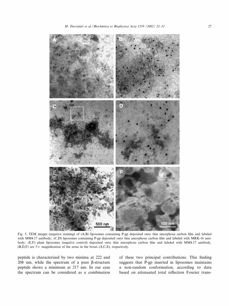
Fig. 5. TEM images (negative staining) of (A,B) liposomes containing P-gp deposited onto thin amorphous carbon ¢lm and labeledwith MM4.17 antibody; (C,D) liposomes containing P-gp deposited onto thin amorphous carbon ¢lm and labeled with MRK-16 anti-body; (E,F) plain liposomes (negative control) deposited onto thin amorphous carbon ¢lm and labeled with MM4.17 antibody.(B,D,F) are 5U magni¢cation of the areas in the boxes (A,C,E), respectively.
BBAMEM 78186 24-1-02
M. Diociaiuti et al. / Biochimica et Biophysica Acta 1559 (2002) 21^31 27

form infrared measurements, where P-gp is describedas constituted by K-helices and L-structures [3,18].The CD spectrum relative to liposomes without P-gp did not show any particular CD signal in thesame spectral region (data not shown).
The mechanisms of opening and fusion of lipo-somes onto several kinds of hydrophobic surfaces,were studied in detail and are summarized in Fig. 4[9,19]. As it can be observed the deposition of lipo-somes onto hydrophobic surfaces, as amorphous car-bon (paths A) or L^B ¢lm (path B), leads to theformation of a lipid double layer following two dif-ferent mechanisms.
The ¢rst substrate we used was amorphous car-bon, generally prepared as a very thin ¢lm (about50 nm), suitable for TEM analysis. Fig. 5A,B showsTEM images of liposomes containing P-gp after thedeposition onto amorphous carbon surface. Typicalround-shaped liposomes were not visible, suggestingthat they opened and fused each other, thus leadingto the formation of a lipid double layer. In this sam-ple, P-gp was labeled employing the highly reactivespeci¢c monoclonal antibody MM4.17, and revealedby goat anti-mouse immunoglobulins conjugatedwith 5 nm gold particles. A large number of goldparticles (black dots in the ¢gure), labeling the pro-tein molecules incorporated in the lipid bilayer canbe observed. This result clearly indicates that thereactivity of epitope, recognized by mAb MM4.17and localized on the fourth loop of the P-gp molecule[11], was not compromised by the mechanism of lipo-some opening and fusion. This result was con¢rmedby the labeling of the same sample by mAb MRK-16(Fig. 5C,D). This antibody recognizes epitopes lo-cated on the ¢rst and fourth loops of P-gp [12] andwas revealed by goat anti-mouse immunoglobulinsconjugated with 5 nm gold particles.
Even though in this ¢gure it is possible to observefew whole liposomes (about 250 nm diameter), thebackground looks like the lipid bilayer in Fig. 5A,B.In fact, the only di¡erence between the two samplesis the antibody used for labeling. A negative control,performed on liposomes containing P-gp labeled withan irrelevant antibody, did not display gold particles(data not shown). Labeling performed by highly spe-ci¢c mAbs MM4.17 and MRK-16 clearly indicatedthat, at least in the surroundings of their reactiveepitopes, P-gp maintained its structural features. It
is noteworthy that both mAb MM4.17 and MRK-16recognize P-gp in living cells.
The lower number of gold particles detected in thesample labeled with mAb MRK-16 as compared tothe sample labeled with mAb MM4.17 suggests thatthe epitopes recognized by mAb MRK-16 were lessaccessible to the antibody. The lower reactivity ofmAb MRK-16, when compared to that of mAbMM4.17, was previously reported in P-gp labelingexperiments of living MDR tumor cells [8].
In agreement with other studies, it can be hypothe-sized that the presence of proteins in the liposomeperturbs the original unstressed organization of thelipid bilayer [20,21] and facilitates liposome openingand fusion processes. This hypothesis appears to besupported by our TEM observation, under the sameexperimental conditions, of P-gp-free liposomes. Fig.5E,F shows the presence of many unopened lipo-somes that can be easily recognized by their roundedshape. This ¢nding is consistent with a structuralstability of protein-free liposomes higher than thatof P-gp-containing liposomes. As a further negativecontrol of labeling experiments, liposomes without P-gp were labeled with mAbs MM4.17 and/or MRK16.As expected, the presence of very few gold particles(Fig. 5E,F) was revealed, indicating that neither aspeci¢c nor aspeci¢c bond occurred in the sample.The isolated gold particles were very likely due toresidual secondary antibodies remaining after thewashing procedure.
The second hydrophobic substrate we used was thesurface of DPPC L^B monolayer deposited ontomica, extremely £at and suitable for AFM analysis.AFM allows us to accurately characterize the lipidsurface, not only in the plane (x^y direction) buteven in the z-direction. Fig. 6 shows AFM imagesof lipid bilayers formed after the fusion of liposomeswith the L^B monolayer.
Fig. 6A,B refers to samples containing P-gp la-beled with mAb MM4.17 and 10 nm gold particles.In this ¢gure, the presence of white spots of about 30nm diameter is clearly evident. These spots appearconsiderably larger than the nominal diameter ofgold particles: this could be due both to the presenceof the antibody molecules and the AFM tip convo-lution e¡ect. Like in TEM images (Fig. 5A,B) manygold particles (white spots) can be observed, con¢rm-ing the presence of P-gp in the lipid bilayer. Two
BBAMEM 78186 24-1-02
M. Diociaiuti et al. / Biochimica et Biophysica Acta 1559 (2002) 21^3128
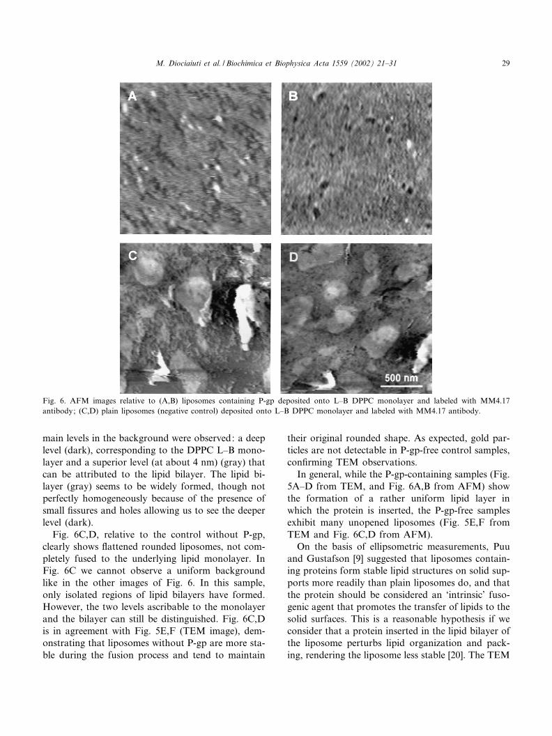
main levels in the background were observed: a deeplevel (dark), corresponding to the DPPC L^B mono-layer and a superior level (at about 4 nm) (gray) thatcan be attributed to the lipid bilayer. The lipid bi-layer (gray) seems to be widely formed, though notperfectly homogeneously because of the presence ofsmall ¢ssures and holes allowing us to see the deeperlevel (dark).
Fig. 6C,D, relative to the control without P-gp,clearly shows £attened rounded liposomes, not com-pletely fused to the underlying lipid monolayer. InFig. 6C we cannot observe a uniform backgroundlike in the other images of Fig. 6. In this sample,only isolated regions of lipid bilayers have formed.However, the two levels ascribable to the monolayerand the bilayer can still be distinguished. Fig. 6C,Dis in agreement with Fig. 5E,F (TEM image), dem-onstrating that liposomes without P-gp are more sta-ble during the fusion process and tend to maintain
their original rounded shape. As expected, gold par-ticles are not detectable in P-gp-free control samples,con¢rming TEM observations.
In general, while the P-gp-containing samples (Fig.5A^D from TEM, and Fig. 6A,B from AFM) showthe formation of a rather uniform lipid layer inwhich the protein is inserted, the P-gp-free samplesexhibit many unopened liposomes (Fig. 5E,F fromTEM and Fig. 6C,D from AFM).
On the basis of ellipsometric measurements, Puuand Gustafson [9] suggested that liposomes contain-ing proteins form stable lipid structures on solid sup-ports more readily than plain liposomes do, and thatthe protein should be considered an `intrinsic' fuso-genic agent that promotes the transfer of lipids to thesolid surfaces. This is a reasonable hypothesis if weconsider that a protein inserted in the lipid bilayer ofthe liposome perturbs lipid organization and pack-ing, rendering the liposome less stable [20]. The TEM
Fig. 6. AFM images relative to (A,B) liposomes containing P-gp deposited onto L^B DPPC monolayer and labeled with MM4.17antibody; (C,D) plain liposomes (negative control) deposited onto L^B DPPC monolayer and labeled with MM4.17 antibody.
BBAMEM 78186 24-1-02
M. Diociaiuti et al. / Biochimica et Biophysica Acta 1559 (2002) 21^31 29

and AFM observations reported in this paper con-¢rm this hypothesis. The absence of P-gp in the con-trol samples hinders the fusion process between lipo-somes deposited onto carbon ¢lm (TEM) or betweenliposomes and the L^B-covered mica substrate(AFM). Our observations clearly show that lipo-somes without P-gp keep their original close struc-ture in both cases, demonstrating the essential role ofthe protein in the fusion process.
Moreover, the comparison between results ob-tained by TEM and AFM prompts some considera-tions. In TEM micrographs, the presence of a highnumber of gold particles reveals that the P-gp epi-topes (Fig. 5A,B), localized in the loops protrudingfrom the cellular membrane [5], maintain their nativeorientation during the fusion process between lipo-somes on carbon ¢lm, and remain available for thereaction with the speci¢c monoclonal antibody. Onthe contrary, the lower number of gold particles ob-served in AFM images (Fig. 6A,B) could be associ-ated with a di¡erent behavior of the protein to main-tain its conformational state when carrier liposomesinteract with a lipid L^B monolayer. This can beascribed to a di¡erent opening mechanism of theliposomes onto carbon ¢lm and L^B ¢lm. It couldbe hypothesized that the double-layer formation oc-curring in the presence of a well-packed and -orderedlipid L^B ¢lm, is a more di¤cult mechanism inwhich the protein transfer from the liposome to theforming supported lipid bilayer is not favored.
4. Conclusions
Both enzymatic assays and CD measurements in-dicate the successful P-gp puri¢cation and reconsti-tution in liposomes. Moreover, TEM and AFM al-lowed us to obtain useful information about themechanisms of opening and fusion of liposomesonto two di¡erent hydrophobic substrates: amor-phous carbon and DPPC L^B monolayer. Our dataare in good agreement with the mechanisms pro-posed in literature, suggesting that the presence ofP-gp plays an important role in stability of the lipo-some. P-gp probably acts as a fusogenic agent, de-creasing the lipid organization and packing.
The epitope reactivity, studied by immunogold la-beling, is preserved after P-gp insertion in the sup-
ported lipid bilayer formed onto both substrates. Fi-nally, information about P-gp aggregation state canbe obtained by observing the distribution of the goldparticles. In all the ¢gures relative to samples contain-ing P-gp inserted in lipid bilayers, performed by TEM(Fig. 5A^D) and AFM (Fig. 6A,B), gold particlesappear as isolated units. The absence of couples orclusters of gold particles in the micrographs allows usto rule out the presence of P-gp dimers or oligomers.
Acknowledgements
The authors are greatly indebted to Dr. M. Cian-friglia for valuable discussions. This work was parti-ally supported by Italian `AIDS RESEARCH' grant.
References
[1] P. van der Valk, C.K. van Kalken, H. Ketelaars, H.J. Brox-sterman, G. Sche¡er, C.M. Kuiiper, T. Tsuruo, J. Lankelma,C.J.L.M. Meijer, H.M. Pinedo, R. Scheper, Ann. Oncol. 1(1990) 56^64.
[2] L.J. Goldstein, I. Pastan, Crit. Rev. Oncol. Hematol. 12(1992) 243^253.
[3] F.J. Sharom, R. Liu, Y. Romsicki, Biochem. Cell. Biol. 76(1998) 695^708.
[4] M.F. Rosenberg, R. Callaghan, R.C. Ford, C.F. Higgins,J. Biol. Chem. 272 (1997) 10685^10694.
[5] C.F. Higgins, R. Callagan, K.J. Linton, M.F. Rosenberg,R.C. Ford, Semin. Cancer Biol. 8 (1997) 135^142.
[6] P.M. Jones, A.M. George, Eur. J. Biochem. 267 (2000)5298^5305.
[7] T.W. Loo, D.M. Clarke, J. Biol. Chem. 27 (1996) 27482^27492.
[8] A. Molinari, M. Cianfriglia, S. Meschini, A. Calcabrini, G.Arancia, Int. J. Cancer 59 (1994) 789^795.
[9] G. Puu, I. Gustafson, Biochim. Biophys. Acta 1327 (1997)149^161.
[10] G. Roberts, Langmuir^Blodgett Films, Plenum Press, NewYork, 1990.
[11] M. Cianfriglia, M.C. Willingham, M. Tombesi, G.V. Sca-gliotti, G. Frasca, A. Chersi, Int. J. Cancer 56 (1994) 153^160.
[12] E. Georges, T. Tsurno, V. Luig, J. Biol. Chem. 268 (1993)1792^1798.
[13] M. Dong, F. Penin, L.G. Baggetto, J. Biol. Chem. 271(1996) 28875^28883.
[14] U.K. Laemmli, Nature 227 (1970) 680^685.[15] J.L. Rigaud, B. Pitard, D. Levy, Biochim. Biophys. Acta
1231 (1995) 223^240.
BBAMEM 78186 24-1-02
M. Diociaiuti et al. / Biochimica et Biophysica Acta 1559 (2002) 21^3130

[16] P. Gonzalo, B. Sontag, D. Guillot, J.P. Reboud, Anal. Bio-chem. 225 (1995) 178^180.
[17] T. Watanabe, N. Kokubu, S.B. Charnick, M. Naito, T.Tsuruo, D. Cohen, Br. J. Pharm. 122 (1997) 241^248.
[18] N. Sonveaux, A.B. Shapiro, E. Goormaghtigh, V. Ling, J.M.Ruysschaert, J. Biol. Chem. 271 (1996) 24617^24624.
[19] C.A. Keller, K. Glasmastar, V.P. Zhdanov, B. Kasemo,Phys. Rev. Lett. 84 (2000) 5443^5446.
[20] B.K. Jap, M. Zulauf, T. Scheybani, A. Hefti, W. Baumein-ster, U. Aebi, Ultramicroscopy 46 (1992) 45^84.
[21] J.N. Israelachvili, S. Marcelja, R.G. Horn, Q. Rev. Biophys.13 (1980) 121^200.
BBAMEM 78186 24-1-02
M. Diociaiuti et al. / Biochimica et Biophysica Acta 1559 (2002) 21^31 31