Peripherally inserted central catheter in critically ill patients
47
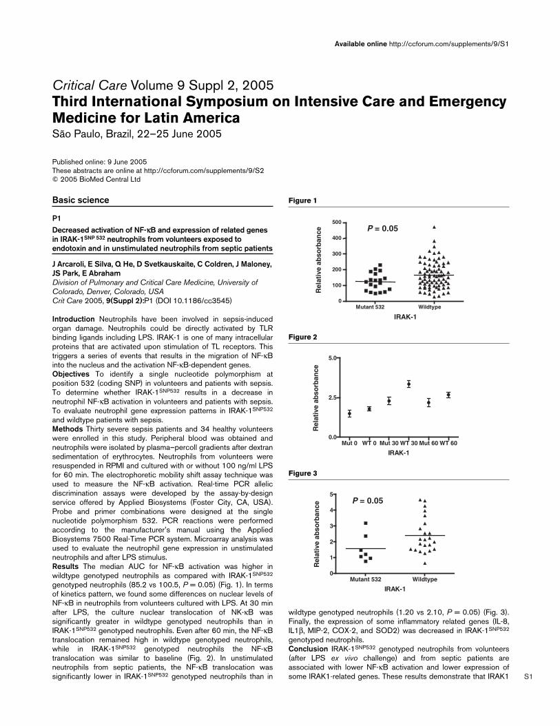
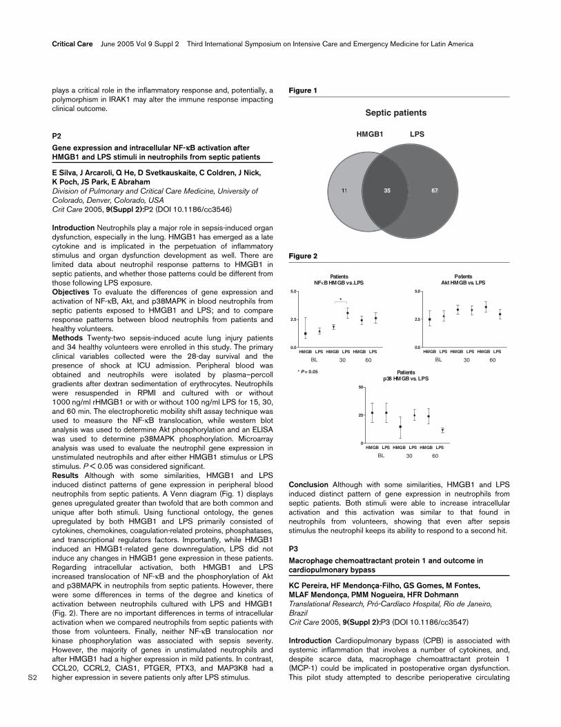
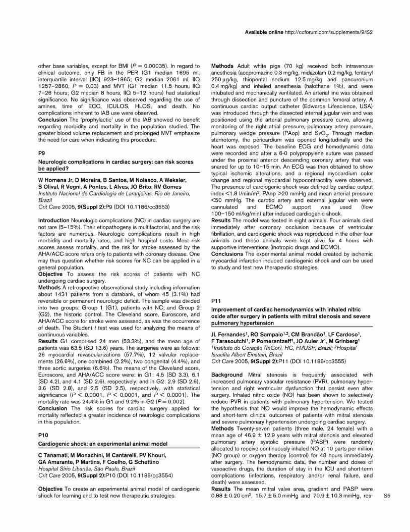
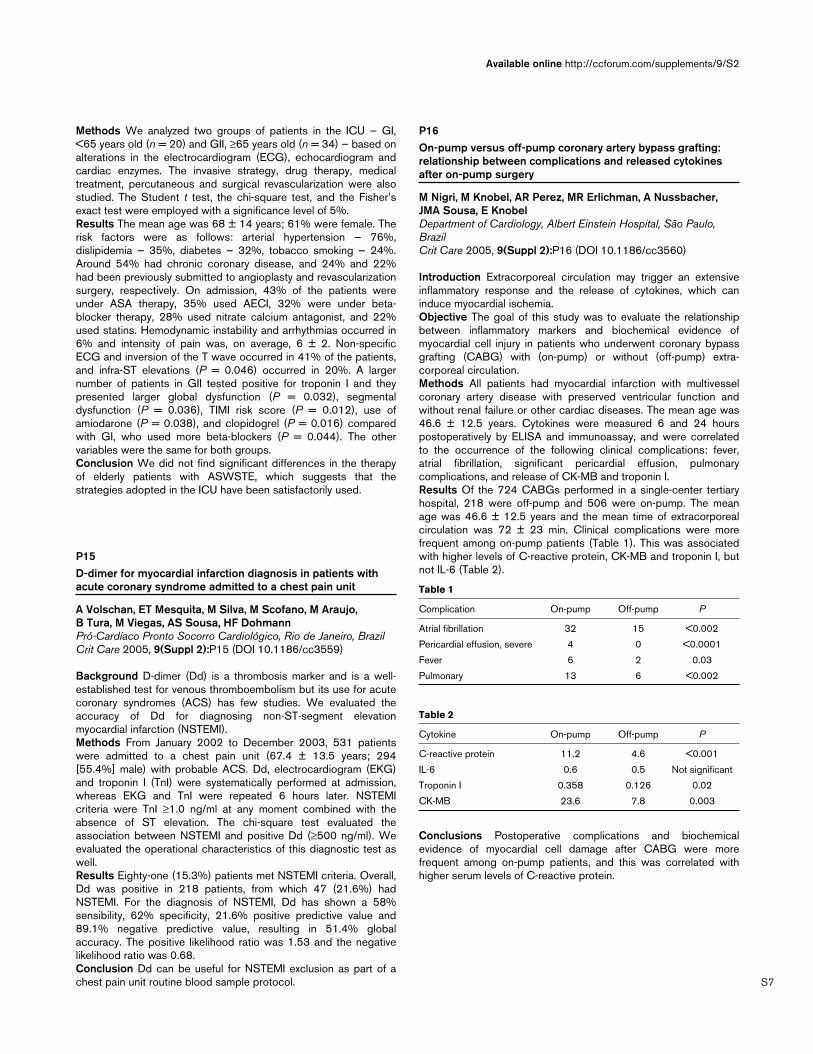
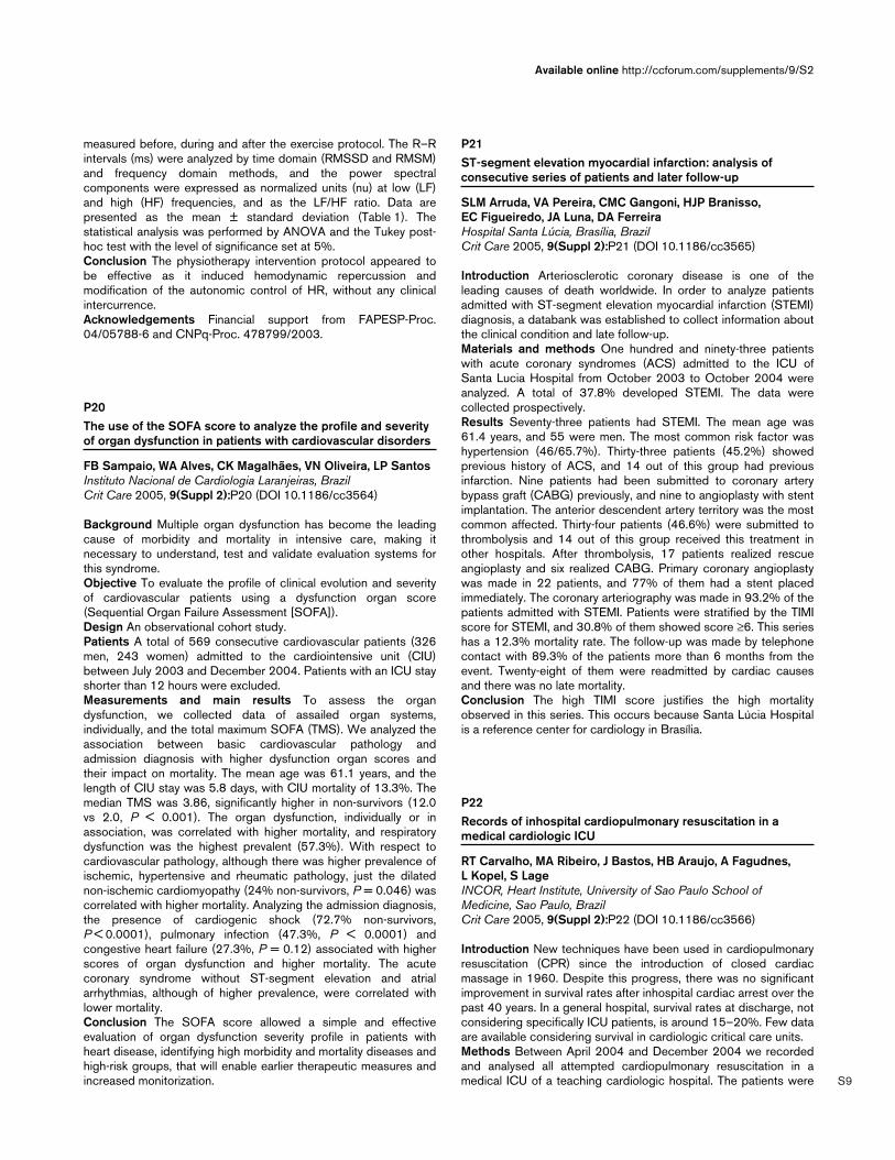
S1 Available online http://ccforum.com/supplements/9/S1 Critical Care Volume 9 Suppl 2, 2005 Third International Symposium on Intensive Care and Emergency Medicine for Latin America São Paulo, Brazil, 22–25 June 2005 Published online: 9 June 2005 These abstracts are online at http://ccforum.com/supplements/9/S2 © 2005 BioMed Central Ltd Basic science P1 Decreased activation of NF-κB and expression of related genes in IRAK-1 SNP 532 neutrophils from volunteers exposed to endotoxin and in unstimulated neutrophils from septic patients J Arcaroli, E Silva, Q He, D Svetkauskaite, C Coldren, J Maloney, JS Park, E Abraham Division of Pulmonary and Critical Care Medicine, University of Colorado, Denver, Colorado, USA Crit Care 2005, 9(Suppl 2):P1 (DOI 10.1186/cc3545) Introduction Neutrophils have been involved in sepsis-induced organ damage. Neutrophils could be directly activated by TLR binding ligands including LPS. IRAK-1 is one of many intracellular proteins that are activated upon stimulation of TL receptors. This triggers a series of events that results in the migration of NF-κB into the nucleus and the activation NF-κB-dependent genes. Objectives To identify a single nucleotide polymorphism at position 532 (coding SNP) in volunteers and patients with sepsis. To determine whether IRAK-1 SNP532 results in a decrease in neutrophil NF-κB activation in volunteers and patients with sepsis. To evaluate neutrophil gene expression patterns in IRAK-1 SNP532 and wildtype patients with sepsis. Methods Thirty severe sepsis patients and 34 healthy volunteers were enrolled in this study. Peripheral blood was obtained and neutrophils were isolated by plasma–percoll gradients after dextran sedimentation of erythrocytes. Neutrophils from volunteers were resuspended in RPMI and cultured with or without 100 ng/ml LPS for 60 min. The electrophoretic mobility shift assay technique was used to measure the NF-κB activation. Real-time PCR allelic discrimination assays were developed by the assay-by-design service offered by Applied Biosystems (Foster City, CA, USA). Probe and primer combinations were designed at the single nucleotide polymorphism 532. PCR reactions were performed according to the manufacturer’s manual using the Applied Biosystems 7500 Real-Time PCR system. Microarray analysis was used to evaluate the neutrophil gene expression in unstimulated neutrophils and after LPS stimulus. Results The median AUC for NF-κB activation was higher in wildtype genotyped neutrophils as compared with IRAK-1 SNP532 genotyped neutrophils (85.2 vs 100.5, P = 0.05) (Fig. 1). In terms of kinetics pattern, we found some differences on nuclear levels of NF-κB in neutrophils from volunteers cultured with LPS. At 30 min after LPS, the culture nuclear translocation of NK-κB was significantly greater in wildtype genotyped neutrophils than in IRAK-1 SNP532 genotyped neutrophils. Even after 60 min, the NF-κB translocation remained high in wildtype genotyped neutrophils, while in IRAK-1 SNP532 genotyped neutrophils the NF-κB translocation was similar to baseline (Fig. 2). In unstimulated neutrophils from septic patients, the NF-κB translocation was significantly lower in IRAK-1 SNP532 genotyped neutrophils than in wildtype genotyped neutrophils (1.20 vs 2.10, P = 0.05) (Fig. 3). Finally, the expression of some inflammatory related genes (IL-8, IL1β, MIP-2, COX-2, and SOD2) was decreased in IRAK-1 SNP532 genotyped neutrophils. Conclusion IRAK-1 SNP532 genotyped neutrophils from volunteers (after LPS ex vivo challenge) and from septic patients are associated with lower NF-κB activation and lower expression of some IRAK1-related genes. These results demonstrate that IRAK1 Figure 2 Mut 0 WT 0 Mut 30 WT 30 Mut 60 WT 60 0. 0 2. 5 5.0 IRAK-1 Relative absorbance Figure 3 Mutant 532 Wildty pe 0 1 2 3 4 5 IRAK-1 P = 0.05 Relative absorbance Figure 1 Mutant 53 2 Wildtyp e 0 10 0 20 0 30 0 40 0 500 P = 0.05 IRAK-1 Relative absorbance
-
Upload
independent -
Category
Documents
-
view
4 -
download
0
Transcript of Peripherally inserted central catheter in critically ill patients
S1
Available online http://ccforum.com/supplements/9/S1
Critical Care Volume 9 Suppl 2, 2005Third International Symposium on Intensive Care and EmergencyMedicine for Latin AmericaSão Paulo, Brazil, 22–25 June 2005
Published online: 9 June 2005These abstracts are online at http://ccforum.com/supplements/9/S2© 2005 BioMed Central Ltd
Basic science
P1Decreased activation of NF-κκB and expression of related genesin IRAK-1SNP 532 neutrophils from volunteers exposed toendotoxin and in unstimulated neutrophils from septic patients
J Arcaroli, E Silva, Q He, D Svetkauskaite, C Coldren, J Maloney,JS Park, E AbrahamDivision of Pulmonary and Critical Care Medicine, University ofColorado, Denver, Colorado, USACrit Care 2005, 9(Suppl 2):P1 (DOI 10.1186/cc3545)
Introduction Neutrophils have been involved in sepsis-inducedorgan damage. Neutrophils could be directly activated by TLRbinding ligands including LPS. IRAK-1 is one of many intracellularproteins that are activated upon stimulation of TL receptors. Thistriggers a series of events that results in the migration of NF-κBinto the nucleus and the activation NF-κB-dependent genes.Objectives To identify a single nucleotide polymorphism atposition 532 (coding SNP) in volunteers and patients with sepsis.To determine whether IRAK-1SNP532 results in a decrease inneutrophil NF-κB activation in volunteers and patients with sepsis.To evaluate neutrophil gene expression patterns in IRAK-1SNP532
and wildtype patients with sepsis.Methods Thirty severe sepsis patients and 34 healthy volunteerswere enrolled in this study. Peripheral blood was obtained andneutrophils were isolated by plasma–percoll gradients after dextransedimentation of erythrocytes. Neutrophils from volunteers wereresuspended in RPMI and cultured with or without 100 ng/ml LPSfor 60 min. The electrophoretic mobility shift assay technique wasused to measure the NF-κB activation. Real-time PCR allelicdiscrimination assays were developed by the assay-by-designservice offered by Applied Biosystems (Foster City, CA, USA).Probe and primer combinations were designed at the singlenucleotide polymorphism 532. PCR reactions were performedaccording to the manufacturer’s manual using the AppliedBiosystems 7500 Real-Time PCR system. Microarray analysis wasused to evaluate the neutrophil gene expression in unstimulatedneutrophils and after LPS stimulus.Results The median AUC for NF-κB activation was higher inwildtype genotyped neutrophils as compared with IRAK-1SNP532
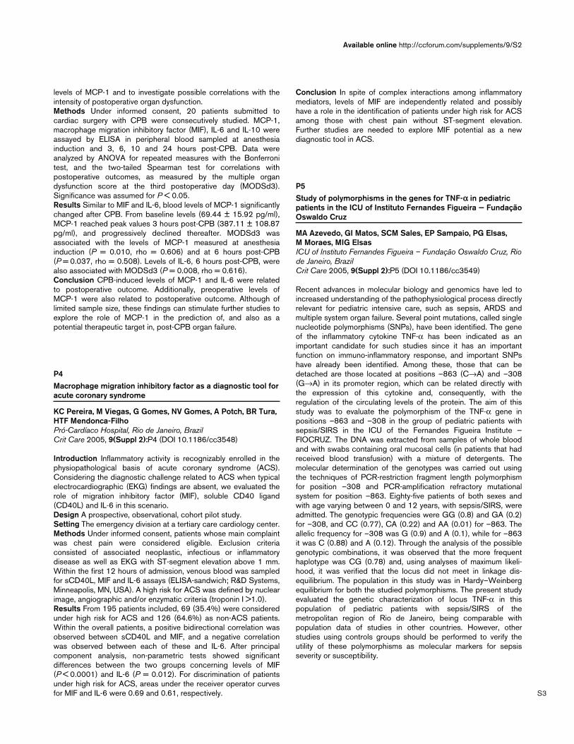
genotyped neutrophils (85.2 vs 100.5, P = 0.05) (Fig. 1). In termsof kinetics pattern, we found some differences on nuclear levels ofNF-κB in neutrophils from volunteers cultured with LPS. At 30 minafter LPS, the culture nuclear translocation of NK-κB wassignificantly greater in wildtype genotyped neutrophils than inIRAK-1SNP532 genotyped neutrophils. Even after 60 min, the NF-κBtranslocation remained high in wildtype genotyped neutrophils,while in IRAK-1SNP532 genotyped neutrophils the NF-κBtranslocation was similar to baseline (Fig. 2). In unstimulatedneutrophils from septic patients, the NF-κB translocation wassignificantly lower in IRAK-1SNP532 genotyped neutrophils than in
wildtype genotyped neutrophils (1.20 vs 2.10, P = 0.05) (Fig. 3).Finally, the expression of some inflammatory related genes (IL-8,IL1β, MIP-2, COX-2, and SOD2) was decreased in IRAK-1SNP532
genotyped neutrophils.Conclusion IRAK-1SNP532 genotyped neutrophils from volunteers(after LPS ex vivo challenge) and from septic patients areassociated with lower NF-κB activation and lower expression ofsome IRAK1-related genes. These results demonstrate that IRAK1
Figure 2
Mut 0 WT 0 Mut 30 WT 30 Mut 60 WT 600.0
2.5
5.0
IRAK-1
Rel
ativ
e ab
sorb
ance
Figure 3
Mutant 532 Wildtype0
1
2
3
4
5
IRAK-1
P = 0.05
Rel
ativ
e ab
sorb
ance
Figure 1
Mutant 532 Wildtype0
100
200
300
400
500P = 0.05
IRAK-1R
elat
ive
abso
rban
ce
S2
Critical Care June 2005 Vol 9 Suppl 2 Third International Symposium on Intensive Care and Emergency Medicine for Latin America
plays a critical role in the inflammatory response and, potentially, apolymorphism in IRAK1 may alter the immune response impactingclinical outcome.
P2Gene expression and intracellular NF-κκB activation afterHMGB1 and LPS stimuli in neutrophils from septic patients
E Silva, J Arcaroli, Q He, D Svetkauskaite, C Coldren, J Nick, K Poch, JS Park, E AbrahamDivision of Pulmonary and Critical Care Medicine, University ofColorado, Denver, Colorado, USACrit Care 2005, 9(Suppl 2):P2 (DOI 10.1186/cc3546)
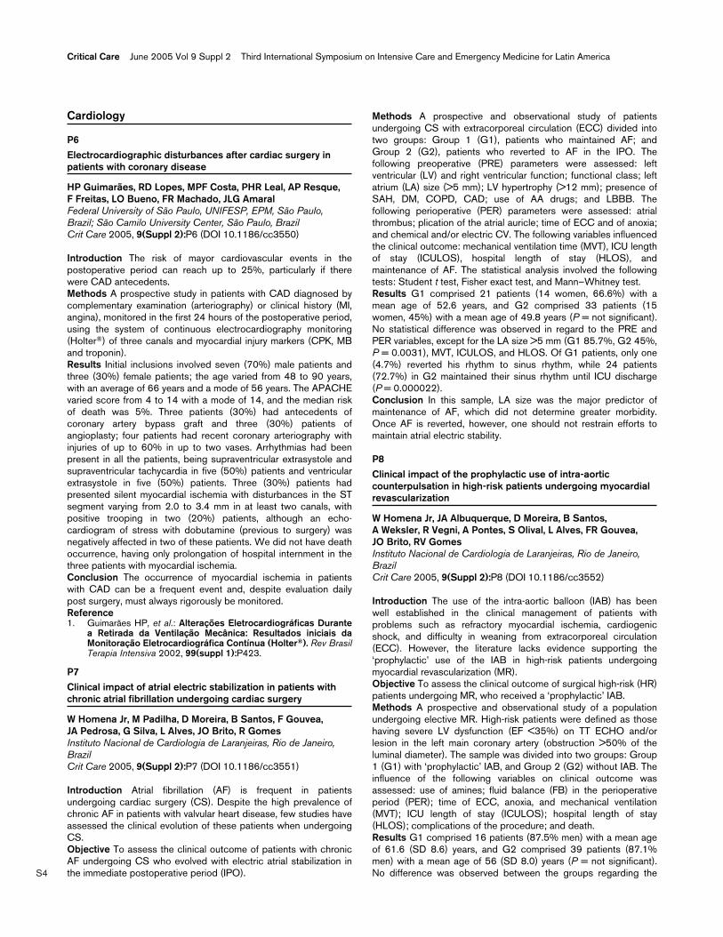
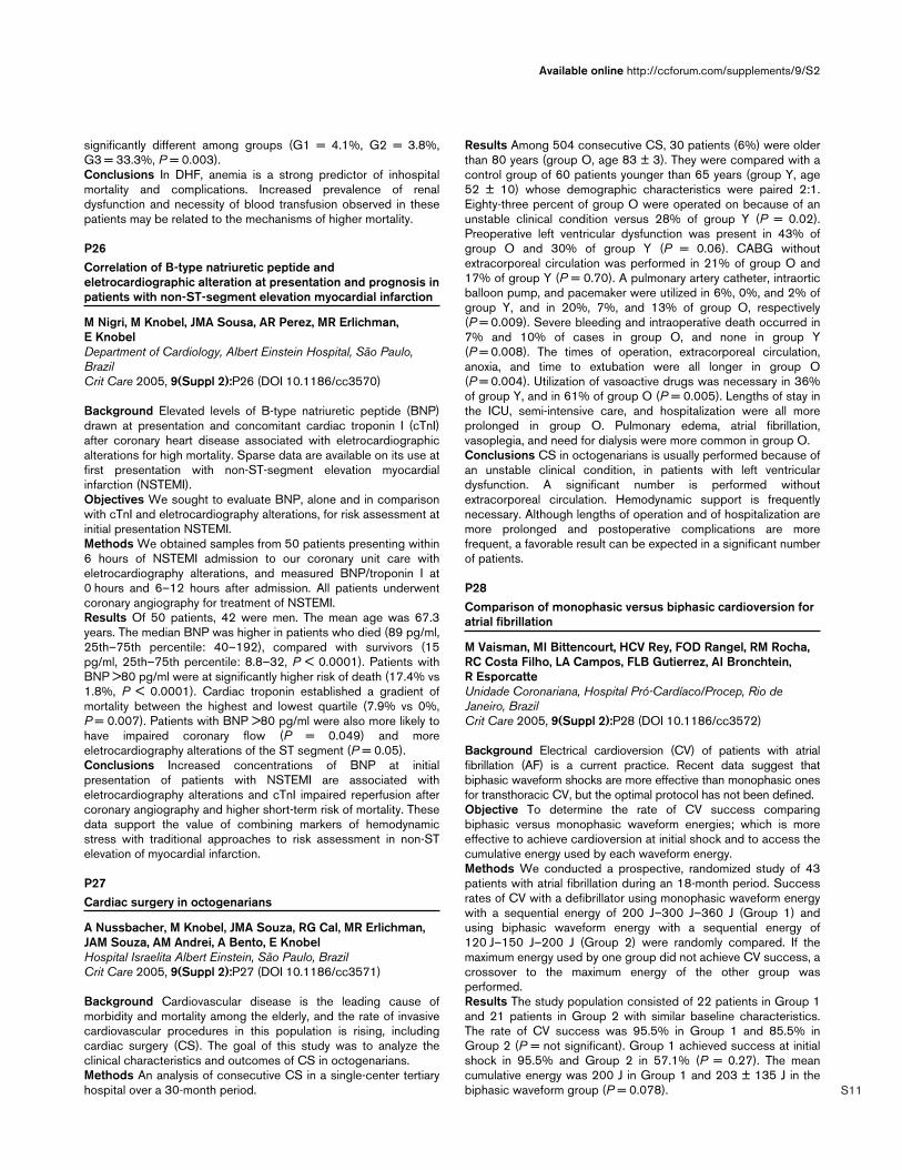
Introduction Neutrophils play a major role in sepsis-induced organdysfunction, especially in the lung. HMGB1 has emerged as a latecytokine and is implicated in the perpetuation of inflammatorystimulus and organ dysfunction development as well. There arelimited data about neutrophil response patterns to HMGB1 inseptic patients, and whether those patterns could be different fromthose following LPS exposure.Objectives To evaluate the differences of gene expression andactivation of NF-κB, Akt, and p38MAPK in blood neutrophils fromseptic patients exposed to HMGB1 and LPS; and to compareresponse patterns between blood neutrophils from patients andhealthy volunteers.Methods Twenty-two sepsis-induced acute lung injury patientsand 34 healthy volunteers were enrolled in this study. The primaryclinical variables collected were the 28-day survival and thepresence of shock at ICU admission. Peripheral blood wasobtained and neutrophils were isolated by plasma–percollgradients after dextran sedimentation of erythrocytes. Neutrophilswere resuspended in RPMI and cultured with or without1000 ng/ml rHMGB1 or with or without 100 ng/ml LPS for 15, 30,and 60 min. The electrophoretic mobility shift assay technique wasused to measure the NF-κB translocation, while western blotanalysis was used to determine Akt phosphorylation and an ELISAwas used to determine p38MAPK phosphorylation. Microarrayanalysis was used to evaluate the neutrophil gene expression inunstimulated neutrophils and after either HMGB1 stimulus or LPSstimulus. P < 0.05 was considered significant.Results Although with some similarities, HMGB1 and LPSinduced distinct patterns of gene expression in peripheral bloodneutrophils from septic patients. A Venn diagram (Fig. 1) displaysgenes upregulated greater than twofold that are both common andunique after both stimuli. Using functional ontology, the genesupregulated by both HMGB1 and LPS primarily consisted ofcytokines, chemokines, coagulation-related proteins, phosphatases,and transcriptional regulators factors. Importantly, while HMGB1induced an HMGB1-related gene downregulation, LPS did notinduce any changes in HMGB1 gene expression in these patients.Regarding intracellular activation, both HMGB1 and LPSincreased translocation of NF-κB and the phosphorylation of Aktand p38MAPK in neutrophils from septic patients. However, therewere some differences in terms of the degree and kinetics ofactivation between neutrophils cultured with LPS and HMGB1(Fig. 2). There are no important differences in terms of intracellularactivation when we compared neutrophils from septic patients withthose from volunteers. Finally, neither NF-κB translocation norkinase phosphorylation was associated with sepsis severity.However, the majority of genes in unstimulated neutrophils andafter HMGB1 had a higher expression in mild patients. In contrast,CCL20, CCRL2, CIAS1, PTGER, PTX3, and MAP3K8 had ahigher expression in severe patients only after LPS stimulus.
Conclusion Although with some similarities, HMGB1 and LPSinduced distinct pattern of gene expression in neutrophils fromseptic patients. Both stimuli were able to increase intracellularactivation and this activation was similar to that found inneutrophils from volunteers, showing that even after sepsisstimulus the neutrophil keeps its ability to respond to a second hit.
P3Macrophage chemoattractant protein 1 and outcome incardiopulmonary bypass
KC Pereira, HF Mendonça-Filho, GS Gomes, M Fontes, MLAF Mendonça, PMM Nogueira, HFR DohmannTranslational Research, Pró-Cardíaco Hospital, Rio de Janeiro,BrazilCrit Care 2005, 9(Suppl 2):P3 (DOI 10.1186/cc3547)
Introduction Cardiopulmonary bypass (CPB) is associated withsystemic inflammation that involves a number of cytokines, and,despite scarce data, macrophage chemoattractant protein 1(MCP-1) could be implicated in postoperative organ dysfunction.This pilot study attempted to describe perioperative circulating
Figure 1
Septic patients
HMGB1 LPS
11 35 67
Figure 2
PatientsNFκB HMGB vs. LPS
HMGB LPS HMGB LPS HMGB LPS0.0
2.5
5.0
BL 30 60
*
* P = 0.05
PatientsAkt HMGB vs. LPS
HMGB LPS HMGB LPS HMGB LPS0.0
2.5
5.0
BL 30 60
Patientsp38 HMGB vs. LPS
HMGB LPS HMGB LPS HMGB LPS0
25
50
BL 30 60
S3
Available online http://ccforum.com/supplements/9/S2
levels of MCP-1 and to investigate possible correlations with theintensity of postoperative organ dysfunction.Methods Under informed consent, 20 patients submitted tocardiac surgery with CPB were consecutively studied. MCP-1,macrophage migration inhibitory factor (MIF), IL-6 and IL-10 wereassayed by ELISA in peripheral blood sampled at anesthesiainduction and 3, 6, 10 and 24 hours post-CPB. Data wereanalyzed by ANOVA for repeated measures with the Bonferronitest, and the two-tailed Spearman test for correlations withpostoperative outcomes, as measured by the multiple organdysfunction score at the third postoperative day (MODSd3).Significance was assumed for P < 0.05.Results Similar to MIF and IL-6, blood levels of MCP-1 significantlychanged after CPB. From baseline levels (69.44 ± 15.92 pg/ml),MCP-1 reached peak values 3 hours post-CPB (387.11 ± 108.87pg/ml), and progressively declined thereafter. MODSd3 wasassociated with the levels of MCP-1 measured at anesthesiainduction (P = 0.010, rho = 0.606) and at 6 hours post-CPB(P = 0.037, rho = 0.508). Levels of IL-6, 6 hours post-CPB, werealso associated with MODSd3 (P = 0.008, rho = 0.616).Conclusion CPB-induced levels of MCP-1 and IL-6 were relatedto postoperative outcome. Additionally, preoperative levels ofMCP-1 were also related to postoperative outcome. Although oflimited sample size, these findings can stimulate further studies toexplore the role of MCP-1 in the prediction of, and also as apotential therapeutic target in, post-CPB organ failure.
P4Macrophage migration inhibitory factor as a diagnostic tool foracute coronary syndrome
KC Pereira, M Viegas, G Gomes, NV Gomes, A Potch, BR Tura,HTF Mendonca-FilhoPró-Cardíaco Hospital, Rio de Janeiro, BrazilCrit Care 2005, 9(Suppl 2):P4 (DOI 10.1186/cc3548)
Introduction Inflammatory activity is recognizably enrolled in thephysiopathological basis of acute coronary syndrome (ACS).Considering the diagnostic challenge related to ACS when typicalelectrocardiographic (EKG) findings are absent, we evaluated therole of migration inhibitory factor (MIF), soluble CD40 ligand(CD40L) and IL-6 in this scenario.Design A prospective, observational, cohort pilot study.Setting The emergency division at a tertiary care cardiology center.Methods Under informed consent, patients whose main complaintwas chest pain were considered eligible. Exclusion criteriaconsisted of associated neoplastic, infectious or inflammatorydisease as well as EKG with ST-segment elevation above 1 mm.Within the first 12 hours of admission, venous blood was sampledfor sCD40L, MIF and IL-6 assays (ELISA-sandwich; R&D Systems,Minneapolis, MN, USA). A high risk for ACS was defined by nuclearimage, angiographic and/or enzymatic criteria (troponin I >1.0).Results From 195 patients included, 69 (35.4%) were consideredunder high risk for ACS and 126 (64.6%) as non-ACS patients.Within the overall patients, a positive bidirectional correlation wasobserved between sCD40L and MIF, and a negative correlationwas observed between each of these and IL-6. After principalcomponent analysis, non-parametric tests showed significantdifferences between the two groups concerning levels of MIF(P < 0.0001) and IL-6 (P = 0.012). For discrimination of patientsunder high risk for ACS, areas under the receiver operator curvesfor MIF and IL-6 were 0.69 and 0.61, respectively.
Conclusion In spite of complex interactions among inflammatorymediators, levels of MIF are independently related and possiblyhave a role in the identification of patients under high risk for ACSamong those with chest pain without ST-segment elevation.Further studies are needed to explore MIF potential as a newdiagnostic tool in ACS.
P5Study of polymorphisms in the genes for TNF-αα in pediatricpatients in the ICU of Instituto Fernandes Figueira — FundaçãoOswaldo Cruz
MA Azevedo, GI Matos, SCM Sales, EP Sampaio, PG Elsas, M Moraes, MIG ElsasICU of Instituto Fernandes Figueira – Fundação Oswaldo Cruz, Riode Janeiro, BrazilCrit Care 2005, 9(Suppl 2):P5 (DOI 10.1186/cc3549)
Recent advances in molecular biology and genomics have led toincreased understanding of the pathophysiological process directlyrelevant for pediatric intensive care, such as sepsis, ARDS andmultiple system organ failure. Several point mutations, called singlenucleotide polymorphisms (SNPs), have been identified. The geneof the inflammatory cytokine TNF-α has been indicated as animportant candidate for such studies since it has an importantfunction on immuno-inflammatory response, and important SNPshave already been identified. Among these, those that can bedetached are those located at positions –863 (C→A) and –308(G→A) in its promoter region, which can be related directly withthe expression of this cytokine and, consequently, with theregulation of the circulating levels of the protein. The aim of thisstudy was to evaluate the polymorphism of the TNF-α gene inpositions –863 and –308 in the group of pediatric patients withsepsis/SIRS in the ICU of the Fernandes Figueira Institute —FIOCRUZ. The DNA was extracted from samples of whole bloodand with swabs containing oral mucosal cells (in patients that hadreceived blood transfusion) with a mixture of detergents. Themolecular determination of the genotypes was carried out usingthe techniques of PCR-restriction fragment length polymorphismfor position –308 and PCR-amplification refractory mutationalsystem for position –863. Eighty-five patients of both sexes andwith age varying between 0 and 12 years, with sepsis/SIRS, wereadmitted. The genotypic frequencies were GG (0.8) and GA (0.2)for –308, and CC (0.77), CA (0.22) and AA (0.01) for –863. Theallelic frequency for –308 was G (0.9) and A (0.1), while for –863it was C (0.88) and A (0.12). Through the analysis of the possiblegenotypic combinations, it was observed that the more frequenthaplotype was CG (0.78) and, using analyses of maximum likeli-hood, it was verified that the locus did not meet in linkage dis-equilibrium. The population in this study was in Hardy–Weinbergequilibrium for both the studied polymorphisms. The present studyevaluated the genetic characterization of locus TNF-α in thispopulation of pediatric patients with sepsis/SIRS of themetropolitan region of Rio de Janeiro, being comparable withpopulation data of studies in other countries. However, otherstudies using controls groups should be performed to verify theutility of these polymorphisms as molecular markers for sepsisseverity or susceptibility.
S4
Cardiology
P6Electrocardiographic disturbances after cardiac surgery inpatients with coronary disease
HP Guimarães, RD Lopes, MPF Costa, PHR Leal, AP Resque, F Freitas, LO Bueno, FR Machado, JLG AmaralFederal University of São Paulo, UNIFESP, EPM, São Paulo,Brazil; São Camilo University Center, São Paulo, BrazilCrit Care 2005, 9(Suppl 2):P6 (DOI 10.1186/cc3550)
Introduction The risk of mayor cardiovascular events in thepostoperative period can reach up to 25%, particularly if therewere CAD antecedents.Methods A prospective study in patients with CAD diagnosed bycomplementary examination (arteriography) or clinical history (MI,angina), monitored in the first 24 hours of the postoperative period,using the system of continuous electrocardiography monitoring(Holter®) of three canals and myocardial injury markers (CPK, MBand troponin).Results Initial inclusions involved seven (70%) male patients andthree (30%) female patients; the age varied from 48 to 90 years,with an average of 66 years and a mode of 56 years. The APACHEvaried score from 4 to 14 with a mode of 14, and the median riskof death was 5%. Three patients (30%) had antecedents ofcoronary artery bypass graft and three (30%) patients ofangioplasty; four patients had recent coronary arteriography withinjuries of up to 60% in up to two vases. Arrhythmias had beenpresent in all the patients, being supraventricular extrasystole andsupraventricular tachycardia in five (50%) patients and ventricularextrasystole in five (50%) patients. Three (30%) patients hadpresented silent myocardial ischemia with disturbances in the STsegment varying from 2.0 to 3.4 mm in at least two canals, withpositive trooping in two (20%) patients, although an echo-cardiogram of stress with dobutamine (previous to surgery) wasnegatively affected in two of these patients. We did not have deathoccurrence, having only prolongation of hospital internment in thethree patients with myocardial ischemia.Conclusion The occurrence of myocardial ischemia in patientswith CAD can be a frequent event and, despite evaluation dailypost surgery, must always rigorously be monitored.Reference1. Guimarães HP, et al.: Alterações Eletrocardiográficas Durante
a Retirada da Ventilação Mecânica: Resultados iniciais daMonitoração Eletrocardiográfica Contínua (Holter®). Rev BrasilTerapia Intensiva 2002, 99(suppl 1):P423.
P7Clinical impact of atrial electric stabilization in patients withchronic atrial fibrillation undergoing cardiac surgery
W Homena Jr, M Padilha, D Moreira, B Santos, F Gouvea, JA Pedrosa, G Silva, L Alves, JO Brito, R GomesInstituto Nacional de Cardiologia de Laranjeiras, Rio de Janeiro,BrazilCrit Care 2005, 9(Suppl 2):P7 (DOI 10.1186/cc3551)
Introduction Atrial fibrillation (AF) is frequent in patientsundergoing cardiac surgery (CS). Despite the high prevalence ofchronic AF in patients with valvular heart disease, few studies haveassessed the clinical evolution of these patients when undergoingCS.Objective To assess the clinical outcome of patients with chronicAF undergoing CS who evolved with electric atrial stabilization inthe immediate postoperative period (IPO).
Methods A prospective and observational study of patientsundergoing CS with extracorporeal circulation (ECC) divided intotwo groups: Group 1 (G1), patients who maintained AF; andGroup 2 (G2), patients who reverted to AF in the IPO. Thefollowing preoperative (PRE) parameters were assessed: leftventricular (LV) and right ventricular function; functional class; leftatrium (LA) size (>5 mm); LV hypertrophy (>12 mm); presence ofSAH, DM, COPD, CAD; use of AA drugs; and LBBB. Thefollowing perioperative (PER) parameters were assessed: atrialthrombus; plication of the atrial auricle; time of ECC and of anoxia;and chemical and/or electric CV. The following variables influencedthe clinical outcome: mechanical ventilation time (MVT), ICU lengthof stay (ICULOS), hospital length of stay (HLOS), andmaintenance of AF. The statistical analysis involved the followingtests: Student t test, Fisher exact test, and Mann–Whitney test.Results G1 comprised 21 patients (14 women, 66.6%) with amean age of 52.6 years, and G2 comprised 33 patients (15women, 45%) with a mean age of 49.8 years (P = not significant).No statistical difference was observed in regard to the PRE andPER variables, except for the LA size >5 mm (G1 85.7%, G2 45%,P = 0.0031), MVT, ICULOS, and HLOS. Of G1 patients, only one(4.7%) reverted his rhythm to sinus rhythm, while 24 patients(72.7%) in G2 maintained their sinus rhythm until ICU discharge(P = 0.000022).Conclusion In this sample, LA size was the major predictor ofmaintenance of AF, which did not determine greater morbidity.Once AF is reverted, however, one should not restrain efforts tomaintain atrial electric stability.
P8Clinical impact of the prophylactic use of intra-aorticcounterpulsation in high-risk patients undergoing myocardialrevascularization
W Homena Jr, JA Albuquerque, D Moreira, B Santos, A Weksler, R Vegni, A Pontes, S Olival, L Alves, FR Gouvea, JO Brito, RV GomesInstituto Nacional de Cardiologia de Laranjeiras, Rio de Janeiro,BrazilCrit Care 2005, 9(Suppl 2):P8 (DOI 10.1186/cc3552)
Introduction The use of the intra-aortic balloon (IAB) has beenwell established in the clinical management of patients withproblems such as refractory myocardial ischemia, cardiogenicshock, and difficulty in weaning from extracorporeal circulation(ECC). However, the literature lacks evidence supporting the‘prophylactic’ use of the IAB in high-risk patients undergoingmyocardial revascularization (MR).Objective To assess the clinical outcome of surgical high-risk (HR)patients undergoing MR, who received a ‘prophylactic’ IAB.Methods A prospective and observational study of a populationundergoing elective MR. High-risk patients were defined as thosehaving severe LV dysfunction (EF <35%) on TT ECHO and/orlesion in the left main coronary artery (obstruction >50% of theluminal diameter). The sample was divided into two groups: Group1 (G1) with ‘prophylactic’ IAB, and Group 2 (G2) without IAB. Theinfluence of the following variables on clinical outcome wasassessed: use of amines; fluid balance (FB) in the perioperativeperiod (PER); time of ECC, anoxia, and mechanical ventilation(MVT); ICU length of stay (ICULOS); hospital length of stay(HLOS); complications of the procedure; and death.Results G1 comprised 16 patients (87.5% men) with a mean ageof 61.6 (SD 8.6) years, and G2 comprised 39 patients (87.1%men) with a mean age of 56 (SD 8.0) years (P = not significant).No difference was observed between the groups regarding the
Critical Care June 2005 Vol 9 Suppl 2 Third International Symposium on Intensive Care and Emergency Medicine for Latin America
S5
other base variables, except for BMI (P = 0.00035). In regard toclinical outcome, only FB in the PER (G1 median 1695 ml,interquartile interval [IIQ] 923–1865; G2 median 2061 ml, IIQ1257–2860, P = 0.03) and MVT (G1 median 11.5 hours, IIQ7–26 hours; G2 median 8 hours, IIQ 5–12 hours) had statisticalsignificance. No significance was observed regarding the use ofamines, time of ECC, ICULOS, HLOS, and death. Nocomplications inherent to IAB use were observed.Conclusion The ‘prophylactic’ use of the IAB showed no benefitregarding morbidity and mortality in the population studied. Thegreater blood volume replacement and prolonged MVT emphasizethe need for care when indicating this procedure.
P9Neurologic complications in cardiac surgery: can risk scoresbe applied?
W Homena Jr, D Moreira, B Santos, M Nolasco, A Weksler, S Olival, R Vegni, A Pontes, L Alves, JO Brito, RV GomesInstituto Nacional de Cardiologia de Laranjeiras, Rio de Janeiro,BrazilCrit Care 2005, 9(Suppl 2):P9 (DOI 10.1186/cc3553)
Introduction Neurologic complications (NC) in cardiac surgery arenot rare (5–15%). Their etiopathogeny is multifactorial, and the riskfactors are numerous. Neurologic complications result in highmorbidity and mortality rates, and high hospital costs. Most riskscores assess mortality, and the risk for stroke assessed by theAHA/ACC score refers only to patients with coronary disease. Onemay thus question whether risk scores for NC can be applied in ageneral population.Objective To assess the risk scores of patients with NCundergoing cardiac surgery.Methods A retrospective observational study including informationabout 1431 patients from a databank, of whom 45 (3.1%) hadreversible or permanent neurologic deficit. The sample was dividedinto two groups: Group 1 (G1), patients with NC; and Group 2(G2), the historic control. The Cleveland score, Euroscore, andAHA/ACC score for stroke were assessed, as was the occurrenceof death. The Student t test was used for analyzing the means ofcontinuous variables.Results G1 comprised 24 men (53.3%), and the mean age ofpatients was 63.5 (SD 13.6) years. The surgeries were as follows:26 myocardial revascularizations (57.7%), 12 valvular replace-ments (26.6%), one combined (2.2%), two congenital (4.4%), andthree aortic surgeries (6.6%). The means of the Cleveland score,Euroscore, and AHA/ACC score were: in G1: 4.5 (SD 3.3), 6.1(SD 4.2), and 4.1 (SD 2.6), respectively; and in G2: 2.9 (SD 2.6),3.6 (SD 2.8), and 2.5 (SD 2.5), respectively, with statisticalsignificance (P < 0.0001, P < 0.0001, and P < 0.0001). Themortality rate was 24.4% in G1 and 9.2% in G2 (P = 0.002).Conclusion The risk scores for cardiac surgery applied formortality reflected a greater incidence of neurologic complicationsin this population.
P10Cardiogenic shock: an experimental animal model
C Tanamati, M Monachini, M Cantarelli, PV Khouri, GA Amarante, P Martins, F Coelho, G SchettinoHospital Sírio Libanês, São Paulo, BrazilCrit Care 2005, 9(Suppl 2):P10 (DOI 10.1186/cc3554)
Objective To create an experimental animal model of cardiogenicshock for learning and to test new therapeutic strategies.
Methods Adult white pigs (70 kg) received both intravenousanesthesia (acepromazine 0.3 mg/kg, midazolam 0.2 mg/kg, fentanyl250 µg/kg, thiopental sodium 12.5 mg/kg and pancuronium0.4 mg/kg) and inhaled anesthesia (halothane 1%), and wereintubated and mechanically ventilated. An arterial line was obtainedthrough dissection and puncture of the common femoral artery. Acontinuous cardiac output catheter (Edwards Lifescience, USA)was introduced through the dissected internal jugular vein and waspositioned using the arterial pulmonary pressure curve, allowingmonitoring of the right atrial pressure, pulmonary artery pressure,pulmonary wedge pressure (PAop) and SvO2. Through mediansternotomy, the pericardium was opened longitudinally and theheart was exposed. The baseline ECG and hemodynamic datawere recorded and after a 6-0 polypropylene suture was passedunder the proximal anterior descending coronary artery that wassnared for up to 10–15 min. An ECG was then obtained to showtypical ischemic alterations, and a regional myocardium colorchange and regional myocardial hypocontractility were observed.The presence of cardiogenic shock was defined by cardiac outputindex <1.8 l/min/m2, PAop >20 mmHg and mean arterial pressure<50 mmHg. The carotid artery and external jugular vein werecannulated and ECMO support was used (flow100–150 ml/kg/min) after induced cardiogenic shock.Results The model was tested in eight animals. Four animals diedimmediately after coronary occlusion because of ventricularfibrillation, and cardiogenic shock was reproduced in the other fouranimals and these animals were kept alive for 4 hours withsupportive interventions (inotropic drugs and ECMO).Conclusions The experimental animal model created by ischemicmyocardial infarction induced cardiogenic shock and can be usedto study and test new therapeutic strategies.
P11Improvement of cardiac hemodynamics with inhaled nitricoxide after surgery in patients with mitral stenosis and severepulmonary hypertension
JL Fernandes1, RO Sampaio1,2, CM Brandão1, LF Cardoso1, F Tarasoutchi1, P Pomerantzeff1, JO Auler Jr1, M Grinberg1
1Instituto do Coração (InCor), HC, FMUSP, Brazil; 2HospitalIsraelita Albert Einstein, BrazilCrit Care 2005, 9(Suppl 2):P11 (DOI 10.1186/cc3555)
Background Mitral stenosis is frequently associated withincreased pulmonary vascular resistance (PVR), pulmonary hyper-tension and right ventricular dysfunction that persist even aftersurgery. Inhaled nitric oxide (NO) has been shown to selectivelyreduce PVR in patients with pulmonary hypertension. We testedthe hypothesis that NO would improve the hemodynamic effectsand short-term clinical outcomes of patients with mitral stenosisand severe pulmonary hypertension undergoing cardiac surgery.Methods Twenty-seven patients (three male, 24 female) with amean age of 46.9 ± 12.9 years with mitral stenosis and elevatedpulmonary artery systolic pressure (PASP) were randomlyallocated to receive continuously inhaled NO at 10 parts per million(NO group) or oxygen therapy (control) for 48 hours immediatelyafter surgery. The hemodynamic data, the number and doses ofvasoactive drugs, the duration of stay in the ICU and short-termcomplications (infections, respiratory and/or renal failure, anddeath) were assessed.Results The mean mitral valve area, gradient and PASP were0.88 ± 0.20 cm2, 15.7 ± 5.0 mmHg and 70.9 ± 10.3 mmHg, res-
Available online http://ccforum.com/supplements/9/S2

S6
pectively, for all patients. After 48 hours, patients receiving NOshowed an increased cardiac index compared with patientsreceiving oxygen therapy, with a reduction in the number ofvasoactive drugs used. There was a significant reduction in PASPin both groups compared with preoperative levels but nodifferences were observed between the groups. A tendencytowards a reduction in pulmonary vascular resistance, ICU stayand acute complications was observed in the NO group but didnot reach statistical significance.
Table 1
Group (n = 27) NO (n = 14) O2 (n = 13) P
Cardiac index (l/min/m2) 3.93 ± 0.9 2.65 ± 1.3 0.03
Number of vasoactive drugs used (n) 2.15 ± 0.15 2.57 ± 0.17 0.05
ICU stay (days) 5.88 ± 3.0 6.85 ± 2.3 0.4
Complications — infections, renal 25 50 0.36insufficiency, death (%)
Conclusions Use of inhaled NO immediately after surgery inpatients with mitral stenosis and severe pulmonary hypertensionimproves cardiac hemodynamics and may have clinical benefits inshort-term outcomes.
P12Fast-track program in cardiovascular surgery
FF Haag Jr, CA Gonnelli, R Costa, J Paes Leme, L Fukuhara, A Girardi, C Dal Pont, E Oppi, V Haadad, R Simões, G Santos, L Puig, N Stolf4º Central ICU, Beneficência Portuguesa de São Paulo Hospital,São Paulo, BrazilCrit Care 2005, 9(Suppl 2):P12 (DOI 10.1186/cc3556)
Objective To study a group of patients included in a fast-trackprogram after cardiovascular surgery concerning the medical,economical, psychological and dynamic conditions of the protocolin the ICU.Materials and methods Seventy patients operated on fromAugust to December 2000 were included. Inclusion criteria were:age, no operation events, hemodynamic stability and no co-morbidity. Early extubation was achieved using bendiazepanantagonist (Flumazenil) and respiratory physiotherapy withnoninvasive ventilation (CPAP or BIPAP). ICU discharge was onthe first postoperative day.Results Among the 70 patients, 57% were male with a mean ageof 56.2 years. With regard to the type of operation, 74% weresubmitted to coronary bypass surgery, 17.1% to valve surgery, and8.9% to another type of operation. The average extubation timewas 153 min; 22% had hypertension and 2.8% were reintubated.From the psychological point of view, 95% of patients consideredthe shorter ICU stay satisfactory. With regard to the dynamics ofthe ICU, there was a 50% decrease in duration of ICU stay, and anincrease of 30% in patient admission and a reduction of 40% incost. No patient had significant clinical complication and no onewas readmitted.Conclusion A reduction of ICU stay was possible in selectedpatients with satisfactory medical and psychological conditions, aswell as cost containment and greater availability of beds.
P13Gap care in diagnostic and prognostic evaluation of chest painin the elderly
R Gamarski, EV Freitas, KL Mohallem, MP Araujo, MV Nogueira, ET MesquitaHospital Pró-Cardíaco/Procep, Rio De Janeiro, BrazilCrit Care 2005, 9(Suppl 2):P13 (DOI 10.1186/cc3557)
Introduction Despite the greater prevalence of coronary disease,aortic pathology and pulmonary thrombolysis in elderly patients,some studies have shown under-utilization of diagnostic andtherapeutic resources in this age group.Methods A total of 541 patients (220 [46%] female) attended theHospital Pró-Cardíaco Chest Pain Center, Rio de Janeiro, Brazil,from January to December 2004. The patients were divided intofour age groups: I: <65 years, n = 264 (48.7%); II: between 65and 74 years, n = 131 (24.2%); III: between 75 and 84 years,n = 104 (19.2%); and IV: >85 years, n = 42 (7.7%).Diagnostic and/or risk stratification tests (treadmill stress test,myocardial scintigraphy, pulmonary scintigraphy, stress echo-cardiogram, angio-tomography, angio-magnetic resonance, trans-esophageal echocardiography, coronariography) were analyzedand patients were divided into two groups: DIAG (patients with atleast one test done) and NO DIAG (patients without any testdone). The intrahospital mortality (MORT) rate was also analyzedand compared between the age groups.Results Table 1 shows the diagnostic test evaluation and theintrahospital mortality rate according to age group.
Table 1
<65 years 65–74 years 75–84 years >85 years P value
NO DIAG 60 (22.7%) 25 (19.1%) 34 (32.7%) 27 (64.3%) <0.001
DIAG 204 (77.3%) 106 (80.9%) 70 (67.3%) 15 (35.7%) <0.001
MORT 0 (0%) 1 (0.7%) 2 (1.9%) 1 (2.3%) 0.01
Conclusion Elderly patients, especially the ‘oldest old’, that cometo the emergency room with chest pain have a greater likelihood ofdischarge without any diagnostic and/risk stratification test beingperformed, compared with younger patients. The intrahospitalmortality rate increased with age. These findings show a gap incare of the elderly with chest pain, with in turn may be associatedwith a worse prognosis in that population.
P14Treatment of acute coronary syndrome without ST-segmentelevations in the elderly
GMM Oliveira, PH Godoy, RR Luiz, CBM Cárcano, RA Nascimento, ML BrandãoFederal University of Rio de Janeiro, Prontocor Lagoa, Rio deJaneiro, BrazilCrit Care 2005, 9(Suppl 2):P14 (DOI 10.1186/cc3558)
Introduction Studies have shown the efficacy of new strategies inacute coronary syndrome without ST-segment elevations(ACSWSTE). However, its implementation does not seemsatisfactory, especially in elderly patients.Objective To analyze the employment of the guidelines in thetreatment of ACSWSTE.
Critical Care June 2005 Vol 9 Suppl 2 Third International Symposium on Intensive Care and Emergency Medicine for Latin America
S7
Methods We analyzed two groups of patients in the ICU — GI,<65 years old (n = 20) and GII, ≥65 years old (n = 34) — based onalterations in the electrocardiogram (ECG), echocardiogram andcardiac enzymes. The invasive strategy, drug therapy, medicaltreatment, percutaneous and surgical revascularization were alsostudied. The Student t test, the chi-square test, and the Fisher’sexact test were employed with a significance level of 5%.Results The mean age was 68 ± 14 years; 61% were female. Therisk factors were as follows: arterial hypertension — 76%,dislipidemia — 35%, diabetes — 32%, tobacco smoking — 24%.Around 54% had chronic coronary disease, and 24% and 22%had been previously submitted to angioplasty and revascularizationsurgery, respectively. On admission, 43% of the patients wereunder ASA therapy, 35% used AECI, 32% were under beta-blocker therapy, 28% used nitrate calcium antagonist, and 22%used statins. Hemodynamic instability and arrhythmias occurred in6% and intensity of pain was, on average, 6 ± 2. Non-specificECG and inversion of the T wave occurred in 41% of the patients,and infra-ST elevations (P = 0.046) occurred in 20%. A largernumber of patients in GII tested positive for troponin I and theypresented larger global dysfunction (P = 0.032), segmentaldysfunction (P = 0.036), TIMI risk score (P = 0.012), use ofamiodarone (P = 0.038), and clopidogrel (P = 0.016) comparedwith GI, who used more beta-blockers (P = 0.044). The othervariables were the same for both groups.Conclusion We did not find significant differences in the therapyof elderly patients with ASWSTE, which suggests that thestrategies adopted in the ICU have been satisfactorily used.
P15D-dimer for myocardial infarction diagnosis in patients withacute coronary syndrome admitted to a chest pain unit
A Volschan, ET Mesquita, M Silva, M Scofano, M Araujo, B Tura, M Viegas, AS Sousa, HF DohmannPró-Cardíaco Pronto Socorro Cardiológico, Rio de Janeiro, BrazilCrit Care 2005, 9(Suppl 2):P15 (DOI 10.1186/cc3559)
Background D-dimer (Dd) is a thrombosis marker and is a well-established test for venous thromboembolism but its use for acutecoronary syndromes (ACS) has few studies. We evaluated theaccuracy of Dd for diagnosing non-ST-segment elevationmyocardial infarction (NSTEMI).Methods From January 2002 to December 2003, 531 patientswere admitted to a chest pain unit (67.4 ± 13.5 years; 294[55.4%] male) with probable ACS. Dd, electrocardiogram (EKG)and troponin I (TnI) were systematically performed at admission,whereas EKG and TnI were repeated 6 hours later. NSTEMIcriteria were TnI ≥1.0 ng/ml at any moment combined with theabsence of ST elevation. The chi-square test evaluated theassociation between NSTEMI and positive Dd (≥500 ng/ml). Weevaluated the operational characteristics of this diagnostic test aswell.Results Eighty-one (15.3%) patients met NSTEMI criteria. Overall,Dd was positive in 218 patients, from which 47 (21.6%) hadNSTEMI. For the diagnosis of NSTEMI, Dd has shown a 58%sensibility, 62% specificity, 21.6% positive predictive value and89.1% negative predictive value, resulting in 51.4% globalaccuracy. The positive likelihood ratio was 1.53 and the negativelikelihood ratio was 0.68.Conclusion Dd can be useful for NSTEMI exclusion as part of achest pain unit routine blood sample protocol.
P16On-pump versus off-pump coronary artery bypass grafting:relationship between complications and released cytokinesafter on-pump surgery
M Nigri, M Knobel, AR Perez, MR Erlichman, A Nussbacher,JMA Sousa, E KnobelDepartment of Cardiology, Albert Einstein Hospital, São Paulo,BrazilCrit Care 2005, 9(Suppl 2):P16 (DOI 10.1186/cc3560)
Introduction Extracorporeal circulation may trigger an extensiveinflammatory response and the release of cytokines, which caninduce myocardial ischemia.Objective The goal of this study was to evaluate the relationshipbetween inflammatory markers and biochemical evidence ofmyocardial cell injury in patients who underwent coronary bypassgrafting (CABG) with (on-pump) or without (off-pump) extra-corporeal circulation.Methods All patients had myocardial infarction with multivesselcoronary artery disease with preserved ventricular function andwithout renal failure or other cardiac diseases. The mean age was46.6 ± 12.5 years. Cytokines were measured 6 and 24 hourspostoperatively by ELISA and immunoassay, and were correlatedto the occurrence of the following clinical complications: fever,atrial fibrillation, significant pericardial effusion, pulmonarycomplications, and release of CK-MB and troponin I.Results Of the 724 CABGs performed in a single-center tertiaryhospital, 218 were off-pump and 506 were on-pump. The meanage was 46.6 ± 12.5 years and the mean time of extracorporealcirculation was 72 ± 23 min. Clinical complications were morefrequent among on-pump patients (Table 1). This was associatedwith higher levels of C-reactive protein, CK-MB and troponin I, butnot IL-6 (Table 2).
Table 1
Complication On-pump Off-pump P
Atrial fibrillation 32 15 <0.002
Pericardial effusion, severe 4 0 <0.0001
Fever 6 2 0.03
Pulmonary 13 6 <0.002
Table 2
Cytokine On-pump Off-pump P
C-reactive protein 11.2 4.6 <0.001
IL-6 0.6 0.5 Not significant
Troponin I 0.358 0.126 0.02
CK-MB 23.6 7.8 0.003
Conclusions Postoperative complications and biochemicalevidence of myocardial cell damage after CABG were morefrequent among on-pump patients, and this was correlated withhigher serum levels of C-reactive protein.
Available online http://ccforum.com/supplements/9/S2

S8
P17Prediction of right ventricular dysfunction in patients withpulmonary embolism
A Volschan, M Knibel, PCP Sousa, ML Toscano, J Pantoja, MMagalhães, R Gaetano, M Calafiori, J Mansur, G Nobre, onbehalf of the investigators of Estudo Multicêntrico de EmboliaPulmonarCrit Care 2005, 9(Suppl 2):P17 (DOI 10.1186/cc3561)
Purpose Right ventricular dysfunction (RVD) is associated withadverse events in patients with pulmonary embolism (PE). An earlydiagnosis of RVD is thus necessary and may lead to a moreaggressive approach. We investigated clinical, electrocardio-graphic and chest X-ray variables in patients with confirmedpulmonary embolism and proposed a model for prediction of RVD.Methods A multicenter cohort included 625 patients, from January1998 to May 2003, admitted with diagnosis of pulmonary embolismconfirmed by: pulmonary angiography, helical computer tomography,magnetic resonance, echocardiography or lung scan. From 550patients who had a 2D echocardiogram, 191 (34%) met RVDcriteria (ventricular dilatation or hypokinesia). We investigated 28clinical (risk factors, signs and symptoms), electrocardiographic andchest X-ray findings in those patients for correlation with RVD. Afterunivariate analysis, we selected variables (P < 0.20) for logisticregression. C-statistics were determined and the independentvariables were applied for building a model for RVD prediction.Results In univariate analysis, gender, recent surgery (<30 days),chronic cor pulmonale, chest pain, tachycardia (>100 beats/min),syncope, tachypnea (>20 breaths/min), arterial hypotension(systolic BP <90 mmHg), cyanosis, right axis deviation, S1Q3T3pattern, right bundle block (RBB) and T-wave inversion (V1–V4leads) were selected for multivariate analysis. In logistic regression,recent surgery (OR = 0.37, P = 0.004), tachypnea (OR = 1.89,P = 0.001), hypotension (OR = 2.00, P = 0.002), S1Q3T3 pattern(OR = 3.33, P < 0.001), RBB (OR = 2.22, P < 0.001) and T-waveinversion (V1–V4 leads) (OR = 2.80, P < 0.01) were independentvariables for prediction of RVD. The C-statistic was 0.71 and theresulting model presented a linear trend (P < 0.01).Conclusion In this proposed model, simple bedside informationsuch as recent surgery, tachypnea, hypotension, S1Q3T3 pattern,RBB and T-wave inversion can be used to predict RVD in patientswith PE in clinical practice.
P18Pulmonary acute edema: analysis of morbid-mortality in the ICU
SLM Arruda, VA Pereira, CMC Gangoni, EC Figueiredo, HJP Branisso, DA Ferreira, MA PiauilinoHospital Santa Lúcia, Brasília, BrazilCrit Care 2005, 9(Suppl 2):P18 (DOI 10.1186/cc3562)
Introduction In order to analyze morbidity and mortality associatedwith pulmonary acute edema, a protocol was established with aprospective data collection.
Materials and methods From October 2003 to October 2004,2012 patients were admitted to the ICU of Santa Lucia Hospitaland 705 (35%) to the cardiologic ICU. All patients were analyzedprospectively and 39 patients had pulmonary acute edema as theprimary cause of admission.Results Thirty-nine patients had pulmonary acute edema. Themean age was 72.9 years old, 21 were women and 18 were men.Thirty-one (88.6%) out of this group had a history of high bloodpressure and 24 (61.5%) related previous admissions by cardiacdiseases. Thirteen (34.2%) patients were admitted with systolicblood pressure (SBP) ≥200 mmHg and 11 (28.4%) had SBPbetween 160 and 199 mmHg. Thirteen patients used sodiumnitroprusside. The most frequent symptoms were respiratoryinsufficiency, observed in 79.5%, and tachyarrhythmia (56.4%).Seventeen (43.6%) patients needed intubation and mechanicalventilation. Four were submitted to non-invasive mechanicalventilation (NIMV) and 15 patients needed vasoactive drugs. Themean ICU stay was 9.4 days. The mortality rate was 28.2%.Conclusion The high mortality associated with the high number ofpatients that needed tracheal intubation and ventilatory prothesisshows the severity of disease. NIMV becomes an importanttherapeutic option to avoid intubation in selected patients.
P19Heart rate variability of patients with acute myocardialinfarction submitted to a physiotherapy intervention 24 hoursafter the cardiac event: phase I of cardiac rehabilitation
MDB Santos, FC Hiss, RC Melo, RMM Verzola, MM Canotilho, L Oliveira, LEB Martins, E Silva, A Borghi-Silva, AM CataiNúcleo de Pesquisa em Exercício Físico (NUPEF) DeFISIO,Universidade Federal de São Carlos, São Carlos, Brazil;Department of Internal Medicine, FMRP, USP, Ribeirão Preto,Brazil; Irmandade Santa Casa de Misericórdia de São Carlos,Brazil; FEF-UNICAMP, Campinas, Brazil; FACIS-UNIMEP,Piracicaba, BrazilCrit Care 2005, 9(Suppl 2):P19 (DOI 10.1186/cc3563)
Purpose To evaluate the heart rate (HR) variability at rest, duringthe deep breathing test (DBT) and during an exercise protocol, inpatients with acute myocardial infarction (AMI) submitted to ahospital physiotherapy intervention 24 hours after the cardiacevent. Additionally, to evaluate the safety of the protocol applied.Methods and results Eight male patients (mean age 50 ± 12years), admitted to the Coronary Care Unit of the Irmandade SantaCasa de Misericórdia de São Carlos with noncomplicated AMI,were studied. All patients were hemodynamically stable and usedconventional medications. The patients were submitted to thehospital physiotherapy intervention 24 hours after their admission.The instantaneous HR was acquired and monitored by an HRmonitor (Polar®S810i) during 10 min at rest pre-exercise, 4 min ofDBT, 5 min of exercise protocol (active hand and foot exercises,and active-assisted lower extremities exercises) and 10 min at restpost-exercise in the supine position. The blood pressure (BP) was
Critical Care June 2005 Vol 9 Suppl 2 Third International Symposium on Intensive Care and Emergency Medicine for Latin America
Table 1 (abstract P19)
RMSSD (ms) RMSM (ms) LF/HF ratio LF (nu) HF (nu) HR (beats/min) SBP (mmHg) DBP (mmHg)
Pre rest 20 ± 9 31 ± 14 0.8 ± 0.6 41 ± 16 58 ± 16 65 ± 6 104 ± 12 70 ± 6DBT 49± 21* 58 ± 28† 1.8 ± 1.3 56 ± 24 44 ± 24 65 ± 6 – –Exercise 20 ± 12 36 ± 15 2.4 ± 1.6‡ 66 ± 14‡ 34 ± 14‡ 72 ± 6‡ 113 ±15‡ 74 ± 6Post rest 24 ± 16 32 ± 17 1.0 ± 0.7 45 ± 16 55 ± 16 65 ± 6 104 ± 14 73 ± 7
* P < 0.05, DBT vs pre rest and post rest; † P < 0.05, DBT vs pre-rest; ‡ P < 0.05, exercise vs pre rest and post rest.
S9
measured before, during and after the exercise protocol. The R–Rintervals (ms) were analyzed by time domain (RMSSD and RMSM)and frequency domain methods, and the power spectralcomponents were expressed as normalized units (nu) at low (LF)and high (HF) frequencies, and as the LF/HF ratio. Data arepresented as the mean ± standard deviation (Table 1). Thestatistical analysis was performed by ANOVA and the Tukey post-hoc test with the level of significance set at 5%.Conclusion The physiotherapy intervention protocol appeared tobe effective as it induced hemodynamic repercussion andmodification of the autonomic control of HR, without any clinicalintercurrence.Acknowledgements Financial support from FAPESP-Proc.04/05788-6 and CNPq-Proc. 478799/2003.
P20The use of the SOFA score to analyze the profile and severityof organ dysfunction in patients with cardiovascular disorders
FB Sampaio, WA Alves, CK Magalhães, VN Oliveira, LP SantosInstituto Nacional de Cardiologia Laranjeiras, BrazilCrit Care 2005, 9(Suppl 2):P20 (DOI 10.1186/cc3564)
Background Multiple organ dysfunction has become the leadingcause of morbidity and mortality in intensive care, making itnecessary to understand, test and validate evaluation systems forthis syndrome.Objective To evaluate the profile of clinical evolution and severityof cardiovascular patients using a dysfunction organ score(Sequential Organ Failure Assessment [SOFA]).Design An observational cohort study.Patients A total of 569 consecutive cardiovascular patients (326men, 243 women) admitted to the cardiointensive unit (CIU)between July 2003 and December 2004. Patients with an ICU stayshorter than 12 hours were excluded.Measurements and main results To assess the organdysfunction, we collected data of assailed organ systems,individually, and the total maximum SOFA (TMS). We analyzed theassociation between basic cardiovascular pathology andadmission diagnosis with higher dysfunction organ scores andtheir impact on mortality. The mean age was 61.1 years, and thelength of CIU stay was 5.8 days, with CIU mortality of 13.3%. Themedian TMS was 3.86, significantly higher in non-survivors (12.0vs 2.0, P < 0.001). The organ dysfunction, individually or inassociation, was correlated with higher mortality, and respiratorydysfunction was the highest prevalent (57.3%). With respect tocardiovascular pathology, although there was higher prevalence ofischemic, hypertensive and rheumatic pathology, just the dilatednon-ischemic cardiomyopathy (24% non-survivors, P = 0.046) wascorrelated with higher mortality. Analyzing the admission diagnosis,the presence of cardiogenic shock (72.7% non-survivors,P < 0.0001), pulmonary infection (47.3%, P < 0.0001) andcongestive heart failure (27.3%, P = 0.12) associated with higherscores of organ dysfunction and higher mortality. The acutecoronary syndrome without ST-segment elevation and atrialarrhythmias, although of higher prevalence, were correlated withlower mortality.Conclusion The SOFA score allowed a simple and effectiveevaluation of organ dysfunction severity profile in patients withheart disease, identifying high morbidity and mortality diseases andhigh-risk groups, that will enable earlier therapeutic measures andincreased monitorization.
P21ST-segment elevation myocardial infarction: analysis ofconsecutive series of patients and later follow-up
SLM Arruda, VA Pereira, CMC Gangoni, HJP Branisso, EC Figueiredo, JA Luna, DA FerreiraHospital Santa Lúcia, Brasília, BrazilCrit Care 2005, 9(Suppl 2):P21 (DOI 10.1186/cc3565)
Introduction Arteriosclerotic coronary disease is one of theleading causes of death worldwide. In order to analyze patientsadmitted with ST-segment elevation myocardial infarction (STEMI)diagnosis, a databank was established to collect information aboutthe clinical condition and late follow-up.Materials and methods One hundred and ninety-three patientswith acute coronary syndromes (ACS) admitted to the ICU ofSanta Lucia Hospital from October 2003 to October 2004 wereanalyzed. A total of 37.8% developed STEMI. The data werecollected prospectively.Results Seventy-three patients had STEMI. The mean age was61.4 years, and 55 were men. The most common risk factor washypertension (46/65.7%). Thirty-three patients (45.2%) showedprevious history of ACS, and 14 out of this group had previousinfarction. Nine patients had been submitted to coronary arterybypass graft (CABG) previously, and nine to angioplasty with stentimplantation. The anterior descendent artery territory was the mostcommon affected. Thirty-four patients (46.6%) were submitted tothrombolysis and 14 out of this group received this treatment inother hospitals. After thrombolysis, 17 patients realized rescueangioplasty and six realized CABG. Primary coronary angioplastywas made in 22 patients, and 77% of them had a stent placedimmediately. The coronary arteriography was made in 93.2% of thepatients admitted with STEMI. Patients were stratified by the TIMIscore for STEMI, and 30.8% of them showed score ≥6. This serieshas a 12.3% mortality rate. The follow-up was made by telephonecontact with 89.3% of the patients more than 6 months from theevent. Twenty-eight of them were readmitted by cardiac causesand there was no late mortality.Conclusion The high TIMI score justifies the high mortalityobserved in this series. This occurs because Santa Lúcia Hospitalis a reference center for cardiology in Brasília.
P22Records of inhospital cardiopulmonary resuscitation in amedical cardiologic ICU
RT Carvalho, MA Ribeiro, J Bastos, HB Araujo, A Fagudnes, L Kopel, S LageINCOR, Heart Institute, University of Sao Paulo School ofMedicine, Sao Paulo, BrazilCrit Care 2005, 9(Suppl 2):P22 (DOI 10.1186/cc3566)
Introduction New techniques have been used in cardiopulmonaryresuscitation (CPR) since the introduction of closed cardiacmassage in 1960. Despite this progress, there was no significantimprovement in survival rates after inhospital cardiac arrest over thepast 40 years. In a general hospital, survival rates at discharge, notconsidering specifically ICU patients, is around 15–20%. Few dataare available considering survival in cardiologic critical care units.Methods Between April 2004 and December 2004 we recordedand analysed all attempted cardiopulmonary resuscitation in amedical ICU of a teaching cardiologic hospital. The patients were
Available online http://ccforum.com/supplements/9/S2

S10
64 ± 20 years. Forty-four (62.8%) were male and 26 (37.2%) werefemale. Diagnosis at admission to the ICU were: cardiogenicand/or septic shock, 37.2%; heart failure (NYHA IV), 20%; acutecoronary syndrome, 21.4%; acute respiratory failure, 10%; others,11.4%. Associated diseases: acute renal failure, 68.5%; hyper-tension, 60%; diabetes mellitus, 37.2%; COPD, 10%; infection,91.5%. Using an Utstein-based template, data were collectedimmediately after each resuscitation, by physicians who haveperformed CPR. They were previously certified in AdvancedCardiac Life Support.Results Seventy cardiopulmonary arrests were recorded in 50patients. Of these, 49% returned to spontaneous circulation and4% had hospital discharge. Twelve patients had more than oneevent. The first pulse rhythm was divided as follows: VF/VT(14.08%), asystole (19.7%), PEA (66.22%). Predictive factors ofreturn to spontaneous circulation in univariate analysis were: timefrom ICU admission to cardiopulmonary arrest ≤7 days (P = 0.03),age <75 years (P = 0.003), time of CPR <18 min (P = 0.0001). Inmultivariate analysis, only the time from admission to the ICU tocardiopulmonary arrest ≤7 days was predictive of return tospontaneous circulation (P = 0.015, odds ratio 1.19, 95% CI0.6–5.9).Conclusion Survival after CPR in cardiac patients is poor.Considering our population, it is lower than that observed ingeneral hospital patients. These data could help physicians inattempting resuscitation, and patients and families in making end-of-life decisions.
P23Correlation between B-type natriuretic peptide and N-terminalpro-brain natriuretic peptide in patients presenting to anemergency department with decompensated heart failure
H Villacorta, ET Mesquita, M MonteraEmergency Department, Hospital Pró-Cardíaco, Rio De Janeiro,BrazilCrit Care 2005, 9(Suppl 2):P23 (DOI 10.1186/cc3567)
Background B-type natriuretic peptide (BNP) and N-terminal pro-brain natriuretic peptide (NT-proBNP) have been used to assesspatients with heart failure. Few studies have compared bothmarkers in patients who present to the emergency department(ED) with decompensated heart failure (HF).Methods We studied 40 patients who presented to an ED withdecompensated HF diagnosed by means of clinical judgement(Framingham criteria). The mean age was 80.3 ± 10.4 years, and18 (45%) were male. Thirty-four (85%) patients were NYHA classIII or IV. Ischemic HF was present in 25 (62.5%) patients. Themean ejection fraction was 46 ± 19.2%. Both BNP and NT-proBNP were measured at the moment of admission to the ED.The correlation between the two markers was assessed by thePearson coefficient test.Results Mean values for BNP and NT-proBNP were943.2 ± 821.2 and 10,436.7 ± 14,721 pg/ml, respectively. Astrong correlation was observed between the two markers(r = 0.81, P < 0.001). NT-proBNP values were much higher thanBNP values in all patients but one, whose values were 165 pg/mland 1620 pg/ml.Conclusion A good correlation was observed between BNP andNT-proBNP. The ratio between NT-proBNP and BNP was muchhigher than usually reported in outpatient settings. This couldreflect a proportionally greater reduction in renal elimination of NT-proBNP in this population with severe decompensated HF.
P24B-type natriuretic peptide as a risk predictor of long-termoutcomes in heart failure patients
HCV Rey, MI Bittencourt, RM Rocha, FOD Rangel, ALC Marins,MI Garcia, SS Xavier, R EsporcatteUnidade Coronariana, Hospital Pró-Cardíaco/Procep, Rio deJaneiro, BrazilCrit Care 2005, 9(Suppl 2):P24 (DOI 10.1186/cc3568)
Background Hospitalization for decompensated heart failure(DHF) carries a poor prognosis, with frequent readmissions. The B-type natriuretic peptide (BNP) is secreted by an overloaded leftventricle, and the prognostic value of the admission BNP assay hasnot been established for patients with DHF.Objective To determine the prognostic value of admission BNP inpatients hospitalized due to DHF.Methods We conducted a prospective observational cohort studyin 63 consecutive patients admitted to the coronary care unit withDHF between January and December 2003. Clinical features andoutcomes were recorded. BNP was measured on admission andcorrelated with the combined end point of death and readmissionfor DHF. Patients were followed up for at least 12 months.Results Baseline characteristics and main outcomes of this cohortwere: 50.8% of patients were male, mean age was 77.3 years and85.7% of patients were in NYHA class IV. Inhospital mortality was12.7%. Through ROC curve analyses a BNP cutoff level of 1160pg/ml was defined, and on Kaplan–Meier curves it turned out to bestrongly related to death or readmission (P = 0.0076).Conclusion A high admission BNP level is a strong predictor ofdeath or readmission in patients hospitalized for DHF.
P25Is anemia a predictor of inhospital complications and mortalityin decompensated heart failure?
RM Rocha, MI Bittencourt, FOD Rangel, HCV Rey, FAC Ferreira,GLG Almeida Jr, EP Bernardo, CG Salgado, R EsporcatteUnidade Coronariana, Hospital Pró-Cardíaco/Procep, Rio deJaneiro, BrazilCrit Care 2005, 9(Suppl 2):P25 (DOI 10.1186/cc3569)
Background Anemia is a common finding in decompensated heartfailure (DHF) and is associated with high mortality rates.Mechanisms for association between these syndromes are unclearand are probably multifactorial.Objective To identify contributing factors for anemia and itscontribution for a worse prognosis in patients with DHF.Methods From January 2003 to December 2004, we studied acohort of 135 patients (54% male, mean age 76.5 ± 11.08 years,79.6% NYHA class IV) admitted to the coronary care unit due toDHF. They were divided into three groups (G) according toadmission hemoglobin (Hgb) (G1: Hgb >12 g/dl; G2: Hgb =10–12 g/dl; G3: Hgb <10 g/dl), and baseline demographics,laboratory findings, need of blood transfusion, inhospitalcomplications and mortality were compared. Statistical analyseswere performed with the Kruskal–Wallis test (laboratory findings)and Pearson’s chi-square test (other variables).Results Most of the patients were in G1 (54.1%) (G2 = 37.1%;G3 = 8.8%). Patients in G3 (male 66.6%, P = 0.002) had moreprevious history of renal dysfunction (41.7%, P = 0.003), higherlevels of B-type natriuretic peptide (P = 0.03) and D-dimer(P = 0.001), needed more blood transfusions (66.7% of patients,P < 0.0001) and all patients had at least one complication(P = 0.039). Importantly, inhospital mortality rates were
Critical Care June 2005 Vol 9 Suppl 2 Third International Symposium on Intensive Care and Emergency Medicine for Latin America
S11
significantly different among groups (G1 = 4.1%, G2 = 3.8%,G3 = 33.3%, P = 0.003).Conclusions In DHF, anemia is a strong predictor of inhospitalmortality and complications. Increased prevalence of renaldysfunction and necessity of blood transfusion observed in thesepatients may be related to the mechanisms of higher mortality.
P26Correlation of B-type natriuretic peptide andeletrocardiographic alteration at presentation and prognosis inpatients with non-ST-segment elevation myocardial infarction
M Nigri, M Knobel, JMA Sousa, AR Perez, MR Erlichman, E KnobelDepartment of Cardiology, Albert Einstein Hospital, São Paulo,BrazilCrit Care 2005, 9(Suppl 2):P26 (DOI 10.1186/cc3570)
Background Elevated levels of B-type natriuretic peptide (BNP)drawn at presentation and concomitant cardiac troponin I (cTnI)after coronary heart disease associated with eletrocardiographicalterations for high mortality. Sparse data are available on its use atfirst presentation with non-ST-segment elevation myocardialinfarction (NSTEMI).Objectives We sought to evaluate BNP, alone and in comparisonwith cTnI and eletrocardiography alterations, for risk assessment atinitial presentation NSTEMI.Methods We obtained samples from 50 patients presenting within6 hours of NSTEMI admission to our coronary unit care witheletrocardiography alterations, and measured BNP/troponin I at0 hours and 6–12 hours after admission. All patients underwentcoronary angiography for treatment of NSTEMI.Results Of 50 patients, 42 were men. The mean age was 67.3years. The median BNP was higher in patients who died (89 pg/ml,25th–75th percentile: 40–192), compared with survivors (15pg/ml, 25th–75th percentile: 8.8–32, P < 0.0001). Patients withBNP >80 pg/ml were at significantly higher risk of death (17.4% vs1.8%, P < 0.0001). Cardiac troponin established a gradient ofmortality between the highest and lowest quartile (7.9% vs 0%,P = 0.007). Patients with BNP >80 pg/ml were also more likely tohave impaired coronary flow (P = 0.049) and moreeletrocardiography alterations of the ST segment (P = 0.05).Conclusions Increased concentrations of BNP at initialpresentation of patients with NSTEMI are associated witheletrocardiography alterations and cTnI impaired reperfusion aftercoronary angiography and higher short-term risk of mortality. Thesedata support the value of combining markers of hemodynamicstress with traditional approaches to risk assessment in non-STelevation of myocardial infarction.
P27Cardiac surgery in octogenarians
A Nussbacher, M Knobel, JMA Souza, RG Cal, MR Erlichman,JAM Souza, AM Andrei, A Bento, E KnobelHospital Israelita Albert Einstein, São Paulo, BrazilCrit Care 2005, 9(Suppl 2):P27 (DOI 10.1186/cc3571)
Background Cardiovascular disease is the leading cause ofmorbidity and mortality among the elderly, and the rate of invasivecardiovascular procedures in this population is rising, includingcardiac surgery (CS). The goal of this study was to analyze theclinical characteristics and outcomes of CS in octogenarians.Methods An analysis of consecutive CS in a single-center tertiaryhospital over a 30-month period.
Results Among 504 consecutive CS, 30 patients (6%) were olderthan 80 years (group O, age 83 ± 3). They were compared with acontrol group of 60 patients younger than 65 years (group Y, age52 ± 10) whose demographic characteristics were paired 2:1.Eighty-three percent of group O were operated on because of anunstable clinical condition versus 28% of group Y (P = 0.02).Preoperative left ventricular dysfunction was present in 43% ofgroup O and 30% of group Y (P = 0.06). CABG withoutextracorporeal circulation was performed in 21% of group O and17% of group Y (P = 0.70). A pulmonary artery catheter, intraorticballoon pump, and pacemaker were utilized in 6%, 0%, and 2% ofgroup Y, and in 20%, 7%, and 13% of group O, respectively(P = 0.009). Severe bleeding and intraoperative death occurred in7% and 10% of cases in group O, and none in group Y(P = 0.008). The times of operation, extracorporeal circulation,anoxia, and time to extubation were all longer in group O(P = 0.004). Utilization of vasoactive drugs was necessary in 36%of group Y, and in 61% of group O (P = 0.005). Lengths of stay inthe ICU, semi-intensive care, and hospitalization were all moreprolonged in group O. Pulmonary edema, atrial fibrillation,vasoplegia, and need for dialysis were more common in group O.Conclusions CS in octogenarians is usually performed because ofan unstable clinical condition, in patients with left ventriculardysfunction. A significant number is performed withoutextracorporeal circulation. Hemodynamic support is frequentlynecessary. Although lengths of operation and of hospitalization aremore prolonged and postoperative complications are morefrequent, a favorable result can be expected in a significant numberof patients.
P28Comparison of monophasic versus biphasic cardioversion foratrial fibrillation
M Vaisman, MI Bittencourt, HCV Rey, FOD Rangel, RM Rocha,RC Costa Filho, LA Campos, FLB Gutierrez, AI Bronchtein, R EsporcatteUnidade Coronariana, Hospital Pró-Cardíaco/Procep, Rio deJaneiro, BrazilCrit Care 2005, 9(Suppl 2):P28 (DOI 10.1186/cc3572)
Background Electrical cardioversion (CV) of patients with atrialfibrillation (AF) is a current practice. Recent data suggest thatbiphasic waveform shocks are more effective than monophasic onesfor transthoracic CV, but the optimal protocol has not been defined.Objective To determine the rate of CV success comparingbiphasic versus monophasic waveform energies; which is moreeffective to achieve cardioversion at initial shock and to access thecumulative energy used by each waveform energy.Methods We conducted a prospective, randomized study of 43patients with atrial fibrillation during an 18-month period. Successrates of CV with a defibrillator using monophasic waveform energywith a sequential energy of 200 J–300 J–360 J (Group 1) andusing biphasic waveform energy with a sequential energy of120 J–150 J–200 J (Group 2) were randomly compared. If themaximum energy used by one group did not achieve CV success, acrossover to the maximum energy of the other group wasperformed.Results The study population consisted of 22 patients in Group 1and 21 patients in Group 2 with similar baseline characteristics.The rate of CV success was 95.5% in Group 1 and 85.5% inGroup 2 (P = not significant). Group 1 achieved success at initialshock in 95.5% and Group 2 in 57.1% (P = 0.27). The meancumulative energy was 200 J in Group 1 and 203 ± 135 J in thebiphasic waveform group (P = 0.078).
Available online http://ccforum.com/supplements/9/S2

S12
Conclusion In this study, AF cardioversion using biphasicwaveform energy was less effective than a monophasic pulse. Thisresult could be attributed to the initial energy of 200 J used by themonophasic group.
P29Prognostic value of D-dimer in acute heart failure
MI Bittencourt, RM Rocha, HCV Rey, FOD Rangel, FT Oliveira,CG Salgado, MI Garcia, R EsporcatteUnidade Coronariana, Hospital Pró-Cardíaco/Procep, Rio deJaneiro, BrazilCrit Care 2005, 9(Suppl 2):P29 (DOI 10.1186/cc3573)
Background Several factors associated with the pathophysiologyof heart failure (HF) contributed to the occurrence of thrombo-embolic events, such as vascular disease, hypercoagulability andvenous stasis. Many studies showed elevation of coagulationmarkers, including D-dimer, in advanced stages of HF. The role ofD-dimer is still unknown as a long-term prognostic marker in HFpatients.Objectives To evaluate the best value of D-dimer that can predictinhospital death. To determine the prognostic role of D-dimer after1 year of follow-up in patients with decompensated HF.Materials and methods A cohort of 70 patients withdecompensated HF (85.7% in NYHA class IV) admitted to acoronary care unit during the year 2003. The D-dimer wasmeasured in 53 patients (77.2 ± 10.2 years old, 54.7% male,84.9% NYHA class IV) at hospital admission; and it was correlatedwith inhospital death and event-free survival (1 year of follow-upafter baseline hospitalization). We used the ROC curve toestablish the best cutoff for sensibility and specificity for inhospitaldeath, followed by the chi-square test; and also the log rank test toanalyze the Kaplan–Meier curve. We consider P ≤ 0.05 statisticallysignificant.Results The best cutoff point of D-dimer in the ROC curve topredict inhospital death was 1433 mg/dl (P = 0.03), withsensibility = 80%, specificity = 69% and negative predictive value= 97%. After 1 year of follow-up we observed that patients with D-dimer ≥2000 mg/dl during initial hospitalization had the worstprognosis (event-free survival median = 295 days when D-dimer<2000 mg/dl vs 70 days when D-dimer ≥2000 mg/dl, P = 0.03).Conclusions An elevated D-dimer at hospital admission in patientswith decompensated HF seems to have clinical importance,indicating a higher probability of inhospital death and worse event-free survival after 1 year.
P30Impact of myeloperoxidase dosage in acute coronarysyndrome
R Esporcatte, HCV Rey, RM Rocha, MI Bittencourt, CS Salgado,MI Garcia, A Potsch, ALC Marins, HFR Dohmann, HTF Mendonça, FOD RangelUnidade Coronariana, Hospital Pró-Cardíaco/Procep, Rio deJaneiro, BrazilCrit Care 2005, 9(Suppl 2):P30 (DOI 10.1186/cc3574)
Background The leukocyte enzyme myeloperoxidase has beenlinked to the development of lipid-laden soft plaque, the activationof protease cascades affecting the stability and thrombogenicity ofplaque in acute coronary syndromes. A recent study showed itspotential usefulness for risk stratification among patients whopresent with chest pain.
Objective To determine whether measurement of myeloperoxidasecan predict acute myocardial infarction in patients admitted to achest pain unit.Methods From July to December 2004, we conducted aprospective observational cohort study in 140 patients presentingto the emergency department within 24 hours after the onset ofchest pain of suspected cardiac origin. Subjects who were at least21 years old and with no history or clinical evidence ofinflammatory, immunological or neoplasic disease were eligible toparticipate. The demographics, clinical profile and outcomes wererecorded. Admissional myeloperoxidase was measured andcorrelated with clinical outcome.Results The study population consisted of 140 patients (54%male); mean age 63.7 ± 13.9 years, 62.8% with systemichypertension and 27.8% were diabetics. Myocardial infarction wasthe final diagnoses in 9.3% of patients. A cutoff point of 100 pMwas selected with the use of the C statistical method. This cutoffpoint showed a sensitivity of 92%, a negative predictive value of98%, a negative likelihood ratio of 0.19 and an odds ratio of 8.1(P = 0.031). By multiple regression including other conventionalrisk factors, this level of admission myeloperoxidase was identifiedas an independent predictor of myocardial infarction (odds ratio of8.0; P = 0.048).Conclusion Myeloperoxidase measured soon after admission dueto chest pain is a new risk marker for acute coronary syndromes.Its usefulness as a strong independent predictor for acutemyocardial infarction must be considered.
P31Obesity and inhospital prognosis in acute heart failure
FAC Ferreira, MI Bittencourt, RM Rocha, HCV Rey, FOD Rangel,ALC Marins, M Vaisman, GLG Almeida Jr, R EsporcatteUnidade Coronariana, Hospital Pró-Cardíaco/Procep, Rio deJaneiro, BrazilCrit Care 2005, 9(Suppl 2):P31 (DOI 10.1186/cc3575)
Background Previous studies have defined that obesity is a riskfactor for the development of heart failure. However, whetherobesity influences inhospital mortality of patients with heart failureremains unknown.Objectives To analyze the obesity impact, measured by the bodymass index (BMI), on inhospital morbidity and mortality of thepatients with heart failure, and to correlate them with other serummarkers, like BNP and D-dimer.Materials and methods A cohort study with 125 patients withheart failure (mean age = 54 years; 55.2% male, 79.2% NYHAfunctional class VI) admitted to the coronary care unit betweenJanuary 2003 and December 2004. This sample was divided intothree groups according to their BMI (weight:height2): group A —BMI <25, group B — BMI = 25–29.9, and group C — BMI ≥30. Theinhospital complications of incidence, mortality and admissionserum D-dimer and BNP were compared. Risk factors,complications and inhospital mortality, were compared using thelikelihood ratio chi-square test. The Kruskal–Wallis test was usedto correlate the BMI with serum markers and ANOVA. Statisticalsignificance was set at P ≤ 0.05.Results The sample was divided into three groups (group A —56.8%, group B — 29.6% and group C — 13.6%). There was nodifference in age between the two groups (P = 0.14). Diabeteswas more frequent in group B (overweight). Obese patients (groupC) had lower BNP levels (P = 0.01) and D-dimer (P = 0.035).There were no differences between the three groups related tocomplications and inhospital mortality.
Critical Care June 2005 Vol 9 Suppl 2 Third International Symposium on Intensive Care and Emergency Medicine for Latin America

S13
Conclusions The elevation of the BMI is not a predictor ofmortality or inhospital complications among patients with heartfailure. We also observed its relation with lower levels of worseprognostic markers, like BNP and D-dimer.
P32Use of anticoagulation and D-dimer levels in patients withacute heart failure
MI Bittencourt, RM Rocha, HCV Rey, FOD Rangel, FT Oliveira,FLB Gutierrez, M Vaisman, R EsporcatteUnidade Coronariana, Hospital Pró-Cardíaco/Procep, Rio deJaneiro, BrazilCrit Care 2005, 9(Suppl 2):P32 (DOI 10.1186/cc3576)
Introduction Decompensated heart failure (DHF) is associatedwith several coagulation disturbances, including elevation of thecirculating D-dimer levels, contributing to pathophysiology andthromboembolic events. The influence of oral anticoagulant on D-dimer levels in patients with HF has not been established.Objective To verify whether oral anticoagulant treatment wouldinfluence D-dimer levels in patients with DHF.Materials and methods A cohort study with 70 patients admittedwith DHF (85.7% NYHA FC IV) to the coronary care unit during1 year. Of this sample, 53 patients had a D-dimer dosage onadmission and were divided into two groups: group A (GA = 8patients) with oral anticoagulant; and group B (GB = 45 patients)without oral anticoagulant. The Student t test was performed forthe analysis.Results GA and GB were similar with respect to mean age(78.7 ± 11.8 vs 77.0 ± 10.1 years) and male gender (37.5% vs57.7%). The mean INR (GA) was 4.09 ± 2.61. There was nodifference in admissional D-dimer levels between GA and GB(GA = 1326.5 mg/dl vs GB = 1426.2 mg/dl, P = 0.81).Conclusions This study indicated that oral anticoagulant therapydid not influence circulating D-dimer levels, despite adequateanticoagulation, suggesting that this therapy does not completelyprotect against all coagulation abnormalities observed in DHF.
P33Prediction of heart failure by C-reactive protein in patients withacute myocardial infarction
FOD Rangel, HCV Rey, RM Rocha, MI Bittencourt, EP Bernardo,SA Silva, HFR Dohmann, EP Gouvea, R EsporcatteUnidade Coronariana, Hospital Pró-Cardíaco/Procep, Rio deJaneiro, BrazilCrit Care 2005, 9(Suppl 2):P33 (DOI 10.1186/cc3577)
Introduction Inflammatory markers such as C-reactive protein(CRP) have shown a high prognostic value in the setting ofcoronary artery disease and heart failure (HF).Hypothesis To correlate CRP levels in patients admitted withacute myocardial infarction (AMI) with MACE, presence ofpulmonary congestion, ventricular function by echocardiogram, andinhospital (IH) mortality.Methods A prospective cohort of 222 patients (72% male,64 ± 13 years old) were admitted to a tertiary hospital coronarycare unit after an AMI and were treated with primary percutaneouscoronary intervention (PCI) between March 1999 and October2003, and were followed for 31.5 ± 15.3 months. The door toballoon time (DB) was 322 ± 908 min, 70.2% underwent stentplacement and 70.2% were Killip I class. CRP was measured in 91patients at baseline (CRP1) and 24 hours after PCI (CRP2), andDelta PCRt was defined by CRP1 – CRP2. The Mann–Whitney
and Wilcoxon tests were performed and statistical significancewas defined as P ≤ 0.05.Results Higher levels of CRP2 were correlated with higher DB(P = 0.03) and diabetes (P = 0.037). There was no correlationbetween the values of CRP-b and CRP-24 hours and MACE.Higher values of Delta PCRt were correlated with age, Killip classII/III and the presence of left ventricular dysfunction, as well asinhospital mortality. ROC curve analysis (AUC = 0.747; 95% CI =0.602–0.691) correlated Delta CRP and inhospital mortality. Thebest Delta PCRt cutoff was 2.32 mg/dl to predict inhospitalmortality.Conclusion CRP variation in the first 24 hours seems to be arelevant method to predict HF in patients admitted with AMI.
P34Unstable angina and non-ST-segment elevation myocardialinfarction: analysis of consecutive series of patients and laterfollow-up
SLM Arruda, CMC Gangoni, VA Pereira, HJP Branisso, EC Figueiredo, A Achcar, JA LunaHospital Santa Lúcia, Brasília, BrazilCrit Care 2005, 9(Suppl 2):P34 (DOI 10.1186/cc3578)
Introduction According to world statistics, arterioscleroticcoronary disease has been growing worldwide, receiving specialattention in ICUs.Materials and methods One hundred and ninety-three patientswith acute coronary syndromes (ACS) admitted to the ICU ofSanta Lucia Hospital from October 2003 to October 2004 wereanalyzed prospectively. A total 62.2% developed unstable anginaor non-ST-segment elevation myocardial infarction (NSTEMI). Thedata collection was made using interviews, chart reviews and otherexaminations.Results One hundred and twenty patients had NSTEMI orunstable angina. The mean age was 66.3 years and 80 were men.The most common risk factor was hypertension (90/75.0%).Seventy-one (59.2%) patients showed previous history of ACS; 29out of this group had previous myocardial infarction, and 28patients had been submitted to coronary artery bypass graft(CABG) previously. Seventy-seven (64.2%) patients weresubmitted to coronary angiography; 23 of them were submitted toangioplasty with stent implantation and 21 to CABG. The meanICU stay was 3.6 days. The mortality rate was 10%. The follow-upwas made by telephone contact with 94.1% of the patients morethan 6 months from the event. A total 33.3% of them werereadmitted for cardiac causes, and late mortality was 5.9% offollowed-up patients.Conclusion The follow-up reached a high percentage of thepopulation studied and showed a late mortality compatible with themedical literature.
P35Validation of the ADHERE model for risk stratification inpatients with acute heart failure admitted to the critical careunit
HCV Rey, FOD Rangel, MI Bittencourt, RM Rocha, ALC Marins,GLG Almeida Jr, EP Bernardo, CG Salgado, R EsporcatteUnidade Coronariana, Hospital Pró-Cardíaco/Procep, Rio deJaneiro, BrazilCrit Care 2005, 9(Suppl 2):P35 (DOI 10.1186/cc3579)
Background Decompensated heart failure (DHF) is responsiblefor high morbidity and mortality. Estimation of the mortality risk in
Available online http://ccforum.com/supplements/9/S2

S14
patients admitted with DHF helps clinicians guiding care. Modelsfor risk stratification of patients during admission for acute DHF arenot well established. The Acute Decompensated Heart FailureNational Registry (ADHERE) developed a tool for risk stratificationfor patients hospitalized with DHF.Objective To validate the ADHERE risk stratification model in acohort of patients with DHF.Design, setting and patients A cohort study of 137 consecutivepatients admitted to the coronary care unit with a primary diagnosisof DHF was conducted to determine the validity of the ADHERErisk stratification model. Demographics, clinical details, laboratorydata and clinical outcome were recorded. The risk stratificationmodel was applied and correlated with clinical outcomes.Results Baseline characteristics and main outcomes of this cohortwere: 54% of patients were male, mean age was 76.52 ± 11.08years and 79.6% of patients were considered NYHA class IV. Theinhospital mortality was 9.2%. Admission ADHERE risk stratifica-tion was performed and we identified 70.67% of patients as lowrisk and 20.3% in the intermediate-risk group. No one wasclassified in the high-risk group. There was no correlation betweenthe ADHERE risk stratification model and inhospital mortality.Conclusions In our small cohort of patients admitted due to DHF,the ADHERE risk stratification model did not accurately predicthospital mortality.
Hemodynamics/shock
P36Volume expansion with hydroxymethylamide (voluven 6%) inthe immediate postoperative period after heart surgery
FF Haag Jr, C Gonnelli, G Brito, R Bougleux, M Batista, L Lourenço, GVL Cividanes, GG Santos, NAG Stolf4º Central ICU, Beneficência Portuguesa de São Paulo Hospital,São Paulo, BrazilCrit Care 2005, 9(Suppl 2):P36 (DOI 10.1186/cc3580)
Objective To analyze the usage of hydroxymethylamide duringvolume reposition in the immediate postoperative period after heartsurgery.Materials and methods A prospective study in which patientssubmitted to heart surgery had volume expansion with voluven 6%.Only patients with total preserved renal and hepatic function wereincluded. None of them had any clinical or laboratorial signs ofcoagulation disturbances or any occurrence during surgicalprocedure. Voluven 6% was used during the initial phase of thepostoperative period (first 2 hours), with a maximum volume limit of1000 ml per patient. Other aspects were studied such as signs ofbleeding, anaphylactic reactions and time for achieving clinicalstability.Results A total of 192 patients were analyzed; 71.3% were male.Regarding the type of surgery, 77.08% were submitted to CABG,15.10% to valve surgeries and 7.82% to other types ofinterventions (congenital heart disease, aortic aneurysm, etc.). Thetime necessary for hemodynamic stability during infusion wasabout 60 min and the total bleeding amount was consideredacceptable (547 ml in 24 hours). Three patients (5.73%) evolvedto bleeding related to coagulation disturbances caused bytemporary CPB, which justified blood transfusions. There were nosignificant alterations in relation to hematocrit and hemoglobinlevels, or to platelet counting. No pyrogenic or anaphylacticreactions were detected during the study.Conclusion We believe the solution of hydroxymethylamide 6% isa safe and adequate volume expander for use during the recoveryphase of patients submitted to heart surgery.
P37Is intestinal tonometry a reliable method to detect histologicalchanges after small bowel transplantation?
RJ Cruz Jr, CJ Correia, CMF Ribeiro, LF Poli de Figueiredo, M Rocha e SilvaDivision of Applied Physiology, Heart Institute (InCor)/University ofSão Paulo, BrazilCrit Care 2005, 9(Suppl 2):P37 (DOI 10.1186/cc3581)
Introduction Postoperative complications after intestinaltransplantation can be attributed to hypothermic storage andreperfusion injury. In this study, we sought to obtain evidence thatintestinal pCO2 measurement can be a useful method formonitoring graft perfusion and early histological changes aftersmall-bowel transplantation. Additionally, we evaluated the initialeffects of isolated intestinal hypothermic perfusion (IHP) (at 4°C)on mucosal and serosal blood flow distribution, and we correlatedthese findings with other systemic and regional markers ofmesenteric ischemia.Methods Eight dogs (23.3 ± 1.1 kg) were submitted to in situ IHP for30 min, followed by a 180-min reperfusion period. The cardiac output,mesenteric vein and intestinal serosal blood flows (SMVBF and SBF,ultrasonic flowprobe), intestinal mucosal–arterial pCO2 gradient(Dt-apCO2, tonometry) and O2-derived variables were evaluated.Results IHP induced a reduction in SMVBF (579 ± 53 to321 ± 10 ml/min) and SBF (44.7 ± 3.2 to 29.1 ± 5.3 ml/min) andan increase in Dt-apCO2 (2 ± 2.8 to 20.5 ± 4.5 mmHg). Noalterations on systemic metabolic or O2-derived variables wereobserved. The increase of the Dt-apCO2 correlated with the gradeof mucosal injury (Fig. 1).
Conclusion IHP induces a proportional reduction on blood flow inall layers of the intestine, and none of the systemic markers ofsplanchnic ischemia predict the intestinal blood flow disturbancesduring the early phase of intestinal transplantation. In addition,intestinal pCO2 measurement seems to be a useful way formonitoring graft perfusion and histological changes afterhypothermic ischemia and reperfusion.
P38Intestinal blood flow and pCO2 gradients in arterial andvenous mesenteric blood flow obstruction
RJ Cruz Jr, C Correia, CMF Ribeiro, T Harada, LF Poli de Figueiredo, M Rocha e SilvaDivision of Applied Physiology, Heart Institute (InCor)/University ofSão Paulo, BrazilCrit Care 2005, 9(Suppl 2):P38 (DOI 10.1186/cc3582)
Introduction In this study, we evaluated the systemic and regionalpCO2 gradients changes induced by arterial and venous
Critical Care June 2005 Vol 9 Suppl 2 Third International Symposium on Intensive Care and Emergency Medicine for Latin America
Figure 1
0
10
20
30
40 Dt-apCO2 (mmHg)
grade 1 grade 2 grade 3

S15
mesenteric blood flow obstruction. In addition, we sought to obtainevidence that systemic markers of splanchnic hypoperfusion candetect the initial changes after intestinal ischemia induced byarterial or venous blood flow interruption.Methods Fourteen dogs were subjected to 45 min of superiormesenteric artery (SMA-O, n = 7) or vein occlusion (SMV-O,n = 7). Systemic hemodynamic was evaluated through a Swan–Ganz catheter and arterial catheters, while gastrointestinal tractperfusion was evaluated by superior mesenteric vein and serosalblood flows (SMVBF and SBF, ultrasonic flowprobe). Intestinal O2-derived variables, mesenteric–arterial and tonometric–arterial pCO2gradients (Dmv-apCO2 and Dt-apCO2) were calculated.Results A significant decrease in CO and MAP was detected inthe SMV-O group; pCO2 gradients presented a significantincrease in both groups (Fig. 1). The histopathologic injury scoreswere 2.7 ± 0.5 and 4.8 ± 0.2 for the SMA-O and SMV-O groups,respectively.
Conclusion Temporary mesenteric congestion was associatedwith significant hemodynamic and metabolic disturbances. TheDt-apCO2 changes can be detected by systemic markers ofsplanchnic hypoperfusion after temporary SMV occlusion.
P39Inhospital mortality of patients with submassive pulmonaryembolism submitted to thrombolytic therapy in a multicenterstudy
A Volschan, PCP Sousa, ML Toscano, J Pantoja, E Eiras, G Sá, M Knibel, AG Macedo, GM Moraes, MA Lugarinho, onbehalf of the Estudo Multicêntrico de Embolia PulmonarGroupHospital Cardio Trauma e São Lucas do Rio de Janeiro, BrazilCrit Care 2005, 9(Suppl 2):P39 (DOI 10.1186/cc3583)
Purpose Thrombolytic therapy for submassive pulmonaryembolism (SPE) remains controversial due to the lack of evidencefor mortality reduction. We compared inhospital mortality of SPEpatients submitted to r-TPA, unfractioned heparin (UFH) or low-
weight molecular heparin (LWMH) in a prospective multicenterstudy.Methods A multicenter cohort included 625 patients, from January1998 to May 2003, admitted with diagnosis of pulmonaryembolism confirmed by: pulmonary angiography, helical computertomography, magnetic resonance, echocardiography or lung scan.One hundred and thirty-three patients (49% males, 68 ± 15 yearsold) were considered having SPE after associated acute rightventricular dysfunction on a 2D echocardiogram and an absence ofarterial hypotension. Syncope and chronic cor pulmonale were theexclusion criteria. Therapy (r-TPA, UFH or LWMH) was initiatedafter diagnosis, at the physician’s discretion.Results Overall inhospital mortality was 15.4%. Fifty eight patientsreceived UFH, 35 patients had LWMH and 40 patients had r-TPA.The inhospital mortality was 19.3%, 17.6% and 10.8%,respectively (P = 0.54). In univariate analysis, a lower mortality wasobserved among patients with heart failure submitted to r-TPA(P = 0.008), which was non-significant in multivariate analysis(P = 0.75). There was no difference when comparing LMWHversus r-TPA (P = 0.4) and UFH versus r-TPA (P = 0.27).Conclusion In this non-selected SPE population, no significantdifference was found among patients submitted to thrombolytictherapy when compared with heparin for inhospital mortality.
P40Clinical and hemodynamic characteristics of elderly patientswith severe sepsis and septic shock
VS Issa, MFR Silva, JMC Coelho, LU Taniguchi, LM Cruz-Neto,IT VelascoHospital das Clínicas da Faculdade de Medicina da Universidadede São Paulo, BrazilCrit Care 2005, 9(Suppl 2):P40 (DOI 10.1186/cc3584)
Background The number of elderly patients admitted to ICUs ishigh; in this setting, infectious complications are frequent. Clinicaland hemodynamic characteristics of elderly patients with severesepsis and septic shock have not been systematically reviewed inrecent series.Objectives To analyze current clinical and hemodynamiccharacteristics of elderly patients with severe sepsis and septicshock admitted to the ICU.Methods From a prospective cohort of 340 patients admitted to ageneral ICU of a tertiary teaching hospital we analyzed 83 cases ofseptic shock and severe sepsis from June 2003 to November2004. The APACHE II score and the Sequential Organ FailureAssessment (SOFA) score were used as prognostic indexes.Differences were considered significant when P < 0.05.Results Fifty-six (67.4%) patients were male and 27 (32.6%) werefemale; the mean age was 57.1 (16–88) years. The median for theAPACHE II score was 25 (4–43). The mean ICU stay was 25(1–171) days, and 52 (62.7%) patients died. In 40 (48.2%) casesseptic shock/severe sepsis was the cause of ICU admission, andin 43 (51.8%) cases it was a complication during the ICU stay.Thirty-four (41%) patients were over 65 years old. As comparedwith younger patients, there was no difference regarding genderdistribution, APACHE II score, SOFA score (initial, delta ormaximum values), use of pulmonary artery catheter, need ofmechanical ventilation, dialysis, duration of ICU stay or mortality. Asfor the hemodynamic parameters, however, elderly patientspresented a lower cardiac index (4.6 ± 0.9 vs 3.2 ± 1.1, P = 0.01),a lower DO2 (575.6 ± 85.5 vs 430.4 ± 121.1, P = 0.01) and alower VO2 (178.4 ± 65.7 vs 119.9 ± 39.6, P = 0.03). There was atrend toward a lower heart rate among elderly patients(111.7 ± 19.4 vs 90.9 ± 25.5, P = 0.06).
Available online http://ccforum.com/supplements/9/S2
Figure 1
Dt-apCO2 (mmHg)100
80
60
40
20
SMA-OSMV-O
30
20
10
BL Il-45 R30 R60 R120
Dmv-apCO2 (mmHg)

S16
Conclusion In the present study, clinical variables were similar inboth young and elderly patients with severe sepsis/septic shock.However, patients over 65 years old had different hemodynamiccharacteristics. This finding should be considered for the diagnosisand management of elderly patients with severe sepsis and septicshock.
P41In vivo leukocyte–endothelium interactions in rat mesentericmicrovessels after ischemia/reperfusion and sepsis
NK Nakagawa, RA Nogueira, S Sinosaki, SR Shiwa, LF Poli de Figueiredo, P Sannomiya, M Rocha e SilvaResearch Division, Heart Institute (InCor), University of São PauloMedical School, São Paulo, BrazilCrit Care 2005, 9(Suppl 2):P41 (DOI 10.1186/cc3585)
Objective A leukocyte–endothelium interaction is known to be aremarkable event at the beginning of systemic inflammatory responsesyndrome. The aim of this study was to evaluate leukocyte–endothelium interactions in superfused mesenteric postcapillaryvenules after hemorrhagic shock/reperfusion and cecal ligation andpuncture in rats.Methods Thirty-six Wistar rats (200–250 g) were submitted to thefollowing interventions: 0 hours, anesthesia with sodium pento-barbital (50 mg/kg i.p.), hemorrhagic shock (MAP ~40 mmHglasting 1 hour) and reperfusion with lactated Ringer’s solution (3 ×shed blood) + 25% of the shed blood; 24 hours, anesthesia andcecal ligation and puncture; 48 hours, anesthesia, cecal resectionand peritoneal lavage; and 72 hours, anesthesia and intravitalmicroscopy of the mesentery (venule diameter, 15–25 µm).Results Data of leukocyte–endothelium interactions in ratmesenteric microcirculation are presented as the mean ± standarddeviation (Table 1).
Table 1
Leukocyte–endothelium interactions in rat mesentericpostcapillary venules
Adherent Rolling cells/ Migrated cells/ 100 µm cells/
Group n 10 min venule length 5000 µm2
SHAM 4 100 ± 13 3 ± 1 2 ± 1
+ CLP 6 215 ± 25* 15 ± 1* 15 ± 1*
+ CLP + REL 3 106 ± 13 5 ± 1 5 ± 1
HS + 25% 4 215 ± 14* 14 ± 2* 16 ± 1*
+ CLP 5 219 ± 9* 20 ± 2* 16 ± 1*
+ CLP + REL 4 105 ± 12 8 ± 1 8 ± 1
HS + LR 3 175 ± 10* 12 ± 0* 13 ± 1*
+ CLP 3 207 ± 16* 16 ± 1* 16 ± 1*
+ CLP + REL 4 102 ± 12 4 ± 1 15 ± 1*
HS, hemorrhagic shock; 25%, reinfusion of 25% of the shed blood volume;LR, lactated Ringer’s solution; CLP, cecal ligation and puncture; REL, cecalresection and peritoneal lavage. * P < 0.01 compared with SHAM.
Conclusions The double-hit model (ischemia/reperfusion andsepsis) induced a severe inflammatory injury similar to sepsisalone. The inflammatory process was overcome by cecal resectionand peritoneal lavage. Up to 72 hours of reperfusion with lactatedRinger’s solution and 25% of the shed blood volume, inflammationis still evidenced by the increased number of migrated cells in theperivascular tissue.Acknowledgements Supported by PRONEX, FAPESP and UNICID.
P42Mixed and central venous oxygen saturation in patients withseptic shock: is there a difference?
RB Carvalho, FR Machado, HP Guimarães, APR Senna, JLG AmaralDisciplina de Anestesiologia, Dor e Terapia Intensiva,UNIFESP/EPM, São Paulo, BrazilCrit Care 2005, 9(Suppl 2):P42 (DOI 10.1186/cc3586)
Introduction The mixed venous oxygen saturation (mSVO2) is animportant marker of global perfusion in patients with septic shock.There is some evidence that central venous oxygen saturation(cSVO2) can replace mSVO2. However, little is known aboutwhere it should be located: the superior vena cava (ccSVO2) or theright atrium (acSVO2). This study aimed at evaluating thedifferences between mSVO2 and cSVO2 (either from the cava orfrom the atrium) and the impact of them in patient management.Methods We included patients admitted to a tertiary universitaryICU with septic shock that had a Swan–Ganz catheter and acentral venous catheter in place. Each patient was submitted tothree sets of hemodynamic and respiratory monitoring, with aminimal interval of 4 hours. Each set included a blood gas analysisof samples collected from the proximal (acSVO2) and distal port ofthe Swan–Ganz catheter (mSVO2) and also from the central linecatheter (ccSVO2). Each of these samples was analyzed by ablinded critical care physician who decided the hypotheticalmanagement for the patient. Statistical analysis was done using apaired Student t test. Results were considered significant ifP ≤ 0.005.Results We studied 22 sets of measures in seven patients (fivefemale and two male) with a mean age of 60.57 ± 23.25 years.The mean values were 76.47 ± 8.02, 75.54 ± 11.96 and70.90 ± 8.53 for ccSVO2, acSVO2 and mSVO2, respectively.There was a significant difference between ccSVO2 and mSVO2(P = 0.009) and acSVO2 and mSVO2 (P = 0.01), but not betweenccSVO2 and acSVO2 (P = 0.60). The concordances in patientmanagement were 63.2%, 68.2% and 78.9% between ccSVO2and mSVO2, acSVO2 and mSVO2 and ccSVO2 and acSVO2.When only sets with a ccSVO2 below 70 were considered, theconcordance between ccSVO2 and mSVO2 was 75%.Conclusion Our results suggest that blood samples derived from acentral catheter, even if it is located in the right atrium, may be notaccurate enough to be used as a measure of tissue oxygenationand may lead to improper management of the patient, mainly whenthe values are above 70%.
P43Hemodynamic monitoring can be lifesaving in pediatric septicshock
CF Oliveira, GA Costa, DSF Oliveira, EJ Troster, FAC VazInstituto da Criança da FMUSP, São Paulo, BrazilCrit Care 2005, 9(Suppl 2):P43 (DOI 10.1186/cc3587)
Background Pediatric septic shock is usually associated withmultiple factors, including hypovolemia, myocardial depression,vascular failure, endocrine and metabolic disturbances. Eightypercent of the children with fluid refractory septic shock presentwith a low cardiac index. We emphasize the role of invasive andnon-invasive monitoring for children with septic shock, leading tochanges in treatment and prognosis.Case A 5-year-old boy presenting with pneumonia, respiratoryfailure and severe sepsis. At admission, he was tachypneic andtachycardic, with inaudible blood pressure, prolonged capillary
Critical Care June 2005 Vol 9 Suppl 2 Third International Symposium on Intensive Care and Emergency Medicine for Latin America
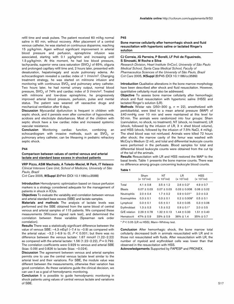
S17
refill time and weak pulses. The patient received 60 ml/kg normalsaline in 60 min, without recovery. After placement of a centralvenous catheter, he was started on continuous dopamine, reaching15 µg/kg/min. Again without significant improvement in arterialblood pressure and perfusion, epinephrine infusion wasassociated, starting with 0.1 µg/kg/min and increasing until1.5 µg/kg/min. At this moment, he had low blood pressure,tachycardia, superior vena cava saturation (SVO2) of 65%, oliguriaand prolonged capillary refill time and, 2 hours later, presented withdesaturation, hepatomegaly and acute pulmonary edema. Anechocardiogram revealed a cardiac index of 1 l/min/m2. Changingtreatment strategy, he was started on milrinone infusion andmonitoring with continuous SVO2 and pulmonary artery catheter.Two hours later, he had normal urinary output, normal bloodpressure, SVO2 of 74% and cardiac index of 3 l/min/m2. Treatedwith milrinone and low-dose epinephrine, he progressivelyimproved arterial blood pressure, perfusion, pulse and mentalstatus. The patient was weaned off vasoactive drugs andmechanical ventilation after 6 days.Discussion Myocardial dysfunction is frequent in children withseptic shock, and it persists even after correction of hypovolemia,acidosis and electrolyte disturbances. Most of the children withseptic shock have a low cardiac index and increased systemicvascular resistance.Conclusion Monitoring cardiac function, combining anechocardiogram with invasive methods, such as SVO2 orpulmonary artery catheter, can be lifesaving in pediatric refractoryseptic shock.
P44Comparison between values of central venous and arteriallactate and standard base excess in shocked patients
VRP Pizzo, ASM Machado, A Toledo-Maciel, M Park, IT VelascoClinical Intensive Care Unit, School of Medicine, University of SãoPaulo, BrazilCrit Care 2005, 9(Suppl 2):P44 (DOI 10.1186/cc3588)
Introduction Hemodynamic optimization based on tissue perfusionmarkers is a strategy considered adequate for the management ofpatients in shock in ICUs.Objectives To evaluate the variability and correlation between venousand arterial standard base excess (SBE) and lactate samples.Materials and methods The analysis of lactate levels wasperformed and the SBE obtained from the same blood of centralvenous and arterial samples of 115 patients. We compared thesemeasurements (Wilcoxon signed rank test), and determined thecorrelation between these variables (Spearman rank ordercorrelation).Results There was a statistically significant difference between thevalue of venous SBE: –4.3 mEq/l (–7.4 to –0.9) as compared withthe arterial value: –3.2 (–6.9 to 0), P < 0.001; but there was nodifference between the venous lactate: 1.67 mmol/l (1.22–2.22)as compared with the arterial lactate: 1.56 (1.22–2.22), P = 0.792.The correlation coefficients were 0.929 to venous and arterial SBE(bias: 0.09) and 0.826 to lactate (bias: –0.024).Discussion The agreement between venous and arterial samplespermits one to use the central venous lactate level similar to thearterial level and their variations. For SBE, the module value wasdifferent between the measurements, otherwise their variation hasgood correlation. As these variations guide the clinical decision, wecan use it as a goal of hemodynamic monitoring.Conclusion It is possible to guide hemodynamic monitoring inshock patients using values of central venous lactate and variationsof SBE.
P45Bone marrow cellularity after hemorrhagic shock and fluidresuscitation with hypertonic saline or lactated Ringer’ssolution
CJ Correia, JG Parreira, P Borelli, LF Poli de Figueiredo, S Sinosaki, M Rocha e SilvaResearch Division, Heart Institute (InCor), University of São PauloMedical School, Santa Casa Medical School, Faculty ofPharmaceutica Sciences of the University of São Paulo, BrazilCrit Care 2005, 9(Suppl 2):P45 (DOI 10.1186/cc3589)
Introduction Qualitative alterations in the bone marrow morphologyhave been described after shock and fluid resuscitation. However,quantitative cellularity must also be addressed.Objective To assess bone marrow cellularity after hemorrhagicshock and fluid resuscitation with hypertonic saline (HSS) andlactated Ringer’s solution (LR).Methods Wistar rats (250–300 g, n = 22), anesthetized withpentobarbital, were bled to a mean arterial pressure (MAP) of±40 mmHg over 10 min and were maintained at this level for50 min. The animals were randomized into four groups: Sham(cannulation, no shock, no treatment), NT (shock, no treatment), LR(shock, followed by the infusion of LR, 3 × shed blood volume),and HSS (shock, followed by the infusion of 7.5% NaCl, 4 ml/kg).The shed blood was not reinfused. Animals were killed 72 hoursafter shock; the marrow cavity of the femur was washed withMcCoy’s Medium (2 ml), and total and differential leukocyte countswere performed in the perfusate. Blood samples for total anddifferential blood leukocyte counts were obtained from the cut tipof the tail of the animals.Results Resuscitation with LR and HSS restored the MAP to thebasal levels. Table 1 presents the bone marrow counts. There wasno difference among groups concerning leukocyte blood counts.
Table 1
Sham NT LR HSS (× 107/ml) (× 107/ml) (× 107/ml) (× 107/ml)
Total 4.1 ± 0.8 3.5 ± 1.2 2.6 ± 0.2* 4.9 ± 0.7
Blasts 0.07 ± 0.03 0.07 ± 0.03 0.03 ± 0.008 0.06 ± 0.02
Neutrophils 2.0 ± 0.4 1.7 ± 0.2 0.9 ± 0.01* 2.0 ± 0.3
Eosinophiles 0.3 ± 0.1 0.3 ± 0.1 0.2 ± 0.009* 0.5 ± 0.1
Lymphoid 0.3 ± 0.1 0.5 ± 0.1 0.3 ± 0.05 0.3 ± 0.06
Erythroblast 1.3 ± 0.3 1.5 ± 0.2 0.9 ± 0.1* 2.0 ± 0.5
G/E relation 2.29 ± 0.78 1.32 ± 0.13 1.44 ± 0.33 1.51 ± 0.40
Hematocrit 47% ± 0.9 33% ± 0.9 36% ± 1.4 35% ± 0.7
* P < 0.05 (LR vs HSS), Mann–Whitney test.
Conclusion After hemorrhagic shock, the bone marrow totalcellularity decreased both in animals resuscitated with LR and inthose not resuscitated with fluids. After resuscitation with LR, thenumber of myeloid and erythroblast cells was lower than thatobserved in the resuscitation with HSS.Acknowledgements Supported by FAPESP and PRONEX.
Available online http://ccforum.com/supplements/9/S2
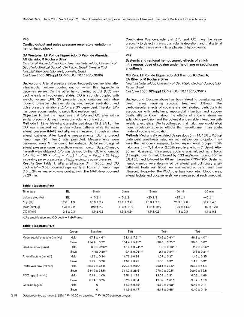
S18
P46Cardiac output and pulse pressure respiratory variation inhemorrhagic shock
GA Westphal, LF Poli de Figueiredo, D Paoli de Almeida, AG Garrido, M Rocha e SilvaDivision of Applied Physiology, Heart Institute, InCor, University ofSão Paulo Medical School, São Paulo, Brazil; General ICU,Hospital Municipal São José, Joinville, BrazilCrit Care 2005, 9(Suppl 2):P46 (DOI 10.1186/cc3590)
Background Arterial pressure values frequently decline later afterintravascular volume contraction, or when this hypovolemiabecomes severe. On the other hand, cardiac output (CO) maydecline early in hypovolemic states. CO is strongly coupled withsystolic volume (SV). SV presents cyclic variations with intra-thoracic pressure changes during mechanical ventilation, andpulse pressure variations (∆Pp) are SV dependent. Thereby, ∆Pphas been recommended to guide fluid replacement.Objective To test the hypothesis that ∆Pp and CO alter with asimilar precocity during intravascular volume contraction.Methods In 12 ventilated and anesthetized dogs (19 ± 2.5 kg), theCO was measured with a Swan–Ganz catheter while the meanarterial pressure (MAP) and ∆Pp were measured through an intra-arterial catheter. After baseline measurements (BL), a gradedhemorrhage (20 ml/min) was promoted. Measurements wereperformed every 5 min during hemorrhage. Digital recordings ofarterial pressure waves by multiparametric monitor (Datex-Ohmeda,Finland) were obtained. ∆Pp was defined by the following formula:∆Pp (%) = 100 × (Ppins – Ppexp) / [(Ppins + Ppexp) / 2]. Ppins,inspiratory pulse pressure and Ppexp, expiratory pulse pressure.Results See Table 1. ∆Pp amplification (P = 0.006) and COdecline (P = 0.02) occurred significantly at 10 min of hemorrhage(15 ± 2% estimated volume contraction). The MAP drop occurredby 20 min.
Conclusion We conclude that ∆Pp and CO have the sameprecocity to detect intravascular volume depletion, and that arterialpressure decreases only in later phases of hypovolemia.
P47Systemic and regional hemodynamic effects of a highintravenous dose of cocaine under halothane or sevofluraneanesthesia
MB Reis, LF Poli de Figueiredo, AG Garrido, RJ Cruz Jr, EA Ribeiro, M Rocha e SilvaHeart Institute, InCor, University of São Paulo Medical School, SãoPaulo, BrazilCrit Care 2005, 9(Suppl 2):P47 (DOI 10.1186/cc3591)
Background Cocaine abuse has been linked to penetrating andblunt trauma requiring surgical treatment. Although thecardiovascular effects of cocaine are well studied, particularly itsassociation with arrhythmia, myocardial infarction and suddendeath, little is known about the effects of cocaine abuse onsplanchnic perfusion and the potential undesirable interaction withvolatile anesthetics. We hypothesized that halothane would elicitmore circulatory adverse effects than sevoflurane in an acutemodel of cocaine intoxication.Methods Mechanically ventilated Beagle dogs (n = 14, 12.8 ± 0.3 kg)underwent anesthesia induction with intravenous propofol. Theywere then randomly assigned to two experimental groups: 1.5%halothane (n = 7, Halo) or 2.25% sevoflurane (n = 7, Sevo). After30 min (Baseline), intravenous cocaine was infused as a bolus(12 mg/kg over 5 min), followed by 0.22 mg/kg/min during 30 min(BL-T35), and followed for 60 min thereafter (T35–T95). Systemichemodynamics were determined by arterial and pulmonary arterycatheters. Portal vein blood flow was measured by a transit timeultrasonic flowprobe. The PCO2 gap (gas tonometry), blood gases,arterial lactate and cocaine levels were measured at each timepoint.
Critical Care June 2005 Vol 9 Suppl 2 Third International Symposium on Intensive Care and Emergency Medicine for Latin America
Table 1 (abstract P46)
Time step BL 5 min 10 min 15 min 20 min 30 min
Volume step (%) BL –10 ± 1 –15 ± 2 –20 ± 3 –35 ± 1 –45 ± 1
∆Pp (%) 12.6 ± 1.9 15.8 ± 2.7 19.7 ± 2.4a 20.8 ± 2.6 21.9 ± 2.6 33.4 ± 4.5
MAP (mmHg) 123 ± 8.2 129 ± 7.0 116 ± 11.9 117 ± 12.2 86 ± 14.3b 80 ± 12.3
CO (l/min) 2.4 ± 0.3 1.9 ± 0.3 1.5 ± 0.3a 1.5 ± 0.3 1.3 ± 0.3 1.1 ± 0.3
a∆Pp amplification and CO decline. bMAP drop.
Table 1 (abstract P47)
Group Baseline T35 T65 T95
Mean arterial pressure (mmHg) Halo 97.3 ± 4.6** 78.1 ± 7.6*,** 73.6 ± 7.6*,** 88.3 ± 4.0**
Sevo 114.7 ± 3.9** 104.4 ± 5.1*,** 96.0 ± 5.7*,** 99.0 ± 5.0**
Cardiac index (l/min) Halo 3.6 ± 0.26** 1.16 ± 0.24*,** 1.3 ± 0.13*,** 2.7 ± 0.16**
Sevo 4.4± 0.30** 2.4 ± 0.26*,** 2.4 ± 0.24*,** 3.6 ± 0.31**
Arterial lactate (mmol/l) Halo 1.69 ± 0.34 1.73 ± 0.34 1.57 ± 0.27 1.45 ± 0.35
Sevo 1.27 ± 0.26 1.52 ± 0.27 1.36 ± 0.37 1.15 ± 0.32
Portal vein flow (ml/min) Halo 584.7 ± 64.0 270.3 ± 23.0* 203.1 ± 26.5* 504.3 ± 41.4
Sevo 534.2 ± 38.5 311.2 ± 28.5* 270.2 ± 29.5* 509.0 ± 35.8
PCO2 gap (mmHg) Halo 5.11 ± 1.09 8.51 ± 1.93 13.59 ± 2.3* 6.06 ± 1.49
Sevo 6.64 ± 0.75 8.23 ± 0.84 12.37 ± 1.81* 9.32 ± 1.19
Cocaine (µg/ml) Halo 0 11.3 ± 0.55* 6.50 ± 0.68* 0.49 ± 0.11
Sevo 0 11.9 ± 0.47* 6.13 ± 0.68* 0.40 ± 0.19
Data presented as mean ± SEM. * P < 0.05 vs baseline; ** P < 0.05 between groups.
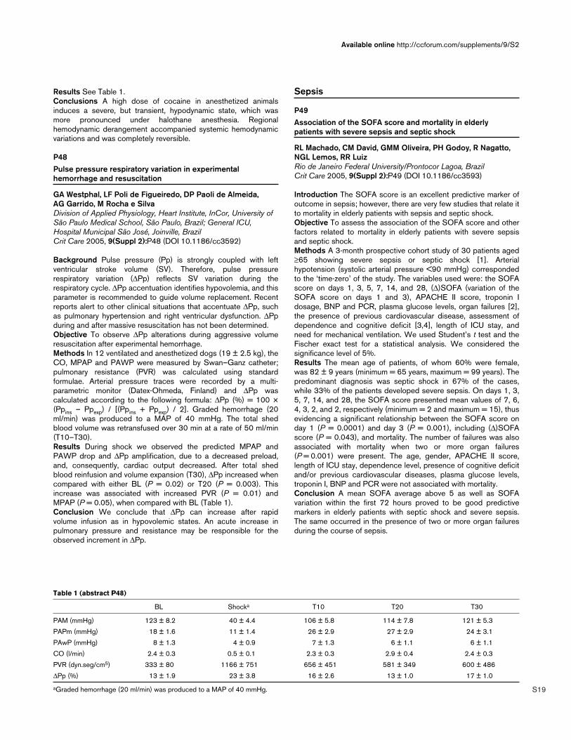
S19
Results See Table 1.Conclusions A high dose of cocaine in anesthetized animalsinduces a severe, but transient, hypodynamic state, which wasmore pronounced under halothane anesthesia. Regionalhemodynamic derangement accompanied systemic hemodynamicvariations and was completely reversible.
P48Pulse pressure respiratory variation in experimentalhemorrhage and resuscitation
GA Westphal, LF Poli de Figueiredo, DP Paoli de Almeida, AG Garrido, M Rocha e SilvaDivision of Applied Physiology, Heart Institute, InCor, University ofSão Paulo Medical School, São Paulo, Brazil; General ICU,Hospital Municipal São José, Joinville, BrazilCrit Care 2005, 9(Suppl 2):P48 (DOI 10.1186/cc3592)
Background Pulse pressure (Pp) is strongly coupled with leftventricular stroke volume (SV). Therefore, pulse pressurerespiratory variation (∆Pp) reflects SV variation during therespiratory cycle. ∆Pp accentuation identifies hypovolemia, and thisparameter is recommended to guide volume replacement. Recentreports alert to other clinical situations that accentuate ∆Pp, suchas pulmonary hypertension and right ventricular dysfunction. ∆Ppduring and after massive resuscitation has not been determined.Objective To observe ∆Pp alterations during aggressive volumeresuscitation after experimental hemorrhage.Methods In 12 ventilated and anesthetized dogs (19 ± 2.5 kg), theCO, MPAP and PAWP were measured by Swan–Ganz catheter;pulmonary resistance (PVR) was calculated using standardformulae. Arterial pressure traces were recorded by a multi-parametric monitor (Datex-Ohmeda, Finland) and ∆Pp wascalculated according to the following formula: ∆Pp (%) = 100 ×(Ppins – Ppexp) / [(Ppins + Ppexp) / 2]. Graded hemorrhage (20ml/min) was produced to a MAP of 40 mmHg. The total shedblood volume was retransfused over 30 min at a rate of 50 ml/min(T10–T30).Results During shock we observed the predicted MPAP andPAWP drop and ∆Pp amplification, due to a decreased preload,and, consequently, cardiac output decreased. After total shedblood reinfusion and volume expansion (T30), ∆Pp increased whencompared with either BL (P = 0.02) or T20 (P = 0.003). Thisincrease was associated with increased PVR (P = 0.01) andMPAP (P = 0.05), when compared with BL (Table 1).Conclusion We conclude that ∆Pp can increase after rapidvolume infusion as in hypovolemic states. An acute increase inpulmonary pressure and resistance may be responsible for theobserved increment in ∆Pp.
Sepsis
P49Association of the SOFA score and mortality in elderlypatients with severe sepsis and septic shock
RL Machado, CM David, GMM Oliveira, PH Godoy, R Nagatto,NGL Lemos, RR LuizRio de Janeiro Federal University/Prontocor Lagoa, BrazilCrit Care 2005, 9(Suppl 2):P49 (DOI 10.1186/cc3593)
Introduction The SOFA score is an excellent predictive marker ofoutcome in sepsis; however, there are very few studies that relate itto mortality in elderly patients with sepsis and septic shock.Objective To assess the association of the SOFA score and otherfactors related to mortality in elderly patients with severe sepsisand septic shock.Methods A 3-month prospective cohort study of 30 patients aged≥65 showing severe sepsis or septic shock [1]. Arterialhypotension (systolic arterial pressure <90 mmHg) correspondedto the ‘time-zero’ of the study. The variables used were: the SOFAscore on days 1, 3, 5, 7, 14, and 28, (∆)SOFA (variation of theSOFA score on days 1 and 3), APACHE II score, troponin Idosage, BNP and PCR, plasma glucose levels, organ failures [2],the presence of previous cardiovascular disease, assessment ofdependence and cognitive deficit [3,4], length of ICU stay, andneed for mechanical ventilation. We used Student’s t test and theFischer exact test for a statistical analysis. We considered thesignificance level of 5%.Results The mean age of patients, of whom 60% were female,was 82 ± 9 years (minimum = 65 years, maximum = 99 years). Thepredominant diagnosis was septic shock in 67% of the cases,while 33% of the patients developed severe sepsis. On days 1, 3,5, 7, 14, and 28, the SOFA score presented mean values of 7, 6,4, 3, 2, and 2, respectively (minimum = 2 and maximum = 15), thusevidencing a significant relationship between the SOFA score onday 1 (P = 0.0001) and day 3 (P = 0.001), including (∆)SOFAscore (P = 0.043), and mortality. The number of failures was alsoassociated with mortality when two or more organ failures(P = 0.001) were present. The age, gender, APACHE II score,length of ICU stay, dependence level, presence of cognitive deficitand/or previous cardiovascular diseases, plasma glucose levels,troponin I, BNP and PCR were not associated with mortality.Conclusion A mean SOFA average above 5 as well as SOFAvariation within the first 72 hours proved to be good predictivemarkers in elderly patients with septic shock and severe sepsis.The same occurred in the presence of two or more organ failuresduring the course of sepsis.
Available online http://ccforum.com/supplements/9/S2
Table 1 (abstract P48)
BL Shocka T10 T20 T30
PAM (mmHg) 123 ± 8.2 40 ± 4.4 106 ± 5.8 114 ± 7.8 121 ± 5.3
PAPm (mmHg) 18 ± 1.6 11 ± 1.4 26 ± 2.9 27 ± 2.9 24 ± 3.1
PAwP (mmHg) 8 ± 1.3 4 ± 0.9 7 ± 1.3 6 ± 1.1 6 ± 1.1
CO (l/min) 2.4 ± 0.3 0.5 ± 0.1 2.3 ± 0.3 2.9 ± 0.4 2.4 ± 0.3
PVR (dyn.seg/cm5) 333 ± 80 1166 ± 751 656 ± 451 581 ± 349 600 ± 486
∆Pp (%) 13 ± 1.9 23 ± 3.8 16 ± 2.6 13 ± 1.0 17 ± 1.0
aGraded hemorrhage (20 ml/min) was produced to a MAP of 40 mmHg.
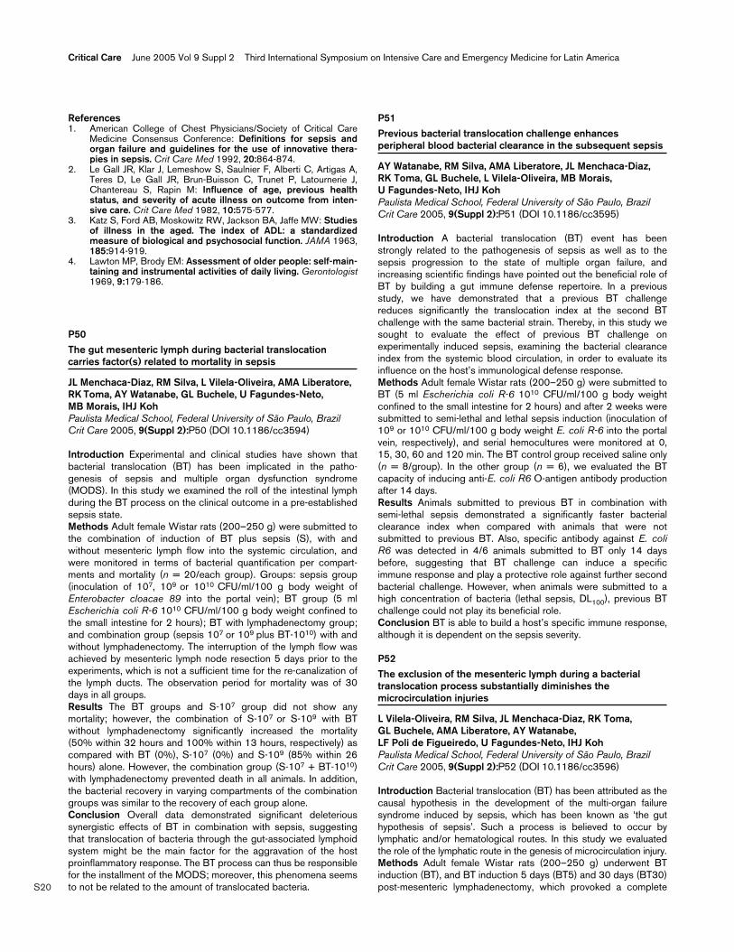
S20
References1. American College of Chest Physicians/Society of Critical Care
Medicine Consensus Conference: Definitions for sepsis andorgan failure and guidelines for the use of innovative thera-pies in sepsis. Crit Care Med 1992, 20:864-874.
2. Le Gall JR, Klar J, Lemeshow S, Saulnier F, Alberti C, Artigas A,Teres D, Le Gall JR, Brun-Buisson C, Trunet P, Latournerie J,Chantereau S, Rapin M: Influence of age, previous healthstatus, and severity of acute illness on outcome from inten-sive care. Crit Care Med 1982, 10:575-577.
3. Katz S, Ford AB, Moskowitz RW, Jackson BA, Jaffe MW: Studiesof illness in the aged. The index of ADL: a standardizedmeasure of biological and psychosocial function. JAMA 1963,185:914-919.
4. Lawton MP, Brody EM: Assessment of older people: self-main-taining and instrumental activities of daily living. Gerontologist1969, 9:179-186.
P50The gut mesenteric lymph during bacterial translocationcarries factor(s) related to mortality in sepsis
JL Menchaca-Diaz, RM Silva, L Vilela-Oliveira, AMA Liberatore,RK Toma, AY Watanabe, GL Buchele, U Fagundes-Neto, MB Morais, IHJ KohPaulista Medical School, Federal University of São Paulo, BrazilCrit Care 2005, 9(Suppl 2):P50 (DOI 10.1186/cc3594)
Introduction Experimental and clinical studies have shown thatbacterial translocation (BT) has been implicated in the patho-genesis of sepsis and multiple organ dysfunction syndrome(MODS). In this study we examined the roll of the intestinal lymphduring the BT process on the clinical outcome in a pre-establishedsepsis state.Methods Adult female Wistar rats (200–250 g) were submitted tothe combination of induction of BT plus sepsis (S), with andwithout mesenteric lymph flow into the systemic circulation, andwere monitored in terms of bacterial quantification per compart-ments and mortality (n = 20/each group). Groups: sepsis group(inoculation of 107, 109 or 1010 CFU/ml/100 g body weight ofEnterobacter cloacae 89 into the portal vein); BT group (5 mlEscherichia coli R-6 1010 CFU/ml/100 g body weight confined tothe small intestine for 2 hours); BT with lymphadenectomy group;and combination group (sepsis 107 or 109 plus BT-1010) with andwithout lymphadenectomy. The interruption of the lymph flow wasachieved by mesenteric lymph node resection 5 days prior to theexperiments, which is not a sufficient time for the re-canalization ofthe lymph ducts. The observation period for mortality was of 30days in all groups.Results The BT groups and S-107 group did not show anymortality; however, the combination of S-107 or S-109 with BTwithout lymphadenectomy significantly increased the mortality(50% within 32 hours and 100% within 13 hours, respectively) ascompared with BT (0%), S-107 (0%) and S-109 (85% within 26hours) alone. However, the combination group (S-107 + BT-1010)with lymphadenectomy prevented death in all animals. In addition,the bacterial recovery in varying compartments of the combinationgroups was similar to the recovery of each group alone.Conclusion Overall data demonstrated significant deleterioussynergistic effects of BT in combination with sepsis, suggestingthat translocation of bacteria through the gut-associated lymphoidsystem might be the main factor for the aggravation of the hostproinflammatory response. The BT process can thus be responsiblefor the installment of the MODS; moreover, this phenomena seemsto not be related to the amount of translocated bacteria.
P51Previous bacterial translocation challenge enhancesperipheral blood bacterial clearance in the subsequent sepsis
AY Watanabe, RM Silva, AMA Liberatore, JL Menchaca-Diaz, RK Toma, GL Buchele, L Vilela-Oliveira, MB Morais, U Fagundes-Neto, IHJ KohPaulista Medical School, Federal University of São Paulo, BrazilCrit Care 2005, 9(Suppl 2):P51 (DOI 10.1186/cc3595)
Introduction A bacterial translocation (BT) event has beenstrongly related to the pathogenesis of sepsis as well as to thesepsis progression to the state of multiple organ failure, andincreasing scientific findings have pointed out the beneficial role ofBT by building a gut immune defense repertoire. In a previousstudy, we have demonstrated that a previous BT challengereduces significantly the translocation index at the second BTchallenge with the same bacterial strain. Thereby, in this study wesought to evaluate the effect of previous BT challenge onexperimentally induced sepsis, examining the bacterial clearanceindex from the systemic blood circulation, in order to evaluate itsinfluence on the host’s immunological defense response.Methods Adult female Wistar rats (200–250 g) were submitted toBT (5 ml Escherichia coli R-6 1010 CFU/ml/100 g body weightconfined to the small intestine for 2 hours) and after 2 weeks weresubmitted to semi-lethal and lethal sepsis induction (inoculation of109 or 1010 CFU/ml/100 g body weight E. coli R-6 into the portalvein, respectively), and serial hemocultures were monitored at 0,15, 30, 60 and 120 min. The BT control group received saline only(n = 8/group). In the other group (n = 6), we evaluated the BTcapacity of inducing anti-E. coli R6 O-antigen antibody productionafter 14 days.Results Animals submitted to previous BT in combination withsemi-lethal sepsis demonstrated a significantly faster bacterialclearance index when compared with animals that were notsubmitted to previous BT. Also, specific antibody against E. coliR6 was detected in 4/6 animals submitted to BT only 14 daysbefore, suggesting that BT challenge can induce a specificimmune response and play a protective role against further secondbacterial challenge. However, when animals were submitted to ahigh concentration of bacteria (lethal sepsis, DL100), previous BTchallenge could not play its beneficial role.Conclusion BT is able to build a host’s specific immune response,although it is dependent on the sepsis severity.
P52The exclusion of the mesenteric lymph during a bacterialtranslocation process substantially diminishes themicrocirculation injuries
L Vilela-Oliveira, RM Silva, JL Menchaca-Diaz, RK Toma, GL Buchele, AMA Liberatore, AY Watanabe, LF Poli de Figueiredo, U Fagundes-Neto, IHJ KohPaulista Medical School, Federal University of São Paulo, BrazilCrit Care 2005, 9(Suppl 2):P52 (DOI 10.1186/cc3596)
Introduction Bacterial translocation (BT) has been attributed as thecausal hypothesis in the development of the multi-organ failuresyndrome induced by sepsis, which has been known as ‘the guthypothesis of sepsis’. Such a process is believed to occur bylymphatic and/or hematological routes. In this study we evaluatedthe role of the lymphatic route in the genesis of microcirculation injury.Methods Adult female Wistar rats (200–250 g) underwent BTinduction (BT), and BT induction 5 days (BT5) and 30 days (BT30)post-mesenteric lymphadenectomy, which provoked a complete
Critical Care June 2005 Vol 9 Suppl 2 Third International Symposium on Intensive Care and Emergency Medicine for Latin America
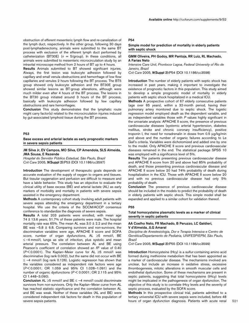
S21
obstruction of efferent mesenteric lymph flow and re-canalization ofthe lymph duct, respectively. In the other group, following 30 dayspost-lymphadenectomy, animals were submitted to the same BTprocess with exclusion of the efferent lymph duct performed bycatheterization (BT30E) (n = 5/group). In these conditions, allanimals were submitted to mesenteric microcirculation study by anintravital microscope method from 2 hours of BT up to 4 hours.Results Animals submitted to BT showed significant injuries.Always, the first lesion was leukocyte adhesion followed bycapillary and small venula obstructions and hemorrhage of low flowcapillaries and venules 2 hours following the BT process. The BT5group showed only leukocyte adhesion and the BT30E groupshowed similar lesions as BT-group alterations, although weremuch milder even after 4 hours of the BT process. The lesions inthe BT30 group initiated around 3 hours of the BT process,basically with leukocyte adhesion followed by few capillaryobstructions and rare hemorrhages.Conclusion This study demonstrates that the lymphatic routemight carry factor(s) related to the microcirculation injuries inducedby gut-associated lymphoid tissue during the BT process.
P53Base excess and arterial lactate as early prognostic markersin severe sepsis patients
JM Silva Jr, EV Campos, MO Silva, CP Amendola, SLS Almeida,JMA Sousa, E RezendeHospital do Servidor Público Estadual, São Paulo, BrazilCrit Care 2005, 9(Suppl 2):P53 (DOI 10.1186/cc3597)
Introduction The development of therapeutic goals depends onaccurate evaluation of the supply of oxygen to organs and tissues.But tissular oxygenation and perfusion are difficult to measure andhave a labile behavior. This study has an objective to evaluate theclinical utility of base excess (BE) and arterial lactate (AL) as earlymarkers of morbidity and mortality in patients with severe sepsisassisted in the emergency department.Methods A contemporary cohort study involving adult patients withsevere sepsis attending the emergency department in a tertiaryhospital. We use the criteria of the SCCM/ACCP consensusconference to standardize the diagnosis of severe sepsis.Results A total 203 patients were enrolled, with mean age74 ± 13.6 years; 51.7% of these patients were male. The hospitalmortality rate was 65%. The mean AL was 3.0 ± 2.2 and the meanBE was –5.8 ± 6.8. Comparing survivors and non-survivors, thediscriminative variables were age, APACHE II score and SOFAscore, number of organ dysfunctions, AL ≥5 mmol/l, BE≤ –4 mmol/l, lungs as site of infection, plus systolic and meanarterial pressure. The correlation between AL and BE usingPearson’s coefficient of correlation showed an R2 value of 0.40(P < 0.0001). The Kaplan–Meier curve for AL ≥5 mmol/l wasdiscriminative (log rank 0.002), but the same did not occur with BE≤ –4 mmol/l (log rank 0.126). Logistic regression has shown thatthe variables considered as independent risk factors were age(P < 0.0001; OR 1.059 and 95% CI 1.028–1.091) and thenumber of organic dysfunctions (P < 0.0001; OR 2.115 and 95%CI 1.448–3.090).Conclusion AL ≥5 mmol/l and BE ≤ –4 mmol/l could discriminatesurvivors from non-survivors. Only the Kaplan–Meier curve from ALhas reached statistic significance and the correlation between ALand BE was weak. Neither of these variables (AL and BE) wereconsidered independent risk factors for death in this population ofsevere sepsis patients.
P54Simple model for prediction of mortality in elderly patientswith septic shock
GMM Oliveira, PH Godoy, MR Pantoja, RR Luiz, RL Machado, A Farias NetoIntensive Care Unit, Prontocor Lagoa, Federal University of Rio deJaneiro, BrazilCrit Care 2005, 9(Suppl 2):P54 (DOI 10.1186/cc3598)
Introduction The number of elderly patients with septic shock hasincreased in past years, making it important to investigate theexistence of prognostic factors in this population. This study aimedto develop a simple prognostic model of mortality in elderlypatients with septic shock hospitalized in a medical ICU.Methods A prospective cohort of 67 elderly consecutive patients(age over 65 years), within a 32-month period, having theirpulmonary artery monitored due to septic shock. The logisticregression model employed death as the dependent variable, andas independent variables those with P values highly significant inthe univariate analysis: APACHE II score, the presence of previouscardiovascular diseases (systemic arterial hypertension, diabetesmellitus, stroke and chronic coronary insufficiency), positivetroponin I, the need for noradrenalin in doses from 0.5 µg/kg/minand above and the number of organic failures according to LeGall’s criteria. Variables were dichotomized and added one by oneto the model. Only APACHE II score and previous cardiovasculardiseases remained in the end. The statistical package SPSS 10was employed with a significance level of 5%.Results The patients presenting previous cardiovascular diseaseand APACHE II score from 20 and above had 85% probability ofdeath, and those presenting previous cardiovascular disease andAPACHE II score below 20 had 74% probability of death duringhospitalization in the ICU. Those with APACHE II score below 20and with no previous cardiovascular disease had only 5%probability of death.Conclusion The presence of previous cardiovascular diseaseshould be included in the models to predict the probability of deathin elderly patients with septic shock. This simple model shall beexpanded and applied to a similar cohort for validation thereof.
P55Total homocysteine plasmatic levels as a marker of clinicalseverity in septic patients
AA Coelho Neto, FR Machado, B Peruzzo, LC Galdieri, V d’Almeida, JLG AmaralDisciplina de Anestesiologia, Dor e Terapia Intensiva e Centro deGenética/Departamento de Pediatria, UNIFESP/EPM, São Paulo,BrazilCrit Care 2005, 9(Suppl 2):P55 (DOI 10.1186/cc3599)
Introduction Homocysteine (Hcy) is a sulfur-containing amino acidformed during methionine metabolism that has been appointed asa marker of cardiovascular disease. The mechanisms involved areunclear, but include an increase in oxidative stress, excessivethrombogenesis, mitotic alterations in smooth muscular cells andendothelial dysfunction. Some of these mechanisms are present inseptic patients, suggesting that total homocysteine (tHcy) levelsmight be implicated in the pathogenesis of organ dysfunction. Theobjective of this study is to correlate tHcy levels and the severity ofseptic process, evaluated by the SOFA score.Methods In this prospective clinical trial, patients admitted to atertiary universital ICU with severe sepsis were included, before 48hours of organ dysfunction diagnosis. Patients with acute renal
Available online http://ccforum.com/supplements/9/S2
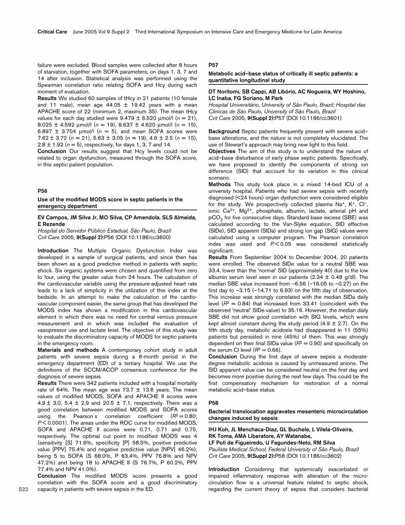
S22
failure were excluded. Blood samples were collected after 8 hoursof starvation, together with SOFA parameters, on days 1, 3, 7 and14 after inclusion. Statistical analysis was performed using theSpearman correlation ratio relating SOFA and Hcy during eachmoment of evaluation.Results We studied 60 samples of tHcy in 21 patients (10 femaleand 11 male), mean age 44.05 ± 19.42 years with a meanAPACHE score of 22 (minimum 2, maximum 35). The mean tHcyvalues for each day studied were 9.479 ± 6.520 µmol/l (n = 21),8.025 ± 4.592 µmol/l (n = 19), 8.637 ± 4.620 µmol/l (n = 15),6.897 ± 3.704 µmol/l (n = 5), and mean SOFA scores were7.62 ± 3.72 (n = 21), 5.63 ± 3.05 (n = 19), 4.6 ± 2.5 (n = 15),2.8 ± 1.92 (n = 5), respectively, for days 1, 3, 7 and 14.Conclusion Our results suggest that Hcy levels could not berelated to organ dysfunction, measured through the SOFA score,in this septic patient population.
P56Use of the modified MODS score in septic patients in theemergency department
EV Campos, JM Silva Jr, MO Silva, CP Amendola, SLS Almeida,E RezendeHospital do Servidor Público Estadual, São Paulo, BrazilCrit Care 2005, 9(Suppl 2):P56 (DOI 10.1186/cc3600)
Introduction The Multiple Organic Dysfunction Index wasdeveloped in a sample of surgical patients, and since then hasbeen shown as a good predictive method in patients with septicshock. Six organic systems were chosen and quantified from zeroto four, using the greater value from 24 hours. The calculation ofthe cardiovascular variable using the pressure-adjusted heart rateleads to a lack of simplicity in the utilization of this index at thebedside. In an attempt to make the calculation of the cardio-vascular component easier, the same group that has developed theMODS index has shown a modification in this cardiovascularelement in which there was no need for central venous pressuremeasurement and in which was included the evaluation ofvasopressor use and lactate level. The objective of this study wasto evaluate the discriminatory capacity of MODS for septic patientsin the emergency room.Materials and methods A contemporary cohort study in adultpatients with severe sepsis during a 6-month period in theemergency department (ED) of a tertiary hospital. We use thedefinitions of the SCCM/ACCP consensus conference for thediagnosis of severe sepsis.Results There were 342 patients included with a hospital mortalityrate of 64%. The mean age was 73.7 ± 13.6 years. The meanvalues of modified MODS, SOFA and APACHE II scores were4.9 ± 3.0, 5.4 ± 2.9 and 20.5 ± 7.1, respectively. There was agood correlation between modified MODS and SOFA scoresusing the Pearson´s correlation coefficient (R2 = 0.80;P < 0.0001). The areas under the ROC curve for modified MODS,SOFA and APACHE II scores were 0.71, 0.71 and 0.75,respectively. The optimal cut point to modified MODS was 4(sensitivity [S] 71.6%, specificity [P] 58.5%, positive predictivevalue [PPV] 75.4% and negative predictive value [NPV] 46.2%),being 5 to SOFA (S 68.0%, P 63.4%, PPV 76.8% and NPV47.2%) and being 18 to APACHE II (S 76.7%, P 60.2%, PPV77.4% and NPV 41.0%).Conclusion The modified MODS score presents a goodcorrelation with the SOFA score and a good discriminatorycapacity in patients with severe sepsis in the ED.
P57Metabolic acid–base status of critically ill septic patients: aquantitative longitudinal study
DT Noritomi, SB Cappi, AB Libório, AC Nogueira, WY Hoshino,LC Inaba, FG Soriano, M ParkHospital Universitário, University of São Paulo, Brazil; Hospital dasClinicas de São Paulo, University of São Paulo, BrazilCrit Care 2005, 9(Suppl 2):P57 (DOI 10.1186/cc3601)
Background Septic patients frequently present with severe acid–base alterations, and the nature is not completely elucidated. Theuse of Stewart’s approach may bring new light to this field.Objectives The aim of this study is to understand the nature ofacid–base disturbance of early phase septic patients. Specifically,we have proposed to identify the components of strong iondifference (SID) that account for its variation in this clinicalscenario.Methods This study took place in a mixed 14-bed ICU of auniversity hospital. Patients who had severe sepsis with recentlydiagnosed (<24 hours) organ dysfunction were considered eligiblefor the study. We prospectively collected plasma Na+, K+, Cl–,ionic Ca2+, Mg2+, phosphate, albumin, lactate, arterial pH andpCO2 for five consecutive days. Standard base excess (SBE) wascalculated according to the Van-Slyke equation. SID effective(SIDe), SID apparent (SIDa) and strong ion gap (SIG) values werecalculated using a computer program. The Pearson correlationindex was used and P < 0.05 was considered statisticallysignificant.Results From September 2004 to December 2004, 20 patientswere enrolled. The observed SIDe value for a neutral SBE was33.4, lower than the ‘normal’ SID (approximately 40) due to the lowalbumin serum level seen in our patients (2.34 ± 0.48 g/dl). Themedian SBE value increased from –6.56 (–16.05 to –0.27) on thefirst day to –3.15 (–14.71 to 6.93) on the fifth day of observation.This increase was strongly correlated with the median SIDa dailylevel (R2 = 0.84) that increased from 33.41 (coincident with theobserved ‘neutral’ SIDe value) to 35.16. However, the median dailySBE did not show good correlation with SIG levels, which werekept almost constant during the study period (4.9 ± 2.7).. On thefifth study day, metabolic acidosis had disappeared in 11 (55%)patients but persisted in nine (45%) of them. This was stronglydependent on their final SIDa value (R2 = 0.90) and specifically onthe serum Cl level (R2 = 0.66).Conclusion During the first days of severe sepsis a moderate-degree metabolic acidosis is caused by unmeasured anions. TheSID apparent value can be considered neutral on the first day andbecomes more positive during the next few days. This could be thefirst compensatory mechanism for restoration of a normalmetabolic acid–base status.
P58Bacterial translocation aggravates mesenteric microcirculationchanges induced by sepsis
IHJ Koh, JL Menchaca-Diaz, GL Buchele, L Vilela-Oliveira, RK Toma, AMA Liberatore, AY Watanabe, LF Poli de Figueiredo, U Fagundes-Neto, RM SilvaPaulista Medical School, Federal University of São Paulo, BrazilCrit Care 2005, 9(Suppl 2):P58 (DOI 10.1186/cc3602)
Introduction Considering that systemically exacerbated orimpaired inflammatory response with alteration of the micro-circulation flow is a universal feature related to septic shock,regarding the current theory of sepsis that considers bacterial
Critical Care June 2005 Vol 9 Suppl 2 Third International Symposium on Intensive Care and Emergency Medicine for Latin America
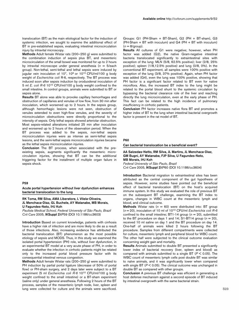
S23
translocation (BT) as the main etiological factor for the induction ofsystemic infection, we sought to examine the additional effect ofBT in pre-established sepsis, evaluating intestinal microcirculationinjury by intravital microscopy.Methods Adult female Wistar rats (200–250 g) were submitted tothe combination induction of sepsis plus BT, and mesentericmicrocirculation of the small bowel was monitored for up to 2 hoursby intravital microscope under general anesthesia (n = 5/eachgroup). Non-lethal, semi-lethal and lethal sepsis were induced byjugular vein inoculation of 107, 109 or 1010 CFU/ml/100 g bodyweight of Escherichia coli R-6, respectively. The BT process wasinduced soon after sepsis induction by oroduodenal inoculation of5 ml E. coli R-6 1010 CFU/ml/100 g body weight confined to thesmall intestine. In control groups, animals were submitted to BT orsepsis alone.Results BT alone was able to provoke capillary hemorrhages andobstruction of capillaries and venulas of low flow, from 30 min afterinoculation, which worsened up to 2 hours. In the sepsis group,although hemorrhagic lesions were not seen, obstruction ofvenules extended to even high-flow venules, and the severity ofmicrocirculation obstructions were directly proportional to theintensity of sepsis. Only lethal sepsis showed arteriolar obstruction.Most sepsis-related alterations initiated 25 min after inoculationand worsened up to 2 hours of the observation period. When theBT process was added to the sepsis, non-lethal sepsismicrocirculation injuries were as intense as semi-lethal sepsislesions, and the semi-lethal sepsis microcirculation injuries becameas the lethal sepsis microcirculation injuries.Conclusion The BT process, when associated with the pre-existing sepsis, augments significantly the mesenteric micro-circulation injuries, showing that BT can be the additionaltriggering factor for the installment of multiple organ failure insepsis shock.
P59Acute portal hypertension without liver dysfunction enhancesbacterial translocation to the lung
RK Toma, RM Silva, AMA Liberatore, L Vilela-Oliveira, JL Menchaca-Diaz, GL Buchele, AY Watanabe, MB Morais, U Fagundes-Neto, IHJ KohPaulista Medical School, Federal University of São Paulo, BrazilCrit Care 2005, 9(Suppl 2):P59 (DOI 10.1186/cc3603)
Introduction Based on current knowledge, patients with cirrhosishave a higher risk of infection and are more likely to die as a resultof those infections. Also, increasing evidence has attributed thebacterial translocation (BT) phenomenon as the most possibleetiology of sepsis and MODS. Thus, in this study we examined theisolated portal hypertension (PH) role, without liver dysfunction, inan experimental BT model at a very acute phase of PH, in order toevaluate whether the infection in cirrhotic patients might be relatedonly to the increased portal blood pressure factor with itsconsequential intestinal venous congestion.Methods Adult female Wistar rats (200–250 g) were submitted toPH induction by partial portal ligature (decrease of 50% of portalflow) or PH-sham surgery, and 2 days later were subject to a BTexperiment (5 ml Escherichia coli R-6 1010 CFU/ml/100 g bodyweight confined to the small intestine) or a BT-sham experiment(saline only) under general anesthesia. Following 2 hours of the BTprocess, samples of the mesenteric lymph node, liver, spleen andlung were collected for culture and the animals were sacrificed.
Groups: G1 (PH-Sham + BT-Sham), G2 (PH + BT-sham), G3(PH-Sham + BT with inoculum) and G4 (PH + BT with inoculum)(n = 8/group).Results All cultures of G1 were negative; however, when PHfactor was added (G2), the native Gram-negative intestinalbacteria translocated significantly to extraintestinal sites withexception of the lung: MLN (5/8, 62.5% positive); liver (2/8, 25%positive); spleen (1/8,12.5% positive) and lung (0/8, 0%). In theconventional BT experiment, all samples were 100% positive withexception of the lung (3/8, 37% positive). Again, when PH factorwas added (G4), even the lung was 100% positive, showing thatPH factor is a significant factor related to BT even for nativemicroflora. Also, the increased BT index to the lung might berelated to the portal blood shunt to the systemic circulation bybypassing the bacterial clearance role of the liver and reachingdirectly the lung microcirculation, even at the early phase of PH.This fact can be related to the high incidence of pulmonaryinsufficiency in cirrhotic patients.Conclusion PH factor increases native flora BT and promotes ahigher index of BT to the lung when intestinal bacterial overgrowthfactor is present in the rat model of BT.
P60Can bacterial translocation be a beneficial event?
AA Salzedas-Netto, RM Silva, JL Martins, JL Menchaca-Diaz,GM Bugni, AY Watanabe, FJP Silva, U Fagundes-Neto, MB Morais, IHJ KohFederal University of São Paulo, BrazilCrit Care 2005, 9(Suppl 2):P60 (DOI 10.1186/cc3604)
Introduction Bacterial migration to extraintestinal sites has beenattributed as the central component of the gut hypothesis ofsepsis. However, some studies have pointed out the beneficialeffect of bacterial translocation (BT) on the host’s acquiredimmune system. In this study we evaluated the role of previous BTon the subsequent BT challenge, examining the BT index toorgans, changes in WBC count at the mesenteric lymph andblood, and clinical outcome.Methods Wistar rats (n = 60) were distributed into: BT group(n = 20), inoculation of 10 ml of 1010 CFU/ml Escherichia coli R-6confined to the small intestine; BT1-14 group (n = 20), submittedto the BT procedure on days 1 and 14; S1-BT14 group (n = 20),received 10 ml saline on day 1 and the BT procedure on day 14.One-half of animals were killed 2 hours following the BTprocedure. Samples from different compartments were collectedfor culture, mesenteric lymph and peripheral blood for WBC count.The other half were subjected to the clinical outcome evaluationconcerning weight gain and mortality.Results Animals submitted to double BT presented a significantlylower index of bacterial recovery (liver, spleen and blood) ascompared with animals submitted to a single BT (P < 0.05). TheWBC count of mesenteric lymph cells post double BT was similarto naïve animals, and it was significantly lower when comparedwith single BT (P < 0.05). The clinical outcome was unchanged indouble BT as compared with other groups.Conclusion A previous BT challenge was efficient in generating ahost defense mechanism against a second episode of BT inducedby intestinal overgrowth with the same bacterial strain.
Available online http://ccforum.com/supplements/9/S2
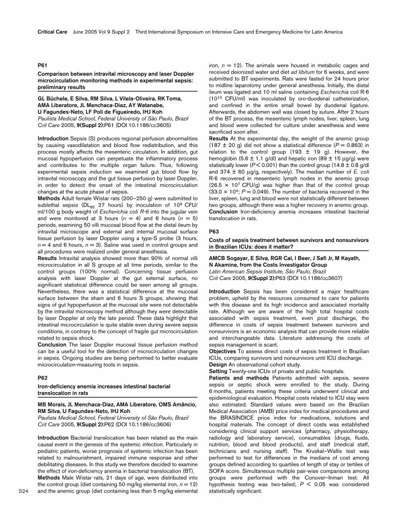
S24
P61Comparison between intravital microscopy and laser Dopplermicrocirculation monitoring methods in experimental sepsis:preliminary results
GL Büchele, E Silva, RM Silva, L Vilela-Oliveira, RK Toma, AMA Liberatore, JL Menchaca-Diaz, AY Watanabe, U Fagundes-Neto, LF Poli de Figueiredo, IHJ KohPaulista Medical School, Federal University of São Paulo, BrazilCrit Care 2005, 9(Suppl 2):P61 (DOI 10.1186/cc3605)
Introduction Sepsis (S) produces regional perfusion abnormalitiesby causing vasodilatation and blood flow redistribution, and thisprocess mostly affects the mesenteric circulation. In addition, gutmucosal hypoperfusion can perpetuate the inflammatory processand contributes to the multiple organ failure. Thus, followingexperimental sepsis induction we examined gut blood flow byintravital microscopy and the gut tissue perfusion by laser Doppler,in order to detect the onset of the intestinal microcirculationchanges at the acute phase of sepsis.Methods Adult female Wistar rats (200–250 g) were submitted tosublethal sepsis (DL85 27 hours) by inoculation of 109 CFU/ml/100 g body weight of Escherichia coli R-6 into the jugular veinand were monitored at 3 hours (n = 4) and 6 hours (n = 5)periods, examining 50 villi mucosal blood flow at the distal ileum byintravital microscope and external and internal mucosal surfacetissue perfusion by laser Doppler using a type-S probe (3 hours,n = 4 and 6 hours, n = 3). Saline was used in control groups andall procedures were realized under general anesthesia.Results Intravital analysis showed more than 90% of normal villimicrocirculation in all S groups at all time periods, similar to thecontrol groups (100% normal). Concerning tissue perfusionanalysis with laser Doppler at the gut external surface, nosignificant statistical difference could be seen among all groups.Nevertheless, there was a statistical difference at the mucosalsurface between the sham and 6 hours S groups, showing thatsigns of gut hypoperfusion at the mucosal site were not detectableby the intravital microscopy method although they were detectableby laser Doppler at only the late period. These data highlight thatintestinal microcirculation is quite stable even during severe sepsisconditions, in contrary to the concept of fragile gut microcirculationrelated to sepsis shock.Conclusion The laser Doppler mucosal tissue perfusion methodcan be a useful tool for the detection of microcirculation changesin sepsis. Ongoing studies are being performed to better evaluatemicrocirculation-measuring tools in sepsis.
P62Iron-deficiency anemia increases intestinal bacterialtranslocation in rats
MB Morais, JL Menchaca-Diaz, AMA Liberatore, OMS Amâncio,RM Silva, U Fagundes-Neto, IHJ KohPaulista Medical School, Federal University of São Paulo, BrazilCrit Care 2005, 9(Suppl 2):P62 (DOI 10.1186/cc3606)
Introduction Bacterial translocation has been related as the maincausal event in the genesis of the systemic infection. Particularly inpediatric patients, worse prognosis of systemic infection has beenrelated to malnourishment, impaired immune response and otherdebilitating diseases. In this study we therefore decided to examinethe effect of iron-deficiency anemia in bacterial translocation (BT).Methods Male Wistar rats, 21 days of age, were distributed intothe control group (diet containing 50 mg/kg elemental iron, n = 12)and the anemic group (diet containing less than 5 mg/kg elemental
iron, n = 12). The animals were housed in metabolic cages andreceived deionized water and diet ad libitum for 6 weeks, and weresubmitted to BT experiments. Rats were fasted for 24 hours priorto midline laparotomy under general anesthesia. Initially, the distalileum was ligated and 10 ml saline containing Escherichia coli R-6(1010 CFU/ml) was inoculated by oro-duodenal catheterization,and confined in the entire small bowel by duodenal ligature.Afterwards, the abdomen wall was closed by suture. After 2 hoursof the BT process, the mesenteric lymph nodes, liver, spleen, lungand blood were collected for culture under anesthesia and weresacrificed soon after.Results At the experimental day, the weight of the anemic group(187 ± 20 g) did not show a statistical difference (P = 0.863) inrelation to the control group (193 ± 19 g). However, thehemoglobin (5.6 ± 1.1 g/dl) and hepatic iron (89 ± 15 µg/g) werestatistically lower (P < 0.001) than the control group (14.8 ± 0.8 g/dland 374 ± 60 µg/g, respectively). The median number of E. coliR-6 recovered in mesenteric lymph nodes in the anemic group(26.5 × 10⁷ CFU/g) was higher than that of the control group(33.0 × 10⁴; P = 0.049). The number of bacteria recovered in theliver, spleen, lung and blood were not statistically different betweentwo groups, although there was a higher recovery in anemic group.Conclusion Iron-deficiency anemia increases intestinal bacterialtranslocation in rats.
P63Costs of sepsis treatment between survivors and nonsurvivorsin Brazilian ICUs: does it matter?
AMCB Sogayar, E Silva, RGR Cal, I Beer, J Safi Jr, M Kayath, N Akamine, from the Costs Investigator GroupLatin American Sepsis Institute, São Paulo, BrazilCrit Care 2005, 9(Suppl 2):P63 (DOI 10.1186/cc3607)
Introduction Sepsis has been considered a major healthcareproblem, upheld by the resources consumed to care for patientswith this disease and its high incidence and associated mortalityrate. Although we are aware of the high total hospital costsassociated with sepsis treatment, even post discharge, thedifference in costs of sepsis treatment between survivors andnonsurvivors is an economic analysis that can provide more reliableand interchangeable data. Literature addressing the costs ofsepsis management is scant.Objectives To assess direct costs of sepsis treatment in BrazilianICUs, comparing survivors and nonsurvivors until ICU discharge.Design An observational cohort study.Setting Twenty-one ICUs of private and public hospitals.Patients and methods Patients admitted with sepsis, severesepsis or septic shock were enrolled to the study. During6 months, patients meeting these criteria underwent clinical andepidemiological evaluation. Hospital costs related to ICU stay werealso estimated. Standard values were based on the BrazilianMedical Association (AMB) price index for medical procedures andthe BRASÍNDICE price index for medications, solutions andhospital materials. The concept of direct costs was establishedconsidering clinical support services (pharmacy, physiotherapy,radiology and laboratory service), consumables (drugs, fluids,nutrition, blood and blood products), and staff (medical staff,technicians and nursing staff). The Kruskal–Wallis test wasperformed to test for differences in the medians of cost amonggroups defined according to quartiles of length of stay or tertiles ofSOFA score. Simultaneous multiple pair-wise comparisons amonggroups were performed with the Conover–Inman test. Allhypothesis testing was two-tailed; P < 0.05 was consideredstatistically significant.
Critical Care June 2005 Vol 9 Suppl 2 Third International Symposium on Intensive Care and Emergency Medicine for Latin America
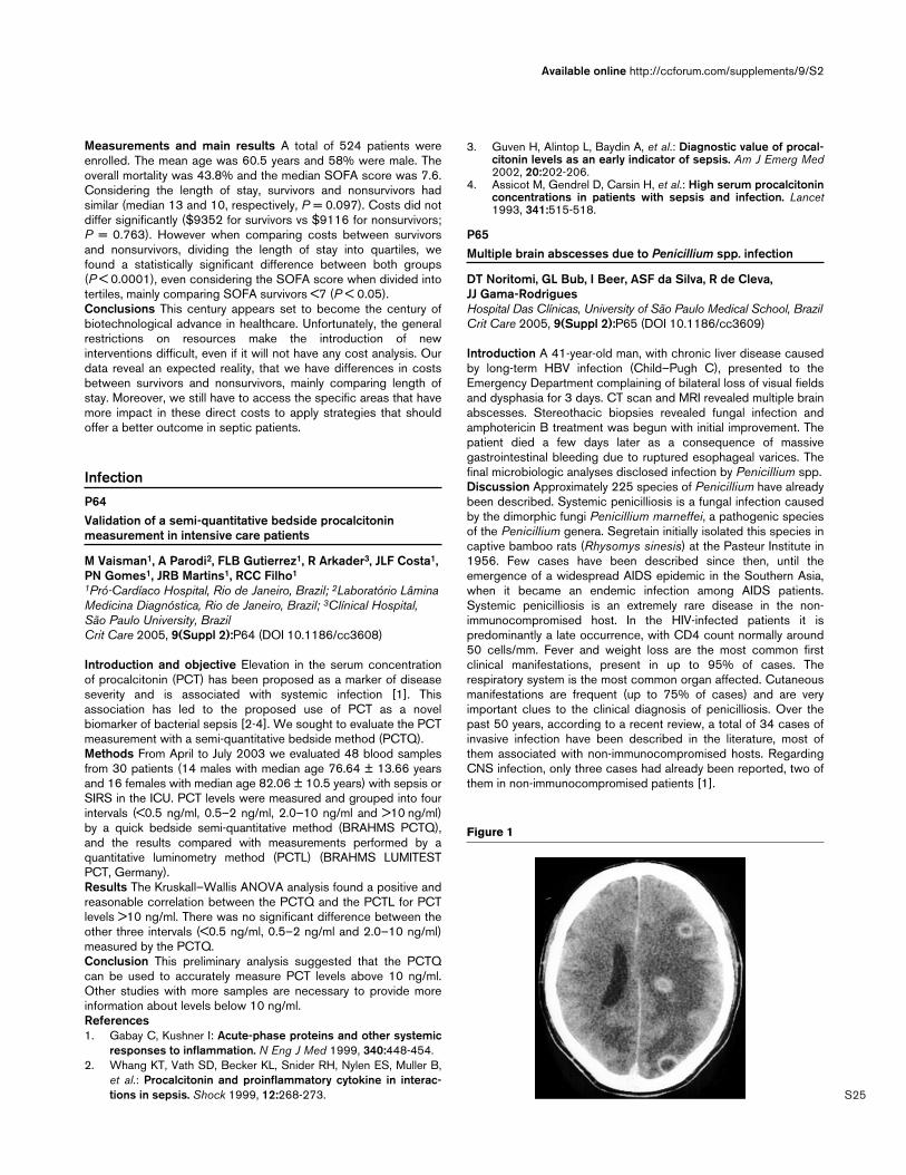
S25
Measurements and main results A total of 524 patients wereenrolled. The mean age was 60.5 years and 58% were male. Theoverall mortality was 43.8% and the median SOFA score was 7.6.Considering the length of stay, survivors and nonsurvivors hadsimilar (median 13 and 10, respectively, P = 0.097). Costs did notdiffer significantly ($9352 for survivors vs $9116 for nonsurvivors;P = 0.763). However when comparing costs between survivorsand nonsurvivors, dividing the length of stay into quartiles, wefound a statistically significant difference between both groups(P < 0.0001), even considering the SOFA score when divided intotertiles, mainly comparing SOFA survivors <7 (P < 0.05).Conclusions This century appears set to become the century ofbiotechnological advance in healthcare. Unfortunately, the generalrestrictions on resources make the introduction of newinterventions difficult, even if it will not have any cost analysis. Ourdata reveal an expected reality, that we have differences in costsbetween survivors and nonsurvivors, mainly comparing length ofstay. Moreover, we still have to access the specific areas that havemore impact in these direct costs to apply strategies that shouldoffer a better outcome in septic patients.
Infection
P64Validation of a semi-quantitative bedside procalcitoninmeasurement in intensive care patients
M Vaisman1, A Parodi2, FLB Gutierrez1, R Arkader3, JLF Costa1,PN Gomes1, JRB Martins1, RCC Filho1
1Pró-Cardíaco Hospital, Rio de Janeiro, Brazil; 2Laboratório LâminaMedicina Diagnóstica, Rio de Janeiro, Brazil; 3Clínical Hospital,São Paulo University, BrazilCrit Care 2005, 9(Suppl 2):P64 (DOI 10.1186/cc3608)
Introduction and objective Elevation in the serum concentrationof procalcitonin (PCT) has been proposed as a marker of diseaseseverity and is associated with systemic infection [1]. Thisassociation has led to the proposed use of PCT as a novelbiomarker of bacterial sepsis [2-4]. We sought to evaluate the PCTmeasurement with a semi-quantitative bedside method (PCTQ).Methods From April to July 2003 we evaluated 48 blood samplesfrom 30 patients (14 males with median age 76.64 ± 13.66 yearsand 16 females with median age 82.06 ± 10.5 years) with sepsis orSIRS in the ICU. PCT levels were measured and grouped into fourintervals (<0.5 ng/ml, 0.5–2 ng/ml, 2.0–10 ng/ml and >10 ng/ml)by a quick bedside semi-quantitative method (BRAHMS PCTQ),and the results compared with measurements performed by aquantitative luminometry method (PCTL) (BRAHMS LUMITESTPCT, Germany).Results The Kruskall–Wallis ANOVA analysis found a positive andreasonable correlation between the PCTQ and the PCTL for PCTlevels >10 ng/ml. There was no significant difference between theother three intervals (<0.5 ng/ml, 0.5–2 ng/ml and 2.0–10 ng/ml)measured by the PCTQ.Conclusion This preliminary analysis suggested that the PCTQcan be used to accurately measure PCT levels above 10 ng/ml.Other studies with more samples are necessary to provide moreinformation about levels below 10 ng/ml.References1. Gabay C, Kushner I: Acute-phase proteins and other systemic
responses to inflammation. N Eng J Med 1999, 340:448-454.2. Whang KT, Vath SD, Becker KL, Snider RH, Nylen ES, Muller B,
et al.: Procalcitonin and proinflammatory cytokine in interac-tions in sepsis. Shock 1999, 12:268-273.
3. Guven H, Alintop L, Baydin A, et al.: Diagnostic value of procal-citonin levels as an early indicator of sepsis. Am J Emerg Med2002, 20:202-206.
4. Assicot M, Gendrel D, Carsin H, et al.: High serum procalcitoninconcentrations in patients with sepsis and infection. Lancet1993, 341:515-518.
P65Multiple brain abscesses due to Penicillium spp. infection
DT Noritomi, GL Bub, I Beer, ASF da Silva, R de Cleva, JJ Gama-RodriguesHospital Das Clínicas, University of São Paulo Medical School, BrazilCrit Care 2005, 9(Suppl 2):P65 (DOI 10.1186/cc3609)
Introduction A 41-year-old man, with chronic liver disease causedby long-term HBV infection (Child–Pugh C), presented to theEmergency Department complaining of bilateral loss of visual fieldsand dysphasia for 3 days. CT scan and MRI revealed multiple brainabscesses. Stereothacic biopsies revealed fungal infection andamphotericin B treatment was begun with initial improvement. Thepatient died a few days later as a consequence of massivegastrointestinal bleeding due to ruptured esophageal varices. Thefinal microbiologic analyses disclosed infection by Penicillium spp.Discussion Approximately 225 species of Penicillium have alreadybeen described. Systemic penicilliosis is a fungal infection causedby the dimorphic fungi Penicillium marneffei, a pathogenic speciesof the Penicillium genera. Segretain initially isolated this species incaptive bamboo rats (Rhysomys sinesis) at the Pasteur Institute in1956. Few cases have been described since then, until theemergence of a widespread AIDS epidemic in the Southern Asia,when it became an endemic infection among AIDS patients.Systemic penicilliosis is an extremely rare disease in the non-immunocompromised host. In the HIV-infected patients it ispredominantly a late occurrence, with CD4 count normally around50 cells/mm. Fever and weight loss are the most common firstclinical manifestations, present in up to 95% of cases. Therespiratory system is the most common organ affected. Cutaneousmanifestations are frequent (up to 75% of cases) and are veryimportant clues to the clinical diagnosis of penicilliosis. Over thepast 50 years, according to a recent review, a total of 34 cases ofinvasive infection have been described in the literature, most ofthem associated with non-immunocompromised hosts. RegardingCNS infection, only three cases had already been reported, two ofthem in non-immunocompromised patients [1].
Available online http://ccforum.com/supplements/9/S2
Figure 1

S26
Conclusion To our knowledge, this is the fourth documented caseof CNS infection by a member of the Penicillium genera in theworld, and the first one in Brazil.Reference1. Lyratzopoulos G: Invasive infection due to Penicillium species
othr than P. maeneffei. J Infect 2002, 45:184-207.
P66Impact of empirical antibiotic therapy and mortality in theelderly with septic shock
GMMO Oliveira, PH Godoy, MR Pantoja, RR Luiz, CE Figueiredo, RR Casemiro, J Regalla, L SolhaIntensive Care Unit, Prontocor da Lagoa, Federal University of RioDe Janeiro, BrazilCrit Care 2005, 9(Suppl 2):P66 (DOI 10.1186/cc3610)
Background There are still questions on early implementation ofcorrect empirical antibiotic therapy and its association withmortality reduction in septic shock (SS).Objective To evaluate the impact of the use of empirical antibioticson elderly mortality with SS in the ICU.Methods A prospective cohort of 67 patients over 65 years oldfollowed-up during 32 months and with pulmonary arterymonitoring due to SS. Cultures were achieved in the first 24 hours.Ventilator and hemodynamic support, volume resuscitation andempirical antibiotic support with large spectrum were alsoemployed. The choice of the antibiotics was based upon aprobable community-acquired or hospital infection, and it wasconsidered adequate when at least one effective drug had beenincluded. Previous diseases, organic failures, and APACHE IIscores were also evaluated. As for the statistical analysis, the t test,the chi-square test and Kaplan–Meier survival curve analyses wereapplied, considering 5% as the significance level.Results The average ranges were: for age (80 ± 7), for APACHE(19 ± 5), for ICU stay (18 ± 9 days), where 51% were women.Among the previous diseases one can point out systemic arterialhypertension in 40%, ischemic heart disease in 31%, stroke in21% and the COPD in 30% of the cases. Pulmonary SS aloneoccurred in 70% of the cases, and in association with urinary SS in27%. The blood cultures were positive in 10% of the samples. TheGram-negative pathogens were responsible for 79% of theinfections, where 36% were due to Pseudomonas. The multidrug-resistant microorganisms represented 8% of the cultures. Thirty-nine deaths occurred during the stay in the ICU. The antibioticsused in the empirical form were correct in 87% of the patients andthey were modified in around 72 hours when clinical worsening orinadequate antimicrobial susceptibility patterns result took place.There was no association between age (P = 0.22) or adequateempirical antibiotic therapy and mortality, but mortality wasassociated with APACHE score (P < 0.001) and organic failures(P = 0.006). The ICU length of stay was not correlated with theuse of adequate empirical antibiotics (P = 0.66).Conclusion The adequate and early empirical antibiotic therapywas not associated with mortality or with the ICU stay of the elderlywith SS. Possibly, the high level of correct choices of the antibioticscheme and its modification due to clinical failure and inadequateantimicrobial susceptibility patterns have contributed to the results.
P67Clinical impact of positive blood cultures in intensive carepatients
VS Issa, MFR Silva, LU Taniguchi, LM Cruz-Neto, IT VelascoHospital das Clínicas da Faculdade de Medicina da Universidadede São Paulo, BrazilCrit Care 2005, 9(Suppl 2):P67 (DOI 10.1186/cc3611)
Background Infectious complications are frequent in patientsadmitted to ICUs, and great effort is made in order to identifypossible infecting microorganisms. In this setting, multiple bloodcultures are usually collected, although their true value is still to beascertained.Objective To ascertain the clinical impact of positive bloodcultures in patients admitted to the ICU.Methods We carried out a prospective cohort study in 340patients in a general ICU of a tertiary hospital from June 2003 toNovember 2004. Clinical characteristics and blood cultures fromall patients were reviewed. Differences were considered significantwhen P < 0.05.Results One hundred and eighty-one (53.2%) patients were male,and the mean age was 51.9 (12–88) years. The initial diagnosiswas respiratory failure in 115 (33.8%), cardiovascular failure in 97(28.5%), neurological in 41 (12.1%), diabetic ketoacidosis in 27(7.9%), postoperatory status in 21 (6.2%) and other conditions in39 (11.5%). The median APACHE score was 17 (range 2–45) and107 patients (31.5%) died in the ICU. Four hundred and eighty-eight blood cultures were drawn from 201 patients; 156 (31.9%)cultures were positive in 93 (46.2%) patients. Gram-positive cocciwere found in 44 (21.9%) patients and Gram-negative bacilli in 25(12.4%) patients. As compared with patients with negative bloodcultures, patients with positive blood culture had a longer ICU stay(32.5 ± 50.5 vs 12 ± 12.3 days, P < 0.001), a higher initial SOFAscore (7.2 ± 1.9 vs 5.6 ± 4.4, P = 0.01), a higher total SOFAscore (152.2 ± 119.8 vs 62.8 ± 76.5, P < 0.001), a highermaximum SOFA score (11.1 ± 4.1 vs 8.3 ± 5.7, P < 0.001), ahigher frequency of central venous catheterization (90.3% vs71.3%, P = 0.002), urinary catheterization (86% vs 6.8%,P = 0.001), invasive blood pressure monitoring (44.1% vs 27.8%,P = 0.06) and a higher frequency of mechanical ventilation (80.6%vs 50.9%, P < 0.001). Mortality was increased in patients withpositive blood cultures (50.5% vs 32.4%, P = 0.009).Conclusion Patients admitted to the ICU that develop positive bloodcultures are more severely ill than patients with negative bloodcultures. The finding of an infecting microorganism in the blood ofintensive care patients does not warrant a better prognosis.
P68Procalcitonin in a semi-quantitative test and C-reactive proteinin the evaluation of postoperative patients admitted to acritical care unit
MO Silva, CP Amendola, S Longui, JM Silva, EV Campos, E RezendeHospital do Servidor Público Estadual, São Paulo, BrazilCrit Care 2005, 9(Suppl 2):P68 (DOI 10.1186/cc3612)
Introduction Procalcitonin and C-reactive protein (CRP) are acutephase proteins produced by stimulation to infectious andinflammatory factors, and are greatly used because they presentmore clinical applicability with practicality and fast detection. Theobjective of this study is to evaluate which of these markers showsbetter capacity to predict the evolution of postoperative patientsduring their stay in the ICU.
Critical Care June 2005 Vol 9 Suppl 2 Third International Symposium on Intensive Care and Emergency Medicine for Latin America

S27
Materials and methods An observational study, performedbetween February and March 2004 in an ICU of a tertiary hospital.Postoperative patients with more than 24 hours of stay in the ICUand older than 18 years were included in the sample. A semi-quantitative test of procalcitonin (BRAHMS Laboratory) wasperformed after the patient enrollment.Results Twenty-six patients were included, 31% submitted tourgent surgery and 69% to elective surgery. The mean age was71.5 ± 10.6 years and 53% were female. The mean APACHE IIscore and SOFA score were 19.2 ± 4.9 and 5.8 ± 2.5, respectively.Every patient presented two or more criteria for systemicinflammatory response syndrome, and the rate of ICU mortality was30.8%. Among the survivors none presented a procalcitonin test≥2 ng/ml, against 57.1% of non-survivors (P = 0.02). The meanvalue of CRP was 13.0 ± 6.2 between survivors and was15.4 ± 7.3 among non-survivors (P = 0.34). The predictivecapacity of these variables, as evaluated by the area under theROC curve, was 0.835 for procalcitonin and 0.634 for CRP.Conclusion The semi-quantitative test of procalcitonin wassuperior to the CRP dosage as a predictor of worse evaluation inthis population. This information would be of utility if provided earlyat the bedside in the identification of patients with potentiallyunfavorable outcome.
P69Analysis of central venous catheter-related bloodstreaminfections in a teaching medical ICU: 2 years of follow-up
L Kopel, RT Carvalho, HBN Araujo, AA Fagundes, M Ribeiro, J Bastos, SG LageClinical Intensive Care Unit, Heart Institute, HCFMUSP, São Paulo,BrazilCrit Care 2005, 9(Suppl 2):P69 (DOI 10.1186/cc3613)
Introduction Central venous catheters (CVCs) are frequently usedin the ICU. Infection, the most frequent complication of CVC use, isassociated with increased morbidity and mortality.Hypothesis The purpose of this study is to describe theepidemiology and to determine infection rates related to CVC use,the causative agents and its influence in ICU stay.Methods We evaluated prospectively (NNISS-CDC/Atlanta) alllaboratory-confirmed CVC-related bloodstream infections (LBSI) inan 18-bed medical ICU of a university centre between January2002 and June 2004. Catheter insertion was performed bycardiology residents. Data collected: number of LBSI, deviceutilization (DU), time of ICU stay, demographic data (sex, age),causative agents and rates of infection/1000 CVC days.Results A total 174 episodes of LBSI were studied. Theyrepresented 28% of all diagnosed ICU infections. The lethality was47% in 30 days (medium ICU mortality was 28%). Ninety-seven(55.7%) episodes occurred in men and 77 (44.3%) in women (P =not significant). Sixty percent of the episodes occurred in patients60–90 years old. A total 20.6% occurred in the first week and 37%after 28 days. The ICU stay of these patients was 26 days (ICUgeneral medium stay was 12 days). The percentage incidences ofthe main infective agents in 2002, 2003 and 2004 were,respectively: S. aureus — 19%, 16.5%, 2.5%; S. epidermidis — 22%,31%, 34%; E. faecalis — 4%, 5%, 7.3%; K. pneumoniae — 21%,16.5%, 14.6%; P. aeroginosa — 13%, 10.5%, 7.3%; other Gram-negatives — 19.4%, 19.4%, 24.5%; Candida sp. — 0.8%, 0%, 9.8%.Conclusions BSI remains an important problem increasing theICU stay and costs. The biphasic incidence calls attention to theneed for better selection for admitting patients from other hospitalsand to improve adherence to standardized infection controlpractices.
P70Renal function profile in patients using polimixin for treatmentof multiresistant Pseudomonas sp. in an ICU
O Messeder, CEC Rolim, MJ Martins, MV Liberato, JM Teles, S Agareno, A FariasReal Sociedade Portuguesa de Beneficência Dezesseis deSetembro, Hospital Português da Bahia, Salvador, BrazilCrit Care 2005, 9(Suppl 2):P70 (DOI 10.1186/cc3614)
Introduction Multidrug-resistant Pseudomonas sp. (MDR-PS) isone of the most frequently isolated microorganisms causinghospital-acquired infections. Polimixin has been the onlytherapeutic option in many such cases. Nephrotoxicity is always aconcern when this antibiotic is being used.Objective The purpose of this study is to describe the impact ofPolimixin use in the renal function of a number of patients.Materials and methods A retrospective series of cases ofpatients with infection caused by MDR-PS, after 3 days or more ofPolimixin use, admitted from December 2002 to August 2004 tothe medical–surgical ICU of a tertiary hospital. The renal functionwas evaluated using the mean of the serum urea and creatinineobtained before and during the Polimixin use. A total of 31 caseswere analyzed. For data analysis, measures of frequency andcentral tendency were used; the Wilcoxon test for comparison ofthe means before and during treatment.Results The mean age of the patients was 66.52 ± 17.86 years,67.7% of male gender. The observed co-morbidities were: arterialhypertension 54.8%, malignant neoplasm 35.4%, diabetes mellitus29.3%, and chronic renal failure (CRF) 19.3%. The length of stayin the ICU was 33.7 ± 28.7 (median 23) days and the averagedays of Polimixin use was 15.19 ± 10.37 (median 14) days. Elevenpatients (35.5%) were on dialysis during the Polimixin use, fivecases started the dialysis during the treatment, three of these hadCRF. Among the patients not on dialysis, only 18 had urea andcreatinine values available for analysis prior to and during thetreatment. The total mean for urea prior to and during the treatmentwas 68.64 ± 49.39 and 69.64 ± 31.90 (P = 0.57) and the totalmean for creatinine before and during treatment was 1.57 ± 1.6and 2.24 ± 3.5 (P = 0.32), respectively.Conclusions In this small sample of critically ill patients, the use ofPolimixin did not demonstrate a significant impact in renal functionas observed by following the urea and creatinine levels.
P71Vancomycin-resistant enterococci in a general ICU of ateaching hospital in São Paulo
MFR Silva, VS Issa, LU Tanaguchi, LM Cruz-Neto, IT VelascoErviço de Emergência Médica, Hospital das Clínicas da Faculdadede Medicina, Universidade de São Paulo, BrazilCrit Care 2005, 9(Suppl 2):P71 (DOI 10.1186/cc3615)
Objective To evaluate the colonization rate and risk factorsassociated with vancomycin-resistant enterococci (VRE) inpatients in a general ICU in a teaching hospital.Methods Rectal swabs were collected from June 2003 toNovember 2004, according to current recommendations of theinfection control committee of our institution. Data from 94 patientswere reviewed for analysis. The variables analyzed were age, timeelapsed until ICU admission, length of ICU stay, Acute Physiologyand Chronic Health Evaluation (APACHE) II score and ICUoutcome. The presence of intravascular catheters, urinary catheter,need of invasive mechanical ventilation (IMV) and dialysis weredetermined. Antimicrobial therapy (vancomycin, imipenen-cilastatin,
Available online http://ccforum.com/supplements/9/S2
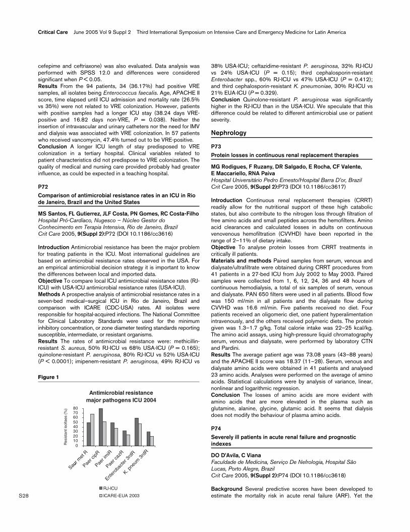
S28
cefepime and ceftriaxone) was also evaluated. Data analysis wasperformed with SPSS 12.0 and differences were consideredsignificant when P < 0.05.Results From the 94 patients, 34 (36.17%) had positive VREsamples, all isolates being Enterococcus faecalis. Age, APACHE IIscore, time elapsed until ICU admission and mortality rate (26.5%vs 35%) were not related to VRE colonization. However, patientswith positive samples had a longer ICU stay (38.24 days VRE-positive and 16.82 days non-VRE, P = 0.038). Neither theinsertion of intravascular and urinary catheters nor the need for IMVand dialysis was associated with VRE colonization. In 57 patientswho received vancomycin, 47.4% turned out to be VRE-positive.Conclusion A longer ICU length of stay predisposed to VREcolonization in a tertiary hospital. Clinical variables related topatient characteristics did not predispose to VRE colonization. Thequality of medical and nursing care provided probably had greaterinfluence, as could be expected in a teaching hospital.
P72Comparison of antimicrobial resistance rates in an ICU in Riode Janeiro, Brazil and the United States
MS Santos, FL Gutierrez, JLF Costa, PN Gomes, RC Costa-FilhoHospital Pró-Cardíaco, Nugesco — Núcleo Gestor doConhecimento em Terapia Intensiva, Rio de Janeiro, BrazilCrit Care 2005, 9(Suppl 2):P72 (DOI 10.1186/cc3616)
Introduction Antimicrobial resistance has been the major problemfor treating patients in the ICU. Most international guidelines arebased on antimicrobial resistance rates observed in the USA. Foran empirical antimicrobial decision strategy it is important to knowthe differences between local and imported data.Objective To compare local ICU antimicrobial resistance rates (RJ-ICU) with USA-ICU antimicrobial resistance rates (USA-ICU).Methods A prospective analysis of antimicrobial resistance rates in aseven-bed medical–surgical ICU in Rio de Janeiro, Brazil andcomparison with ICARE (CDC-USA) rates. All isolates wereresponsible for hospital-acquired infections. The National Committeefor Clinical Laboratory Standards were used for the minimuminhibitory concentration, or zone diameter testing standards reportingsusceptible, intermediate, or resistant organisms.Results The rates of antimicrobial resistance were: methicillin-resistant S. aureus, 50% RJ-ICU vs 68% USA-ICU (P = 0.165);quinolone-resistant P. aeruginosa, 80% RJ-ICU vs 52% USA-ICU(P < 0.0001); imipenem-resistant P. aeruginosa, 49% RJ-ICU vs
38% USA-ICU; ceftazidime-resistant P. aeruginosa, 32% RJ-ICUvs 24% USA-ICU (P = 0.15); third cephalosporin-resistantEnterobacter spp., 60% RJ-ICU vs 47% USA-ICU (P = 0.412);and third cephalosporin-resistant K. pneumoniae, 30% RJ-ICU vs21% EUA-ICU (P = 0.329).Conclusion Quinolone-resistant P. aeruginosa was significantlyhigher in the RJ-ICU than in the USA-ICU. We speculate that thisdifference could be related to different antimicrobial use or patientseverity.
Nephrology
P73Protein losses in continuous renal replacement therapies
MG Rodigues, F Ruzany, DR Salgado, E Rocha, CF Valente, E Maccariello, RNA PaivaHospital Universitário Pedro Ernesto/Hospital Barra D’or, BrazilCrit Care 2005, 9(Suppl 2):P73 (DOI 10.1186/cc3617)
Introduction Continuous renal replacement therapies (CRRT)readily allow for the nutritional support of these high catabolicstates, but also contribute to the nitrogen loss through filtration offree amino acids and small peptides across the hemofilters. Aminoacid clearances and calculated losses in adults on continuousvenovenous hemofiltration (CVVHD) have been reported in therange of 2–11% of dietary intake.Objective To analyse protein losses from CRRT treatments incritically ill patients.Materials and methods Paired samples from serum, venous anddialysate/ultrafiltrate were obtained during CRRT procedures from41 patients in a 27-bed ICU from July 2002 to May 2003. Pairedsamples were collected from 1, 6, 12, 24, 36 and 48 hours ofcontinuous hemodialysis, a total of six samples of serum, venousand dialysate. PAN 650 filters were used in all patients. Blood flowwas 150 ml/min in all patients and the dialysate flow duringCVVHD was 16.6 ml/min. Five patients received no diet. Fourpatients received an oligomeric diet, one patient hyperalimentationintravenously, and the others received polymeric diets. The proteingiven was 1.3–1.7 g/kg. Total calorie intake was 22–25 kcal/kg.The amino acid assays, using high-pressure liquid chromatographyserum, venous and dialysate, were performed by laboratory CTNand Pardini.Results The average patient age was 73.08 years (43–88 years)and the APACHE II score was 18.37 (11–28). Serum, venous anddialysate amino acids were obtained in 41 patients and analysed23 amino acids. Analyses were performed on the average of aminoacids. Statistical calculations were by analysis of variance, linear,nonlinear and logarithmic regression.Conclusion The losses of amino acids are more evident withamino acids that are more elevated in the plasma such asglutamine, alanine, glycine, glutamic acid. It seems that dialysisdoes not modify the behaviour of plasma amino acids.
P74Severely ill patients in acute renal failure and prognosticindexes
DO D’Avila, C VianaFaculdade de Medicina, Serviço De Nefrologia, Hospital SãoLucas, Porto Alegre, BrazilCrit Care 2005, 9(Suppl 2):P74 (DOI 10.1186/cc3618)
Background Several predictive scores have been developed toestimate the mortality risk in acute renal failure (ARF). Yet the
Critical Care June 2005 Vol 9 Suppl 2 Third International Symposium on Intensive Care and Emergency Medicine for Latin America
Figure 1
01020304050607080
Res
ista
nt is
olta
es (
%)
Saur m
et R
u Paer c
ipR
Paer i
miR
Paer c
azR
Enter
obac
ter 3
rdR
K. pne
um 3
rdR
Antimicrobial resistancemajor pathogens ICU 2004
RJ-ICU
ICARE-EUA 2003
S29
accuracy of scores has been shown to vary when applied toforeign populations, and locally developed indexes may becomebetter predictors. The aim of this study was to compare theperformances of three working scores — one locally developed.Patients and methods A total of 194 consecutive patients withARF under intensive care were enrolled at initiation of renalreplacement therapy maintained beyond 24 hours. Liaño’s index(ATN-ISS), Mehta’s index (PICARD), and a locally tested index(IRA-PUC) were properly applied. The discrimination andcalibration of the indexes were evaluated (area under the ROCcurves, goodness of fit and linear regression analysis), and werecompared.Results The population’s observed mortality was high (87.0%),with a median of four (95% CI: 3–4) failing organs at inclusion.Predicted mean mortalities were 86.8, 84.9 and 87.9 (ATN-ISS,PICARD and IRA-PUC, respectively). The areas under the ROCcurves were 0.702, 0.695 and 0.755 (ATN-ISS, PICARD and IRA-PUC, respectively). Goodness-of-fit determination was acceptablefor all, and the linear regression analysis R2 value was 0.954,0.448 and 0.947 (P = 0.015; P = 0.088; P = 0.001) for ATN-ISS,PICARD and IRA-PUC, respectively. Discrimination was lessadequate than previously stated for all tested indexes. Contrastingwith ATN-ISS and IRA-PUC, PICARD was not so well calibrated.Conclusion Discrepancies in characteristics and in mortality ratesamong populations at risk and the original one may decrease theaccuracy of ARF predictive indexes, precluding their extensive use.Acknowledgement Supported by FAPERGS.
P75Prognostic significance of serum creatinine in patients withacute coronary syndromes for prediction of inhospitalmortality
MA Mattos, DG Toledo, CE Mattos, FC de Deus, MHV Assad, BR TuraInstituto Nacional de Cardiologia Laranjeiras, Unidade Coronariana,BrazilCrit Care 2005, 9(Suppl 2):P75 (DOI 10.1186/cc3619)
Context Patients with renal dysfunction are at an increased risk forcardiovascular disease.Objective To evaluate the prognostic significance of serum creatininefor inhospital mortality in patients with acute coronary syndromes.Design An observational study.Patients and methods Included were consecutive patients withacute coronary syndromes admitted alive to the coronary care unitfrom February 2004 to January 2005. The patients were initiallyclassified into three groups on the basis of serum creatinineconcentration measured on admittance. Normal renal function andmild and severe renal dysfunction were defined as serum creatinineconcentrations of <1.2 mg/dl, ≥1.2 but <2.0 mg/dl, and≥2.0 mg/dl, respectively. Patients receiving regular hemodialysiswere excluded from the study. Univariate and multivariate relativerisks (RRs) were calculated for three renal risk quartiles using theserum creatinine concentration on presentation.Results This study included 227 patients. The mean age wasgreater in the severe renal dysfunction group (P = 0.031). Of theanalysed variables — age, sex, diabetes, hypertension, dyslipidemia,previous myocardial infarction, previous coronary angioplasty andcoronary artery bypass graft — only the creatinine level was anindependent predictor of greater inhospital mortality. The inhospitalmortalities of mild and severe renal dysfunction patients weregreater (7.9% and 31.6%, respectively) than that of patientswithout renal dysfunction (2.8%), P = 0.000, with an increased riskof 10 times.
Conclusion In this study, we showed that the creatinine level atadmission is an independent predictor of inhospital mortality inpatients with acute coronary syndromes.
P76Impact of renal scintigraphy in severe patients with acuterenal failure
JF Ferreira, JLF Costa, M Dessen, AM Mesquita, MF Garcez, CT Mesquita, FL Gutierrez, RC Costa-FilhoHospital Pró-Cardíaco/Nugesco — Núcleo Gestor doConhecimento em Terapia Intensiva, Rio de Janeiro, BrazilCrit Care 2005, 9(Suppl 2):P76 (DOI 10.1186/cc3620)
Introduction Acute renal failure (ARF) is frequent in severepatients, producing a poor outcome in the face of its multipleinsults that are individually determined [1,2]. Technetium 99methylenedicysteine scintigraphy (Tc-EC) has a very low plasmaprotein binding and a large volume of distribution. Tc-EC is a goodagent for renal function evaluation, providing an index of tubularfunction and yielding high-quality images [3-5].Objective To evaluate the clinical impact of renal scintigraphy (RC)in the management of ARF.Materials and methods We evaluate all patients in ICUs of ageneral, private hospital, submitted to RC in the period of January2003 to January 2004.Results Thirty-four patients (18 male), 79.56 ± 16.26 years old,with an APACHE II score of 16.94 ± 6.34 (expected mortality25%) were studied. The observed mortality was 29.41%. Seventy-three percent of the patients were anuric for 55.36 ± 13.43 days.Dialyses were used in 85.29% of the population and RC alwayscarried through after 30 days. Seventy-four percent of the patientswere mechanically ventilated during RC with no transport accident.RC changed the nephrology prescription, interrupted the methodor indicated long-term access confection. The radiotracer mostused was Tc-EC in 64.70% of the patients. Twenty percent of thepatients continued in chronic dialysis and 17.64% had recoveredrenal function.Conclusion RC, mainly with Tc-EC, influenced and modified theclinical impression, nephrological decision, and medical care ofsevere ARF patients. RC seems to be a good cost–benefit withsimple accomplishment method to evaluate glomerular filtration andtubular function with an important impact especially in anuric patients.References1. Mehta RL, Chertow GM: J Am Soc Nephrol 2003, 14:2178-
2187.2. Riella MC: Nephrology Primer and Hydroeletrolitycus Disturbs.
4th edition. Rio de Janeiro: Guanabara Koogan; 2003:372-385.3. Kabasakal L: Eur J Nucl Med 2000, 27: 351-357.4. Kabasakal L, Turogh HT, Onsel C, et al.: J Nucl Med 1995,
36:224-228.5. Van Merom CG, Bormans GM, De Roo MJ, et al.: Eur J Nucl Med
1993, 20:738-746.
P77Influence of renal dysfunction on inhospital morbidity andmortality of patients with decompensated heart failure
RM Rocha, MI Bittencourt, FOD Rangel, HCV Rey, FAC Ferreira,EP Bernardo, ALC Marins, M Vaisman, R EsporcatteUnidade Coronariana, Hospital Pró-Cardíaco/Procep, Rio deJaneiro, BrazilCrit Care 2005, 9(Suppl 2):P77 (DOI 10.1186/cc3621)
Background Renal dysfunction (RD) is a clinical conditionassociated with worse inhospital prognosis for patients admitteddue to decompensated heart failure (DHF).
Available online http://ccforum.com/supplements/9/S2
S30
Objectives To evaluate the impact of RD in patients admitted dueto DHF, and its relationship with clinical features and laboratory data,length of stay (LOS), and inhospital complications and mortality.Methods From January 2003 to December 2004, we studied acohort of 137 patients admitted to the coronary care unit due toDHF (79.6% NYHA class IV). The mean age was 76.5 ± 11.08 years,54% male, 29.9% diabetes mellitus, 74.5% systemic hypertension,64% ischemic cardiomyopathy and the mean LOS was14.2 ± 34.6 days. RD was defined as an estimated (Cockcroft)creatinine clearance less than 60 ml/min on admission. Baselinedemographics, laboratory findings, LOS and complications(cardiac arrhythmias, hemorrhage, need of blood transfusions,hemodynamic instability and infections) and mortality rates werecompared. The Mann–Whitney test (laboratory findings and LOS),the Student t test (age) and Pearson’s chi-square test (othervariables) were used.Results A total of 73.4% of the patients with DHF were consideredto have RD. They were older (79.7 ± 9.5 vs 67.1 ± 10.4 years,P < 0.0001), with paradoxically less diabetes (18.1% vs 55.9%,P < 0.0001). On admission, B-type natriuretic peptide (P = 0.021),D-dimer (P = 0.024), hematocrit (36.2 ± 5.6% vs 38.2 ± 4.4%,P = 0.029) and hemoglobin (12.1 ± 1.89 vs 12.9 ± 2.08 g/dl,P = 0.057) were smaller. The need for blood transfusion (21.2% vs5.9%, P = 0.041), and a significant increase of LOS was observedin the RD group (16.3 ± 41.2 vs 8.4 ± 5.7 days, P = 0.013), andhigher inhospital mortality (9.57% vs 5.88%, P = not significant)was observed in the RD group.Conclusions RD is highly prevalent in patients admitted due tosevere heart failure, with a clinical impact on blood transfusion,length of stay, and determining a trend to higher mortality.
P78Analysis of two groups of patients with acute renal failure inthe critical care setting
EJB Mendonça, L Andrade, JP Maciel, S Agareno, JMM Telles,AMC Farias, OHC MessederUTI do Hospital Português, Salvador, Bahia, BrazilCrit Care 2005, 9(Suppl 2):P78 (DOI 10.1186/cc3622)
Introduction Acute renal (ARF) failure has a notable prevalence inthe ICU. When patients are identified by small alterations in thecreatinine levels, the percentage of ARF may reach 25% in thecritically ill.Objective To evaluate ARF in two groups of patients: Group A(GA), patients with ARF at admission to the ICU; Group B (GB),patients developing ARF during the ICU stay.Materials and methods A prospective survey, carried outbetween July 2004 and February 2005 in a tertiary care privatehospital. The sample was composed of two groups. GA: patientswith ARF at admission, creatinine >2.0 mg/dl and normal renalfunction prior to the admission. GB: patients with normal renalfunction, with a 0.5 mg/dl increase in the serum creatinine duringthe ICU stay or if presented with chronic renal failure, defined asserum creatinine >2 and <4.9 mg/dl, an increase of 1.0 mg/dl inserum creatinine levels. Patients with serum creatinine level>5 mg/dl, history of ARF in another hospital or kidney transplantwere excluded.Results Six hundred and eighty patients were admitted to the ICUduring the study period. Sixty-five patients had ARF, 14 in GA and51 in GB. The mean age was 62.79 years and 69.4 years,respectively. The causes of ARF in GA were hypoperfusion(28.6%) and sepsis (21.4%), whereas in GB they werehypoperfusion (35.3%), septic shock (29.4%) and sepsis (19.6%).The prevalence of contrast-induced ARF was 7.1% in GA, and
2.0% in GB (P < 0.05). The mean serum creatinine level was4.2 mg/dl in GA and 2.7 mg/dl in GB (P < 0.05). The mean urineoutput measured 24 hours before the diagnosis of ARF was551.08 ml in GA and 713.37 ml in GB. The APACHE II score was33.94 in GA and 41.76 in GB. The mortality in GA was 28.6%(four patients) and was 58.8% in group B (30 patients) (P < 0.05).The mortality for the ICU in general was 13.2% (90 patients).Twenty-three patients went for dialysis: GA 35.7% (five patients)and GB 35.29% (18 patients). The mean number of sessions was4.78 in GA and 3.2 in GB. Four patients in GA (80%) and 15(83.3%) in GB died in the dialysis group.Conclusions The ARF mortality remains high despite the advancesin technology, notably in patients on dialysis. In some cases theARF diagnosis was delayed, as demonstrated by the serumcreatinine levels in GA.
Pneumology
P79Cuff pressure control of the tracheal tube: a prospective studyin a general ICU
GS Cardoso, HP Guimarães, RD Lopes, PHR Leal, AP Resque,CJ Guedes, FRP Souza, FR Machado, JLG AmaralFederal University of São Paulo, UNIFESP, EPM, São Paulo, BrazilCrit Care 2005, 9(Suppl 2):P79 (DOI 10.1186/cc3623)
Introduction High pressures of the tracheal tube cuffs can impairdamage for restriction of the air flow.Objectives To demonstrate the prevalence of high pressures ofthe tracheal cuff in orotracheal tubes (OTTs) and tracheotomies(TCT). To verify the impact of cuff pressure control on further cuffvolume adjustment.Methods This is a transversal study, which evaluated everytracheally intubated (OTT or TCT) patient in a general orneurosurgery ICU. Daily we have measured cuff pressures with amercury manometer. The pressure goal was 20 mmHg(27.2 cmH2O).Results One hundred and six patients were evaluated, 75 (71%)male. Their age ranged from 16 to 92 years (mode 85 years).Forty-two (39%) patients had undergone TCT and 64 (61%) hadundergone OTT. The first-day APACHE II score ranged from 4 to39 (mode 22) and the average mortality risk was 27.5%. Thelength of time of OTT ranged from 1 to 16 days (mode 8 days) andthat for TCT from 5 to 70 days (mode 5 days). The initial pressuresranged from <20 mmHg to 140 mmHg (mode <20 mmHg). Afterinitial reduction of the cuff volumes, the range of the pressuresdecreased (from <20 mmHg to 80 mmHg).Conclusion Increased prevalence of high pressures of the cuff intracheal tubes justifies frequent monitoring of this parameter,aiming at brightening up the deleterious effect of the drawn-outuse of these tubes.Reference1. Wujtewicz MA, Sawicka W, Sein Anand J, et al.: Monitoring of
tracheal tube cuff pressure in patients treated in intensivetherapy unit and intensive care units. Przegl Lek 2004, 61:353-355.
Critical Care June 2005 Vol 9 Suppl 2 Third International Symposium on Intensive Care and Emergency Medicine for Latin America
S31
P80A new integrative weaning index of discontinuation frommechanical ventilation
SN Nemer, CSV Barbas, JB Caldeira, LM Azeredo, S Machado,R Gago, T Clipes, PR Filho, PCP SouzaHospital de Clínicas de Niterói, Rio de Janeiro, BrazilCrit Care 2005, 9(Suppl 2):P80 (DOI 10.1186/cc3624)
Background and objectives Indexes predicting the weaningoutcome are frequently inaccurate. With the present study, we aimto evaluate the predictive performance of a new index forpredicting the weaning outcome, which we called the integrativeweaning index (IWI).Methods Two hundred and fifty patients of several etiologies in theweaning process that remained up to 24 hours in mechanicalventilation were evaluated (all with PaO2 ≥60 mmHg with FiO2≤0.4 and PEEP ≤8 cmH2O). All patients were submitted to a 2-hour trial of spontaneous breathing. Those who sustained 2 hoursof spontaneous breathing without return to mechanical ventilationin the following 24 hours were considered weaned, while thosewho could not sustain 2 hours of spontaneous breathing orreturned to mechanical ventilation in the following 24 hours wereconsidered not weaned. The frequency/tidal volume ratio (f/Vtratio), the airway occlusion pressure at 0.1 s after the onset ofinspiratory effort (P 0.1), the product of P 0.1 and f/Vt (P 0.1 ×f/Vt), the respiratory rate (RR), the quasi-static compliance of therespiratory system (Cqst,rs), the PaO2/FiO2 ratio and the newintegrative weaning index (IWI = Cqst,rs × SaO2 / f/Vt ratio) wereevaluated in all patients. Arterial blood gas was collected with FiO2in 0.35. The sensitivity, specificity, positive predictive value,negative predictive value and the receiver operating characteristic(ROC) curves were calculated in order to evaluate the predictiveperformance of each index. The nonparametric method of Hanleyand McNeil was used to compare the area under the ROC curvesof each index.Results Two hundred and eighteen patients were weaned, while32 patients were not weaned. The IWI presented the larger areaunder the ROC curves (0.97), followed by the f/Vt ratio (0.90),Cqst,rs (0.89), f/Vt × P 0.1 (0.85), RR (0.80), P 0.1 (0.70) andfinally by the PaO2/FiO2 ratio (0.60). The area under the ROCcurves of the IWI was larger than those for f/Vt ratio (P < 0.0001)and also larger than those for the other indexes (P < 0.0001).Conclusion In our study, even compared with other essentialindexes in the literature, the IWI showed the best criteria forpredicting the weaning outcome. With the obtained results webelieve that with the use of the IWI in other countries we mayfurther prove its accuracy.
P81A comparative study of inspiratory muscle strength,neuromuscular drive to breath and its ratio in weaning outcome
SN Nemer, CSV Barbas, M Andrade, C Geraldo, L Tabajaras, C Coimbra, C Cadilhe, J Dias, PCP SouzaHospital de Clínicas de Niterói, Rio de Janeiro, Brazil; University ofSão Paulo, BrazilCrit Care 2005, 9(Suppl 2):P81 (DOI 10.1186/cc3625)
Background and objectives The inspiratory muscle strength andthe neuromuscular drive to breath, evaluated by the maximalinspiratory pressure (MIP) and airway occlusion pressure (P 0.1),respectively, are important factors in weaning. The aim of this studyis to evaluate the MIP, the P 0.1 and its ratio (P 0.1/MIP) inweaning outcome.
Methods Seventy consecutive patients of several etiologies in theweaning process that remained up to 24 hours in mechanicalventilation were evaluated (all with PaO2 ≥60 mmHg with FiO2≤0.4 and PEEP ≤8 cmH2O). All patients were submitted to a 2-hour trial of spontaneous breathing. Those who sustained 2 hoursof spontaneous breathing without return to mechanical ventilationin the following 24 hours were considered weaned, while thosewho could not sustain 2 hours of spontaneous breathing orreturned to mechanical ventilation in the following 24 hours wereconsidered not weaned. MIP < –25 cmH2O, P 0.1 <4.2 cmH2Oand P 0.1/MIP < 0.14 cmH2O were used to predict the success inweaning outcome. The predictive performance of each index wasevaluated through the sensibility, specificity, positive predictivevalue, negative predictive value and diagnostic accuracy. Theresults were also evaluated by the area under the receiveroperating characteristic (ROC) curves.Results MIP presented an area under the ROC curves smallerthan those for P 0.1 (0.52 ± 0.08 vs 0.76 ± 0.06, respectively;P = 0.004) and also smaller than those for P 0.1/MIP (0.52 ± 0.08vs 0.78 ± 0.06, respectively; P = 0.0006). P 0.1/MIP presentedexcellent predictive performance in weaned patients, withsensibility of 98.08, but with the area under the ROC curves onlyslightly larger than those for P 0.1 (0.78 ± 0.06 vs 0.76 ± 0.06,respectively; P = 0.69).Conclusion In our study, Pi max was the criterion with the worstpredictive performance. P 0.1 was shown to be a very importantcriterion to evaluate the respiratory center output, although withlimitations in evaluating weaning failure. Patients with P 0.1/Pi maxratio >0.14 are not always associated with weaning failure, butvalues <0.14 were highly associated with success in weaningoutcome.
P82A comparison of two alveolar recruitment maneuver approachesin patients with acute respiratory distress syndrome andhemorrhagic stroke with Glasgow Coma Scale ≤≤8
SN Nemer, CSV Barbas, J Caldeiras, L Caldeiras, J Goulart, M Cabral, M Rocha, JM Garcia, PCP SouzaHospital de Clínicas de Niterói, Rio de Janeiro, BrazilCrit Care 2005, 9(Suppl 2):P82 (DOI 10.1186/cc3626)
Background and objectives Alveolar recruitment maneuvers(ARM) are generally not used in acute respiratory distresssyndrome (ARDS) patients in an acute phase of brain injury, aimingto avoid increasing the intracranial pressure (ICP).Methods Sixteen patients with ARDS and hemorrhagic strokewere evaluated. Criteria for admission were: acute onset, bilateralchest radiographic infiltrates, pulmonary-capillary wedge pressure≤18 mmHg, PaO2/FiO2 ratio <200 and Glasgow Coma Scale ≤8with ICP monitorization. Patients were randomized into two similargroups. One group received ARM with CPAP of 35 cmH2O for40 s, and the other group received pressure control ventilation(PCV) with a positive end expiratory pressure (PEEP) of 15 cmH2Oand pressure control above a PEEP of 35 cmH2O for 2 min (tidalrecruitment). The ICP, cerebral perfusion pressure (CPP) andoxygen pulse saturation (SpO2) were similar in both groups beforethe randomization. The fraction of inspired oxygen (FiO2) was keptto 1.0 during the study. The ICP, CPP and SpO2 were measuredbefore and after ARM and were compared by Student’s t test.Mortality was compared by Fisher’s test.Results Initial values of the ICP, CPP and SpO2 were,respectively: 13.38 ± 4.53 mmHg (CPAP group) vs 13.25 ± 3.45(tidal recruitment group), P = 0.95; 82.75 ± 10.37 (CPAP group)vs 84.25 ± 10.37 mmHg (tidal recruitment group), P = 0.73; and
Available online http://ccforum.com/supplements/9/S2
S32
95.75 ± 1.04 (CPAP group) vs 95.0 ± 1.51% (tidal recruitmentgroup), P = 0.26. After ARM, the ICP was higher in the CPAPgroup (20.50 ± 4.75 mmHg vs 13.13 ± 3.56 mmHg; P = 0.003),the CPP was lower in the CPAP group (62.38 ± 9.81 vs79.60 ± 6.80 mmHg; P = 0.001) and the SpO2 was lower in theCPAP group (96.58 ± 1.50 vs 98.25 ± 1.83%; P = 0.045).Mortality was lower in the tidal recruitment group, but was notstatistically different (37.5% vs 50%; P = 0.50)Conclusion Tidal recruitment with a PEEP of 15 cmH2O and pressurecontrol above a PEEP of 35 cmH2O did not affect the ICP anddecreased the CPP, but at safe levels, besides improving oxygenation,it can be done safely in patients with ARDS and brain injury. On theother hand, ARM with a CPAP of 35 cmH2O for 40 s can worsen theICP and CPP, and should be avoided in these patients.
P83Early extubation after heart surgery
FF Haag Jr, C Gonnelli, A Scandelai, G Brito, R Bougleux, A Batalha, R Alves, G Cividanes, F Souto, K Nakiri4º Central ICU, Beneficência Portuguesa de São Paulo Hospital,São Paulo, BrazilCrit Care 2005, 9(Suppl 2):P83 (DOI 10.1186/cc3627)
Objective To analyze early extubation of patients submitted toheart surgery while in the ICU.Materials and methods Aleatory and prospective analysis ofpatients submitted to heart surgery and early extubation immediatelypost operation, with other aspects being studied such as: clinicalevolution, cardiopulmonary complications, time of stay in the ICUand complications related to early extubation. As criteria forinclusion, the main determining factors were hemodynamic stability,consciousness level (Ramsay >3), motor function preservation,normal radiologic pattern and PO2/FiO2 ratio >250. After 1 hour ofICU arrival, patients were submitted to superficialization of theconsciousness level with an antagonist of benzodiazepinics, andthen nebulization with a ‘T’ piece for 30 min, following extubation,and then intermittent noninvasive ventilation support (CPAP). All ofthe patients were monitored with pulse oximetry, series of gasanalysis, clinical evaluation and radiologic control.Results Out of the total number of patients analyzed (n = 100),72% were male. Regarding the type of surgery, there was a higherprevalence of patients submitted to CABG (83%); other typessuch as valve surgeries and congenital corrections totaled 17%.The average time for extubation was 2 hours (ICU arrival untilextubation), and the average period of nebulization was 30 min. Inthis group, 4% went through reintubation because of somnolence.There were no deaths among the patients studied.Conclusion The determination of specific protocols for earlyextubation facilitates a satisfactory evolution with less morbidityamong patients submitted to heart surgery.
P84The influence of positive airway pressure associated withphysiotherapy intervention in phase I of cardiac rehabilitation
A Borghi-Silva, RG Mendes, LMM Sampaio, F Negrini, VAP di Lorenzo, S LuzziNúcleo de Pesquisa em Exercício Físico, UFSCAR, UniversidadeFederal de São Carlos, Brazil; Laboratório de FisioterapiaCardiorespiratória-UNIARA, Araraquara, BrazilCrit Care 2005, 9(Suppl 2):P84 (DOI 10.1186/cc3628)
Objective The objective of this study was to evaluate the effects ofpositive expiratory airway pressure and physiotherapy intervention
(PI) in phase I of cardiac rehabilitation (PP1) on the behavior ofpulmonary function (PF) and inspiratory muscle strength (IMS) inthe postoperative (PO) period of cardiac surgery (CS).Method Twenty-four patients were randomly assigned into twogroups: GEP (n = 8) performed respiratory exercises with positiveexpiratory pressure (EPAP) associated with PP1; and GP1(n = 16) performed PP1 only. The PF was measured by spirometrypreoperatively and at the fifth PO (5PO), and the IMS wasmeasured by the maximal inspiratory pressure (PImax) pre-operatively, at the first PO (1PO) and at 5PO.Results For spirometric variables, as a percentage of predicted,significant reductions were observed when the preoperative wascompared with 1PO for GP1, but for GEP only a difference to thevital capacity was observed (P > 0.05). In relation to thedifferences between the treatments, smaller values were observedin GP1 for peak flow (PFl) at 5PO. We found significant reductionsof PImax preoperatively to 1PO in both groups. However, PImaxshowed a reduction of the preoperative to 5PO situation only inGPI (P < 0.05).Conclusion These data suggest that CS produces reductions ofIMS and PF, and that EPAP associated with PP1 was moreeffective in the reduction of respiratory changes than PP1 isolated.However, the PF did not wholly reverse until 5PO, the continuanceof treatments being necessary after this hospital period.
P85Acute effects of the application of bi-level positive airwaypressure on heart rate variability in healthy young men
CBF Pantoni, MS Reis, RG Mendes, LMM Sampaio, E Silva, AM Catai, A Borghi-SilvaNúcleo de Pesquisa em Exercício Físico (NUPEF) DeFISIO,Universidade Federal de São Carlos, São Carlos, Brazil; UNIARA,Araraquara, Brazil; FACIS-UNIMEP, Piracicaba, BrazilCrit Care 2005, 9(Suppl 2):P85 (DOI 10.1186/cc3629)
Objective To evaluate the acute effects of noninvasive ventilationthrough bi-level positive airway pressure (BiPAP) on heart ratevariability (HRV) in young and healthy men.Methods Eleven young and healthy men, aged 22 ± 2 years, werestudied. The heart rate and R–R intervals (ms) were collected on abeat-to-beat basis from an electrocardiogram recording in the CM5derivation with an electrocardiography monitor (Ecafix TC500). Thesignal was converted by an analogical/digital conversor (Lab.PC+;National Instruments) and processed by a specific routine. The recordwas collected in two situations: 10 min at rest in the sitting positionduring spontaneous breathing; and 10 min at the same position withBiPAP (BiPAP®; Tranquility Respironics) application through a nasalmask. The BiPAP pressures slowly increased the inspiratory pressureup to 15 cmH2O and the expiratory pressure to 8 cmH2O, allowingvoluntary adaptation. The heart rate and its variability were analyzed bythe time domain (TD), through RMSSD (ms) and RMSM (ms) indexes,and by the frequency domain (FD), which was expressed asnormalized units of low frequency (LF), high frequency (HF) and theLF/HF ratio. In addition, the respiratory rate (RR), the arterial oxygensaturation (oxymeter Emai OX-P-10), the end-tidal carbon dioxide(capnometer BCI-1050) and the blood pressure (indirect auscultatorymethods) were measured. The statistical analysis was performed bythe Wilcoxon test with P < 0.05.Results In the TD the RMSSD index was significantly lower duringBiPAP application when compared with the situation without theventilatory support. In the FD, only the LF/HF ratio increasedsignificantly during BiPAP application. In relation to physiologicvariables, lower end-tidal carbon dioxide values (P = 0.001) andhigher SaO2 (P = 0.04) were observed during BiPAP application.
Critical Care June 2005 Vol 9 Suppl 2 Third International Symposium on Intensive Care and Emergency Medicine for Latin America
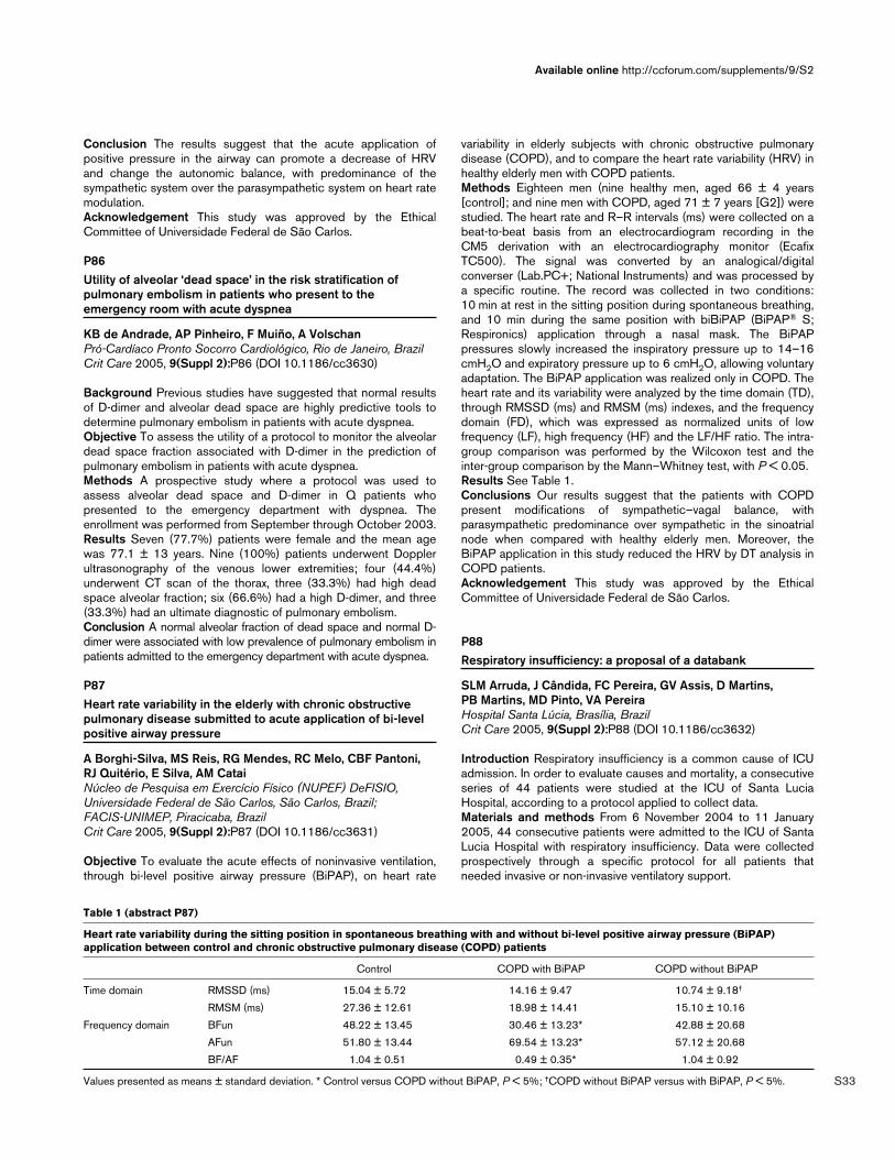
S33
Conclusion The results suggest that the acute application ofpositive pressure in the airway can promote a decrease of HRVand change the autonomic balance, with predominance of thesympathetic system over the parasympathetic system on heart ratemodulation.Acknowledgement This study was approved by the EthicalCommittee of Universidade Federal de São Carlos.
P86Utility of alveolar ‘dead space’ in the risk stratification ofpulmonary embolism in patients who present to theemergency room with acute dyspnea
KB de Andrade, AP Pinheiro, F Muiño, A VolschanPró-Cardíaco Pronto Socorro Cardiológico, Rio de Janeiro, BrazilCrit Care 2005, 9(Suppl 2):P86 (DOI 10.1186/cc3630)
Background Previous studies have suggested that normal resultsof D-dimer and alveolar dead space are highly predictive tools todetermine pulmonary embolism in patients with acute dyspnea.Objective To assess the utility of a protocol to monitor the alveolardead space fraction associated with D-dimer in the prediction ofpulmonary embolism in patients with acute dyspnea.Methods A prospective study where a protocol was used toassess alveolar dead space and D-dimer in Q patients whopresented to the emergency department with dyspnea. Theenrollment was performed from September through October 2003.Results Seven (77.7%) patients were female and the mean agewas 77.1 ± 13 years. Nine (100%) patients underwent Dopplerultrasonography of the venous lower extremities; four (44.4%)underwent CT scan of the thorax, three (33.3%) had high deadspace alveolar fraction; six (66.6%) had a high D-dimer, and three(33.3%) had an ultimate diagnostic of pulmonary embolism.Conclusion A normal alveolar fraction of dead space and normal D-dimer were associated with low prevalence of pulmonary embolism inpatients admitted to the emergency department with acute dyspnea.
P87Heart rate variability in the elderly with chronic obstructivepulmonary disease submitted to acute application of bi-levelpositive airway pressure
A Borghi-Silva, MS Reis, RG Mendes, RC Melo, CBF Pantoni,RJ Quitério, E Silva, AM CataiNúcleo de Pesquisa em Exercício Físico (NUPEF) DeFISIO,Universidade Federal de São Carlos, São Carlos, Brazil;FACIS-UNIMEP, Piracicaba, BrazilCrit Care 2005, 9(Suppl 2):P87 (DOI 10.1186/cc3631)
Objective To evaluate the acute effects of noninvasive ventilation,through bi-level positive airway pressure (BiPAP), on heart rate
variability in elderly subjects with chronic obstructive pulmonarydisease (COPD), and to compare the heart rate variability (HRV) inhealthy elderly men with COPD patients.Methods Eighteen men (nine healthy men, aged 66 ± 4 years[control]; and nine men with COPD, aged 71 ± 7 years [G2]) werestudied. The heart rate and R–R intervals (ms) were collected on abeat-to-beat basis from an electrocardiogram recording in theCM5 derivation with an electrocardiography monitor (EcafixTC500). The signal was converted by an analogical/digitalconverser (Lab.PC+; National Instruments) and was processed bya specific routine. The record was collected in two conditions:10 min at rest in the sitting position during spontaneous breathing,and 10 min during the same position with biBiPAP (BiPAP® S;Respironics) application through a nasal mask. The BiPAPpressures slowly increased the inspiratory pressure up to 14–16cmH2O and expiratory pressure up to 6 cmH2O, allowing voluntaryadaptation. The BiPAP application was realized only in COPD. Theheart rate and its variability were analyzed by the time domain (TD),through RMSSD (ms) and RMSM (ms) indexes, and the frequencydomain (FD), which was expressed as normalized units of lowfrequency (LF), high frequency (HF) and the LF/HF ratio. The intra-group comparison was performed by the Wilcoxon test and theinter-group comparison by the Mann–Whitney test, with P < 0.05.Results See Table 1.Conclusions Our results suggest that the patients with COPDpresent modifications of sympathetic–vagal balance, withparasympathetic predominance over sympathetic in the sinoatrialnode when compared with healthy elderly men. Moreover, theBiPAP application in this study reduced the HRV by DT analysis inCOPD patients.Acknowledgement This study was approved by the EthicalCommittee of Universidade Federal de São Carlos.
P88Respiratory insufficiency: a proposal of a databank
SLM Arruda, J Cândida, FC Pereira, GV Assis, D Martins, PB Martins, MD Pinto, VA PereiraHospital Santa Lúcia, Brasília, BrazilCrit Care 2005, 9(Suppl 2):P88 (DOI 10.1186/cc3632)
Introduction Respiratory insufficiency is a common cause of ICUadmission. In order to evaluate causes and mortality, a consecutiveseries of 44 patients were studied at the ICU of Santa LuciaHospital, according to a protocol applied to collect data.Materials and methods From 6 November 2004 to 11 January2005, 44 consecutive patients were admitted to the ICU of SantaLucia Hospital with respiratory insufficiency. Data were collectedprospectively through a specific protocol for all patients thatneeded invasive or non-invasive ventilatory support.
Available online http://ccforum.com/supplements/9/S2
Table 1 (abstract P87)
Heart rate variability during the sitting position in spontaneous breathing with and without bi-level positive airway pressure (BiPAP)application between control and chronic obstructive pulmonary disease (COPD) patients
Control COPD with BiPAP COPD without BiPAP
Time domain RMSSD (ms) 15.04 ± 5.72 14.16 ± 9.47 10.74 ± 9.18†
RMSM (ms) 27.36 ± 12.61 18.98 ± 14.41 15.10 ± 10.16
Frequency domain BFun 48.22 ± 13.45 30.46 ± 13.23* 42.88 ± 20.68
AFun 51.80 ± 13.44 69.54 ± 13.23* 57.12 ± 20.68
BF/AF 1.04 ± 0.51 0.49 ± 0.35* 1.04 ± 0.92
Values presented as means ± standard deviation. * Control versus COPD without BiPAP, P < 5%; †COPD without BiPAP versus with BiPAP, P < 5%.
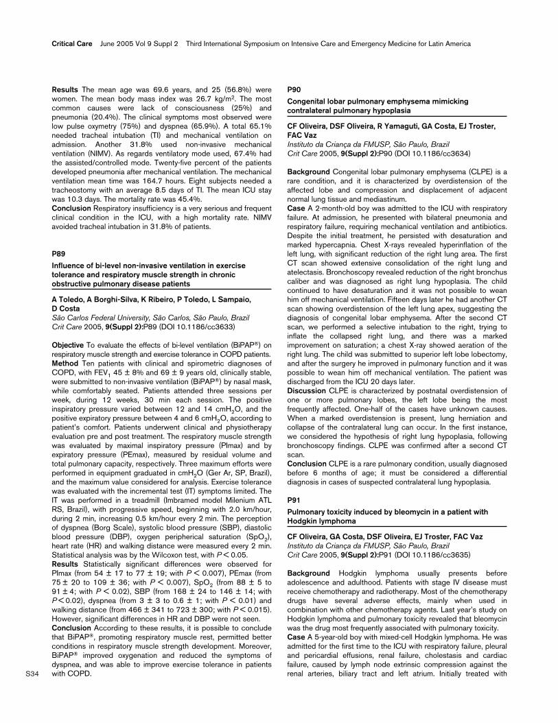
S34
Results The mean age was 69.6 years, and 25 (56.8%) werewomen. The mean body mass index was 26.7 kg/m2. The mostcommon causes were lack of consciousness (25%) andpneumonia (20.4%). The clinical symptoms most observed werelow pulse oxymetry (75%) and dyspnea (65.9%). A total 65.1%needed tracheal intubation (TI) and mechanical ventilation onadmission. Another 31.8% used non-invasive mechanicalventilation (NIMV). As regards ventilatory mode used, 67.4% hadthe assisted/controlled mode. Twenty-five percent of the patientsdeveloped pneumonia after mechanical ventilation. The mechanicalventilation mean time was 164.7 hours. Eight subjects needed atracheostomy with an average 8.5 days of TI. The mean ICU staywas 10.3 days. The mortality rate was 45.4%.Conclusion Respiratory insufficiency is a very serious and frequentclinical condition in the ICU, with a high mortality rate. NIMVavoided tracheal intubation in 31.8% of patients.
P89Influence of bi-level non-invasive ventilation in exercisetolerance and respiratory muscle strength in chronicobstructive pulmonary disease patients
A Toledo, A Borghi-Silva, K Ribeiro, P Toledo, L Sampaio, D CostaSão Carlos Federal University, São Carlos, São Paulo, BrazilCrit Care 2005, 9(Suppl 2):P89 (DOI 10.1186/cc3633)
Objective To evaluate the effects of bi-level ventilation (BiPAP®) onrespiratory muscle strength and exercise tolerance in COPD patients.Method Ten patients with clinical and spirometric diagnoses ofCOPD, with FEV1 45 ± 8% and 69 ± 9 years old, clinically stable,were submitted to non-invasive ventilation (BiPAP®) by nasal mask,while comfortably seated. Patients attended three sessions perweek, during 12 weeks, 30 min each session. The positiveinspiratory pressure varied between 12 and 14 cmH2O, and thepositive expiratory pressure between 4 and 6 cmH2O, according topatient’s comfort. Patients underwent clinical and physiotherapyevaluation pre and post treatment. The respiratory muscle strengthwas evaluated by maximal inspiratory pressure (PImax) and byexpiratory pressure (PEmax), measured by residual volume andtotal pulmonary capacity, respectively. Three maximum efforts wereperformed in equipment graduated in cmH2O (Ger Ar, SP, Brazil),and the maximum value considered for analysis. Exercise tolerancewas evaluated with the incremental test (IT) symptoms limited. TheIT was performed in a treadmill (Imbramed model Milenium ATLRS, Brazil), with progressive speed, beginning with 2.0 km/hour,during 2 min, increasing 0.5 km/hour every 2 min. The perceptionof dyspnea (Borg Scale), systolic blood pressure (SBP), diastolicblood pressure (DBP), oxygen peripherical saturation (SpO2),heart rate (HR) and walking distance were measured every 2 min.Statistical analysis was by the Wilcoxon test, with P < 0.05.Results Statistically significant differences were observed forPImax (from 54 ± 17 to 77 ± 19; with P < 0.007), PEmax (from75 ± 20 to 109 ± 36; with P < 0.007), SpO2 (from 88 ± 5 to91 ± 4; with P < 0.02), SBP (from 168 ± 24 to 146 ± 14; withP < 0.02), dyspnea (from 3 ± 3 to 0.6 ± 1; with P < 0.01) andwalking distance (from 466 ± 341 to 723 ± 300; with P < 0.015).However, significant differences in HR and DBP were not seen.Conclusion According to these results, it is possible to concludethat BiPAP®, promoting respiratory muscle rest, permitted betterconditions in respiratory muscle strength development. Moreover,BiPAP® improved oxygenation and reduced the symptoms ofdyspnea, and was able to improve exercise tolerance in patientswith COPD.
P90Congenital lobar pulmonary emphysema mimickingcontralateral pulmonary hypoplasia
CF Oliveira, DSF Oliveira, R Yamaguti, GA Costa, EJ Troster,FAC VazInstituto da Criança da FMUSP, São Paulo, BrazilCrit Care 2005, 9(Suppl 2):P90 (DOI 10.1186/cc3634)
Background Congenital lobar pulmonary emphysema (CLPE) is arare condition, and it is characterized by overdistension of theaffected lobe and compression and displacement of adjacentnormal lung tissue and mediastinum.Case A 2-month-old boy was admitted to the ICU with respiratoryfailure. At admission, he presented with bilateral pneumonia andrespiratory failure, requiring mechanical ventilation and antibiotics.Despite the initial treatment, he persisted with desaturation andmarked hypercapnia. Chest X-rays revealed hyperinflation of theleft lung, with significant reduction of the right lung area. The firstCT scan showed extensive consolidation of the right lung andatelectasis. Bronchoscopy revealed reduction of the right bronchuscaliber and was diagnosed as right lung hypoplasia. The childcontinued to have desaturation and it was not possible to weanhim off mechanical ventilation. Fifteen days later he had another CTscan showing overdistension of the left lung apex, suggesting thediagnosis of congenital lobar emphysema. After the second CTscan, we performed a selective intubation to the right, trying toinflate the collapsed right lung, and there was a markedimprovement on saturation; a chest X-ray showed aeration of theright lung. The child was submitted to superior left lobe lobectomy,and after the surgery he improved in pulmonary function and it waspossible to wean him off mechanical ventilation. The patient wasdischarged from the ICU 20 days later.Discussion CLPE is characterized by postnatal overdistension ofone or more pulmonary lobes, the left lobe being the mostfrequently affected. One-half of the cases have unknown causes.When a marked overdistension is present, lung herniation andcollapse of the contralateral lung can occur. In the first instance,we considered the hypothesis of right lung hypoplasia, followingbronchoscopy findings. CLPE was confirmed after a second CTscan.Conclusion CLPE is a rare pulmonary condition, usually diagnosedbefore 6 months of age; it must be considered a differentialdiagnosis in cases of suspected contralateral lung hypoplasia.
P91Pulmonary toxicity induced by bleomycin in a patient withHodgkin lymphoma
CF Oliveira, GA Costa, DSF Oliveira, EJ Troster, FAC VazInstituto da Criança da FMUSP, São Paulo, BrazilCrit Care 2005, 9(Suppl 2):P91 (DOI 10.1186/cc3635)
Background Hodgkin lymphoma usually presents beforeadolescence and adulthood. Patients with stage IV disease mustreceive chemotherapy and radiotherapy. Most of the chemotherapydrugs have several adverse effects, mainly when used incombination with other chemotherapy agents. Last year’s study onHodgkin lymphoma and pulmonary toxicity revealed that bleomycinwas the drug most frequently associated with pulmonary toxicity.Case A 5-year-old boy with mixed-cell Hodgkin lymphoma. He wasadmitted for the first time to the ICU with respiratory failure, pleuraland pericardial effusions, renal failure, cholestasis and cardiacfailure, caused by lymph node extrinsic compression against therenal arteries, biliary tract and left atrium. Initially treated with
Critical Care June 2005 Vol 9 Suppl 2 Third International Symposium on Intensive Care and Emergency Medicine for Latin America
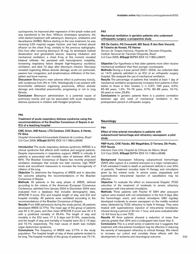
S35
cyclosporine, he improved after regression of the lymph nodes andwas transferred to the floor. Without cholestasis symptoms, thechild started treatment with adriamycin, bleomycin, vinblastine anddacarbazine (AVBD). Before starting on the new treatment, he wasreceiving 0.50 inspired oxygen fraction, and had minimal pleuraleffusion on the chest X-ray, similarly to the previous radiographs.One hour after receiving bleomycin (6 mg), he developed markeddesaturation and generalized seizure, requiring intubation andmechanical ventilation; a chest X-ray at this moment revealedbilateral infiltrate. He persisted with hemodynamic instability,worsening respiratory failure despite high-frequency oscillatoryventilation, and died 13 days after bleomycin infusion. Necropsyrevealed organizing diffuse alveolar damage, acute cor pulmonale,passive liver congestion, and lymphomatous infiltration of the liver,spleen and bone marrow.Discussion Bleomycin’s main adverse effect is pulmonary toxicity,with incidences from 3% to 10%. Histologically it can present withbronchiolitis obliterans organizing pneumonia, diffuse alveolardamage and interstitial pneumonitis, progressing or not to lungfibrosis.Conclusion Bleomycin administration is a potential cause ofpulmonary toxicity and can be associated with acute respiratorydistress syndrome in children with Hodgkin lymphoma.
P92Treatment of acute respiratory distress syndrome using therecommendations of the Brazilian Consensus of Sepsis in anICU of a teaching hospital
CMC Grion, IAM Kauss, LTQ Cardoso, CVC Bueno, A Herek, FV SilvaHospital Universitário/Universidade Estadual de Londrina, BrazilCrit Care 2005, 9(Suppl 2):P92 (DOI 10.1186/cc3636)
Introduction The acute respiratory distress syndrome (ARDS) is aclinical syndrome that affects both medical and surgical patients.Despite improvements in respiratory support, ARDS in critically illpatients is associated with mortality rates of between 40% and65%. The Brazilian Consensus of Sepsis has recently proposedventilation strategies that include low tidal volumes, high PEEPlevels and recruitment maneuvers to increase the homogeneity ofinflation of the lung.Objective To determine the frequency of ARDS and to describethe outcome adopting the recommendations of the BrazilianConsensus of Sepsis.Methods All patients in the early phase of ARDS, definedaccording to the criteria of the American–European ConsensusConference, admitted from January 2004 to December 2004 wereevaluated from a database that includes demographic data,APACHE II score, ICU and hospital length of stay, and ICU andhospital mortality. All patients were ventilated according to therecommendations of the Brazilian Consensus of Sepsis.Results From 938 admissions during the study period, 26 patientsdeveloped ARDS (2.77%). The mean age of this group of patientswas 50 ± 22 years, and their mean APACHE II score was 20 ± 6with a predicted mortality of 35.4%. The length of stay andmortality in the ICU were 17 ± 3 days and 61.5%, respectively,and the length of stay and mortality in hospital were 22 ± 13 daysand 73.1%, respectively. The main cause of death was multipleorgan dysfunction syndrome.Conclusions The frequency of ARDS was 2.77% in the studypopulation. The hospital length of stay of these patients tended tobe long. The hospital mortality of this group of patients was 73.1%.
P93Mechanical ventilation in geriatric patients who underwentorthopaedic surgery: a prospective study
IRS De Castro, PG Coscarelli, MD Saud, R dos Reis Visconti, M Teixeira de Novais, FS ViannaServiço de Terapia Intensiva, Hospital de Tráumato-Ortopedia,Instituto Nacional de Tráumato-Ortopedia, BrazilCrit Care 2005, 9(Suppl 2):P93 (DOI 10.1186/cc3637)
Objective Our hypothesis is that older patients more often receivemechanical ventilation than their younger counterparts.Methods During a 4-year period (2001–2004), we collected dataon 1410 patients admitted to an ICU of an orthopedic surgeryhospital. We analyzed the use of mechanical ventilation.Results The percentage of patients that needed at least 1 day ofmechanical ventilation progressively increases from patients in theirsixties to those in their nineties (r = 0.97): <65 years, 3.5%;65–69 years, 1.2%; 70–79 years, 3.7%; 80–89 years, 16.1%;90 years or more, 25.8%.Conclusions In geriatric patients there is a positive correlationbetween age and need of mechanical ventilation in thepostoperative period of orthopedic surgery.
Neurology
P94Effect of intra-arterial nimodipine in patients withsubarachnoid hemorrhage and refractory vasospasm: a pilotstudy
PMP Kurtz, CHE Falcão, MD Magalhães, D Terrana, DA Prado,PCP SouzaIntensive Care Unit, Hospital de Clínicas de Niteroi, BrazilCrit Care 2005, 9(Suppl 2):P94 (DOI 10.1186/cc3638)
Background Vasospasm following subarachnoid hemorrhage(SAH) after rupture of a cerebral aneurysm is a major complication.If left untreated it leads to death or permanent deficits in over 20%of patients. Treatment includes triple H therapy and nimodipinegiven by the enteral route. In severe cases, angioplasty andsuperselective intra-arterial injection of vasodilators may beeffective.Objective To evaluate the effect on transcranial Doppler (TCD)velocities of the treatment of moderate to severe refractoryvasospasm with intra-arterial nimodipine.Methods Three patients with Fischer IV SAH after aneurysmrupture were treated with coil embolization in the first 3 days afterthe hemorrhagic event, and enteral nimodipine (360 mg/day)developed moderate to severe vasospasm on the middle cerebralartery (detected by TCD) refractory to triple H therapy. They weretreated with superselective injection of intra-arterial nimodipine,infused during a period of 30 min–1 hour, and were evaluated after12–24 hours by a new TCD.Results All three patients showed a reduction of mean flowvelocity greater than 30% and a Lindergaard ratio lower than 3.Conclusions This is a small series of patients but it suggests thattreatment with intra-arterial nimodipine may be effective in reducingthe severity of vasospasm refractory to clinical therapy. We intendto increase our cohort and correlate these effects with thedevelopment of ischemia and neurological outcome.
Available online http://ccforum.com/supplements/9/S2
S36
P95Cerebrovascular autoregulation, CO2 reactivity and outcome inpatients with severe traumatic brain injury: a pilot study
PMP Kurtz, MD Magalhães, D Terrana, DA Prado, PCP SouzaIntensive Care Unit, Hospital de Clínicas de Niteroi, BrazilCrit Care 2005, 9(Suppl 2):P95 (DOI 10.1186/cc3639)
Background Impaired cerebral autoregulation is frequent aftersevere traumatic head injury, and impaired CO2 reactivity may beassociated with poor outcome.Objective To evaluate the incidence, asymmetry and relation tooutcome of impaired autoregulation and CO2 reactivity.Methods Five patients with severe head injury were prospectivelyassessed in the first 24 hours after trauma for the presence ofimpaired autoregulation and CO2 reactivity. Four patients weremonitored for invasive arterial blood pressure (ABP), intracranialpressure, cerebral perfusion pressure (CPP), jugular bulb oxymetryand transcranial Doppler (TCD) flow velocities and pulsatility index.One patient was monitored only for ABP and TCD parameters.Autoregulation was evaluated by flow velocity response to CPPand ABP changes induced by noradrenaline infusion. The CO2reactivity was assessed by flow velocity response to ventilatormanipulation of the minute volume. Outcome was assessed withGlasgow Outcome Scale at discharge from hospital and after3 months.Results Autoregulation was globally impaired in three of the fivepatients and impaired in one hemisphere in one patient. CO2reactivity was globally impaired in one patient and impaired in onehemisphere in one patient. Three patients died, two of themprobably associated with the severity of brain injury and one ofseptic shock. The two other patients are still in the hospital.Conclusion We present here a small series of patients, but theresults suggest that impaired autoregulation is indeed common inthese patients, which may have implications in therapy. We intendto increase our cohort and to correlate autoregulation and CO2reactivity with outcome soon.
P96Protein S-100b as a screening tool for the need of head CTscan after minor head trauma
LF Poli de Figueiredo, C Simao Filho, C Hauser, W Mutschler,P BiberthalerDepartment of Surgery, Federal University of São Paulo, Brazil;Emergency Department, João XXIII Hospital, Belo Horizonte, Brazil;Department of Surgery and Traumatology, Ludwig-Maximilians-University, Munich, GermanyCrit Care 2005, 9(Suppl 2):P96 (DOI 10.1186/cc3640)
Introduction Systematic use of head CT scan after minor headtrauma may lead to a high incidence of negative examinations andhigh costs. On the other hand, undetected intracranial lesions maybe life threatening. Neuronal protein S-100b into the circulation hasbeen suggested as a specific indication of neuronal damage. Wetested the hypothesis that protein S-100b is a useful and cost-effective screening tool for the management of minor head traumas.Methods Fifty consecutive patients sustaining isolated minor headtrauma were prospectively evaluated in the emergency room byroutine head CT scan and blood sampling for protein S-100bmeasurement, using an immunoluminescence test kit. Seventeennormal healthy individuals served as negative controls. Data arepresented as the median and 25th–75th percentiles.Results Patients reached the emergency room 45 min(30–62 min) after minor head trauma. Six patients had relevant
post-traumatic lesions at the initial head CT (12%) and werethereby counted as positive (CT+). The median systemic concen-tration of S-100b in those patients was 0.75 µg/l (0.61–6.5 µg/l),which was significantly different (U-test, P = 0.011) from themedian concentration, 0.26 µg/l (0.12–0.65 µg/l), of those withoutpost-traumatic lesions in the initial head CT scan (CT–). Asensitivity of 100%, a specificity of 20%, a positive predictive valueof 15% and a negative predictive value of 100% were detected.Conclusions Protein S-100b has a very high sensitivity andnegative predictive value, and could have an important role in rulingout the need for CT scan after minor head trauma. This may be ofclinical relevance, particularly in countries in which trauma isepidemic and medical resources are limited, such as in Brazil.Acknowledgements This work was supported by CAPES and theDeutsche Forschungs-Gemeinschaft, Sonderforschungs-bereich469 of the Ludwig-Maximilians-University Munich.
Nutrition/metabolism
P97The effects of enteral feeding with eicosapentaenoic acid,gamma-linolenic acid and antioxidants in patients with sepsis
A Pontes-ArrudaHospital Antônio Prudente, Fortaleza, Ceará, BrazilCrit Care 2005, 9(Suppl 2):P97 (DOI 10.1186/cc3641)
Introduction Nutritional support with diets containing eicosa-pentaenoic acid (EPA), gamma-linolenic acid (GLA) and anti-oxidants can improve lung microvascular permeability, oxygenationand cardiopulmonary function, by modulating proinflammatoryeicosanoid (leukotriene B4, prostaglandin E, thromboxane B2)production from arachdonic acid [1]. This diet can improve gasexchange and clinical outcomes in comparison with a standardcontrol diet in patients with ARDS [2]. This study investigateswhether an EPA + GLA + antioxidant enriched enteral diet canimprove outcomes and reduce mortality in patients with sepsis.Materials and methods This are the preliminary results of aprospective, randomized, controlled trial. Thirty patients withclinical diagnosis of either sepsis, severe sepsis or septic shockand under mechanical ventilation were enrolled in this study andwere randomized for a high-fat, low-carbohydrate enteral nutritionformula or an enteral diet enriched with EPA + LGA + antioxidants(Oxepa; Abbott Laboratories). In association with sepsis standardsof care, patients received enteral formula during mechanicalventilation. In all patients included in this study, enteral feeding wasdelivered at a constant rate to achieve a minimum of 50% of BasalEnergy Expenditure (BEE) (Harris–Benedict equation) × 1.3 withinthe first 24 hours and, if tolerated, a minimum of 75% of BEE × 1.3within 72 hours of initiation of enteral feeding until completeweaning from the ventilator.Results Septic patients fed EPA + GLA + antioxidants maintainedhigher oxygenation status (P = 0.001), more ventilator-free days(P = 0.001), more ICU-free days (P = 0.02) and lower mortalityrates (P = 0.03).Conclusion This study suggests that an enteral diet containingEPA, GLA and elevated antioxidants helps to downregulate thesynthesis of proinflammatory mediators and contributes to restorehomeostasis of the septic patient. The beneficial effects of this dietsuggest that this enteral nutrition formula would be a usefuladjuvant therapy in the clinical management of sepsis.References1. Pacht, et al.: Crit Care Med 2003, 31:491-500.2. Gadek, et al.: Crit Care Med 1999, 27:1409-1420.
Critical Care June 2005 Vol 9 Suppl 2 Third International Symposium on Intensive Care and Emergency Medicine for Latin America
S37
P98How long does it takes to begin nutrition support in the ICU?
C Belo, MG RodriguesQuinta D’or Hospital, Rio de Janeiro, BrazilCrit Care 2005, 9(Suppl 2):P98 (DOI 10.1186/cc3642)
Introduction Nutritional support is very important in critically illpatients and many studies have been performed trying to optimizethe nutritional therapy and to minimize the muscular catabolism ofthis group of patients.Objective The present study demonstrated how long it takes tobegin and to complete nutritional support in critically ill patients inthe ICU, compared with clinical and surgical patients, and analyzedthe complications and the causes of discontinuation of nutritionalsupport in the ICU.Materials and methods A prospective analysis of 85 patients (32women and 53 men), 68 clinical and 17 surgical diagnoses,admitted to a 41-bed ICU from July 2004 to February 2005. Thepresent study analyzed the time to begin and to completenutritional support, the reason for discontinuation, thecomplications and the position of the catheter to provide thenutritional support.Results The patient APACHE II score was 16.6 and 17 forsurgical and clinical patients, respectively. The time to beginnutrition support was 3.52 and 2.1 days for surgical and clinicalpatients, respectively. The clinical patients had a gastric catheter in51 patients and a catheter in the jejune position in 17 patients. Thediet used was polymeric in 60 patients and oligomeric in eightpatients; discontinuation of nutrition occurred in nine cases(13.4%) (four mesenteric ischemia, three hemodynamic instability,one oral nutrition and one gastrointestinal hemorrhage), anddiarrhea occurred in 20 patients (29.41%), but it was auto limitedand the average was 1.61 episodes/day. Gastroparesis occurredin 14 patients (20.6%) but all of them except one resolved in 1 daywithout needing a change of the catheter position and withoutrelation to the catheter position. Eighteen patients died (26.47%).The surgical patients had a gastric catheter in 10 patients and acatheter in the jejune position in seven patients; discontinuation ofnutrition occurred in three cases (17.64%) (two mesentericischemia, one hemodynamic instability), and diarrhea occurred ineight patients (47%), but it was auto limited and the average was3.05 episodes/day. Gastroparesis occurred in four patients(23.5%) but all of them resolved in 1 day without needing tochange the catheter position; all of them were related tohemodynamic instability. Four patients died (23.5%).Conclusion The time to beginning nutrition support was shorter inthe clinical group compared with the surgical group in spite ofcomplete nutritional support in surgical patients being shorter. Wedid not observe too many complications and the majority receivedthe nutritional support proposed. Diarrhea was more prevalent inthe surgical group but was not involved in discontinuation ofnutrition. The mortality was similar in both groups.
P99Plasma amino acids in critically ill patients
MG Rodigues, F Ruzany, DR Salgado, E Rocha, CF Valente, E Maccariello, RNA PaivaHospital Universitário Pedro Ernesto/Hospital Barra D’or, BrazilCrit Care 2005, 9(Suppl 2):P99 (DOI 10.1186/cc3643)
Introduction Stress states, sepsis and trauma markedly increaseprotein catabolism in skeletal muscle, gut, and connective tissue.
Catabolism is potentiated by a sepsis-induced decrease in aminoacid uptake in skeletal muscle. One-third of the total amino acidoutput from skeletal muscle is glutamine and one-third is alanine.Objective To demonstrate the concentration of plasma aminoacids in critically ill patients.Materials and methods A prospective analysis of 41 patients (17women and 24 men) admitted to a 27-bed ICU from July 2002 toMay 2003. Twenty-three amino acids were analysed in eachpatient up to 24 hours of admission in the ICU. The amino acidassays, using high-pressure liquid chromatography, wereperformed by laboratory CTN and Pardini.Results The average patient age was 73.08 years (43–88 years)and the APACHE II score was 18.37 (11–28). Plasma amino acidswere obtained in 41 patients and 23 amino acids were analysed(µmol/l). The total value of plasma amino acids was4257.475 µmol/l (average). The doses of plasma amino acidswere: aspartic acid 75.95, glutamic acid 384.45, alanine 587.1,arginine 84.45, asparagine 78.5, cystine 497.55, phenylalanine179.2, glycine 497.55, glutamine 413.25, hydroxyproline 58.25,histidine 84.55, isoleucine 90.325, leucine 195.5, lysine 190.05,methionine 48.85, ornithine 118.65, proline 201.5, serine 156.75,taurine 167.25, tyrosine 127.85, threonine 167, tryptophan51.925, and valine 278.975.Conclusion The concentrations of alanine, glutamic acid, asparticacid, phelalanine and arginine were more elevated than the otheramino acids. Glutamine was demonstrated to be decreased,probably, as an essential amino acid in sepsis or catabolic patients,greatly consumed.
P100Amino acid adherence in continuous renal replacementtherapies
MG Rodrigues, F Ruzany, DR Salgado, E Rocha, CF Valente, E Maccariello, RNA PaivaHospital Universitário Pedro Ernesto/Hospital Barra D’or, BrazilCrit Care 2005, 9(Suppl 2):P100 (DOI 10.1186/cc3644)
Introduction Continuous renal replacement therapies (CRRT)readily allow for the nutritional support of these high catabolicstates, but also contribute to the nitrogen loss through filtration offree amino acids and small peptides across the hemofilters, andpossible adherence of amino acids.Objective To evaluate amino acid adherence in CRRT.Materials and methods Paired samples from serum, venous anddialysate/ultrafiltrate were obtained during CRRT procedures from41 patients in a 27-bed ICU from July 2002 to May 2003. Pairedsamples were collected from 1, 6, 12, 24, 36 and 48 hours ofcontinuous hemodialysis, a total of six samples of serum, venousand dialysate. PAN 650 filters were used in all patients. The bloodflow was 150 ml/min in all patients and the dialysate flow duringCVVHD was 16.6 ml/min.Amino acid assays, using high-pressure liquid chromatographyserum, venous and dialysate, were performed by laboratory CTNand Pardini. Adherence was calculated as: clearance hemodialysis(CH) / serum clearance (SC); CH = (16.6 × dialysis) / serumamino acids; SC = 150 × (serum amino acids – venous aminoacids) / dialysis amino acids.Results The average patient age was 73.08 years (43–88 years)and the APACHE II score was 18.37 (11–28). Serum, venous anddialysate amino acids were obtained in 41 patients and 23 aminoacids were analysed (µmol/l): aspartic acid, glutamic acid, alanine,
Available online http://ccforum.com/supplements/9/S2
S38
arginine, asparagine, cystine, phenylalanine, glycine, glutamine,hydroxyproline, histidine, isoleucine, leucine, lysine, methionine,ornithine, proline, serine, taurine, tyrosine, threonine, tryptophan,valine. Analyses were performed on the average of the aminoacids. Adherence was not statistically significant, and did notcorrelate with time of hemodialysis or with serum amino acids.Statistical calculations were made by analysis of variance, linear,nonlinear and logarithmic regression: glutamine (R2 = 0.978691062),alanine (R2 = 0.575243703), taurine (R2 = 0.612666425), valine(R2 = 0.743575525).Conclusion We should not estimate the protein necessity in aseptic or trauma patient on CHVVD based exclusively on the aminoacid loss. Other factors, such as the adsorption of amino acids,may contribute to the continuous catabolism seen in these criticallyill patients.
P101Glutamine serum, adherence and losses in continuous renalreplacement therapies in critically ill patients
MG Rodigues, F Ruzany, DR Salgado, E Rocha, CF Valente, E Maccariello, RNA PaivaHospital Universitário Pedro Ernesto/Hospital Barra D’or, Rio deJaneiro, BrazilCrit Care 2005, 9(Suppl 2):P101 (DOI 10.1186/cc3645)
Introduction Continuous renal replacement therapies (CRRT)readily allow for the nutritional support of these high catabolicstates, but also contribute to the nitrogen loss through filtration offree amino acids and small peptides across the hemofilters. Aminoacid clearances and calculated losses in adults on continuousvenovenous hemofiltration (CVVHD) have been reported in therange of 2–11% of dietary intake. Glutamine is an very importantamino acid in critically ill patients.Objective To analyse glutamine serum, losses and adherence fromCRRT treatments in critically ill patients.Materials and methods Paired samples from serum, venous anddialysate/ultrafiltrate were obtained during CRRT procedures from41 patients in a 27-bed ICU from July 2002 to May 2003. Pairedsamples were collected from 1, 6, 12, 24, 36 and 48 hours ofcontinuous hemodialysis, a total of six samples of serum, venousand dialysate. PAN 650 filters were used in all patients. The bloodflows was 150 ml/min in all patients and the dialysate flow duringCVVHD was 16.6 ml/min. Amino acid assays, using high-pressureliquid chromatography serum, venous and dialysate, wereperformed by laboratory CTN and Pardini.Results The average patient age was 73.08 years (43–88 years)and the APACHE II score was 18.37 (11–28). Serum, venous anddialysate amino acids were obtained in 41 patients and glutaminewas analysed (µmol/l). Analyses were performed on the average ofamino acids. Statistical calculations were made by analysis ofvariance, linear, nonlinear and logarithmic regression.Conclusion Glutamine is very important in critically ill patients.Glutamine was demonstrated to be decreased, probably, as anessential amino acid in sepsis or catabolic patients, greatlyconsumed. We should not estimate the protein necessity in aseptic or trauma patient on CHVVD based exclusively on the aminoacid loss. Other factors, such as the adsorption of amino acids,may contribute to the continuous catabolism seen in these criticallyill patients.
P102Results of an intensive insulin infusion protocol in diabetic andnondiabetic critically ill patients
PN Gomes, JLF Costa, JRB Martins, F Gutierrez, L Mousalem, S Simões, SEM Pereira, AMF Mesquita, LS Araújo, M Rosemblat, RC CostaHospital Pró-Cardíaco, Rio De Janeiro, BrazilCrit Care 2005, 9(Suppl 2):P102 (DOI 10.1186/cc3646)
Introduction The implementation of an intensive insulin infusionprotocol (IIIP) has recently been shown to improve results in thecritically ill.Methods We retrospectively reviewed the clinical charts andspecific nutritional records from the Nutrition Support Team of all thepatients admitted to the ICU and submitted to an IIIP during 1 year (1January–31 December 2004). On all patients we collected:demographics (age, sex), maximum SOFA score, clinicalcharacteristics (diabetes mellitus, glucose levels above 130 mg/dl orbelow 60 mg/dl) and risk factors to hyperglycemia (drugs, infection).Results Over the study period, 37 patients received IIIP. All thesepatients presented infection and were submitted to mechanicalventilation. Twelve patients were diabetic and 25 were nondiabetic.A total of 10,239 glucose estimations were performed during444 days. The demographics were well matched. The maximumSOFA score was bigger in the diabetic group (10 ± 2 vs 8 ± 3).We found 32% of hyperglycemic levels and 0.28% of hypo-glycemia in the whole population. In the diabetic group, theincidence of hyperglycemia and hypoglycemia were, respectively,37% and 0.44%, while in the nondiabetic group they were 29%and 0.21%.Discussion The literature provides similar trends in the safetyprofile of IIIP in the critically ill. It seems necessary to observe thedifferences between diabetic and nondiabetic patients.References1. Van den Berghe G, Wouters P, Weekers F, et al.: N Engl J Med
2001, 345:1359-13672. Krinsley JS: Mayo Clin Proc 2004, 79:992-1000.
P103Enteral nutrition in septic shock in the elderly: do the waitingtime to start and the achieved basal energy expenditureinterfere with mortality?
PH Godoy, GMM Oliveira, EB Lameu, MR Pantoja, RR Luiz, CC Seneghini, M BandãoProntocor Lagoa, Federal University of Rio de Janeiro, BrazilCrit Care 2005, 9(Suppl 2):P103 (DOI 10.1186/cc3647)
Background The earliest starting point of enteral nutrition (EN), aswell as the basal energy expenditure (BEE), achieved within asshort a waiting time as possible seems to benefit quite a largenumber of critical patients.Objectives To analyze when to start EN and the BEE achieved inthe elderly hospitalized with a septic shock diagnosis, and to verifyits association with mortality in these patients.Methods A prospective cohort conducted within 32 months andwith 67 patients over 65 years old in the ICU with septic shock,where 59 of these patients had EN. The APACHE II score wasdetermined in every patient. The following variables were analyzedin this group: the time taken to start the NE, the BEE (achieved ornot), and the time to reach the BEE in those who had it. Thesevariables were correlated with death and it was still observedwhether there was correlation between the starting time point of
Critical Care June 2005 Vol 9 Suppl 2 Third International Symposium on Intensive Care and Emergency Medicine for Latin America
S39
EN and the achieved BEE. The statistical tests used were the t testand the chi-square test, considering 5% as the significance level.Results The average age was 80 ± 7 (minimum = 66, maximum =96) years, the APACHE II average score was 18 ± 5 (minimum =8, maximum = 28), and the time average to start EN was 80 ± 53hours (minimum of 12 hours and maximum of 240 hours). Theachieved BEE occurred in 69.5% (n = 41) of the patients and thetime average to reach it was 115 ± 56 hours (minimum of 72 hoursand maximum of 360 hours). Death was associated with time tostart EN (P = 0.001) and with the non-achieved BEE (P < 0.001).However, there was no correlation with time to reach BEE(P = 0.22). The time to start EN did not show association with theachieved BEE (P = 0.08).Conclusions Initiating EN as soon as possible and the BEE whenachieved in this group of patients showed some benefit. The timeto achieve the BEE does not seem to have correlation withmortality in these patients. The time to start NE did not haveassociation with the BEE achieved in this sample.
Nursing
P104Peripherally inserted central catheter in critically ill patients
LCR Lamblet, LR Guastelli, DF Moura Jr, M Alves, AC Bittencourt, APP Teixeira, E KnobelHospital Israelita Albert Einstein, São Paulo, BrazilCrit Care 2005, 9(Suppl 2):P104 (DOI 10.1186/cc3648)
Introduction The use of the vascular catheters is one of the mostimportant and required interventions in ICUs. One of these optionsis the peripherally inserted central catheter (PICC), which isinserted by habilitated nurses in patients who are in theseconditions in the ICU.Background and objectives To evaluate the use of this type ofcatheter in an adult ICU in the city of São Paulo, to checkindication, insertion, maintenance and its complication rate.Methods A prospective and descriptive study performed during 1year from March 2003 to March 2004. From 89 evaluations, 40PICC were inserted.Results The main indication for the use of PICC was theadministration of antibiotics, followed by difficulty in venous accessand the administration of medications that act in the vascularsystem. Eighty-five percent of the catheters were used in the Semi-ICU. The great majority of patients were taken off the catheter justafter the completion of the treatment (85%). There were two casesof phlebitis, three cases in which the catheter was accidentallyremoved and one case of obstruction.Conclusions The PICC has importance and application inintensive therapy, being one more therapeutic option, with a lowrange of mechanical and infectious complications. It is necessaryfor institutional training to have adequate maintenance andmanipulation.References1. O’Grady NP, Alexander M, Dellinger EP, et al.: Guidelines for the
prevention of intravascular catheter-related infections.Centers for Disease Control and Prevention. MMWR RecommRep 2002, 51:1-29.
2. Black IH, Blosser AS, Murray WB: Central venous pressuremeasurements: peripherally inserted catheters versus cen-trally inserted catheters. Crit Care Med 2000, 18:3833-3836.
3. Griffiths VR, Philpot P: Peripherally inserted central catheters(PICCs): do they have a role in the care of the critically illpatient. Intensive Crit Care Nurs 2002, 18:37-47.
P105Nasoenteral feeding tube placement: indications and nurseskills
SCPL Shiramizo, PE Almeida, LR Guastelli, M Durão Jr, DF Moura Jr, OFP SantosHospital Israelita Albert Einstein, São Paulo, BrazilCrit Care 2005, 9(Suppl 2):P105 (DOI 10.1186/cc3649)
Introduction Nutrition therapy (NT) is a powerful tool to assist thepatients’ nutritional needs mainly in the ICU, where the patientsusually have high energy expenditures. The route mostly used forNT is the nasoenteral feeding tube (NFT), being easier and withlower costs. It can be accomplished by the nursing staff underdirect vision by the nasal route (after training) or throughendoscopy. The nasoenteral insertion is not free fromcomplications, and these can be classified as: mechanics(obstruction of the probe, accidental exit, migration of the tube),infectious and psychological (anxiety, depression). The NFT issuitable for small and medium periods of NT (about 6 weeks).Objectives To classify the indication of the NFT in the ICU; toidentify the complications and the professional that accomplishedthe insertion.Results During the period from January 2005 to 23 February 2005we studied 83 patients receiving NT, 49% of them male.Successful first introduction was achieved in 35 patients (42%).The indication of NFT was NT in 98% of cases: 58% of them werereplaced, and the reasons were 16 obstructions of the tube (33%),17 accidental exits (35%) and 15 migrations (32%). The nursingstaff accomplished the tube insertion 80% of times. There were5% with complications; all of them were nasal bleeding,considered a minor complication.Conclusion The main reason for NFT insertion was NT initiation.Nursing staff have great capability for tube placement and weidentify clear requests for improvement interventions to avoid alarge number of tube replacements.
Epidemiology/quality of life/administration
P106Electrocardiographic analysis for telemedicine based on alow-cost software approach
LK Pimenta Cury1, PH Garcia-Mansur1, JB Destro-Filho1, E Santos-Resende2, W Salomão-Rocha2
1School of Electronic Engineering, Federal University of Uberlandia,Campus Santa Monica, Uberlandia, Brazil; 2UniversityHospital/Federal University of Uberlandia, BrazilCrit Care 2005, 9(Suppl 2):P106 (DOI 10.1186/cc3650)
Introduction Trends in telemedicine applied for clinical cardiologyinvolve techniques such as the transmission of Holter monitoringrecordings, as well as of electrocardiographic (ECG)measurements in the ICU. Both techniques enable more efficientclinical procedures, especially in the case of acute myocardialinfarction [1]. However, this transmission is a complex task, sincedifferent file formats must be handled by the system, thus requiringconversion from one format to the other.Objective To propose a low-cost software tool for the analysis ofECG recordings of different file formats, which enable processingof data measured in the ICU or arising from telemedicine systems.Materials and methods Twenty ECG recordings, taken fromacute myocardial infarction patients by a telemedicine system,were stored as .pdf files. We have considered just the derivations
Available online http://ccforum.com/supplements/9/S2

S40
Avf, II and III. In the following, they were analysed by our softwaretool to estimate the variation of the area of the ST segment. First,the .pdf files were converted to eight-bit .bmp files, as images with2000 × 1600 pixel resolution, in a grey scale. Axes are defined inthe image to extract a data file, which may be converted into anydesired format. This data file was processed by two differentmethods, in order to estimate the variation of the ST-segment area.The first method employs vertical axis scanning whereas thesecond method carries out standard integration, based on a finite-interval technique.Results The two methods presented the following average errorsfor the estimation: 1.80% for derivation Avf, 1.27% for derivation IIand 1.38% for derivation III. Average variances were 7000 formethod 1 (axis scanning) and 7200 for method 2 (standardintegration).Conclusion Simple and cost-effective methods for the analysis ofECG recordings arising from a telemedicine system, which may beused in current computer configurations in the ICU, weresuccessfully employed to estimate the variation of the ST-segmentarea for myocardial infarction patients. Method 1 presents thelowest variance.Reference1. Heilbron EL: Advances in modern electrocardiographic equip-
ment for long-term ambulatory monitoring. Cardiac Electro-physiol Rev 2002, 6:185-189.
P107Resuscitative status of patients during their stay in the ICU:changing decision-making to end-of-life care
MR Erlichman, A Caiuby, ALM Silva, LFA Camargo, RGR Cal, A Taub, PBA Andreoli, FP Almeida, E KnobelInstituto Israelita de Ensino E Pesquisa Albert Einstein, IntensiveCare Unit, Albert Einstein Hospital, São Paulo, BrazilCrit Care 2005, 9(Suppl 2):P107 (DOI 10.1186/cc3651)
Objective To verify whether the status of treatment in terms ofresuscitative effort changes during the stay of critical patients inintensive care.Methods We retrospectively analyzed 2108 patients admitted tothe ICU from 1 January 2003 to 31 December 2003. During thisperiod, the resuscitative status (no limitation of care, do notresuscitate and comfort care) at the moment of admission and inthe morning medical visit on the day of death were recorded andcompared. Numerical variables were compared using the Student ttest and categorical variables with the chi-square test. A level ofsignificance of 5% was required to consider some difference assignificant.Results Death occurred in 204 (9.7%) patients and dead patientswere older (69.2 ± 17.6 vs 63.1 ± 18.5 years, P < 0.001) and hada longer stay in the ICU (45 ± 13.8 vs 4 ± 8.3 days, P < 0,001)than the total admission patients. The APACHE score was notdifferent (20.1 vs 21.2, P = 0.06). The resuscitative status ispresented in Table 1.
Table 1
Resuscitative status at admission and on the day of death
Admission Death Resuscitative status (n = 2108) (n = 204) P
No limitation of care 2094 (99.3%) 103 (50.5%) <0.001
Do not resuscitate 2 (0.1%) 12 (5.9%)
Comfort care 12 (0.6%) 89 (43.6%)
Conclusion The change of the resuscitative status from admissionto the day of death may reflect a change in the medical decision,involving the families, to end-of-life care, limiting measures thatprolong life after failure of intensive support.
P108Analysis of the stressful factors in an ICU
LC Albuquerque, AM Almeida, AGV Bitencourt, RM Vieira de Mello, AM Farias, AS Souza Filho, OH Messeder,JM Teles, M DantasHospital Português, Salvador, Bahia, BrazilCrit Care 2005, 9(Suppl 2):P108 (DOI 10.1186/cc3652)
Background The hospital atmosphere, notably in the ICU, submitsthe patient to psychological experiences that frequently influencethem after ICU discharge. It is important to identify the stressfulfactors contributing to the development of this phenomenon.Objectives To identify and to stratify the stressful factors, in thepatient’s perspective, their relatives and the health careprofessionals, at an ICU.Methods A descriptive cross-sectional survey, carried out betweenJune and November 2004 in a general ICU of a private hospital. Thesample was composed of three groups: group 1 (G1) was formed bypatients, group 2 (G2) by patient’s relatives and group 3 (G3) by ICUhealth care personnel responsible for the included patients. In orderto identify and stratify the stressful factors, we used the Intensive CareUnit Environmental Stressor Scale (ICUESS), composed of 40assigned items scaled 1 (not stressful) to 4 (very stressful). The threegroups answered in agreement with their perception of stressfulfactors for the patient. For each individual, a Total Stress Score (TSS)was calculated from the sum of all the answers of the scale.Results Thirty individuals were included in each group. The meanage of the three groups was: 57.30 ± 17.61 years for G1;41.43 ± 12.19 for G2; and 40.82 ± 20.20 for G3. The mean TSSwas 62.63 ± 14.01 for the patients; 91.10 ± 30.91 for the relatives;and 99.30 ± 21.60 for the health care professionals. A larger TSSwas associated with patients without religion, that have moreexpensive health insurance and those that had attended university.Among the relatives, the parents had larger values of TSS. Therespiratory therapists were the professionals that obtained a largerTSS and the smallest average was found among the doctors. Theaverage of the patients’ TSS was lower than that of the relatives andthat of the health care professionals. There was no statisticallydifference between the latter two groups (family and professionals).Conclusion The perception of the main stressful factors wasdifferent among the three groups, especially when patients werecompared with the relatives and health care professionals. Thesegroups (family and professionals) believe that some factors are morestressful for the patient than the patient themself would have stated.
P109Intensive care medicine teaching in a Brazilian medicalschool: the student’s perspective
LC Albuquerque, AM Almeida, AGV Bitencourt, TM Godinho,MV Liberato, FC Cabral-Oliveira, ABG Azevedo, CEC Rolim,APSS Neves, MJ Martins, JPM Silva, PN Rocha, JM Telles, PAJ Santos, NM Filgueiras Filho, SA Souza FilhoUniversidade Federal da Bahia, Faculdade de Medicina da Bahia,Salvador, BrazilCrit Care 2005, 9(Suppl 2):P109 (DOI 10.1186/cc3653)
Introduction Several medical schools do not have a specificdiscipline for the teaching of intensive care medicine (ICM) topics
Critical Care June 2005 Vol 9 Suppl 2 Third International Symposium on Intensive Care and Emergency Medicine for Latin America
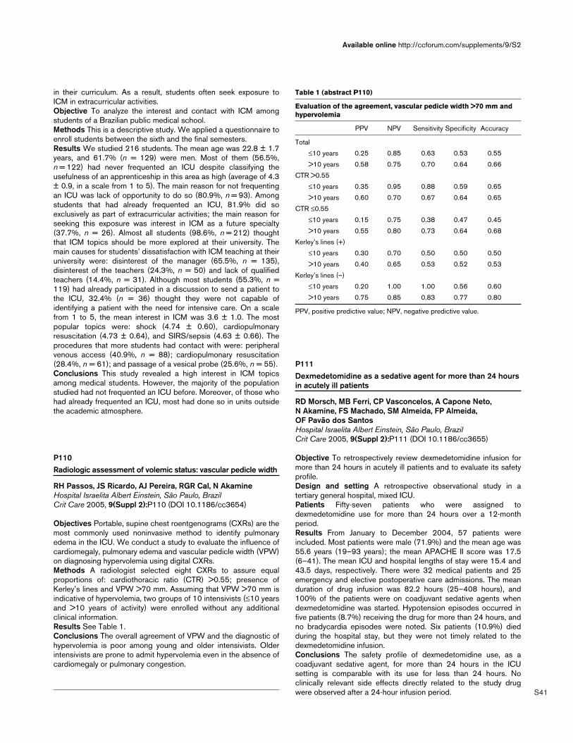
S41
in their curriculum. As a result, students often seek exposure toICM in extracurricular activities.Objective To analyze the interest and contact with ICM amongstudents of a Brazilian public medical school.Methods This is a descriptive study. We applied a questionnaire toenroll students between the sixth and the final semesters.Results We studied 216 students. The mean age was 22.8 ± 1.7years, and 61.7% (n = 129) were men. Most of them (56.5%,n = 122) had never frequented an ICU despite classifying theusefulness of an apprenticeship in this area as high (average of 4.3± 0.9, in a scale from 1 to 5). The main reason for not frequentingan ICU was lack of opportunity to do so (80.9%, n = 93). Amongstudents that had already frequented an ICU, 81.9% did soexclusively as part of extracurricular activities; the main reason forseeking this exposure was interest in ICM as a future specialty(37.7%, n = 26). Almost all students (98.6%, n = 212) thoughtthat ICM topics should be more explored at their university. Themain causes for students’ dissatisfaction with ICM teaching at theiruniversity were: disinterest of the manager (65.5%, n = 135),disinterest of the teachers (24.3%, n = 50) and lack of qualifiedteachers (14.4%, n = 31). Although most students (55.3%, n =119) had already participated in a discussion to send a patient tothe ICU, 32.4% (n = 36) thought they were not capable ofidentifying a patient with the need for intensive care. On a scalefrom 1 to 5, the mean interest in ICM was 3.6 ± 1.0. The mostpopular topics were: shock (4.74 ± 0.60), cardiopulmonaryresuscitation (4.73 ± 0.64), and SIRS/sepsis (4.63 ± 0.66). Theprocedures that more students had contact with were: peripheralvenous access (40.9%, n = 88); cardiopulmonary resuscitation(28.4%, n = 61); and passage of a vesical probe (25.6%, n = 55).Conclusions This study revealed a high interest in ICM topicsamong medical students. However, the majority of the populationstudied had not frequented an ICU before. Moreover, of those whohad already frequented an ICU, most had done so in units outsidethe academic atmosphere.
P110Radiologic assessment of volemic status: vascular pedicle width
RH Passos, JS Ricardo, AJ Pereira, RGR Cal, N AkamineHospital Israelita Albert Einstein, São Paulo, BrazilCrit Care 2005, 9(Suppl 2):P110 (DOI 10.1186/cc3654)
Objectives Portable, supine chest roentgenograms (CXRs) are themost commonly used noninvasive method to identify pulmonaryedema in the ICU. We conduct a study to evaluate the influence ofcardiomegaly, pulmonary edema and vascular pedicle width (VPW)on diagnosing hypervolemia using digital CXRs.Methods A radiologist selected eight CXRs to assure equalproportions of: cardiothoracic ratio (CTR) >0.55; presence ofKerley’s lines and VPW >70 mm. Assuming that VPW >70 mm isindicative of hypervolemia, two groups of 10 intensivists (≤10 yearsand >10 years of activity) were enrolled without any additionalclinical information.Results See Table 1.Conclusions The overall agreement of VPW and the diagnostic ofhypervolemia is poor among young and older intensivists. Olderintensivists are prone to admit hypervolemia even in the absence ofcardiomegaly or pulmonary congestion.
Table 1 (abstract P110)
Evaluation of the agreement, vascular pedicle width >70 mm andhypervolemia
PPV NPV Sensitivity Specificity Accuracy
Total
≤10 years 0.25 0.85 0.63 0.53 0.55
>10 years 0.58 0.75 0.70 0.64 0.66
CTR >0.55
≤10 years 0.35 0.95 0.88 0.59 0.65
>10 years 0.60 0.70 0.67 0.64 0.65
CTR ≤0.55
≤10 years 0.15 0.75 0.38 0.47 0.45
>10 years 0.55 0.80 0.73 0.64 0.68
Kerley’s lines (+)
≤10 years 0.30 0.70 0.50 0.50 0.50
>10 years 0.40 0.65 0.53 0.52 0.53
Kerley’s lines (–)
≤10 years 0.20 1.00 1.00 0.56 0.60
>10 years 0.75 0.85 0.83 0.77 0.80
PPV, positive predictive value; NPV, negative predictive value.
P111Dexmedetomidine as a sedative agent for more than 24 hoursin acutely ill patients
RD Morsch, MB Ferri, CP Vasconcelos, A Capone Neto, N Akamine, FS Machado, SM Almeida, FP Almeida, OF Pavão dos SantosHospital Israelita Albert Einstein, São Paulo, BrazilCrit Care 2005, 9(Suppl 2):P111 (DOI 10.1186/cc3655)
Objective To retrospectively review dexmedetomidine infusion formore than 24 hours in acutely ill patients and to evaluate its safetyprofile.Design and setting A retrospective observational study in atertiary general hospital, mixed ICU.Patients Fifty-seven patients who were assigned todexmedetomidine use for more than 24 hours over a 12-monthperiod.Results From January to December 2004, 57 patients wereincluded. Most patients were male (71.9%) and the mean age was55.6 years (19–93 years); the mean APACHE II score was 17.5(6–41). The mean ICU and hospital lengths of stay were 15.4 and43.5 days, respectively. There were 32 medical patients and 25emergency and elective postoperative care admissions. The meanduration of drug infusion was 82.2 hours (25–408 hours), and100% of the patients were on coadjuvant sedative agents whendexmedetomidine was started. Hypotension episodes occurred infive patients (8.7%) receiving the drug for more than 24 hours, andno bradycardia episodes were noted. Six patients (10.9%) diedduring the hospital stay, but they were not timely related to thedexmedetomidine infusion.Conclusions The safety profile of dexmedetomidine use, as acoadjuvant sedative agent, for more than 24 hours in the ICUsetting is comparable with its use for less than 24 hours. Noclinically relevant side effects directly related to the study drugwere observed after a 24-hour infusion period.
Available online http://ccforum.com/supplements/9/S2
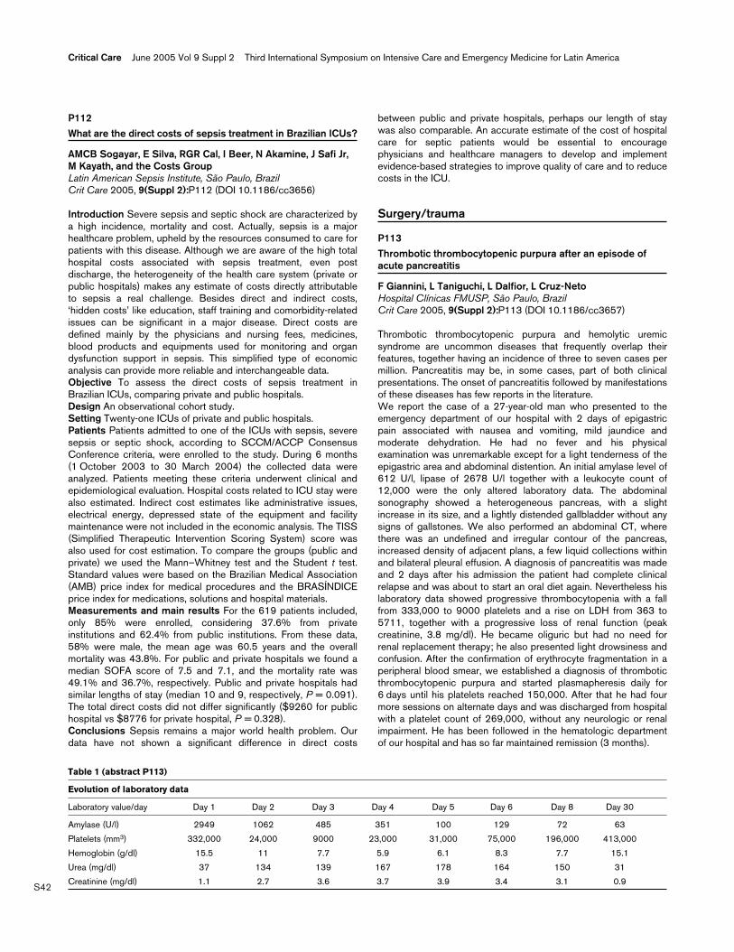
S42
P112What are the direct costs of sepsis treatment in Brazilian ICUs?
AMCB Sogayar, E Silva, RGR Cal, I Beer, N Akamine, J Safi Jr,M Kayath, and the Costs GroupLatin American Sepsis Institute, São Paulo, BrazilCrit Care 2005, 9(Suppl 2):P112 (DOI 10.1186/cc3656)
Introduction Severe sepsis and septic shock are characterized bya high incidence, mortality and cost. Actually, sepsis is a majorhealthcare problem, upheld by the resources consumed to care forpatients with this disease. Although we are aware of the high totalhospital costs associated with sepsis treatment, even postdischarge, the heterogeneity of the health care system (private orpublic hospitals) makes any estimate of costs directly attributableto sepsis a real challenge. Besides direct and indirect costs,‘hidden costs’ like education, staff training and comorbidity-relatedissues can be significant in a major disease. Direct costs aredefined mainly by the physicians and nursing fees, medicines,blood products and equipments used for monitoring and organdysfunction support in sepsis. This simplified type of economicanalysis can provide more reliable and interchangeable data.Objective To assess the direct costs of sepsis treatment inBrazilian ICUs, comparing private and public hospitals.Design An observational cohort study.Setting Twenty-one ICUs of private and public hospitals.Patients Patients admitted to one of the ICUs with sepsis, severesepsis or septic shock, according to SCCM/ACCP ConsensusConference criteria, were enrolled to the study. During 6 months(1 October 2003 to 30 March 2004) the collected data wereanalyzed. Patients meeting these criteria underwent clinical andepidemiological evaluation. Hospital costs related to ICU stay werealso estimated. Indirect cost estimates like administrative issues,electrical energy, depressed state of the equipment and facilitymaintenance were not included in the economic analysis. The TISS(Simplified Therapeutic Intervention Scoring System) score wasalso used for cost estimation. To compare the groups (public andprivate) we used the Mann–Whitney test and the Student t test.Standard values were based on the Brazilian Medical Association(AMB) price index for medical procedures and the BRASÍNDICEprice index for medications, solutions and hospital materials.Measurements and main results For the 619 patients included,only 85% were enrolled, considering 37.6% from privateinstitutions and 62.4% from public institutions. From these data,58% were male, the mean age was 60.5 years and the overallmortality was 43.8%. For public and private hospitals we found amedian SOFA score of 7.5 and 7.1, and the mortality rate was49.1% and 36.7%, respectively. Public and private hospitals hadsimilar lengths of stay (median 10 and 9, respectively, P = 0.091).The total direct costs did not differ significantly ($9260 for publichospital vs $8776 for private hospital, P = 0.328).Conclusions Sepsis remains a major world health problem. Ourdata have not shown a significant difference in direct costs
between public and private hospitals, perhaps our length of staywas also comparable. An accurate estimate of the cost of hospitalcare for septic patients would be essential to encouragephysicians and healthcare managers to develop and implementevidence-based strategies to improve quality of care and to reducecosts in the ICU.
Surgery/trauma
P113Thrombotic thrombocytopenic purpura after an episode ofacute pancreatitis
F Giannini, L Taniguchi, L Dalfior, L Cruz-NetoHospital Clínicas FMUSP, São Paulo, BrazilCrit Care 2005, 9(Suppl 2):P113 (DOI 10.1186/cc3657)
Thrombotic thrombocytopenic purpura and hemolytic uremicsyndrome are uncommon diseases that frequently overlap theirfeatures, together having an incidence of three to seven cases permillion. Pancreatitis may be, in some cases, part of both clinicalpresentations. The onset of pancreatitis followed by manifestationsof these diseases has few reports in the literature.We report the case of a 27-year-old man who presented to theemergency department of our hospital with 2 days of epigastricpain associated with nausea and vomiting, mild jaundice andmoderate dehydration. He had no fever and his physicalexamination was unremarkable except for a light tenderness of theepigastric area and abdominal distention. An initial amylase level of612 U/l, lipase of 2678 U/l together with a leukocyte count of12,000 were the only altered laboratory data. The abdominalsonography showed a heterogeneous pancreas, with a slightincrease in its size, and a lightly distended gallbladder without anysigns of gallstones. We also performed an abdominal CT, wherethere was an undefined and irregular contour of the pancreas,increased density of adjacent plans, a few liquid collections withinand bilateral pleural effusion. A diagnosis of pancreatitis was madeand 2 days after his admission the patient had complete clinicalrelapse and was about to start an oral diet again. Nevertheless hislaboratory data showed progressive thrombocytopenia with a fallfrom 333,000 to 9000 platelets and a rise on LDH from 363 to5711, together with a progressive loss of renal function (peakcreatinine, 3.8 mg/dl). He became oliguric but had no need forrenal replacement therapy; he also presented light drowsiness andconfusion. After the confirmation of erythrocyte fragmentation in aperipheral blood smear, we established a diagnosis of thromboticthrombocytopenic purpura and started plasmapheresis daily for6 days until his platelets reached 150,000. After that he had fourmore sessions on alternate days and was discharged from hospitalwith a platelet count of 269,000, without any neurologic or renalimpairment. He has been followed in the hematologic departmentof our hospital and has so far maintained remission (3 months).
Critical Care June 2005 Vol 9 Suppl 2 Third International Symposium on Intensive Care and Emergency Medicine for Latin America
Table 1 (abstract P113)
Evolution of laboratory data
Laboratory value/day Day 1 Day 2 Day 3 Day 4 Day 5 Day 6 Day 8 Day 30
Amylase (U/l) 2949 1062 485 351 100 129 72 63
Platelets (mm3) 332,000 24,000 9000 23,000 31,000 75,000 196,000 413,000
Hemoglobin (g/dl) 15.5 11 7.7 5.9 6.1 8.3 7.7 15.1
Urea (mg/dl) 37 134 139 167 178 164 150 31
Creatinine (mg/dl) 1.1 2.7 3.6 3.7 3.9 3.4 3.1 0.9

S43
P114Abdominal hypertension and compartmental syndrome inintensive care medicine
HP Guimarães, A Raimondi, PHR Leal, RD Lopes, AP Resque,GK Barcelos, MB Peruzzo, FR Machado, JLG AmaralFederal University of São Paulo, UNIFESP, EPM, São Paulo, BrazilCrit Care 2005, 9(Suppl 2):P114 (DOI 10.1186/cc3658)
Introduction and objectives The objective of this trial was tocompare abdominal hypertension and abdominal compartmentalsyndrome (ACS) in patients under risk for these pathologies in theICU.Methods Eighty-nine patients with monitoring indications for intra-abdominal pressure (IAP) based on clinical or surgical admissiondiagnosis in the ICU were included. Measurements via an indirectmethod through the vesical catheter were made at three distincttimepoints: at admission, and at 6 hours and 12 hours.Results The incidence of ACS was 14.6% (13 patients); in thetotal sample, the male sex prevailed at 63% (56 patients). Thepatients had been distributed into two groups: with ACS andwithout ACS. Gastric surgery diagnosis predominated in bothgroups (84.6%/57.9%; P < 0.067). Global mortality was 30.3%(27 patients): 53.8% (seven patients) in the group with ACS and26.3% (20 patients) in the group without ACS; P < 0.046. Table 1describes the sample.
Table 1
Present Absent Compartmental (n = 13), (n = 76), syndrome average/standard average/standard P value
Weight 78.3/10.4 79.4/25 0.876
Height 1.66/0.04 1.67/0.09 0.696
APACHE 16.1/7.6 16.5/8.1 0.869
IAP admission 28.2/6 15.6/6.3 <0.0001
IAP 6 hours 27.8/9.2 15.8/6.3 <0.0001
IAP 12 hours 29.6/9.4 15.9/6.2 <0.0001
IAP Average 28.5/6.6 15.7/5.8 <0.0001
Conclusion Abdominal hypertension and ACS are frequentdiagnostics in intensive care medicine; superior average values ofthe IAP at admission, at 6 hours and at 12 hours to 28 mmHg arecorrelated with the biggest incidence of ACS and greater mortality.
P115Outcome of bariatric surgical patients admitted to the ICU
FVC de Marco, ALG Guimarães, WM Imanishi, M Catania, NKG Souza, RK Kikko, PMC BrunoIntensive Care Unit, Hospital Gastroclinica, São José dos Campos,BrazilCrit Care 2005, 9(Suppl 2):P115 (DOI 10.1186/cc3659)
Background The problem of obesity has reached epidemicproportions and the number of bariatric procedures is increasing.There are few data regarding the outcome of these patients in theintensive care environment.Objective The aim of this study was to evaluate the outcome ofbariatric surgical patients admitted to the ICU.Setting An eight-bed surgical ICU in a 50-bed private hospital.Methods From April 2003 to February 2005 we prospectivelyfollowed bariatric surgical patients admitted to the ICU. Theoutcome and ICU resource utilization were recorded. The
APACHE II score was calculated (QuaTI System softwaredatabase; Dixtal, SP, Brazil).Results There were 304 patients (240 female) in 313 consecutiveICU admissions. The mean age was 36.9 ± 10.9 years. The meanBMI was 44.10 ± 5.2 kg/m2. Of these patients, 302 (99.3%) wereadmitted in the immediate postoperative period for a primaryprocedure. A total of 252 patients (82.9%) underwent Roux-en-Ygastric bypass (16 laparoscopic) and 52 patients (17.1%)underwent biliopancreatic diversion (two laparoscopic). In 20(6.4%) of 313 admissions, the length of stay was >24 hours. TheAPACHE II score was 10.0 ± 9.2. The mean LOS was 7.8 ± 16.8days. The main reasons for unplanned ICU admissions (n = 11)were bowel occlusion (n = 2), anastomotic leakage (n = 2), gastro-intestinal bleeding (n = 2), wound infection (n = 1), pulmonaryembolism (n = 1), acute pulmonary edema (n = 1), massiveatelectasis (n = 1) and perforated bowel (n = 1). There was needfor mechanical ventilation in 22 (7.0%) admissions, a pulmonaryartery catheter in two (0.6%), dialysis therapy in one (0.3%) andparenteral nutrition in one (0.3%). Only three patients havedeveloped complications (anaphylactic reaction, respiratoryacidosis, cetoacidosis) during the immediate postoperative periodfor a primary procedure (<48 hours) and only one patient hasnever been discharged from the ICU. The 28-day ICU mortality ratewas 0.65% (n = 2) and the overall ICU mortality rate was 0.98%(n = 3).Conclusions Based on these preliminary results, we conclude thatbariatric surgical patients in the immediate postoperative period fora primary procedure are very low risk ICU patients.
P116Dexmedetomidine for short-term and long-term sedation:amplifying its applicability
I Beer, AT Maciel, JMC Coelho, R de ClevaSurgical Intensive Care Unit, Department of Gastroenterology,Discipline of Digestive Surgery, Hospital das Clinicas, University ofSão Paulo, BrazilCrit Care 2005, 9(Suppl 2):P116 (DOI 10.1186/cc3660)
Background The α2-agonist dexmedetomidine (DEX) it is aneffective agent for the management of sedation and analgesia aftercardiac, general, orthopedic, head and neck, oncologic andvascular surgery in the ICU. DEX is currently approved as asedative for up to 24 hours use in the ICU, but the number ofreports showing safe long-term therapy with very good tolerabilityis increasing. The α2-agonists may also attenuate hypertension,anxiety, agitation, tachycardia and fever during benzodiazepine andopioid withdrawal [1].Objective To describe the experience with DEX in the surgicalICU, for early postoperative and long-term sedation of critically illpatients and for withdrawal syndrome management.Design and setting A retrospective review of medical records andpresentation of case-series (n = 13) from a surgical ICU (14 beds)of a university hospital from August 2004 to February 2005.Results The 13 critically ill patients (eight male, five female; age41–83 years) who received DEX were elective or complicatedpostoperative patients who required sedation and mechanicalventilation in the ICU for a variety of medical problems. We haveincluded one case of withdrawal syndrome after interruption ofprolonged midazolam and fentanyl infusion, and two cases ofbariatric surgery (rarely reported in literature). The patientsreceived DEX infusion during at least 6 hours. The mean time ofdrug administration was 45 hours (6–336 hours). Only mild falls inarterial pressure occurred in some patients, but in none of themwas DEX discontinued (managed with fluid challenge or a slight
Available online http://ccforum.com/supplements/9/S2
S44
increase in vasoactive drug). Only three patients required a singledose of fentanyl (100 µg) as rescue analgesia.Conclusion DEX is an effective and safe sedative agent that hasanalgesic properties and predictable cardiovascular effects, andmay benefit a heterogeneous population of critically ill patients forshort-term (early postoperative period) and long-term sedation. Webelieve that a continuous infusion of a α2-agonist agent forsedation-induced withdrawal syndrome management is highlyapplicable.Reference1. Multz AS: Prolonged dexmedetomidine infusion as an adjunct
in treating sedation-induced withdrawal. Anesth Analg 2003;96:1054-1055.
P117Conventional tracheostomy in critically ill patients: a safeprocedure
CL Mendes, F Negri, L Holmes, HBM Almeida, BM Vieira, IP Amorim, CLR Moreira, EQR Moura, MVD Marchi, SDS LuzUti Adulto, Hospital Universitário, UFPB, Cidade Universitária, JoãoPessoa, BrazilCrit Care 2005, 9(Suppl 2):P117 (DOI 10.1186/cc3661)
Introduction Tracheostomy is a very usual procedure in critically illpatients and is assumed to have a low rate of complications whenexecuted by experienced hands. We did not have data about thisissue in our setting, so we decided to perform a prospective trial inorder to observe the main characteristics of tracheostomies madein critically ill patients in two of our centers: a general ICU and atrauma ICU.Materials and methods We performed a prospective andobservational trial during the period between 28 May 2004 and 16December 2004 in two ICUs of two tertiary hospitals.Results During this period, 601 patients were admitted: 291patients in the trauma ICU and 310 patients in the general ICU.Sixty-six patients (10.98%) were submitted to tracheostomia in thetwo centers, 40 (60.6%) in the trauma ICU and 26 (39.4%) in thegeneral ICU. Forty-four (66.7%) were male. Acute stroke(ischaemic or haemorragic) was the main cause of admission tothe ICUs among the tracheostomized patients (28.8%), followedby brain trauma (21.2%). The major cause of admission in thegeneral ICU was respiratory insufficiency (38.4%), and was braininjury (32.5%) in the trauma unit. The mean Glasgow, SOFA andAPACHE II scores at the day of tracheostomy were 7.5 ± 3.19, 5.4± 2.01 and 19.2 ± 5.46, respectively. The main indication totracheostomy (90.9%) was prolonged invasive ventilation (90.9%).The tracheostomies were exclusively performed by an experiencedhead and neck surgeon in the two centers. Tracheostomies wereperformed inside the ICU setting in the trauma ICU and in theoperation room in the other. There were no differences in the rateof complications between the two centers. The rate ofintraoperative and early complications was very low. There werethree minor bleeding complications (4.5%). The mean time ofinvasive ventilation was 20.9 ± 10.5 days. The mean time in theICU was 22.7 ± 10.39 days. The mortality rate was 31.8% andnone of the deaths was related to the tracheostomies.Conclusions Despite the fact that it was a procedure performed invery critically ill patients, tracheostomy was associated with veryfew minor complications in this sample. We hypothesized that thislow rate of complications is due in part to the very high expertise ofthe operators involved in the realization of conventionaltracheostomies in the two centers.
P118Blood transfusion in geriatric patients who underwentorthopaedic surgery: a prospective study
PG Coscarelli, IRS de Castro, MD Saud, R dos Reis Visconti, M Teixeira de NovaisInstituto Nacional de Tráuma, To-Ortopedia, Rio de Janeiro, BrazilCrit Care 2005, 9(Suppl 2):P118 (DOI 10.1186/cc3662)
Objective Our hypothesis is that older patients more often receivepacked red blood cells, motivated for comorbidity.Methods During a 4-year period (2001–2004) we collected dataon 1064 patients admitted to an ICU of an orthopaedic surgeryhospital. We analyzed the use of packages of red blood cells(PRBC), the quantity of packages, and comorbidities. Weexcluded patients that received more than 4 PRBC.Results The percentage of patients that received PRBCprogressively increases from patients in their sixties to theirnineties (r = 0.99): <65 years, 30.6%; 65–69 years, 20.7%;70–79 years, 33.2%; 80–89 years, 45.3%; 90 years or more,64.7%. There was no increase in the number of packages as theage advances: <65 years, 1.81 packs; 65–69 years, 1.83;70–79 years, 1.93; 80–89 years, 1.9; 90 years or more, 2.18. Nodifference was found in the postoperative hematocrit: <65 years,33.1%; 65–69 years, 33.6%; 70–79 years, 34.8%; 80–89 years,32.4%; 90 years or more, 32.9%. We did not find any relationshipof the use of PRBC and cardiovascular comorbidities (χ2 = 0.35;P > 0.05).Conclusions There was a positive correlation between age andnecessity of hemotransfusion in the postoperative period oforthopedic surgeries, but it was no correlated to the cardiovascularcomorbidity.
P119Mortality in elderly patients with hip fractures: a retrospectivecohort study
ACR Chaves, MA Dutra, GC Geraldino, MGS Ghilardi, HV Carrasco, SMR CarvalhoFaculdade de Medicina de Marília, Hospital Santa Casa deMisericórdia de Marília, BrazilCrit Care 2005, 9(Suppl 2):P119 (DOI 10.1186/cc3663)
Introduction The purpose of this study is to gather a population ofelderly patients with hip fractures who have been through surgeryhospitalized in the ICU, and to establish the validity of the APACHEII score as a tool in determining the risk of death.Methods A retrospective cohort study was developed. During theperiod January–December 2003, patients were checked through arevision of medical histories up to the time when they were out ofthe hospital or up to their death. Initially, 65 patients aged above60 years were included, an average of 85 (68–98) years, of which46 (71%) were male. The depending variables were the rate ofmortality and the APACHE II score. The APACHE II scale wasevaluated by logistic regression procedure with the help of theStatisDirect software 1.617 for Windows. We adopted a level of5% of probability of rejection of the hypothesis of nullity.Results The APACHE II score was 13 (6–28) and the mortalitywas 9.2% taking into consideration that 25% of the patients hadan APACHE II score >16. The length of stay in the ICU was 9.8(2–38) days. It was observed that the predictive model had asensitivity of 66.7% and a specificity of 98.3%.Conclusions The risk (odds ratio) for elderly patients who hadbeen through hip fracture surgery was 1.4 (1.1–1.7) times greaterfor a score of APACHE II greater than 16. We consider that the
Critical Care June 2005 Vol 9 Suppl 2 Third International Symposium on Intensive Care and Emergency Medicine for Latin America

S45
APACHE II score was a valid tool in determining the risk of death inthis kind of patient.
P120Dexmedetomidine for sedation following cardiovascularsurgery: a two different loading doses
L Kopel, RT Carvalho, HBN Araujo, AA Fagundes, M Ribeiro, J Bastos, SG LageClinical Intensive Care Unit, Heart Institute, HCFMUSP, São Paulo,BrazilCrit Care 2005, 9(Suppl 2):P120 (DOI 10.1186/cc3664)
Introduction Dexmedetomidine (DEX) is a selective alpha-2-adrenergic agonist used for sedation of patients in ICUs.Hypothesis The purpose of this study was to evaluate the efficacyand safety of DEX administration after cardiovascular surgery usingtwo different loading doses.Methods This was a prospective, multicenter (three sites),randomized, open study. Patients were randomized to either aloading dose of 6 µg/kg/hour (group I) for 10 min or 3 µg/kg/hour(group II) for 20 min, with a total loading dose of 1 µg/kg, followedby a maintenance infusion of 0.4 µg/kg/hour, adjusted accordingto sedation level from 0.2 to 0.7 µg/kg/hour (Ramsay sedationscale >3 for mechanically ventilated patients and >2 afterextubation). The sedation period with DEX ranged from 6 to24 hours, beginning in the first hour after the patient’s arrival in thepostoperative ICU.Results Forty-two patients were studied after electivecardiovascular surgery (34 CABG, four aortic aneurysms, fourvalvular heart surgery). Eighty-eight percent of patients demon-strated no need for additional sedative drugs during mechanicalventilation. Twenty-six percent of patients received analgesicswhile intubated. The level of sedation, evaluated based on theRamsay sedation scale, did not show any difference between thetwo groups during the 24-hour infusion. No differences have beenfound in the heart rate, systolic blood pressure, central venouspressure and pulse oxymeter saturation between the two groupsduring the 24-hour period.Conclusions Our study indicates that DEX is an effective and safedrug to be used in the postoperative period of cardiovascularsurgery, independent of the loading dose rate infusion.
P121Rhabdomyolysis and acute renal failure after bariatric surgery:severe forms of crush injury
I Beer, R de Cleva, J Faintuch, M Matsuda, B Zilberstein, JJ Gama-RodriguesSurgical Intensive Care Unit, Department of Gastroenterology,Hospital das Clinicas, University of São Paulo School of Medicine,BrazilCrit Care 2005, 9(Suppl 2):P121 (DOI 10.1186/cc3665)
Background Rhabdomyolysis is one of the leading causes ofacute renal failure (ARF). The most commonly described crushinjury is that which affects victims of natural disasters such asearthquakes, mining or industrial accidents, war, or any othersituation resulting in the collapse of buildings and falling debris. Itmay also affect patients who, because of an altered level ofconsciousness, crush a part of the body with their own weight,such as after drug overdose or stroke. Surgical positioning is amuch less common etiology, with few cases reported in the worldliterature implicating it as a cause. Among the risk factors for thedevelopment of postoperative RML, surgical time and overweight
are the most important. A few cases of RML with acute renal failurehave been reported in morbidly obese patients having surgery forseveral reasons, and another few cases recently reported at thesame time in patients undergoing gastric bypass [1,2]. As thenumber of bariatric procedures by laparotomy or laparoscopy hasbeen growing fast in the past few years, mainly in Westerncountries, an increasing incidence of this complication may beseen. Besides, in the learning curve of laparoscopic procedures, alonger surgical time (up to 4 or 5 hours) is another risk factor [3].Strategies to avoid and to treat this complication and theconsequences must be applied in all perioperative periods.Objective To report a series of six cases (five male, one female) ofsevere rhabdomyolysis after bariatric surgery from a surgical ICU(14 beds) of a university hospital.Conclusions Bariatric surgery, and probably other surgicalprocedures in morbidly obese patients, is a significant form ofcrush injury (overweight and prolonged immobilization on theoperating table) leading to postoperative rhabdomyolysis(maximum CPK value: 29,150 ± 17,129 IU/l) and reversiblemyohemoglobinuric acute renal failure (66.6% of cases). Fluidexpansion, urine alkalinization and renal replacement therapy arethe mainstays of treatment. Cases of complete crush syndrome(hypotension, coagulopathy, acute renal failure) and death (0%)are rare according to our series and the specific literature.References1. Torres Vilalobos G, Kimura E, Mosqueda JL, García-García G,
Cherit D, Herrera MF: Pressure-induced rhabdomyolysis afterbariatric surgery. Obes Surg 2003, 13:297-301.
2. Bostanjian D, Anthone GJ, Hamoui N, Crookes PF: Obes Surg2003, 13:302-305.
3. Schirmer BD: Surg Clin North Am 2000, 80:1253-1267.
P122A new model of uncontrolled hepatic hemorrhage from trocarinsertion simulating iatrogenic lesions during video-assistedsurgeries
MO Taha, JH Soares, PS Venerando, ACC Gandolfi, JB Tsuchya, A Caricati-Neto, LF Poli de FigueiredoDepartment of Surgery and Department of Pharmacology, FederalUniversity of São Paulo, BrazilCrit Care 2005, 9(Suppl 2):P122 (DOI 10.1186/cc3666)
Background Visceral iatrogenic lesions during video-assistedsurgeries may occur during the blind insertion of trocars. Life-threatening hemorrhage and conversion to open surgery mayoccur. The use of topical hemostatic agents may control thehemorrhage and avoid the need for laparotomy. Our goal is todevelop a model simulating liver injury produced by trocarinsertion.Methods Ten male Landrace pigs weighing 16–20 kg wereanesthetized and submitted to pneumoperitonium using CO2 forintroduction of a laparoscopic camera into the abdominal cavity.Under direct vision, the anterior surface of the right hepatic lobeliver was perforated by a trocar. The animals were previouslydivided into two groups: controls (n = 5) and heparin (n = 5), inwhich 200 UI/kg heparin was administrated intravenously 10 minbefore the liver lesion, simulating a coagulopathic scenario. Before(0 min) and 10 or 20 min after induction of the liver lesion,hematologic and hemodynamic parameters including hematocrit(Ht), mean arterial pressure (MAP), cardiac output (l/min) and intra-abdominal blood loss (ml) were determined.Results See Table 1 and Fig. 1.Conclusion We conclude that trocar-induced liver injury promoteslimited blood loss, discrete hypotension and a minor decrease incardiac output. The use of heparin promotes a significant increase
Available online http://ccforum.com/supplements/9/S2

S46
in blood loss and severe hemodynamic compromise. Both modelsmay be useful to test interventions to stop bleeding and to reversehemodynamic instability due to uncontrolled hemorrhage.
P123Hemodynamic patterns in trauma patients
MC Oliveira, A Réa-NetoCEPETI, Hospital do Trabalhador da UFPR, Curitiba, BrazilCrit Care 2005, 9(Suppl 2):P123 (DOI 10.1186/cc3667)
Introduction The main cause of shock in trauma patients ishypovolemia secondary to blood loss; other causes are cardiactamponade, tension pneumothorax, myocardial contusion andneurogenic shock. However, there are several patients who remainin shock after adequate fluid resuscitation and without othercauses of shock.Objective To identify the hemodynamic pattern of patients withtrauma and shock, after hemorrhagic control and correct fluidadministration.Materials and methods We included consecutive patientsadmitted to a trauma ICU, from 1 January 2003 to 31 December2004, who had hemorrhagic control of the trauma lesions and weretreated with fluid and remained dependent on vasoactive drugs.Results Eighty-five patients, or 6.2% of the patients admittedduring the recruitment, met the criteria. The mortality was 59%,against 34% (P < 0.01) of the population without the criteria. Themain cause of death was refractory shock. The mean crystalloidused in the first 24 hours was 9015 ml (±3202 ml), the meanhydroxyethyl starch was 1463 ml (±1868 ml), the mean red cellpack administration was 3 U (±3.43 U) and the mean fresh frozenplasma was 2 U (±2.82 U). Twenty-five patients (30%) weresubmitted to invasive monitoring with a pulmonary arterial catheter.The mean PAOP was 16 mmHg (±5.67 mmHg) but six patientshad a PAOP below 12 mmHg; the mean CI was 5.3 l/m2
(±2.3 l/m2) but five patients had CI <3.0 l/m2; and the mean SVRIwas 1132 dyn/m2/s (±508 dyn/m2/s).
Conclusion The presence of shock was associated with highmortality. The most prevalent hemodynamic pattern washyperdynamic shock; but 25% (six patients) had hypovolemia evenafter vigorous fluid resuscitation, and 12.5% (three patients) hadhypodynamic shock. The invasive monitoring of the pulmonaryartery was important to diagnose the class of shock and to identifythe presence of residual hypovolemia.
P124The incidence of organ dysfunction and failure in patients withabdominal trauma
MC Oliveira, A Réa-NetoCEPETI, Hospital do Trabalhador Da UFPR, Curitiba, BrazilCrit Care 2005, 9(Suppl 2):P124 (DOI 10.1186/cc3668)
Introduction Trauma patients have a high mortality and more oftendevelop organ failure with poor outcome.Objective To study the incidence of organ dysfunction and failure,and the impact on mortality of patients who were admitted to anICU with abdominal trauma without other lesions.Materials and methods We studied patients admitted to a traumaICU from 1 January 2001 to 31 December 2003. The SequentialOrgan Failure Assessment score at admission was measured; weconsidered points 1 and 2 of the score as dysfunction and points 3and 4 as failure.Results A total of 77 patients were studied and the mortality was31%; the mortality of the general population with multiple traumas(725 patients) was 39%. The incidence of organ dysfunction andfailure, with the correspondent mortality, is illustrated in Table 1.
Conclusion The frequency of coagulation and renal dysfunctionand failure is higher in patients with abdominal trauma than inpatients with multiple trauma. The presence of renal,cardiovascular and hematological failures was associated withhigher mortality in patients with abdominal trauma than in patientswith multiple trauma.
Critical Care June 2005 Vol 9 Suppl 2 Third International Symposium on Intensive Care and Emergency Medicine for Latin America
Table 1
Group 0 min 10 min 20 min
Ht (%) Control 26.6 ± 0.9 26.6 ± 1.0 26.4± 0.9
Heparin 28.4 ± 0.6 25.2 ± 1.7 26.6 ± 1.6
MAP (mmHg) Control 89.4 ± 6.9 76.4 ± 8 76.4 ± 4
Heparin 87 ± 14 44 ± 17* 42.2 ± 17*
Cardiac output (l/min) Control 5.1 ± 0.7 4.0 ± 0.6 4.23 ± 07
Heparin 5.8 ± 0.4 2.7 ± 1.1* 2.73 ± 1.4*
Data presented as the mean ± SEM; * P < 0.05 compared with controls.
Figure 1
0
200
400
600
800
1000
1200
*HeparinControl
Blo
od
Lo
st (
ml)
Table 1
Incidence of organ dysfunction and failure in abdominal traumapatients
Dysfunction Failure
Incidence Mortality Incidence Mortality (%) (%) (%) (%)
Respiration Abdominal 76 33 18 43
Multiple trauma 63 22 24 60
Coagulation Abdominal 41 50 11 100
Multiple trauma 25 44 2 82
Liver Abdominal 35 38 – –
Multiple trauma 20 38 1 40
Cardiovascular Abdominal – – 30 100
Multiple trauma 3 58 21 61
Renal Abdominal 35 62 18 100
Multiple trauma 10 56 4 71

S47
P125The incidence of organ dysfunction and failure in traumapatients
MC Oliveira, A Réa-NetoCEPETI, Hospital do Trabalhador da UFPR, Curitiba, BrazilCrit Care 2005, 9(Suppl 2):P125 (DOI 10.1186/cc3669)
Introduction Trauma patients have a high mortality and more oftendevelop organ failure with worse outcome.Objective To study the incidence of organ dysfunction and failure,and the impact on mortality in patients who were admitted to anICU with multiple trauma.Materials and methods We studied patients consecutivelyadmitted to a trauma ICU from 1 January 2001 to 31 December2003. The Sequential Organ Failure Assessment score atadmission was measured; we considered points 1 and 2 of thescore as dysfunction and points 3 and 4 as failure.Results: A total of 802 patients were studied; the mortality of thegeneral population without organ dysfunction was 39.65%. Theincidence of organ dysfunction and failure, with the correspondentmortality, is illustrated in Table 1.
Conclusion The presence of organ dysfunction worsened theoutcome of patients with multiple trauma; the respiratorydysfunction is more prevalent than others but with a lower impacton mortality, followed by coagulation and liver dysfunction. Thepresence of coagulation, respiratory failure and cardiovascularfailure is associated with higher mortality than liver or renal failure.
Available online http://ccforum.com/supplements/9/S2
Table 1
Incidence of organ dysfunction and failure in major trauma
Dysfunction Failure
Incidence Mortality Incidence Mortality Major trauma (%) (%) (%) (%)
Respiration 63 26 25 79
Coagulation 26 50 2.5 84
Liver 23 38.6 1,7 54
Cardiovascular 12.6 53 12 77.8
Renal 11.5 59 4.8 60