
Concealed entrainment as a guide for catheter ablation of ventricular tachycardia in patients with...
12
678 ELECTROPHYSIOLOGIC STUDIES lACC Vol. 17, No.3 March 1. 1991:678-89 Concealed Entrainment As a Guide for Catheter Ablation of Ventricular Tachycardia in Patients With Prior Myocardial Infarction FRED MORADY, MD, FACC, ALAN KADISH, MD, FACC, SHIMON ROSENHECK, MD, HUGH CALKINS, MD, WILLIAM H. KOU, MICHAEL DE BUITLEIR, MD, JOAO SOUSA, MD Ann Arbor, Michigan Fifteen consecutive patients with drug-refractory, recurrent, sus- tained, monomorphic ventricular tachycardia and a history of remote myocardial infarction underwent catheter ablation of ventricular tachycardia. Shocks of 100 to 300 J were delivered to sites at which pacing during ventricular tachycardia resulted in concealed entrainment, in which the ventricular tachycardia accelerated to the pacing rate, there was a long stimulus to QRS interval and there was no change in the configuration of the QRS complex during pacing at several rates compared with the config- uration during ventricular tachycardia, thus identifying a zone of slow conduction in the reentrant circuit. Concealed entrainment was demonstrated in nine (60%) of 15 patients, and the stimulus to QRS intervals were 90 to 400 ms. At sites of concealed entrainment, the endocardial activation time relative to the QRS complex during ventricular tachycardia ranged from - 125 to + SO ms, the timing of the local electrogram relative to the QRS complex was the same during entrainment as during ventricular tachycardia and the pace map during sinus rhythm was discordant with that of the ventricular tachycardia in Anecdotal reports (1-5) based on a small number of patients show that ventricular pacing during ventricular tachycardia may allow localization of a zone of slow conduction within the reentrant circuit of ventricular tachycardia in patients with coronary artery disease. One method of identifying zones of slow conduction is the phenomenon of "concealed entrainment," in which the ventricular tachycardia acceler- ates to the pacing rate, there is a long interval between the pacing stimulus and the QRS complex influenced by that stimulus, and there is no change in the configuration of the QRS complex during pacing compared with that during ventricular tachycardia (3). The initial reports (1-5) suggest that the zone of slow conduction is an appropriate site at which to direct transcatheter shocks aimed at ablating ven- tricular tachycardia. The purpose of the present study was to assess the value From the Department of Internal Medicine, Division of Cardiology, University of Michigan Medical Center, Ann Arbor, Michigan. Manuscript received March 18. 1990; revised manuscript received August 28, 1990, accepted September II, 1990. Address for reprints: Fred Morady, MD, Division of Cardiology, BIF245, University of Michigan Hospital, Ann Arbor, Michigan 48109-0022. © 1991 by the American College of Cardiology seven patients. In the six patients in whom a site of concealed entrainment could not be identified, the target site for ablation was selected on the basis of identification of an isolated mid- diastolic potential, activation mapping and pace mapping. The mean (±SD) cumulative number of joules delivered to the target site was 306 ± 140. A successful long-term clinical outcome was achieved in 9 of the 15 patients (mean follow-up 20 ± 7 months). The clinical success rate was the same whether the target site was selected on the basis of concealed entrainment (five of nine, 56%) or on the basis of the other mapping techniques (four of six, 67%). In conclusion, the responses to pacing suggest that sites at which there is concealed entrainment may be located within a zone of slow conduction in the ventricular tachycardia reentry circuit, although not necessarily in an area critical for the maintenance of reentry. The long-term clinical efficacy of catheter ablation tar- geted to sites of concealed entrainment is about 60%, similar to the results achieved when conventional mapping techniques are used. (J Am Coil CardioI1991;17:678-89) of concealed entrainment as a guide for catheter ablation of ventricular tachycardia in patients with coronary artery disease. The specific issues addressed were: 1) How often can a site at which there is concealed entrainment be identified in patients with prior myocardial infarction who have recurrent, sustained, monomorphic ventricular tachy- cardia? 2) What is the relation between the phenomenon of concealed entrainment and other markers of the site of origin of ventricular tachycardia? 3) What are the results of cath- eter ablation of ventricular tachycardia using concealed entrainment to identify the target site for ablation? Methods Patients (Table 1). The subjects of this study were 15 consecutive patients with coronary artery disease and a history of remote myocardial infarction who underwent an attempt at catheter ablation because of recurrent, sustained, monomorphic ventricular tachycardia refractory to pharma- cologic therapy. In each patient, the recurrent episodes of monomorphic ventricular tachycardia had been documented to have the same configuration on a 12 lead electrocardio- gram (EeG). There were 10 men and 5 women with a mean 0735-1097/91/$3.50
-
Upload
independent -
Category
Documents
-
view
0 -
download
0
Transcript of Concealed entrainment as a guide for catheter ablation of ventricular tachycardia in patients with...

678
ELECTROPHYSIOLOGIC STUDIES
lACC Vol. 17, No.3 March 1. 1991:678-89
Concealed Entrainment As a Guide for Catheter Ablation of Ventricular Tachycardia in Patients With Prior Myocardial Infarction
FRED MORADY, MD, FACC, ALAN KADISH, MD, FACC, SHIMON ROSENHECK, MD, HUGH CALKINS, MD, WILLIAM H. KOU, MICHAEL DE BUITLEIR, MD, JOAO SOUSA, MD
Ann Arbor, Michigan
Fifteen consecutive patients with drug-refractory, recurrent, sustained, monomorphic ventricular tachycardia and a history of remote myocardial infarction underwent catheter ablation of ventricular tachycardia. Shocks of 100 to 300 J were delivered to sites at which pacing during ventricular tachycardia resulted in concealed entrainment, in which the ventricular tachycardia accelerated to the pacing rate, there was a long stimulus to QRS interval and there was no change in the configuration of the QRS complex during pacing at several rates compared with the configuration during ventricular tachycardia, thus identifying a zone of slow conduction in the reentrant circuit.
Concealed entrainment was demonstrated in nine (60%) of 15 patients, and the stimulus to QRS intervals were 90 to 400 ms. At sites of concealed entrainment, the endocardial activation time relative to the QRS complex during ventricular tachycardia ranged from - 125 to + SO ms, the timing of the local electrogram relative to the QRS complex was the same during entrainment as during ventricular tachycardia and the pace map during sinus rhythm was discordant with that of the ventricular tachycardia in
Anecdotal reports (1-5) based on a small number of patients show that ventricular pacing during ventricular tachycardia may allow localization of a zone of slow conduction within the reentrant circuit of ventricular tachycardia in patients with coronary artery disease. One method of identifying zones of slow conduction is the phenomenon of "concealed entrainment," in which the ventricular tachycardia accelerates to the pacing rate, there is a long interval between the pacing stimulus and the QRS complex influenced by that stimulus, and there is no change in the configuration of the QRS complex during pacing compared with that during ventricular tachycardia (3). The initial reports (1-5) suggest that the zone of slow conduction is an appropriate site at which to direct transcatheter shocks aimed at ablating ventricular tachycardia.
The purpose of the present study was to assess the value
From the Department of Internal Medicine, Division of Cardiology, University of Michigan Medical Center, Ann Arbor, Michigan.
Manuscript received March 18. 1990; revised manuscript received August 28, 1990, accepted September II, 1990.
Address for reprints: Fred Morady, MD, Division of Cardiology, BIF245, University of Michigan Hospital, Ann Arbor, Michigan 48109-0022.
© 1991 by the American College of Cardiology
seven patients. In the six patients in whom a site of concealed entrainment could not be identified, the target site for ablation was selected on the basis of identification of an isolated middiastolic potential, activation mapping and pace mapping. The mean (±SD) cumulative number of joules delivered to the target site was 306 ± 140. A successful long-term clinical outcome was achieved in 9 of the 15 patients (mean follow-up 20 ± 7 months). The clinical success rate was the same whether the target site was selected on the basis of concealed entrainment (five of nine, 56%) or on the basis of the other mapping techniques (four of six, 67%).
In conclusion, the responses to pacing suggest that sites at which there is concealed entrainment may be located within a zone of slow conduction in the ventricular tachycardia reentry circuit, although not necessarily in an area critical for the maintenance of reentry. The long-term clinical efficacy of catheter ablation targeted to sites of concealed entrainment is about 60%, similar to the results achieved when conventional mapping techniques are used.
(J Am Coil CardioI1991;17:678-89)
of concealed entrainment as a guide for catheter ablation of ventricular tachycardia in patients with coronary artery disease. The specific issues addressed were: 1) How often can a site at which there is concealed entrainment be identified in patients with prior myocardial infarction who have recurrent, sustained, monomorphic ventricular tachycardia? 2) What is the relation between the phenomenon of concealed entrainment and other markers of the site of origin of ventricular tachycardia? 3) What are the results of catheter ablation of ventricular tachycardia using concealed entrainment to identify the target site for ablation?
Methods Patients (Table 1). The subjects of this study were 15
consecutive patients with coronary artery disease and a history of remote myocardial infarction who underwent an attempt at catheter ablation because of recurrent, sustained, monomorphic ventricular tachycardia refractory to pharmacologic therapy. In each patient, the recurrent episodes of monomorphic ventricular tachycardia had been documented to have the same configuration on a 12 lead electrocardiogram (EeG). There were 10 men and 5 women with a mean
0735-1097/91/$3.50

JACC Vol. 17. No.3 MORADY ET AL. 679 March 1. 1991 :678-89 CONCEALED ENTRAINMENT
Table 1. Characteristics of 15 Patients
PI. Age (yr)1 Infarct 1st Onset of Episodes of VT Current Drug No. Gender Location LVEF VT(mo.) (no.) Therapy
70/M Ant OJ6 1.5 4 (incess x 2 wk) Amio 2 70/M Ant 0.18 2 3 (incess x 2 wk) Amio. PA 3 61/M Inf OJ2 2 5 Amio 4 60/F Ant 0.16 24 12 Amio 5 66/F Ant 0.30 30 10 Encainide 6 73/M Ant OJ7 15 (incess x I wk) Amio 7 751F Inf OJO 60 > 25 (incess x3 days) Flecainide 8 62/M Inf 0.40 120 \0 (incess x3 days) None 9 61/F Ant 0.39 40 >25 Amio
\0 68/M Inf 0.25 13 >25 Amio 11 65/F Ant 0.40 36 >25 Amio 12 52/M Ant 0.27 24 6 Amio \3 78/M Ant. inf 0.25 120 >25 Amio. PA 15 63/M Inf OJO >25 Amio
Amio = amiodarone; Ant = anterior; incess = incessant; F = female; Inf = inferior; LVEF = left ventricular ejection fraction; M = male; PA = procainamide; VT = ventricular tachycardia.
age (±SD) of 66 ± 7 years. As assessed from the Q wave pattern on the ECG and the results of cardiac catheterization, eight patients had had an anterior myocardial infarction, six patients an inferior infarction and one patient both an anterior and an inferior infarction. Mean left ventricular ejection fraction as determined by contrast ventriculography was 0.31 ± 0.08. The first onset of sustained ventricular tachycardia occurred a mean of 32 ± 40 months before the ablation attempt. The number of documented episodes of sustained, monomorphic ventricular tachycardia ranged from 3 to >25.
Therapy with mean of 4. 7 ± 1.5 antiarrhythmic drugs had been unsuccessful in controlling the ventricular tachycardia, including at least 2 months of therapy with amiodarone in 14 of the 15 patients. Three patients had undergone subendocardial resection or aneurysectomy, or both, 2 to 5 years earlier. At the time of mapping and ablation, all patients except one were being treated with one or more antiarrhythmic drugs that had been partially effective in controlling ventricular tachycardia or that had been effective in completely suppressing some tachycardia configurations. Two patients in this series were the subjects of an earlier report (3).
Electrophysiologic study protocol. Each patient gave informed consent. Electrophysiologic studies were performed with the patient in the fasting, unsedated state. The study protocol was approved by the Human Research Committee. Quadripolar electrode catheters (6F) were inserted into a femoral vein and positioned against the right ventricular apex and the right ventricular septum or outflow tract. A previously unused 6F quadripolar electrode catheter (USCI, 0.5 or 1 cm interelectrode spacing) inserted percutaneously into a femoral or brachial artery was positioned in the left ventricle and used for mapping and ablation. Heparin (3,000 U) was administered intravenously after the catheter was positioned in the left ventricle, and an additional 1000 U were administered every hour.
The surface ECG leads and intracardiac electrograms were recorded at a paper speed of 100 mmls on a Siemens Elema Mingograph-7 recorder. The filter settings for the intracardiac recordings were 50 to 500 Hz. Pacing was performed with a programmable stimulator (Bloom Associates, Ltd.). The pacing stimuli were 2 ms in duration. Right ventricular pacing and programmed ventricular stimulation were performed at a current strength of twice late diastolic threshold, which was always ::;0.8 rnA. In the left ventricle, local stimulation thresholds ranged from 0.5 to 40 rnA, and current strengths of 1 to 45 rnA were used for pacing.
Mapping. A 6F quadripolar electrode catheter with 1 cm interelectrode spacing was used for left ventricular mapping and ablation in the first six patients in this series, and in these patients the distal two electrodes were used for recording or pacing and the proximal two electrodes were used for recording local electrograms during pacing. In the subsequent nine patients, a 6F quadripolar electrode catheter with 0.5 cm interelectrode spacing was used; to minimize the distance between the pacing site and the local recording site, electrodes 1 (distal tip) and 3 were used for recording local electrograms during ventricular tachycardia or for pacing and electrodes 2 and 4 were used for recording during paCIng.
Right and left ventricular mapping sites were designated according to a scheme previously described by Josephson et al. (6). Mapping sites were identified by biplane fluoroscopy. Five to 10 left ventricular sites were mapped in each patient, the limiting factor being either the inability to manipulate the mapping catheter to all areas of the left ventricle or reluctance to move the catheter once a site believed to be an appropriate target site for ablation was identified.
The mapping process consisted of recording the local endocardial electrogram during ventricular tachycardia and pacing at that site during ventricular tachycardia and, if possible, during sinus rhythm. In two patients, pacing during sinus rhythm was not possible because the ventricular tachy-

680 MORADY ET AL. CONCEALED ENTRAINMENT
I~ S
350ms 420ms
11YVVWV"f'{\('{Y
aVR~
av~fl.f\JlflA/VVVv\A
avF¥VVVVVVYVVV A
1 ~
V~
420 420 I nl~--~n.I~----~-----:i~
III
cardia was incessant and because ventricular capture could not be achieved after ablation when sinus rhythm had been restored. Trains of 2:: 10 stimuli were introduced in synchronous fashion such that the first pacing stimulus resulted in ventricular capture. At least two pacing cycle lengths were used, one being 20 to 50 ms shorter than the ventricular tachycardia cycle length and the other 70 to lOO ms shorter. The intracardiac electrograms and all 12 leads of the ECG were recorded during pacing. Whenever possible, pacing was performed at the same cycle lengths during sinus rhythm, and the 12 lead ECG was recorded.
At sites where an isolated mid-diastolic potential was present during ventricular tachycardia, right ventricular pacing was performed during ventricular tachycardia and initiation and termination of the ventricular tachycardia was attempted to assess whether the isolated mid-diastolic potential could be dissociated from the ventricular tachycardia.
BP
lACC Vol. 17. No.3 March 1, 1991:678-89
1
III i ~\/~)~)~J B . I It' V' I I
Figure 1. Concealed entrainment in Patient 11. A, 12 lead electrocardiogram, recorded 6 leads at a time, during pacing at a cycle length of 350 ms at site 6-8, in the setting of ventricular tachycardia with a cycle length of 420 ms. Shown are the last 6 stimuli of a train of 15 stimuli. The ventricular tachycardia accelerates to the pacing rate and the last entrained QRS complex (marked x) is separated from the last pacing stimulus (S) by an interval of 290 ms. The ventricular tachycardia resumes at its original cycle length of 420 ms after the last entrained QRS complex. The QRS complex during pacing is identical to that during ventricular tachycardia in each of the 12 leads. The time lines in all figures indicate I s intervals. B, The last three stimuli (S) of a pacing train at a cycle length of 350 ms at site 6-8 during ventricular tachycardia. Shown are leads V I' t, intracardiac electrograms from the right ventricular septum (RVS), right ventricular apex (RV A) and site 6-8 in the left ventricle (LV 6-8), the arterial pressure tracing (BP) and lead III. The stimulus to QRS interval during pacing is 290 ms and the endocardial activation time relative to the QRS complex during ventricular tachycardia at the pacing site is 0 (dashed line). C, When the pacing cycle length is shortened to 3 IO ms the stimulus to QRS interval increases to 350 ms and there is again no change in the QRS configuration from ventricular tachycardia to pacing.
Selection of site for catheter ablation. The primary criterion used to select the target site for ablation was concealed entrainment, in which pacing during ventricular tachycardia resulted in acceleration of the QRS complexes to the pacing rate, a long interval (>90 ms) between each pacing stimulus and the QRS complex resulting from that stimulus, and an identical QRS configuration in all 12 ECG leads during ventricular tachycardia and during pacing (Fig, 1), These findings suggest that the pacing site is in a zone of slow conduction bounded by inexcitable areas of block between the pacing site and the exit site of the reentrant circuit (3). In classic entrainment (7,8), pacing rates that are only slightly faster than the ventricular tachycardia rate may result in minimal fusion of the Q RS complexes because of collision of wave fronts close to the pacing site in the antidromic direction, and this may mimic concealed entrainment (Fig. 2), However, with classic entrainment, more rapid pacing

JACC Vol. 17. No.3 March I. 1991:678-89
results in progressive fusion of the QRS complexes as the site of collision of wave fronts moves away from the pacing site and more of the ventricle is depolarized directly by the pacing stimuli (Fig. 2) (7,8). Therefore, to rule out progressive fusion typical of classic entrainment, our criteria for concealed entrainment included absence of any change in configuration of the QRS complexes as the pacing rate increased.
If a site at which there was concealed entrainment could not be identified, the next criterion used to select a target site for catheter ablation was the presence of an isolated middiastolic potential that could not be dissociated from ventricular tachycardia. A previous report demonstrated that these mid-diastolic potentials may represent critical components of the ventricular tachycardia reentrant circuit (9).
In the absence of any site at which there was either concealed entrainment or an isolated mid-diastolic potential, the criteria used to select an ablation site consisted of 1) the earliest presystolic endocardial activation during ventricular tachycardia (10,11) and 2) concordance in the configuration of QRS complexes during pacing and during ventricular tachycardia (pace-mapping) (12,13). These conventional mapping criteria are believed to identify sites close to or at the "exit site" of the ventricular tachycardia reentrant circuit (14).
Catheter ablation technique. The distal electrode of the ablation catheter was connected to the cathodal output of a defibrillator (Airshields). A 16 cm patch electrode (R2 Corp.) connected to the anodal output of the defibrillator was positioned on the left side of the chest as close as possible to the ablation site. After general anesthesia with sodium methohexital, 100 to 300 J shocks were delivered that had a damped sinusoidal configuration. The inducibility of ventricular tachycardia was assessed by programmed ventricular stimulation with 1 to 3 extra stimuli at the right ventricular apex using basic drive cycle lengths of 600 and 400 ms 15 to 30 min afterward.
Follow-up. After the ablation procedure, patients were monitored and treatment was continued with the same antiarrhythmic drugs being used at the time of the ablation attempt. A follow-up electrophysiologic test was performed after 7 to 10 days to determine the short-term results of ablation. The stimulation protocol used to evaluate the results of ablation consisted of 1 to 3 extrastimuli using basic drive cycle lengths of 600 and 400 ms at the right ventricular apex and septum or outflow tract. The entire stimulation protocol was completed in each patient. The patients were then followed up long-term, with at least 1 year of follow-up in patients who had a successful outcome. A successful clinical outcome was defined as the lack of symptomatic ventricular tachycardia during follow-up in the absence of any additional therapy beyond the antiarrhythmic drugs with which the patient was being treated at the time of the ablation procedure. Follow-up information was obtained by direct interviews with the patients or their survivors.
MORADY ET AL. 681 CONCEALED ENTRAINMENT
BP
III
BP
Figure 2. Classic entrainment in Patient 11. A, Lead V I and I, intracardiac electrograms from the right ventricular apex (RV A) and site 5 in the left ventricle (L V5), the arterial pressure (BP) tracing and lead III. The last three pacing stimuli (S) of a pacing train at site 5 at a cycle length of 350 ms are shown. As was the case at site 6-8, there is a long stimulus to QRS interval. The QRS configuration during pacing and during ventricular tachycardia is similar. B, When the pacing cycle length is shortened to 300 ms, the QRS changes drastically to a right bundle branch block configuration, except for the last entrained QRS complex, which is not fused and which has the same left bundle branch block configuration as the ventricular tachycardia. The progressive QRS fusion that accompanies a shortening of the pacing cycle length is typical of classic entrainment and allows differentiation from concealed entrainment.
Analysis of data. Statistical comparisons were performed using Fisher's exact test. Values are expressed as mean ± 1 SD. A p value <0.05 was considered significant.
Results Characteristics of ventricular tachycardia (Table 2). Five
patients had incessant ventricular tachycardia, which had been present for 3 days to 3 weeks, at the time of the ablation procedure. In the other 10 patients, the clinical ventricular tachycardia was inducible by programmed stimulation. The ventricular tachycardia had a left bundle branch block configuration in eight patients and a right bundle branch block configuration in seven. The ventricular tachycardia

682 MORADY ET AL. CONCEALED ENTRAINMENT
Table 2. Characteristics of Clinical Ventricular Tachycardia in 15 Patients
Cycle ES Needed Pt. Length BBB to Induce No. (ms) Pattern Axis VT
1 500 Right Superior Incessant 2 510 Right Superior Incessant
460 Left Superior 1 4 440 Right Superior 1 5 390 Right Superior 2 6 440 Right Superior Incessant 7 500 Left Inferior Incessant 8 340 Left Superior Incessant 9 570 Right Inferior 2
10 610 Left Superior 2 11 420 Left Superior 12 400 Left Superior 2 13 390 Left Superior 14 390 Left Superior 15 500 Right Superior
BBB = bundle branch block; ES = extrastimuli; VT = ventricular tachycardia.
cycle length ranged from 340 to 610 ms (mean 454 ± 78). Systolic blood pressure during ventricular tachycardia ranged from 80 to 130 mm Hg.
Sites with concealed entrainment (Table 3). Nine (60%) of 15 patients had a site at which there was concealed entrainment. The stimulus to QRS interval during pacing ranged from 90 to 400 ms, and within individual patients varied by as much as 70 ms, depending on the pacing cycle length. In most patients, the stimulus to QRS interval increased as the
Table 3. Results of Pace-Mapping
Pt. Concealed No. Site Entrainment
9 Yes 2 2 Yes
1-2 Yes 4 5-7 No
5t No 5 7 No 6 No 7 4-6 Yes 8 4 Yes 9 10 No
10 4-6 Yes 11 6-8 Yes 12 No 13 6 Yes 14 5-6 Yes 15 5 No
Target Site
S-QRS* EAT (ms) (ms)
350-400 +50 280-350 None
90 -40 -30 +50
-110 -60
320-350 0 120 CEA
-40 100-110 -50 290-350 0
-50 270-300 + 10 120-140 -125
-80
JACC Vol. 17. No.3 March 1, 1991:678-89
pacing cycle length shortened (Fig. 1B and lC). The stimulation thresholds at sites of concealed entrainment varied between 1 and 40 rnA.
The endocardial activation times relative to the onset of the surface QRS complex at sites of concealed entrainment ranged from -125 to +50 ms. In the five patients in whom the stimulus to QRS interval was ;::::270 ms during concealed entrainment, the endocardial activation time at the same site was between 0 and +50 ms (Fig. IB). In contrast, among four patients in whom the stimulus to QRS interval during concealed entrainment was 90 to 120 ms, the endocardial activation time at the sites of concealed entrainment ranged from -40 to -125 ms in three patients (Fig. 3); one patient had continuous electrical activity during diastole (Fig. 4). The timing of the local electrogram relative to the QRS complex at sites of concealed entrainment was the same during concealed entrainment as during ventricular tachycardia (Fig. I and 3). An isolated mid-diastolic potential was present during ventricular tachycardia at a site of concealed entrainment in only one patient.
In five of nine patients. the earliest endocardial activation time that could be identified was at a site adjacent to the site of concealed entrainment; the endocardial activation time at these sites was 15 to 80 ms earlier than that recorded at sites of concealed entrainment (Table 3).
Pace-mapping during sinus rhythm at sites of concealed entrainment demonstrated QRS complexes that were not concordant with those during ventricular tachycardia in any of the 12 ECG leads in seven of the nine patients (Fig. 4). In the other two patients. in whom the stimulus to QRS interval was 280 to 400 ms. sinus rhythm pace-mapping was not
Site of Earliest EAT
EAT Pace Map+ IMDP Site (ms)
NA No 10 -30 NA No -50
0 No 1-2 -40 12 No 5 -30 12 Yes 5 -30 11 No 7 -110 8 Yes -60 0 Yes 4 -30 0 No 4 CEA 9 No 8-10 -75 0 No 4-6 -50 0 No 5-6 CEA
11 No -50 0 No 4-6 -30 0 No 6-8 -140
11 Yes 5 -80
*Stimulus-to-QRS interval during concealed entrainment; tSecond target site. used after unsuccessful attempt at first site; :j:Number of ECG leads out of 12 in which the QRS complex was the same during ventricular tachycardia as during pacing; §An endocardial electrogram was not present. CEA = continuous electrical activity; EAT = endocardial activation time; IMDP = isolated mid-diastolic potential; NA = not available because ventricular tachycardia was incessant and because capture not possible after sinus rhythm restored by ablative shocks.

JACC Vol. 17, No.3 March I. 1991:678-89
possible. The stimulus to QRS interval in pace maps obtained during sinus rhythm ranged from 20 to 100 ms (mean 48 ± 28).
Patients in whom concealed entrainment was not found. In 6 of the 15 patients, a site at which there was concealed entrainment could not be identified. The earliest endocardial activation time during ventricular tachycardia relative to the onset of the QRS complex varied between - 30 and -140 ms (Table 3). A site at which there was an isolated mid-diastolic potential was identified in three of the six patients.
Pace-mapping at the sites of earliest endocardial activation revealed concordance between QRS complexes during ventricular tachycardia and during pacing in 8 to 12 leads of the EeG (Table 3).
Short-term results of catheter ablation (Tables 4 and 5). One to four shocks of 100 to 300 J were delivered to a left ventricular target site in each of the 15 patients. Shocks were also delivered to a second site in two patients in whom the ablation attempt was unsuccessful at the first site. Therefore, shocks were delivered to a total of 17 target sites in the 15 patients (Table 4). A mean of 306 ± 140 J was delivered at each site. Programmed stimulation 15 to 30 min after delivery of the ablative shocks demonstrated that the clinical ventricular tachycardia was no longer inducible in 12 of the 15 patients.
The mean peak creatine kinase MB fraction after ablation was 38 ± 12 IV/liter. The ablation attempts did not result in any complications.
At the time of the 7- to JO-day follow-up electrophysiologic test, the clinical ventricular tachycardia was inducible in 5 patients and not inducible in 10 (Table 5). The clinical ventricular tachycardia was no longer inducible in four (44%) of nine patients who received shocks at a target site at which concealed entrainment was demonstrated. The clinical ventricular tachycardia was no longer inducible in six of six patients in whom a target site was selected on the basis of endocardial activation time, pace-mapping or presence of an isolated mid-diastolic potential (p < 0.05). In 8 of the 15 patients, one or two types of monomorphic, sustained ventricular tachycardia were induced that had not been documented to occur spontaneously.
Long-term follow-up (Table 5). A successful clinical outcome was achieved in nine patients followed up for a mean of 20 ± 7 months (range 12 to 36). The clinical success rate was no different in patients in whom a site of concealed entrainment was demonstrated (five of nine. 56%) than in patients in whom a site of concealed entrainment could not be identified (four of six, 67%).
Among the five patients in whom the clinical ventricular tachycardia was still inducible at the 7- to IO-day follow-up electrophysiologic test, four were treated with additional drugs, an antitachycardia pacemaker (Patient 10). an automatic implantable cardioverter-defibrillator (Patients 12 and 13), or subendocardial resection (Patient II) (Table 5). The clinical ventricular tachycardia in Patient 11 had a left bundle branch block configuration and a superior axis; however, in
MORADY ET AL. CONCEALED ENTRAINMENT
" aVR~ S
320ms 390ms
aVL J\J\JIPJIJ\!IMA aVF~
A'
320ms 390ms
V5~ S
I
683
~~ r"'I r"'I ~ N /\ ,/\ r Figure 3. Concealed entrainment in Patient 14. A, The 12 lead ECG, recorded 6 leads at a time, during pacing at a cycle length of 320 ms at site 5-6 in the setting of ventricular tachycardia with a cycle length of 390 ms. The last entrained QRS complex (marked x) is separated from the last pacing stimulus (S) by an interval of 140 ms. B, Leads V I and I, intracardiac electrograms recorded at the right ventricular apex (RVA, gain setting 2.5 mm/mV) and at site 5-6 in the left ventricle (LV 5-6, gain settings of 10 and 80 mm/mV), the arterial pressure (BP) tracing and lead III. Pacing at site 5-6 was performed at a cycle length of 360 ms. As is typical of concealed entrainment, the QRS complexes during pacing and during ventricular tachycardia are identical. The stimulus to QRS interval is 140 ms, which is shorter than the intervals of 280 to 400 ms noted in some patients during concealed entrainment. The endocardial activation time at LV 5-6 during ventricular tachycardia is -125 ms.
the operating room. the only ventricular tachycardias induced were ventricular flutter and two types of monomorphic ventricular tachycardia, one with a left bundle branch block configuration and inferior axis and the other with a right bundle branch block configuration and superior axis. An extensive visually directed resection of endocardial scar tissue at the basal portions of the inferior wall and septum was performed, and no ventricular tachycardia could be induced postoperatively.
Patient 7, the fifth patient in whom the clinical ventricular

684 MORADY ET AL. CONCEALED ENTRAINMENT
/~~ ~V----J1~/----'V------~~ , 300 300 300 340 340 340
-':~'L--..L-..-\ __________ _ 10mm/mv
BP
III
r~ A'
~lr0VL~VG~
~C:-~LCC BP
c
tachycardia was still inducible at the 7- to IO-day electrophysiologic test. had a five-year history of frequent recurrences of symptomatic ventricular tachycardia and 3 days of incessant ventricular tachycardia before the ablation attempt. She did not receive additional therapy and was judged to have had a successful clinical outcome because of the resolution of incessant ventricular tachycardia and the absence of symptomatic ventricular tachycardia during 17 months of follow-up. This patient's ventricular tachycardia had been incessant at the time of the ablation attempt and was inducible only with triple extrastimuli in the follow-up electrophysiologic test.
Among the 10 patients in whom the clinical ventricular tachycardia was no longer inducible at the follow-up e/ectrophysiologic test, 8 had a successful clinical outcome. including 5 in whom at least one type of nonclinical monomorphic ventricular tachycardia could be induced (Table 5). Two of the 10 patients in whom the clinical ventricular
JACC Vol. 17. No, 3 March I, 1991:678-89
::;«~~11rtmm ;~~!W~~~M~; v41~V~J~~vJ~q~v~WvV~v \;\;\~~~~~~WVvV(I(V V5~ NJVVJJjJ)JflJJ0I}JWvlv
Figure 4. Concealed entrainment in Patient 8. who had incessant ventricular tachycardia. A, Format is as in Figure 3, Pacing at a cycle length of 300 ms at site 4 (LV4) during ventricular tachycardia (cycle length 340 ms) resulted in stimulus to QRS interval to 120 ms and no change in the QRS configuration. Continuous electrical activity is recorded during ventricular tachycardia at pacing site LV 4. B, Simultaneous recording of leads V 1 through V 6 during pacing at incremental rates at site 4. After the last pacing stimulus (S). ventricular tachycardia resumes at its original cycle length of 340 ms. There is no fusion of the QRS complexes during pacing at a cycle length of either 300 or 250 ms. This is typical of concealed entrainment and rules out classic entrainment. C, Incessant ventricular tachycardia was terminated by ablative shocks delivered at site 4 (LV4), allowing pacing during sinus rhythm at the same site at which concealed entrainment had been demonstrated. When pacing was performed during ventricular tachycardia (A and B). the QRS complexes always had the same left bundle branch block configuration as the ventricular tachycardia. In marked contrast. pacing at the same site in the setting of sinus rhythm resulted in a right bundle branch block configuration. The discordant response to pacing at the same site during ventricular tachycardia and during sinus rhythm validates the conclusion that the ventricular tachycardia had been entrained. The stimulus to QRS interval is 100 ms. suggesting that the pacing site was in an area in which conduction was abnormal even during sinus rhythm.
tachycardia was no longer inducible were nevertheless judged as having had an unsuccessful clinical outcome; these patients had several recurrences of symptomatic ventricular tachycardia without documentation of the configuration; therefore. a recurrence of the original clinical ventricular tachycardia could not be ruled out.
Predictors of clinical outcome. A successful clinical outcome was associated with the presence of incessant ventricular tachycardia; the success rate was 100% among the 5 patients who had incessant ventricular tachycardia, compared with 40% among the 10 patients who did not have incessant ventricular tachycardia (p < 0.05),
The clinical outcome was not associated with any characteristics of the target site, including the presence or absence of concealed entrainment, the magnitude of the stimulus to QRS interval during concealed entrainment. the quality of the pace map. the endocardial activation time or the presence of continuous electrical activity or an isolated
JACC Vol. 17. No.3 March I. 1991:678-89
MORADY ET AL. 685 CONCEALED ENTRAINMENT
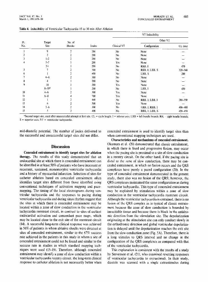
Table 4. Inducibility of Ventricular Tachycardia 15 to 30 min After Ablation
VT Inducibility
Pt. Target No. of Other VT
No. Site Shocks Joules Clinical VT Configuration CL(ms)
I 9 2 200 No None 2 2 2 200 No None 3 1-2 2 200 No None 4 5-7 200 Yes None
5+ 200 No RBB. I 470 5 7 2 300 No RBB. I; LBB. S 320-340 6 2 400 No LBB.S 280 7 4-6 2 300 No None 8 4 200 No None 9 10 200 Yes None
8-10' 200 No LBB.l 430 10 4-6 2 300 Yes None 11 6-8 4 700 Yes None 12 2 400 No RBB. I: LBB. I 280-350 13 6 2 500 Yes None 14 5-6 2 300 No LBB. I; RBB. I 400-480 15 5 2 400 No RBB. I: LBB. S 420-450
'Second target site. used after unsuccessful attempt at first site. CL = cycle length: I = inferior axis: LBB = left bundle branch: RB; = right bundle branch; S = superior axis; VT = ventricular tachycardia.
mid-diastolic potential. The number of joules delivered to the successful and unsuccessful target sites did not differ.
Discussion Concealed entrainment to identify target sites for ablation
therapy. The results of this study demonstrated that an endocardial site at which there is concealed entrainment can be identified in at least 50% of patients who have incessant or recurrent, sustained, monomorphic ventricular tachycardia and a history of myocardial infarction. Selection of sites for catheter ablation based on concealed entrainment often identifies target sites different from those identified using conventional techniques of activation mapping and pacemapping. The timing of the local electrograms during ventricular tachycardia and the responses to pacing during ventricular tachycardia and during sinus rhythm suggest that the sites at which there is concealed entrainment may be located within a zone of slow conduction in the ventricular tachycardia reentrant circuit, in contrast to sites of earliest endocardial activation and concordant pace maps, which may be located close to the exit site of the reentrant circuit (14). A successful long-term clinical response was achieved in 56% of patients in whom ablative shocks were directed at sites of concealed entrainment, similar to the 67% success rate achieved in the patients in this study in whom a site of concealed entrainment could not be found and similar to the success rate in studies in which standard mapping techniques were used (15-19). Therefore, although concealed entrainment may identify a zone of slow conduction within a ventricular tachycardia reentry circuit, the long-term clinical response to catheter ablation is not necessarily better when
concealed entrainment is used to identify target sites than when conventional mapping techniques are used.
Characteristics and mechanisms of concealed entrainment. Okumura et al. (20) demonstrated that classic entrainment, in which there is fixed and progressive fusion, may occur when the pacing site is proximal to a site of slow conduction in a reentry circuit. On the other hand. if the pacing site is distal to the zone of slow conduction, there may be concealed entrainment, in which no fusion occurs and the QRS complexes have purely a paced configuration (20). In the type of concealed entrainment demonstrated in the present study, there also was no fusion of the QRS; however, the QRS complexes maintained the same configuration as during ventricular tachycardia. This type of concealed entrainment may be explained by stimulation within a zone of slow conduction in the ventricular tachycardia reentrant circuit. Although the ventricular tachycardia is entrained, there is no fusion of the QRS complex as is typical of classic entrainment because the zone of slow conduction is bounded by inexcitable tissue and because there is block in the antidromic direction from the stimulation site. The depolarization originating at the stimulation site can only conduct slowly in the orthodromic direction and global ventricular depolarization is delayed until the depolarization reaches the exit site from the slow conduction zone (Fig. 5A). Therefore, there is a long stimulus to QRS interval and no change in the configuration of the QRS complexes as compared with that of the ventricular tachycardia.
This explanation is consistent with the results of a study by Stevenson et al. (21), who examined resetting responses of ventricular tachycardia to extrastimuli. In their study, diastole was scanned with a single extrastimulus during

686 MORADY ET AL. CONCEALED ENTRAINMENT
ventricular tachycardia, which. from a mechanistic standpoint. is analogous to pacing with trains of stimuli at incremental rates, as was done in the present study. Based on computer simulations of reentrant circuits, these investigators (21) concluded that whenever all capturing stimuli were followed after a delay by early QRS complexes that had the same configuration as that of the ventricular tachycardia. the pacing site was within a slowly conducting central pathway in the reentry circuit.
Although the long stimulus to QRS interval and lack of QRS fusion that occur during concealed entrainment may suggest that the site of stimulation is within a zone of slow conduction. theoretically the zone of slow conduction need not be a critical component of the reentry circuit. Findings typical of concealed entrainment may occur if a zone of slow conduction bounded by inexcitable tissue is located outside the reentrant circuit but is connected to it. However. in each of the patients in whom concealed entrainment was demonstrated. the timing of the endocardial electrogram relative to the QRS complex was similar during concealed entrainment and during the undisturbed ventricular tachycardia. If the pacing site is located in a zone of slow conduction that is a superfluous "blind alley." the local electrogram at the pacing site would be expected to occur earlier relative to the QRS complex during pacing than during ventricular tachycardia (Fig. 5B). On the other hand. if the pacing site is located in a zone of slow conduction that is an "alternate pathway." the response to pacing might be indistinguishable from the response when the pacing site is located in a critical zone of slow conduction (Fig. 5C).
lACC Vol. 17. No.3 March 1, 1991:678-89
Relation to endocardial activation times and pace mapping. Whereas localization of ventricular tachycardia conventionally has been based on identification of presystolic endocardial activation, the endocardial activation times at sites of concealed entrainment were 0 to + 50 ms in our patients who had a stimulus to QRS interval ::::280 ms. In addition, pace-mapping at sites of concealed entrainment demonstrated complete discordance with ventricular tachycardia. Because presystolic endocardial activity and a pace map concordant with ventricular tachycardia are characteristic of endocardial sites that are close to the exit site of a reentrant circuit, the absence of presystolic endocardial activity and concordant pace maps at sites of concealed entrainment suggest that these sites were located at some distance from the exit site. In accord with this possibility are the results of computerized mapping studies (22) in dogs that demonstrated late endocardial activation times at the proximal portion of the slow conduction zone during ventricular tachycardia.
Properties of the zone of slow conduction. In several patients, decremental conduction in the presumed zone of slow conduction was demonstrated by pacing at incremental rates. Stevenson et al. (21), by introducing single stimuli during ventricular tachycardia. also showed that the zone of slow conduction has decremental properties. In contrast, Almendral et al. (23) examined the responses to entrainment at incremental rates and observed a fixed conduction time between pacing stimuli and the local electrogram at the site of origin of the ventricular tachycardia; they concluded that decremental conduction properties were absent. This appar-
Table S. Inducibility of Ventricular Tachycardia 7 to 10 Days After Ablation and Long-Term Follow-Up
VT Inducibility
Pt. Other VTs Follow-up
No. Clinical VT Configuration CL(ms) Therapy Months Comments
Patients with a successful clinical outcome I No LBB,S 450 Amio 22 Died ofCHF* 2 No None Amio 18 Died ofCHF* 3 No None Amio 36 Alive & well 4 No RBB, I 520 Amio 23 Alive & well 5 No LBB,S 280 Encainide 22 Alive & well 6 No RBI 320 Amio 18 Alive & well 7 Yes None Flecainide 17 Alive & well 8 No None None 13 Alive & well 9 No LBB,S 430 None 12 Alive & well
Patients with an unsuccessful clinical outcome 10 Yes None Q,ATPM 7 Many VT episodes, sudden death II Yes RBB, S 390 SER 22 Alive & well, no more VT 12 No RBB, I; LBB, I 250-280 Amio, AICD 19 Recurrent VT x 2 13 Yes None Amio, AICD 12 Recurrent VT x I 14 Yes RBB, I; LBB, I 400-480 Amio, PA 8 Recurrent VT x I 15 No RBB, I; LBB, S 420-530 Amio, Q Many VT episodes, sudden death
*These two patients had preexisting severe congestive heart failure (CHF). AICD = automatic implantable cardioverter-defibrillator, ATPM antitachycardia pacemaker; CHF = congestive heart failure; Q = quinidine; SER = subendocardial resection; other abbreviations are in Tables I and 4.

JACC Vol. 17. No.3 March 1, 1991 :678-89
Figure 5. A figure-8 model of reentrant circuit and two variations. The ~batched 3I"l'a'i represent areas of conduction block. AlTows with solid 1m indicate normal conduction and arrows with dotted 1m indicate slow conduction. The recording and pacing site is at location x. The QRS complexes and local electrograms at location x are shown below each corresponding reentrant circuit. Although a figure-8 model is used here, the explanations would also apply to other models of reentry, such as the ring model. A, Site x is in the proximal portion of a zone of slow conduction critical for the maintenance of reentry. Global ventricular depolarization and the onset of the QRS complex occur when the wave of depolarization reaches the exit site (E). The local electrogram at site x occurs within the QRS complex because the waves of depolarization reenter the zone of slow conduction before global ventricular activation is complete. When pacing is instituted during ventricular tachycardia at site x, there is block in the antidromic direction and slow conduction to the exit site in the orthodromic direction. This results in a long stimulus to QRS interval. The timing of the local electrogram relative to the QRS complex is similar during ventricular tachycardia and during pacing because the sequence of activation between site x and the exit site is the same under both circumstances. B, Site x is located in a "blind alley," or "dead end" pathway, that is connected to the critical zone of slow conduction. Although site x is within a zone of slow conduction, this area is not critical for maintaining reentry. During ventricular tachycardia, site x is activated at approximately the same time as the wave of depolarization in the critical zone of slow conduction reaches the exit site. Accordingly, the local electrogram is synchronous with the QRS complex. However, during pacing, activation begins at site x and proceeds slowly toward the exit site. Therefore, the local electrogram at site x occurs earlier relative to the QRS complex during pacing than during ventricular tachycardia. C, Site x is located in a zone of slow conduction that serves as an alternative pathway that is not critical for the maintenance of reentry. In this case, activation between site x and the exit site occurs in the same orthodromic fashion during ventricular tachycardia and during pacing and, therefore, the timing of the local electrogram relative to the QRS complex is the same under both circumstances. The response of pacing does not allow differentiation ofthis type of alternate pathway from a critical zone of slow conduction.
VENTRICULAR TACHYCARDIA
PACING
A.r. ~
~ )
C.
E ................ ·x .. ..
~ ~ X h ....
X... he
Blind Alley
~ s s
'-;(~ iiiiiiiiiiiiiii ') AlIem,Ie '(I' iiiiIiiiiiiii ') ~PathWaY~
~ ~ s s
MORADY ET AL. 687 CONCEALED ENTRAINMENT
ent discrepancy has at least two possible explanations. Almost all of the patients in the present study and in the study of Stevenson et al. (21) were being treated with antiarrhythmic drugs, which may have induced or accentuated the decremental properties. Second, pacing stimuli were introduced within the presumed reentrant circuit in both studies, whereas the pacing site was in the right ventricle in the study by Almendral et al. (23). Decremental conduction may be more difficult to demonstrate when the pacing site is distant from the reentrant circuit.
Isolated mid-diastolic potentials. In a study by Fitzgerald et al. (9), target sites for catheter ablation of ventricular tachycardia were selected on the basis of isolated middiastolic potentials that could not be dissociated from the tachycardia. These potentials were believed to represent critical components of the ventricular tachycardia reentrant circuit. An isolated mid-diastolic potential was identified in 7 of 14 ventricular tachycardias, and in these cases the ventricular tachycardia reentrant circuit was successfully ablated. Perhaps the sites of which isolated mid-diastolic potentials were recorded would have demonstrated concealed entrainment; however this is unknown because pacing was not performed at these sites (9) . In the present study, an isolated mid-diastolic potential that could not be dissociated from the ventricular tachycardia was recorded at the target site in four patients, and the ablation procedure was successful in three of them. However, in only one of these four patients did pacing during ventricular tachycardia at the site of the isolated mid-diastolic potential result in concealed entrainment. This may suggest that the site at which isolated mid-diastolic potentials are recorded may not always be directly within a critical component of the reentrant circuit. Although ablative shocks targeted at a site at which isolated mid-diastolic potentials are recorded appear to have a high success rate, the injury induced by the shocks may be diffuse enough to ablate the reentrant circuit even if the target site is adjacent to the circuit but not directly within it.
Electrophysiologic versus clinical criteria for assessing outcome. In most patients there was concordance between the effects of the ablation attempt on the inducibility of the target ventricular tachycardia and the long-term clinical outcome. In eight of nine patients who had a successful clinical outcome, the clinical ventricular tachycardia that had been the target of ablation was no longer inducible after ablation. The exception was Patient 7, who had a dramatic clinical response even though the original clinical ventricular tachycardia was inducible 1 week after ablation. However, whereas the ventricular tachycardia had been incessant at the time of the ablation, it was inducible only by programmed stimulation with three extrastimuli in the follow-up electrophysiologic test. The reason for the discordant electrophysiologic and clinical responses is unknown. Although the ventricular tachycardia reentrant circuit was not ablated in this patient, the shocks may have modified the myocardium adjacent to the circuit in such a way as to suppress spontaneous initiation of ventricular tachycardia.

688 MORADY ET AL. CONCEALED ENTRAINMENT
In five of nine patients who had a successful clinical outcome, one or more types of monomorphic ventricular tachycardia different from the ablated ventricular tachycardia were inducible after ablation. These ventricular tachycardias were considered nonclinical because they had never been documented to occur spontaneously. The successful clinical outcome in these patients during 12 to 22 months of follow-up suggests that such nonclinical ventricular tachy- -cardias may not have clinical significance and do not require therapy.
In two patients the clinical outcome was considered unsuccessful although the original ventricular tachycardia that had been the ablation target was not inducible I week after ablation. These two patients had multiple recurrences of ventricular tachycardia that were not documented, and recurrences of the original clinical ventricular tachycardia could not be ruled out.
Predictors of successful outcome. Among the several clinical and electrophysiologic variables examined, the only one significantly associated with clinical outcome was incessant ventricular tachycardia, and each of the five patients with this arrhythmia had a successful long-term clinical outcome. Whatever characteristics of a reentrant circuit account for incessancy of ventricular tachycardia may also make the circuit more amenable to ablation.
Shocks delivered to a site at which there was concealed entrainment may have been unable to ablate ventricular tachycardia for several possible reasons. The zone of slow conduction identified by concealed entrainment may have been an "alternate pathway" that was not critical to the maintenance of reentry (Fig. 5C). The target sites may have been within a critical zone of slow conduction, but the injury induced by the ablative shocks may have been inadequate to abolish conduction through these zones. This might occur if the critical zone of slow conduction at the target site was wide instead of a narrow isthmus, if scarring and fibrosis at the target site limited the extent of injury caused by the ablative shocks, or ifthe delivered energy of the shocks was insufficient.
Limitations. To rule out classic entrainment, our criteria for concealed entrainment included the absence of QRS fusion at incremental pacing rates. However, when the pacing rate is rapid, it is conceivable that the antidromic wave front from a pacing site within a zone of slow conduction could exit the reentrant circuit at its entrance before colliding with the orthodromic wave front, and this might result in fusion of the QRS complex. In accord with this possibility, Stevenson et al. (21) demonstrated with computer simulations that if a pacing site is within a reentrant circuit but near its entrance, a single stimulus late in diastole during ventricular tachycardia might not alter the QRS configuration, whereas an early stimulus might do so. Therefore, a zone of slow conduction might have been detected in a larger percentage of patients in this study if less stringent criteria for the absence of Q RS fusion had been used. On the other hand, the occurrence of QRS fusion during entrain-
JACC Vol. 17, No.3 March I, 1991 :678-89
ment would not have allowed differentiation of pacing sites within and outside the zone of slow conduction.
A second limitation of this study is that once a site of concealed entrainment was identified, additional mapping usually was not performed for fear of being unable to again locate the desired target site if the catheter was moved. Therefore, mapping data obtained from sites other than target sites were often incomplete, especially when a suitable target site was identified early in the mapping process.
This study was not designed to compare the results of catheter ablation guided by concealed entrainment with those of other mapping techniques. Although the long-term efficacy was similar whether target sites were selected on the basis of concealed entrainment, isolated mid-diastolic potentials or endocardial activation times and pace-mapping, these mapping techniques were not assigned in random fashion but were prioritized.
Another limitation of this study is that some of the results are operator-dependent. The percent of patients in whom concealed entrainment can be demonstrated or the earliest endocardial activation time which can be found may be in large part dependent on the skill and tenacity of the person manipulating the mapping catheter. Therefore, although a site at which there was concealed entrainment was identified in 60% of the patients in this series, the percent may have been higher in other hands.
In this study, we have presumed that a markedly prolonged stimulus to QRS interval during concealed entrainment reflects conduction in a zone of slow conduction. However, as pointed out by Almendral et al. (23), a long conduction time does not prove slow conduction; it may reflect a longer pathway of activation. Direct measurements of conduction velocity would be necessary to distinguish slow conduction velocity from normal velocity over a long pathway.
Conclusions. An endocardial site at which there is concealed entrainment during ventricular tachycardia can be identified in at least 50% of patients who have incessant or recurrent, sustained, monomorphic ventricular tachycardia and a history of myocardial infarction. The responses to pacing during ventricular tachycardia and during sinus rhythm suggest that the phenomenon of concealed entrainment may identify a zone of slow conduction that is either within or connected to the ventricular tachycardia reentry circuit. If the timing of the endocardial electrogram relative to the QRS complex at the site of concealed entrainment is the same during entrainment and during ventricular tachycardia, this may indicate that the zone of slow conduction is a critical link in the reentry circuit and therefore a suitable
'target site for catheter ablation. Sites at which there is concealed entrainment often are not the same sites at which the earliest endocardial activation time, a concordant pace map, or an isolated mild-diastolic potential are found.
The long-term clinical efficacy of catheter ablation targeted to sites of concealed entrainment is about 60%, similar to the clinical results achieved with conventional mapping

JACC Vol. 17, No.3 March I, 1991:678-89
techniques. However, catheter ablation guided by concealed entrainment was uniformly successful in the subgroup of patients in this study who had incessant ventricular tachycardia, suggesting that concealed entrainment may be particularly useful as a guide for ablation of this type of tachycardia. Whether anyone of the various techniques for identifying target sites for catheter ablation will lead to better clinical results than the others remains to be determined.
We are grateful to Marion Maguire and Joan Stea for typing the manuscript and to the staff of the electrophysiology laboratory for their technical assistance.
References I. Stevenson WG, Weiss J, Wiener I, Wohlgelernter D, Yeatman L.
Localization of slow conduction in a ventricular circuit: implications for catheter ablation. Am Heart J 1987;1 14: 1253-8.
2. Stevenson WG, Weiss IN. Wiener I, et al. Resetting of ventricular tachycardia: implications for localizing the area of slow conduction. J Am Coli CardioI1988;11:522-9.
3. Morady F, Frank R, Kou WHo et al. Identification and catheter ablation of a zone of slow conduction in the reentrant circuit of ventricular tachycardia in humans. J Am Coli Cardiol 1988;1 1:775-82.
4. Borggrefe AP. Martinez-Rubio A, Breithardt G. Termination ofre-entrant ventricular tachycardia of subthreshold stimulus applied to the zone of slow conduction. Eur Heart J 1988;9: 1146-50.
5. Frank R. Tonet JL. Kounde S. Farenq G. Fontaine G. Localization of the area of slow conduction during ventricular tachycardia. In: Brugada P. Wellens HJJ, eds. Cardiac Arrhythmias: Where To Go From Herery New York: Futura Publishing. 1987: 191-208.
6. Josephson ME. Horowitz LN, Spielman SR. Waxman HL. Greenspan AM. Role of catheter mapping in the preoperative evaluation of ventricular tachycardia. Am J Cardiol 1982:49:207-20.
7. Okumura K, Olshansky B, Henthorn RW. Epstein AE. Plumb VJ. Waldo AL. Demonstration of the presence of slow conduction during sustained ventricular tachycardia in man: use of transient entrainment of the tachycardia. Circulation 1987 ;75 :369-78.
8. Henthorn RW, Okumura K, Olshansky B, Plumb VJ. Hess PG, Waldo AL. A fourth criterion for transient entrainment: the electrogram equivalent of progressive fusion. Circulation 1988;77: 1003-12.
9. Fitzgerald DM, Friday KJ, Yeung Lai Wah JA, Lazzara R, Jackman WM. Electrogram patterns predicting successful catheter ablation of ventricular tachycardia. Circulation 1988;77:806-14.
MORADY ET AL. 689 CONCEALED ENTRAINMENT
10. Josephson ME, Horowitz LN, Farshidi A, Spear JE, Kastor JA, Moore EN. Recurrent sustained ventricular tachycardia. 2. Endocardial mapping. Circulation 1978:57:440-7.
II. Horowitz LN. Josephson ME, Harken AH. Epicardial and endocardial activation during sustained ventricular tachycardia in man. Circulation 1980;61: 1227-37.
12. Waxman HL. Josephson ME. Ventricular activation during ventricular endocardial pacing. I. Electrocardiographic pattern related to the site of pacing. Am J Cardiol 1982;50: 1-10.
13. Josephson ME, Waxman HL. Cain ME, Gardner MJ, Buxton AE. Ventricular activation during ventricular endocardial pacing. II. Role of pace-mapping to localize origin of ventricular tachycardia. Am J Cardiol 1982;50: 11-22.
14. Stevenson WG, Weiss IN, Wiener I, Nadamanee K. Slow conduction in the infarct scar: relevance to the occurrence, detection, and ablation of ventricular reentry circuits resulting from myocardial infarction. Am Heart J 1989;117:452-64.
15. Belhassen B, Miller HI. Geller E. Laniado S. Transcatheter electric shock ablation of ventricular tachycardia. J Am Coli Cardiol 1986;7: 1347-55.
16. Morady F, Scheinman MM, DiCarlo LA, et al. Catheter ablation of ventricular tachycardia with intracardiac shocks: results in 33 patients. Circulation 1987:5: 1037-49.
17. Fontaine G, Tonet JL, Frank R, et al. Treatment ofresistant ventricular tachycardia by endocavitary fulguration associated with antiarrhythmic therapy. In: Fontaine G, Scheinman MM, eds. Ablation in Cardiac Arrhythmias. New York: Futura Publishing, 1987:337-45.
18. Garan H, Kuchar D, Freeman C, Finkelstein D, Ruskin IN. Early assessment of the effect of map-guided transcatheter intracardiac electric shock on sustained ventricular tachycardia secondary to coronary artery disease. Am J CardioI1988;61:1018-23.
19. Borggrefe M. Breithardt G, Podczeck A, Rohner D, Budde T, MartinezRubio A. Catheter ablation of ventricular tachycardia using defibrillator pulses: electrophysiological findings and long-term results. Eur Heart J 1989;10:591-601.
20. Okumura K, Henthorn RW, Epstein AE, Plumb VJ, Waldo AL. Further observations on transient entrainment: importance of pacing site and properties of the components of the reentry circuit. Circulation 1985;72: 1293-1307.
2J. Stevenson WG, Nademanee K. Weiss IN, et al. Programmed electrical stimulation at potential ventricular reentry circuit sites: comparison of observations in humans with predictions from computer simulations. Circulation 1989;80:793-806.
22. EI-Sherif N. Mehra R, Gough WB. Zeiler RH. Reentrant ventricular arrhythmias in the late myocardial infarction period: interruption of reentrant circuits by cryothermal techniques. Circulation 1983;68:644-56.
23. Almendral JM, Gottlieb CD, Rosenthal ME, et al: Entrainment of ventricular tachycardia: explanation for surface electrographic phenomena by analysis of electrograms recorded within the tachycardia circuit. Circulation 1988:77:569-80.