REVERSAL OF DOXORUBICIN-INDUCED CARDIAC METABOLIC DAMAGE BY?-CARNITINE

Mitochondrial localization and activity of P-glycoprotein indoxorubicin-resistant K562 cells
Eliza Munteanu a, Mireille Verdier a, Fabienne Grandjean-Forestier a, Christophe Stenger a,Chantal Jayat-Vignoles b, Sylvie Huet c, Jacques Robert c, Marie-Helene Ratinaud a,*a Laboratoire de Physiologie Mitochondriale, EA 3842 Faculte de Medecine, 2 rue du Dr Marcland, 87025 Limoges Cedex, Franceb Institut de Biotechnologie, Faculte des Sciences & Techniques, 123 Av. A. Thomas, 87060 Limoges Cedex, Francec Laboratoire de Pharmacologie des Agents Anticancereux, Institut Bergonie, 229 cours de l’Argonne, 33076 Bordeaux Cedex, France
b i o c h e m i c a l p h a r m a c o l o g y 7 1 ( 2 0 0 6 ) 1 1 6 2 – 1 1 7 4
a r t i c l e i n f o
Article history:
Received 27 October 2005
Accepted 9 January 2006
Keywords:
Multidrug resistance
P-glycoprotein
Mitochondria
Doxorubicin
Intracellular P-gp
Abbreviations:
ABC, ATP-binding cassette protein
FITC, fluorescein isothiocyanate
LRP, lung resistance protein
MDR, multidrug resistance
PE, phycoerythrin
P-gp, P-glycoprotein 170
a b s t r a c t
It is now well-established that P-glycoprotein 170 (P-gp), an efflux pump involved in
multidrug resistance (MDR) is overexpressed at the plasma membrane of doxorubicin-
resistant K562 leukemia cells. Nevertheless, several results suggested: (i) that P-gp-mediated
drug efflux was not the only mechanism involved in resistance; (ii) that intracellular
compartments could accumulate the drug, preventing it from reaching its nuclear targets;
(iii) that agents able to reverse multidrug resistance may lead to intracellular drug redis-
tribution. We have studied the localization of P-gp in mitochondria as well as its functional
properties in this compartment. Using several monoclonal antibodies (MoAbs) directed
against different P-gp epitopes, a protein was detected in the cytoplasm of two doxorubicin-
resistant K562 sublines and, by confocal laser scanning microscopy, this protein was shown
to co-localize in the Golgi apparatus and in mitochondria, in equivalent proportions. Purified
mitochondria were isolated from K562 cell variants; the presence of a protein of about
170 kDa and reacting with several anti-P-gp antibodies was assessed in MDR cells by
Western blotting and flow cytometry. Functional assays have shown that mitochondrial
P-gp was involved in doxorubicin accumulation inside the organelle but not in its efflux,
suggesting an orientation of P-gp in the mitochondrial membrane inverse to that observed
in the plasma membrane. A potential role for mitochondrial P-gp in MDR cells would be to
protect the nucleus from doxorubicin. This is the first demonstration of the presence and
functional activity of P-gp in mitochondria of MDR cells.
# 2006 Elsevier Inc. All rights reserved.
avai lable at www.sc iencedi rec t .com
journal homepage: www.e lsev ier .com/ locate /b iochempharm
1. Introduction
Resistance to antineoplastic drugs represents a major obstacle
to the success of cancer chemotherapy. Frequently, drug
resistance concerns simultaneously a variety of chemically
unrelated compounds, giving rise to multidrug resistance
(MDR) phenotypes. Different mechanisms of MDR have been
* Corresponding author. Tel.: +33 555 435 849; fax: +33 555 435 983.E-mail address: [email protected] (M.-H. Ratinaud).
0006-2952/$ – see front matter # 2006 Elsevier Inc. All rights reserveddoi:10.1016/j.bcp.2006.01.006
identified, involving especially common drug detoxification
pathways or alterations in common drug targets [1]. The first
and the best characterized MDR mechanism is due to the over
expression of a transmembrane active pump called P-
glycoprotein 170 (P-gp), a member of the ATP-binding cassette
(ABC) protein super family [2]. This protein of 1280 amino acids
is able to expel various agents, such as anthracyclines, out of
.

b i o c h e m i c a l p h a r m a c o l o g y 7 1 ( 2 0 0 6 ) 1 1 6 2 – 1 1 7 4 1163
the cell. The overexpression of P-gp can be due to the
amplification of the MDR1 gene or to a positive transcriptional
regulation. Overexpression of P-gp occurs in vitro in numerous
drug-selected cell lines as well as in vivo in different types of
solid tumors and leukemias.
In the past decade, the existence of alternative and/or
additional mechanisms of multidrug resistance became
obvious. Several ABC proteins, such as the MDR-related
protein (MRP1) or the breast cancer resistance protein (BCRP)
have been described [3]. Non-ABC proteins may also be
involved, such as the lung resistance protein (LRP), which may
play a role in the nuclear–cytoplasmic transport of cytotoxic
agents [4]. In previous studies, we have shown that the MDR
phenotype of doxorubicin-selected variants of the human
myeloid leukemia cell line K562 was related to the over-
expression of P-gp [5], occurring concomitantly with the
disappearance of LRP protein [6]. In addition, it has been
observed that the reversal of MDR in K562-resistant cells was
not complete with P-gp inhibitors, such as verapamil or
cyclosporine A [5], but the expression of oiher transporters
was not likely to be involved in the accumulation and efflux of
doxorubicin [6]. Thus, it could be hypothesized that oiher
mechanisms of resistance were operative in these cell lines,
especially through intracellular redistribution of drugs, as
suggested by other studies (see for review [7]).
Although primarily located in the plasma membrane, P-gp
has been detected also in the nucleus [8], in the Golgi
apparatus [9] as well as in unidentified organelles [10–12].
Bennis et al. [5] observed a preferential accumulation of
doxorubicin in subcellular components distinct from nucleus
in doxorubicin-resistant K562 cells. The reversal of the MDR
phenotype by quinine in these cells was not accompanied by
the restoration of its cellular accumulation, but by the drug
redistribution from these sequestration compartments to the
nucleus. Quinine is a P-gp inhibitor, but it is also known to
antagonize the mitochondrial permeability transition pore
(PTP or megachannel). This multiprotein complex is involved
in the transport of small molecules between the cytoplasm
and the mitochondrial matrix and in the execution phase of
apoptosis [13]. In addition, a study suggested that daunor-
ubicin was sequestered in mitochondria in a MDR K562 cell
variant [14]. These authors observed that the green fluores-
cence of daunorubicin was located in the perinuclear region
and at the cell periphery. On the other hand, a similar
distribution of the fluorescence was observed with rhodamine
123, a mitochondrial fluorescent probe. Thus, this organelle
appears to be a good candidate for doxorubicin sequestration
in K562 MDR cells, possibly implying the megachannel and/or
P-gp for accumulation and efflux of the drug. In view of this
hypothesis, it was necessary to clarify the possible involve-
ment of mitochondria as a drug sequestration compartment.
This property could be related to mitochondrial expression of
P-gp.
In this purpose, we analyzed simultaneously by confocal
microscopy, after immunostaining with monoclonal antibo-
dies (MoAbs), the expression of intracellular P-gp in the Golgi
apparatus and in mitochondria. In addition, Western blot and
immunocytometric assays were carried out on isolated whole
mitochondria from K562-sensitive and doxorubicin-resistant
cell lines, with different anti-P-gp monoclonal antibodies.
These analyses demonstrated the presence of P-gp in
mitochondria of K562-resistant cell lines. Functional assays
were performed on isolated whole mitochondria to assess
their capacity for doxorubicin sequestration.
2. Materials and methods
2.1. Cell line and culture conditions
Doxorubicin-sensitive K562 cells and their doxorubicin-resis-
tant counterparts [5,6] were grown in suspension in RPMI 1640
medium containing 10% (v/v) heat-inactivated fetal calf serum
(Eurobio) and 2 mM L-glutamine (Eurobio, France). Cells were
incubated at 37 8C in a humidified atmosphere containing 5%
CO2 The resistant cell lines were obtained by progressive
adaptation of the parental sensitive cells (K562/S) to 0.2 mg/ml
of doxorubicin for K562/0.2R cells, and then further to 0.5 mg/
ml of doxorubicin for K562/0.5R cells [6]. Before the experi-
ments, the resistant cell lines were maintained without drug
for at least 8 weeks for a complete elimination of doxorubicin.
In this way, all the modifications observed were not due to a
direct effect of doxorubicin to cells, but were related to the cell
phenotype.
2.2. Monoclonal antibodies
For immunostaining studies of P-gp, we have used several
monoclonal antibodies, reacting specifically with different
epitopes of human P-gp. Two MoAbs recognize P-pg external
epitopes. The MoAb named UIC2 [15], an IgG2a which
recognizes a conformational epitope, was purchased from
Beckman-Coulter (Marseille, France) and was used either
uncoupled or directly coupled to phycoerythrin (PE). The F4
MoAb [16], an IgG1 which binds to an epitope located in the N-
terminal extracellular loop, was purchased from Sigma–
Aldrich Chimie (Saint-Quentin-Fallavier, France). A third
MoAb, C494 [17], an IgG2a reacting with a P-pg internal epitope,
was purchased from Dako (Trappes, France). Immunostaining
of mitochondria was performed with M117, an IgG1 obtained
from Leinco Technologies (Saint-Louis, USA), which recog-
nizes a 60 kDa non-glycosylated human mitochondrial protein
[18]. Immunostaining of the Golgi apparatus was performed
with an anti-Golgi antibody, 58 K, an IgG1 recognizing a 58 kDa
protein (Sigma–Aldrich Chimie). A fluorescein isothiocyanate
(FITC)-conjugated goat anti-mouse antibody (Pharmingen,
San Diego, CA, USA) was used as secondary antibody.
2.3. Expression of external and total P-gp by flowcytometry
Immunofluorescent staining of doxorubicin-sensitive and -
resistant K562 cells was first achieved to determine the
concentration of UIC2 necessary to saturate all P-gp sites. For
the measurement of total P-gp (that accessible on the plasma
membrane and the intracellular one), cell fixation and
permeabilization were performed before staining with the
Intrastain kit (Dako) according to manufacturer’s instructions.
This was not done for the study of the plasma membrane-
associated P-gp since immunostaining was carried out on

b i o c h e m i c a l p h a r m a c o l o g y 7 1 ( 2 0 0 6 ) 1 1 6 2 – 1 1 7 41164
intact living cells. In both cases, before staining, cells were
washed twice in phosphate-buffered saline (PBS). Then,
7 � 105 cells/sample were incubated for 30 min at 4 8C with
different concentrations of the primary MoAb (UIC2), ranging
from 0.5 to 5 mg. This was done before fixation for plasma
membrane P-gp and after cell fixation and permeabilization
for total P-gp. Cells were then washed twice in PBS containing
1% bovine serum albumin (PBS–BSA) and the pellet was
resuspended in the presence of 4 mg of the secondary antibody
diluted in PBS–BSA. After 30 min incubation at 4 8C, cells were
washed twice in PBS–BSA and resuspended in 500 ml of PBS–
paraformaldehyde (PFA) 1%. An isotypic control (IgG2a, Beck-
man-Coulter) was processed to determine non-specific bind-
ing. Cell fluorescence was analyzed with a FACS Vantage flow
cytometer (Becton-Dickinson, Grenoble, France). For each
sample, 104 cells were analyzed after elimination of dead
cells, debris and aggregates, with a flow rate of 1000 cells/s. For
each immunostaining, the mean fluorescence signal of the
corresponding negative control was subtracted from the mean
fluorescence signal of the assay.
2.4. In situ analysis of P-gp expression by confocal laserscanning microscopy
For single immunostaining of doxorubicin-sensitive and -
resistant K562 cells, two staining protocols were used: (i) for P-
gp expressed on the plasma membrane, 2.5 � 105 cells/sample
were stained with UIC2-FITC (3 mg/assay); after washing, cells
were fixed with PBS–PFA, and counter-staining of the nucleus
was realized with propidium iodide (PI) (5 mg/assay) and (ii) for
P-gp expressed intracellularly and on the plasma membrane
(total P-gp), cells were first fixed and permeabilized with the
Intrastain kit, and P-gp was then stained with UIC2-FITC (3 mg/
assay); after washing, a counter-staining of nucleus was
realized with PI (5 mg/assay).
For the co-localization experiments, cells were first fixed
and permeabilized with the Intrastain kit. Mitochondria and
the Golgi apparatus were, respectively, stained with M117
(25 ml/assay) and 58 K (2 mg/assay) MoAbs, which were
revealed by a 4 mg of the secondary antibody diluted in PBS–
BSA. Immunostaining of P-gp was revealed with PE-associated
UIC2 (3 mg/assay).
Two confocal imaging analyzers were used. In a first set of
experiments, we employed an ACAS 570 (Meridian, Okemos,
MI, USA), located in Institut Pasteur, Paris, consisting of a
200 mW argon ion laser (Coherent 92-S, Palo Alto, CA, USA),
coupled with an attenuator 7% filter, and emitting at 488 nm.
An optical microscope (Olympus, Tokyo, Japan) was equipped
with an oil immersion objective lens (�63). Collection of the
FITC green fluorescence of the Golgi apparatus or mitochon-
dria was carried out through a band-pass filter (530 � 15 nm)
and PE orange-red fluorescence of P-gp was collected through
a high-pass filter (575 nm). In a second set of experiments,
samples were analyzed using a fluorescence microscope
(Axiovert 200M, Zeiss), equipped with laser scanning confocal
imaging system (LSM 510 Meta, Zeiss), comprising one 30 mW
argon and two He–Neon lasers. After immunostaining of P-gp
with a FITC-conjugated MoAb and counter-staining of nucleus
with PI, green and red fluorescence signals were collected,
respectively, with BP 505–530 and 560–615 nm emission filters.
For the double immunostaining of mitochondria and P-gp
(FITC green and PE orange-red fluorescences), the Lambda
mode was used; images were collected to each 10.7 nm after
excitation at 488 nm, in order to reduce fluorescence over-
lapping. Image processing was obtained with a LSM 510
computer software (Zeiss).
2.5. Preparation of isolated mitochondria from K562 cells
Mitochondria were prepared according to Bourgeron et al. [19]
as modified by Denis-Gay et al. [20]. The purification degree of
mitochondria obtained by this method was previously
checked, especially concerning the absence of debris originat-
ing from other cellular membrane structures [21]. Further
purification of mitochondria was avoided since it has been
shown that it increased the risk of undesirable selection of a
particular fraction of the mitochondrial population, poten-
tially leading to the loss of altered mitochondria [22]. All steps
of the purification were performed at 4 8C. Cells (5 � 106) were
collected by centrifugation (600 � g for 10 min at 4 8C) and were
then washed twice in Tris–HCl 10 mM, pH 7.4, containing
100 mM sucrose, 1 mM EGTA, 20 mM MOPS and 1 mg/ml BSA.
The pellet was resuspended in the buffer solution supple-
mented with 10 mM triethanolamine, 5% (v/v) Percoll and
0.1 mg/ml digitonin, for 5 min at 4 8C. Finally, the mixture was
homogenized in a Potter homogenizer (10 strokes, 1000 rpm)
before being diluted at the ratio of 1/5 in Tris–HCl 10 mM, pH
7.4, containing 300 mM sucrose, 1 mM EGTA, 20 mM MOPS and
1 mg/ml BSA, and centrifuged at 2500 � g for 5 min at 4 8C. The
supernatant, containing mitochondria, was collected,
whereas the pellet was resuspended in the last buffer and
re-centrifuged. The supernatants were combined and cen-
trifuged at 10,000 � g for 10 min at 4 8C in order to collect
mitochondria in the pellet. Isolated mitochondria were
washed twice in the same conditions before being resus-
pended in hypertonic Tris–HCl 10 mM, pH 7.4, containing 0.3 M
mannitol, 10 mM KCl, 5 mM MgCl2, 10 mM KH2PO4, 1 mg/ml
BSA and 10 mM succinate. Mitochondrial integrity was
assessed by a respiratory functional test according to
Bourgeron et al. [19]. The mitochondrial fraction could be
then used for the different analyses described below.
2.6. Western blot analysis of P-gp expression in whole cellsand mitochondria
Detection and analysis of P-gp expression from cells and
mitochondrial fractions isolated from parental and MDR
variant cell lines were performed by immunoblotting. Equiva-
lent amounts of cell proteins (80 mg) were suspended in a
denaturing buffer (Tris–HCl 10 mM, pH 8, containing 5%
glycerol, 2.5% SDS, 5% b-mercaptoethanol and 0.005% bro-
mophenol blue). Proteins from cells or mitochondria were
resolved by SDS-PAGE (8%) electrophoresis gels, and electro-
blotted onto PVDF membranes (Amersham). After 2 h incuba-
tion at 4 8C with 1% blocking reagent, the membranes were
incubated overnight at room temperature with the F4 MoAb
for K562 whole cell lysates. Mtochondrial proteins were
resolved under the same conditions as whole cell lysates
using either 60 mg of proteins and the F4 MoAb (15.6 mg/ml).
Blots were revealed by chemoluminescence using a rabbit

b i o c h e m i c a l p h a r m a c o l o g y 7 1 ( 2 0 0 6 ) 1 1 6 2 – 1 1 7 4 1165
anti-mouse peroxidase-labeled secondary antibody (1.3 mg/
ml) purchased from Dako, with the chemiluminescence
blotting substrate kit (Roche, Meylan, France) according to
manufacturer’s instructions, and evaluated with the Image
station 440 CF from Kodak (Boston, MA, USA).
2.7. P-gp expression in isolated mitochondria by flowcytometry
For each assay, an aliquot of isolated whole mitochondria
(50 mg proteins) from sensitive and resistant K562 cells was
resuspended in 500 ml hypertonic buffer. Mitochondria were
incubated for 1 h at 4 8C with different concentrations of the
primary MoAb (ranging from 0.5 to 6 mg for UIC2, 1 to 8 mg for
F4 and 0.5 to 12 mg for C494). Optimal labeling of organelles was
obtained with 6 mg of UIC2, 8 mg of F4 and 6 mg of the FITC-
coupled secondary antibody, as described above for intact
cells. For labeling of mitochondria with C494, a particular
protocol was used. This MoAb cross-reacts with pyruvate
carboxylase (PC), an abundant mitochondrial enzyme [23]. The
non-specific immunoreactivity was abolished by pre-incubat-
ing C494 in the presence of an excess of the synthetic-specific
peptide (TLEG). Briefly, C494 (12 mg) was pre-incubated for 1 h
at 4 8C in distilled water without (control C494) or in presence
(TLEG C494) of 10–100-fold molar excess of PC epitope-specific
peptide. Then, the mitochondria samples were stained either
with control C494 or TLEG C494 as described. All washes were
performed in the hypertonic buffer. An isotypic control
(Beckman-Coulter) was processed to determine non-specific
binding. To present P-glycoprotein expression, we applied the
consensus method for P-gp detection in human hematological
malignancies [24,25]. Thus, the mean fluorescence was
expressed as the ratio of the fluorescence signal of the assay
versus the fluorescence signal of the corresponding negative
control.
2.8. Functional assay of mitochondrial P-gp by flowcytometry
Whole isolated mitochondria (50 mg proteins in hypertonic
buffer) from sensitive and resistant lines were divided in test
tubes to evaluate mitochondrial autofluorescence as well as
the uptake and efflux of doxorubicin into and out of
organelles, either without any other drug, or in the presence
of specific MoAbs (4 mg of UIC2 or 1.5 mg of F4), or in the
presence of inhibitors (5 mM cyclosporine A or 10 mM dex-
verapamil or 50 mM quinine). Pre-incubations were performed
at 20 8C for 1 h (MoAbs) or 30 min (inhibitors). Then,
doxorubicin (10 mM final) was added to all samples, which
were further incubated for 1 h at 20 8C, avoiding light
exposure. After incubation, the samples were kept on ice for
a few minutes until flow cytometric analysis. Concomitantly,
in order to estimate doxorubicin efflux from mitochondria,
2 ml of hypertonic buffer were added in appropriate samples.
Mitochondria were centrifuged for 5 min at 450 � g at 4 8C and
washed once more with 2 ml of buffer, then diluted in 500 ml of
buffer at 20 8C. All tubes were incubated for 1 h at 20 8C. The
fluorescence of doxorubicin was measured in each sample (for
uptake and retention evaluation), on 10,000 isolated mito-
chondria with a flow rate of 500 events/s on a FACS Vantage
flow cytometer (Becton-Dickinson), after elimination of debris
and aggregates.
2.9. Statistics
An unpaired Student’s t-test of the StatviewTM software was
used for all experiments. Coefficients of variation (for
cytometric or microscopic analyses) corresponded to standard
deviation/mean and were expressed in percentage.
3. Results
3.1. Studies in whole cells
We first investigated the intracellular expression of P-gp in
doxorubicin-sensitive and MDR K562 cells by flow cytometry,
using UIC2. P-gp expression was estimated on the plasma
membrane in comparison with its total expression in whole
cells (Fig. 1 and Table 1). On plasma membranes (mb-P-gp), the
protein was undetectable in K562-sensitive cells and strongly
expressed in resistant cells. Despite a lower level of resistance,
P-gp expression at the membrane level was 2.35-fold higher in
K562/0.2R than in K562/0.5R (p < 0.05). In whole cells, P-gp was
barely detected in sensitive K562 cells, and strongly expressed
in both K562-resistant lines. It was not possible to determine
the saturating concentration of UIC2 in K562-sensitive cells,
even with high quantities of MoAb (up to 10 mg), because
fluorescence was always parallel to that of the isotypic control.
As a consequence, it is likely that UIC2 binding was not specific
in K562-sensitive cells. In resistant cells, total P-gp expression
was 1.97-fold higher in K562/0.2R than K562/0.5R ( p < 0.05).
The proportion between external and internal P-gp was
slightly and not significantly different in the two variants:
about 57% of total P-gp was associated to the plasma
membrane of K562/0.2R cells and 48% for K562/0.5R cells.
Similar results were obtained with F4 MoAb.
The expression of membrane-associated P-gp and total P-
gp was also studied by confocal microscopy with UIC2. In order
to obtain a better discrimination of P-gp expression, a counter-
staining of the nucleus was realized with addition of
propidium iodide just before microscopic analyses. As
expected, no expression was observed on the plasma
membrane of sensitive cells (Fig. 2A), whereas, in both MDR
variants, membrane-associated P-gp gave a strong signal
(Fig. 2B and C). Total P-gp expression was then evaluated in
whole cells after permeabilization and staining with UIC2
MoAb. In sensitive cells, P-gp gave a faint signal in the
cytoplasm (Fig. 2D). In both MDR variants, P-gp was strongly
expressed at the membrane level and in the cytoplasm of the
cells (Fig. 2E and F). The total P-gp label was lower in the K562/
0.5R variant than in the K562/0.2R.
In order to test the hypothesis of the presence of
mitochondrial P-gp in K562 MDR cells, various experiments
were carried out. The expression of P-gp at the mitochondrial
level was sought by confocal microscopy in whole cells. After
fixation and permeabilization, P-gp was stained with PE-
conjugated UIC2 and mitochondria were stained with M117
and revealed by a FITC-coupled secondary antibody. Non-
specific binding in the three K562 cell lines, estimated with

b i o c h e m i c a l p h a r m a c o l o g y 7 1 ( 2 0 0 6 ) 1 1 6 2 – 1 1 7 41166
Fig. 1 – Example of representative histograms obtained by flow cytometry with K562 cell lines (sensitive and resistant
variants). For each staining of plasma membrane P-gp (mb-P-gp) (living cells) or total P-gp (fixed cells), 7 � 105 cells were
incubated for 30 min at 4 8C with 3 mg UIC2 MoAb. This was done before fixation for plasma membrane P-gp and after cell
fixation and permeabilization with the Intrastain kit (Dako) for total P-gp. After washing, the pellet was resuspended with
4 mg of the secondary antibody coupled to FITC. After 30 min incubation at 4 8C, cells were washed twice and resuspended
in 500 ml of PBS–PFA 1%. An isotypic control (IgG2a) was processed to determine non-specific binding. The relative mean of
green fluorescence was evaluated on 104 cells for each sample after elimination of dead cells, debris and aggregates.
Cumulative data are shown in Table 1. (For interpretation of the references to color in this figure caption, the reader is
referred to the web version of the article.)

b i o c h e m i c a l p h a r m a c o l o g y 7 1 ( 2 0 0 6 ) 1 1 6 2 – 1 1 7 4 1167
Table 1 – Flow cytometric analyses of plasma membrane and whole cell P-gp expression in K562 cell lines
Cell lines K562/S K562/0.2R K562/0.5R
mb-P-gp expression (UIC2) 0.95 � 0.05 150 � 16*** 63.7 � 6.0***,y
Total P-gp expression (UIC2) 37.7 � 8.2 262 � 20*** 133 � 14*,y
mb-P-gp (mean value) (%) 2.5 58 48
Intracellular P-gp (mean value) (%) 97.5 42 52
For each staining of plasma membrane P-gp (mb-P-gp) (living cells) or total P-gp (fixed cells), 7 � 105 cells/sample were washed twice in PBS. For
the measurement of total P-gp, cell fixation and permeabilization were performed with the Intrastain kit (Dako) before staining with MoAbs. In
all cases, cells were incubated for 1 h at 4 8C with the UIC2 MoAb (3 mg/assay). Cells were then washed twice and the pellet was resuspended
with 4 mg of the secondary antibody coupled to FITC. After 30 min incubation at 4 8C, cells were washed twice and resuspended in 500 ml of
PBS–PFA 1%. An isotypic control (IgG2a, Beckman-Coulter) was processed to determine non-specific binding. The degree of P-gp expression was
evaluated as arithmetic mean fluorescence intensity for each cell line, relative to the isotypic control. Each assay was done at least on four
independent experiments.* Significance of resistant cells vs. sensitive cells, p < 0.05.*** Significance of resistant cells vs. sensitive cells, p < 0.001.y Significance of 0.5R cells vs. 0.2R cells, p < 0.05.
irrelevant MoAbs, produced a very faint fluorescence (data not
shown). In sensitive K562/S cells, a bright green fluorescence
was detected, characteristics for mitochondria, while a very
faint orange-red signal appeared throughout the cytoplasm
(Fig. 3A). In contrast, in K562-resistant cells (Fig. 3B and C), a
bright green fluorescence and a bright orange-red fluorescence
appeared localized in the cytoplasm. In the merge, it was
possible to discriminate between the orange-red fluorescence
of P-gp (PE), the green fluorescence of mitochondria (FITC) and
the yellow fluorescence corresponding to an overlapping of
FITC and PE, indicated by red arrows, and which can be
interpreted as P-gp expression at the mitochondrial level. The
intracellular green and red fluorescence localization appeared
variable in both K562-resistant sublines. Nevertheless, it was
possible to observe a similar overlapping of FITC and PE
fluorescence signals in both resistant K562 cells, as shown in
histograms of green or orange-red fluorescence (line profiles).
For the evaluation of the percentage of total P-glycoprotein,
which appeared localized in mitochondria, we analyzed at
Fig. 2 – Detection of membrane and total P-gp in doxorubicin-re
Plasma membrane P-gp in K562/S (A), K562/0.2R (B) and K562/0.
with UIC2-FITC (3 mg/assay). After washing and cell fixation, pro
fluorescence of the nucleus. Total P-gp was detected after cell fix
(3 mg/assay) in K562/S (D), K562/0.2R (E) and K562/0.5R (F). After
with PI (5 mg/assay). In both cases, immunofluorescence was an
The P-gp green and the nucleus red fluorescence signals were c
emission filters, on confocal optical sections of 0.45 mm. (For int
the reader is referred to the web version of the article.)
least 10 cells, each at least on 10 slices. This percentage was
22 � 7% in K562/0.2R and 15 � 5% in K562/0.5R, respectively.
The dispersion of the values was confirmed by the orange-red
fluorescence semi-quantification of P-gp, with the Meridian
confocal imaging analyzer, and was achieved on at least 100
cells for each staining. The relative intensity was 1435 � 355
(CV = 25%) and 750 � 399 (CV = 53%), respectively, for K562/
R0.2 and K562/R0.5 cells. These values are analogous to those
obtained with cytometric analyses of P-gp expression (CV = 44
and 51% for K562/R0.2 and K562/R0.5 cells, respectively).
Nevertheless, the coefficient of correlation obtained when
plotting P-gp versus mitochondria was highly significant for
the two resistant cell lines (0.96451 and 0.93148 for K562/R0.2
and K562/R0.5 cells, respectively). This demonstrates a
correlation between the expression of P-gp and the quantity
of mitochondria in a given cell.
In order to evaluate the relative importance of the
expression of P-gp at the mitochondrial level, we also analyzed
the localization of P-gp in the Golgi apparatus. In sensitive
sistant K562 cells by confocal laser scanning microscopy.
5R (C) cells was detected by staining of 7 � 105 cells/sample
pidium iodide (5 mg/assay) was added and produced a red
ation and permeabilization before staining with UIC2-FITC
washing, a counter-staining of the nucleus was realized
alyzed with the confocal microscope LSM 510 Meta (Zeiss).
ollected, respectively, with BP 505–530 nm and LP 560 nm
erpretation of the references to color in this figure caption,

b i o c h e m i c a l p h a r m a c o l o g y 7 1 ( 2 0 0 6 ) 1 1 6 2 – 1 1 7 41168
Fig. 3 – Expression of P-gp in mitochondria of K562 MDR cells as determined by confocal laser scanning microscopy. Cells
(2.5 � 105 per sample) were first fixed and permeabilized with the Intrastain kit. Mitochondria were stained with 25 ml/
assay of M117 antibody and revealed with a FITC-conjugated goat anti-mouse IgG1 antibody (4 mg). Immunostaining of P-gp
was obtained with PE-associated UIC2 (3 mg/assay). Samples were analyzed with a confocal laser scanning microscope LSM
510 Meta (Zeiss), equipped with laser ion argon emitting at 488 nm. The Lambda mode was used, images were carried out
each 10.7 nm in order to reduce fluorescence overlapping. The green and red fluorescences, respectively, of mitochondria
and P-gp were collected with BP 505–530 nm and LP 560 nm emission filters, on confocal optical sections of 0.45 mm. (A)
K562/S, (B) K562/0.2R and (C) K562/0.5R cells. (For interpretation of the references to color in this figure caption, the reader is
referred to the web version of the article.)
K562/S cells, only a bright green fluorescence was detected,
corresponding to Golgi apparatus (Fig. 4A). The orange
fluorescence of P-gp and the green fluorescence of the Golgi
apparatus were observed in resistant K562 cells (Fig. 4B and C),
and distinct yellow fluorescence signals were detected,
corresponding to P-gp expression at the level of the Golgi
apparatus. The percentage of total P-glycoprotein present the
Golgi apparatus was highly variable according to the cell and
the slice analyzed (at least 14 slices for each cell). Its mean
value was 27% in K562/0.2R (range: 18–56%) and 39% in K562/
0.5R (range: 10–62%). Nevertheless, the coefficient of correla-
tion obtained when plotting P-gp versus Golgi apparatus was
highly significant for both resistant cell lines (0.90813 and
0.89365 for K562/R0.2 and K562/R0.5 cells, respectively).
Therefore, there is also a correlation between the expression
of P-gp and the amount of Golgi-specific structures.
3.2. Studies in isolated mitochondria
We have then researched the presence and functionality of P-
gp in isolated mitochondria from sensitive and doxorubicin-
resistant K562 cells. P-gp expression was first evidenced by

b i o c h e m i c a l p h a r m a c o l o g y 7 1 ( 2 0 0 6 ) 1 1 6 2 – 1 1 7 4 1169
Fig. 4 – Expression of P-gp in Golgi apparatus, as determined
by confocal laser scanning microscopy. Cells (2.5 � 105 per
sample) were first fixed and permeabilized with the
Intrastain kit. The Golgi apparatus was stained with 2 mg/
assay of anti-Golgi 58 K antibody and revealed with a FITC-
conjugated goat anti-mouse IgG1 antibody (4 mg).
Immunostaining of P-gp was obtained with PE-associated
UIC2 (3 mg/assay). Samples were analyzed with a confocal
laser scanning microscope ACAS 570 (Meridian) equipped
with laser ion argon emitting at 488 nm. The green
fluorescence of FITC was collected through a band-pass
filter (530 W 15 nm) and the orange fluorescence of PE was
collected through a high-pass filter (575 nm). Several optical
sections (0.2 mm) of MDR K562 cells are shown. (A) K562/S,
(B) K562/0.2R and (C) K562/0.5R cells. (For interpretation of
the references to color in this figure caption, the reader is
referred to the web version of the article.)
Fig. 5 – Western blot analysis of P-gp expression in cells
and mitochondria from K562 cell lines. Proteins from
whole cell or mitochondrial lysates were resolved by SDS-
PAGE (8%) P-gp expression was detected with two different
primary antibodies, then revealed with a rabbit anti-
mouse peroxidase-labeled secondary antibody. Detection
with F4 (15.6 mg/ml) in: (A) whole cells (lane 1: K562/S, lane
2: K562/0.2R and lane 3: K562/0.5R) and (B) mitochondria
isolated from K562/S (lane 4), K562/0.2R (lane 5) and K562/
0.5R (lane6).
Western blotting and revelation with F4, which specifically
recognizes an extracellular epitope of P-gp (Fig. 5A). P-gp was
not present in mitochondria isolated from K562-sensitive
cells, but a significant expression was detected in mitochon-
dria isolated from K562/0.2R and K562/0.5R (Fig. 5B), with a Mr
of 170 kDa, identical to that observed in whole cell lysates. P-
gp expression in whole isolated mitochondria was also studied
by flow cytometry, with three MoAbs recognizing different
external (UIC2 and F4) and internal (C494) epitopes (Fig. 6 and
Table 2). Since C494 cross-reacts with pyruvate carboxylase, a
mitochondrial enzyme, it was necessary to eliminate non-
specific binding. The problem has been solved by pre-
incubating the MoAb with an excess of TLEG peptide. The
process significantly reduced the organelle staining by
38.9 � 6.2% for mitochondria from K562-sensitive cells,
45.9 � 8.1 and 55.6 � 10.2%, respectively, for mitochondria
from K562/0.2R and K562/0.5R cells. For the three antibodies,
the fluorescence intensity of isolated mitochondria from
parental sensitive K562 cells was very low and was similar
to that obtained with irrelevant MoAbs (ratio close to 1). In
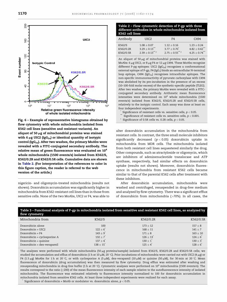
contrast, with all MoAbs, P-gp expression was detected in
mitochondria isolated from the two resistant cell lines. As
plasma membrane P-gp, mitochondrial P-gp expression was
always higher for K562/0.2R than for K562/0.5R. Moreover, for
both resistant lines, mitochondrial P-gp staining was similar
with the two MoAbs recognizing external epitopes (UIC2 and
F4), the specific TLEG-C494 MoAb giving the highest relative
fluorescence signal.
The functionality of P-gp expressed in mitochondria, from
whole organelles isolated from K562 cells, has been evaluated
by flow cytometric analyses of doxorubicin accumulation and
efflux. In addition, the effects of modulators were studied,
using either specific MoAbs (UIC2 and F4), or small molecule
inhibitors (cyclosporine A, dex-verapamil and quinine).
Doxorubicin accumulation in mitochondria isolated from
the sensitive parental cell line (K562/S) significantly increased
(p < 0.05) when incubated in the presence of anti-P-gp MoAbs
or of inhibitors (Table 3). Prior incubation with antibodies did
not modify subsequent drug accumulation. Cyclosporine A,
dex-verapamil and quinine induced an increase of about 40%
in doxorubicin uptake. A similar change was also observed for
b i o c h e m i c a l p h a r m a c o l o g y 7 1 ( 2 0 0 6 ) 1 1 6 2 – 1 1 7 41170
Fig. 6 – Example of representative histograms obtained by
flow cytometry with whole mitochondria isolated from
K562 cell lines (sensitive and resistant variants). An
aliquot of 50 mg of mitochondrial proteins was stained
with 6 mg UIC2 (IgG2a) or identical quantity of isotypic
control (IgG2a). After two washes, the primary MoAbs were
revealed with a FITC-conjugated secondary antibody. The
relative mean of green fluorescence was evaluated on 104
whole mitochondria (1500 events/s) isolated from K562/S,
K562/0.2R and K562/0.5R cells. Cumulative data are shown
in Table 2. (For interpretation of the references to color in
this figure caption, the reader is referred to the web
version of the article.)
Table 2 – Flow cytometric detection of P-gp with threedifferent antibodies in whole mitochondria isolated fromK562 cell lines
Antibody UIC2 F4 C494
K562/S 1.08 � 0.07 1.12 � 0.16 1.23 � 0.24
K562/0.2R 3.29 � 0.13*** 3.77 � 0.70* 4.82 � 0.62***
K562/0.5R 2.39 � 0.13***,y 2.75 � 0.55***,y 4.25 � 0.74***
An aliquot of 50 mg of mitochondrial proteins was stained with
MoAbs: 6 mg UIC2, or 8 mg F4 or 12 mg C494. These MoAbs recognise
different P-pg epitopes: UIC2 (IgG2a) recognises a conformational
external epitope of P-gp, F4 (IgG1) binds an extracellular N-terminal
loop epitope, C494 (IgG2a) recognises intracellular epitopes. The
non-specific immunoreactivity of pyruvate carboxylase with C494
was abolished by its pre-incubation in the presence of an excess
(10–100-fold molar excess) of the synthetic-specific peptide (TLEG).
After two washes, the primary MoAbs were revealed with a FITC-
conjugated secondary antibody. Arithmetic mean fluorescence
intensities were determined on 104 whole mitochondria (1500
events/s) isolated from K562/S, K562/0.2R and K562/0.5R cells,
relatively to the isotypic control. Each assay was done at least on
four independent experiments.* Significance of resistant cells vs. sensitive cells, p < 0.05.*** Significance of resistant cells vs. sensitive cells, p < 0.001.y Significance of 0.5R cells vs. 0.2R cells, p < 0.05.
nigericin- and oligomycin-treated mitochondria (results not
shown). Doxorubicin accumulation was significantly higher in
mitochondria from K562-resistant cell lines than in those from
sensitive cells. None of the two MoAbs, UIC2 or F4, was able to
Table 3 – Functional analysis of P-gp in mitochondria isolatedflow cytometry
Mitochondria from K562/S
Doxorubicin alone 100
Doxorubicin + UIC2 122 � 6*
Doxorubicin + F4 143 � 9*
Doxorubicin + cyclosporine A 132 � 7*
Doxorubicin + quinine 137 � 4*
Doxorubicin + dex-verapamil 138 � 15*
The analyses were performed with whole mitochondria (50 mg protein
studied the accumulation and efflux of doxorubicin (1 h at 10 mM, 20 8C). P
F4 (1.5 mg) MoAbs for 1 h at 20 8C, or with cyclosporine A (5 mM), dex
fluorescence of doxorubicin (drug accumulation) was then measured
resuspending mitochondria in drug-free buffer (1 h at 20 8C). Cytometric
results correspond to the ratio (�200) of the mean fluorescence intensity
mitochondria. The fluorescence was estimated relatively to fluoresce
mitochondria isolated from sensitive K562 cells. At least three independ* Significance of doxorubicin + MoAb or modulator vs. doxorubicin alon
alter doxorubicin accumulation in the mitochondria from
resistant cells. In contrast, the three small molecule inhibitors
significantly decreased (p < 0.05) doxorubicin uptake in
mitochondria from MDR cells. The mitochondria isolated
from both resistant cell lines sequestered similarly the drug.
Other compounds, such as atractyloside or oligomycin, which
are inhibitors of adeninenucleotide translocase and ATP
synthase, respectively, had similar effects on doxorubicin
uptake (results not shown). Moreover, doxorubicin fluores-
cence in mitochondria from resistant K562 cells became
similar to that of the parental K562 cells after treatment with
these inhibitors.
After doxorubicin accumulation, mitochondria were
washed and centrifuged, resuspended in drug-free medium
and analyzed by flow cytometry. There was a significant efflux
of doxorubicin from mitochondria (>70%). In all cases, the
from sensitive and resistant K562 cell lines, as analyzed by
K562/0.2R K562/0.5R
173 � 12 157 � 8
168 � 11 141 � 7
171 � 8 163 � 10
126 � 13* 124 � 6*
130 � 1* 130 � 3*
125 � 4* 128 � 6*
s/sample) isolated from K562/S, K562/0.2R and K562/0.5R cells; we
rior incubations of mitochondria were carried out with UIC2 (4 mg) or
-verapamil (10 mM) or quinine (50 mM), for 30 min at 20 8C. Mean
by flow cytometry. Drug efflux was estimated after washing and
analyses were performed on 104 mitochondria (1500 events/s). The
of each sample relative to the autofluorescence intensity of isolated
nce intensity normalized to 100 for doxorubicin accumulation in
ent experiments were realized for each assay.
e, p < 0.05.

b i o c h e m i c a l p h a r m a c o l o g y 7 1 ( 2 0 0 6 ) 1 1 6 2 – 1 1 7 4 1171
residual relative fluorescence of doxorubicin was comparable
in mitochondria isolated from K562/S cells (34.0 � 3.4%), from
K562/0.2R cells (34.8 � 4.7%) and from K562/0.5R cells
(30.5 � 2.9%), and the addition of MoAbs or drugs did not
modify this phenomenon.
4. Discussion
Several mechanisms can explain MDR phenotypes, often
involving drug detoxification by means of the overexpression
of plasma membrane ABC proteins, such as P-gp, MRP1 or BCRP
[1–3]. These proteins, and possibly other proteins such as LRP,
may also be responsible for drug redistribution within the cells
[7]. Doxorubicin-resistant K562 cells are commonly used as a
model for MDR studies, particularly via P-gp overexpression.
We have already shown that inhibition of P-gp activity by
modulators does not completely restore sensitivity of K562 MDR
cells to doxorubicin and a redistribution of this drug has been
observed in the K562/0.2R variant [5]. These results suggest the
involvement of other resistance mechanisms in these cells,
possibly through intracellular drug redistribution.
Drug localization in cytoplasmic compartments of MDR
cells has been observed by several authors by confocal laser
scanning microscopy, using mainly doxorubicin because of its
fluorescent properties [5,7–11]. Some studies on intracellular
expression of P-gp have been reported, aiming at the
clarification of the relationship between cytoplasmic localiza-
tion and the multidrug resistance phenotype [8–12]. In
different cell lines, P-gp has been detected in the nucleus
[8], in the Golgi apparatus [9] as well as in unidentified vesicles,
possibly lysosomes or recycling endosomes [10–12]. In cells
transfected with the MDR1 gene, P-gp was detected in vesicles
located around the periphery of nucleus [26], suggesting a
mitochondrial pattern. Gong et al. [14] have shown that
accumulation of daunorubicin occurred in mitochondria-like
organelles in K562-resistant cells and suggested a role for P-gp
in drug intracellular transport. Shapiro et al. [12] and Ferrao
et al. [27] demonstrated the involvement of P-gp in drug
compartmentalization in leukemic cell lines and patient
samples. It appears, therefore, that cytoplasmic localization
could be involved in the sequestration of doxorubicin in
organelles, preventing it to reach its nuclear targets. It could
also correspond to a storage pool of P-gp for maintaining a
steady-state level of surface P-gp for a novel mechanism of
drug sequestration in K562 myeloid leukemic cells.
In favor of a role for mitochondria in doxorubicin
sequestration in MDR cells, in addition to the works already
mentioned [14,27], is the fact that some MDR modulators, such
as cyclosporine A or quinine, which are able to mediate
doxorubicin redistribution in MDR cells are also known as
antagonists of the mitochondrial megachannel [13]. As a
consequence, mitochondria may appear as a good candidate
for doxorubicin sequestration in K562 MDR cells. In view of
this hypothesis, we have studied in detail the possible
mitochondrial location of P-gp and the involvement of
mitochondria as a drug sequestration compartment.
Using flow cytometry, we first verified that P-gp was
expressed on the plasma membrane and in whole cells of the
different K562 variants. We noticed a low-level, non-saturable
binding of UIC2 in sensitive K562 whole cells, parallel to the
binding of an isotypic antibody, and which is irrelevant for this
reason. We also noticed, using three different MoAbs, that the
variant with the lowest degree of resistance (K562/0.2R)
contained a higher amount of P-gp than the variant with
the highest degree of resistance, both at the plasma
membrane level and in whole cells. Such a discrepancy
between the level of resistance and the amount of P-gp has
been already mentioned in various cell lines and is probably
related to the multifactorial nature of MDR [28].
Using confocal microscopy, a strong P-gp staining appeared
on the plasma membrane of the two resistant cell lines, with
more intensity for K562/0.2R cells, confirming cytometric
analyses. In addition, intracellular expression of P-gp was also
noticed, preferentially accumulated in restricted areas of the
cytoplasm. Such a distribution has already been observed in
MDR cellular models and attributed to the Golgi apparatus [9]
or unknown vesicles [12,26].
Dual fluorescence study allowed a positive approach of P-
gp localization. In K562-sensitive cells, the simultaneous
labeling of P-gp and mitochondria provided neither the
orange-red fluorescence signal corresponding to P-gp, nor
the yellow fluorescence signal corresponding to P-gp and
mitochondrial co-expression. In contrast, P-gp appeared
localized in mitochondria of K562-resistant cells and
accounted for about 20% of the total P-gp signal in the two
sublines. A significant orange-red signal, corresponding to
intracellular P-gp alone, indicated that P-gp was also present
in other intracellular compartments of resistant cells, such as
lysosomes and recycling endosomes. With simultaneous
labeling of P-gp and the Golgi apparatus, a marked yellow
fluorescence was detected in both K562-resistant variants,
corresponding to 27–39% of intracellular P-gp, but was not
observed in sensitive cells, confirming the observations of
Molinari et al. [9]. The localization of P-gp in the Golgi
apparatus may reflect both a processing and transport
compartment of P-gp from the endoplasmic reticulum to
the plasma membrane and an intracellular sequestration
compartment [29]. The important variability in the proportion
of Golgi apparatus or mitochondria-associated P-gp can be
attributed to the asynchronous growth of the cells and their
study at different steps of the cell cycle. It has been shown that
the level of expression of P-gp and its distribution changed
during the cell cycle [30,31]. We observed that, in cells
undergoing mitosis, no P-gp was detected at the plasma
membrane level of K562-resistant cells (results not shown).
The presence of P-gp was assessed by Western blotting of
mitochondria isolated from resistant cell lines with two
different antibodies, able to stain a 170 kDa protein. Five
mammalian ABC proteins have been identified in mitochon-
dria [32]. They correspond to ‘‘half-transporters’’ and have a
size (65–94 kDa) much lower than that of P-gp (170 kDa). The
conventional SDS-PAGE technique we used allows to exclude
the detection of a dimeric mammalian mitochondrial ABC
transporter corresponding its functional structure [33]. In
addition, the homology of mitochondrial ABC transporters
with P-gp is relatively low (less than 50%), and is important
only in the ATP-binding domain, whereas the epitopes
recognized by all MoAbs used in this study are localized in
other domains [15–17]. These features exclude the possibility

b i o c h e m i c a l p h a r m a c o l o g y 7 1 ( 2 0 0 6 ) 1 1 6 2 – 1 1 7 41172
that the mitochondrial P-gp evidenced in this study could be the
mitochondrial ABC transporters. The purification of mitochon-
dria by differential centrifugation is a widely used technique
[19,20], which generally provides organelles with a high degree
of purity (�90%), rendering unlikely a significant contamination
of our preparations by P-gp originating from another cellular
localization [21,22]. However, one cannot exclude a slight
difference between plasma membrane P-gp and mitochondrial
P-gp, especially at the level of the glycanic moiety.
P-gp expression in whole isolated mitochondria was
investigated by flow cytometry with several MoAbs. For
staining with C494, a specific pre-treatment allows to avoid
reactivity with pyruvate carboxylase, a mitochondrial protein
not related to P-gp [23]. Mitochondria from K562-sensitive cells
do not contain detectable P-gp. In contrast, in mitochondria
from both K562-resistant cell lines, P-gp expression was
observed whatever the MoAb used. From the flow cytometry
data, we cannot conclude about the relative amounts of
mitochondrial P-gp in the two K562-resistant cell lines
because the fluorescence signals were similar excepted with
UIC2. In addition, in mitochondria from both K562-resistant
cell lines, fluorescence intensity was similar with F4 and UIC2
and higher with C494, which was not the case for plasma
membrane P-gp. This could be due to a difference in epitope
accessibility by the MoAbs and suggests an opposite topology
in mitochondria and in the plasma membrane.
Mitochondrial P-gp functionality was investigated in the
presence of several MoAbs and inhibitors. Anti-P-gp MoAbs
and inhibitors enhanced doxorubicin uptake in mitochondria
from sensitive cells, a phenomenon that cannot be attributed
to P-gp inhibition, since it is absent in these mitochondria. As
shown by several authors, doxorubicin spontaneously binds
numerous mitochondrial targets: DNA, cardiolipin and
respiratory chain complexes (see for review [34]). The effect
of MoAbs and small inhibitors can involve an alteration of
membrane conformation leading to a decrease in the fixation
Fig. 7 – A hypothetic scheme for mitochondrial topology and fu
the results obtained concerning mitochondrial P-gp expression
of doxorubicin to its mitochondrial targets. In mitochondria
isolated from K562-resistant cells, in contrast, which accu-
mulate more doxorubicin than those from sensitive cells, the
presence of inhibitors decreased this accumulation while the
antibodies had no significant effect. These results are
important because both tend to suggest that mitochondrial
P-gp has a topology inverse to that observed in plasma
membrane: (i) it provides an increased accumulation of the
drug in the organelle; (ii) ‘‘extracellular’’ MoAbs have no effect
on this accumulation, probably because they cannot reach any
binding site; (iii) small inhibitors, which can cross the
mitochondrial membrane, reduce drug accumulation in the
organelle; (iv) oligomycin, which blocks ATP synthesis, also
reduces mitochondrial drug accumulation. The residual
fluorescence after washing was similar in mitochondria
isolated from all three cell lines, even in the presence of P-
gp inhibitors. Drug retention appeared, therefore, indepen-
dent from P-gp expression.
One can propose, as a general conclusion, that mitochon-
drial P-gp expressed in K562-resistant cells behaves as a
unidirectional pump, oriented in a direction opposite to that of
the plasma membrane. This orientation is compatible with
that of another mitochondrial ABC transporters, M-ABC1,
which is embedded in the inner mitochondrial membrane and
exposes its ATP-binding domain in the mitochondrial inter-
membrane space [35]. At the level of the plasma membrane,
doxorubicin uptake is mostly passive and extrusion is active
through P-gp action [36]. In mitochondria, doxorubicin uptake
would be P-gp-dependent and specifically inhibited by P-gp
reverters, whereas its exit would be essentially passive. A
potential role for mitochondrial P-gp in MDR cells would be to
protect the nucleus from doxorubicin. Nevertheless, the drug
uptake in mitochondria from MDR K562 cell lines could lead to
oxidative damage in relation with its interaction with
mitochondrial components. In fact, due to low capacities of
organelles to repair oxidative insults, the free oxygen radicals
nctionality. A hypothesis was proposed in agreement with
and function.

b i o c h e m i c a l p h a r m a c o l o g y 7 1 ( 2 0 0 6 ) 1 1 6 2 – 1 1 7 4 1173
can induce lipid peroxidations, bioenergetic failure, DNA
mutations, membrane disorders and mitochondrial dysfonc-
tions [34]. Drug detoxification would then be the usual purpose
of vesicular traffic and secretion/and or autophagy of damaged
organelles such as mitochondria [37]. An increase in the
number of autophagic vacuoles, favoring the removal of
damaged intracellular macro molecules, is observed in cancer
cells in response to drug treatment [38].
A model of P-gp function at the plasma membrane and at
the mitochondrial levels is proposed in Fig. 7. Sequestration of
anticancer drugs by an active process in this organelle could
contribute to the multidrug resistance phenotype and explain
several features often observed, such as intracellular drug
tolerance [24] or drug redistribution upon the action of
modulators like quinine [5]. Such drug sequestration has
already been observed in cytoplasmic vesicles of K562-
resistant cells, although the compartment involved in this
process was not identified [12,26], despite the fact that the
Golgi apparatus and lysosomes clearly contribute to drug
sequestration [39]. The fact that cyclosporine A and quinine
are not pure inhibitors of P-gp but can also interact with the
mitochondrial permeability transition pore may be confound-
ing. One cannot exclude that the effects observed with these
compounds are due to their interference with the pore, or that
mitochondrial P-gp is bound to this macromolecular complex.
This would explain the differential effects of verapamil and
quinine, which remained unexplained [5].
Our results raise different questions to be solved in the
future about the intracellular trafficking of P-gp and the import
of P-gp in mitochondria. Other points remain to be under-
stood, such as the possible relationship between the P-gp
expression, its localization, and its function with other
proteins of multidrug resistance family. The sequential
expression of different proteins involved in MDR phenotype
varies according to the degree of resistance of K562 cells [6].
Moreover, Larsen et al. [7] suggested that the degree of P-gp
over expression could influence the organization and function
of other membrane proteins such as ion transporters. Such
features are now to be taken in consideration for a complete
understanding of the role of P-gp in the MDR phenotype.
Acknowledgments
The authors express thanks to Helene Kiefer-Biazizzo, Vincent
Denis and Claire Carrion for their technical expertise in
confocal imaging. Financial support of this work and fellow-
ships were provided by the Ministere Francais des Affaires
Etrangeres, the Government of Romania, the Ministere
Francais de l’Enseignement Superieur et de la Recherche
and the Conseil Regional du Limousin.
r e f e r e n c e s
[1] Gottesman MM. Mechanisms of cancer drug resistance.Annu Rev Med 2002;53:615–27.
[2] Gottesman MM, Fojo T, Bates SE. Multidrug resistance incancer: role of ATP-dependent transporters. Nat Rev Cancer2002;2:48–58.
[3] Litman T, Brangi M, Hudson E, Fetsch P, Abati A, Ross DD,et al. The multidrug-resistant phenotype associated withoverexpression of the new ABC half-transporter, MXR(ABCG2). J Cell Sci 2000;113:2011–21.
[4] Scheffer GL, Wijngaard PLJ, Flens MJ, Izquierdo MA, SlovakML, Pinedo HM, et al. The drug resistance related proteinLRP is a major vault protein. Nat Med 1995;l:578–82.
[5] Bennis S, Ichas F, Robert J. Differential effects of verapamiland quinine on the reversal of doxorubicin resistance in ahuman leukemia cell line. Int J Cancer 1995;62:283–90.
[6] Grandjean F, Bremaud L, Verdier M, Robert J, Ratinaud MH.Sequential gene expression of P-glycoprotein (P-gp),multidrug resistance-associated protein (MRP) and lungresistance protein (LRP): functional activity of P-gp and MRPpresent in the doxorubicin-resistant human K562 cell lines.Anticancer Drugs 2001;12:247–58.
[7] Larsen AK, Escargueil AE, Skladanowski A. Resistancemechanisms associated with altered intracellulardistribution of anticancer agents. Pharmacol Ther2000;85:217–29.
[8] Baldini N, Scotlandi K, Serra M, Shikita T, Zini N, OgnibeneA, et al. Nuclear immunolocalization of P-glycoprotein inmultidrug-resistant cell lines showing similar mechanismsof doxorubicin distribution. Eur J Cell Biol 1995;68:226–39.
[9] Molinari A, Cianfriglia M, Meschini S, Calcabrini A, AranciaG. P-glycoprotein expression in the Golgi apparatus ofmultidrug resistant cells. Int J Cancer 1994;9:789–95.
[10] Sognier MA, Zhang Y, Eberle RL, Sweet KM, Altenberg GA,Belli JA. Sequestration of doxorubicin in vesicles in amultidrug-resistant cell line (LZ-l00). Biochem Pharmacol1994;48:391–401.
[11] Bour-Dill C, Gramain MP, Merlin JL, Marchal S, Guillemin F.Determination of intracellular organelles implicated indaunorubicin cytoplasmic sequestration in multidrug-resistant MCF-7 cells using fluorescence microscopy imageanalysis. Cytometry 2000;39:16–25.
[12] Shapiro AB, Fox K, Lee P, Yang YD, Ling V. Functionalintracellular P-glycoprotein. Int J Cancer 1998;76:857–64.
[13] Kroemer G. The mitochondrial permeability transition porecomplex as a pharmacological target. An introduction. CurrMed Chem 2003;10:1469–72.
[14] Gong Y, Wang Y, Chen F, Han J, Miao J, Shao N, et al.Identification of the subcellular localization ofdaunorubicin in multidrug-resistant K562 cell line. LeukRes 2000;24:769–74.
[15] Mechetner EB, Roninson IB. Efficient inhibition of P-glycoprotein-mediated multidrug resistance with amonoclonal antibody. Proc Natl Acad Sci USA 1992;89:5824–8.
[16] Chu TM, Kawinski E, Lin TH. Characterization of anewmonoclonal antibody F4 detecting cell surface epitope andP-glycoprotein in drug-resistant human tumor cell lines.Hybridoma 1993;12:417–29.
[17] Georges E, Bradley G, Gariepy J, Ling V. Detection of P-glycoprotein isoforms by gene-specific monoclonalantibodies. Proc Natl Acad Sci USA 1990;87:152–6.
[18] Dussossoy D, Carayon P, Feraut D, Belugou S, Combes T,Canat X, et al. Developement of a monoclonal antibody toimmuno-cytochemical analysis of the cellular localizationof the peripheral benzodiazepine receptor. Cytometry1996;24:39–48.
[19] Bourgeron T, Chretien D, Rotig A, Munnich A, Rustin P.Isolation and characterization of mitochondria fromhuman B lymphoblastoid cell lines. Biochem Biophys ResCommun 1992;186:16–23.
[20] Denis-Gay M, Petit JM, Mazat JP, Ratinaud MH.Modifications of oxido-reductase activities in adriamycin-resistant leukaemia K562 cells. Biochem Pharmacol1998;56:451–7.

b i o c h e m i c a l p h a r m a c o l o g y 7 1 ( 2 0 0 6 ) 1 1 6 2 – 1 1 7 41174
[21] Petit JM, Huet O, Gallet PF, Maftah A, Ratinaud MH, Julien R.Direct analysis and significance of cardiolipin transversedistribution in mitochondrial inner membranes. Eur JBiochem 1994;220:871–9.
[22] Rustin P, Chretien D, Bourgeron T, Gerard B, Rotig A,Saudubray JM, et al. Biochemical and molecularinvestigations in respiratory chain deficiencies. Clin ChimActa 1994;228:35–51.
[23] Rao VV, Anihony DC, Piwnica-Worms D. MDR1 gene-specific monoclonal antibody C494 cross-reacts withpyruvate carboxylase. Cancer Res 1994;54:1536–41.
[24] Huet S, Marie JP, Gualde N, Robert J. Reference method fordetection of P-gp mediated multidrug resistance in humanhaematological malignancies: a method validated by thelaboratories of the French drug resistance network.Cytometry 1998;34:248–56.
[25] Huet S, Marie JP, Laurand A, Robert J. Major improvement ofthe reference method of the French drug resistancenetwork for P-glycoprotein detection in humanhaematological malignancies. Leuk Res 2005;29:1029–37.
[26] Rajagopal A, Simon SM. Subcellular localization andactivity of multidrug resistance proteins. Mol Biol Cell2003;14:3389–99.
[27] Ferrao P, Sincock P, Cole S, Ashman L. Intracellular P-gpcontributes to functional drug efflux and resistance inacute myeloid leukemia. Leuk Res 2001;25:395–405.
[28] Huet S, Schott B, Robert J. P-glycoprotein overexpressioncannot explain the complete doxorubicin-resistancephenotype in rat glioblastoma cell lines. Br J Cancer1992;65:538–44.
[29] Fu D, Bebawy M, Kable EPW, Roufogalis BD. Dynamic andintracellular trafficking of P-glycoprotein-EGFP fusion
protein: implications in multidrug resistance in cancer. Int JCancer 2004;109:174–81.
[30] Ramachandran C, Mead D, Wellham LL, Sauerteig A,Krishan A. Expression of drug resistance-associated mdr-1,GST pi, and topoisomerase II genes during cell cycletraverse. Biochem Pharmacol 1995;49:545–52.
[31] Zhang W, Ling V. Cell cycle-dependent turn-over of P-glycoprotein in multidrug-resistant cells. J Cell Physiol2000;184:17–26.
[32] Lill R, Kispal G. Mitochondrial ABC transporters. ResMicrobiol 2001;152:331–40.
[33] Graf SA, Haigh SE, Corson ED, Shirihai OS. Targeting,import, and dimerization of a mammalian mitochondrialATP binding cassette (ABC) transporter, ABCB10 (ABC-me). JBiol Chem 2004;279:42954–63.
[34] Jung K, Reszka R. Mitochondria as subcellular targets forclinical useful anthracyclines. Adv Drug Deliv Rev2001;49:87–105.
[35] Zhang F, Hogue DL, Liu L, Fisher CL, Hui D, Childs S, et al.M-ABC2, a new human mitochondrial ATP-binding cassettemembrane protein. FEBS Lett 2000;478:89–94.
[36] Eytan GD. Mechanism of multidrug resistance in relation topassive membrane permeation. Biomed Pharmacother2005;59:90–7.
[37] Ogier-Denis E, Codogno P. Autophagy: a barrier or anadaptive response to cancer. Biochem Biophys Acta2003;1603:113–28.
[38] Cuervo AM. Autophagy: in sickness and in health. TrendsCell Biol 2004;14:70–7.
[39] Gong Y, Duvvuri M, Krise JP. Separate roles for the Golgiapparatus and lysosomes in the sequestration of drugs inthe multidrug-resistant human leukemic cell line HL-60. JBiol Chem 2003;278:50234–9.
Copyright © 2022 FDOKUMEN