Adenosine triphosphate-dependent transport of doxorubicin, daunomycin, and vinblastine in human...
8
Adenosine Triphosphate-dependent Transport of Doxorubicin, Daunomycin, and Vinblastine in Human Tissues by a Mechanism Distinct from the P-Glycoprotein Sanjay Awasthi, * Sharad S. Singhal, * Sanjay K. Srivastava,t Piotr Zimniak, Krishna K. Bajpai,t Manju Saxena,t Rashmi Sharma,t Stephen A. Ziller 111,0 Eugene P. Frenkel,11 Shivendra V. Singh," Nong G. He,* and Yogesh C. Awasthit *Department of Internal Medicine, Division of Hematology and Oncology, and tDepartment of Human Biological Chemistry and Genetics, University of Texas Medical Branch, Galveston, Texas 77555; §Department ofMedicine, and Department of Biochemistry and Molecular Biology, University ofArkansas for Medical Sciences, Little Rock, Arkansas 72205; 1Department of Internal Medicine, The University of Texas Southwestern Medical Center, Dallas, Texas 75235; and 'Mercy Hospital of Pittsburgh, Pittsburgh, Pennsylvania 15219 Abstract Previous studies have demonstrated that a human glutathione conjugate transporter, designated as dinitrophenyl-S-glutathi- one ATPase (DNP-SG ATPase), catalyzed ATP hydrolysis in the presence of several amphiphilic compounds other than glu- tathione conjugates (Singhal, S. S., R. Sharma, S. Gupta, H. Ahmad, P. Zimniak, A. Radominska, R. Lester, and Y. C. Awasthi. 1991. FEBSIFed. Eur. Biochem. Soc.J Lett. 281:255- 257). We now demonstrate that DNP-SG ATPase purified from human lung and erythrocyte membranes catalyzed the hydrolysis of ATP in the presence of doxorubicin and its metab- olites. Doxorubicin-stimulated ATP hydrolysis by DNP-SG ATPase was saturable with respect to doxorubicin (Km 1.2 and 2.8 MM for the lung and erythrocyte enzymes, respectively). Antibodies against DNP-SG ATPase immunoprecipitated the ATP hydrolyzing activity stimulated by doxorubicin, its metab- olites, and glutathione conjugates. Inside out vesicles prepared from erythrocyte membranes took up doxorubicin, daunomy- cin, and vinblastine in an ATP-dependent manner. The uptake was linear with respect to time and vesicle protein, was depen- dent on ATP and magnesium, was inhibited by heavy metal salts or by heating the vesicles, and was sensitive to both osmo- larity and orientation of the vesicles. The transport had an acti- vation energy of 13 kcal/mol, was saturable with respect to both doxorubicin and ATP (K. values of 1.8 ,M and 1.9 mM, respectively), and was competitively inhibited by glutathione conjugates as well as by a number of amphiphiles such as dauno- mycin or vinblastine. Transport was diminished upon coating the vesicles with antibodies against DNP-SG ATPase. Incorpo- ration of increasing amounts of purified DNP-SG ATPase into the vesicles resulted in a linear increase in transport of doxoru- bicin. These studies demonstrated for the first time that a mem- brane protein that catalyzed the transport of anionic amphiphi- lic molecules such as glutathione conjugates could also mediate the transport of weakly cationic antitumor antibiotic, doxorubi- cin. Notably, the Km of transport was in the range of doxorubi- Address correspondence to Yogesh C. Awasthi, Ph.D., Department of Human Biological Chemistry and Genetics, 2.138 Medical Research Building, University of Texas Medical Branch, Galveston, TX 77555- 1067. Receivedfor publication 21 July 1993 and in revisedform 15 Sep- tember 1993. cin concentration achievable in human serum after intravenous dosing of doxorubicin. (J. Clin. Invest. 1994.93:958-965.) Key words: doxorubicin * biological transport - glutathione * drug resistance * ethacrynic acid Introduction Doxorubicin hydrochloride (DOX', Adriamycin®; Farmitalia, Milan, Italy) is an effective cytoreductive agent for chemother- apy of a variety of neoplasms, but its efficacy is limited by inherent or acquired resistance to its cytocidal effects in cancer cells ( 1). Studies of DOX resistance have shown that cultured malignant cells resistant to DOX frequently exhibit a multi- drug resistant (MDR) phenotype associated with the ATP-de- pendent efflux of a number of amphiphilic cytotoxins such as DOX, vincristine, and vinblastine (2-7). A number of these malignant cell lines with the MDR phenotype overexpress the P-glycoprotein, a 170-kD glycoprotein product of the mdr- 1 gene (5, 8). The P-glycoprotein has been demonstrated to me- diate the ATP-dependent efflux of DOX and other amphiphilic drugs (3-8). Several studies have also documented the exis- tence of an MDR phenotype in malignant cell lines which do not overexpress the P-glycoprotein (9-13). Increased efflux of DOX from MDR cells which do not overexpress P-glycopro- tein has also been demonstrated (5, 11-13), suggesting that efflux mechanism(s) distinct from P-glycoprotein may be in- volved in the exclusion of DOX or similar amphiphilic com- pounds. ATP-dependent transport of oxidized glutathione and con- jugates of GSH and xenobiotics across human erythrocyte membrane is known ( 14-16), and ATPases catalyzing hydroly- sis of ATP in the presence of conjugates of xenobiotics and GSH ( 17) and oxidized glutathione ( 18) have been identified in human erythrocyte membrane. A transporter of glutathi- one-xenobiotic (GS-X) conjugates (16, 17) present in the membrane of human erythrocytes and other cells (19) cata- lyzed ATP hydrolysis in presence of not only GS-X conjugates but also in the presence of several structurally unrelated amphi- philic compounds including estradiol glucuronide, bilirubin ditaurate, lithocholic acid 3-O-glucuronide, and leukotriene C4 methyl ester (20, 21 ). We hypothesize that this GS-X conju- 1. Abbreviations used in this paper: DNP-SG, dinitrophenyl-S-glutathi- one; DOX, doxorubicin, EA-SG conjugate, ethacrynic acid-glutathi- one conjugate; G3PD, glyceraldehyde-3-phosphate dehydrogenase; GST, glutathione S-transferase; GS-X conjugate, glutathione-xeno- biotic conjugate; IOV, inside out vesicle; MDR, multidrug resistant; ROV, right side out vesicle. 958 Awasthi et al. J. Clin. Invest. © The American Society for Clinical Investigation, Inc. 0021-9738/94/03/0958/08 $2.00 Volume 93, March 1994, 958-965
-
Upload
independent -
Category
Documents
-
view
0 -
download
0
Transcript of Adenosine triphosphate-dependent transport of doxorubicin, daunomycin, and vinblastine in human...

Adenosine Triphosphate-dependent Transport of Doxorubicin, Daunomycin, andVinblastine in Human Tissues by a Mechanism Distinct from the P-GlycoproteinSanjay Awasthi, * Sharad S. Singhal, * Sanjay K. Srivastava,t Piotr Zimniak, Krishna K. Bajpai,t Manju Saxena,tRashmi Sharma,t Stephen A. Ziller 111,0 Eugene P. Frenkel,11 Shivendra V. Singh," Nong G. He,* and Yogesh C. Awasthit*Department of Internal Medicine, Division ofHematology and Oncology, and tDepartment ofHuman Biological Chemistry andGenetics, University of Texas Medical Branch, Galveston, Texas 77555; §Department ofMedicine, and Department ofBiochemistryand Molecular Biology, University ofArkansas for Medical Sciences, Little Rock, Arkansas 72205;1Department ofInternal Medicine, The University of Texas Southwestern Medical Center, Dallas,Texas 75235; and 'Mercy Hospital ofPittsburgh, Pittsburgh, Pennsylvania 15219
Abstract
Previous studies have demonstrated that a human glutathioneconjugate transporter, designated as dinitrophenyl-S-glutathi-one ATPase (DNP-SG ATPase), catalyzed ATP hydrolysis inthe presence of several amphiphilic compounds other than glu-tathione conjugates (Singhal, S. S., R. Sharma, S. Gupta, H.Ahmad, P. Zimniak, A. Radominska, R. Lester, and Y. C.Awasthi. 1991. FEBSIFed. Eur. Biochem. Soc.J Lett. 281:255-257). We now demonstrate that DNP-SG ATPase purifiedfrom human lung and erythrocyte membranes catalyzed thehydrolysis ofATP in the presence of doxorubicin and its metab-olites. Doxorubicin-stimulated ATP hydrolysis by DNP-SGATPase was saturable with respect to doxorubicin (Km 1.2 and2.8 MM for the lung and erythrocyte enzymes, respectively).Antibodies against DNP-SG ATPase immunoprecipitated theATP hydrolyzing activity stimulated by doxorubicin, its metab-olites, and glutathione conjugates. Inside out vesicles preparedfrom erythrocyte membranes took up doxorubicin, daunomy-cin, and vinblastine in an ATP-dependent manner. The uptakewas linear with respect to time and vesicle protein, was depen-dent on ATP and magnesium, was inhibited by heavy metalsalts or by heating the vesicles, and was sensitive to both osmo-larity and orientation of the vesicles. The transport had an acti-vation energy of 13 kcal/mol, was saturable with respect toboth doxorubicin and ATP (K. values of 1.8 ,M and 1.9 mM,respectively), and was competitively inhibited by glutathioneconjugates as well as by a number ofamphiphiles such as dauno-mycin or vinblastine. Transport was diminished upon coatingthe vesicles with antibodies against DNP-SG ATPase. Incorpo-ration of increasing amounts of purified DNP-SG ATPase intothe vesicles resulted in a linear increase in transport of doxoru-bicin. These studies demonstrated for the first time that a mem-brane protein that catalyzed the transport of anionic amphiphi-lic molecules such as glutathione conjugates could also mediatethe transport of weakly cationic antitumor antibiotic, doxorubi-cin. Notably, the Km of transport was in the range of doxorubi-
Address correspondence to Yogesh C. Awasthi, Ph.D., Department ofHuman Biological Chemistry and Genetics, 2.138 Medical ResearchBuilding, University of Texas Medical Branch, Galveston, TX 77555-1067.
Receivedfor publication 21 July 1993 and in revisedform 15 Sep-tember 1993.
cin concentration achievable in human serum after intravenousdosing of doxorubicin. (J. Clin. Invest. 1994.93:958-965.) Keywords: doxorubicin * biological transport - glutathione * drugresistance * ethacrynic acid
Introduction
Doxorubicin hydrochloride (DOX', Adriamycin®; Farmitalia,Milan, Italy) is an effective cytoreductive agent for chemother-apy of a variety of neoplasms, but its efficacy is limited byinherent or acquired resistance to its cytocidal effects in cancercells ( 1). Studies ofDOX resistance have shown that culturedmalignant cells resistant to DOX frequently exhibit a multi-drug resistant (MDR) phenotype associated with the ATP-de-pendent efflux of a number of amphiphilic cytotoxins such asDOX, vincristine, and vinblastine (2-7). A number of thesemalignant cell lines with the MDR phenotype overexpress theP-glycoprotein, a 170-kD glycoprotein product of the mdr- 1gene (5, 8). The P-glycoprotein has been demonstrated to me-diate the ATP-dependent efflux ofDOX and other amphiphilicdrugs (3-8). Several studies have also documented the exis-tence of an MDR phenotype in malignant cell lines which donot overexpress the P-glycoprotein (9-13). Increased efflux ofDOX from MDR cells which do not overexpress P-glycopro-tein has also been demonstrated (5, 11-13), suggesting thatefflux mechanism(s) distinct from P-glycoprotein may be in-volved in the exclusion of DOX or similar amphiphilic com-pounds.
ATP-dependent transport ofoxidized glutathione and con-jugates of GSH and xenobiotics across human erythrocytemembrane is known ( 14-16), and ATPases catalyzing hydroly-sis of ATP in the presence of conjugates of xenobiotics andGSH ( 17) and oxidized glutathione ( 18) have been identifiedin human erythrocyte membrane. A transporter of glutathi-one-xenobiotic (GS-X) conjugates (16, 17) present in themembrane of human erythrocytes and other cells (19) cata-lyzed ATP hydrolysis in presence of not only GS-X conjugatesbut also in the presence ofseveral structurally unrelated amphi-philic compounds including estradiol glucuronide, bilirubinditaurate, lithocholic acid 3-O-glucuronide, and leukotriene C4methyl ester (20, 21 ). We hypothesize that this GS-X conju-
1. Abbreviations used in thispaper: DNP-SG, dinitrophenyl-S-glutathi-one; DOX, doxorubicin, EA-SG conjugate, ethacrynic acid-glutathi-one conjugate; G3PD, glyceraldehyde-3-phosphate dehydrogenase;GST, glutathione S-transferase; GS-X conjugate, glutathione-xeno-biotic conjugate; IOV, inside out vesicle; MDR, multidrug resistant;ROV, right side out vesicle.
958 Awasthi et al.
J. Clin. Invest.© The American Society for Clinical Investigation, Inc.0021-9738/94/03/0958/08 $2.00Volume 93, March 1994, 958-965

gate transporter, previously designated as DNP-SG ATPasebecause of its routinely used model substrate dinitrophenyl-S-glutathione (DNP-SG) (16, 17), may be involved in the en-ergy-dependent efflux of other structurally unrelated amphi-philic compounds such as DOX and may thus be a contribu-tory factor in the mechanism(s) for DOX resistance inmalignant cells not overexpressing the P-glycoprotein. This re-port demonstrates that DNP-SG ATPase purified from humanerythrocytes and lung tissue stimulated ATP hydrolysis in thepresence of DOX and its metabolites. Furthermore, we havedemonstrated that DOX, daunomycin, and vinblastine weretaken up by inside out vesicles (IOVs) prepared from mem-branes ofhuman erythrocytes (which do not express the P-gly-coprotein) through an ATP-dependent process. The uptake ofDOX was competitively inhibited by substrates of the GS-Xpump (DNP-SG ATPase) as well as by prototypical substratesof the P-glycoprotein transporter. The transport of DOX wasalso inhibited by polyclonal antibodies specific to DNP-SGATPase. Incorporation ofpurified DNP-SG ATPase into eryth-rocyte IOVs more than doubled the rate of DOX transport.These studies suggest a close similarity between the transportmechanisms ofDOX and GS-X conjugates. Possible implica-tions of these findings in the mechanisms of drug resistanceand strategies of cancer chemotherapy are discussed in thisreport.
Methods
Materials. Use of human tissues was approved by the InstitutionalReview Board. Blood collected from normal subjects and stored for < 1wk was obtained from the Blood Bank, and human lung samples fromadult subjects without known pulmonary disorders deceased < 8 hwere collected from the autopsy service. Sources ofreagents used in thepresent studies were the same as described previously (21). DOX(AdriamycinO), doxorubicinone, dihydrodoxorubicinone, deoxydox-orubicinone, dihydrodoxorubicin, and vinblastine sulfate were gener-ously provided by Dr. Sergio Penco (Adria Labs, Milan, Italy). Mitox-antrone and daunorubicin were generously provided by Lederle Labo-ratories (Pearl River, NY) and by Wyeth Laboratories (Philadelphia,PA), respectively. [14'4C]Doxorubicin hydrochloride (sp act 57mCi/mmol) and [G-3H]vinblastine sulfate (sp act 11 Ci/mmol) werepurchased from Amersham Corp. (Arlington Heights, IL). [y-32P]-ATP and [G-3Hjdaunomycin (sp act 1.7 Ci/mmol) were procuredfrom Du Pont/New England Nuclear (Boston, MA). (±)-Verapamilhydrochloride was purchased from Sigma Chemical Co. (St. Louis,MO). Taurodeoxycholate and taurolithocholate (sodium salts) werepurchased from Calbiochem-Novabiochem Corp. (La Jolla, CA). Theconjugate of l-chloro-2,4-dinitrobenzene and GSH, 2,4-dinitrophenyl-S-glutathione (DNP-SG), was synthesized enzymatically, purified,and authenticated according to our previously published method (22)and was linked to CNBr-activated Sepharose 4B according to a methoddescribed previously (21 ). GSH conjugate ofethacrynic acid (EA-SG)was synthesized and authenticated as described by us previously (23).Polyclonal antibodies against human erythrocyte DNP-SG ATPaseand glyceraldehyde 3-phosphate dehydrogenase, raised in rabbits, werethe same as those used in previous studies ( 19). The IgG fractions wereisolated from these antibodies and the preimmune serum by DEAE-cellulose ion-exchange chromatography and were purified further us-ing a column of protein A bound to CNBr-activated Sepharose 6MB.
Enzyme assays andpurification ofDNP-SGA TPase. ATPase activ-ity in the presence of DNP-SG, DOX, and other substrates was deter-mined according to the method used by us previously (21 ). 1 mU ofenzyme activity was defined as 1 nmol ATP hydrolysis/min at 37°C.Purifications ofDNP-SG ATPase from human lung were performed at4VC according to a previously described protocol (21). The details of
purification of DNP-SG ATPase from human erythrocytes have alsobeen reported in our previous studies ( 19). Because of interference bylubrol PX (Sigma Chemical Co.) used during purification, protein wasestimated by the method of Minamide and Bamburg (24). SDS-PAGE, Western blot analysis, and immunotitration ofDNP-SG ATP-ase were performed by methods used by us previously ( 19, 21 ).
Preparation ofIOVs and right side out vesicles (ROVs) from hu-man erythrocyte membranes. Preparation of human erythrocyte vesi-cles was carried out by the method of Steck and Kant (25) in whichhuman erythrocyte ghosts prepared according to the method ofDodgeet al. (26) were diluted in 40 vol of 0.5 mM sodium phosphate buffer,pH 8.0, and were allowed to incubate overnight at 4VC. The pelletobtained by centrifugation at 28,000 g for 1 h was resuspended in thesame buffer, and vesiculation was performed by repeatedly passing thesuspension through a 27-gauge needle. Vesiculation performed in pres-ence or absence of 250 mM sucrose resulted in vesicles with similaryield and transport properties. The IOVs were purified either by den-sity gradient centrifugation at 105,000 g for 2 h over a dextran barrier( 1.03 g/ml) during which the ROVs and unsealed ghosts pelleted to thebottom, or by passing the mixture of vesicles over a column ( 1 cm X 5cm) of wheat germ agglutinin bound to CNBr-activated Sepharose 4B.In the latter procedure, the ROVs were almost quantitatively retainedby the column, whereas the IOVs were recovered in the flow-throughfraction of the column. The ROVs were eluted from the column with50 mM methyl-a-D-mannopyranoside. The purities of the IOVs andROVs were estimated by comparing acetylcholinesterase activities inthe erythrocyte ghosts and the vesicles using the assay described byEllman et al. (27). Both IOVs and ROVs were finally suspended inbuffer containing 250 mM sucrose in 10 mM Tris-HCl, pH 7.4.
Transport studies with DOX and other P-glycoprotein substrates.For the transport studies, stock solutions of 10 mM MgCl2 and 20mMATP were prepared in buffer containing 250 mM sucrose and 10 mMTris-HCl, pH 7.4. The pH ofthe ATP solution was adjusted to 7.4 with0.1 N NaOH. The osmolarity of all solutions was measured using amicroosmometer (model 3MO; Baxter Scientific, McGraw Park, IL).The reaction mixture consisted of IOV protein (0.5-14 sg), 10 mMTris-HCI, pH 7.4, 250 mM sucrose, 4 mM MgCI2, and either 2 mMATP or an equiosmolar concentration of NaCl (3 mM). Since ROVsprepared by wheat germ agglutinin affinity chromatography containedmethyl-a-D-mannopyranoside, the experiments in which the transportactivities of the IOVs and ROVs were compared included equal con-centrations of methyl-a-D-mannopyranoside in incubation mixturescontaining IOVs. To start the reaction, 20 ,g of radiolabeled [14-'4C]-DOX (75,000 cpm/nmol) dissolved in Tris/sucrose buffer was addedto achieve a final volume of reaction mixture of 100 zd. The reactionmixtures were incubated at 370 for 10 min, and free DOX was sepa-rated from the vesicles. Because DOX is a freely membrane-permeableamphiphilic compound, a method for the rapid separation of IOVsfrom free DOX to minimize the leakage ofDOX from IOVs was neces-sary. We initially used Penefsky's method (28) in which the separationofthe IOVs from free DOX was achieved by Sephadex G-50 size exclu-sion chromatography in a 1-ml syringe by centrifugation. The resultsobtained by this method, however, were not readily reproducible be-tween experiments and varied significantly probably because of smalldifferences in filtration times and possibly because of differences in thepacking of the Sephadex beads. We conjectured that these differencescould arise due to the leakage ofDOX from vesicles into the surround-ing medium because as the IOVs are separated from free DOX, theconcentration gradient of DOX between the IOVs and surroundingbuffer increased, causing an increased leakage ofDOX from the IOVs.To minimize the diffusion ofDOX from the IOVs, we devised a rapidmembrane filtration method employing a 96-well plate multiscreenassay system (Millipore Corp., Bedford, MA) in which 0.45-Mm poresize nitrocellulose membranes are individually attached to the bottomof each of the wells. Rapid filtration ofthe incubation mixtures used intransport studies to separate free DOX from the IOVs was achieved byattaching the plate to a vacuum device. During standardization of thismethod, we were able to get complete separation of free DOX and
Doxorubicin Transport by a Glutathione Conjugate Transporter 959

Table I. Purification ofDNP-SG A TPasefrom Human Lung
Fraction Volume Activity* Protein Specific activity Yield Purification
mU/mgml mU/ml Total (mU) mg/ml protein % Fold
Membrane 10 17.4 174.0 10.1 1.7 100Lubrol extract 9 17.2 154.8 10.2 1.7 89DNP-SG elute 15 1.5 22.5 0.084 18.0 13 11G3PD immunoaffinity
unadsorbed fraction 16 1.3 20.8 0.062 21.0 12 12
40 g human lung as starting material was used for the purification.* 1 mU of enzyme catalyzed 1 nmol ATP hydrolyzed/min at 370C.
IOVs within 15 s from the incubation mixtures containing up to 140gg/ml of vesicle protein. Nonspecific binding of DOX to cellulose ni-trate membrane was minimized by washing the membrane with 3.4mM DOX before experiments and was unaffected by the presence orabsence of ATP. After filtration, the bottoms of the membranes wereblotted dry with filter paper, were punched out and placed in liquidscintillation counting vials containing 10 ml of scintillation fluid (Hy-drofluor/Betafluor, 1:1 [vol/vol]; National Diagnostics, Atlanta, GA,and Manville, NJ, respectively), were vortexed thoroughly, were al-lowed to stand for 1 h at room temperature, and were counted in aliquid scintillation counter (LS-6800; Beckman Instruments, Inc., Ful-lerton, CA). ATP-dependent uptake ofDOX was determined by sub-tracting the radioactivity (cpm) of the control without ATP from thatof the experimental containing ATP, and the transport of DOX wascalculated in terms of picomoles per minute per milligram IOV pro-tein. The transport ofdaunomycin and vinblastine were performed in asimilar manner using [G-3H]daunomycin (sp act 52,000 cpm/nmol)and [G-3H]vinblastine sulfate (sp act 46,000 cpm/nmol).
Kinetic studies and inhibition ofDOX transport. The kinetics oftransport of DOX, daunomycin, and vinblastine were measured inseparate experiments at various concentrations of ATP or these sub-strates keeping the other fixed, and double reciprocal plots of 1/v vs1 / [S] were generated to determine Km values. The inhibitory effect ofthe GSH conjugates (DNP-SG and EA-SG), and ofbilirubin ditaurate,taurolithocholate, taurodeoxycholate, mitoxantrone, daunorubicin, ve-rapamil, vinblastine, and cyclosporin A on the transport ofDOX wasinvestigated by comparing its transport in the absence and presence ofdifferent concentrations of these compounds. The reaction mixtureswere preincubated with the inhibitor for 5 min at 37°C before additionof the substrates. The I50 values (the concentration of the inhibitorgiving 50% inhibition of the rate ofDOX transport) were determinedfrom the plots of percentage of control DOX transport vs inhibitorconcentration. The nature of inhibition was determined by double re-ciprocal plots; the Ki was determined by the replots ofthe double recip-rocal plots.
Reconstitution experiments. Erythrocyte IOVs were sonicated in asonifier cell disruptor at 40 W for 1 min on ice in the presence ofincreasing amounts of purified DNP-SG ATPase protein. Equalamounts of albumin were added to control vesicles and treated simi-larly. After a 2-h incubation at 4°C, these vesicle preparations weresubjected to wheat germ agglutinin chromatography as describedabove. DOX transport studies were performed as described above onthe IOVs obtained in the unadsorbed fraction ofwheat germ agglutininchromatography as described in this section.
Results
The results of purification of DNP-SG ATPase from humanlung, presented in Table I, indicate that the enzyme could bepurified with an overall yield of 13%, similar to that previously
observed during purification of this enzyme from muscle (21).A major portion ofactivity was lost at the affinity chromatogra-phy step, probably because of enzyme inactivation. Traces ofglyceraldehyde-3-phosphate dehydrogenase (G3PD) whichhave previously been found to be associated with DNP-SGATPase purified from human erythrocytes ( 19) were removedby immunoaffinity chromatography using anti-G3PD antibod-ies linked to CNBr-activated Sepharose 4B. The purified en-zyme recovered in the flow-through fraction ofthe immunoaf-finity column showed a single band on SDS-PAGE (Fig. 1 A)with a subunit Mr value of 38 kD, which was similar to theMr value of human erythrocyte and human muscle DNP-SGATPase reported by us previously ( 19, 21 ). In Western blots(Fig. 1 B), the enzyme was recognized by polyclonal antibodiesraised in rabbits against human erythrocyte DNP-SG ATPase.The specific activity of the purified enzyme from lung towardsDNP-SG was comparable with that reported for the erythro-cyte and muscle enzymes ( 19, 21). In parallel experiments,human erythrocyte DNP-SG ATPase was also purified. ATPhydrolysis catalyzed by the lung and erythrocyte enzymes inthe presence of various S-alkyl- and S-aryl-glutathione conju-gates (Table II) was similar to that previously reported for hu-man muscle DNP-SG ATPase (21 ). In general, the long alkylchain conjugates exhibited higher activity than the conjugates
A
66K-
30K- ..4. .:,Pg
14-4K-1
B
80K-
32 5K-
18 5K-2 1 2 3 4
Figure 1. Homogeneity and immunoreactivity of purified humanlung DNP-SG ATPase. SDS-PAGE of the purified human lungDNP-SG ATPase (A) with standard protein markers and purifiedhuman lung DNP-SG ATPase present in lanes I and 2, respectively.Immunoreactivity ofhuman lung DNP-SG ATPase in Western blotanalysis with prestained standard marker proteins, DNP-SG affinityeluate, unadsorbed fraction ofanti-G3PD immunoaffinity column,and crude membrane lubrol extract present in lanes 1-4, respectively.
960 Awasthi et al.

Table II. Activity ofDNP-SG A TPase towards Various Substrates
Specific activity* K. V.
Activators Lung Erythrocytes Lung Erythrocytes Lung Erythrocytes
nmol/minmU/mg protein AM per milligram protein
DNP-SG 20.3 23.1 125.2 80.1 33 32Doxorubicin 25.9 48.4 1.2 2.8 25 48Doxorubicinone 24.8 29.4 5.9 5.8 40 48Dihydrodoxorubicinone 27.2 38.7 3.4 5.2 30 51Deoxydoxorubicinone 22.5 32.5 2.9 7.6 33 63Dihydrodoxorubicin 18.6 39.0 3.5 2.0 28 53S-methyl glutathione 8.3 9.4 ND ND ND NDS-(n-propyl) glutathione 9.0 9.8 ND ND ND NDS-(n-pentyl) glutathione 12.7 14.2 ND ND ND NDS-(n-decyl) glutathione 13.9 15.0 ND ND ND NDS-(p-chlorophenacyl) glutathione 15.9 18.8 ND ND ND NDS-(p-nitrobenzyl) glutathione 18.1 23.9 ND ND ND ND
* 1 mU of enzyme catalyzed, 1 nmol ATP hydrolyzed/min at 370C. ND, not determined. Values represent mean of n = 3 determinations; therelative standard deviation was <4% in all cases.
with shorter alkyl chain. These results indicated that the lungenzyme was similar and probably identical to the DNP-SGATPase characterized previously from human erythrocytesand muscle (19,21).
Catalysis ofA TP hydrolysis in the presence ofDOX. Puri-fied human lung DNP-SG ATPase not only catalyzed the hy-drolysis of ATP in the presence of DOX and its metabolites,but comparatively higher ATPase activity of the enzyme wasobserved with these compounds as compared to that with GSHconjugates (Table II). DOX-stimulated ATP hydrolysis byDNP-SG ATPase purified from both erythrocytes and lung waslinear with the amount of the purified enzyme used and withthe incubation time for at least 60 min. The results of theseexperiments with the erythrocyte enzyme are presented in Fig.2, A and B; similar results were obtained with the lung enzyme.DOX-stimulated ATPase activity ofthe enzyme was abolishedwhen the enzyme was preheated at 90'C for 5 min, a resultconsistent with the previously reported heat inactivation ofthisenzyme (20).
The DOX- and doxorubicinone-stimulated ATP hydrolyz-ing activity ofthe purified erythrocyte enzyme was found to beimmunoprecipitated (77 and 9 1%, respectively) by anti-DNP-SG ATPase antibodies in immunotitration experiments inwhich preimmune serum did not significantly affect the activ-ity (Fig. 2 C). In parallel experiments, the ATPase activitystimulated by the GSH conjugate (DNP-SG) used as a positivecontrol was also immunoprecipitated to the extent of82% (Fig.2 C). Similar results were obtained with the purified enzymefrom human lung (data not presented).
ATP hydrolysis by the human lung and erythrocyte DNP-SG ATPase by DOX and its metabolites was saturable at higherconcentrations of these compounds. Linearity ofdouble recip-rocal plots was observed for both human lung and erythrocyteenzymes with DOX and its metabolites as substrates (data notpresented). The Km and Vma. values ofthe erythrocyte and lungenzymes for each compound are shown in Table II. The Kmvalues of the erythrocyte and lung enzymes for these com-pounds were much lower than those for GS-X conjugates and
ranged from 1.2 to 7.6 gM. It is important to note that the Kmvalues ofthe enzymes forDOX and its metabolites were withinthe range of peak serum DOX concentrations achievable withconventional doses of DOX during chemotherapy.
Transport ofDOX. IOVs prepared from erythrocyte mem-branes by the method described by Steck and Kant (25) were- 80% pure as determined by acetylcholinesterase latency(27). Results of transport studies with the IOVs made in theabsence or presence of 250 mM sucrose were similar, with theexception that the DOX transport activity was more stable at4°C in the IOVs vesiculated in the presence of sucrose. Beforeperforming transport studies, osmolarity was measured on all
80
°340E A
2 4Protein (ug)
, 1.5
%.50 B
20 40 60W
Time (min)~.1001
U50
50 100 150Antiserum (alI)
Figure 2. Human eryth-rocyte DNP-SG ATPaseactivity. DOX-stimu-lated ATPase activity ofthe purified transporterwith respect to varyingconcentrations of pro-tein and with respect totime ofincubation ofthe reaction mixture arepresented in A and B,respectively. DOX con-centration used in thesestudies was 3.6,uM. Theimmunoprecipitation ofDNP-SG (*), doxoru-bicin (+), and doxoru-bicinone (o) stimulatedATPase activity of thepurified enzyme withanti-DNP-SG ATPaseantibodies and the effect
of preimmune serum (.) on DOX-stimulated ATPase activity arepresented in C. The concentrations of DNP-SG, doxorubicin, anddoxorubicinone used in these studies were 120, 4, and 20 ,uM, re-spectively.
Doxorubicin Transport by a Glutathione Conjugate Transporter 961
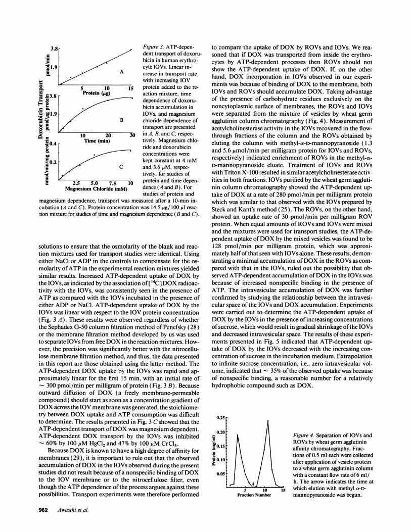
Figure 3. ATP-depen-dent transport of doxoru-bicin in human erythro-cyte IOVs. Linear in-crease in transport ratewith increasing IOV
j5 protein added to the re-
action mixture, timedependence of doxoru-bicin accumulation inIOVs, and magnesiumchloride dependence oftransport are presented
30 in A, B, and C, respec-
tively. Magnesium chlo-ride and doxorubicinconcentrations were
kept constant at 4 mMand 3.6,uM, respec-
tively, for studies ofprotein and time depen-dence (A and B). Forstudies of protein and
magnesium dependence, transport was measured after a 10-min in-cubation (A and C). Protein concentration was 14.5 ,tg/ 100 ,ul reac-
tion mixture for studies of time and magnesium dependence (B and C).
solutions to ensure that the osmolarity of the blank and reac-
tion mixtures used for transport studies were identical. Usingeither NaCl or ADP in the controls to compensate for the os-
molarity ofATP in the experimental reaction mixtures yieldedsimilar results. Increased ATP-dependent uptake of DOX bythe IOVs, as indicated by the association of [ 14C ]DOX radioac-tivity with the IOVs, was consistently seen in the presence ofATP as compared with the IOVs incubated in the presence ofeither ADP or NaCl. ATP-dependent uptake of DOX by theIOVs was linear with respect to the IOV protein concentration(Fig. 3 A). These results were observed regardless of whetherthe Sephadex G-50 column filtration method of Penefsky (28)or the membrane filtration method developed by us was usedto separate IOVs from free DOX in the reaction mixtures. How-ever, the precision was significantly better with the nitrocellu-lose membrane filtration method, and thus, the data presentedin this report are those obtained using the latter method. TheATP-dependent DOX uptake by the IOVs was rapid and ap-
proximately linear for the first 15 min, with an initial rate of- 300 pmol/min per milligram of protein (Fig. 3 B). Becauseoutward diffusion of DOX (a freely membrane-permeablecompound) should start as soon as a concentration gradient ofDOX across the IOV membrane was generated, the stoichiome-try between DOX uptake and ATP consumption was difficultto determine. The results presented in Fig. 3 C showed that theATP-dependent transport ofDOX was magnesium dependent.ATP-dependent DOX transport by the IOVs was inhibited- 60% by 100,uM HgCl2 and 47% by 100 ,tM CrCl3.
Because DOX is known to have a high degree ofaffinity formembranes (29), it is important to rule out that the observedaccumulation ofDOX in the IOVs observed during the presentstudies did not result because ofa nonspecific binding ofDOXto the IOV membrane or to the nitrocellulose filter, even
though the ATP dependence ofthe process argues against thesepossibilities. Transport experiments were therefore performed
to compare the uptake of DOX by ROVs and IOVs. We rea-
soned that if DOX was transported from inside the erythro-cytes by ATP-dependent processes then ROVs should notshow the ATP-dependent uptake of DOX. If, on the otherhand, DOX incorporation in IOVs observed in our experi-ments was because of binding ofDOX to the membrane, bothIOVs and ROVs should accumulate DOX. Taking advantageof the presence of carbohydrate residues exclusively on thenoncytoplasmic surface of membranes, the ROVs and IOVswere separated from the mixture of vesicles by wheat germ
agglutinin column chromatography (Fig. 4). Measurement ofacetylcholinesterase activity in the IOVs recovered in the flow-through fractions of the column and the ROVs obtained byeluting the column with methyl-a-D-mannopyranoside (1.3and 5.6 ,mol/min per milligram protein for IOVs and ROVs,respectively) indicated enrichment of ROVs in the methyl-a-D-mannopyranoside eluate. Treatment of IOVs and ROVswith Triton X- 100 resulted in similar acetylcholinesterase activ-ities in both fractions. IOVs purified by the wheat germ aggluti-nin column chromatography showed the ATP-dependent up-
take ofDOX at a rate of 280 pmol/min per milligram proteinwhich was similar to that observed with the IOVs prepared bySteck and Kant's method (25). The ROVs, on the other hand,showed an uptake rate of 30 pmol/min per milligram ROVprotein. When equal amounts of ROVs and IOVs were mixedand the mixtures were used for transport studies, the ATP-de-pendent uptake ofDOX by the mixed vesicles was found to be128 pmol/min per milligram protein, which was approxi-mately halfofthat seen with IOVs alone. These results, demon-strating a minimal accumulation ofDOX in the ROVs as com-
pared with that in the IOVs, ruled out the possibility that ob-served ATP-dependent accumulation ofDOX in the IOVs wasbecause of increased nonspecific binding in the presence ofATP. The intravesicular accumulation of DOX was furtherconfirmed by studying the relationship between the intravesi-cular space of the IOVs and DOX accumulation. Experimentswere carried out to determine the ATP-dependent uptake ofDOX by the IOVs in the presence ofincreasing concentrationsof sucrose, which would result in gradual shrinkage ofthe IOVsand decreased intravesicular space. The results of these experi-ments presented in Fig. 5 indicated that ATP-dependent up-take of DOX by the IOVs decreased with the increasing con-centration ofsucrose in the incubation medium. Extrapolationto infinite sucrose concentration, i.e., zero intravesicular vol-ume, indicated that 35% ofthe observed uptake was because
of nonspecific binding, a reasonable number for a relativelyhydrophobic compound such as DOX.
0.25r
0.20O
E 0.15-E
o O.10
0.05
Fraction Number
Figure 4. Separation of IOVs andROVs by wheat germ agglutininaffinity chromatography. Frac-tions of 0.5 ml each were collectedafter application of vesicle proteinto a wheat germ agglutinin columnwith a constant flow rate of 6 ml /h. The arrow indicates the time atwhich elution with methyl a-D-
mannopyranoside was begun.
962 Awasthi et al.
3.8-
'61.90. ~~~~A
a" 5 10e 3 8 Protein (ig).Ev..
-1.9O0 ~~~~~B
10 2000.4 Time (min)-
0.2 -
E / ~~~~C2.5 5.0 7.5
Magnesium Chloride (mM)

Figure 5. Osmotic sensitivity ofo4 / DOX transport in erythrocyte
IOVs. Total ATP-dependent up-3 take of DOX after 30 min was
measured in the presence of 250-,,2 / 950 mM sucrose, and the uptake
was plotted vs the inverse of su-~ 1 crose concentration. Linear regres-
sion was used to extrapolate the-2 3 4 DOX binding to the vesicles at in-
1/[Sucrose] (M-') finite sucrose concentration.
The dependence of DOX uptake on ATP and on propermembrane sidedness indicated that the transport was mediatedby an ATP-dependent pump. This was further verified by de-termining the activation energy ofthe transport, which is com-patible with an enzymatic process, and by establishing the heatsensitivity of the process. The ATP-dependent DOX transportby IOVs was shown to be temperature dependent with rates of2 1, 127, and 217 pmol/min per milligram protein at 4, 20, and37°C, respectively, yielding an activation energy of - 13 kcal/mol. Inactivation of the transporter upon heating was demon-strated in two separate experiments. IOVs preheated at 45°Cfor 1 and 4 h transported DOX at 14.0 and 2.5 pmol/min permilligram protein, corresponding to 6 and 1% ofcontrol trans-port rate, respectively. In other experiments, IOVs were im-mersed in boiling water for 5 min, were cooled to room temper-ature, and were revesiculated by sonication. IOVs were thenseparated from the mixture of vesicles using a wheat germ ag-glutinin column which quantitatively retained ROVs. Thetransport of DOX in the heat-inactivated IOVs thus preparedwas completely abolished (< 1% ofcontrol). The kinetic char-acterization of the transport mechanism for DOX during thepresent studies included the determination of the Km and V,,mvalues for its substrates, DOX and ATP (Fig. 6, A and B, respec-tively). The Km ofthe transporter for ATP was found to be 1.85mM, which was in the same range as that reported for GSHconjugate transporter ( 16, 17, 21 ). The Km of the transporterfor DOX was found to be 1.7 MM, which was closely similar tothe Km values (Table II) of the ATP-hydrolyzing activity ofDNP-SG ATPase forDOX purified from either erythrocytes or
..-.-, lnp-SG Figure 6. Kinetics ofDOX transport in hu-
025 0.50 man erythrocyte IOVs.DOX1 (SM-') Competitive inhibition
ofDOX transport byDNP-SG (A) and theKm of ATP for DOXtransport (B) are pre-sented. The concentra-tion ofDOX used for
2.0 4.0 determination of the Km1[ATPJ (mM') ofATP was 3.6 MM.
lung (1.7 MuM). Furthermore, these Km values were within therange of peak DOX serum concentrations after conventionalintravenous dosing ofDOX in humans (30) during cancer che-motherapy. In a manner similar to that of DOX transport, asaturable ATP-dependent transport by the IOVs was also ob-served for daunomycin (Km = 3.3 ,M, and Vm. = 200 pmol/min per milligram protein) and vinblastine (Km = 2.9 ,gM, andVma = 212 pmol/min per milligram protein). Since the pres-ence of DNP-SG ATPase has been demonstrated in all thehuman tissues examined so far, these results strongly suggestedthat this transport mechanism may be an important and physio-logically relevant protective mechanism against the amphiphi-lic cytotoxins such as DOX, daunomycin, or vinblastine.
Inhibition ofDOX transport by substrates ofP-glycoprotein,GSH conjugates, and the antibodies against DNP-SG A TPase.The overlap between the transport mechanism(s) for DOXand GSH conjugates was corroborated by the results ofexperi-ments showing that the ATP-dependent uptake of DOX byIOVs was competitively inhibited by DNP-SG (Fig. 6 A) andEA-SG (data not presented). The inhibitory effects of severalclasses of amphiphilic compounds including anthracyclines(mitoxantrone and daunorubicin), vinca alkaloid (vinblas-tine), calcium channel blocker (verapamil), bilirubin conju-gate (bilirubin ditaurate), bile acid conjugates (taurolithocho-late and taurodeoxycholate), and cyclosporin A on the trans-port ofDOX were also examined. The inhibition constants andnature of inhibition for these compounds are presented in Ta-ble III. It is noteworthy that the known substrates of P-glyco-protein (vinblastine, daunomycin, vincristine, and mitoxan-trone) and the known substrates ofthe DNP-SG ATPase (bili-rubin ditaurate, DNP-SG, and EA-SG) were found to inhibitDOX transport in a competitive manner. In contrast, the am-phiphilic bile salts which do not stimulate ATP hydrolysis byDNP-SG ATPase (20) were not inhibitory towards this trans-porter at concentrations up to 100 MM used in these studies.Interestingly, cyclosporin A, a compound which can reverseMDR in cells with or without P-glycoprotein overexpression,also inhibited this enzyme, albeit in a noncompetitive manner.Additional evidence for the shared transport mechanisms ofDOX and GSH conjugates was provided by a significant inhibi-tion of DOX transport in IOVs preincubated for 5 min with
Table III. Inhibition ofDOX Transport by Various AmphiphilicCompounds
Type of inhibitionInhibitors I50* K, with respect to DOX
AM AM
Vinblastine 12.0 9.5 CompetitiveVerapamil 10.0 8.3 CompetitiveMitoxantrone 15.0 12.0 CompetitiveBilirubin ditaurate 85.0 60.0 CompetitiveDaunorubicin 3.0 1.8 CompetitiveCyclosporin A 90.0 75.0 NoncompetitiveTaurolithocholate Not inhibitoryt,Taurodeoxycholate Not inhibitory -
* The 10 value is the concentration of inhibitor giving 50% inhibitionofDOX transport. Details are given in the text. $ Concentrationsof these compounds up to 100 giM were used in inhibition studies.
Insulin-degrading Enzyme 963
2.0

polyclonal antibodies against the human erythrocyte DNP-SGATPase. Whereas - 45% inhibition of the transport of DOXwas observed in the IOVs coated with anti-DNP-SG ATPaseantibodies, the preimmune serum showed no effect on thetransport ofDOX by the IOVs (Fig. 7). Taken together, theseresults provided strong evidence that the ATP-dependent trans-port of GSH conjugates and DOX from human erythrocytes,lung, and possibly other tissues was mediated through sharedmechanism(s) and that DNP-SG ATPase was a common com-ponent of this mechanism(s).
Reconstitution studies. The purified DNP-SG ATPase pro-tein used for reconstitution studies was essentially homoge-neous as seen in SDS-PAGE and was the only protein recog-nized by anti-DNP-SG ATPase antibodies in Western blotanalysis. This protein was not recognized by the C2 19 antibody(which recognizes P-glycoprotein) in Western blot analysis(data not presented). The results of DOX transport studies inIOVs reconstituted in the presence of increasing amounts ofpurified DNP-SG ATPase are presented in Fig. 8. These resultsshow a linear increase in DOX transport activity in IOVs re-constituted in the presence of increasing amounts of the puri-fied DNP-SG ATPase. Activity was unaffected in IOVs recon-stituted in the presence of increasing amounts of albumin.
Discussion
Although the DNP-SG ATPase was initially thought to be atransporter of GS-X conjugates ( 17), structurally unrelatedanionic compounds were later added to its list of substrates(20). Present studies indicate a much broader role for theDNP-SG ATPase as a xenobiotic transporter because we nowdemonstrate that the weakly cationic compounds (doxorubi-cin, dihydrodoxorubicin, daunomycin, and vinblastine) anduncharged compounds (doxorubicinone, deoxydoxorubicin-one, and dihydrodoxorubicinone) also stimulate ATP hydroly-sis by DNP-SG ATPase, and that DOX, daunomycin, andvinblastine are transported across the erythrocyte membranes(which do not express mdr- 1 gene product P-glycoprotein)through an ATP-dependent transport mechanism most likelymediated by DNP-SG ATPase. This exceptionally broad sub-strate specificity and its wide tissue distribution indicate thatthis versatile transport mechanism may function normally asan important efflux mechanism for both xenobiotics and endo-biotics. Its ability to transport the lipophilic xenobiotics andthe amphiphilic products of phase II detoxification puts it in aunique position. This is probably the only cellular defense sys-
Figure 7. The effect of anti-DNP-SG ATPase antibodieson the rate of DOX transportin erythrocyte IOVs. DOXtransport rate for the first 10min at 3.6 jiM DOX and 14.5jig IOV protein in the presenceof equal amounts of eitherpreimmune serum (-) or anti-DNP-SG antibody (+) proteinare shown as the percentage ofcontrol containing neither thepreimmune serum nor the
4o so anti-DNP-SG ATPase anti-body.
C.
C
E.W
Ernw
c
X
10 20 30 40Purified Dnp-SG ATPase (*g)
Figure 8. Transport ofDOX in eryth-rocyte IOVs reconstituted with puri-fied erythrocyte DNP-SG ATPase.DOX transport rate of erythrocyteIOVs stored for < 1 wk at 4VC andsonicated in the presence of varyingamounts of the purified DNP-SGATPase protein (o) or equal quanti-ties ofalbumin (.). DOX and IOVprotein concentrations used in thesestudies were 3.6 AM and 14 ,ug/ 100,Iu reaction mixture, respectively.
tem against toxic compounds characterized so far which maybe involved in the protection against the toxic effects ofamphi-philic toxins as well as their phase I and II biotransformationproducts.
The wide tissue distribution ( 19) and ability to transportDOX with a Km in the range of clinically achievable serumDOX concentrations is particularly interesting, suggesting thatthis transport mechanism may be important in protecting nor-mal or malignant tissues from the toxic effects of DOX. Thecytotoxicity ofDOX is closely related to the degree of its intra-cellular accumulation (31 ). Increased expression of P-glyco-protein, the best characterized mechanism for decreasing intra-cellular accumulation of DOX (3-8), has been shown toconfer resistance to DOX (4, 8). The GS-X transport mecha-nism appears to be similar to the P-glycoprotein and its relatedtransporters not only because of its ability to transport DOXwith a relatively low Km and its wide substrate specificity, butalso because, similar to P-glycoprotein, it is present in the cana-licular membrane ofhepatocytes (32). Thus, in a manner anal-ogous to the P-glycoprotein, the GS-X conjugate transportmechanism may also function to ameliorate the cytotoxicity ofDOX, daunomycin, vinblastine, and possibly other struc-turally unrelated amphiphilic compounds. Several malignantcell lines displaying the MDR phenotype have been describedwhich appear to actively extrude DOX without expressing theP-glycoprotein (8-13). It is also interesting to note that a re-cent study (7) comparing the kinetics ofDOX efflux betweenintact malignant cells with or without P-glycoprotein overex-pression has shown that although the initial rate ofDOX effluxwas greater in malignant cells overexpressing the P-glycopro-tein, the subsequent steady state rate ofDOX efflux was equalin cells with or without P-glycoprotein expression (7), whichsuggests that xenobiotic transport mechanisms distinct fromthe P-glycoprotein may also play a role in mediating energy-de-pendent efflux of DOX. The results of present studies providestrong support to this contention.
The cytotoxicity ofDOX towards drug-resistant malignantcells expressing the P-glycoprotein can be enhanced by multi-ple, structurally unrelated, relatively noncytotoxic com-pounds, presumably through competitive inhibition of the P-glycoprotein-mediated effilux ofDOX (3-5). Because we haveshown that GS-X conjugates are competitive inhibitors ofDOX transport by this GS-X conjugate transporter, drugs suchas ethacrynic acid which undergo extensive conjugation withGSH may also enhance the cytotoxicity of DOX. The coad-ministration of DOX with ethacrynic acid or other relativelynontoxic drugs which are metabolized to GS-X conjugates canthus be a novel approach for modulating the antineoplastic
=100-0
vC-
EuSW 50aC.E
10 20 30Antiserum (il
964 Awasthi et al.

effects ofDOX. Since the relatively high expression ofglutathi-one S-transferases (GSTs) in many malignant tissues (33) mayresult in more rapid conversion of certain drugs (such as etha-crynic acid) to their GS-X conjugates, combinations of suchdrugs and DOX may be effective in selectively enhancing thecytotoxicity of DOX towards malignant cells overexpressingGSTs. This combination could be particularly useful for thetreatment of drug-resistant malignancies overexpressing GST-ix since ethacrynic acid is a preferred substrate of GST-7r, theoverexpression of which has been shown to decrease the effi-cacy ofethacrynic acid in enhancing the cytotoxicity ofalkylat-ing agents (34). Further studies are required to explore thesepossibilities for enhancing the cytotoxic effects of DOX inMDR phenotypes.
Acknowledgments
This study was supported in part by U.S. Public Health Servicegrant GM-32304 awarded by the National Institute for General Medi-cine Sciences to Y. C. Awasthi, and Veterans Administration Meritreview to P. Zimniak. S. Awasthi thanks Dr. D. W. Powell, Chairman,Internal Medicine, University of Texas Medical Branch for researchfunds.
References
1. Chabner, B. A., and C. E. Myers. 1989. Clinical pharmacology of cancerchemotherapy. In Cancer Principles and Practice of Oncology. V. T. DeVita, S.Hellman, and S. A. Rosenberg, editors. J. B. Lippincott Co., Philadelphia. 349-420.
2. Juliano, R. L., and V. Ling. 1976. A surface glycoprotein modulating drugpermeability in Chinese hamster ovary cell mutants. Biochim. Biophys. Acta.455:152-162.
3. Horio, M., M. M. Gottesman, and I. Pastan. 1988. ATP-dependent trans-port of vinblastine in vesicles from human multidrug-resistant cells. Proc. Nati.Acad. Sci. USA. 85:3580-3584.
4. Biedler, J. L. 1992. Genetic aspects of multidrug resistance. Cancer(Phila.). 70(Suppl. 6):1799-1809.
5. Nielson, D., and T. Skovsgaard. 1992. P-glycoprotein as multidrug trans-porter a critical review of current multidrug resistant cell lines. Biochim.Biophys. Acta. 1139:169-183.
6. Spoelstra, E. C., H. V. Westerhoff, H. Dekker, and J. Lankelma. 1992.Kinetics ofdaunorubicin transport by P-glycoprotein of intact cancer cells. Eur.J. Biochem. 207:567-579.
7. Roepe, P. D. 1992. Analysis ofthe steady-state and initial rate ofdoxorubi-cin efflux from a series of multidrug-resistant cells expressing different levels ofP-glycoprotein. Biochemistry. 3 1:12555-12564.
8. Roninson, I. B., J. E. Chin, K. Choi, P. Gros, D. E. Houseman, A. Fojo,D. W. Shen, M. M. Gottesman, and I. Pastan. 1986. Isolation of human mdrDNA sequences amplified in multidrug-resistant KB carcinoma cells. Proc. NatI.Acad. Sci. USA. 83:4583-4590.
9. Chen, Y. N., L. A. Mickley, A. M. Schwartz, E. M. Acton, J. Hwang, and A.T. Fojo. 1990. Characterization ofadriamycin resistant human breast cancer cellswhich display over-expression ofa novel resistance-related membrane protein. J.Biol. Chem. 265:10073-10080.
10. Slovak, M. L., G. A. Hoeltge, W. S. Dalton, and J. M. Trent. 1988.Pharmacologic and biologic evidence for differing mechanism of doxorubicinresistance in two human tumor cell lines. Cancer Res. 48:2793-2797.
11. Coley, H. M., P. Workman, and P. R. Twentyman. 1991. Retention ofactivity by selected anthracyclines in a multidrug resistant human large cell lungcarcinoma line without P-glycoprotein hyperexpression. Br. J. Cancer. 63:351-357.
12. Versantvoort, C. H. M., H. J. Broxterman, H. M. Pinedo, E. G. E. deVries, N. Feller, C. M. Kuiper, and J. Lankelma. 1992. Energy-dependent pro-cesses involved in reduced drug accumulation in multidrug-resistant human lungcancer cell lines without P-glycoprotein expression. Cancer Res. 52:17-23.
13. Baas, F., A. P. M. Jongsma, H. J. Broxterman, R. J. Arecici, D. House-
man, G. L. Scheffler, A. Riethorst, M. van Groenigen, A. W. M. Nieuwint, and H.Joenje. 1990. Non-P-glycoprotein mediated mechanisms for multidrug resis-tance precede P-glycoprotein expression during in-vitro selection for doxorubicinresistance in a human lung cancer cell line. Cancer Res. 50:5392-5398.
14. Srivastava, S. K., and E. Beutler. 1969. The transport ofoxidized glutathi-one from human erythrocytes. J. Biol. Chem. 244:9-16.
15. Kondo, T., G. L. Dale, and E. Beutler. 1981. Studies on glutathionetransport utilizing inside-out vesicles prepared from human erythrocytes. Bio-chim. Biophys. Acta. 645:132-136.
16. LaBelle, E. F., S. V. Singh, S. K. Srivastava, and Y. C. Awasthi. 1986.Dinitrophenyl glutathione efflux from human erythrocytes is a primary activeATP-dependent transport. Biochem. J. 238:443-449.
17. LaBelle, E. F., S. V. Singh, H. Ahmad, L. Wronski, S. K. Srivastava, andY. C. Awasthi. 1988. A novel dinitrophenylglutathione-stimulated ATPase ispresent in human erythrocyte membranes. FEBS (Fed. Eur. Biochem. Soc.) Lett.228:53-56.
18. Kondo, T., Y. Kawakani, N. Taniguchi, and E. Beutler. 1987. Glutathi-one disulfide-stimulated Mg2+-ATPase ofhuman erythrocyte membranes. Proc.Natl. Acad. Sci. USA. 84:7373-7377.
19. Sharma, R., S. Gupta, S. V. Singh, R. D. Medh, H. Ahmad, E. F. LaBelle,and Y. C. Awasthi. 1990. Purification and characterization of dinitrophenylglu-tathione ATPase of human erythrocytes and its expression in other tissues. Bio-chem. Biophys. Res. Commun. 171:155-161.
20. Singhal, S. S., R. Sharma, S. Gupta, H. Ahmad, P. Zimniak, A. Rado-minska, R. Lester, and Y. C. Awasthi. 1991. The anionic conjugates of bilirubinand bile salts stimulate ATP hydrolysis by S-(dinitrophenyl)glutathione ATPaseof human erythrocyte. FEBS (Fed. Eur. Biochem. Soc.) Lett. 281:255-257.
21. Saxena, M., S. S. Singhal, S. Awasthi, S. V. Singh, E. F. LaBelle, P. Zim-niak, and Y. C. Awasthi. 1992. Dinitrophenyl S-glutathione ATPase purifiedfrom human muscle catalyzes ATP hydrolysis in presence of leukotrienes. Arch.Biochem. Biophys. 298:231-237.
22. Awasthi, Y. C., H. S. Garg, D. D. Dao, C. A. Partridge, and S. K. Srivas-tava. 1981. Enzymatic conjugation of erythrocyte glutathione with l-chloro-2,4-dinitrobenzene: the fate ofglutathione conjugate in erythrocytes and the effect ofglutathione depletion on hemoglobin. Blood. 58:733-738.
23. Awasthi, S., S. K. Srivastava, F. Ahmad, H. Ahmad, and G. A. S. Ansari.1993. Interactions of glutathione S-transferase ir with ethacrynic acid and itsglutathione conjugate. Biochim. Biophys. Acta. 1164:173-178.
24. Minamide, L. S., and J. R. Bamburg. 1990. A filter paper dye-bindingassay for quantitative determination of protein without interference from reduc-ing agents or detergents. Anal. Biochem. 190:66-70.
25. Steck, T. L., and J. A. Kant. 1974. Preparation ofimpermeable ghosts andinside-out vesicles from human erythrocyte membranes. Methods Enzymol.31A:172-180.
26. Dodge, J. T., C. Mitchell, and D. J. Hanahan. 1963. The preparation andchemical characteristics of hemoglobin-free ghosts ofhuman erythrocytes. Arch.Biochem. Biophys. 100: 1 19-130.
27. Ellman, G. L., K. D. Courtney, V. Andres, and R. M. Featherstone. 1961.A new and rapid colorimetric determination ofacetylcholinesterase activity. Bio-chem. Pharmacol. 7:88-95.
28. Penefsky, H. S. 1977. Reversible binding ofPi by beefheart mitochondrialadenosine triphosphatase. J. Biol. Chem. 252:2891-2899.
29. Awasthi, S., R. Sharma, Y. C. Awasthi, J. A. Belli, and E. P. Frenkel. 1992.The relationship ofdoxorubicin binding to membrane lipids with drug resistance.Cancer Lett. 63:109-116.
30. Benjamin, R. S. 1974. Pharmacokinetics of adriamycin (NSC-123127) inpatients with sarcomas. Cancer Chemother. Rep. 58:271-273.
31. Keizer, H. G., G. J. Schuurhuid, H. J. Broxterman, J. Lankelma, W. G. E.J. Schoonen, J. van Rijn, H. M. Pinedoand, and H. Joenje. 1989. Correlation ofmultidrug resistance with decreased accumulation, altered subcellular drug distri-bution and increased P-glycoprotein expression in cultured SW-1573 humanlung tumor cells. Cancer Res. 49:2988-2993.
32. Zimniak, P., S. A. Ziller, I. Panfil, A. Radominska, H. Wolters, F.Kuipers, R. Sharma, M. Saxena, M. T. Moslen, M. Vore, et al. 1991. Identifica-tion of an anion-transport ATPase that catalyzed glutathione conjugate-depen-dent ATP hydrolysis in canalicular plasma membranes from normal rats and ratswith conjugated hyperbilirubinemia (GY mutant). Arch. Biochem. Biophys.292:534-538.
33. Tsuchida, S., and K. Sato. 1992. Glutathione transferases and cancer. Crit.Rev. Biochem. Mol. Biol. 27:337-384.
34. Kuzmich, S., L. A. Vanderveer, W. S. Walsh, F. P. LaCreta, and K. D.Tew. 1992. Increased levels of glutathione S-transferase-2r transcript as mecha-nism of resistance to ethacrynic acid. Biochem. J. 281:219-224.
Doxorubicin Transport by a Glutathione Conjugate Transporter 965