Role of beneficial microbes with nitrogen and phosphorous ...
Beneficial effects of adenosine triphosphate-sensitive K+ channel opener on liver ischemia/reperfusion injury
Mateus Antunes Nogueira, Ana Maria Mendonça Coelho, Sandra Nassa Sampietre, Rosely Antunes Patzina, Fabiano Pinheiro da Silva, Luiz Augusto Carneiro D'Albuquerque, Marcel Cerqueira Cesar Machado
Mateus Antunes Nogueira, Ana Maria Mendonça Coelho, Sandra Nassa Sampietre, Rosely Antunes Patzina, Fabiano Pinheiro da Silva, Luiz Augusto Carneiro D’Albuquerque, Marcel Cerqueira Cesar Machado, Department of Gastroenter-ology (LIM/37), Medical School, University of Sao Paulo, Sao Paulo 01246903, BrazilMateus Antunes Nogueira, Ana Maria Mendonça Coelho, Sandra Nassa Sampietre, Rosely Antunes Patzina, Fabiano Pinheiro da Silva, Luiz Augusto Carneiro D’Albuquerque, Marcel Cerqueira Cesar Machado, Department of Clinical Emergency (LIM/51), Medical School, University of Sao Paulo, Sao Paulo 01246903, BrazilAuthor contributions: Nogueira MA and Machado MCC de-signed research; Coelho AMM, Sampietre SN and Patzina RA performed research; D’Albuquerque LAC provided material and laboratory equipment, revised the manuscript; Nogueira MA, Coelho AMM, Pinheiro da Siva F and Machado MCC analyzed data; Coelho AMM and Machado MCC wrote the paper.Supported by Fundação de Amparo à Pesquisa do Estado de São Paulo, No. 2010/19078-1Correspondence to: Marcel Cerqueira Cesar Machado, MD, Professor of Medicine, Department of Clinical Emergency (LIM/51), Medical School, University of Sao Paulo, R. Peixoto Gomide, 515 13 andar 01409001, Sao Paulo 01246903, Brazil. [email protected]: +55-11-32891188 Fax: +55-11-32891188Received: March 28, 2014 Revised: May 28, 2014Accepted: July 11, 2014Published online: November 7, 2014
AbstractAIM: To investigate the effect of diazoxide administra-tion on liver ischemia/reperfusion injury.
METHODS: Wistar male rats underwent partial liver ischemia performed by clamping the pedicle from the medium and left anterior lateral segments for 1 h un-der mechanical ventilation. They were divided into 3 groups: Control Group, rats submitted to liver manipu-lation, Saline Group, rats received saline, and Diazoxide
Group, rats received intravenous injection diazoxide (3.5 mg/kg) 15 min before liver reperfusion. 4 h and 24 h after reperfusion, blood was collected for determination of aspartate transaminase (AST), alanine transaminase (ALT), tumor necrosis factor (TNF-α), interleukin-6 (IL-6), interleukin-10 (IL-10), nitrite/nitrate, creatinine and tumor growth factor-β1 (TGF-β1). Liver tissues were assembled for mitochondrial oxidation and phos-phorylation, malondialdehyde (MDA) content, and his-tologic analysis. Pulmonary vascular permeability and myeloperoxidase (MPO) were also determined.
RESULTS: Four hours after reperfusion the diazoxide group presented with significant reduction of AST (2009 ± 257 U/L vs 3523 ± 424 U/L, P = 0.005); ALT (1794 ± 295 U/L vs 3316 ± 413 U/L, P = 0.005); TNF-α (17 ± 9 pg/mL vs 152 ± 43 pg/mL, P = 0.013; IL-6 (62 ± 18 pg/mL vs 281 ± 92 pg/mL); IL-10 (40 ± 9 pg/mL vs 78 ± 10 pg/mL P = 0.03), and nitrite/nitrate (3.8 ± 0.9 µmol/L vs 10.2 ± 2.4 µmol/L, P = 0.025) when compared to the saline group. A significant reduction in liver mitochondrial dysfunction was observed in the diazoxide group com-pared to the saline group (P < 0.05). No differences in liver MDA content, serum creatinine, pulmonary vascular permeability and MPO activity were observed between groups. Twenty four hours after reperfusion the diazoxide group showed a reduction of AST (495 ± 78 U/L vs 978 ± 192 U/L, P = 0.032); ALT (335 ± 59 U/L vs 742 ± 182 U/L, P = 0.048), and TGF-β1 (11 ± 1 ng/mL vs 17 ± 0.5 ng/mL, P = 0.004) serum levels when compared to the saline group. The control group did not present altera-tions when compared to the diazoxide and saline groups.
CONCLUSION: Diazoxide maintains liver mitochondrial function, increases liver tolerance to ischemia/reper-fusion injury, and reduces the systemic inflammatory response. These effects require further evaluation for using in a clinical setting.
© 2014 Baishideng Publishing Group Inc. All rights reserved.
RESEARCH REPORT
Submit a Manuscript: http://www.wjgnet.com/esps/Help Desk: http://www.wjgnet.com/esps/helpdesk.aspxDOI: 10.3748/wjg.v20.i41.15319
15319 November 7, 2014|Volume 20|Issue 41|WJG|www.wjgnet.com
World J Gastroenterol 2014 November 7; 20(41): 15319-15326 ISSN 1007-9327 (print) ISSN 2219-2840 (online)
© 2014 Baishideng Publishing Group Inc. All rights reserved.
Key words: Liver ischemia/reperfusion; Diazoxide; K+ channel opener; Mitochondrial ATP-sensitive potassium channel; Liver mitochondria
Core tip: Diazoxide is a selective mitoKATP channel opener and has a protective effect against organ isch-emia/reperfusion (I/R) injury. This report shows that di-azoxide maintains liver mitochondrial function, increases liver tolerance to I/R injury, and reduces the systemic inflammatory response. Since diazoxide has also a hypotensive effect its administration may also reduce bleeding in liver surgery during hepatic parenchyma transection. These effects require further evaluation for use in a clinical setting.
Nogueira MA, Coelho AMM, Sampietre SN, Patzina RA, Pinhei-ro da Silva F, D’Albuquerque LAC, Machado MCC. Beneficial effects of adenosine triphosphate-sensitive K+ channel opener on liver ischemia/reperfusion injury. World J Gastroenterol 2014; 20(41): 15319-15326 Available from: URL: http://www.wjgnet.com/1007-9327/full/v20/i41/15319.htm DOI: http://dx.doi.org/10.3748/wjg.v20.i41.15319
INTRODUCTIONThe physiological function of mitochondrial ATP-sensitive potassium channel (mitoKATP) is to permit K+ transport into the mitochondrial matrix and therefore maintain its volume[1]. This mitochondrial channel has been identified in many tissues, such as heart[2], brain[3], skeletal muscle[4] and liver[5].
This mitoKATP channel has been related to the preconditioning protection of the ischemic heart[6]. It was also observed that the preconditioning-like effect of morphine in the intact heart can be the result of mito-KATP channel activation[7].
Diazoxide is a selective mitoKATP channel opener and could have a protective effect against organ isch-emia/reperfusion (I/R) injury. In fact a protective effect has been demonstrated in the heart[8]. This protective effect is related to mitoKATP opening since other drugs with this action also reduce the heart I/R injury[9].
The protective effect of diazoxide in I/R injury has been reported in several organs, such as brain[10] and spi-nal cord[11] in which mitoKATP has been identified. Since mitoKATP channels are present in liver mitochondria[12] it is conceivable that diazoxide may have a protective ef-fect on liver I/R injury. In the present study, we evaluated the effect of diazoxide on local and systemic effects of liver I/R injury.
MATERIALS AND METHODSAnimalsSeventy adult male Wistar rats weighing 230-270 g housed in individual cages in a 12 h dark-light controlled environment were used for the experimental protocol.
Rats had free access to standard rat chow and water. The experimental protocol was approved by the Ethics Com-mittee for Animal Research from the Medical School of São Paulo University and received humanized care ac-cording to the criteria outlined in the Guide for the Care and Use of Laboratory Animals (Institute of Laboratory Animal Resources, Commission on Life Sciences and National Research Council. National Academic Press, Washington, DC, 1996.)
Experimental design The rats were randomly submitted to the following ex-perimental protocols: Control Group (n = 18), rats un-derwent laparotomy and liver manipulation; Saline Group (n = 26), rats received intravenous injection (iv) saline 15 min before liver ischemia; Diazoxide Group (n = 26), rats received iv diazoxide (Sigma Chemical CO, St. Louis, MO, USA) (3.5 mg/kg) 15 min before liver ischemia. Saline and Diazoxide groups received the same iv volume in mL/kg.
Surgical procedure and sample collectionThe animals were anesthetized with intra-peritoneal ketamine (Cristalia, São Paulo, Brazil) (30 mg/kg) and xylazine (Bayer, São Paulo, Brazil) (30 mg/kg) and sub-mitted to orotracheal intubation, and ventilated with a tidal volume of 0.08 mL/g body weight, at a respiratory rate of 60/min, and FiO2 of 0.21 (Small Animal Ven-tilator model 683, Harvard Apparatus, Holliston, MA, USA). During the surgical procedure, body temperature was monitored using a rectal digital thermometer (YSI Precision 4000A Thermometer, USA), being maintained at 37 ℃. Median laparotomy was performed and the hepatic pedicles of the median and left anterolateral seg-ments were isolated, exposed and clamped with a non-traumatic microvascular bulldog clamp during 1 h to induce ischemia to 70% of the total liver volume. In this model, intestinal congestion is avoided, allowing the pos-sibility to study the effects of isolated liver ischemia. The incision was closed, and after a 60-min ischemic period, the abdomen was reopened allowing clamp removal and liver reperfusion[13,14].
After liver reperfusion, rats were re-anesthetized for blood sampling through cardiac puncture and killed by exsanguination. At 4 h and 24 h after reperfusion, blood was collected for determination of aspartate aminotrans-ferase (AST), alanine aminotransferase (ALT), tumor ne-crosis factor (TNF-α), interleukin-6 (IL-6), interleukin-10 (IL-10), tumor growth factor-β1 (TGF-β1), nitrite/nitrate and creatinine. Hepatic tissues were assembled for evalu-ation of mitochondrial oxidation and phosphorylation, malondialdehyde (MDA) content, and histologic analysis. The liver tissue for post-ischemic analysis was obtained from the median and left anterolateral segments previ-ously submitted to I/R injury. Lungs were perfused via the trachea with 30-50 mL of 0.9% NaCl at 10 mL/min, using a syringe pump (model 975) from Harvard Appara-tus, and fragments were harvested and divided for analy-
15320 November 7, 2014|Volume 20|Issue 41|WJG|www.wjgnet.com
Nogueira MA et al . Diazoxide on liver ischemia/reperfusion injury
sis of microvascular permeability and myeloperoxidase (MPO) activity. No mortality was observed in this model of partial liver ischemia.
Serum AST and ALT levels Serum AST and ALT levels were measured to assess the extent of hepatocellular injury. The enzyme activities were assayed by using the optimized ultraviolet method (COBAS MIRA) from Roche (Roche Diagnostics, Rot-krenz, Switzerland). Results are expressed as units per liter (U/L).
Liver mitochondrial oxidation and phosphorylation activitiesLiver mitochondria were prepared as previously de-scribed[15]. Briefly, rat livers were rapidly excised and placed in medium containing 250 mmol/L sucrose, 10 mmol/L Tris-HCl, and 1 mmol/L EGTA, pH 7.3, at 4 ℃. The tissue was scissor-minced and homogenized in ice using a Teflon Potter homogenizer. The homogenate was cen-trifuged at 600 g for 10 min. The supernatant was centri-fuged for 10 min at 10000 g to obtain the mitochondrial pellet. A mitochondrial suspension containing 30-40 mg/mL of mitochondrial protein was prepared, and stored on ice before the assay of mitochondrial respiration.
The mitochondrial oxygen consumption was polaro-graphically[16] measured using a Gilson 5/6H Oxygraph (Gilson Medical Eletronics, Inc., Middleton, WI) in a closed reaction vessel fitted with a Clark oxygen elec-trode (Yellow Springs Instruments Co., Yellow Springs, OH) at 28 ℃. The incubation medium consisted of 120 mmol/L KCl, 2 mmol/L sodium phosphate, 10 µmol/L rotenone, and 1 mmol/L EGTA [Ethylene glycol-bis(2-aminoethylether)-N,N,N’,N’-tetraacetic acid], and was buffered at pH 7.3 with 5 mmol/L Tris-HCl. Mitochon-dria were energized with potassium succinate as substrate at a final concentration of 10 mmol/L. After a brief equilibration period, state 3 (activated state, S3) respira-tion was induced by the addition of 280 nmol adenosine diphosphate (ADP). The added ADP was phosphory-lated to adenosine triphosphate (ATP) and the state 4 (basal state, S4) respiration was then measured. The oxy-gen consumption ratio in the presence of ADP to that in absence (respiratory control rate, RCR) and the ADP/O ratio were calculated as indices of mitochondrial oxida-tion and phosphorylation activities[17].
RCR = Oxygen consumption in the S3/oxygen con-sumption in the S4.
ADP/O = Moles of ATP formed from ADP per atom of oxygen consumed.
S3 and S4 were measured and reported as nmol oxygen per milligram mitochondrial protein per minute. Mitochondria protein content was determined by the method of Lowry et al[18].
Lipid peroxidation analysisMDA formation was used to indicate the occurrence of lipid peroxidation in the tissues and was estimated as thio-
barbituric acid-reactive substances (TBARS). Liver tissues (100 mg/mL) were homogenized in 1.15% KCl buffer, and centrifuged at 14000 g for 20 min. An aliquot of the supernatant was then added to a reaction mixture consist-ing of 1.5 mL 0.8% thiobarbituric acid, 200 µL 8.1% (v/v) sodium dodecyl sulfate, 1.5 mL 20% acetic acid (pH 3.5), and 600 µL distilled water. The mixture was then heated at 90 ℃ for 45 min. After cooling to room temperature, the samples were cleared by centrifugation (10000 g for 10 min), and the absorbance was measured at 532 nm using malondialdehyde bis (dimethyl acetyl) as external standard. The content of lipid peroxides was expressed as nmol MDA per mg of protein[19].
Serum levels of nitrite-nitrate Serum levels of nitrite-nitrate were determining using a commercial assay kit (R and D Systems Inc, MN, USA) according to the manufacturer’s guidelines.
Determination of inflammatory mediatorsSerum levels of TNF-α, IL-6, IL-10, and TGF-β1 were determined by ELISA using commercial kits (Invitrogen, CA, USA).
Histological analysis of the liverLiver samples were fixed in 10% buffered formalin for standard HE staining. Histological evaluation of the liver sections was performed by the same pathologist in a blinded manner. The severity of histological injury was analyzed according to the scoring system proposed by Quireze et al[20].
Lung tissue microvascular permeability analysisIncreases in lung microvascular permeability were quan-tified by the Evans blue dye (EBD) extravasation tech-nique as described previously[18]. EBD 20 mg/kg body weight was injected via dorsal penial vein 15 min before euthanasia. After collecting blood sampling, lungs were perfused with 30-50 mL of NaCl 0.9% at 10 mL/min, using a syringe pump (model 975) from Harvard Appara-tus, and weighed. One small fragment was dried at 60 ℃ for calculation of total dry weight. To extract the dye, the lung was incubated with formamide, 4 mL/mg of tissue, for 24 h at room temperature. The concentration of EBD extracted into formamide was quantified spec-trophotometrically at 620 nm using the Ultra Microplate Reader ELX 808 from Bio-Tek Instruments (Winooski, VT). The results are expressed as microgram of EBD per gram of dry weight tissue. Expression of results as a function of dry weight avoids underevaluation due to edema formation[21].
Lung tissue MPO activityLung MPO activity was used as an indicator of the neu-trophil content in lung parenchyma. Samples of 300 mg wet lung tissue were homogenized with a polytron ho-mogenizer (Polytron PT-2100 homogenizer, Kinematica AG, Luzern, Switzerland) for 60 s in 1 mL of sodium
15321 November 7, 2014|Volume 20|Issue 41|WJG|www.wjgnet.com
Nogueira MA et al . Diazoxide on liver ischemia/reperfusion injury
15322 November 7, 2014|Volume 20|Issue 41|WJG|www.wjgnet.com
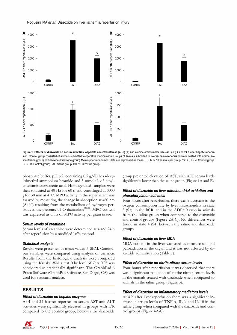
group presented elevation of AST, with ALT serum levels significantly lower than the saline group (Figure 1A and B).
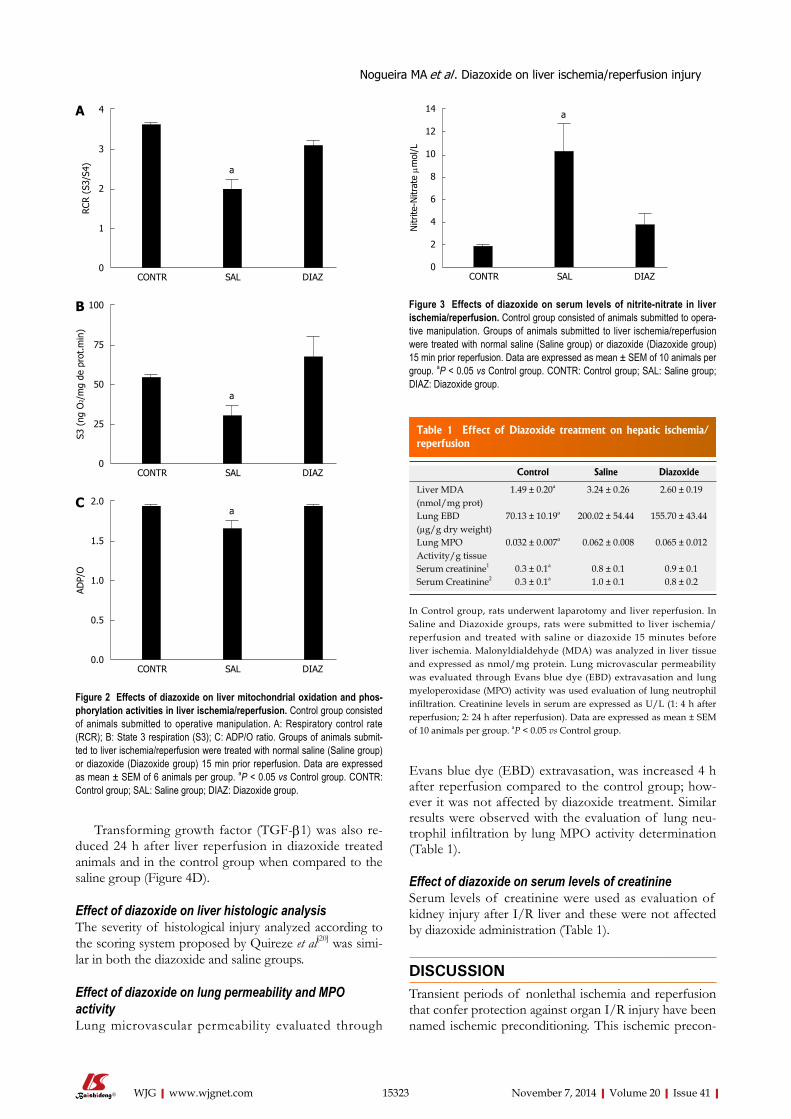
Effect of diazoxide on liver mitochondrial oxidation and phosphorylation activitiesFour hours after reperfusion, there was a decrease in the oxygen consumption rate by liver mitochondria in state 3 (S3), in the RCR, and in the ADP/O ratio in animals from the saline group when compared to the diazoxide and control groups (Figure 2A-C). No differences were found in state 4 (S4) between the saline and diazoxide groups.
Effect of diazoxide on liver MDAMDA content in the liver was used as measure of lipid peroxidation in the organ and it was not affected by di-azoxide administration (Table 1).
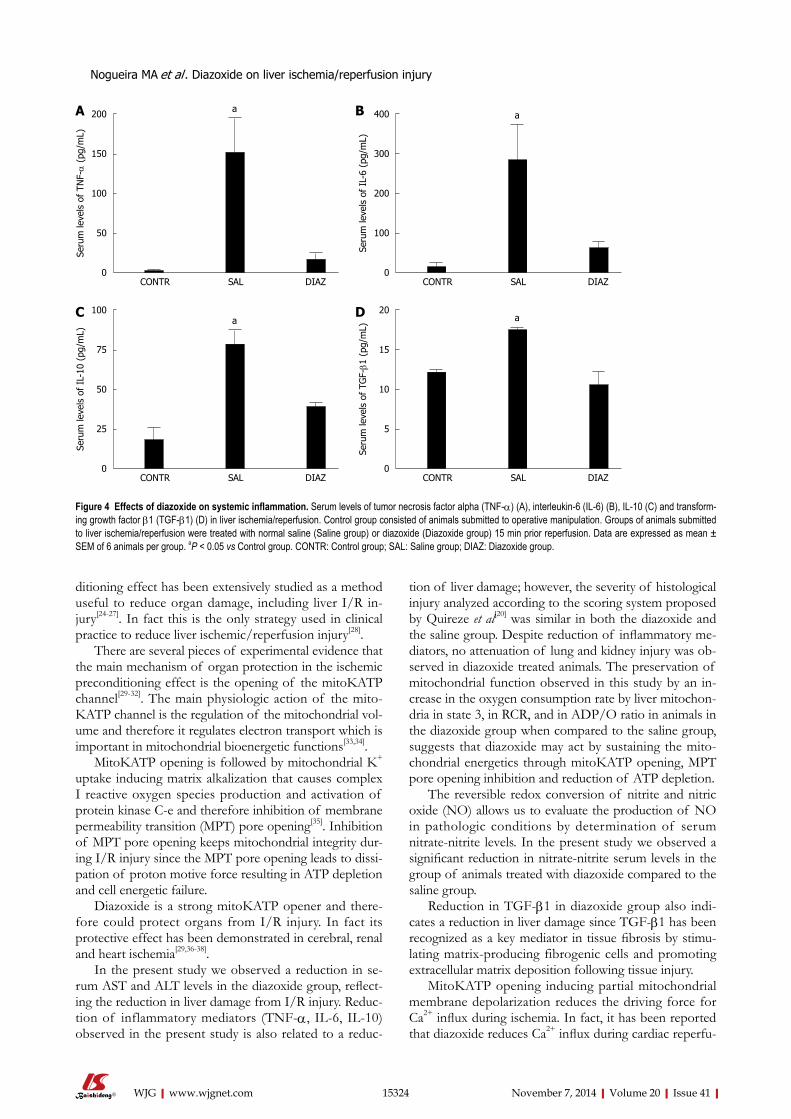
Effect of diazoxide on nitrite-nitrate serum levelsFour hours after reperfusion it was observed that there was a significant reduction of nitrite-nitrate serum levels in the animals treated with diazoxide when compared to animals in the saline group (Figure 3).
Effect of diazoxide on inflammatory mediators levelsAt 4 h after liver reperfusion there was a significant in-crease in serum levels of TNF-α, IL-6, and IL-10 in the saline group when compared with the diazoxide and con-trol groups (Figure 4A-C).
phosphate buffer, pH 6.2, containing 0.5 g/dL hexadecy-ltrimethyl ammonium bromide and 5 mmol/L of ethyl-enediaminetetraacetic acid. Homogenized samples were then sonicated at 40 Hz for 60 s, and centrifuged at 3000 g for 30 min at 4 ℃. MPO activity in the supernatant was assayed by measuring the change in absorption at 460 nm (A460) resulting from the metabolism of hydrogen per-oxide in the presence of O-dianisidine[22,23]. MPO content was expressed as units of MPO activity per gram tissue.
Serum levels of creatinineSerum levels of creatinine were determined at 4 and 24 h after reperfusion by a modified Jaffe method.
Statistical analysisResults were presented as mean values ± SEM. Continu-ous variables were compared using analysis of variance. Results from the histological analysis were compared using the Kruskal-Wallis test. The level of P < 0.05 was considered as statistically significant. The GraphPad 6 Prism Software (GraphPad Software, San Diego, CA) was used for statistical analysis.
RESULTSEffect of diazoxide on hepatic enzymes At 4 and 24 h after reperfusion serum AST and ALT activities were significantly elevated in groups with I/R compared to the control group; however the diazoxide
4000
3000
2000
1000
0CONTR SAL DIAZ
AST
4 h
afte
r re
perf
usio
n (U
/L)
a
c
4000
3000
2000
1000
0CONTR SAL DIAZ
ALT
4 h
afte
r re
perf
usio
n (U
/L)
a
c
Figure 1 Effects of diazoxide on serum activities. Aspartate aminotransferase (AST) (A) and alanine aminotransferase (ALT) (B) 4 and 24 h after hepatic reperfu-sion. Control group consisted of animals submitted to operative manipulation. Groups of animals submitted to liver ischemia/reperfusion were treated with normal sa-line (Saline group) or diazoxide (Diazoxide group) 15 min prior reperfusion. Data are expressed as mean ± SEM of 10 animals per group. a,cP < 0.05 vs Control group. CONTR: Control group; SAL: Saline group; DIAZ: Diazoxide group.
A B
1500
1000
500
0CONTR SAL DIAZ
AST
24 h
aft
er r
eper
fusi
on (
U/L
)
a
1000
750
500
250
0CONTR SAL DIAZ
ALT
24 h
aft
er r
eper
fusi
on (
U/L
)
a
Nogueira MA et al . Diazoxide on liver ischemia/reperfusion injury
15323 November 7, 2014|Volume 20|Issue 41|WJG|www.wjgnet.com
Transforming growth factor (TGF-β1) was also re-duced 24 h after liver reperfusion in diazoxide treated animals and in the control group when compared to the saline group (Figure 4D).
Effect of diazoxide on liver histologic analysisThe severity of histological injury analyzed according to the scoring system proposed by Quireze et al[20] was simi-lar in both the diazoxide and saline groups.
Effect of diazoxide on lung permeability and MPO activityLung microvascular permeability evaluated through
Evans blue dye (EBD) extravasation, was increased 4 h after reperfusion compared to the control group; how-ever it was not affected by diazoxide treatment. Similar results were observed with the evaluation of lung neu-trophil infiltration by lung MPO activity determination (Table 1).
Effect of diazoxide on serum levels of creatinineSerum levels of creatinine were used as evaluation of kidney injury after I/R liver and these were not affected by diazoxide administration (Table 1).
DISCUSSIONTransient periods of nonlethal ischemia and reperfusion that confer protection against organ I/R injury have been named ischemic preconditioning. This ischemic precon-
Table 1 Effect of Diazoxide treatment on hepatic ischemia/reperfusion
Control Saline Diazoxide
Liver MDA (nmol/mg prot)
1.49 ± 0.20a 3.24 ± 0.26 2.60 ± 0.19
Lung EBD(µg/g dry weight)
70.13 ± 10.19a 200.02 ± 54.44 155.70 ± 43.44
Lung MPO Activity/g tissue
0.032 ± 0.007a 0.062 ± 0.008 0.065 ± 0.012
Serum creatinine1 0.3 ± 0.1a 0.8 ± 0.1 0.9 ± 0.1Serum Creatinine2 0.3 ± 0.1a 1.0 ± 0.1 0.8 ± 0.2
In Control group, rats underwent laparotomy and liver reperfusion. In Saline and Diazoxide groups, rats were submitted to liver ischemia/reperfusion and treated with saline or diazoxide 15 minutes before liver ischemia. Malonyldialdehyde (MDA) was analyzed in liver tissue and expressed as nmol/mg protein. Lung microvascular permeability was evaluated through Evans blue dye (EBD) extravasation and lung myeloperoxidase (MPO) activity was used evaluation of lung neutrophil infiltration. Creatinine levels in serum are expressed as U/L (1: 4 h after reperfusion; 2: 24 h after reperfusion). Data are expressed as mean ± SEM of 10 animals per group. aP < 0.05 vs Control group.
4
3
2
1
0CONTR SAL DIAZ
RCR
(S3
/S4) a
A
100
75
50
25
0CONTR SAL DIAZ
S3 (
ng O
2 /m
g de
pro
t.m
in)
a
B
2.0
1.5
1.0
0.5
0.0CONTR SAL DIAZ
ADP/
O
aC
Figure 2 Effects of diazoxide on liver mitochondrial oxidation and phos-phorylation activities in liver ischemia/reperfusion. Control group consisted of animals submitted to operative manipulation. A: Respiratory control rate (RCR); B: State 3 respiration (S3); C: ADP/O ratio. Groups of animals submit-ted to liver ischemia/reperfusion were treated with normal saline (Saline group) or diazoxide (Diazoxide group) 15 min prior reperfusion. Data are expressed as mean ± SEM of 6 animals per group. aP < 0.05 vs Control group. CONTR: Control group; SAL: Saline group; DIAZ: Diazoxide group.
14
12
10
8
6
4
2
0CONTR SAL DIAZ
Nitr
ite-N
itrat
e µm
ol/L
a
Figure 3 Effects of diazoxide on serum levels of nitrite-nitrate in liver ischemia/reperfusion. Control group consisted of animals submitted to opera-tive manipulation. Groups of animals submitted to liver ischemia/reperfusion were treated with normal saline (Saline group) or diazoxide (Diazoxide group) 15 min prior reperfusion. Data are expressed as mean ± SEM of 10 animals per group. aP < 0.05 vs Control group. CONTR: Control group; SAL: Saline group; DIAZ: Diazoxide group.
Nogueira MA et al . Diazoxide on liver ischemia/reperfusion injury
15324 November 7, 2014|Volume 20|Issue 41|WJG|www.wjgnet.com
ditioning effect has been extensively studied as a method useful to reduce organ damage, including liver I/R in-jury[24-27]. In fact this is the only strategy used in clinical practice to reduce liver ischemic/reperfusion injury[28].
There are several pieces of experimental evidence that the main mechanism of organ protection in the ischemic preconditioning effect is the opening of the mitoKATP channel[29-32]. The main physiologic action of the mito-KATP channel is the regulation of the mitochondrial vol-ume and therefore it regulates electron transport which is important in mitochondrial bioenergetic functions[33,34].
MitoKATP opening is followed by mitochondrial K+ uptake inducing matrix alkalization that causes complex I reactive oxygen species production and activation of protein kinase C-e and therefore inhibition of membrane permeability transition (MPT) pore opening[35]. Inhibition of MPT pore opening keeps mitochondrial integrity dur-ing I/R injury since the MPT pore opening leads to dissi-pation of proton motive force resulting in ATP depletion and cell energetic failure.
Diazoxide is a strong mitoKATP opener and there-fore could protect organs from I/R injury. In fact its protective effect has been demonstrated in cerebral, renal and heart ischemia[29,36-38].
In the present study we observed a reduction in se-rum AST and ALT levels in the diazoxide group, reflect-ing the reduction in liver damage from I/R injury. Reduc-tion of inflammatory mediators (TNF-α, IL-6, IL-10) observed in the present study is also related to a reduc-
tion of liver damage; however, the severity of histological injury analyzed according to the scoring system proposed by Quireze et al[20] was similar in both the diazoxide and the saline group. Despite reduction of inflammatory me-diators, no attenuation of lung and kidney injury was ob-served in diazoxide treated animals. The preservation of mitochondrial function observed in this study by an in-crease in the oxygen consumption rate by liver mitochon-dria in state 3, in RCR, and in ADP/O ratio in animals in the diazoxide group when compared to the saline group, suggests that diazoxide may act by sustaining the mito-chondrial energetics through mitoKATP opening, MPT pore opening inhibition and reduction of ATP depletion.
The reversible redox conversion of nitrite and nitric oxide (NO) allows us to evaluate the production of NO in pathologic conditions by determination of serum nitrate-nitrite levels. In the present study we observed a significant reduction in nitrate-nitrite serum levels in the group of animals treated with diazoxide compared to the saline group.
Reduction in TGF-β1 in diazoxide group also indi-cates a reduction in liver damage since TGF-β1 has been recognized as a key mediator in tissue fibrosis by stimu-lating matrix-producing fibrogenic cells and promoting extracellular matrix deposition following tissue injury.
MitoKATP opening inducing partial mitochondrial membrane depolarization reduces the driving force for Ca2+ influx during ischemia. In fact, it has been reported that diazoxide reduces Ca2+ influx during cardiac reperfu-
200
150
100
50
0CONTR SAL DIAZ
Seru
m le
vels
of
TNF-α
(pg
/mL)
a 400
300
200
100
0CONTR SAL DIAZ
Seru
m le
vels
of
IL-6
(pg
/mL)
a
100
75
50
25
0CONTR SAL DIAZ
Seru
m le
vels
of
IL-1
0 (p
g/m
L)
a20
15
10
5
0CONTR SAL DIAZ
Seru
m le
vels
of
TGF-β1
(pg
/mL)
a
Figure 4 Effects of diazoxide on systemic inflammation. Serum levels of tumor necrosis factor alpha (TNF-α) (A), interleukin-6 (IL-6) (B), IL-10 (C) and transform-ing growth factor β1 (TGF-β1) (D) in liver ischemia/reperfusion. Control group consisted of animals submitted to operative manipulation. Groups of animals submitted to liver ischemia/reperfusion were treated with normal saline (Saline group) or diazoxide (Diazoxide group) 15 min prior reperfusion. Data are expressed as mean ± SEM of 6 animals per group. aP < 0.05 vs Control group. CONTR: Control group; SAL: Saline group; DIAZ: Diazoxide group.
Nogueira MA et al . Diazoxide on liver ischemia/reperfusion injury
A B
C D
15325 November 7, 2014|Volume 20|Issue 41|WJG|www.wjgnet.com
sion[38]. Since intracellular Ca2+ overload may cause mi-tochondrial damage, the beneficial effect of diazoxide in liver I/R injury may also be related to reduction in hepa-tocyte intracellular Ca2+. The suppression of hepatocyte calcium overload by diazoxide recently demonstrated[12] is therefore an additional effect of diazoxide protecting liver mitochondria from I/R injury. The study of other activators of these mitoKATP channels may provide new therapeutic strategies for treatment of liver I/R injury.
In conclusion diazoxide administration by mitoKATP opening maintains liver mitochondrial function, increases liver tolerance to I/R injury, and reduces systemic inflam-matory response. Since diazoxide has also a hypotensive effect its administration may also reduce bleeding in liver surgery during hepatic parenchyma transection. These effects require further evaluation before use in a clinical setting.
COMMENTSBackgroundThe mitochondrial ATP-sensitive potassium channel (mitoKATP) channel has been related to preconditioning protection in several organs including the liver. Diazoxide is a selective mitoKATP opener which has a protective effect on liver ischemia/reperfusion (I/R) injury; however the mechanism of this protection is not well understood.Research frontiersThe authors demonstrated in this study that diazoxide protects the liver from I/R injury by reduction of mitochondrial dysfunction and also by significant reduction of inflammatory cytokines.Innovations and breakthroughsAlthough the protective effects of diazoxide on liver I/R injury had been previ-ously shown, we demonstrated that these effects include mitochondrial function preservation and also a significant reduction in inflammatory cytokines. The au-thors concluded that diazoxide reduces mitochondrial dysfunction and reduces inflammatory cytokines protecting the liver but has no effect on distant organ damage.ApplicationsDiazoxide as a hypotensive drug can be used during liver resection reducing bleeding and also protecting liver from I/R injury. Peer reviewThis study evaluated the effects of mitochondrial K+ channel opener diazoxide on liver I/R injury using a rat in vivo warm I/R model. It shows that diazoxide de-creased AST and ALT release after hepatic warm I/R but did not protect against liver histological changes and lung and kidney injury. It provides some interest-ing data on the protective effects of diazoxide after hepatic warm I/R in rats.
REFERENCES 1 Kicińska A, Szewczyk A. Protective effects of the potassium
channel opener-diazoxide against injury in neonatal rat ventricular myocytes. Gen Physiol Biophys 2003; 22: 383-395 [PMID: 14986888]
2 Noma A. ATP-regulated K+ channels in cardiac muscle. Na-ture 1983; 305: 147-148 [PMID: 6310409 DOI: 10.1038/305147a0]
3 Bajgar R, Seetharaman S, Kowaltowski AJ, Garlid KD, Paucek P. Identification and properties of a novel intracel-lular (mitochondrial) ATP-sensitive potassium channel in brain. J Biol Chem 2001; 276: 33369-33374 [PMID: 11441006 DOI: 10.1074/jbc.M103320200]
4 Debska G, Kicinska A, Skalska J, Szewczyk A, May R, Elger CE, Kunz WS. Opening of potassium channels modulates mitochondrial function in rat skeletal muscle. Biochim Bio-phys Acta 2002; 1556: 97-105 [PMID: 12460666 DOI: 10.1016/
S0005-2728(02)00340-7]5 Inoue I, Nagase H, Kishi K, Higuti T. ATP-sensitive K+
channel in the mitochondrial inner membrane. Nature 1991; 352: 244-247 [PMID: 1857420 DOI: 10.1038/352244a0]
6 Wang L, Kinnear C, Hammel JM, Zhu W, Hua Z, Mi W, Caldarone CA. Preservation of mitochondrial structure and function after cardioplegic arrest in the neonate using a selective mitochondrial KATP channel opener. Ann Thorac Surg 2006; 81: 1817-1823 [PMID: 16631678 DOI: 10.1016/j.athoracsur.2005.11.029]
7 Liang BT, Gross GJ. Direct preconditioning of cardiac myocytes via opioid receptors and KATP channels. Circ Res 1999; 84: 1396-1400 [PMID: 10381891 DOI: 10.1161/01.RES.84.12.1396]
8 Wakahara N, Katoh H, Yaguchi Y, Uehara A, Satoh H, Terada H, Fujise Y, Hayashi H. Difference in the cardiopro-tective mechanisms between ischemic preconditioning and pharmacological preconditioning by diazoxide in rat hearts. Circ J 2004; 68: 156-162 [PMID: 14745152]
9 Oldenburg O, Yang XM, Krieg T, Garlid KD, Cohen MV, Grover GJ, Downey JM. P1075 opens mitochondrial K(ATP) channels and generates reactive oxygen species resulting in cardioprotection of rabbit hearts. J Mol Cell Cardiol 2003; 35: 1035-1042 [PMID: 12967626 DOI: 10.1016/S0022-2828(03)00151-2]
10 Lenzsér G, Kis B, Bari F, Busija DW. Diazoxide precondi-tioning attenuates global cerebral ischemia-induced blood-brain barrier permeability. Brain Res 2005; 1051: 72-80 [PMID: 16004973 DOI: 10.1016/j.brainres.2005.05.064]
11 Roseborough G, Gao D, Chen L, Trush MA, Zhou S, Wil-liams GM, Wei C. The mitochondrial K-ATP channel opener, diazoxide, prevents ischemia-reperfusion injury in the rab-bit spinal cord. Am J Pathol 2006; 168: 1443-1451 [PMID: 16651612 DOI: 10.2353/ajpath.2006.050569]
12 Nakagawa Y, Yoshioka M, Abe Y, Uchinami H, Ohba T, Ono K, Yamamoto Y. Enhancement of liver regeneration by ad-enosine triphosphate-sensitive K+ channel opener (diazoxide) after partial hepatectomy. Transplantation 2012; 93: 1094-1100 [PMID: 22466787 DOI: 10.1097/TP.0b013e31824ef1d1]
13 Yoshizumi T, Yanaga K, Soejima Y, Maeda T, Uchiyama H, Sugimachi K. Amelioration of liver injury by ischaemic pre-conditioning. Br J Surg 1998; 85: 1636-1640 [PMID: 9876065 DOI: 10.1046/j.1365-2168.1998.00917.x]
14 Figueira ER, Bacchella T, Coelho AM, Sampietre SN, Molan NA, Leitão RM, Machado MC. Timing-dependent protection of hypertonic saline solution administration in experimental liver ischemia/reperfusion injury. Surgery 2010; 147: 415-423 [PMID: 20004454 DOI: 10.1016/j.surg.2009.10.018]
15 Coelho AM, Machado MC, Sampietre SN, Leite KR, Oliveira VL, Pinotti HW. Hepatic damage during acute pancreatitis in the rat. Braz J Med Biol Res 1997; 30: 947-953 [PMID: 9361723 DOI: 10.1590/S0100-879X1997000800006]
16 Estabrook RW. Mitochondrial respiratory control and the polarographic measurement of ADP/O ratios. Estabrook and ME Pullman: Methods in enzymology. New York: Aca-demic Press, 1967: 41-47
17 CHANCE B, WILLIAMS GR. A simple and rapid assay of oxidative phosphorylation. Nature 1955; 175: 1120-1121 [PMID: 14394122 DOI: 10.1038/1751120a0]
18 Lowry OH, Rosebrough NJ, Farr AL, Randall RJ. Protein measurement with the Folin phenol reagent. J Biol Chem 1951; 193: 265-275 [PMID: 14907713]
19 Soriano FG, Liaudet L, Szabó E, Virág L, Mabley JG, Pacher P, Szabó C. Resistance to acute septic peritonitis in poly(ADP-ribose) polymerase-1-deficient mice. Shock 2002; 17: 286-292 [PMID: 11954828 DOI: 10.1097/00024382-200204000-00008]
20 Quireze C, Montero EF, Leitão RM, Juliano Y, Fagundes DJ, Poli-de-Figueiredo LF. Ischemic preconditioning prevents apoptotic cell death and necrosis in early and intermediate
Nogueira MA et al . Diazoxide on liver ischemia/reperfusion injury
COMMENTS
15326 November 7, 2014|Volume 20|Issue 41|WJG|www.wjgnet.com
phases of liver ischemia-reperfusion injury in rats. J Invest Surg 2006; 19: 229-236 [PMID: 16835137 DOI: 10.1080/08941930600778206]
21 Jancar S, De Giaccobi G, Mariano M, Mencia-Huerta JM, Sirois P, Braquet P. Immune complex induced pancreatitis: effect of BN 52021, a selective antagonist of platelet-activat-ing factor. Prostaglandins 1988; 35: 757-770 [PMID: 2969600 DOI: 10.1016/0090-6980(88)90148-7]
22 Goldblum SE, Wu KM, Jay M. Lung myeloperoxidase as a measure of pulmonary leukostasis in rabbits. J Appl Physiol (1985) 1985; 59: 1978-1985 [PMID: 3001018]
23 Warren JS, Yabroff KR, Mandel DM, Johnson KJ, Ward PA. Role of O2- in neutrophil recruitment into sites of der-mal and pulmonary vasculitis. Free Radic Biol Med 1990; 8: 163-172 [PMID: 2158935 DOI: 10.1016/0891-5849(90)90089-2]
24 Kume M, Yamamoto Y, Saad S, Gomi T, Kimoto S, Shimabu-kuro T, Yagi T, Nakagami M, Takada Y, Morimoto T, Yama-oka Y. Ischemic preconditioning of the liver in rats: implica-tions of heat shock protein induction to increase tolerance of ischemia-reperfusion injury. J Lab Clin Med 1996; 128: 251-258 [PMID: 8783632 DOI: 10.1016/S0022-2143(96)90026-8]
25 Yellon DM, Baxter GF, Garcia-Dorado D, Heusch G, Sumer-ay MS. Ischaemic preconditioning: present position and fu-ture directions. Cardiovasc Res 1998; 37: 21-33 [PMID: 9539854 DOI: 10.1016/S0008-6363(97)00214-9]
26 Peralta C, Prats N, Xaus C, Gelpí E, Roselló-Catafau J. Pro-tective effect of liver ischemic preconditioning on liver and lung injury induced by hepatic ischemia-reperfusion in the rat. Hepatology 1999; 30: 1481-1489 [PMID: 10573528]
27 Zhang WX, Yin W, Zhang L, Wang LH, Bao L, Tuo HF, Zhou LF, Wang CC. Preconditioning and postconditioning reduce hepatic ischemia-reperfusion injury in rats. Hepatobi-liary Pancreat Dis Int 2009; 8: 586-590 [PMID: 20007074]
28 Petrowsky H, McCormack L, Trujillo M, Selzner M, Jo-chum W, Clavien PA. A prospective, randomized, con-trolled trial comparing intermittent portal triad clamping versus ischemic preconditioning with continuous clamp-ing for major liver resection. Ann Surg 2006; 244: 921-928; discussion 928-930 [PMID: 17122617 DOI: 10.1097/01.sla.0000246834.07130.5d]
29 Rahgozar M, Willgoss DA, Gobé GC, Endre ZH. ATP-dependent K+ channels in renal ischemia reperfusion injury. Ren Fail 2003; 25: 885-896 [PMID: 14669848 DOI: 10.1081/
JDI-120026024]30 Facundo HT, Carreira RS, de Paula JG, Santos CC, Ferranti
R, Laurindo FR, Kowaltowski AJ. Ischemic preconditioning requires increases in reactive oxygen release independent of mitochondrial K+ channel activity. Free Radic Biol Med 2006; 40: 469-479 [PMID: 16443162 DOI: 10.1016/j.freeradbiomed.2005.08.041]
31 Facundo HT, de Paula JG, Kowaltowski AJ. Mitochondrial ATP-sensitive K+ channels are redox-sensitive pathways that control reactive oxygen species production. Free Radic Biol Med 2007; 42: 1039-1048 [PMID: 17349931]
32 Nishida H, Sato T, Fukasawa M, Miyazaki M, Nakaya H. Oxy-tocin potentiates the opening of mitochondrial ATPsensitive K channels and reduces infarct size in rabbit hearts. J Pharmacol Sci 2007; 103 Suppl I: 102P [DOI: 10.1124/jpet.108.136218]
33 Nishida H, Matsumoto A, Tomono N, Hanakai T, Harada S, Nakaya H. Biochemistry and physiology of mitochon-drial ion channels involved in cardioprotection. FEBS Lett 2010; 584: 2161-2166 [PMID: 20035754 DOI: 10.1016/j.febslet.2009.12.033]
34 Nishida H, Sato T, Ogura T, Nakaya H. New aspects for the treatment of cardiac diseases based on the diversity of func-tional controls on cardiac muscles: mitochondrial ion chan-nels and cardioprotection. J Pharmacol Sci 2009; 109: 341-347 [PMID: 19270424 DOI: 10.1254/jhs.08R24FM]
35 Garlid KD, Costa AD, Quinlan CL, Pierre SV, Dos Santos P. Cardioprotective signaling to mitochondria. J Mol Cell Cardiol 2009; 46: 858-866 [PMID: 19118560 DOI: 10.1016/j.yjmcc.2008.11.019]
36 Domoki F, Bari F, Nagy K, Busija DW, Siklós L. Diazoxide prevents mitochondrial swelling and Ca2+ accumulation in CA1 pyramidal cells after cerebral ischemia in newborn pigs. Brain Res 2004; 1019: 97-104 [PMID: 15306243 DOI: 10.1016/j.brainres.2004.05.088]
37 Iwai T, Tanonaka K, Koshimizu M, Takeo S. Preservation of mitochondrial function by diazoxide during sustained ischaemia in the rat heart. Br J Pharmacol 2000; 129: 1219-1227 [PMID: 10725271 DOI: 10.1038/sj.bjp.0703148]
38 González G, Zaldívar D, Carrillo E, Hernández A, García M, Sánchez J. Pharmacological preconditioning by diazoxide downregulates cardiac L-type Ca(2+) channels. Br J Phar-macol 2010; 161: 1172-1185 [PMID: 20636393 DOI: 10.1111/j.1476-5381.2010.00960.x]
P- Reviewer: Zhong Z S- Editor: Ding Y L- Editor: O’Neill M E- Editor: Wang CH
Nogueira MA et al . Diazoxide on liver ischemia/reperfusion injury
© 2014 Baishideng Publishing Group Inc. All rights reserved.
Published by Baishideng Publishing Group Inc8226 Regency Drive, Pleasanton, CA 94588, USA
Telephone: +1-925-223-8242Fax: +1-925-223-8243
E-mail: [email protected] Desk: http://www.wjgnet.com/esps/helpdesk.aspx
http://www.wjgnet.com
I S S N 1 0 0 7 - 9 3 2 7
9 7 7 1 0 07 9 3 2 0 45
4 1
Copyright © 2022 FDOKUMEN