Trypanosoma brucei brucei: Biochemical characterization of ecto-nucleoside triphosphate...
9
Experimental Parasitology 115 (2007) 315–323 www.elsevier.com/locate/yexpr 0014-4894/$ - see front matter © 2006 Elsevier Inc. All rights reserved. doi:10.1016/j.exppara.2006.09.002 Trypanosoma brucei brucei: Biochemical characterization of ecto-nucleoside triphosphate diphosphohydrolase activities Milane de Souza Leite a , Rachel Thomaz a , Fábio Vasconcelos Fonseca a , Rogério Panizzutti b , Anibal E. Vercesi c , José Roberto Meyer-Fernandes a,¤ a Instituto de Bioquímica Médica, CCS, Universidade Federal do Rio de Janeiro, Cidade Universitária, Rio de Janeiro, RJ, Brazil b Departamento de Anatomia, Instituto de Ciências Biomédicas, CCS, Universidade Federal do Rio de Janeiro, Cidade Universitária, Rio de Janeiro, RJ, Brazil c Departamento de Patologia Clínica, Faculdade de Ciências Médicas, Universidade Estadual de Campinas, Campinas, SP, Brazil Received 18 May 2006; received in revised form 28 August 2006; accepted 1 September 2006 Available online 4 December 2006 Abstract In this work we describe the ability of living cells of Trypanosoma brucei brucei to hydrolyze extracellular ATP. In these intact para- sites there was a low level of ATP hydrolysis in the absence of any divalent metal (4.72 § 0.51 nmol Pi £ 10 ¡7 cells £ h ¡1 ). The ATP hydro- lysis was stimulated by MgCl 2 and the Mg-dependent ecto-ATPase activity was 27.15 § 2.91 nmol Pi £ 10 ¡7 cells £ h ¡1 . This stimulatory activity was also observed when MgCl 2 was replaced by MnCl 2 . CaCl 2 and ZnCl 2 were also able to stimulate the ATPase activity, although less than MgCl 2 . The apparent K m for ATP was 0.61 mM. This ecto-ATPase activity was insensitive to inhibitors of other ATP- ase and phosphatase activities. To conWrm that this Mg-dependent ATPase activity is an ecto-ATPase activity, we used an impermeable inhibitor, DIDS (4, 4-diisothiocyanostylbene 2-2-disulfonic acid), as well as suramin, an antagonist of P 2 purinoreceptors and inhibitor of some ecto-ATPases. These two reagents inhibited the Mg 2+ -dependent ATPase activity in a dose-dependent manner. Living cells sequentially hydrolyzed the ATP molecule generating ADP, AMP and adenosine, and supplementation of the culture medium with ATP was able to sustain the proliferation of T. brucei brucei as well as adenosine supplementation. Furthermore, the E-NTPDase activity of T. brucei brucei is modulated by the availability of purines in the medium. These results indicate that this surface enzyme may play a role in the salvage of purines from the extracellular medium in T. brucei brucei. © 2006 Elsevier Inc. All rights reserved. Index Descriptors and Abbreviations: Trypanosoma brucei brucei; Ecto-ATPase; Adenosine 1. Introduction Parasitic protozoa of the Trypanosoma brucei subgroup are the causative agents of African sleeping sickness in man and the related livestock disease, nagana. These parasites have a digenetic life cycle, with two main stages: the blood- stream form that lives in the bloodstream of its mammalian host and the procyclic form that lives in the insect vector (tsetse Xy) with changes in metabolism, morphology and membrane composition (Vickerman, 1986; Clayton and Michels, 1996). The plasma membrane of cells may contain enzymes whose active sites face the external medium rather than the cytoplasm. The activities of these enzymes, referred to as ecto-enzymes, can be measured using intact cells (Fernandes et al., 1997; Meyer-Fernandes, 2002; Pin- heiro et al., 2006). Knowledge about interactions between components of the external surface of the Trypanosoma brucei brucei and the cellular elements of the host is of obvi- ous importance for the understanding of the complex pathology of nagana disease. Cell membrane ecto-ATPases are integral membrane glycoproteins that are millimolar divalent cation-depen- dent, low speciWcity enzymes that hydrolyze all nucleoside * Corresponding author. Fax: +55 21 2270 8647. E-mail address: [email protected] (J.R. Meyer-Fernandes).
-
Upload
independent -
Category
Documents
-
view
0 -
download
0
Transcript of Trypanosoma brucei brucei: Biochemical characterization of ecto-nucleoside triphosphate...

Experimental Parasitology 115 (2007) 315–323
www.elsevier.com/locate/yexpr
Trypanosoma brucei brucei: Biochemical characterizationof ecto-nucleoside triphosphate diphosphohydrolase activities
Milane de Souza Leite a, Rachel Thomaz a, Fábio Vasconcelos Fonseca a, Rogério Panizzutti b, Anibal E. Vercesi c, José Roberto Meyer-Fernandes a,¤
a Instituto de Bioquímica Médica, CCS, Universidade Federal do Rio de Janeiro, Cidade Universitária, Rio de Janeiro, RJ, Brazilb Departamento de Anatomia, Instituto de Ciências Biomédicas, CCS, Universidade Federal do Rio de Janeiro, Cidade Universitária,
Rio de Janeiro, RJ, Brazilc Departamento de Patologia Clínica, Faculdade de Ciências Médicas, Universidade Estadual de Campinas, Campinas, SP, Brazil
Received 18 May 2006; received in revised form 28 August 2006; accepted 1 September 2006Available online 4 December 2006
Abstract
In this work we describe the ability of living cells of Trypanosoma brucei brucei to hydrolyze extracellular ATP. In these intact para-sites there was a low level of ATP hydrolysis in the absence of any divalent metal (4.72§ 0.51 nmol Pi£ 10¡7 cells£h¡1). The ATP hydro-lysis was stimulated by MgCl2 and the Mg-dependent ecto-ATPase activity was 27.15§ 2.91 nmol Pi£ 10¡7 cells£h¡1. This stimulatoryactivity was also observed when MgCl2 was replaced by MnCl2. CaCl2 and ZnCl2 were also able to stimulate the ATPase activity,although less than MgCl2. The apparent Km for ATP was 0.61 mM. This ecto-ATPase activity was insensitive to inhibitors of other ATP-ase and phosphatase activities. To conWrm that this Mg-dependent ATPase activity is an ecto-ATPase activity, we used an impermeableinhibitor, DIDS (4, 4�-diisothiocyanostylbene 2�-2�-disulfonic acid), as well as suramin, an antagonist of P2 purinoreceptors and inhibitorof some ecto-ATPases. These two reagents inhibited the Mg2+-dependent ATPase activity in a dose-dependent manner. Living cellssequentially hydrolyzed the ATP molecule generating ADP, AMP and adenosine, and supplementation of the culture medium with ATPwas able to sustain the proliferation of T. brucei brucei as well as adenosine supplementation. Furthermore, the E-NTPDase activity of T.brucei brucei is modulated by the availability of purines in the medium. These results indicate that this surface enzyme may play a role inthe salvage of purines from the extracellular medium in T. brucei brucei.© 2006 Elsevier Inc. All rights reserved.
Index Descriptors and Abbreviations: Trypanosoma brucei brucei; Ecto-ATPase; Adenosine
1. Introduction
Parasitic protozoa of the Trypanosoma brucei subgroupare the causative agents of African sleeping sickness in manand the related livestock disease, nagana. These parasiteshave a digenetic life cycle, with two main stages: the blood-stream form that lives in the bloodstream of its mammalianhost and the procyclic form that lives in the insect vector(tsetse Xy) with changes in metabolism, morphology andmembrane composition (Vickerman, 1986; Clayton and
* Corresponding author. Fax: +55 21 2270 8647.E-mail address: [email protected] (J.R. Meyer-Fernandes).
0014-4894/$ - see front matter © 2006 Elsevier Inc. All rights reserved.doi:10.1016/j.exppara.2006.09.002
Michels, 1996). The plasma membrane of cells may containenzymes whose active sites face the external medium ratherthan the cytoplasm. The activities of these enzymes,referred to as ecto-enzymes, can be measured using intactcells (Fernandes et al., 1997; Meyer-Fernandes, 2002; Pin-heiro et al., 2006). Knowledge about interactions betweencomponents of the external surface of the Trypanosomabrucei brucei and the cellular elements of the host is of obvi-ous importance for the understanding of the complexpathology of nagana disease.
Cell membrane ecto-ATPases are integral membraneglycoproteins that are millimolar divalent cation-depen-dent, low speciWcity enzymes that hydrolyze all nucleoside

316 M. de Souza Leite et al. / Experimental Parasitology 115 (2007) 315–323
triphosphates (Plesner, 1995; Kirley, 1997; Meyer-Fernan-des et al., 1997). The identity and the function of ecto-ATP-ases have been reviewed and the nomenclature of “E-typeATPases” was proposed to describe these enzymes (Plesner,1995). More recently, these enzymes were grouped intoecto-nucleoside triphosphate diphosphohydrolase (E-NTP-Dase) family (Zimmermann, 2001). Their physiological roleis still unknown. Nevertheless, several hypotheses havebeen suggested, such as (i) protection from cytolytic eVectsof extracellular ATP (Filippini et al., 1990; Zanovello et al.,1990; Steinberg and Di Virgilio, 1991), (ii) regulation ofectokinase substrate concentration (Plesner, 1995), (iii) ter-mination of purinergic signaling (Weisman et al., 1996;Westfall et al., 1997), (iv) involvement in signal transduc-tion (Margolis et al., 1990; Dubyak and El-Moatassim,1993; Yagi et al., 1994), and (v) involvement in cellularadhesion (Cheung et al., 1993; Dzhandzhugazyan andBock, 1993; Stout et al., 1995; Kirley, 1997).
Here, we show the presence of a Mg2+-dependent E-NTP-Dase activity on the cell surface of living Trypanosoma bru-cei and characterize the properties of this enzyme.Additionally, we investigated the sequential hydrolysis ofthe ATP molecule (ATP!ADP!AMP! adenosine) byliving T. brucei brucei. Finally, we also demonstrated thatthis E-NTPDase activity decreased along the 144 h of T.brucei brucei grown in vitro and respond to purine starva-tion indicating that this activity may play a role in the sal-vage of purines from the extracellular medium.
2. Materials and methods
2.1. Culture methods
Trypanosoma brucei brucei procyclic forms (Iltar 1 pro-cyclics) were grown at 28 °C in SDM-79 medium (Cunning-ham, 1977) supplemented with hemin (7.5 mg/mL) and 10%fetal bovine serum. For growth under conditions with con-trolled purine concentrations, procyclic cells were grown inpurine free trypanosome medium (PFTM) supplementedwith 10% (w:v) dialysed fetal bovine serum (Gibco) andadenosine or ATP as required. As previously described byDe Koning et al. (2000), one litre of PFTM consisted of130 mL RPMI 1640, 7 g S-MEM, 8 mL MEM (50£) aminoacids w:o glutamine, 6 mL MEM non-essential amino acids,265�M CaCl2 5.5 mM glucose, 8 g Hepes, 5 g Mops,23.8 mM NaHCO3, 0.9 mM sodium pyruvate, 2.2 mM L-ala-nine, 475 �M L-arginine, 2.0 mM L-glutamine, 470 mM L-methionine, 480 mM L-phenylalanine, 5.2 mM L-proline,570�M L-serine, 2.9 mM L-threonine, 550 �M L-tyrosine,230�M glucosamine HCl, 9.1 �M folic acid, 15 �M p-ami-nobenzoic acid, 0.8�M biotin, 80 �M sodium acetate, and11.5�M hemin (pH 7.3 (NaOH)). Two to three days afterinoculations, cells were collected by centrifugation, washedtwice and kept in 50 mM Tris–HCl, pH 7.2, 20 mM KCl,and 100 mM sucrose. Cellular viability was assessed, beforeand after incubations, by motility and Trypan blue dyeexclusion. For Trypan staining the cells were incubated in
the presence of 0.01% Trypan blue for 10 min in the buVerused in each experiment. The viability was not aVectedunder the conditions employed here.
2.2. Ecto-ATPase activity measurements
Intact cells were incubated for 1 h at 30 °C in 0.5 mL of amixture containing, unless otherwise speciWed, 50 mM Tris–HCl, pH7.2, 20 mM KCl, 100 mM sucrose, 5.0 mM ATP,and 108 cells/mL, in the absence or in the presence of5.0 mM MgCl2. The Mg2+-dependent ecto-ATPase activitywas calculated from the total activity, measured in the pres-ence of 5 mM MgCl2, minus the basal activity, measured inthe absence of MgCl2. The ATPase activity was determinedby measuring the hydrolysis of [�-32P]ATP (104 Bq/nmolATP) (Saad-Nehme et al., 2000). The experiments werestarted by the addition of living cells and terminated by theaddition of 1.0 mL of a cold mixture containing 25% char-coal in 0.1 M HCl. The tubes were then centrifuged at 1500gfor 10 min at 4 °C. Aliquots (0.5 mL) of the supernatantscontaining the released 32Pi were transferred to scintillationvials containing 9.0 mL of scintillation Xuid. The ATPaseactivity was calculated by subtracting the non-speciWc ATPhydrolysis measured in the absence of cells. The ATPhydrolysis was linear with time under the assay conditionsused and was proportional to the cell number. In the exper-iments where other nucleotides were used, the hydrolyticactivities measured under the same conditions describedabove were assayed spectrophotometrically by measuringthe release of Pi from the nucleotides (Lowry and Lopes,1946). The values obtained for ATPase activities measuredusing both methods (spectrophotometric and radioactive)were exactly the same. In the experiments where high con-centrations of Mn2+, Zn2+, Ca2+, and Sr2+ were tested, pos-sible precipitates formed were checked as previouslydescribed (Meyer-Fernandes and Vieyra, 1988). Under theconditions employed, in the reaction medium containing50 mM Tris–HCl, pH 7.2, 20 mM KCl, 100 mM sucrose and5.0 mM ATP, no phosphate precipitates were observed inthe presence of these cations.
2.3. Reverse-phase HPLC analysis
The HPLC system consisted of LC-10At pump, FCV-10AL solvent mixer, DGU-14A degasser, SPD-M10Adiodearray detector, and a CLASS-LC10A (version 1.41)computing integrator, all of Shimadzu (Kyoto, Japan). TheXow rate was maintained at 2 mL/min. The separation ofthe nucleotides and nucleosides was achieved by ion-pairreversed-phase chromatography on an analytical SupelcosilLC-18 (46£ 250 mm, 5 �m particle diameter; Supelco, St.Louis, USA) equipped with a guard column Supelguard(4£20 mm, 5 �m, Supelco). The eluents, 50 mM KH2PO4,50 mM K2HPO4, 4 mM TBAB, and 10% methanol adjustedto pH 6.0 with H3PO4 were prepared in the day of use andWltered through a 0.22-�m Wlter (Millipore). The methodol-ogy used was modiWed of the original protocol proposed by

M. de Souza Leite et al. / Experimental Parasitology 115 (2007) 315–323 317
Kawamoto et al. (1998) for the best separation and repeat-ability under ours conditions. The adenine nucleotides ofinterest were separated (retention times, min: adenosine,4.52§0.10; AMP, 3.61§0.07; ADP, 5.30§0.18; ATP,7.47§0.24) and detected by UV spectroscopy at 254 nm.For calibration graphs, Wve replicate determinations ateach concentrations of a standard mixture were assessed.The calibration graphs were constructed by plotting thepeak area ratios against amounts inject. The hydrolysis ofATP and generations of ADP and AMP was determinateincubating 108 cells/mL in 0.5 mL of a mixture containing50 mM Tris–HCl, pH 7.2, 20 mM KCl, 100 mM sucrose,5 mM MgCl2 and 100 mM ATP. After 5, 15, 30, 45, 60, and120 min, aliquots of 200 �l was taken and load in the systemfor the separation. The amount of nucleotides was calcu-lated using the peak area ratio in the calibration graph.
2.4. Statistical analysis
All experiments were performed in triplicates, with simi-lar results obtained in at least three separate cell suspen-sions. Apparent Km and Vmax values were calculated using anonlinear regression analysis of the data to the Michaelis–Menten equation. Statistical signiWcance was determined byStudent’s t test. SigniWcance was considered as P < 0.05.Data from the pH curve were analyzed by means ofANOVA One Way followed by the Tukey test using thePrism computer software (Graphpad Software Inc., SanDiego, CA, USA).
2.5. Chemicals
All reagents were purchased from E. Merck (D-6100Darmstadt, Germany) or Sigma–Aldrich (Sigma Co. StLouis, MO). [�32P]ATP was prepared as described by
Glynn and Chappell (1964). Distilled water was deionizedusing a MilliQ system of resins (Millipore Corp., Bedford,MA) and was used in the preparation of all solutions.
3. Results
Trypanosoma brucei brucei procyclic forms, whose via-bility was assessed before and after the reactions by motil-ity and by Trypan blue dye exclusion, presented low ATPhydrolysis (4.72§ 0.51 nmol Pi£ 10¡7 cells£h¡1) in theabsence of any divalent metal (1 mM EDTA). Ecto-ATP-ases are usually activated by divalent cations, such as Ca2+
and Mg2+. Therefore, we evaluated whether the ecto-ATP-ase activity observed in T. brucei brucei was inXuenced bythe addition of such ionic components. At pH 7.2, the addi-tion of 5 mM MgCl2 stimulated the ATP hydrolysis and theMg2+-dependent ecto-ATPase activity [diVerence betweentotal (measured in the presence of 5 mM MgCl2) minusbasal ecto-ATPase activity (measured in the presence of1 mM EDTA)] present in these parasites hydrolyzed ATPat 27.15§ 2.91 nmol Pi/h£108 cells (Fig. 1). In order tocheck the possibility that the observed ATP hydrolysis wasthe result of secreted soluble enzymes, as observed for otherparasites (Smith et al., 1997), we prepared a reaction mix-ture with cells that were incubated in the absence of ATP.Subsequently, the suspension was centrifuged to removecells and the supernatant was checked for ATPase activity.This supernatant failed to show ATP hydrolysis either inthe absence or in the presence of MgCl2 (data not shown).These results also rule out the possibility that the ATPaseactivity here described could be from lysed T. brucei bruceicells.
The inXuence of other divalent cations on the T. bruceibrucei ecto-ATPase activity was evaluated by the determi-nation of the rate of ATP hydrolysis in the presence of
Fig. 1. Stimulation of the ecto-ATPase activity in T. brucei brucei by divalent cations. (A) InXuence of diVerent divalent cations on the ecto-ATPase activ-ities in intact cells of T. brucei brucei. Cells were incubated for 1 h at 30 °C in a reaction medium (control) containing 50 mM Tris–HCl buVer, pH 7.2,100 mM sucrose, 20 mM KCl, 108 cells/mL, and 5 mM ATP [�-32P]ATP (speciWc activity D 104 Bq/nmol ATP). Chloride salts of the ions described in theWgure were added at a Wnal concentration of 5 mM. Alternatively, the medium was supplemented with EDTA, in order to avoid interference. Asterisksdenote signiWcant diVerences (P < 0.05) after comparison with the enzyme activity in the presence of EDTA. (B) MgCl2 enhanced enzyme activity in adose-dependent pattern, as determined after incubation of living cells under the conditions described in (A). (C) Km determination. Experiments were per-formed under the conditions described in (A), in reaction media supplemented with varying concentrations of ATP. The ATPase activity was measured atdiVerent periods of time and ATP hydrolysis did not exceed 10%. The curve represents the Wt of experimental data by nonlinear regression using theMichaelis–Menten equation. Data are means § standard errors of three determinations with diVerent cell suspensions.

318 M. de Souza Leite et al. / Experimental Parasitology 115 (2007) 315–323
5 mM CaCl2, MnCl2, ZnCl2, and SrCl2. As shown inFig. 1A, Ca2+, Zn2+, and Mn2+ stimulated the surface ATP-ase activity, while Sr2+ did not. The addition of Mg2+ andMn2+ together did not increase the rate of ATP hydrolysisobserved in the presence of Mg2+ or Mn2+ only. Mg2+ posi-tively modulated the enzyme activity in a dose-dependentmanner (Fig. 1B). At 5 mM ATP, half maximal stimulationof ATP hydrolysis was obtained in the presence of 0.60 mMMgCl2 and an apparent Km for ATP corresponding to1.57 mM was determined (Fig. 1C).
The time course of ATP hydrolysis (Fig. 2A) by theT. brucei brucei Mg2+-dependent ecto-ATPase was linear forat least 60 min (r2D0.9882). Similarly, in assays to determinethe inXuence of cell density (Fig. 2B), the Mg2+-dependentactivity measured over 60 min was linear over a nearly Wve-fold range of cell density (r2D0.9977). All these experimentswere performed with intact cells, suggesting that thedescribed Mg2+-dependent ATPase is an ecto-enzyme. Thishypothesis was investigated following previous deWnitionsof ecto-enzymes which, according to several authors (Know-les, 1988; Barbacci et al., 1996) are inhibited by impermeableinhibitors. To conWrm this hypothesis, ATP hydrolysis byT. brucei brucei was performed in the presence of an extra-cellular impermeant inhibitor 4,4�-diisothiocyanostylbene2,2�-disulfonic acid (DIDS) (Knowles, 1988; Barbacci et al.,1996; Meyer-Fernandes et al., 1997) and a trypanocidalpharmaceutical suramin (Docampo and Moreno, 2003),which is also an ecto-ATPase inhibitor (Ziganshin et al.,1995; Meyer-Fernandes et al., 2004). As shown in Fig. 3(open circles), the Mg2+-dependent ATPase activity ofT. brucei brucei was completely inhibited by DIDS in adose-dependent manner, indicating its ecto-enzymaticnature. On the other hand, suramin only inhibited 50% ofthe ATPase activity (Fig. 3, closed circles). This result sug-gests that at least two enzymes could be involved with theATP hydrolysis on the external surface of T. brucei brucei.
Trypanosoma brucei brucei procyclic forms express sur-face acid phosphatases (Fernandes et al., 1997, 2003), whichcould contribute to ATP hydrolysis. To evaluate whetherphosphate release from ATP was inXuenced by phospha-tase activities, diVerent experimental approaches were
followed, as presented in Fig. 4 and Table 1. We showedthat the increase of pH inhibited the phosphatase activitypresent on the external surface of T. brucei brucei (Fernan-des et al., 1997, 2003). On the other hand the Mg2+-depen-dent ATPase activity was not modiWed by the increased ofthe pH (Fig. 4). Several inhibitors of phosphatases andother classes of ATPases were tested in order to exclude thepossibility that the ATP hydrolysis was due to the men-tioned enzymes. Table 1 shows that sodium Xuoride (NaF),tartrate and ammonium molybdate, potent inhibitors ofacid phosphatase (Fernandes et al., 1997; Gomes et al.,2006), had no eVect on ATPase activity. Levamizole, a spe-ciWc inhibitor of alkaline phosphatases (Van Belle, 1976),also failed to inhibit the ATP hydrolysis catalyzed by intactT. brucei brucei. In addition, the lack of response to p-nitro-phenylphosphate (p-NPP), a substrate for phosphatase
Fig. 3. EVect of increasing concentrations of DIDS and suramin on theMg2+-dependent ecto-ATPase activity of intact cells of T. brucei brucei.Cells were incubated for 1 h at 30 °C in the same reaction medium (Finalvolume 0.5 mL) as that described in the legend of Fig. 1, in the absence orin the presence of 5 mM MgCl2 with increasing concentrations of DIDS(open circles) or suramin (closed circles). The Mg2+-dependent ecto-ATP-ase activity was calculated from the total activity, measured in the pres-ence of 5 mM MgCl2, minus the basal activity, measured in the absence ofMgCl2. Data are means § SE of three determinations with diVerent cellsuspensions.
Fig. 2. Time course (A) and cell density dependence (B) of the Mg2+-dependent ecto-ATPase activity of intact cells of T. brucei brucei. Cells were incubatedfor diVerent periods of time (A) or for 1 h (B) at 30 °C, in a reaction medium containing 50 mM Tris–HCl buVer, pH 7.2, in the absence or in the presenceof 5 mM MgCl2. The Mg2+-dependent ecto-ATPase activity was calculated from the total activity, measured in the presence of 5 mM MgCl2, minus thebasal activity, measured in the absence of MgCl2. Data are means § SE of three determinations with diVerent cell suspensions.

M. de Souza Leite et al. / Experimental Parasitology 115 (2007) 315–323 319
activity (Table 1), indicated that this enzyme did not con-tribute to the observed ATP hydrolysis. The Mg-dependentATPase activity was insensitive to oligomycin, one inhibi-tor of mitochondrial Mg-ATPase (Meyer-Fernandes et al.,1997); baWlomycin, a V-ATPase inhibitor (Bowman et al.,1988); ouabain, a Na+/K+-ATPase inhibitor (Caruso-Neveset al., 1998a); furosemide, a Na+-ATPase inhibitor (Caruso-
Fig. 4. EVect of pH on the ecto-ATPase activity of intact cells of T. bruceibrucei. Cells were incubated for 1 h at 30 °C in a reaction medium contain-ing 100 mM sucrose, 20 mM KCl, 108 cells/mL, 5 mM ATP [32P]ATP (spe-ciWc activityD 104 Bq/nmol ATP), and 50 mM Tris–HCl buVer, adjustedto pH values between 6.8 and 8.4 with HCl and Tris, in the absence or inthe presence of 5 mM MgCl2. The Mg2+-dependent ecto-ATPase activitywas calculated from the total activity, measured in the presence of 5 mMMgCl2, minus the basal activity, measured in the absence of MgCl2. In thispH range the cells were viable throughout the course of the experiments.There was no statistically signiWcant diVerences in ATPase activitybetween pH 6.8 and 8.4. Data are means § SE of three determinationswith diVerent cell suspensions.
Table 1InXuence of various agents on ATP hydrolysis by intact cells of T. bruceibrucei
Note. ATPase activity was measured at pH 7.2 in the standard assay describedunder in Section 2 with 5 mM ATP. The ATPase activity is expressed as thepercentage of that measured under control conditions, i.e., without other addi-tions. The Mg2+-dependent ATPase (28.8§1.2 nmolPi£10¡7 cells£h¡1)activity was taken as 100%. Data are means§SE of triplicate of three deter-minations with diVerent cell suspensions.¤ p < 0.05 values signiWcantly diVerent from control by unpaired student
t test.
Additions Relative activity(%)
Control 100.0§ 4.6Levamizole (1.0 mM) 110.0§ 1.6Molybdate (1.0 mM) 107.4§ 5.7Naf (1.0 mM) 108.1§ 1.6Tartrate (10.0 mM) 114.0§ 1.0Vanadate (1.0 mM) 84.9 § 0.4p-NPP (5.0 mM) 85.2 § 0.9Ouabain (1.0 mM) 113.1§ 1.2Furosemide (1.0 mM) 80.1 § 0.3BaWlomycin (1.0 �M) 113.0§ 1.6Oligomicyn (2.0 �M) 105.0§ 1.65�-AMP (5.0 mM) 88.7 § 0.5Dipiridamole (10.0 �M) 89.1 § 2.7ADP (5.0 mM) 50.1 § 0.6¤
NaN3 (10.0 mM) 41.7 § 0.8¤
Neves et al., 1998b); as well as to vanadate, which is apotent inhibitor of P-ATPases (Sodre et al., 2000). Dipyrid-amole, a nucleoside transporter antagonist (Lemmens et al.,1996) also failed to inhibit the ATPase activity (Table 1). Apossible explanation for the ATP hydrolysis was that 5�nucleotidase, another enzyme present on the external sur-face of T. brucei brucei (Fig. 5) could be responsible for thishydrolysis. However, the lack of response to molybdate(Table 1), a potent inhibitor of 5�-nucleotidase (Gottlieband Dwyer, 1983) and AMP (Table 1) the substrate for thisenzyme indicated that a 5� nucleotidase did not contributefor the observed ATP hydrolysis. On the other hand,sodium azide an inhibitor of some ecto-ATPDases andADP inhibited the ATP hydrolysis suggesting that the ATPhydrolysis would be catalyzed by an authentic ecto-nucleo-side triphosphate diphosphohydrolase.
The ability of the T. brucei brucei ecto-enzyme to hydro-lyze other nucleotides was also evaluated. Table 2 showsthat ITP, ATP, CTP, GTP, and UTP were the preferredsubstrates for the surface enzyme, although it alsoeYciently hydrolyzed TTP and ADP. The ATP:ADPhydrolysis ratio was 1:0.7, a ratio very similar to thatobserved to E-NTPDase type 1, a plasma membrane-asso-ciated ecto-enzyme (Zimmermann, 2001). The observationthat molybdate, a potent inhibitor of 5�-nucleotidase (Gott-lieb and Dwyer, 1983), did not inhibit ATP hydrolysis(Table 1) discard that the AMP hydrolysis by T. brucei bru-cei (Fig. 5) was catalyzed by the same enzyme. These dataconWrm that a 5� nucleotidase activity also present on thesurface of T. brucei brucei (Fig. 5) together with the nucleo-side triphosphate diphosphohydrolase characterized heremight sequentially dephosphorylate ATP to adenosine:ATP!ADP!AMP! adenosine, making adenosineavailable to T. brucei brucei from nucleotides which,
Fig. 5. Ecto-phosphohydrolase activities of intact cells of T. brucei brucei.Cells were incubated for 1 h at 30 °C in the same reaction medium (Wnalvolume: 0.5 mL) as that described in the legend of Fig. 1, in the presence of5 mM of either ATP, ADP, or 5�-AMP. Bars: total activity measured inthe presence of 5 mM MgCl2. In these experiments ATP hydrolysis weremeasured using the same spectrophotometric assay (described in Section2) for Pi release as that used for the other nucleotides. Data aremeans § SE of three determinations with diVerent cell suspensions.
320 M. de Souza Leite et al. / Experimental Parasitology 115 (2007) 315–323
because of their charge, are not permeable to the plasmamembrane.
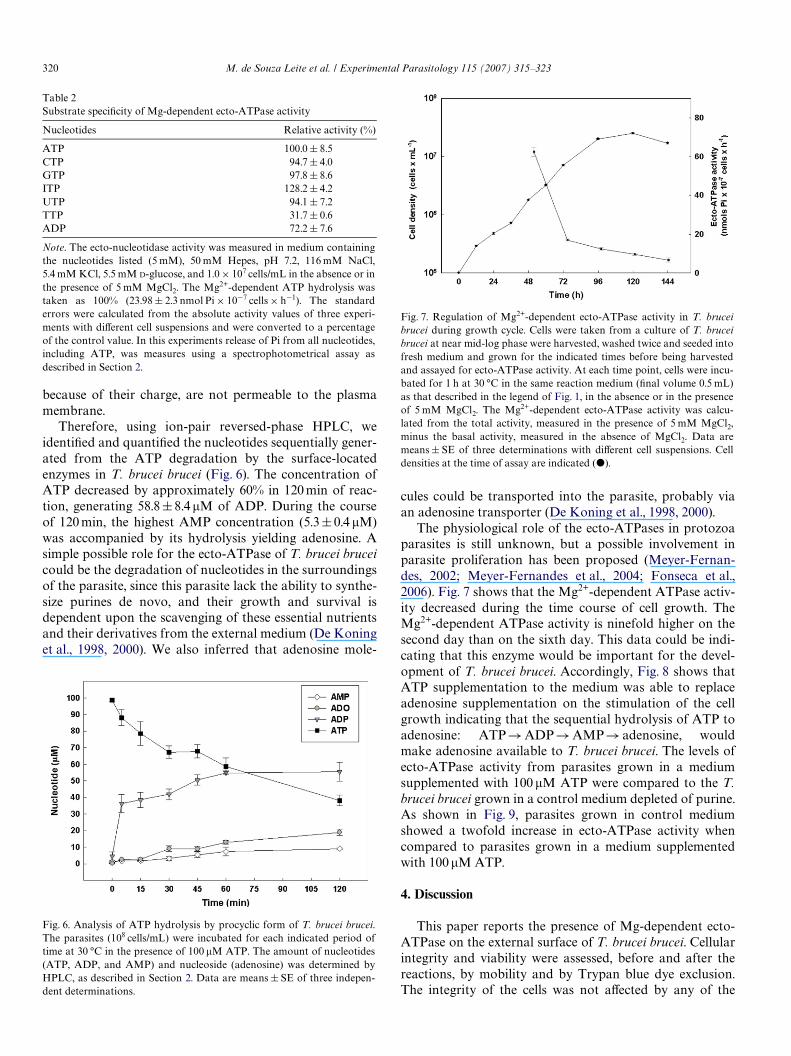
Therefore, using ion-pair reversed-phase HPLC, weidentiWed and quantiWed the nucleotides sequentially gener-ated from the ATP degradation by the surface-locatedenzymes in T. brucei brucei (Fig. 6). The concentration ofATP decreased by approximately 60% in 120 min of reac-tion, generating 58.8§8.4 �M of ADP. During the courseof 120 min, the highest AMP concentration (5.3§0.4 �M)was accompanied by its hydrolysis yielding adenosine. Asimple possible role for the ecto-ATPase of T. brucei bruceicould be the degradation of nucleotides in the surroundingsof the parasite, since this parasite lack the ability to synthe-size purines de novo, and their growth and survival isdependent upon the scavenging of these essential nutrientsand their derivatives from the external medium (De Koninget al., 1998, 2000). We also inferred that adenosine mole-
Table 2Substrate speciWcity of Mg-dependent ecto-ATPase activity
Note. The ecto-nucleotidase activity was measured in medium containingthe nucleotides listed (5 mM), 50 mM Hepes, pH 7.2, 116 mM NaCl,5.4 mM KCl, 5.5 mM D-glucose, and 1.0£ 107 cells/mL in the absence or inthe presence of 5 mM MgCl2. The Mg2+-dependent ATP hydrolysis wastaken as 100% (23.98§ 2.3 nmol Pi£ 10¡7 cells £ h¡1). The standarderrors were calculated from the absolute activity values of three experi-ments with diVerent cell suspensions and were converted to a percentageof the control value. In this experiments release of Pi from all nucleotides,including ATP, was measures using a spectrophotometrical assay asdescribed in Section 2.
Nucleotides Relative activity (%)
ATP 100.0 § 8.5CTP 94.7 § 4.0GTP 97.8 § 8.6ITP 128.2 § 4.2UTP 94.1 § 7.2TTP 31.7 § 0.6ADP 72.2 § 7.6
Fig. 6. Analysis of ATP hydrolysis by procyclic form of T. brucei brucei.The parasites (108 cells/mL) were incubated for each indicated period oftime at 30 °C in the presence of 100 �M ATP. The amount of nucleotides(ATP, ADP, and AMP) and nucleoside (adenosine) was determined byHPLC, as described in Section 2. Data are means § SE of three indepen-dent determinations.
cules could be transported into the parasite, probably viaan adenosine transporter (De Koning et al., 1998, 2000).
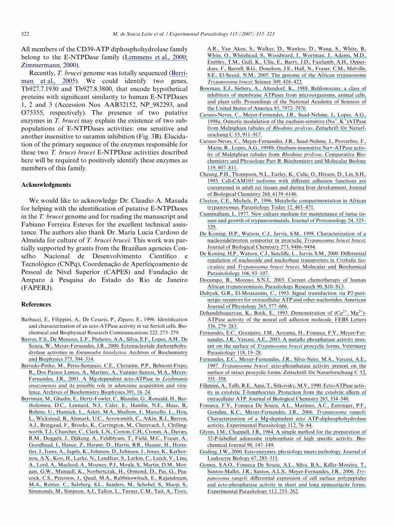
The physiological role of the ecto-ATPases in protozoaparasites is still unknown, but a possible involvement inparasite proliferation has been proposed (Meyer-Fernan-des, 2002; Meyer-Fernandes et al., 2004; Fonseca et al.,2006). Fig. 7 shows that the Mg2+-dependent ATPase activ-ity decreased during the time course of cell growth. TheMg2+-dependent ATPase activity is ninefold higher on thesecond day than on the sixth day. This data could be indi-cating that this enzyme would be important for the devel-opment of T. brucei brucei. Accordingly, Fig. 8 shows thatATP supplementation to the medium was able to replaceadenosine supplementation on the stimulation of the cellgrowth indicating that the sequential hydrolysis of ATP toadenosine: ATP!ADP!AMP! adenosine, wouldmake adenosine available to T. brucei brucei. The levels ofecto-ATPase activity from parasites grown in a mediumsupplemented with 100 �M ATP were compared to the T.brucei brucei grown in a control medium depleted of purine.As shown in Fig. 9, parasites grown in control mediumshowed a twofold increase in ecto-ATPase activity whencompared to parasites grown in a medium supplementedwith 100 �M ATP.
4. Discussion
This paper reports the presence of Mg-dependent ecto-ATPase on the external surface of T. brucei brucei. Cellularintegrity and viability were assessed, before and after thereactions, by mobility and by Trypan blue dye exclusion.The integrity of the cells was not aVected by any of the
Fig. 7. Regulation of Mg2+-dependent ecto-ATPase activity in T. bruceibrucei during growth cycle. Cells were taken from a culture of T. bruceibrucei at near mid-log phase were harvested, washed twice and seeded intofresh medium and grown for the indicated times before being harvestedand assayed for ecto-ATPase activity. At each time point, cells were incu-bated for 1 h at 30 °C in the same reaction medium (Wnal volume 0.5 mL)as that described in the legend of Fig. 1, in the absence or in the presenceof 5 mM MgCl2. The Mg2+-dependent ecto-ATPase activity was calcu-lated from the total activity, measured in the presence of 5 mM MgCl2,minus the basal activity, measured in the absence of MgCl2. Data aremeans § SE of three determinations with diVerent cell suspensions. Celldensities at the time of assay are indicated (�).

al Parasitology 115 (2007) 315–323 321
conditions used in the assays. The external location of theATP-hydrolyzing site is supported by its sensitivity to theimpermeable inhibitor DIDS (Fig. 4, panel A) (Knowles,1988; Barbacci et al., 1996; Meyer-Fernandes et al., 1997).Moreover, a battery of inhibitors for other ATPases thathave intracellular ATP binding sites showed no eVect onthe ecto-ATPase activity (Table 1). The Mg-dependentATPase activity reported in this work could not be attrib-uted to a mitochondrial ATPase, since this activity was
Fig. 8. Growth of procyclic T. brucei brucei in the presence of adenosine orATP. Procyclic cells growing in mid-log phase were harvested, washedtwice in Purine free trypanosome medium (PFTM) without Purine supple-ment and seeded at 105 cells/mL in various medium as indicated. Data aremeans § SE of three determinations in triplicate with diVerent cell suspen-sions. Samples were taken at regular intervals and cell density determinedin triplicate, using a hemocytometer.
Fig. 9. Mg2+-dependent ecto-ATPase activity in T. brucei brucei procyclicsafter 72 h in purine free trypanosome medium (PFTM) or in PFTM sup-plemented with ATP during growth cycle. Parasites were seeded intoPFTM (gray bars) or in PFTM supplemented with 100 �M ATP (blackbars) and grown for 72 h. Cells were harvested and incubated for 1 h at30 °C in the same reaction medium (Wnal volume 0.5 mL) as that describedin the legend of Fig. 1, in the absence or in the presence of 5 mM MgCl2.The Mg2+-dependent ecto-ATPase activity was calculated from the totalactivity, measured in the presence of 5 mM MgCl2, minus the basal activ-ity, measured in the absence of MgCl2. Data are means§ SE of threedeterminations with diVerent cell suspensions.
insensitive to oligomycin (Table 1), a known F-typeATPase inhibitor (Meyer-Fernandes et al., 1997). Since theMg-dependent ecto-ATPase activity was also insensitive tovanadate (Table 1), the possibility that this activity was dueto a P-ATPase present on the surface of the plasma mem-brane was discarded. For these reasons, we assign an ecto-localization for the Mg-dependent ATPase activitydescribed here. ATP hydrolysis could not be due to a phos-phatase activity present on the external surface of T. bruceibrucei membrane (Fernandes et al., 1997, 2003), because asshown in Table 1 potent inhibitors for phosphatase activi-ties were not capable of modify the Mg-dependent ecto-ATPase activity. The Mg-dependent ecto-ATPase activitydescribed here could not be attributed to a 5�-nucleotidase,since the ATP hydrolysis was not inhibited by ammoniummolybdate, a potent inhibitor of 5� nucleotidase (Gottlieband Dwyer, 1983) (Table 1).
Most of the ecto-ATPases are Mg2+ or Ca2+ stimulated(Plesner, 1995). The addition of CaCl2, ZnCl2, MgCl2, andMnCl2 to the extracellular medium stimulated the ecto-ATPase activity (Fig. 1A). The ecto-ATPase activity pres-ent in T. brucei brucei hydrolyses ATP, ITP, GTP, CTP,and UTP at high rates (Table 2) as also observed with theecto-ATPase present on the surface of Trypanosoma cruzi(Meyer-Fernandes et al., 2004) and Trypanosoma rangeli(Fonseca et al., 2006). ADP was also recognized as sub-strate, indicating that this enzyme is an authentic nucleo-side triphosphate diphosphohydrolase as described in othercells (Wang and Guidotti, 1996; Barros et al., 2000; Fons-eca et al., 2006).
Trypanosoma brucei brucei, as well as Leishmania ama-zonensis, are pathogens which cannot synthesize purines denovo (De Koning et al., 2000; Berredo-Pinho et al., 2001). Ithas been postulated that these ecto-ATPases in protozoaparasites could play a role in the salvage of purines fromthe host cells (Berredo-Pinho et al., 2001; Meyer-Fernan-des, 2002). The ability of T. brucei brucei to hydrolyze ATP,ADP, and AMP (Fig. 5) might sequentially dephosphory-late ATP to adenosine: ATP!ADP!AMP! adenosine,indicating that this enzyme in T. brucei brucei might play arole in the salvage of purines from extracellular medium.
The recently identiWed family of ecto-nucleoside triphos-phate diphosphohydrolases (E-NTPDase family) containsmultiple members that diVer in their substrate speciWcitiesand cellular locations (Zimmermann, 1999). It has beendemonstrated that mammalian membrane-associated ecto-ATPDase was homologous to human CD39 (or E-NTP-Dase1), a lymphoid cell activation antigen (Handa andGuidotti, 1996). Wang and Guidotti discovered that CD39has sequence homology with a potato apyrase and thatCD39 has apyrase activity (Wang and Guidotti, 1996).Their work led to the identiWcation of a family ofecto-ATPases that are related in sequence but vary intheir membrane topology and tissue distribution (Plesner,1995; Zimmermann, 1999; Goding, 2000). Further charac-terization of cloned members of proteins related toCD39 allowed the suggestion of a unifying nomenclature.
M. de Souza Leite et al. / Experiment
322 M. de Souza Leite et al. / Experimental Parasitology 115 (2007) 315–323
All members of the CD39-ATP diphosphohydrolase familybelong to the E-NTPDase family (Lemmens et al., 2000;Zimmermann, 2000).
Recently, T. brucei genome was totally sequenced (Berri-man et al., 2005). We could identify two genes,Tb927.7.1930 and Tb927.8.3800, that encode hypotheticalproteins with signiWcant similarity to human E-NTPDases1, 2 and 3 (Accession Nos. AAB32152, NP_982293, andO75355, respectively). The presence of two putativeenzymes in T. brucei may explain the existence of two sub-populations of E-NTPDases activities: one sensitive andanother insensitive to suramin inhibition (Fig. 3B). Elucida-tion of the primary sequence of the enzymes responsible forthese two T. brucei brucei E-NTPDase activities describedhere will be required to positively identify these enzymes asmembers of this family.
Acknowledgments
We would like to acknowledge Dr. Claudio A. Masudafor helping with the identiWcation of putative E-NTPDasesin the T. brucei genome and for reading the manuscript andFabiano Ferreira Esteves for the excellent technical assis-tance. The authors also thank Dr. Maria Lucia Cardoso deAlmeida for culture of T. brucei brucei. This work was par-tially supported by grants from the Brazilian agencies Con-selho Nacional de Desenvolvimento CientíWco eTecnológico (CNPq), Coordenação de Aperfeiçoamento dePessoal de Nível Superior (CAPES) and Fundação deAmparo à Pesquisa do Estado do Rio de Janeiro(FAPERJ).
References
Barbacci, E., Filippini, A., De Cesaris, P., Ziparo, E., 1996. IdentiWcationand characterization of an ecto-ATPase activity in rat Sertoli cells. Bio-chemical and Biophysical Research Communications 222, 273–279.
Barros, F.S., De Menezes, L.F., Pinheiro, A.A., Silva, E.F., Lopes, A.H., DeSouza, W., Meyer-Fernandes, J.R., 2000. Ectonucleotide diphosphohy-drolase activities in Entamoeba histolytica. Archives of Biochemistryand Biophysics 375, 304–314.
Berredo-Pinho, M., Peres-Sampaio, C.E., Chrispim, P.P., Belmont-Firpo,R., Dos Passos Lemos, A., Martiny, A., Vannier-Santos, M.A., Meyer-Fernandes, J.R., 2001. A Mg-dependent ecto-ATPase in Leishmaniaamazonensis and its possible role in adenosine acquisition and viru-lence. Archives of Biochemistry Biophysics 391, 16–24.
Berriman, M., Ghedin, E., Hertz-Fowler, C., Blandin, G., Renauld, H., Bar-tholomeu, D.C., Lennard, N.J., Caler, E., Hamlin, N.E., Haas, B.,Bohme, U., Hannick, L., Aslett, M.A., Shallom, J., Marcello, L., Hou,L., Wickstead, B., Alsmark, U.C., Arrowsmith, C., Atkin, R.J., Barron,A.J., Bringaud, F., Brooks, K., Carrington, M., Cherevach, I., Chilling-worth, T.J., Churcher, C., Clark, L.N., Corton, C.H., Cronin, A., Davies,R.M., Doggett, J., Djikeng, A., Feldblyum, T., Field, M.C., Fraser, A.,Goodhead, I., Hance, Z., Harper, D., Harris, B.R., Hauser, H., Hoste-tler, J., Ivens, A., Jagels, K., Johnson, D., Johnson, J., Jones, K., Kerhor-nou, A.X., Koo, H., Larke, N., Landfear, S., Larkin, C., Leech, V., Line,A., Lord, A., Macleod, A., Mooney, P.J., Moule, S., Martin, D.M., Mor-gan, G.W., Mungall, K., Norbertczak, H., Ormond, D., Pai, G., Pea-cock, C.S., Peterson, J., Quail, M.A., Rabbinowitsch, E., Rajandream,M.A., Reitter, C., Salzberg, S.L., Sanders, M., Schobel, S., Sharp, S.,Simmonds, M., Simpson, A.J., Tallon, L., Turner, C.M., Tait, A., Tivey,
A.R., Van Aken, S., Walker, D., Wanless, D., Wang, S., White, B.,White, O., Whitehead, S., Woodward, J., Wortman, J., Adams, M.D.,Embley, T.M., Gull, K., Ullu, E., Barry, J.D., Fairlamb, A.H., Opper-does, F., Barrell, B.G., Donelson, J.E., Hall, N., Fraser, C.M., Melville,S.E., El-Sayed, N.M., 2005. The genome of the African trypanosomeTrypanosoma brucei. Science 309, 416–422.
Bowman, E.J., Siebers, A., Altendorf, K., 1988. BaWlomycins: a class ofinhibitors of membrane ATPases from microorganisms, animal cells,and plant cells. Proceedings of the National Academy of Sciences ofthe United States of America 85, 7972–7976.
Caruso-Neves, C., Meyer-Fernandes, J.R., Saad-Nehme, J., Lopes, A.G.,1998a. Osmotic modulation of the ouabain-sensitive (Na+, K+)ATPasefrom Malpighian tubules of Rhodnius prolixus. Zeitschrift för Naturf-orschung C 53, 911–917.
Caruso-Neves, C., Meyer-Fernandes, J.R., Saad-Nehme, J., Proverbio, F.,Marin, R., Lopes, A.G., 1998b. Ouabain-insensitive Na+-ATPase activ-ity of Malpighian tubules from Rhodnius prolixus. Comparative Bio-chemistry and Physiology Part B: Biochemistry and Molecular Biology119, 807–811.
Cheung, P.H., Thompson, N.L., Earley, K., Culic, O., Hixson, D., Lin, S.H.,1993. Cell-CAM105 isoforms with diVerent adhesion functions arecoexpressed in adult rat tissues and during liver development. Journalof Biological Chemistry 268, 6139–6146.
Clayton, C.E., Michels, P., 1996. Metabolic compartmentation in Africantrypanosomes. Parasitology Today 12, 465–471.
Cunningham, I., 1977. New culture medium for maintenance of tsetse tis-sues and growth of trypanosomatids. Journal of Protozoology 24, 325–329.
De Koning, H.P., Watson, C.J., Jarvis, S.M., 1998. Characterization of anucleoside/proton symporter in procyclic Trypanosoma brucei brucei.Journal of Biological Chemistry 273, 9486–9494.
De Koning, H.P., Watson, C.J., SutcliVe, L., Jarvis, S.M., 2000. DiVerentialregulation of nucleoside and nucleobase transporters in Crithidia fas-ciculata and Trypanosoma brucei brucei. Molecular and BiochemicalParasitolology 106, 93–107.
Docampo, R., Moreno, S.N.J., 2003. Current chemotherapy of humanAfrican trypanosomiasis. Parasitology Research 90, S10–S13.
Dubyak, G.R., El-Moatassim, C., 1993. Signal transduction via P2-puri-nergic receptors for extracellular ATP and other nucleotides. AmericanJournal of Physiology 265, 577–606.
Dzhandzhugazyan, K., Bock, E., 1993. Demonstration of (Ca2+, Mg2+)-ATPase activity of the neural cell adhesion molecule. FEBS Letters336, 279–283.
Fernandes, E.C., Granjeiro, J.M., Aoyama, H., Fonseca, F.V., Meyer-Fer-nandes, J.R., Vercesi, A.E., 2003. A metallo phosphatase activity pres-ent on the surface of Trypanosoma brucei procyclic forms. VeterinaryParasitology 118, 19–28.
Fernandes, E.C., Meyer-Fernandes, J.R., Silva-Neto, M.A., Vercesi, A.E.,1997. Trypanosoma brucei: ecto-phosphatase activity present on thesurface of intact procyclic forms. Zeitschrift för Naturforschung C 52,351–358.
Filippini, A., TaVs, R.E., Agui, T., Sitkovsky, M.V., 1990. Ecto-ATPase activ-ity in cytolytic T-lymphocytes. Protection from the cytolytic eVects ofextracellular ATP. Journal of Biological Chemistry 265, 334–340.
Fonseca, F.V., Fonseca De Souza, A.L., Mariano, A.C., Entringer, P.F.,Gondim, K.C., Meyer-Fernandes, J.R., 2006. Trypanosoma rangeli:Characterization of a Mg-dependent ecto ATP-diphosphohydrolaseactivity. Experimental Parasitology 112, 76–84.
Glynn, I.M., Chappell, J.B., 1964. A simple method for the preparation of32-P-labelled adenosine triphosphate of high speciWc activity. Bio-chemical Journal 90, 147–149.
Goding, J.W., 2000. Ecto-enzymes: physiology meets pathology. Journal ofLeukocyte Biology 67, 285–311.
Gomes, S.A.O., Fonseca De Souza, A.L., Silva, B.A., KiVer-Moreira, T.,Santos-Mallet, J.R., Santos, A.L.S., Meyer-Fernandes, J.R., 2006. Try-panosoma rangeli: diVerential expression of cell surface polypeptidesand ecto-phosphatase activity in short and long epimastigote forms.Experimental Parasitology 112, 253–262.

M. de Souza Leite et al. / Experimental Parasitology 115 (2007) 315–323 323
Gottlieb, M., Dwyer, D.M., 1983. Evidence for distinct 5�- and 3�-nucleotid-ase activities in the surface membrane fraction of Leishmania donovanipromastigotes. Molecular and Biochemical Parasitology 7, 303–317.
Handa, M., Guidotti, G., 1996. PuriWcation and cloning of a soluble ATP-diphosphohydrolase (apyrase) from potato tubers (Solanum tubero-sum). Biochemical and Biophysical Research Communications 218,916–923.
Kawamoto, Y., Shinozuka, K., Kunitomo, M., Haginaka, J., 1998. Deter-mination of ATP and its metabolites released from rat caudal artery byisocratic ion-pair reversed-phase high-performance liquid chromatog-raphy. Analytical Biochemistry 262, 33–38.
Kirley, T.L., 1997. Complementary DNA cloning and sequencing of thechicken muscle ecto-ATPase. Homology with the lymphoid cell activa-tion antigen CD39. Journal of Biological Chemistry 272, 1076–1081.
Knowles, A.F., 1988. DiVerential expression of ecto-Mg2+-ATPase andecto-Ca2+-ATPase activities in human hepatoma cells. Archives of Bio-chemistry and Biophysics 263, 264–271.
Lemmens, R., VanduVel, I.L., Teuchy, H., Culic, O., 1996. Regulation ofproliferation of LLC-MK2 cells by nucleosides and nucleotides: therole of ecto-enzymes. Biochemical Journal 316, 551–557.
Lemmens, R., VanduVel, L., Kittel, A., Beaudoin, A.R., Benrezzak, O., Sev-igny, J., 2000. Distribution, cloning, and characterization of porcinenucleoside triphosphate diphosphohydrolase-1. European Journal ofBiochemistry 267, 4106–4114.
Lowry, O.H., Lopes, J., 1946. The determination of inorganic phosphate inthe presence of labile phosphate esters. Journal of Biological Chemistry162, 421–428.
Margolis, B., Zilberstein, A., Franks, C., Felder, S., Kremer, S., Ullrich, A.,Rhee, S.G., Skorecki, K., Schlessinger, J., 1990. EVect of phospholipaseC-gamma overexpression on PDGF-induced second messengers andmitogenesis. Science 248, 607–610.
Meyer-Fernandes, J.R., 2002. Ecto-ATPases in protozoa parasites: lookingfor a function. Parasitology International 51, 299–303.
Meyer-Fernandes, J.R., Dutra, P.M., Rodrigues, C.O., Saad-Nehme, J.,Lopes, A.H., 1997. Mg-dependent ecto-ATPase activity in Leishmaniatropica. Archives of Biochemistry and Biophysics 341, 40–46.
Meyer-Fernandes, J.R., Saad-Nehme, J., Peres-Sampaio, C.E., Belmont-Firpo, L., Bisaggio, D.F., Do Couto, L.C., Fonseca De Souza, A.L.,Lopes, A.H., Souto-Padron, T., 2004. A Mg-dependent ecto-ATPase isincreased in the infective stages of Trypanosoma cruzi. ParasitologyResearch 93, 567–576.
Meyer-Fernandes, J.R., Vieyra, A., 1988. Pyrophosphate formation fromacetyl phosphate and orthophosphate: evidence for heterogeneouscatalysis. Archives of Biochemistry and Biophysics 266, 132–141.
Plesner, L., 1995. Ecto-ATPases: identities and functions. InternationalReview of Cytology 158, 141–214.
Pinheiro, C.M., Martins-Duarte, E.S., Ferraro, R.B., Fonseca DeSouza, A.L., Gomes, M.T., Lopes, A.H.C.S., Vannier-Santos, M.A.,Santos, A.L.S., Meyer-Fernandes, J.R., 2006. Leishmania amazonen-sis: biological and biochemical characterization of ecto-nucleosidetriphosphate diphosphohydrolase activities. Experimental Parasi-tology 114, 16–25.
Saad-Nehme, J., Silva, J.L., Meyer-Fernandes, J.R., 2000. Carbohydratesprotect mitochondrial FoF1ATPase complex against thermal inactiva-tion. Zeitschrift Fur Naturforschung C 55, 594–599.
Smith Jr., T.M., Kirley, T.L., Hennessey, T.M., 1997. A soluble ecto-ATP-ase from Tetrahymena thermophila: puriWcation and similarity to themembrane-bound ecto-ATPase of smooth muscle. Archives of Bio-chemistry and Biophysics 337, 351–359.
Sodre, C.L., Moreira, B.L., Nóbrega, F.B., Gadelha, F.R., Meyer-Fernan-des, J.R., Dutra, P.M., Vercesi, A.E., Lopes, A.H., Scofano, H.M., Bar-rabin, H., 2000. Characterization of the intracellular (Ca2+) poolsinvolved in the calcium homeostasis in Herpetomonas sp. promasti-gotes. Archives of Biochemistry and Biophysics 380, 85–91.
Steinberg, T.H., Di Virgilio, F., 1991. Cell-mediated cytotoxicity: ATPas an eVector and the role of target cells. Current Opinion in Immu-nology 3, 71–75.
Stout, J.G., Strobel, R.S., Kirley, T.L., 1995. Properties of and proteinsassociated with the extracellular ATPase of chicken gizzard smoothmuscle. A monoclonal antibody study. Journal of Biological Chemistry270, 11845–11850.
Van Belle, H., 1976. Alkaline phosphatase. I. Kinetics and inhibition bylevamisole of puriWed isoenzymes from humans. Clinical Chemistry 22,972–976.
Vickerman, K., 1986. Parasitology: Clandestine sex in trypanosomes.Nature 322, 113–114.
Wang, T.F., Guidotti, G., 1996. CD39 is an ecto-(Ca2+, Mg2+)-apyrase.Journal of Biological Chemistry 271, 9898–9901.
Weisman, G.A., Turner, J.T., Fedan, J.S., 1996. Structure and function ofP2 purinocepters. Journal of Pharmacology and Experimental Thera-peutics 277, 1–9.
Westfall, T.D., Kennedy, C., Sneddon, P., 1997. The ecto-ATPase inhibitorARL 67156 enhances parasympathetic neurotransmission in the guinea-pig urinary bladder. European Journal of Pharmacology 329, 169–173.
Yagi, K., Nishino, I., Eguchi, M., Kitagawa, M., Miura, Y., Mizoguchi, T.,1994. Involvement of ecto-ATPase as an ATP receptor in the stimula-tory eVect of extracellular ATP on NO release in bovine aorta endothe-lial cells. Biochemistry and Biophysical Research Communication 203,1237–1243.
Zanovello, P., Bronte, V., Rosato, A., Pizzo, P., Di Virgilio, F., 1990.Responses of mouse lymphocytes to extracellular ATP. II. Extracellu-lar ATP causes cell type-dependent lysis and DNA fragmentation.Journal of Immunology 145, 1545–1550.
Ziganshin, A.U., Ziganshina, L.E., Bodin, P., Bailey, D., Burnstock, G.,1995. EVects of P2-purinoceptor antagonists on ecto-nucleotidaseactivity of guinea-pig vas deferens cultured smooth muscle cells. Bio-chemistry and Molecular Biology International 36, 863–869.
Zimmermann, H., 1999. Two novel families of ectonucleotidases: molecu-lar structures, catalytic properties and a search for function. Trends inPharmacological Sciences 20, 231–236.
Zimmermann, H., 2000. Extracellular metabolism of ATP and other nucleo-tides. Naunyn Schmiedebergs Archives of Pharmacology 362, 299–309.
Zimmermann, H., 2001. Ectonucleotidases: some recent developments anda note on nomenclature. Drug Development Research 52, 44–56.