Identification of novel snRNA-specific Sm proteins that bind selectively to U2 and U4 snRNAs in...
14
Identification of novel snRNA-specific Sm proteins that bind selectively to U2 and U4 snRNAs in Trypanosoma brucei ITAI DOV TKACZ, 1 YANIV LUSTIG, 1 MICHAEL ZEEV STERN, 1 MOSHE BITON, 1 MALI SALMON-DIVON, 1 ANISH DAS, 2 VIVIAN BELLOFATTO, 2 and SHULAMIT MICHAELI 1 1 The Mina & Everard Goodman Faculty of Life Sciences, Bar-Ilan University, Ramat-Gan 52900, Israel 2 Department of Microbiology and Molecular Genetics, UMDNJ–New Jersey Medical School, International Center for Public Health, Newark, New Jersey 07103, USA ABSTRACT In eukaryotes the seven Sm core proteins bind to U1, U2, U4, and U5 snRNAs. In Trypanosoma brucei, Sm proteins have been implicated in binding both spliced leader (SL) and U snRNAs. In this study, we examined the function of these Sm proteins using RNAi silencing and protein purification. RNAi silencing of each of the seven Sm genes resulted in accumulation of SL RNA as well as reduction of several U snRNAs. Interestingly, U2 was unaffected by the loss of SmB, and both U2 and U4 snRNAs were unaffected by the loss of SmD3, suggesting that these snRNAs are not bound by the heptameric Sm complex that binds to U1, U5, and SL RNA. RNAi silencing and protein purification showed that U2 and U4 snRNAs were bound by a unique set of Sm proteins that we termed SSm (specific spliceosomal Sm proteins). This is the first study that identifies specific core Sm proteins that bind only to a subset of spliceosomal snRNAs. Keywords: trypanosome; snRNA; Sm proteins; SL RNA; Lsm INTRODUCTION Sm proteins are core proteins in mammals and yeast that bind the spliceosomal U1, U2, U4, and U5 snRNAs (Will and Lu ¨ hrmann 2001). The Sm proteins are characterized by bipartite sequence motifs termed Sm1 and Sm2 (Hermann et al. 1995; Se ´raphin 1995) that function in protein–protein interactions (Hermann et al. 1995). The Sm proteins bind to the Sm-site, which consists of a single-stranded uridine- rich region that is usually flanked by two stem–loop structures (Branlant et al. 1982). The seven Sm proteins SmB, D1, D2, D3, E, F, and G bind to the Sm-site, forming the Sm core (Branlant et al. 1982; Liautard et al. 1982). In the absence of U snRNA, three stable complexes of Sm proteins, D3/B, D1/D2, and E/F/G, are found (Hermann et al. 1995; Raker et al. 1996). None of the individual Sm proteins binds stably to RNA; instead, the Sm proteins interact via their Sm 1 and 2 motifs. The RNA binding site is generated as a consequence of Sm heteromeric formation (Raker et al. 1996). The crystal structures of two Sm protein complexes, D3/B and D1/D2, show that these proteins have a common fold containing an N-terminal helix, followed by a five-stranded antiparallel b-sheet (Kambach et al. 1999). In addition to the core Sm proteins, other proteins car- rying Sm motifs have been identified in many eukaryotes. These proteins are designated Lsm (Like-Sm) proteins. Two distinct complexes containing these proteins exist. One complex, composed of Lsm2p–Lsm8p, binds the 39-end poly(U) tract of U6 snRNA, and is required for U6 snRNP formation (Terns et al. 1992; Achsel et al. 1999). The binding of Lsm2p–Lsm8p to the 39 U-rich single-stranded region of U6 facilitates U6 RNP formation and assembly of this snRNA with U4 into the di-snRNP and probably the tri-snRNP com- plexes (Achsel et al. 1999; Mayes et al. 1999; Salgado-Garrido et al. 1999). The second complex, consisting of Lsm1p–Lsm7p, functions in mRNA decay in the cytoplasm, mostly during the decapping step (Bouveret et al. 2000). Additional Lsm proteins exist; these include those that form the U7 snRNP, which functions in 39-end processing of histone mRNA. In this case, the canonical D1 and D2 Sm proteins are replaced by Lsm10 and Lsm11 (Pillai et al. 2001, 2003). Trypanosomes are protozoan parasites that diverged very early in the eukaryotic lineage (Stevens and Gibson 1999). Reprint requests to: Shulamit Michaeli, The Mina & Everard Goodman Faculty of Life Sciences, Bar-Ilan University, Geha Street, Ramat-Gan 52900, Israel; e-mail: [email protected]; fax: 972-3-5351824. Article published online ahead of print. Article and publication date are at http://www.rnajournal.org/cgi/doi/10.1261/rna.174307. 30 RNA (2007), 13:30–43. Published by Cold Spring Harbor Laboratory Press. Copyright Ó 2007 RNA Society.
Transcript of Identification of novel snRNA-specific Sm proteins that bind selectively to U2 and U4 snRNAs in...
Identification of novel snRNA-specific Sm proteins
that bind selectively to U2 and U4 snRNAs inTrypanosoma brucei
ITAI DOV TKACZ,1 YANIV LUSTIG,1 MICHAEL ZEEV STERN,1 MOSHE BITON,1 MALI SALMON-DIVON,1
ANISH DAS,2 VIVIAN BELLOFATTO,2 and SHULAMIT MICHAELI1
1The Mina & Everard Goodman Faculty of Life Sciences, Bar-Ilan University, Ramat-Gan 52900, Israel2Department of Microbiology and Molecular Genetics, UMDNJ–New Jersey Medical School, International Center for Public Health,Newark, New Jersey 07103, USA
ABSTRACT
In eukaryotes the seven Sm core proteins bind to U1, U2, U4, and U5 snRNAs. In Trypanosoma brucei, Sm proteins have beenimplicated in binding both spliced leader (SL) and U snRNAs. In this study, we examined the function of these Sm proteins usingRNAi silencing and protein purification. RNAi silencing of each of the seven Sm genes resulted in accumulation of SL RNA aswell as reduction of several U snRNAs. Interestingly, U2 was unaffected by the loss of SmB, and both U2 and U4 snRNAs wereunaffected by the loss of SmD3, suggesting that these snRNAs are not bound by the heptameric Sm complex that binds to U1,U5, and SL RNA. RNAi silencing and protein purification showed that U2 and U4 snRNAs were bound by a unique set of Smproteins that we termed SSm (specific spliceosomal Sm proteins). This is the first study that identifies specific core Sm proteins thatbind only to a subset of spliceosomal snRNAs.
Keywords: trypanosome; snRNA; Sm proteins; SL RNA; Lsm
INTRODUCTION
Sm proteins are core proteins in mammals and yeast thatbind the spliceosomal U1, U2, U4, and U5 snRNAs (Willand Luhrmann 2001). The Sm proteins are characterized bybipartite sequence motifs termed Sm1 and Sm2 (Hermannet al. 1995; Seraphin 1995) that function in protein–proteininteractions (Hermann et al. 1995). The Sm proteins bindto the Sm-site, which consists of a single-stranded uridine-rich region that is usually flanked by two stem–loopstructures (Branlant et al. 1982). The seven Sm proteinsSmB, D1, D2, D3, E, F, and G bind to the Sm-site, formingthe Sm core (Branlant et al. 1982; Liautard et al. 1982). Inthe absence of U snRNA, three stable complexes of Smproteins, D3/B, D1/D2, and E/F/G, are found (Hermannet al. 1995; Raker et al. 1996). None of the individual Smproteins binds stably to RNA; instead, the Sm proteinsinteract via their Sm 1 and 2 motifs. The RNA binding siteis generated as a consequence of Sm heteromeric formation
(Raker et al. 1996). The crystal structures of two Sm proteincomplexes, D3/B and D1/D2, show that these proteins havea common fold containing an N-terminal helix, followed bya five-stranded antiparallel b-sheet (Kambach et al. 1999).
In addition to the core Sm proteins, other proteins car-rying Sm motifs have been identified in many eukaryotes.These proteins are designated Lsm (Like-Sm) proteins. Twodistinct complexes containing these proteins exist. Onecomplex, composed of Lsm2p–Lsm8p, binds the 39-endpoly(U) tract of U6 snRNA, and is required for U6 snRNPformation (Terns et al. 1992; Achsel et al. 1999). The bindingof Lsm2p–Lsm8p to the 39 U-rich single-stranded region ofU6 facilitates U6 RNP formation and assembly of this snRNAwith U4 into the di-snRNP and probably the tri-snRNP com-plexes (Achsel et al. 1999; Mayes et al. 1999; Salgado-Garridoet al. 1999). The second complex, consisting of Lsm1p–Lsm7p,functions in mRNA decay in the cytoplasm, mostly during thedecapping step (Bouveret et al. 2000). Additional Lsm proteinsexist; these include those that form the U7 snRNP, whichfunctions in 39-end processing of histone mRNA. In this case,the canonical D1 and D2 Sm proteins are replaced by Lsm10and Lsm11 (Pillai et al. 2001, 2003).
Trypanosomes are protozoan parasites that diverged veryearly in the eukaryotic lineage (Stevens and Gibson 1999).
rna1743 Tkacz et al. ARTICLE RA
Reprint requests to: Shulamit Michaeli, The Mina & Everard GoodmanFaculty of Life Sciences, Bar-Ilan University, Geha Street, Ramat-Gan52900, Israel; e-mail: [email protected]; fax: 972-3-5351824.
Article published online ahead of print. Article and publication date areat http://www.rnajournal.org/cgi/doi/10.1261/rna.174307.
30 RNA (2007), 13:30–43. Published by Cold Spring Harbor Laboratory Press. Copyright � 2007 RNA Society.
JOBNAME: RNA 13#1 2006 PAGE: 1 OUTPUT: Thursday December 7 04:29:57 2006
csh/RNA/127813/rna1743
These parasites possess very exotic RNA processing mech-anisms such as trans-splicing and RNA editing (Liang et al.2003; Simpson et al. 2003). Trypanosomes possess the fullset of U snRNAs that function in splicing (Liang et al.2003). These RNAs, as opposed to U snRNAs in other eu-karyotes, carry various cap structures. U1 (Schnare andGray 1999; Djikeng et al. 2001; Palfi et al. 2005), U2, andU4 possess a tri-methyl guanosine cap (Mottram et al.1989). U5 has a 59 phosphate end (Dungan et al. 1996;Xu et al. 1997; Bell and Bindereif 1999). The U6 cap isa g-methyl cap that is identical to the cap found in othereukaryotes (Mottram et al. 1989). The spliced leader (SL)RNA has a unique cap that is composed of m7G togetherwith four hypermodified nucleotides, which comprises acap-4 (Bangs et al. 1992). Like U snRNAs in other eukaryotes,trypanosome snRNAs carry Sm-sites, but this site is verydegenerate in the U2 and U4 snRNAs (Mottram et al. 1989).
The first trypanosome Sm protein was identified as an SLRNA core protein (Goncharov et al. 1999), and later the fullset of seven Sm proteins was identified (Palfi et al. 2000).The trypanosome Sm proteins carry bipartite Sm motifsincluding interesting deviations from the Sm consensus(Goncharov et al. 1999; Palfi et al. 2000). The C terminus ofthe Sm proteins lacks the RG dipeptide repeats in whichthe arginine of this motif is methylated in the mammalianproteins. This finding may explain in part why trypano-some Sm proteins are not recognized by anti-Sm antibodies(Palfi et al. 2000). To elucidate the repertoire and functionof Sm proteins, two Sm proteins, SmE and SmD1, weresilenced by RNAi (Mandelboim et al. 2003). As expected,the silencing of these proteins decreased the level ofsnRNAs, but surprisingly, the level of SL RNA increasedmore than 10-fold. The SL RNA that accumulates duringsilencing lacks the +4 cap nucleotide and forms a stableRNP complex called SL RNP-C (Mandelboim et al. 2003).Lsm proteins were also identified in Trypanosoma brucei(Liu et al. 2004). Two of these proteins (homologs to Lsm3pand Lsm8p) were found to be essential for U6 stability andmRNA decay (Liu et al. 2004).
Relatively little is known about snRNP biogenesis intrypanosomes. Specifically, it is not clear in which cellularcompartment this process takes place. The U snRNAs aretranscribed in trypanosomes by RNA polymerase III fromextragenic tRNA promoters (Nakaar et al. 1994). In con-trast, the SL RNA is transcribed by an RNA polymeraseII-dependent extragenic promoter (Gilinger and Bellofatto2001; Das et al. 2005). Strong evidence was provided forcoupling of the unique hypermethylated capping of SLRNA and its transcription (Mair et al. 2000). Most recentlytwo distinct methyltransferases that carry out the 29-O9-methylations at positions +3 and +4 and +2 of the cap-4were identified and localized to the nucleus (Arhin et al.2006a,b) . Despite the fact that many factors involved in SLRNA biogenesis are found within the nucleus, studies fromrecent years suggested that SL RNA biogenesis, neverthe-
less, involves a cytoplasmic phase (Mandelboim et al. 2003;Zeiner et al. 2003). It was suggested that export of SL RNAto the cytoplasm is mediated by XPO1 (Zeiner et al. 2003).The finding that SL RNA accumulated in the cytoplasmduring Sm depletion, and that snRNP assembly occurs inthe cytoplasm in metazoa, led to the proposal that SL RNAassembly with Sm proteins takes place in the cytoplasm intrypanosomes (Mandelboim et al. 2003). Most recently,inspecting the localization of SL RNA during early timepoints of Sm depletion indicated that SL RNA first accu-mulates in the nucleus. Only after massive accumulation,the SL RNA migrates to the cytoplasm, forming speckles aspreviously described. RNAi of XPO1 also demonstrates thatthis factor does not function in SL RNA transport (Bitonet al. 2006). These recent data suggest that SL RNA assemblywith Sm proteins most probably takes place in the nucleus.
In this study, we examined the function of all seven Smproteins in trypanosomes and three additional proteinscarrying Sm domains. Surprisingly, the two proteins similarto known Lsm proteins (Liu et al. 2004) were found tobe unusual Sm proteins that bind selectively to either U2or U4 snRNPs. We termed these proteins SSm (specificspliceosomal Sm proteins), to distinguish them from pre-viously characterized Sm and Lsm proteins. Using RNAisilencing and affinity-tagging of Sm and SSm proteins, wedetermined the selectivity of their binding to SL andsnRNAs. This is the first study to describe U spliceosomalsnRNPs (U2, U4) that have unique core Sm proteins. Thisfinding contributes to our understanding how trypanosomesselectively distinguish and control the assembly of core pro-teins on snRNAs, which function in splicing but harbor dif-ferent cap structures.
RESULTS
Silencing of Sm core proteins results in an increase inthe steady-state level of undermethylated SL RNAand differential decrease in U snRNAs
In our previous studies, silencing of either SmE or SmD1 inT. brucei revealed a very characteristic phenotype: U snRNAlevels were reduced, although SL RNA levels increased10-fold (Mandelboim et al. 2003). To examine whether al-terations in other Sm proteins generated this phenotype,additional Sm proteins were individually down-regulatedby RNAi. In the silencing constructs, T7 promoters wereused to produce the gene-specific dsRNA under tetracyclineregulation (Wang et al. 2000). Parasites stopped growingwithin 3 d after dsRNA induction in each case (data notshown). RNA was prepared from uninduced cells and fromcells after 2 d of induction, and analyzed using snRNA-specific probes (Fig. 1A). Quantification of data by densi-tometry from three independent experiments is shown inFigure 1B. In each case, the reduction of the specific Smprotein resulted in an increase in SL RNA amounts and
Trypanosome SM proteins
www.rnajournal.org 31
JOBNAME: RNA 13#1 2006 PAGE: 2 OUTPUT: Thursday December 7 04:29:57 2006
csh/RNA/127813/rna1743
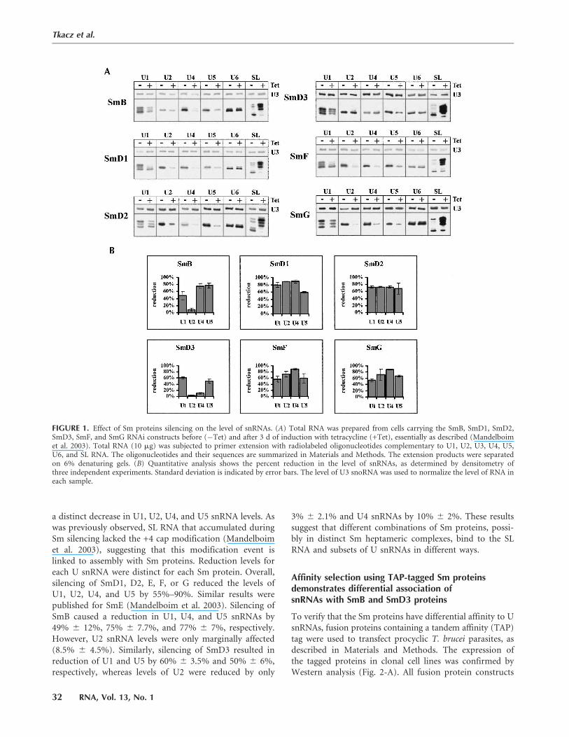
a distinct decrease in U1, U2, U4, and U5 snRNA levels. Aswas previously observed, SL RNA that accumulated duringSm silencing lacked the +4 cap modification (Mandelboimet al. 2003), suggesting that this modification event islinked to assembly with Sm proteins. Reduction levels foreach U snRNA were distinct for each Sm protein. Overall,silencing of SmD1, D2, E, F, or G reduced the levels ofU1, U2, U4, and U5 by 55%–90%. Similar results werepublished for SmE (Mandelboim et al. 2003). Silencing ofSmB caused a reduction in U1, U4, and U5 snRNAs by49% 6 12%, 75% 6 7.7%, and 77% 6 7%, respectively.However, U2 snRNA levels were only marginally affected(8.5% 6 4.5%). Similarly, silencing of SmD3 resulted inreduction of U1 and U5 by 60% 6 3.5% and 50% 6 6%,respectively, whereas levels of U2 were reduced by only
3% 6 2.1% and U4 snRNAs by 10% 6 2%. These resultssuggest that different combinations of Sm proteins, possi-bly in distinct Sm heptameric complexes, bind to the SLRNA and subsets of U snRNAs in different ways.
Affinity selection using TAP-tagged Sm proteinsdemonstrates differential association ofsnRNAs with SmB and SmD3 proteins
To verify that the Sm proteins have differential affinity to UsnRNAs, fusion proteins containing a tandem affinity (TAP)tag were used to transfect procyclic T. brucei parasites, asdescribed in Materials and Methods. The expression ofthe tagged proteins in clonal cell lines was confirmed byWestern analysis (Fig. 2-A). All fusion protein constructs
FIGURE 1. Effect of Sm proteins silencing on the level of snRNAs. (A) Total RNA was prepared from cells carrying the SmB, SmD1, SmD2,SmD3, SmF, and SmG RNAi constructs before (�Tet) and after 3 d of induction with tetracycline (+Tet), essentially as described (Mandelboimet al. 2003). Total RNA (10 mg) was subjected to primer extension with radiolabeled oligonucleotides complementary to U1, U2, U3, U4, U5,U6, and SL RNA. The oligonucleotides and their sequences are summarized in Materials and Methods. The extension products were separatedon 6% denaturing gels. (B) Quantitative analysis shows the percent reduction in the level of snRNAs, as determined by densitometry ofthree independent experiments. Standard deviation is indicated by error bars. The level of U3 snoRNA was used to normalize the level of RNA ineach sample.
Tkacz et al.
32 RNA, Vol. 13, No. 1
JOBNAME: RNA 13#1 2006 PAGE: 3 OUTPUT: Thursday December 7 04:29:58 2006
csh/RNA/127813/rna1743
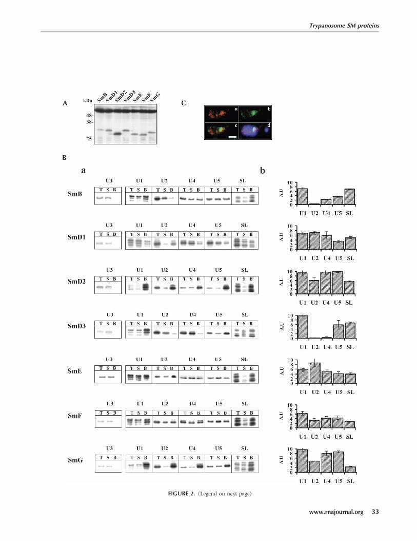
FIGURE 2. (Legend on next page)
Trypanosome SM proteins
www.rnajournal.org 33
JOBNAME: RNA 13#1 2006 PAGE: 4 OUTPUT: Thursday December 7 04:30:20 2006
csh/RNA/127813/rna1743
Fig. 2 live 4/C
produced polypeptides of the expected molecular mass.The expression level of the different tagged proteins wasvery similar, with the exception of SmD2, which was highlyexpressed. To determine which snRNAs are associated withspecific Sm proteins, fusion proteins along with boundRNA were affinity-purified based on the tight association ofthe protein A, within the TAP tag, to IgG-conjugated beads.RNA was extracted from the bound material by phenol,and the specific snRNAs were identified by primer exten-sion (Fig. 2B). The different efficiency of selection can bevisualized by comparing the amount of RNA selected onthe beads with an aliquot of the total amount of the snRNApresent in the sample. The data enable us to examine therelative efficiency of selection of each of the snRNAs by thedifferent proteins, as well as to compare the efficiency ofselection of the snRNAs among the different Sm proteins.Great variation exists in the efficiency of affinity selectionamong the different tagged proteins. Quantitating theefficiency of selection of each snRNA from three indepen-dent experiments in arbitrary units is shown in Figure 2B-b.The tagged proteins SmD2, SmD3, and SmG selectedthe particles in the same efficiency (z10 arbitrary units),presenting 10%–15% of snRNPs present in the extract.Tagged SmD1, SmB, SmE, and SmF selected only 5%–7%of the snRNPs. Despite differences in the efficacy ofselection, results demonstrate high specificity in the abilityof the tagged proteins to select the different snRNAs. Theresults clearly show that SmD1, D2, E, F, and G are boundto U1, U2, U4, and U5 snRNAs. These protein–RNAinteractions are specific, as U3 snoRNA was not detectedin any of the Sm-protein-dependent purifications. Thecharacteristic association of RNA and Sm proteins areconsistent with the data shown in Figure 1, indicating thatSmD1, D2, E, F, and G proteins are important for theassembly/ stabilization of the U1, U2, U4, U5, and SL RNA-containing snRNPs. Conversely, both Sm B and D3 didnot bind to U2 snRNA. The U2 snRNP was not selectedby SmB in contrast to the selection of U1, U4, U5,and SL RNA. Moreover, the absence of these two pro-teins in the RNAi experiments did not lead to a decreasein U2 snRNA levels, as shown in Figure 1. SmD3 didnot bind to U2 or U4 snRNAs, but did bind to U1 andU5 snRNAs. These results are in agreement with the
effect of silencing the SmD3 by RNAi, as shown inFigure 1.
Localization and assembly of Sm tagged proteinsto their cognate snRNPs
To investigate the source of variation leading to the dif-ference in the efficiency of selection, several factors such asthe level of expression, toxicity created by the expression ofthe tagged protein, and assembly of the tagged protein intosnRNP complexes were examined. The effect of expressionof the tagged proteins on cell viability and growth rate wasexamined in cells expressing two tagged Sm proteins, SmGand SmD1, which select the snRNPs efficiently or poorly,respectively. The results indicate no difference in the growthrate upon induction of tagged-Sm protein synthesis, sug-gesting that expression of these proteins is not toxic to thecells (data not shown).
We next examined the localization of the tagged proteins.To this end, an SmE-GFP construct was prepared. Theexpression of the protein is constitutive and is driven bythe EP promoter (explained in Materials and Methods).The results indicate that the SmE tagged protein is concen-trated mainly in the nucleus in a large ‘‘dot,’’ but also insmaller ‘‘dots’’ (like speckles) (see Fig. 2C-b). Hybridiza-tion with SL RNA suggests that the single ‘‘dot’’ containedthe majority of the SL RNA (Fig. 2C-a). The merged imagesof the SmE-GFP and SL RNA without or with nuclearstaining, respectively, are presented in Figure 2C, panelsc and d. Results suggest that the majority of the SL RNAbound to Sm proteins is confined to a distinct compartmentwithin the nucleus.
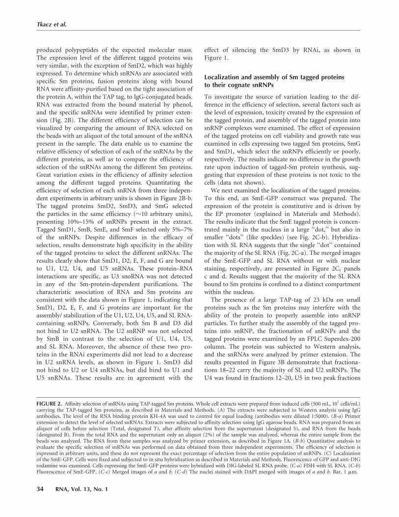
The presence of a large TAP-tag of 23 kDa on smallproteins such as the Sm proteins may interfere with theability of the protein to properly assemble into snRNPparticles. To further study the assembly of the tagged pro-teins into snRNP, the fractionation of snRNPs and thetagged proteins were examined by an FPLC Superdex-200column. The protein was subjected to Western analysis,and the snRNAs were analyzed by primer extension. Theresults presented in Figure 3B demonstrate that fractiona-tions 18–22 carry the majority of SL and U2 snRNPs. TheU4 was found in fractions 12–20, U5 in two peak fractions
FIGURE 2. Affinity selection of snRNAs using TAP-tagged Sm proteins. Whole cell extracts were prepared from induced cells (500 mL, 107 cells/mL)carrying the TAP-tagged Sm proteins, as described in Materials and Methods. (A) The extracts were subjected to Western analysis using IgGantibodies. The level of the RNA binding protein KH-4A was used to control for equal loading (antibodies were diluted 1:5000). (B-a) Primerextension to detect the level of selected snRNAs. Extracts were subjected to affinity selection using IgG agarose beads. RNA was prepared from analiquot of cells before selection (Total, designated T), after affinity selection from the supernatant (designated S), and RNA from the beads(designated B). From the total RNA and the supernatant only an aliquot (2%) of the sample was analyzed, whereas the entire sample from thebeads was analyzed. The RNA from these samples was analyzed by primer extension, as described in Figure 1A. (B-b) Quantitative analysis toevaluate the specific selection of snRNAs was performed on data obtained from three independent experiments. The efficiency of selection isexpressed in arbitrary units, and these do not represent the exact percentage of selection from the entire population of snRNPs. (C) Localizationof the SmE-GFP. Cells were fixed and subjected to in situ hybridization as described in Materials and Methods. Fluorescence of GFP and anti-DIGrodamine was examined. Cells expressing the SmE-GFP proteins were hybridized with DIG-labeled SL RNA probe. (C-a) FISH with SL RNA. (C-b)Fluorescence of SmE-GFP. (C-c) Merged images of a and b. (C-d) The nuclei stained with DAPI merged with images of a and b. Bar, 1 mm.
Tkacz et al.
34 RNA, Vol. 13, No. 1
JOBNAME: RNA 13#1 2006 PAGE: 5 OUTPUT: Thursday December 7 04:31:25 2006
csh/RNA/127813/rna1743
(14–16 and 20–22), whereas the U6 was also found in twopeak fractions (12–14 and 18–22). The two peaks observedfor U5 and U6 snRNA suggest that these RNAs are presentin monoparticles as well as in multimeric particles. Indeed,dimeric U4/U6 and trimeric U4/U5/U6 were recently ob-served in trypanosomes by sucrose gradient fractionation(Liang et al. 2006), and these particles most probably arefound in fractions 14–16 carrying U4/U5/U6 snRNAs.
The tagged SmE protein that is common to all snRNPswas found in fractions 12–24, which are enriched in all thesnRNPs and SL RNP (Fig. 3A). No signal was observed infractions 32 and above, where free protein should fraction-ate, suggesting that all the tagged cellular proteins arebound to snRNAs. Sm proteins and all snRNAs includingSL RNA and the Y-structure intermediate were also foundin fractions 12–14 (Fig. 3A,B). These fractions contain largespliceosomal complexes carrying also splicing factorssuch as SF1 and U2AF65 (D. Mualem, M.Z. Stern, andS. Michaeli, unpubl.). When compared to the distributionof SmE, the tagged SmB protein, which binds only to U1,U4, U5, and SL RNA, was less broadly distributed (Fig. 3A).
The fractionation of the tagged Sm proteins resemblesthe fractionation of a tagged Prp31 protein (Fig. 3A). This
protein is associated with U4 and the tri-snRNP complex(Liang et al. 2006). The Prp31 was tagged at the N terminuswith a BB2 epitope. The second allele was disrupted, andtherefore all the cellular Prp31 is epitope-tagged (Lianget al. 2006). The fractionation of Sm- and Prp31-taggedproteins suggest that based on these examples, regardlessof the tagging procedure, abundant RNA binding proteinssuch as Sm core proteins or particle-specific proteins(Prp31) are assembled efficiently into spliceosomal com-plexes. We cannot rule out the possibility that tagged pro-teins that did not assemble with snRNAs were subjected toproteolysis.
Our results also do not show correlation between thelevel of expression of the tagged protein and the efficiencyof selection of snRNPs. For instance, SmD2 is highly ex-pressed compared to other Sm proteins, yet it selects thesnRNAs as well as SmG, which is expressed at lower levels(compared to SmD2). All these data suggest that the dif-ferential efficacy of affinity selection may reflect the abilityof the IgG to bind to the protein A domain within thetagged protein when the protein is assembled in the snRNPcomplex.
Bioinformatics searches identified threenovel Sm-like proteins
The differential binding of U2 and U4 to core Sm proteinsprompted us to search for additional proteins harboringSm motifs. We previously have described seven proteins inthe T. brucei genome that contain Sm motifs. Silencing twoof these proteins, Lsm3 and Lsm8, results in destabilizationof U6 snRNA and mRNA. Therefore, these proteins arebona fide Lsm proteins (Liu et al. 2004). Bioinformaticsanalysis was expanded, and three data sets were preparedfor trypanosome proteins carrying Sm motifs. These in-cluded the Sm core proteins (Palfi et al. 2000), Lsm pro-teins (Liu et al. 2004), and three proteins that we termedSSm (see below). The bioinformatics analysis was per-formed using the program s-search from the GCG package(version 11.1; Accelrys Inc.), which conducts a rigorousSmith-Waterman search for similarity between a querysequence and a group of sequences of the same type. Thepurpose of these searches was to identify the relatedness ofthe SSm proteins to known Sm or Lsm proteins. Theprotein SSm2-1 (Tb927.6.4340) was previously designatedLsm5 (analogous to SmE) (Liu et al. 2004). When theSSm2-1 was compared to all the Sm core proteins, it wasclear that this protein was related to the following proteins:SmF with the highest score, then SmD2 and SmB. How-ever, when SmB was the query against the SSm proteins, theSSm2-1 protein was selected with the highest score. Wenext performed pairwise alignments between SSm2-1 andeach of the Sm proteins specified above. The results in-dicate that when SSm2-1 is compared with SmF, the second
FIGURE 3. Fractionation of extracts from cells expressing TAP-tagged Sm proteins and the Prp31-BB2 tagged protein on FPLC.Whole cell extracts were prepared from cells (500 mL, 107 cells/mL)expressing the fusions as described in Materials and Methods. (A)Proteins were subjected to Western analysis using IgG or anti-BB2antibodies. (T) Designates 2% of total extract loaded on the column.Twenty percent of each fraction was analyzed. The identity of theprotein examined is indicated. The elution positions of markerproteins BSA (66 kDa) and b-amylase (200 kDa) are indicated witharrows. (B) Primer extension to analyze the fractionation of snRNPs.RNA was extracted from every other fraction and analyzed by primerextension for the levels of U1, U2, U4, U5, U6 SL RNA the Y structureintermediate.
Trypanosome SM proteins
www.rnajournal.org 35
JOBNAME: RNA 13#1 2006 PAGE: 6 OUTPUT: Thursday December 7 04:31:25 2006
csh/RNA/127813/rna1743
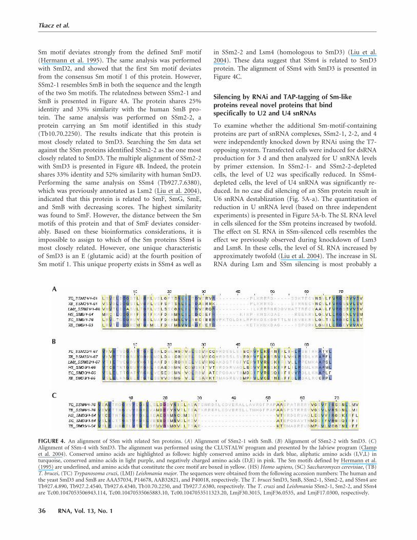
Sm motif deviates strongly from the defined SmF motif(Hermann et al. 1995). The same analysis was performedwith SmD2, and showed that the first Sm motif deviatesfrom the consensus Sm motif 1 of this protein. However,SSm2-1 resembles SmB in both the sequence and the lengthof the two Sm motifs. The relatedness between SSm2-1 andSmB is presented in Figure 4A. The protein shares 25%identity and 33% similarity with the human SmB pro-tein. The same analysis was performed on SSm2-2, aprotein carrying an Sm motif identified in this study(Tb10.70.2250). The results indicate that this protein ismost closely related to SmD3. Searching the Sm data setagainst the SSm proteins identified SSm2-2 as the one mostclosely related to SmD3. The multiple alignment of SSm2-2with SmD3 is presented in Figure 4B. Indeed, the proteinshares 33% identity and 52% similarity with human SmD3.Performing the same analysis on SSm4 (Tb927.7.6380),which was previously annotated as Lsm2 (Liu et al. 2004),indicated that this protein is related to SmF, SmG, SmE,and SmB with decreasing scores. The highest similaritywas found to SmF. However, the distance between the Smmotifs of this protein and that of SmF deviates consider-ably. Based on these bioinformatics considerations, it isimpossible to assign to which of the Sm proteins SSm4 ismost closely related. However, one unique characteristicof SmD3 is an E (glutamic acid) at the fourth position ofSm motif 1. This unique property exists in SSm4 as well as
in SSm2-2 and Lsm4 (homologous to SmD3) (Liu et al.2004). These data suggest that SSm4 is related to SmD3protein. The alignment of SSm4 with SmD3 is presented inFigure 4C.
Silencing by RNAi and TAP-tagging of Sm-likeproteins reveal novel proteins that bindspecifically to U2 and U4 snRNAs
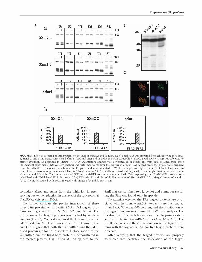
To examine whether the additional Sm-motif-containingproteins are part of snRNA complexes, SSm2-1, 2-2, and 4were independently knocked down by RNAi using the T7-opposing system. Transfected cells were induced for dsRNAproduction for 3 d and then analyzed for U snRNA levelsby primer extension. In SSm2-1- and SSm2-2-depletedcells, the level of U2 was specifically reduced. In SSm4-depleted cells, the level of U4 snRNA was significantly re-duced. In no case did silencing of an SSm protein result inU6 snRNA destablization (Fig. 5A-a). The quantitation ofreduction in U snRNA level (based on three independentexperiments) is presented in Figure 5A-b. The SL RNA levelin cells silenced for the SSm proteins increased by twofold.The effect on SL RNA in SSm-silenced cells resembles theeffect we previously observed during knockdown of Lsm3and Lsm8. In these cells, the level of SL RNA increased byapproximately twofold (Liu et al. 2004). The increase in SLRNA during Lsm and SSm silencing is most probably a
FIGURE 4. An alignment of SSm with related Sm proteins. (A) Alignment of SSm2-1 with SmB. (B) Alignment of SSm2-2 with SmD3. (C)Alignment of SSm-4 with SmD3. The alignment was performed using the CLUSTALW program and presented by the Jalview program (Clampet al. 2004). Conserved amino acids are highlighted as follows: highly conserved amino acids in dark blue, aliphatic amino acids (I,V,L) inturquoise, conserved amino acids in light purple, and negatively charged amino acids (D,E) in pink. The Sm motifs defined by Hermann et al.(1995) are underlined, and amino acids that constitute the core motif are boxed in yellow. (HS) Homo sapiens, (SC) Saccharomyces cerevisiae, (TB)T. brucei, (TC) Trypanosoma cruzi, (LMJ) Leishmania major. The sequences were obtained from the following accession numbers: The human andthe yeast SmD3 and SmB are AAA57034, P14678, AAB32821, and P40018, respectively. The T. brucei SmD3, SmB, SSm2-1, SSm2-2, and SSm4 areTb927.4.890, Tb927.2.4540, Tb927.6.4340, Tb10.70.2250, and Tb927.7.6380, respectively. The T. cruzi and Leishmania SSm2-1, Sm2-2, and SSm4are Tc00.1047053506943.114, Tc00.10470535065883.10, Tc00.1047053511323.20, LmjF30.3015, LmjF36.0535, and LmjF17.0300, respectively.
Tkacz et al.
36 RNA, Vol. 13, No. 1
JOBNAME: RNA 13#1 2006 PAGE: 7 OUTPUT: Thursday December 7 04:31:30 2006
csh/RNA/127813/rna1743
Fig. 4 live 4/C
secondary effect, and stems from the inhibition in trans-splicing due to the reduction in the level of the spliceosomalU snRNAs (Liu et al. 2004).
To further elucidate the precise interactions of thesethree SSm proteins with specific RNAs, TAP-tagged pro-teins were generated for SSm2-1, 2-2, and SSm4. Theexpression of the tagged proteins was verified by Westernanalysis (Fig. 5B). We next examined the localization of theGFP-fused SSm 2-1. The images presented in Figure 5, C-aand C-b, suggest that both the U2 snRNA and the GFP-fused protein are found in speckles. Colocalization of theU2 snRNA and the fused SSm protein is demonstrated inthe merged pictures (Fig. 5C-c,C-d). As opposed to the
SmE that was confined to a large dot and numerous speck-les, the SSm was found only in speckles.
To examine whether the TAP-tagged proteins are asso-ciated with the cognate snRNAs, extracts were fractionatedin an FPLC Superdex-200 column, and the distribution ofthe tagged proteins was examined by Western analysis. Thelocalization of the particles was examined by primer exten-sion with U2 and U4 snRNA probes (Fig. 6A-a,A-b). Theresults demonstrate the cofractionation of the tagged pro-teins with the cognate RNAs. No free tagged proteins wereobserved.
After verifying that the tagged proteins are properlyassembled into particles, the association of the tagged
FIGURE 5. Effect of silencing of SSm proteins on the level of snRNAs and SL RNA. (A-a) Total RNA was prepared from cells carrying the SSm2-1, SSm2-2, and SSm4 RNAi constructs before (�Tet) and after 3 d of induction with tetracycline (+Tet). Total RNA (10 mg) was subjected toprimer extension, as described in Figure 1A. (A-b) Quantitative analysis was performed as in Figure 1B, from data obtained from threeindependent experiments. (B) Western analysis was performed to monitor the expression of SSm-TAP tagged proteins. Extracts were preparedfrom the cells after tetracycline induction with 50 ng/mL, and were subjected to Western analysis with IgG. The level of 4A-KH was used tocontrol for the amount of protein in each lane. (C) Localization of SSm2-1. Cells were fixed and subjected to in situ hybridization, as described inMaterials and Methods. The fluorescence of GFP and anti-DIG rodamine was examined. Cells expressing the SSm2-1-GFP protein werehybridized with DIG-labeled U2 RNA probe. (C-a) FISH with U2 snRNA. (C-b) Fluorescence of SSm2-1-GFP. (C-c) Merged images of a and b.(C-d) The nuclei stained with DAPI merged with images of a and b. Bar, 1 mm.
Trypanosome SM proteins
www.rnajournal.org 37
JOBNAME: RNA 13#1 2006 PAGE: 8 OUTPUT: Thursday December 7 04:31:51 2006
csh/RNA/127813/rna1743
Fig. 5 live 4/C
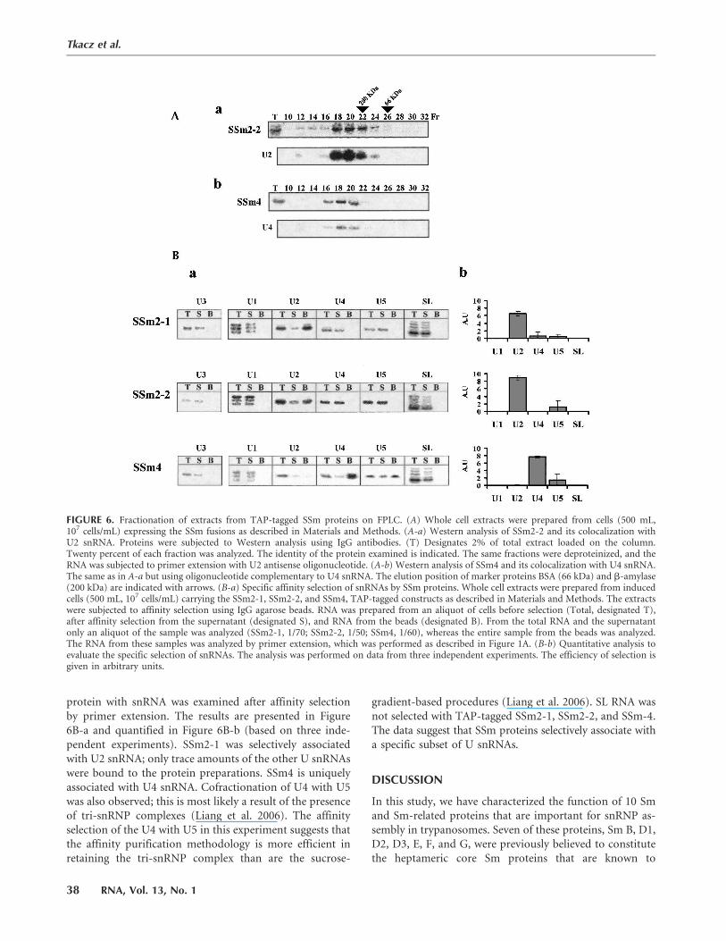
protein with snRNA was examined after affinity selectionby primer extension. The results are presented in Figure6B-a and quantified in Figure 6B-b (based on three inde-pendent experiments). SSm2-1 was selectively associatedwith U2 snRNA; only trace amounts of the other U snRNAswere bound to the protein preparations. SSm4 is uniquelyassociated with U4 snRNA. Cofractionation of U4 with U5was also observed; this is most likely a result of the presenceof tri-snRNP complexes (Liang et al. 2006). The affinityselection of the U4 with U5 in this experiment suggests thatthe affinity purification methodology is more efficient inretaining the tri-snRNP complex than are the sucrose-
gradient-based procedures (Liang et al. 2006). SL RNA wasnot selected with TAP-tagged SSm2-1, SSm2-2, and SSm-4.The data suggest that SSm proteins selectively associate witha specific subset of U snRNAs.
DISCUSSION
In this study, we have characterized the function of 10 Smand Sm-related proteins that are important for snRNP as-sembly in trypanosomes. Seven of these proteins, Sm B, D1,D2, D3, E, F, and G, were previously believed to constitutethe heptameric core Sm proteins that are known to
FIGURE 6. Fractionation of extracts from TAP-tagged SSm proteins on FPLC. (A) Whole cell extracts were prepared from cells (500 mL,107 cells/mL) expressing the SSm fusions as described in Materials and Methods. (A-a) Western analysis of SSm2-2 and its colocalization withU2 snRNA. Proteins were subjected to Western analysis using IgG antibodies. (T) Designates 2% of total extract loaded on the column.Twenty percent of each fraction was analyzed. The identity of the protein examined is indicated. The same fractions were deproteinized, and theRNA was subjected to primer extension with U2 antisense oligonucleotide. (A-b) Western analysis of SSm4 and its colocalization with U4 snRNA.The same as in A-a but using oligonucleotide complementary to U4 snRNA. The elution position of marker proteins BSA (66 kDa) and b-amylase(200 kDa) are indicated with arrows. (B-a) Specific affinity selection of snRNAs by SSm proteins. Whole cell extracts were prepared from inducedcells (500 mL, 107 cells/mL) carrying the SSm2-1, SSm2-2, and SSm4, TAP-tagged constructs as described in Materials and Methods. The extractswere subjected to affinity selection using IgG agarose beads. RNA was prepared from an aliquot of cells before selection (Total, designated T),after affinity selection from the supernatant (designated S), and RNA from the beads (designated B). From the total RNA and the supernatantonly an aliquot of the sample was analyzed (SSm2-1, 1/70; SSm2-2, 1/50; SSm4, 1/60), whereas the entire sample from the beads was analyzed.The RNA from these samples was analyzed by primer extension, which was performed as described in Figure 1A. (B-b) Quantitative analysis toevaluate the specific selection of snRNAs. The analysis was performed on data from three independent experiments. The efficiency of selection isgiven in arbitrary units.
Tkacz et al.
38 RNA, Vol. 13, No. 1
JOBNAME: RNA 13#1 2006 PAGE: 9 OUTPUT: Thursday December 7 04:32:25 2006
csh/RNA/127813/rna1743
associate with all U snRNAs in other eukaryotic cells. Wehave demonstrated that different sets of Sm proteins bindto different U snRNAs to form specific snRNPs. Our intra-cellular localization studies reveal that in trypanosomes, Smproteins are present in the nucleus of parasites, possibly inspecific subnuclear locales. Three newly identified proteinscarrying Sm motifs, SSm2-1, 2-2, and 4, unique to try-panosomes, are part of the core Sm assembly on specificsnRNAs. These proteins localize to nuclear speckles that aredistinct from the nuclear compartment where SL RNA Smassembly most probably takes place.
In yeast and mammals, the same complement of core Smproteins binds U1, U2, U4, and U5 to the snRNP complexnecessary for spliceosome function (Will and Luhrmann2001). Mammalian Sm proteins form three subcomplexes,namely, D1/D2, D3/B, and E/F/G, in in vitro reconstitutionexperiments (Hermann et al. 1995; Raker et al. 1996). Twoadditional core Sm proteins exist in mammals; one is anisoform of SmB (van Dam et al. 1989), and another an iso-form of the same protein that localizes exclusively to neu-rons and is known as N protein (Schmauss et al. 1989).These proteins most probably also assemble into snRNPcomplexes in the cytoplasm, but it is currently unknownwhether particles carrying these special Sm proteins haveunique functions in regulated or constitutive splicing.
Results suggest that different snRNPs in T. brucei as-semble using different subsets of core Sm proteins. This wasobserved most strikingly in the case of the U2 and U4snRNPs. In the U2 snRNP, the SmB/D3 protein complex isreplaced by the SSm2-1 and SSm2-2 proteins. It was notsurprising to find a pairwise swap, since SmB/D3 normallyassociate into a subcomplex during Sm assembly (Rakeret al. 1996). However, this does not seem to be the case forthe U4 snRNP, as SmB is retained in the U4 snRNP, butSmD3 is replaced by the unique SSm4 protein. SmB asso-ciates with U4 snRNP but not with U2 snRNP, indicatingthat an SmB can function independently from SmD3. Thisconclusion is based on the following results: (1) silencing ofSmD3 reduced the level of U4; (2) tagged SmD3 did notselect U4; (3) SmB silencing reduced the level of U4 snRNA;and (4) tagged SmB binds U4 snRNA. Taken together, thesedata suggest that trypanosome SmB may have more thanone partner. Surprisingly, the trypanosome SmB does notdeviate substantially from its counterpartners in othereukaryotes (Palfi et al. 2000) to explain its promiscuityin interactions with different Sm and SSm proteins.
Although the results regarding the effect on snRNAs inRNAi-silenced cells and the differential binding of the TAP-tagged proteins to snRNAs are consistent with a uniquecore composition of the U2 and U4 snRNPs, mass-spectrometry analysis of pure core Sm heptameric com-plexes is necessary. Such analysis is needed, as the efficiencyof IgG-based capture of Sm complexes containing differentU snRNAs varied. This may stem from the different avail-ability of the tagged protein to bind to the IgG-containing
resin, as binding likely is affected by steric hindrance bythe three-dimensional configuration of the RNA–proteincomplex.
What is the precise function of SSm proteins in trypa-nosomes? Two striking unique properties of trypanosomeU snRNAs, compared to these RNAs in other eukaryotes,may explain the requirement for these extra proteins. First,snRNAs that participate in splicing possess different caps;the SL RNA has a special cap-4 structure; U1, U2, and U4snRNAs possess a trimethylguanosine cap; and U5 does nothave a cap while U6, as in other eukaryotes, possesses ag-methyl cap. Second, as opposed to eukaryotic snRNAs(U1, U2, U4, and U snRNAs) whose Sm-sites comply withthe Sm consensus PuAU4–6GPu (Liautard et al. 1982), sev-eral of the trypanosome Sm-sites are degenerate and deviatestrongly from this consensus. The T. brucei Sm binding siteof the SL RNA deviates slightly (AAUCUGG) from thecanonical Sm-site, because of the C substitution in theU-rich region (Bruzik et al. 1988). Also, the T. brucei U5Sm-recognition site differs from the consensus and isAACUUUG, also carrying the C substitution (Dungan et al.1996; Xu et al. 1997). In contrast, the Sm-site deviationsfound in trypanosome U2 and U4 snRNAs (Mottram et al.1989) involve substitution of pyrimidine with purines.Specifically, the T. brucei U2 and U4 sites are AACUGUUGand AAGUUG, respectively. Recognition of these degener-ate Sm-sites likely requires SSm proteins. Indeed, U2 andU4, which have the most degenerate Sm-sites, bind theunique SSm proteins. These findings are reminiscent of thefinding about the mammalian U7 snRNP. In U7 snRNP,SmD1 and SmD2 are replaced by two unique Sm-like pro-teins, Lsm10 and Lsm11. Interestingly, Lsm10 and 11binding requires the degenerate Sm-site found in the U7snRNA; replacing this site with a consensus Sm-site blocksLsm10 and 11 binding to U7 snRNA. Whereas in the caseof mammalian U7 the deviations of the Sm-site are in the39 half of the Sm-site, in trypanosomes the changes aremore extensive and also exist in the 59 half of the Sm-site,thus suggesting that the differential binding of Sm/SSmmight be more complex than in the case of the U7 snRNP.Experiments are in progress to determine whether thedegenerate Sm-site is the only factor that helps discriminateSm from SSm binding by performing in vitro binding andreconstitution of U2 snRNP harboring a canonical Sm-site,and of SL RNA carrying the degenerate U2 snRNA site.Most recently it was found that Lsm 10 and 11 do notundergo dimethylarginine modification. This may explainin part the specificity of these proteins to the degenerateSm-site (Azzouz et al. 2005). In trypanosomes, the specificinteractions of the SSm over Sm core proteins cannot beattributed to dimethyl modification, since most of the try-panosome Sm proteins lack the RG repeats required forthese modifications.
Our studies suggest that an SL RNA Sm-specific proteinmay not exist, since seven Sm core proteins were shown to
Trypanosome SM proteins
www.rnajournal.org 39
JOBNAME: RNA 13#1 2006 PAGE: 10 OUTPUT: Thursday December 7 04:32:47 2006
csh/RNA/127813/rna1743
bind SL RNA in this study. The subcellular site of SL RNAassembly with Sm proteins is currently subject to debate.It was initially proposed that since cap-4 formation iscotranscriptional, SL RNA assembly takes place in thenucleus (Mair et al. 2000). Later it was suggested thatXPO1, an export factor, may mediate the transport of theSL RNA from the nucleus to the cytoplasm, suggesting thatat least part of the assembly process may occur in thecytoplasm (Zeiner et al. 2003). The observation of large cy-toplasmic SL RNA speckles (SL RNP-C) during Sm de-pletion supports the cytoplasmic assembly of SL RNA. Thefact that the SL RNA in SL RNP-C lacks the +4 capmodification is also reminiscent of the linkage that exists inmammals between the formation of the trimethylguanosinecap and cytoplasmic Sm assembly (Mandelboim et al. 2003).However, recent data from our laboratory suggest thatseveral hours following depletion of Sm proteins, defectiveSL RNA lacking the +4 cap modification accumulates in thenucleus, suggesting that Sm assembly takes place in thiscompartment (Biton et al. 2006). Furthermore, the meth-yltransferase that directs modification on SL RNA at the+3 and +4 positions, as well as the enzyme that carries themodification at +2 position, were recently identified andare both nuclear proteins (Arhin et al. 2006a,b). All theserecent data strongly suggest that SL RNA assembly withSm proteins takes place in the nucleus. The distinctnuclear ‘‘dot’’ carrying the SmE-GFP fused protein thatcolocalizes with the SL RNA most probably is the sitewhere SL RNP is produced. These data agree with therecent observation that SL RNA transcription takes placein a distinct nuclear structure that appears as ‘‘dot-like’’under confocal microscopy (Dossin and Schenkman2005). Interestingly, the localization of the SSm2-1 pro-tein in the nucleus suggests that U2 snRNP does not existin the SL RNA factory, but rather in small speckles, similarto the localization of splicing-factor speckles recentlyobserved in T. brucei (Liang et al. 2006). ‘‘Speckles’’ inmetazoa are dynamic structures in which both the proteinand RNA components cycle continuously to other nuclearlocations. They are a reservoir for splicing factors andsnRNPs that are recruited to the site of transcription/splicing of pre-mRNA (Lamond and Spector 2003). Theunique compartment carrying SL RNA observed in thisstudy may be the site of production as well as storage forSL RNP.
In summary, this study identifies the core Sm proteinsthat bind the SL RNA and the SSm proteins that bindspecifically to U2 and U4 snRNAs. SSm proteins mayassist trypanosomes in sorting the different snRNAs toacquire different cap structures, and to correctly and ef-ficiently assemble Sm proteins despite having degenerateSm sites. Localization of SSm and Sm proteins suggeststhat the majority of SL RNA is found in the cell in aspecial nuclear compartment that might be the site forSL RNP production.
MATERIALS AND METHODS
Oligonucleotides
3977: 5-CGCTTTCGCTTCCCAC, antisense, complementary topositions 54–68 of U1 snRNA.
3974: 5-GAACAGTTTAATAAC, antisense, complementary topositions 31–45 of U2 snRNA.
3976: 5-TGCCGTTCATCGAAC, antisense, complementaryto positions 107–121 of U3 snRNA.
2593: 5-CAAACTTTCCCCGAAGGA, antisense, complementaryto positions 76–93 of U4 snRNA.
3975: 5-CCGCTCGAGGACACCCCAAACTTT, antisense, com-plementary to positions 48–72 of U5 snRNA.
19282: 5-AGCTATATCTCTCGAA, antisense, complementary topositions 81–95 of U6 snRNA.
1098: 5-GGGAGCTTCTCATAC, antisense, complementary topositions 40–54 of SL snRNA.
P013B10: 59-AACTAACGCTATTATTA, sense, complementaryto positions 1–17 of SL RNA used for PCR amplification ofthe 161-bp fragment for in situ hybridization.
0244C02: 59-TTAATACGACTCACTATAGGGAGAAAAAAAATAAAAAAAATA, antisense, complementary to positions 143–161of SL RNA used for PCR amplification of the 161-bp fragmentfor in situ hybridization.
11,240,946 TBU2–5S: 59-ATATCTTCTCGGCTATTTAG, sense,complementary to positions 112–131 of U2 snRNA used for PCRamplification of the 148-bp fragment for in situ hybridization.
9001890: 59-ACCGTCGCGCTCCGTCCGGA, antisense, comple-mentary to positions 240–259 of U2 snRNA used for PCR am-plification of the 148-bp fragment for in situ hybridization.
21113: 59-AACTAACGCTATTATTA, sense, complementary topositions 1–17 of SL RNA.
9091: 59-GCAGGAACCAACAGCACAATGCG, antisense, comple-mentary to positions 90–110 of SL RNA.
11240946: 59-ATATCTTCTCGGCTATTTAG, sense, complemen-tary to positions 1–20 of U2 snRNA.
11643974: 59-GAACAGTTTAATAAC, antisense, complementaryto positions 31–45 of U2 snRNA.
Plasmids
For RNAi silencing of the Sm and SSm proteins, PCR fragments,generated from the following oligonucleotides, were inserted intothe pZJM vector:
SmB; 7414: 59-CCGCTCGAGATGCTTCACAACATCAACCG, sense,from position 16–35 including an XhoI site.
7415: 59-CCCAAGCTTTCAAAGTTTGCTGGTTGTGC, antisense,complementary to positions 262–280 including a HindIII site.
SmD2; 0943: 59-CCGCTCGAGGAGGAACCCACTAAATTGCAGC,sense, from positions 10–31 including an XhoI site.
0944: 59-CCCAAGCTTTCCCGCAACACGAGGTTAAAG, antisense,complementary to positions 201–220 including a HindIII site.
SmD3; 1036: 59-CCGCTCGAGAAGGTGCTGTCCGATGCTG,sense, from positions 25–43 including an XhoI site.
1037: 59-CCCAAGCTTCACCAAATCCCTTTCCGTC, antisense,complementary to positions 298–316 including a HindIII site.
SmF; 7416: 59-CCGCTCGAGATGGATGCAAATGTACCG, sense,from positions 1–18 including an XhoI site.
Tkacz et al.
40 RNA, Vol. 13, No. 1
JOBNAME: RNA 13#1 2006 PAGE: 11 OUTPUT: Thursday December 7 04:32:47 2006
csh/RNA/127813/rna1743
7417: 59-CCCAAGCTTTGGTACTTCCCGAATGTACAAC, anti-
sense, complementary to positions 198–219, including a HindIII
site.SmG; 0945: 59-CCGCTCGAGTGAACCACTTCATGGAGAAGC,
sense, from positions 29–49 including an XhoI site.0946: 59-CCCAAGCTTTATCTTCGTTCTCGACAGTACCC, anti-
sense, complementary to positions 159–180 including a HindIIIsite.
SSm2-1; 13777408: 59-CCGCTCGAGGTGTGGATTTGGATGACG,
sense, from positions 56–73 including an XhoI site.13777409: 59-CCCAAGCTTTGTGTCCAAACTTTGACTCG, anti-
sense, complementary to positions 323–341 including a HindIIIsite.
SSm2-2; 0633: 59-CCGCTCGAGTCCCTTACGTGCCATTTGTG,
sense, from positions 21–40 including an XhoI site.0633: 59-CCCAAGCTTAGACAAATTCTTCCAGTCCAGG, anti-
sense, complementary to positions 255–276 including a HindIII
site.SSm4; 13238819: 59-CCGCTCGAGTCGTGTTGTCTCTGCACCA
CTG, sense, from positions 162–183 including an XhoI site.13238820: 59-CCCAAGCTTCTCCATGCGATTCTTCTGCC, anti-
sense, complementary to positions 508–527 including a HindIII
site.For TAP-tagging the Sm and SSm proteins, PCR fragments, gen-
erated from the following oligonucleotides, were inserted into
the pLew79-MHTAP vector:SmB-TAP; 4771: 59-CCCAAGCTTATGGGCCACC AAAATATGCT,
sense, from positions 1–20 including a HindIII site.4772: 59-CCCGGATCCATCGCGTTTCCGCTTGG, antisense, com-
plementary to positions 310–330 including a BamHI site.SmD1-TAP; 1165: 59-CCCAAGCTTATGCCCGCGGCGGAGT,
sense, from positions 1–20 including a HindIII site.1164: 59-CCCGGATCCCTCATTTGATCGCTCCGTCC, antisense,
complementary to positions 298–318 including a BamHI site.SmD2-TAP; 1707: 59-CCCAAGCTTATGTCTGGTGAGGAACCC
ACT, sense, from positions 1–21 including a HindIII site.1849: 59-GCCGCAGTGGGATCCTACAGCGCTCTGCGGCAA,
antisense, complementary to positions 316–332 including a
BamHI site.SmD3-TAP; 1162: 59-CCCAAGCTTATGAACACGGAGGGGCT,
sense, from positions 1–18 including a HindIII site.1163: 59-CCCGGATCCCTTCTTTGGCTTCTTACGG, antisense,
complementary to positions 327–345 including a BamHI site.SmE-TAP; 9047: 5-CCCAAGCTTATGAGCGTCACAACAAAGCA,
sense, from positions 1–20 including a HindIII site.9048: 5-CGCGGATCCTATGCCAATAGGGTGAATGACAC, anti-
sense, complementary to positions 236–258 including a BamHI
site.SmF-TAP; 1156: 59-CCCAAGCTTATGGATGCAAATGTACCG,
sense, from positions 1–20 including a HindIII site.1157: 59-CCCGGATCCTTCCTTTGGTACTTCCC, antisense, comple-
mentary to positions 210–225 including a BamHI site.SmG-TAP; 1158: 59-CCCAAGCTTATGCCCGCCAAGAAGCG,
sense, from positions 1–17 including a HindIII site.1159: 59-CCCGGATCCCACTTCAAGACCGACAATATCC, anti-
sense, complementary to positions 224–246 including a BamHI
site.SSm2-1-TAP; 1768: 59-CCCAAGCTTATGTCTGTCAAATCCGA
GCG, sense, from positions 1–20 including a HindIII site.
1769: 59-CGCGGATCCTCGGAGGGGAGTGTCCAAACTTTG,antisense, complementary to positions 323–341 including aBamHI site.
SSm2-2-TAP; 4773: 59-CCCAAGCTTATGGAAGGTG TAGCTGATAT, sense, from positions 1–20 including a HindIII site.
4774: 59-CCCGGATCCTAGGAGTAGCTTTTTCTTT, anti-sense, complementary to positions 373–393 including a BamHIsite.
SSm4-TAP; 9043: 59-CCCAAGCTTATGTCGGTAGAGGCGAGTAG, sense, from positions 1–20 including a HindIII site.
9044: 59-TTATGAGCTGGATCCCTCTTTCTTTGCTTTGGT,antisense, complementary to positions 613–628 including aBamHI site.
For the GFP constructs, PCR fragments coding for Sm and SSmproteins were generated from the following oligonucleotides andinserted into the puC19 vector:
Sm2-1-GFP; 4584: 5-GGGCCGCATGCATGTCTGTCAAATCCGAGCG, sense, complementary to positions 1–20 including anSphI site.
4583: 5-GGGCCTCTAGAGCTCGGAGGGGAGTGTCCAAAC, anti-sense, complementary to positions 332–351 including an XbaIsite.
SmE-GFP; 4590: 5-GGGCCGCATGCATGAGCGTCACAACAAAGCAG, sense, complementary to positions 1–21 including anSphI site.
4589: 5-GGGCCTCTAGAGCTATGCCAATAGGGTGAATG, anti-sense, complementary to positions 240–258 including anXbaI site.
Construct generation and preparation oftransgenic parasites
To obtain the silencing constructs in the T7 opposing system, thegenes were amplified by PCR using the primers listed above, andthe products were cloned into the pZJM vector (Wang et al. 2000).After they were linearized, the plasmids were transfected toT. brucei 29–13 cells, and transformants were selected and clonedas previously described (Mandelboim et al. 2003). To generate theTAP-tagged versions, the genes were amplified with the primersspecified above. The fragments were cloned into pLew79-MHTAP(carrying also Myc-His tags between the protein and the TAP-tag,kindly provided by M. Parsons and B. Jensen, Seattle BiomedicalInstitute, Washington). In the case of SmD2, pLew79-TAP wasused (Schnaufer et al. 2003). After verifying the sequence, theplasmids were transfected to T. brucei 29–13 as described above.To generate the GFP-fused proteins, the gene was amplified witha forward primer carrying the SphI site (two nucleotides wereadded to generate the open reading frame, ORF) and a reverseprimer carrying the XbaI site as described above. The product wascloned into a pUC19 vector carrying the GFP. The pUC19-GFPwas generated by cloning the genes using SacI and BamHI sitesfrom a BlueScript vector carrying the GFP gene that was excisedfrom pEGFP-N1 (Clontech) using BamHI and NotI. The pUC19-GFP-Sm proteins were then cloned into the T. brucei expressionvector, pXS2 (Bangs et al. 1996). The plasmids were linearizedwith MluI, which drives their integration into the tubulin locus(Bangs et al. 1996).
Trypanosome SM proteins
www.rnajournal.org 41
JOBNAME: RNA 13#1 2006 PAGE: 12 OUTPUT: Thursday December 7 04:32:48 2006
csh/RNA/127813/rna1743
TAP-tag purification of snRNPs associated withSm and SSm proteins
Expression of the tagged proteins was induced for 60 h withtetracycline (50 ng/mL). In this vector, the production of the fusedprotein is under the regulation of the tetracycline repressor(Schnaufer et al. 2003). The genes encoding all Sm proteins wereamplified by PCR and cloned into the TAP-expression vector.Cells (300–500 mL z 107 cells/mL) were harvested and washedwith PBS. Extracts and affinity selection with IgG-agarose beadswere performed as recently described (Lustig et al. 2005). The beadswere deproteinized by phenol extraction, and the released RNAwas divided in five equal parts and analyzed by primer extension,as previously described (Mandelboim et al. 2003), with thesnRNA-specific primers specified above.
In situ hybridization and confocal microscopy ofGFP-fused proteins and immunofluorescence
PCR for preparing the digoxigeneine-11 (DIG) labeled DNA probe,was performed under standard conditions using the Expand highfidelity enzyme (Roche Molecular Biochemicals), as recently de-scribed (Mandelboim et al. 2003; Lustig et al. 2005). After fixation,hybridization, and washing as described (Lustig et al. 2005), thecells were incubated with DAPI (49-6-diamidino-2-phenylindole)for 5 min to stain the nucleus. Finally, the cells were visualizedwith a Zeiss LSM 510 META inverted microscope.
Fractionation of snRNPs by FPLC
Extracts were prepared as previously described (Mandelboim et al.2003). Whole cell extracts from 5 3 109 cells were loaded ona Superdex 200 gel filtration column (Amersham BioSciences),equilibrated with 20 mM HEPES (pH 7.9), 10 mM MgCl2, 150mM KCl, and 5 mM b-mercaptoethanol at a flow rate of 0.5 mL/min. Fractions of 0.5 mL were collected. RNA and protein forevery other fraction (10–32) were analyzed by Western andNorthern analyses. The elutions of bovine serum albumin(66 kDa) and b-amylase (200 kDa) were used as markers tofollow the fractionation.
ACKNOWLEDGMENTS
We thank Marylin Parsons and Bryan Jensen for the TAP-Myc-Hisconstruct. We thank Vered Ozeri for her help in designing theGFP-fusions. This study was supported by a United States-Israel-Binational Science Foundation grant and by an InternationalResearch Scholars grant from the Howard Hughes Medical Insti-tute to S.M.
Received June 1, 2006; accepted September 28, 2006.
REFERENCES
Achsel, T., Brahms, H., Kastner, B., Bachi, A., Wilm, M., andLuhrmann, R. 1999. A doughnut-shaped heteromer of humanSm-like proteins binds to the 39-end of U6 snRNA, thereby facili-tating U4/U6 duplex formation in vitro. EMBO J. 18: 5789–5802.
Arhin, G.K., Li, H., Ullu, E., and Tschudi, C. 2006a. A protein relatedto the vaccinia virus cap-specific methyltransferase VP39 is
involved in cap 4 modification in Trypanosoma brucei. RNA 12:53–62.
Arhin, G.K., Ullu, E., and Tschudi, C. 2006b. 29-O-Methylation ofposition 2 of the trypanosome spliced leader cap 4 is mediated bya 48 kDa protein related to vaccinia virus VP39. Mol. Biochem.Parasitol. 147: 137–139.
Azzouz, T.N., Pillai, R.S., Dapp, C., Chari, A., Meister, G.,Kambach, C., Fischer, U., and Schumperli, D. 2005. Toward anassembly line for U7 snRNPs: Interactions of U7-specific Lsmproteins with PRMT5 and SMN complexes. J. Biol. Chem. 280:34435–34440.
Bangs, J.D., Crain, P.F., Hashizume, T., McCloskey, J.A., andBoothroyd, J.C. 1992. Mass spectrometry of mRNA cap 4 fromtrypanosomatids reveals two novel nucleosides. J. Biol. Chem. 267:9805–9815.
Bangs, J.D., Brouch, E.M., Ransom, D.M., and Roggy, J.L. 1996. Asoluble secretory reporter system in Trypanosoma brucei. Studies onendoplasmic reticulum targeting. J. Biol. Chem. 271: 18387–18393.
Bell, M. and Bindereif, A. 1999. Cloning and mutational analysis ofthe Leptomonas seymouri U5 snRNA gene: Function of the Sm sitein core RNP formation and nuclear localization. Nucleic Acids Res.27: 3986–3994.
Biton, M., Mandelboim, M., Arvatz, G., and Michaeli, S. 2006. RNAiinterference of XPO1 and Sm genes and their effect on the splicedleader RNA in Trypanosoma brucei. Mol. Biochem. Parasitol. 150:132–143.
Bouveret, E., Rigaut, G., Shevchenko, A., Wilm, M., and Seraphin, B.2000. A Sm-like protein complex that participates in mRNAdegradation. EMBO J. 19: 1661–1671.
Branlant, C., Krol, A., Ebel, J.P., Lazar, E., Haendler, B., and Jacob, M.1982. U2 RNA shares a structural domain with U1, U4, and U5RNAs. EMBO J. 1: 1259–1265.
Bruzik, J.P., Van Doren, K., Hirsh, D., and Steitz, J.A. 1988. Transsplicing involves a novel form of small nuclear ribonucleoproteinparticles. Nature 335: 559–562.
Clamp, M., Cuff, J., Searle, S.M., and Barton, G.J. 2004. The JalviewJava alignment editor. Bioinformatics 20: 426–427.
Das, A., Zhang, Q., Palenchar, J.B., Chatterjee, B., Cross, G.A., andBellofatto, V. 2005. Trypanosomal TBP functions with the multi-subunit transcription factor tSNAP to direct spliced-leader RNAgene expression. Mol. Cell. Biol. 25: 7314–7322.
Djikeng, A., Ferreira, L., D’Angelo, M., Dolezal, P., Lamb, T.,Murta, S., Triggs, V., Ulbert, S., Villarino, A., Renzi, S., et al.2001. Characterization of a candidate Trypanosoma brucei U1small nuclear RNA gene. Mol. Biochem. Parasitol. 113: 109–115.
Dossin, F.M. and Schenkman, S. 2005. Actively transcribing RNApolymerase II concentrates on spliced leader genes in the nucleusof Trypanosoma cruzi. Eukaryot. Cell 4: 960–970.
Dungan, J.M., Watkins, K.P., and Agabian, N. 1996. Evidence for thepresence of a small U5-like RNA in active trans-spliceosomes ofTrypanosoma brucei. EMBO J. 15: 4016–4029.
Gilinger, G. and Bellofatto, V. 2001. Trypanosome spliced leader RNAgenes contain the first identified RNA polymerase II gene promoterin these organisms. Nucleic Acids Res. 29: 1556–1564.
Goncharov, I., Palfi, Z., Bindereif, A., and Michaeli, S. 1999.Purification of the spliced leader ribonucleoprotein particle fromLeptomonas collosoma revealed the existence of an Sm protein intrypanosomes—Cloning the SmE homologue. J. Biol. Chem. 274:12217–12221.
Hermann, H., Fabrizio, P., Raker, V.A., Foulaki, K., Hornig, H.,Brahms, H., and Luhrmann, R. 1995. snRNP Sm proteins sharetwo evolutionarily conserved sequence motifs which are involvedin Sm protein–protein interactions. EMBO J. 14: 2076–2088.
Kambach, C., Walke, S., Young, R., Avis, J.M., de la Fortelle, E.,Raker, V.A., Luhrmann, R., Li, J., and Nagai, K. 1999. Crystalstructures of two Sm protein complexes and their implications forthe assembly of the spliceosomal snRNPs. Cell 96: 375–387.
Lamond, A.I. and Spector, D.L. 2003. Nuclear speckles: A model fornuclear organelles. Nat. Rev. Mol. Cell Biol. 4: 605–612.
Tkacz et al.
42 RNA, Vol. 13, No. 1
JOBNAME: RNA 13#1 2006 PAGE: 13 OUTPUT: Thursday December 7 04:32:49 2006
csh/RNA/127813/rna1743
Liang, X.H., Haritan, A., Uliel, S., and Michaeli, S. 2003. trans and cissplicing in Trypanosomatids: Mechanism, factors, and regulation.Eukaryot. Cell 2: 830–840.
Liang, X.H., Liu, Q., Liu, L., Tschudi, C., and Michaeli, S. 2006.Analysis of spliceosomal complexes in Trypanosoma brucei andsilencing of two splicing factors Prp31 and Prp43. Mol. Biochem.Parasitol. 145: 29–39.
Liautard, J.P., Sri-Widada, J., Brunel, C., and Jeanteur, P. 1982.Structural organization of ribonucleoproteins containing smallnuclear RNAs from HeLa cells. Proteins interact closely witha similar structural domain of U1, U2, U4 and U5 small nuclearRNAs. J. Mol. Biol. 162: 623–643.
Liu, Q., Liang, X.H., Uliel, S., Belahcen, M., Unger, R., andMichaeli, S. 2004. Identification and functional characterizationof Lsm proteins in Tryapnosoma brucei. J. Biol. Chem. 279:18210–18219.
Lustig, Y., Goldshmidt, H., Uliel, S., and Michaeli, S. 2005. TheTrypanosoma brucei signal recognition particle lacks the Alu-domain-binding proteins: purification and functional analysis ofits binding proteins by RNAi. J. Cell Sci. 118: 4551–4562.
Mair, G., Ullu, E., and Tschudi, C. 2000. Cotranscriptional cap 4formation on the Trypanosoma brucei spliced leader RNA. J. Biol.Chem. 275: 28994–28999.
Mandelboim, M., Barth, S., Biton, M., Liang, X.H., and Michaeli, S.2003. Silencing of Sm proteins in Trypanosoma brucei by RNAinterference captured a novel cytoplasmic intermediate in splicedleader RNA biogenesis. J. Biol. Chem. 278: 51469–51478.
Mayes, A.E., Verdone, L., Legrain, P., and Beggs, J.D. 1999. Charac-terization of Sm-like proteins in yeast and their association withU6 snRNA. EMBO J. 18: 4321–4331.
Mottram, J., Perry, K.L., Lizardi, P.M., Luhrmann, R., Agabian, N.,and Nelson, R.G. 1989. Isolation and sequence of four smallnuclear U RNA genes of Trypanosoma brucei subsp. brucei: Iden-tification of the U2, U4, and U6 RNA analogs. Mol. Cell. Biol. 9:1212–1223.
Nakaar, V., Dare, A.O., Hong, D., Ullu, E., and Tschudi, C. 1994.Upstream tRNA genes are essential for expression of small nuclearand cytoplasmic RNA genes in trypanosomes. Mol. Cell. Biol. 14:6736–6742.
Palfi, Z., Lucke, S., Lahm, H.W., Lane, W.S., Kruft, V., Bragado-Nilsson, E., Seraphin, B., and Bindereif, A. 2000. The spliceosomalsnRNP core complex of Trypanosoma brucei: Cloning and func-tional analysis reveals seven Sm protein constituents. Proc. Natl.Acad. Sci. 97: 8967–8972.
Palfi, Z., Schimanski, B., Gunzl, A., Lucke, S., and Bindereif, A. 2005.U1 small nuclear RNP from Trypanosoma brucei: A minimal U1snRNA with unusual protein components. Nucleic Acids Res. 33:2493–2503.
Pillai, R.S., Will, C.L., Luhrmann, R., Schumperli, D., and Muller, B.2001. Purified U7 snRNPs lack the Sm proteins D1 and D2 but
contain Lsm10, a new 14 kDa Sm D1-like protein. EMBO J. 20:5470–5479.
Pillai, R.S., Grimmler, M., Meister, G., Will, C.L., Luhrmann, R.,Fischer, U., and Schumperli, D. 2003. Unique Sm core structure ofU7 snRNPs: Assembly by a specialized SMN complex and the roleof a new component, Lsm11, in histone RNA processing. Genes &Dev. 17: 2321–2333.
Raker, V.A., Plessel, G., and Luhrmann, R. 1996. The snRNP coreassembly pathway: Identification of stable core protein hetero-meric complexes and an snRNP subcore particle in vitro. EMBO J.15: 2256–2269.
Salgado-Garrido, J., Bragado-Nilsson, E., Kandels-Lewis, S., andSeraphin, B. 1999. Sm and Sm-like proteins assemble in two relatedcomplexes of deep evolutionary origin. EMBO J. 18: 3451–3462.
Schmauss, C., McAllister, G., Ohosone, Y., Hardin, J.A., andLerner, M.R. 1989. A comparison of snRNP-associated Sm-autoantigens: Human N, rat N and human B/B9. Nucleic AcidsRes. 17: 1733–1743.
Schnare, M.N. and Gray, M.W. 1999. A candidate U1 small nuclearRNA for trypanosomatid protozoa. J. Biol. Chem. 274: 23691–23694.
Schnaufer, A., Ernst, N.L., Palazzo, S.S., O’Rear, J., Salavati, R., andStuart, K. 2003. Separate insertion and deletion subcomplexes of theTrypanosoma brucei RNA editing complex. Mol. Cell 12: 307–319.
Seraphin, B. 1995. Sm and Sm-like proteins belong to a large family:Identification of proteins of the U6 as well as the U1, U2, U4 andU5 snRNPs. EMBO J. 14: 2089–2098.
Simpson, L., Sbicego, S., and Aphasizhev, R. 2003. Uridine insertion/deletion RNA editing in trypanosome mitochondria: A complexbusiness. RNA 9: 265–276.
Stevens, J.R. and Gibson, W. 1999. The molecular evolution oftrypanosomes. Parasitol. Today 15: 432–437.
Terns, M.P., Lund, E., and Dahlberg, J.E. 1992. 39-End-dependentformation of U6 small nuclear ribonucleoprotein particles inXenopus laevis oocyte nuclei. Mol. Cell. Biol. 12: 3032–3040.
van Dam, A., Winkel, I., Zijlstra-Baalbergen, J., Smeenk, R., andCuypers, H.T. 1989. Cloned human snRNP proteins B and B9differ only in their carboxy-terminal part. EMBO J. 8: 3853–3860.
Wang, Z., Morris, J.C., Drew, M.E., and Englund, P.T. 2000. In-hibition of Trypanosoma brucei gene expression by RNA interfer-ence using an integratable vector with opposing T7 promoters.J. Biol. Chem. 275: 40174–40179.
Will, C.L. and Luhrmann, R. 2001. Spliceosomal UsnRNP biogenesis,structure and function. Curr. Opin. Cell Biol. 13: 290–301.
Xu, Y., Ben Shlomo, H., and Michaeli, S. 1997. The U5 RNA oftrypanosomes deviates from the canonical U5 RNA: The Lepto-monas collosoma U5 RNA and its coding gene. Proc. Natl. Acad.Sci. 94: 8473–8478.
Zeiner, G.M., Sturm, N.R., and Campbell, D.A. 2003. Exportin 1mediates nuclear export of the kinetoplastid spliced leader RNA.Eukaryot. Cell 2: 222–230.
Trypanosome SM proteins
www.rnajournal.org 43
JOBNAME: RNA 13#1 2006 PAGE: 14 OUTPUT: Thursday December 7 04:32:51 2006
csh/RNA/127813/rna1743