Requirement of Restoration in the Avoidance of Releases of ...
Upload
independentCategory
view
0download
0
HMBA Releases P-TEFb from HEXIM1 and 7SKsnRNA via PI3K/Akt and Activates HIVTranscriptionXavier Contreras, Matjaz Barboric, Tina Lenasi, B. Matija Peterlin
*
Departments of Medicine, Microbiology, and Immunology, Rosalind Russell Medical Research Center, University of California San Francisco, San Francisco, California, United
States of America
Hexamethylene bisacetamide (HMBA) is a potent inducer of cell differentiation and HIV production in chronicallyinfected cells. However, its mechanism of action remains poorly defined. In this study, we demonstrate that HMBAactivates transiently the PI3K/Akt pathway, which leads to the phosphorylation of HEXIM1 and the subsequent releaseof active positive transcription elongation factor b (P-TEFb) from its transcriptionally inactive complex with HEXIM1and 7SK small nuclear RNA (snRNA). As a result, P-TEFb is recruited to the HIV promoter to stimulate transcriptionelongation and viral production. Despite the continuous presence of HMBA, the released P-TEFb reassembles rapidlywith 7SK snRNA and HEXIM1. In contrast, a mutant HEXIM1 protein that cannot be phosphorylated and released fromP-TEFb and 7SK snRNA via the PI3K/Akt pathway antagonizes this HMBA-mediated induction of viral production. Thus,our studies reveal how HIV transcription is induced by HMBA and suggest how modifications in the equilibriumbetween active and inactive P-TEFb could contribute to cell differentiation.
Citation: Contreras X, Barboric M, Lenasi T, Peterlin BM (2007) HMBA releases P-TEFb from HEXIM1 and 7SK snRNA via PI3K/Akt and activates HIV transcription. PLoS Pathog3(10): e146. doi:10.1371/journal.ppat.0030146
Introduction
Highly active antiretroviral therapy (HAART) has proveneffective against progression toAIDS. Indeed, the viral loads canbe lowered to undetectable levels in peripheral blood of HIV-infected individuals with this treatment. However, the persis-tence of latently infected cells in these patients prevents theircure. Indeed, these cells harbor integrated proviral genomes,which are insensitive to HAART and can be reactivated upontreatment interruption. Thus, one of the major therapeuticgoals is to purge these latent reservoirs of HIV.
Proviral latency is established predominantly at the level oftranscription [1,2]. Reactivating viral replication shouldrender HIV susceptible to HAART and immune elimination.To this end, initial attempts included treatments with growthfactors such as IL-2 or the activation of T cells with anti-CD3antibodies, which failed to eradicate HIV and resulted indeleterious side effects [3,4]. Therefore, alternative ap-proaches towards the reactivation of HIV must be developed.They should not induce a global stimulation of lymphocyteproliferation but activate specifically HIV transcription. Ofnote, prostratin, a compound that activates protein kinase C(PKC) and NF-jB [5,6], as well as IL-7, a key factor inlymphocyte homeostasis [7], can activate HIV transcription.In addition, the inhibition of histone deacetylases (HDACs),whose recruitment to the HIV promoter has been associatedwith transcriptional repression [8], can also activate viraltranscription in peripheral blood mononuclear cells (PBMCs)from HAART-treated patients using valproic acid [9]. How-ever, this compound is a weak HDAC inhibitor and despiteencouraging results obtained in four patients [10], the latentreservoir was not reduced in patients receiving this drugchronically for neurological conditions [11].
Interestingly, hexamethylene bisacetamide (HMBA), which
is a hybrid bipolar compound that induces terminal differ-entiation and apoptosis in transformed cells in culture[12,13], reactivates viral production in chronically infectedcell lines [14,15]. This activation occurs at the level oftranscription and is independent of NF-jB but requiresSp1-binding sites in the HIV promoter [15]. However, themechanism by which HMBA induces HIV transcriptionremains unknown. One possible mechanism could involveincreased DNA accessibility and induction of nucleosomeremodeling [16]. However, HMBA neither inhibits HDACsnor increases histone acetylation [17]. Alternatively, HMBAcould mediate its effects on viral transcription via theactivation of cellular kinases. Indeed, PKC and calciumpathways are activated by HMBA [18]. In addition, suber-oylanilide hydroxamic acid (SAHA), a bipolar compound thatis structurally similar to HMBA, activates Akt [19,20].Importantly, HMBA increases greatly the expression ofHMBA-induced protein 1 (HEXIM1) [21,22] and its homologHEXIM2, which, in concert with 7SK small nuclear RNA
Editor: John A. T. Young, The Salk Institute for Biological Studies, United States ofAmerica
Received June 19, 2007; Accepted August 21, 2007; Published October 12, 2007
Copyright: � 2007 Contreras et al. This is an open-access article distributed underthe terms of the Creative Commons Attribution License, which permits unrestricteduse, distribution, and reproduction in any medium, provided the original authorand source are credited.
Abbreviations: Cdk9, cyclin-dependent kinase 9; ChIP, chromatin immunopreci-pitation assay; CycT1, cyclin T1; HAART, highly active antiretroviral therapy; HDAC,histone deacetylase; HEXIM, HMBA-induced protein; HMBA, hexamethylenebisacetamide; LC, large complex; PBL, peripheral blood lymphocyte; PBMC,peripheral blood mononuclear cell; PI3K, phosphatidylinositol-3-kinase; PKC,protein kinase C; P-TEFb, positive transcription elongation factor b; RNAPII, RNApolymerase II; SAHA, suberoylanilide hydroxamic acid; SC, small complex; snRNA,small nuclear RNA; TBD, CycT-binding domain
* To whom correspondence should be addressed. E-mail: [email protected]
PLoS Pathogens | www.plospathogens.org October 2007 | Volume 3 | Issue 10 | e1461459
(snRNA), inhibit and sequester the positive transcriptionelongation factor b (P-TEFb) in its transcriptionally inactivecomplex (large complex [LC]) [23–28]. Besides the LC, P-TEFb, which is composed of cyclin-dependent kinase 9 (Cdk9)and cyclin T1 (CycT1), predominantly binds Brd4 [29,30] or isin a free heterodimeric form (small complex [SC]). As such, P-TEFb is transcriptionally active and can be recruited to theHIV promoter to stimulate viral transcription elongation byphosphorylating the C-terminal domain of RNA polymeraseII (RNAPII) and negative transcription elongation factors[31,32]. Thus, the exposure of cells to HMBA leads to twoseemingly opposite phenotypes, the induction of HIV tran-scription and increased levels of HEXIM1 and HEXIM2.However, it is possible that these events do not occursimultaneously. Indeed, the partitioning of P-TEFb intoinactive and active complexes is dynamic, and several stress-inducing agents disrupt the LC and result in the release of P-TEFb [23,30,33]. Of note, in the course of our studies, it wasdemonstrated that HMBA also disrupts the LC, whichresulted in cellular differentiation [34]. However, the natureof this disruption remains unknown.
In this study, we investigated the mechanism by whichHMBA induces viral production. We found that HMBAactivates Akt transiently via the phosphatidylinositol-3-kinase(PI3K), leading to the concomitant disruption of the LC andthe recruitment of P-TEFb to the HIV promoter. Impor-tantly, the inhibition of the PI3K/Akt pathway in chronicallyinfected cell lines or the expression of a mutant HEXIM1protein that could not be phosphorylated by Akt, and wasresistant to HMBA-mediated disruption of the LC, antago-nized the induction of HIV transcription by HMBA. Thus,our studies reveal how HMBA releases P-TEFb to reactivateviral production from latency.
Results
HMBA Induces Viral Production in Chronically InfectedCell Lines and Resting CD4þ T Cells
Since chronically infected U1 (monocytic lineage), ACH-2,and JDK (lymphocytic lineages) cells represent model systems
for proviral latency in the host, we first examined effects ofHMBA on the production of HIV particles in these cells(Figure 1A). Cells were stimulated with increasing concen-trations of HMBA and the release of viral particles wasmeasured by p24 ELISA after 24 h in their supernatants. Asexpected, HMBA induced viral production in a dose-depend-ent manner in all cells examined (Figure 1A). At the optimalconcentration of HMBA, viral production in U1, ACH-2, andJDK cells increased by 135-, 25-, and 220-fold, respectively.Since viral production increased the most in JDK cells thatbear an integrated HIV genome lacking two NF-jB bindingsites, this stimulatory effect of HMBA did not depend on NF-jB. At higher concentrations, HMBA was less efficient atinducing viral production, most likely due to its pronouncedcytotoxic effects as determined by trypan blue staining andFACS analyses (not shown). In addition, HMBA induced anincrease of the luciferase activity from HeLa cells thatcontained an integrated luciferase gene under the controlof the HIV promoter (not shown). We conclude that HMBAinduces viral production in chronically infected cell lines,most likely through an NF-kB-independent activation of HIVtranscription.To confirm these effects on viral production in primary
cells, peripheral blood lymphocytes (PBLs) were activated for3 d, infected with HIV-1LAI, and rested for 11 d. Next, restingCD4þ T cells were isolated using anti-CD4 magnetic beadsand washed extensively. By FACS, these cells expressedneither CD25 nor HLA-DR (not shown). Before the additionof HMBA, we observed few if any viral particles in thesupernatants of these selected cells (Figure 1B, control). Incontrast, in the presence of HMBA, levels of p24 increased 7-fold in their supernatants (Figure 1B, HMBA). Thus, HMBAalso activates viral production in resting CD4þ T cells.
HMBA Disrupts Transiently the LCPrevious studies established that HMBA activates HIV
transcription [14,15,34]. Moreover, HIV gene expressiondepends critically on the kinase activity of P-TEFb [31].Finally, HMBA induces the expression of HEXIM1 andHEXIM2, which together with 7SK snRNA inhibit P-TEFbin the LC [35]. Thus, HMBA could disrupt the LC, albeittransiently, to activate P-TEFb for the stimulation of HIVtranscription. Therefore, we analyzed the LC and SC byglycerol gradient sedimentation analyses with anti-HEXIM1,HEXIM2, and P-TEFb antibodies at various times in Jurkatcells that were treated with HMBA (Figure 1C). As describedpreviously, the presence of HEXIM1/2, CycT1, and Cdk9 inhigh molecular weight fractions is characteristic of theirpresence in the LC with 7SK snRNA [25]. As presented inFigure 1C, HMBA induced the disruption of the LC (fractions5, 6, and 7), starting as soon as 30 min and reaching amaximum at 1 h after the addition of HMBA. Since HEXIM1,HEXIM2, and Cdk9 reassociated into the LC as soon as 6 hafter the addition of HMBA, this disruption was transient.Notably, the reassociation of P-TEFb with HEXIM1 was moreefficient than the reassociation with HEXIM2. Also, whereasoverall amounts of HEXIM1 and HEXIM2 increased after 6 hof stimulation, levels of Cdk9 did not change significantly.Quantitative data presented in Figure 1D demonstrate thatratios between LC and SC (LC/SC) were minimal at 1 h andcame back to pre-treatment values at 6 h post-stimulation.Thus, whereas HMBA disrupts the LC transiently in Jurkat
PLoS Pathogens | www.plospathogens.org October 2007 | Volume 3 | Issue 10 | e1461460
HMBA Activates PI3K, Akt, and HIV Production
Author Summary
The reservoir of HIV in infected people remains an insurmountableproblem in the era of highly active antiretroviral therapy. Thus, thevirus persists despite the best possible treatment. HIV hides in manycells and tissues, where its genome is not expressed. Thus, neitherdrugs nor the immune system can eradicate it from the body. Onehope is to activate the production of HIV in these reservoirs in thepresence of optimal treatment. Strategies aimed at activatinghematopoetic cells and thus viral replication have been tried andfailed. In this report, we targeted a specific host transcriptionalcomplex that is essential for the transcription of HIV genome. Itsactivation should not lead to generalized stimulation of the immunesystem. Indeed, paradoxically, hexamethylene bisacetamide (HMBA)and related compounds lead to cellular differentiation andapoptosis. By studying properties of these differentiation agents,we discovered that they activate transiently transcription of HIV, beit in stable cell lines or in primary infected cells. Thus, compoundsrelated to HMBA, some of which have now been approved forclinical use, could be tried to diminish or eliminate the reservoir ofHIV in optimally treated infected individuals.

cells, a prolonged exposure to this compound leads to theestablishment of a new equilibrium between LC and SC.
HMBA Induces Viral Production via the Activation of PI3K
and AktTo determine the mechanism by which HMBA induced
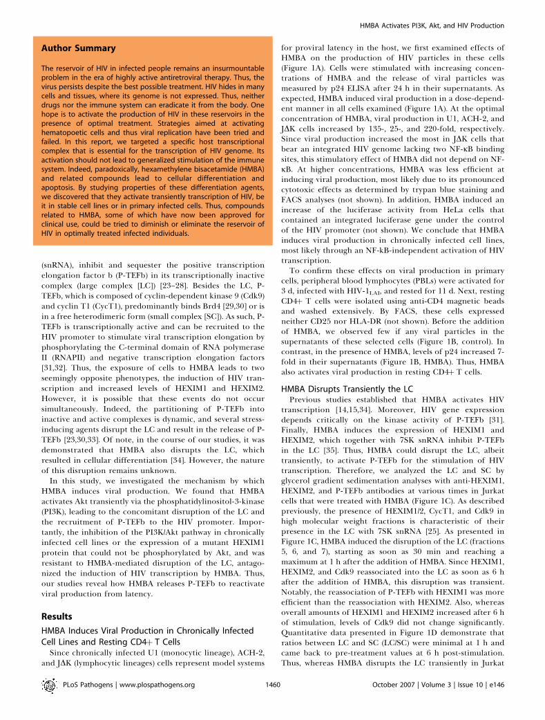
such a rapid, transient disruption of the LC, we reasoned thatHMBA could act by activating cellular signaling pathways.Since SAHA is similar in structure to HMBA [13,17] and itactivates the PI3K/Akt pathway [20], we hypothesized thatHMBA could do the same. Indeed, HMBA activated Akttransiently in Jurkat cells as determined with an antibody thatrecognizes the active, phosphorylated form of Akt (Figure2A). This activation which was detected after 30 min ofstimulation, decreased significantly at 6 h (Figure 2A, lanes 2–4). Moreover, 24 h later, levels of activated Akt were even
lower than those in untreated cells (compare lanes 1 and 5).Importantly, HMBA-mediated activation of Akt was inhibitedwhen cells were pre-incubated with the PI3K inhibitorLY294002 (Figure 2A, lane 6) or the Akt inhibitor 8 (AI8,Figure 2A, lane 7). Thus, similar to SAHA, HMBA induces atransient activation of Akt via PI3K and results in a long-terminhibition of this signaling pathway. Importantly, the kineticsof Akt activation correlated nicely with the kinetics ofdisruption of the LC.To investigate this activation of Akt further, we next asked
whether these kinases were required for the stimulatory effectof HMBA on viral production. We pre-incubated U1 cellswith AI8 or LY294002 prior to the stimulation with HMBAand measured viral production as in Figure 1A. In these cells,HMBA stimulated viral production by approximately 150-fold (Figure 1A, lane 2). Critically, in U1 cells, inhibitors of
Figure 1. HMBA Transiently Disrupts the LC
(A) U1, Ach-2, and JDK cells were stimulated with increasing concentrations of HMBA (1, 5, 10, and 20 mM) and viral production was assessed at 48 h inthe supernatant using p24 ELISA.(B) Resting CD4þT cells, which were isolated from PBMCs that were infected with HIV-1LAI and rested for 11 d were treated or not with HMBA (1 mM) for3 d. Viral production was assessed using p24 ELISA.(C) Total cell lysates of Jurkat cells stimulated or not with HMBA (5 mM) for 30 min, 1 h, 2 h, 6 h, and 24 h, were subjected to glycerol gradientsedimentation analyses (10%–30%), and the fractions were analyzed by western blotting using HEXIM1, HEXIM2, and Cdk9 antibodies. Numbers belowthe western blots correspond to fractions from glycerol gradient analyses.(D) LC/SC represents the ratio of cumulated intensities of fractions 5, 6, and 7 (LC) over intensities of fractions 1, 2, and 3 (SC). Values were normalized toratios obtained in unstimulated cells.doi:10.1371/journal.ppat.0020146.g001
PLoS Pathogens | www.plospathogens.org October 2007 | Volume 3 | Issue 10 | e1461461
HMBA Activates PI3K, Akt, and HIV Production
PI3K and Akt reduced the HMBA-induced viral productionby 97% and 90%, respectively (Figure 2B, lanes 3 and 4). Incontrast, these inhibitors had only minor effects on basallevels of viral production (lanes 5 and 6). Consistent withthese results, the expression of a mutant dominant negativeform of Akt (DN-Akt) also prevented the stimulation of viralproduction by HMBA in U1 cells (Figure 2C, compare lanes 3
and 4). In addition, similar effects were observed in ACH-2and JDK cells (not shown). Taken together, these resultsdemonstrate that a transient activation of the PI3K/Aktpathway is required for the HMBA-induced viral productionin chronically infected cell lines.
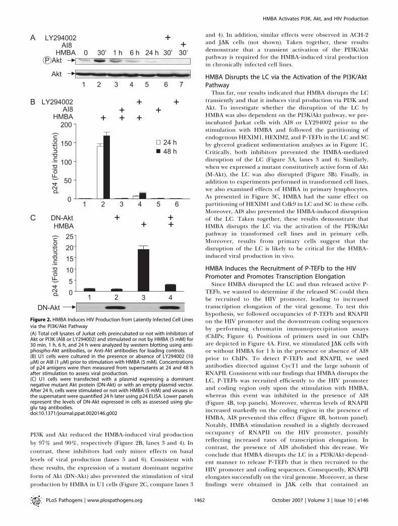
HMBA Disrupts the LC via the Activation of the PI3K/AktPathwayThus far, our results indicated that HMBA disrupts the LC
transiently and that it induces viral production via PI3K andAkt. To investigate whether the disruption of the LC byHMBA was also dependent on the PI3K/Akt pathway, we pre-incubated Jurkat cells with AI8 or LY294002 prior to thestimulation with HMBA and followed the partitioning ofendogenous HEXIM1, HEXIM2, and P-TEFb in the LC and SCby glycerol gradient sedimentation analyses as in Figure 1C.Critically, both inhibitors prevented the HMBA-mediateddisruption of the LC (Figure 3A, lanes 3 and 4). Similarly,when we expressed a mutant constitutively active form of Akt(M-Akt), the LC was also disrupted (Figure 3B). Finally, inaddition to experiments performed in transformed cell lines,we also examined effects of HMBA in primary lymphocytes.As presented in Figure 3C, HMBA had the same effect onpartitioning of HEXIM1 and Cdk9 in LC and SC in these cells.Moreover, AI8 also prevented the HMBA-induced disruptionof the LC. Taken together, these results demonstrate thatHMBA disrupts the LC via the activation of the PI3K/Aktpathway in transformed cell lines and in primary cells.Moreover, results from primary cells suggest that thedisruption of the LC is likely to be critical for the HMBA-induced viral production in vivo.
HMBA Induces the Recruitment of P-TEFb to the HIVPromoter and Promotes Transcription ElongationSince HMBA disrupted the LC and thus released active P-
TEFb, we wanted to determine if the released SC could thenbe recruited to the HIV promoter, leading to increasedtranscription elongation of the viral genome. To test thishypothesis, we followed occupancies of P-TEFb and RNAPIIon the HIV promoter and the downstream coding sequencesby performing chromatin immunoprecipitation assays(ChIPs; Figure 4). Positions of primers used in our ChIPsare depicted in Figure 4A. First, we stimulated JDK cells withor without HMBA for 1 h in the presence or absence of AI8prior to ChIPs. To detect P-TEFb and RNAPII, we usedantibodies directed against CycT1 and the large subunit ofRNAPII. Consistent with our findings that HMBA disrupts theLC, P-TEFb was recruited efficiently to the HIV promoterand coding region only upon the stimulation with HMBA,whereas this event was inhibited in the presence of AI8(Figure 4B, top panels). Moreover, whereas levels of RNAPIIincreased markedly on the coding region in the presence ofHMBA, AI8 prevented this effect (Figure 4B, bottom panel).Notably, HMBA stimulation resulted in a slightly decreasedoccupancy of RNAPII on the HIV promoter, possiblyreflecting increased rates of transcription elongation. Incontrast, the presence of AI8 abolished this decrease. Weconclude that HMBA disrupts the LC in a PI3K/Akt-depend-ent manner to release P-TEFb that is then recruited to theHIV promoter and coding sequences. Consequently, RNAPIIelongates successfully on the viral genome. Moreover, as thesefindings were obtained in JDK cells that contained an
Figure 2. HMBA Induces HIV Production from Latently Infected Cell Lines
via the PI3K/Akt Pathway
(A) Total cell lysates of Jurkat cells preincubated or not with inhibitors ofAkt or PI3K (AI8 or LY294002) and stimulated or not by HMBA (5 mM) for30 min, 1 h, 6 h, and 24 h were analyzed by western blotting using anti-phospho-Akt antibodies, or Anti-Akt antibodies for loading controls.(B) U1 cells were cultured in the presence or absence of LY294002 (10lM) or AI8 (1 lM) prior to stimulation with HMBA (5 mM). Concentrationsof p24 antigens were then measured from supernatants at 24 and 48 hafter stimulation to assess viral production.(C) U1 cells were transfected with a plasmid expressing a dominantnegative mutant Akt protein (DN-Akt) or with an empty plasmid vector.After 24 h, cells were stimulated or not with HMBA (5 mM) and viruses inthe supernatant were quantified 24 h later using p24 ELISA. Lower panelsrepresent the levels of DN-Akt expressed in cells as assessed using glu-glu tag antibodies.doi:10.1371/journal.ppat.0020146.g002
PLoS Pathogens | www.plospathogens.org October 2007 | Volume 3 | Issue 10 | e1461462
HMBA Activates PI3K, Akt, and HIV Production
integrated HIV provirus lacking NF-jB binding sites, theseresults suggest further that the stimulation of HIV tran-scription by HMBA can occur independently of NF-jB.
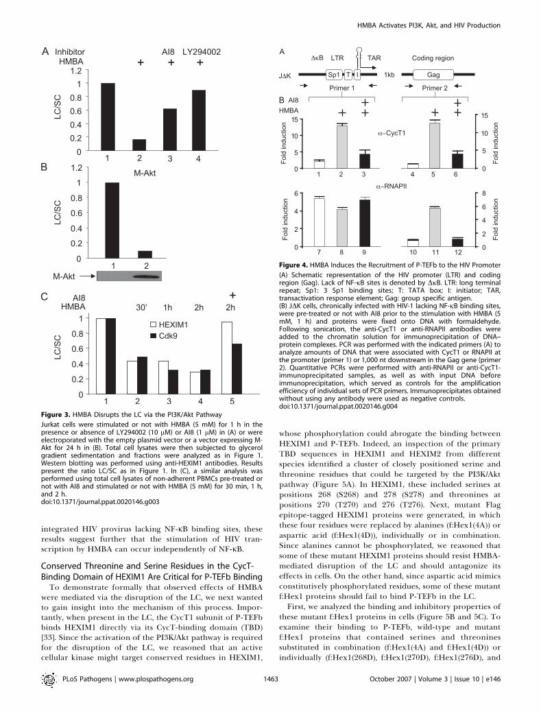
Conserved Threonine and Serine Residues in the CycT-Binding Domain of HEXIM1 Are Critical for P-TEFb Binding
To demonstrate formally that observed effects of HMBAwere mediated via the disruption of the LC, we next wantedto gain insight into the mechanism of this process. Impor-tantly, when present in the LC, the CycT1 subunit of P-TEFbbinds HEXIM1 directly via its CycT-binding domain (TBD)[33]. Since the activation of the PI3K/Akt pathway is requiredfor the disruption of the LC, we reasoned that an activecellular kinase might target conserved residues in HEXIM1,
whose phosphorylation could abrogate the binding betweenHEXIM1 and P-TEFb. Indeed, an inspection of the primaryTBD sequences in HEXIM1 and HEXIM2 from differentspecies identified a cluster of closely positioned serine andthreonine residues that could be targeted by the PI3K/Aktpathway (Figure 5A). In HEXIM1, these included serines atpositions 268 (S268) and 278 (S278) and threonines atpositions 270 (T270) and 276 (T276). Next, mutant Flagepitope-tagged HEXIM1 proteins were generated, in whichthese four residues were replaced by alanines (f:Hex1(4A)) oraspartic acid (f:Hex1(4D)), individually or in combination.Since alanines cannot be phosphorylated, we reasoned thatsome of these mutant HEXIM1 proteins should resist HMBA-mediated disruption of the LC and should antagonize itseffects in cells. On the other hand, since aspartic acid mimicsconstitutively phosphorylated residues, some of these mutantf:Hex1 proteins should fail to bind P-TEFb in the LC.First, we analyzed the binding and inhibitory properties of
these mutant f:Hex1 proteins in cells (Figure 5B and 5C). Toexamine their binding to P-TEFb, wild-type and mutantf:Hex1 proteins that contained serines and threoninessubstituted in combination (f:Hex1(4A) and f:Hex1(4D)) orindividually (f:Hex1(268D), f:Hex1(270D), f:Hex1(276D), and
Figure 3. HMBA Disrupts the LC via the PI3K/Akt Pathway
Jurkat cells were stimulated or not with HMBA (5 mM) for 1 h in thepresence or absence of LY294002 (10 lM) or AI8 (1 lM) in (A) or wereelectroporated with the empty plasmid vector or a vector expressing M-Akt for 24 h in (B). Total cell lysates were then subjected to glycerolgradient sedimentation and fractions were analyzed as in Figure 1.Western blotting was performed using anti-HEXIM1 antibodies. Resultspresent the ratio LC/SC as in Figure 1. In (C), a similar analysis wasperformed using total cell lysates of non-adherent PBMCs pre-treated ornot with AI8 and stimulated or not with HMBA (5 mM) for 30 min, 1 h,and 2 h.doi:10.1371/journal.ppat.0020146.g003
Figure 4. HMBA Induces the Recruitment of P-TEFb to the HIV Promoter
(A) Schematic representation of the HIV promoter (LTR) and codingregion (Gag). Lack of NF-jB sites is denoted by DjB. LTR: long terminalrepeat; Sp1: 3 Sp1 binding sites; T: TATA box; I: initiator; TAR,transactivation response element; Gag: group specific antigen.(B) JDK cells, chronically infected with HIV-1 lacking NF-jB binding sites,were pre-treated or not with AI8 prior to the stimulation with HMBA (5mM, 1 h) and proteins were fixed onto DNA with formaldehyde.Following sonication, the anti-CycT1 or anti-RNAPII antibodies wereadded to the chromatin solution for immunoprecipitation of DNA–protein complexes. PCR was performed with the indicated primers (A) toanalyze amounts of DNA that were associated with CycT1 or RNAPII atthe promoter (primer 1) or 1,000 nt downstream in the Gag gene (primer2). Quantitative PCRs were performed with anti-RNAPII or anti-CycT1-immunoprecipitated samples, as well as with input DNA beforeimmunoprecipitation, which served as controls for the amplificationefficiency of individual sets of PCR primers. Immunoprecipitates obtainedwithout using any antibody were used as negative controls.doi:10.1371/journal.ppat.0020146.g004
PLoS Pathogens | www.plospathogens.org October 2007 | Volume 3 | Issue 10 | e1461463
HMBA Activates PI3K, Akt, and HIV Production
f:Hex1(278D)) were expressed in Jurkat cells and immuno-precipitated using anti-Flag agarose beads. The presence ofP-TEFb in immunoprecipitations was followed by antibodiesdirected against CycT1. As expected, whereas wild-type andmutant f:Hex1(4A) proteins bound P-TEFb, the mutantf:Hex1(4D) protein failed to do so (Figure 5B, lanes 2–4).Interestingly, whereas the mutant f:Hex1(268D) andf:Hex1(276D) proteins did not alter these interactions, themutant f:Hex1(270D) and f:Hex1(278D) proteins did not
interact as potently (67% and 62% inhibition, respectively) asthe wild-type protein with P-TEFb, suggesting that T270 andS278 are important for this interaction. Levels of Flagepitope-tagged proteins in our immunoprecipitations weresimilar (Figure 5B, lower panel). Moreover, individual alaninesubstitutions did not affect the binding of the mutant f:Hex1proteins to P-TEFb (not shown). Additionally, in contrast tothe wild-type and mutant f:Hex1(4A) proteins, the mutantf:Hex1(4D) protein failed to bind P-TEFb in the LC asdetermined by glycerol gradient sedimentation analyses (notshown; see below).Next, these features were supported further by transcrip-
tional assays in cells (Figure 5C). We used a classical DNA-tethering system, which consists of the plasmid reporterpG6TAR that contains six Gal4 DNA binding sites and theplasmid effector Gal4.CycT1. The recruitment of Gal4.CycT1to the promoter results in the P-TEFb-dependent activationof transcription that is sensitive to the inhibition by HEXIM1[28]. Indeed, the mutant f:Hex1(4A) protein inhibited thetranscriptional activation by Gal4.CycT1 even more robustlywhen compared to the wild-type f:Hex1 protein (Figure 5C,lanes 1–3). In contrast, the mutant f:Hex1(4D) protein, whichfailed to bind P-TEFb, did not inhibit this transcriptionalactivation (Figure 5C, lane 4). Similar results were obtainedwith mutant f:Hex1(270D) and f:Hex1(278D) proteins. Also,levels of Flag epitope-tagged proteins were similar (Figure 5C,lower panel). Collectively, these results indicate that T270 andS278 in the TBD of HEXIM1 are important for its binding toP-TEFb in cells. Furthermore, they suggest that these residuescould be phosphorylated by Akt, leading to the disruption ofthe LC and a subsequent release of the transcriptionallyactive P-TEFb.
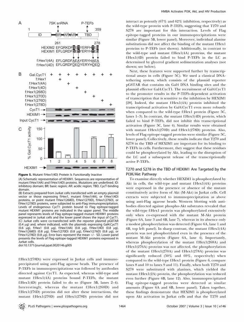
T270 and S278 in the TBD of HEXIM1 Are Targeted by thePI3K/Akt PathwayTo examine directly whether HEXIM1 is phosphorylated by
Akt in cells, the wild-type and mutant f:Hex1(4A) proteinswere expressed in the presence or absence of the mutantconstitutively active form of Akt (M-Akt) in Jurkat cells. Cellextracts were subjected to immunoprecipitation as aboveusing anti-Flag agarose beads. Western blotting with anti-bodies directed against phospho-Akt substrates revealed thatthe wild-type f:Hex1 protein was phosphorylated extensivelyonly when co-expressed with the mutant M-Akt protein(Figure 6A, lane 3 and 6B, lane 7), whereas in its absence onlya modest phosphorylation was detected (Figure 6A, lane 1 and6B, top left panel). In sharp contrast, the mutant f:Hex1(4A)protein was not phosphorylated even in the presence of themutant M-Akt protein (Figure 6A, lane 4). Importantly,whereas phosphorylation of the mutant f:Hex1(268A) andf:Hex1(276A) proteins was not affected, the phosphorylationof the mutant f:Hex1(270A) and f:Hex1(278A) proteins wassignificantly reduced (50% and 69%, respectively) whencompared to the wild-type f:Hex1 protein (Figure 6, comparelanes 8 and 10 to lanes 9 and 11). Finally, when both T270 andS278 were substituted with alanines, which yielded themutant f:Hex1(2A) protein, the phosphorylation was reducedeven further (Figure 6B, lane 12). Also, immunoprecipitatedFlag epitope-tagged proteins were detected at similaramounts (Figure 6A and 6B, lower panel). Taken together,these findings demonstrate that HEXIM1 is phosphorylatedupon Akt activation in Jurkat cells and that the T270 and
Figure 5. Mutant f:Hex1(4D) Protein Is Functionally Inactive
(A) Schematic representation of HEXIM1. Sequences are representative ofmutant f:Hex1(4A) and f:Hex1(4D) proteins. Mutations are underlined. ID:inhibitory domain; BR: basic region; AR: acidic region; TBD, CycT-bindingdomain.(B) Extracts prepared from Jurkat cells transfected with an empty plasmidvector or those expressing f:Hex1, mutant f:Hex1(4A), or f:Hex1(4D)proteins, or point mutant f:Hex1(268D), f:Hex1(270D), f:Hex1(276D), orf:Hex1(278D) proteins, were subjected to anti-Flag immunoprecipitation.Levels of endogenous CycT1 protein bound to Flag epitope-taggedmutant HEXIM1 proteins are indicated in the upper panel. The middlepanel represents levels of Flag epitope-tagged mutant HEXIM1 proteinsexpressed in Jurkat cells and the lower panel shows the input of CycT1.(C) Jurkat cells were co-transfected with the reporter plasmid pG6TAR(0.4 lg) and, where indicated, with the plasmids expressing Gal4.CycT1(0.6 lg), f:Hex1 (0.8 lg), f:Hex1(4A) (0.8 lg), f:Hex1(4D) (0.8 lg),f:Hex1(268D) (0.8 lg), f:Hex1(270D) (0.8 lg), f:Hex1(276D) (0.8 lg), orf:Hex1(278D) (0.8 lg). Error bars represent the meanþ/� SD. Lower panelpresents the levels of Flag epitope-tagged HEXIM1 proteins expressed inJurkat cells.doi:10.1371/journal.ppat.0020146.g005
PLoS Pathogens | www.plospathogens.org October 2007 | Volume 3 | Issue 10 | e1461464
HMBA Activates PI3K, Akt, and HIV Production

S278 are responsible for these effects. In addition, theseresults suggest that the phosphorylation of residues in theTBD of HEXIM1 via the PI3K/Akt pathway disrupts the LC.
Conserved Threonines and Serines in the TBD of HEXIM1Mediate Its Release from the LC
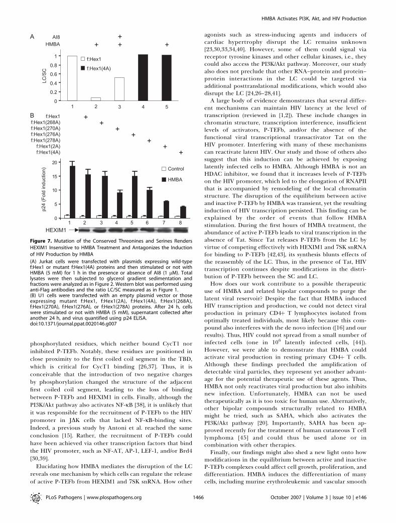
To determine whether conserved threonines and serineswere required for the HMBA-mediated disruption of the LC,we expressed the wild-type or the mutant f:Hex1(4A) proteinsin Jurkat cells. As in Figure 1, the partitioning of f:Hex1proteins in the LC and SC in untreated or HMBA-treatedcells was followed by glycerol gradient sedimentation analyses(Figure 7A). Similar to endogenous HEXIM1 proteins, thewild-type f:Hex1 protein was released from the LC uponHMBA stimulation and the Akt inhibitor AI8 prevented thiseffect (Figure 7A, lanes 1–3). Critically, the mutant f:Hex1(4A)protein was not responsive to HMBA treatment andremained in the LC even after the stimulation (Figure 7A,lanes 4 and 5). Taken together, these results demonstrate thatthe conserved threonines and serines in the TBD play a majorrole in regulating the release of HEXIM1 from the LC uponHMBA stimulation of cells.
HMBA Stimulation of Viral Production Depends on T270and S278 in the TBD of HEXIM1
Finally, the use of the mutant f:Hex1(4A) protein thatresisted the HMBA-mediated disruption of the LC enabled usto examine directly the importance of this phosphorylationfor the stimulation of viral production. Wild-type or mutantf:Hex1(4A) proteins were expressed in U1 cells that were leftuntreated or treated with HMBA, and the production of newviral particles was measured as above. As presented in Figure7B, the expression of the mutant f:Hex1(4A) proteinantagonized effects of HMBA profoundly, as its inductionof viral production was decreased by 75% (Figure 7B, lane 8).Consistently, individual substitutions of the threonine andserine at positions 270 and 278 with alanines reduced viralproduction by 50% and 40% in the presence of HMBA(Figure 7B, lanes 4 and 6), and mutation of both residues(f:Hex1(2A)) resulted in an inhibition comparable to that bythe mutant f:Hex1(4A) protein (Figure 7B, compare lanes 7and 8). In contrast, HMBA induced viral production robustlyand the expression of the wild-type f:Hex1 protein had only amodest effect (Figure 7B, lanes 1 and 2). Also, levels of these
Flag epitope-tagged proteins were similar as were basal levelsof viral production in these cells (Figure 7B, lower panel andlanes 1–8). Taken together, these results indicate that HMBAinduces viral production via the PI3/Akt pathway and thedisruption of the LC via the phosphorylation of the T270 andS278 of HEXIM1.
Discussion
In this study, we demonstrated that HMBA reactivates viralproduction in chronically infected cells via the transientrelease of active P-TEFb from the LC. These events dependedon the transient activation of the PI3K/Akt pathway that ledto the phosphorylation of T270 and S278 in HEXIM1. In turn,the released P-TEFb was recruited to the HIV promoter tostimulate transcription elongation. Importantly, the inhib-ition of the PI3K/Akt pathway and the transient expression ofmutant HEXIM1 proteins that could not be phosphorylatedand released from the LC via Akt antagonized the HMBA-mediated induction of viral production. Therefore, weprovide a mechanism by which HMBA targets the pivotalcellular co-factor P-TEFb for the reactivation of HIV.Thus far, a regulatory mechanism that releases P-TEFb
from HEXIM1 and 7SK snRNA has not been determined.Here, we provide evidence that HMBA accomplishes this taskby activating the PI3K/Akt pathway. Since inhibitors of bothkinases and the expression of the mutant DN-Akt proteinresulted in an identical phenotype, PI3K and Akt are involvedin this release. Moreover, the mutant constitutively active M-Akt protein also released P-TEFb. It is attractive tohypothesize that HMBA, which is a bipolar compound, bindsand activates PI3K directly or via aggregation. In such ascenario, the brief nature of this signal could reflect a rapidmodification of HMBA and/or the sequestration and degra-dation of the signaling complex. The signaling pathwaybetween PI3K and Akt and the subsequent nuclear trans-location of Akt has been described in great detail elsewhere[36]. Since the mutant constitutively active M-Akt protein ledto the phosphorylation of T270 and S278, it is tempting tospeculate that Akt could phosphorylate these residues in theTBD of HEXIM1 directly. This phosphorylation also abro-gated the binding between HEXIM1 and P-TEFb in cells.Indeed, this finding was supported by the analysis of mutantHEXIM1 proteins containing aspartic acids that mimic
Figure 6. T270 and S278 in the TBD of HEXIM1 Are Phosphorylated following the Activation of Akt In Vivo
f:Hex1 protein and mutant f:Hex1(4A) protein in (A), and mutant f:Hex1(268A), f:Hex1(270A), f:Hex1(276A), or f:Hex1(278A) proteins in (B) wereexpressed in Jurkat cells transfected with an empty plasmid vector or a vector expressing M-Akt. After 24 h, protein extracts were subjected toimmunoprecipitation using anti-Flag antibodies, and levels of phospho-Akt substrates (pAktSub) and Flag-tagged expressed proteins were measuredby western blotting.doi:10.1371/journal.ppat.0020146.g006
PLoS Pathogens | www.plospathogens.org October 2007 | Volume 3 | Issue 10 | e1461465
HMBA Activates PI3K, Akt, and HIV Production
phosphorylated residues, which neither bound CycT1 norinhibited P-TEFb. Notably, these residues are positioned inclose proximity to the first coiled coil segment in the TBD,which is critical for CycT1 binding [26,37]. Thus, it isconceivable that the introduction of two negative chargesby phosphorylation changed the structure of the adjacentfirst coiled coil segment, leading to the loss of bindingbetween P-TEFb and HEXIM1 in cells. Finally, although thePI3K/Akt pathway also activates NF-jB [38], it is unlikely thatit was responsible for the recruitment of P-TEFb to the HIVpromoter in JDK cells that lacked NF-jB-binding sites.Indeed, a previous study by Antoni et al. reached the sameconclusion [15]. Rather, the recruitment of P-TEFb couldhave been achieved via other transcription factors that bindthe HIV promoter, such as NF-AT, AP-1, LEF-1, and/or Brd4[30,39].
Elucidating how HMBA mediates the disruption of the LCreveals one mechanism by which cells can regulate the releaseof active P-TEFb from HEXIM1 and 7SK snRNA. How other
agonists such as stress-inducing agents and inducers ofcardiac hypertrophy disrupt the LC remains unknown[23,30,33,34,40]. However, some of them could signal viareceptor tyrosine kinases and other cellular kinases, i.e., theycould also access the PI3K/Akt pathway. Moreover, our studyalso does not preclude that other RNA–protein and protein–protein interactions in the LC could be targeted viaadditional posttranslational modifications, which would alsodisrupt the LC [24,26–28,41].A large body of evidence demonstrates that several differ-
ent mechanisms can maintain HIV latency at the level oftranscription (reviewed in [1,2]). These include changes inchromatin structure, transcription interference, insufficientlevels of activators, P-TEFb, and/or the absence of thefunctional viral transcriptional transactivator Tat on theHIV promoter. Interfering with many of these mechanismscan reactivate latent HIV. Our study and those of others alsosuggest that this induction can be achieved by exposinglatently infected cells to HMBA. Although HMBA is not anHDAC inhibitor, we found that it increases levels of P-TEFbon the HIV promoter, which led to the elongation of RNAPIIthat is accompanied by remodeling of the local chromatinstructure. The disruption of the equilibrium between activeand inactive P-TEFb by HMBA was transient, yet the resultinginduction of HIV transcription persisted. This finding can beexplained by the order of events that follow HMBAstimulation. During the first hours of HMBA treatment, theabundance of active P-TEFb leads to viral transcription in theabsence of Tat. Since Tat releases P-TEFb from the LC byvirtue of competing effectively with HEXIM1 and 7SK snRNAfor binding to P-TEFb [42,43], its synthesis blunts effects ofthe reassembly of the LC. Thus, in the presence of Tat, HIVtranscription continues despite modifications in the distri-bution of P-TEFb between the SC and LC.How does our work contribute to a possible therapeutic
use of HMBA and related bipolar compounds to purge thelatent viral reservoir? Despite the fact that HMBA inducedHIV transcription and production, we could not detect viralproduction in primary CD4þ T lymphocytes isolated fromoptimally treated individuals, most likely because this com-pound also interferes with the de novo infection ([16] and ourresults). Thus, HIV could not spread from a small number ofinfected cells (one in 106 latently infected cells, [44]).However, we were able to demonstrate that HMBA couldactivate viral production in resting primary CD4þ T cells.Although these findings precluded the amplification ofdetectable viral particles, they represent yet another advant-age for the potential therapeutic use of these agents. Thus,HMBA not only reactivates viral production but also inhibitsnew infection. Unfortunately, HMBA can not be usedtherapeutically as it is too toxic for human use. Alternatively,other bipolar compounds structurally related to HMBAmight be tried, such as SAHA, which also activates thePI3K/Akt pathway [20]. Importantly, SAHA has been ap-proved recently for the treatment of human cutaneous T celllymphoma [45] and could thus be used alone or incombination with other therapies.Finally, our findings might also shed a new light onto how
modifications in the equilibrium between active and inactiveP-TEFb complexes could affect cell growth, proliferation, anddifferentiation. HMBA induces the differentiation of manycells, including murine erythroleukemic and vascular smooth
Figure 7. Mutation of the Conserved Threonines and Serines Renders
HEXIM1 Insensitive to HMBA Treatment and Antagonizes the Induction
of HIV Production by HMBA
(A) Jurkat cells were transfected with plasmids expressing wild-typef:Hex1 or mutant f:Hex1(4A) proteins and then stimulated or not withHMBA (5 mM) for 1 h in the presence or absence of AI8 (1 lM). Totallysates were then subjected to glycerol gradient sedimentation andfractions were analyzed as in Figure 2. Western blot was performed usinganti-Flag antibodies and the ratio LC/SC measured as in Figure 1.(B) U1 cells were transfected with an empty plasmid vector or thoseexpressing mutant f:Hex1, f:Hex1(2A), f:Hex1(4A), f:Hex1(268A),f:Hex1(270A), f:Hex1(276A), or f:Hex1(278A) proteins. After 24 h, cellswere stimulated or not with HMBA (5 mM), supernatant collected afteranother 24 h, and virus quantified using p24 ELISA.doi:10.1371/journal.ppat.0020146.g007
PLoS Pathogens | www.plospathogens.org October 2007 | Volume 3 | Issue 10 | e1461466
HMBA Activates PI3K, Akt, and HIV Production
muscle cells, monocytes, lymphocytes, and neurons [46,47].Importantly, HMBA also increases greatly the expression ofHEXIM1 in vascular smooth muscle cells [22] and morerecently, the transient disassembly and reassembly of the LCby HMBA was found to be associated with the differentiationof murine erythroleukemic cells [34]. Indeed, whereas thesustained activation of PI3K and Akt is associated with cellgrowth and proliferation [48], a transient activation of thePI3K/Akt pathway leads rather to cell differentiation andgrowth arrest. This seeming paradox can now be explained bythe feedback mechanism involved in the regulation of P-TEFb. We propose that HMBA first disrupts the LC to releasethe active P-TEFb, which is followed by increased globaltranscription. In very short order, the LC reassembles withabundant new synthesis of HEXIM1 [22,34]. As the pool ofactive P-TEFb decreases, cellular reprogramming follows,leading to cell differentiation. As SAHA also activatestransiently this pathway [20], the role of other bipolarcompounds that are related to HMBA should also beinvestigated for their effects on PI3K and Akt.
Materials and Methods
Cell lines. U1, ACH-2, and J1.1 cells were obtained through theNIH AIDS reagents program. JDK cells were kindly provided byArnold Rabson. Jurkat. U1, ACH-2, J1.1, and JDK cells were grown inRPMI containing penicillin (100 IU/ml), streptomycin (100 lg/ml), and10% FBS at 37 8C with 5% CO2.
Plasmid DNAs. Plasmid reporter pG6TAR and plasmids coding forthe Gal4.CycT1 chimera or f:Hex1 protein were described previously[28]. To construct plasmids coding for the mutant f:Hex1 proteins,the pFlag-CMV-2. HEXIM1 plasmid was subjected to site directedmutagenesis with the QuickChange II XL Site-Directed MutagenesisKit (200521; Stratagene). The plasmids coding for myristoylated Akt(M-Akt) and dominant negative Akt (DN-Akt) were a gift from DavidStokoe and were described previously [49].
Immunoreagents and chemicals. The anti-CycT1 (sc-8127) andanti-Cdk9 (D7) antibodies were obtained from Santa Cruz Biotech-nology. The anti-HEXIM1 and anti-HEXIM2 antibodies weredescribed previously [26]. The antibodies directed against the total(9272), phosphorylated form of Akt (9271), and phospho-Aktsubstrates (9611S) were obtained from Cell Signaling. DN-Akt andM-Akt were a gift from David Stokoe [49]. The anti-Flag M2 (F3165)antibody and the anti-Flag M2 beads (FlagIPT-1) were purchasedfrom Sigma-Aldrich.
HMBA (Sigma-Aldrich) was resuspended in sterile water to obtaina stock solution of 1 M. Chemical inhibitors, Akt inhibitor VIII (AI8),and LY 294002 (Calbiochem), were resuspended in DMSO.
Activation of HIV production in HIV chronically infected cell lines.U1, ACH-2, or JDK cells were plated at 2.105 cells/ml in 24-well plates,supernatant was collected before stimulation with HMBA and thenevery day, and viral release in the supernatant quantified by p24ELISA (PerkinElmer). Baseline levels were of 50 to 100 pg/ml incontrol cells. Pre-treatment with inhibitor was done 1 h beforestimulation by HMBA. Plasmid transfection was done using GenePulser II Electroporator (Bio-Rad).
Transient transfection and CAT reporter gene assay. HeLa cellswere seeded into six-well plates approximately 12 h prior totransfection and transfected with FuGENE6 reagent (1 815 091;Roche Applied Science). CAT enzymatic assays were performed asdescribed [28]. Transcriptional activation of the pG6TAR reporterplasmid by the Gal4.CycT1 chimera was set to 100%. Error bars givestandard errors of the mean.
Immunoprecipitation assay and western blotting. 10 millions ofJurkat cells were lysed in 0.8 ml of lysis buffer A (10 mM Tris-HCl [pH7.4], 150 mM NaCl, 2 mM EDTA, 1% NP-40, 0.1% protease inhibitor)for 0.5 h at 4 8C 20 h post-electroporation. The lysates wereimmunoprecipitated with the anti-Flag M2 beads for 2 h at 4 8C,washed extensively with the lysis buffer A, and the bound proteinswere separated on SDS-PAGE electrophoresis. Western blotting wasperformed with the indicated antibodies and according to thestandard protocols.
Glycerol gradient sedimentation analysis. Glycerol gradients
(10%–30%) were established by pippeting 2 ml of each of theglycerol fractions (10, 15, 20, 25, and 30% v/v) in buffer A (20 mMHEPES [pH 7.9], 0.3 M KCl, 0.2 mM EDTA, 0.1% NP-40) intocentrifugation tubes (Beckman), 331372. Gradients were formed bystanding for 6 h at 4 8C. HeLa cells either transfected with thecorresponding plasmids or not transfected were lysed in 0.5 ml ofbuffer A containing 0.1% protease inhibitor and either 0.5% RNaseinhibitor or RNase A (100 mg/ml final concentration) for 30 min at 48C. The lysates were centrifuged at 10,000g for 10 min and thesupernatants were loaded into tubes with the preformed glycerolgradients. Protein complexes were then fractionated by centrifuga-tion in an SW 41Ti rotor (Beckman) at 38,000 rpm. for 21 h. Tenfractions (1 ml) were collected, precipitated with trichloracetic acidand finally analyzed by immunoblotting with the appropriateantibodies [25]. Films with exposures in the linear range were usedand intensities of each bands were obtained using histograms withPhotoshop. The ratio of proteins present in the LC (fractions 5, 6, and7) versus the ones present in the SC (fractions 1, 2, and 3) wascalculated. The ratio LC/SC was normalized to 1 for unstimulatedcells.
Infection of PBMCs and isolation of resting CD4þ T cells. PBMCswere isolated from buffy coats of healthy HIV negative donors in aFicoll density gradient (Pharmacia). PBMCs were then plated at 5.106
cells per ml in 24-well plates, using RPMI 10% human serum AB.After 30 min, non-adherent cells (PBLs) were isolated and cultured incomplete RPMI (containing penicillin [100 IU/ml], streptomycin [100lg/ml], and 10% FCS) with IL-2 (10 U/ml) and PHA (3 ug/ml). After 3d, cells were treated or not with HMBA (5 mM) in the presence orabsence of Akt inhibitor 8 (AI8).
After isolation, PBLs (107) were activated with PHA (5 lg/ml) andIL-2 (10 U/ml) for 3 d and infected with HIV-1LAI (0.1 ng/ml of p24).They were cultured in RPMI, 10% FCS, supplemented with IL-2 (10U/ml). After 11 d, resting CD4þ T cells were isolated by negativeselection using magnetic beads (Invitrogen). Cells were morethan 95% pure as assessed by FACS. These cells were then culturedin RPMI, 10% FCS. These cells expressed neither CD25 nor HLA-DR.
ChIP assays. ChIP was carried out essentially as describedpreviously [50]. Cross-linking was achieved by incubating 70 millioncells (J1.1 or JDK) in 1% formaldehyde in medium for 10 min at roomtemperature. Cross-linking reactions were stopped by addition ofglycine to a final concentration of 0.125 M. Cells were then pelleted ina conical tube and washed with cold phosphate-buffered saline. Thecell pellets were then resuspended in 1 ml of Lysis buffer (1% SDS, 10mM EDTA, 50 mM Tris-HCl [pH 8.0]) for 10 min on ice and subjectedto sonication to obtain DNA fragments averaging approximately 200to 500 bp in length. One-tenth of the total chromatin solution wasused in each ChIP. Chromatin solutions were precleared with proteinA/G-Sepharose beads and then incubated with the appropriateantibody at 4 8C overnight. Protein A/G-Sepharose beads were thenadded, and the mixture was incubated for another 2 h. The beadswere washed five times in TSE-150 (0.1% SDS, 1% Triton X-100, 2mM EDTA, 20 mM Tris-HCl [pH 8.0], 150 mM NaCl), TSE-500 (likeTSE-150 but with 500 mM NaCl), and buffer III (0.25 M LiCl, 1% NP-40, 1% deoxycholate, 1 mM EDTA, 10 mM Tris-HCl [pH 8.0]), andtwice in Tris-EDTA buffer. Immunocomplexes were eluted from thebeads with elution buffer (1% SDS and 0.5% NaHCO3) for 15 min atRT. The DNA–protein complexes were then treated with proteinaseK, followed by reverse cross-linking at 65 8C 4 h. DNA was extractedwith phenol-chloroform, precipitated with ethanol, and dissolved in30 ll of Tris-EDTA buffer. Two microliters of DNA was used withappropriate primer sets to amplify specific DNA fragments. qPCRwas then performed using the Stratagene MX3000P real-time PCRsystem. Primers used for LTR were described previously [8] andprimers for Gag span a region comprised between nucleotides 1144and 1410. Standard curves for each primer pair was first obtained tocheck for their respective efficiency. Products were quantified usingBrilliant SYBR Green QPCR (Stratagene) according to the manu-facturer’s directions. Relative intensity was calculated and normalizedto the input. Fold-induction was assessed as the immunoprecipitationwith specific antibodies over no antibody control.
Supporting InformationAccession Numbers
The National Center for Biotechnology Information (http://www.ncbi.nlm.nih.gov/) accession numbers for HEXIM1 are NM_006460(nucleotide sequence) and BAA36166 (protein sequence).
PLoS Pathogens | www.plospathogens.org October 2007 | Volume 3 | Issue 10 | e1461467
HMBA Activates PI3K, Akt, and HIV Production

Acknowledgments
We thank Arnold B. Rabson and David Stokoe for providing the JDKcells and the plasmids coding for different forms of Akt. We aregrateful to Matthias Geyer and David Stokoe and to members of thePeterlin laboratory for stimulating discussions and continuoussupport.
Author contributions. All authors conceived and designed theexperiments, analyzed the data, contributed reagents/materials/
analysis tools, and wrote the paper. XC, MB, and TL performed theexperiments.
Funding. This work was supported by grants from the NationalInstitutes of Health to BMP (AI49104 and AI058708). MB wassupported by a grant (106584–36–RFNT) from the AmericanFoundation for AIDS Research (amfAR).
Competing interests. The authors have declared that no competinginterests exist.
References1. Contreras X, Lenasi T, Peterlin BM (2006) HIV latency: present knowledge
and future directions. Future Virology 1: 733–745.2. Lassen K, Han Y, Zhou Y, Siliciano J, Siliciano RF (2004) The multifactorial
nature of HIV-1 latency. Trends Mol Med 10: 525–531.3. Chun TW, Engel D, Mizell SB, Hallahan CW, Fischette M, et al. (1999) Effect
of interleukin-2 on the pool of latently infected, resting CD4þ T cells inHIV-1-infected patients receiving highly active anti-retroviral therapy. NatMed 5: 651–655.
4. Stellbrink HJ, van Lunzen J, Westby M, O’Sullivan E, Schneider C, et al.(2002) Effects of interleukin-2 plus highly active antiretroviral therapyon HIV-1 replication and proviral DNA (COSMIC trial). AIDS 16: 1479–1487.
5. Biancotto A, Grivel JC, Gondois-Rey F, Bettendroffer L, Vigne R, et al.(2004) Dual role of prostratin in inhibition of infection and reactivation ofhuman immunodeficiency virus from latency in primary blood lympho-cytes and lymphoid tissue. J Virol 78: 10507–10515.
6. Williams SA, Chen LF, Kwon H, Fenard D, Bisgrove D, et al. (2004)Prostratin antagonizes HIV latency by activating NF-kappaB. J Biol Chem279: 42008–42017.
7. Wang FX, Xu Y, Sullivan J, Souder E, Argyris EG, et al. (2005) IL-7 is apotent and proviral strain-specific inducer of latent HIV-1 cellularreservoirs of infected individuals on virally suppressive HAART. J ClinInvest 115: 128–137.
8. Williams SA, Chen LF, Kwon H, Ruiz-Jarabo CM, Verdin E, et al. (2006) NF-kappaB p50 promotes HIV latency through HDAC recruitment andrepression of transcriptional initiation. EMBO J 25: 139–149.
9. Ylisastigui L, Coull JJ, Rucker VC, Melander C, Bosch RJ, et al. (2004)Polyamides reveal a role for repression in latency within resting T cells ofHIV-infected donors. J Infect Dis 190: 1429–1437.
10. Lehrman G, Hogue IB, Palmer S, Jennings C, Spina CA, et al. (2005)Depletion of latent HIV-1 infection in vivo: a proof-of-concept study.Lancet 366: 549–555.
11. Siliciano JD, Lai J, Callender M, Pitt E, Zhang H, et al. (2007) Stability of thelatent reservoir for HIV-1 in patients receiving valproic acid. J Infect Dis195: 833–836.
12. Siegel DS, Zhang X, Feinman R, Teitz T, Zelenetz A, et al. (1998)Hexamethylene bisacetamide induces programmed cell death (apoptosis)and down-regulates BCL-2 expression in human myeloma cells. Proc NatlAcad Sci U S A 95: 162–166.
13. Richon VM, Webb Y, Merger R, Sheppard T, Jursic B, et al. (1996) Secondgeneration hybrid polar compounds are potent inducers of transformedcell differentiation. Proc Natl Acad Sci U S A 93: 5705–5708.
14. Vlach J, Pitha PM (1993) Hexamethylene bisacetamide activates the humanimmunodeficiency virus type 1 provirus by an NF-kappa B-independentmechanism. J Gen Virol 74 (Pt 11): 2401–2408.
15. Antoni BA, Rabson AB, Kinter A, Bodkin M, Poli G (1994) NF-kappa B-dependent and -independent pathways of HIV activation in a chronicallyinfected T cell line. Virology 202: 684–694.
16. Klichko V, Archin N, Kaur R, Lehrman G, Margolis D (2006) Hexame-thylbisacetamide remodels the human immunodeficiency virus type 1(HIV-1) promoter and induces Tat-independent HIV-1 expression butblunts cell activation. J Virol 80: 4570–4579.
17. Richon VM, Emiliani S, Verdin E, Webb Y, Breslow R, et al. (1998) A class ofhybrid polar inducers of transformed cell differentiation inhibits histonedeacetylases. Proc Natl Acad Sci U S A 95: 3003–3007.
18. Rajagopalan V, Blankenship J, Thomas DW (2006) 1,6-Diaminohexanecontributes to the hexamethylene bisacetamide-induced erythroid differ-entiation pathway by stimulating Ca2þ release from inositol 1,4,5-tri-sphosphate-sensitive stores and promoting Ca2þ influx. Arch BiochemBiophys 445: 129–137.
19. Chen CS, Weng SC, Tseng PH, Lin HP, Chen CS (2005) Histone acetylation-independent effect of histone deacetylase inhibitors on Akt through thereshufflingofproteinphosphatase1complexes. JBiolChem280: 38879–38887.
20. Liu Y, Denlinger CE, Rundall BK, Smith PW, Jones DR (2006) Suberoyla-nilide hydroxamic acid induces Akt-mediated phosphorylation of p300,which promotes acetylation and transcriptional activation of RelA/p65. JBiol Chem 281: 31359–31368.
21. Ouchida R, Kusuhara M, Shimizu N, Hisada T, Makino Y, et al. (2003)Suppression of NF-kappaB-dependent gene expression by a hexamethylenebisacetamide-inducible protein HEXIM1 in human vascular smooth musclecells. Genes Cells 8: 95–107.
22. Kusuhara M, Nagasaki K, Kimura K, Maass N, Manabe T, et al. (1999)Cloning of hexamethylene-bis-acetamide-inducible transcript, HEXIM1, inhuman vascular smooth muscle cells. Biomed Res 20: 273–279.
23. Yik JH, Chen R, Nishimura R, Jennings JL, Link AJ, et al. (2003) Inhibitionof P-TEFb (CDK9/Cyclin T) kinase and RNA polymerase II transcriptionby the coordinated actions of HEXIM1 and 7SK snRNA. Mol Cell 12: 971–982.
24. Michels AA, Fraldi A, Li Q, Adamson TE, Bonnet F, et al. (2004) Binding ofthe 7SK snRNA turns the HEXIM1 protein into a P-TEFb (CDK9/cyclin T)inhibitor. EMBO J 23: 2608–2619.
25. Yik JH, Chen R, Pezda AC, Zhou Q (2005) Compensatory contributions ofHEXIM1 and HEXIM2 in maintaining the balance of active and inactivepositive transcription elongation factor b complexes for control oftranscription. J Biol Chem 280: 16368–16376.
26. Li Q, Price JP, Byers SA, Cheng D, Peng J, et al. (2005) Analysis of the largeinactive P-TEFb complex indicates that it contains one 7SK molecule, adimer of HEXIM1 or HEXIM2, and two P-TEFb molecules containing Cdk9phosphorylated at threonine 186. J Biol Chem 280: 28819–28826.
27. Byers SA, Price JP, Cooper JJ, Li Q, Price DH (2005) HEXIM2, a HEXIM1-related protein, regulates positive transcription elongation factor bthrough association with 7SK. J Biol Chem 280: 16360–16367.
28. Barboric M, Kohoutek J, Price JP, Blazek D, Price DH, et al. (2005) Interplaybetween 7SK snRNA and oppositely charged regions in HEXIM1 direct theinhibition of P-TEFb. EMBO J 24: 4291–4303.
29. Jang MK, Mochizuki K, Zhou M, Jeong HS, Brady JN, et al. (2005) Thebromodomain protein Brd4 is a positive regulatory component of P-TEFband stimulates RNA polymerase II-dependent transcription. Mol Cell 19:523–534.
30. Yang Z, Yik JH, Chen R, He N, Jang MK, et al. (2005) Recruitment of P-TEFbfor stimulation of transcriptional elongation by the bromodomain proteinBrd4. Mol Cell 19: 535–545.
31. Barboric M, Peterlin BM (2005) A new paradigm in eukaryotic biology: HIVTat and the control of transcriptional elongation. PLoS Biol 3: e76. doi:10.1371/journal.pbio.0030076
32. Shim EY, Walker AK, Shi Y, Blackwell TK (2002) CDK-9/cyclin T (P-TEFb) isrequired in two postinitiation pathways for transcription in the C. elegansembryo. Genes Dev 16: 2135–2146.
33. Michels AA, Nguyen VT, Fraldi A, Labas V, Edwards M, et al. (2003) MAQ1and 7SK RNA interact with CDK9/cyclin T complexes in a transcription-dependent manner. Mol Cell Biol 23: 4859–4869.
34. He N, Pezda AC, Zhou Q (2006) Modulation of a P-TEFb functionalequilibrium for the global control of cell growth and differentiation. MolCell Biol 26: 7068–7076.
35. Zhou Q, Yik JH (2006) The Yin and Yang of P-TEFb regulation:implications for human immunodeficiency virus gene expression andglobal control of cell growth and differentiation. Microbiol Mol Biol Rev70: 646–659.
36. Bader AG, Kang S, Zhao L, Vogt PK (2005) Oncogenic PI3K deregulatestranscription and translation. Nat Rev Cancer 5: 921–929.
37. Blazek D, Barboric M, Kohoutek J, Oven I, Peterlin BM (2005) Oligome-rization of HEXIM1 via 7SK snRNA and coiled-coil region directs theinhibition of P-TEFb. Nucleic Acids Res 33: 7000–7010.
38. Datta SR, Brunet A, Greenberg ME (1999) Cellular survival: a play in threeAkts. Genes Dev 13: 2905–2927.
39. Steger DJ, Workman JL (1997) Stable co-occupancy of transcription factorsand histones at the HIV-1 enhancer. EMBO J 16: 2463–2472.
40. Sano M, Abdellatif M, Oh H, Xie M, Bagella L, et al. (2002) Activation andfunction of cyclin T-Cdk9 (positive transcription elongation factor-b) incardiac muscle-cell hypertrophy. Nat Med 8: 1310–1317.
41. Chen R, Yang Z, Zhou Q (2004) Phosphorylated positive transcriptionelongation factor b (P-TEFb) is tagged for inhibition through associationwith 7SK snRNA. J Biol Chem 279: 4153–4160.
42. Barboric M, Yik JH, Czudnochowski N, Yang Z, Chen R, et al. (2007) Tatcompetes with HEXIM1 to increase the active pool of P-TEFb for HIV-1transcription. Nucleic Acids Res 35: 2003–2012.
43. Schulte A, Czudnochowski N, Barboric M, Schonichen A, Blazek D, et al.(2005) Identification of a cyclin T-binding domain in Hexim1 andbiochemical analysis of its binding competition with HIV-1 Tat. J BiolChem 280: 24968–24977.
44. Chun TW, Carruth L, Finzi D, Shen X, DiGiuseppe JA, et al. (1997)Quantification of latent tissue reservoirs and total body viral load in HIV-1infection. Nature 387: 183–188.
PLoS Pathogens | www.plospathogens.org October 2007 | Volume 3 | Issue 10 | e1461468
HMBA Activates PI3K, Akt, and HIV Production
45. Marks PA (2007) Discovery and development of SAHA as an anticanceragent. Oncogene 26: 1351–1356.
46. Bellan C, De Falco G, Lazzi S, Micheli P, Vicidomini S, et al. (2004) CDK9/CYCLIN T1 expression during normal lymphoid differentiation andmalignant transformation. J Pathol 203: 946–952.
47. De Falco G, Bellan C, D’Amuri A, Angeloni G, Leucci E, et al. (2005) Cdk9regulates neural differentiation and its expression correlates with thedifferentiation grade of neuroblastoma and PNET tumors. Cancer BiolTher 4: 277–281.
48. Hennessy BT, Smith DL, Ram PT, Lu Y, Mills GB (2005) Exploiting the PI3K/AKT pathway for cancer drug discovery. Nat Rev Drug Discov 4: 988–1004.
49. Stokoe D, Stephens LR, Copeland T, Gaffney PR, Reese CB, et al. (1997)Dual role of phosphatidylinositol-3,4,5-trisphosphate in the activation ofprotein kinase B. Science 277: 567–570.
50. Nissen RM, Yamamoto KR (2000) The glucocorticoid receptor inhibitsNFkappaB by interfering with serine-2 phosphorylation of the RNApolymerase II carboxy-terminal domain. Genes Dev 14: 2314–2329.
PLoS Pathogens | www.plospathogens.org October 2007 | Volume 3 | Issue 10 | e1461469
HMBA Activates PI3K, Akt, and HIV Production
Copyright © 2022 FDOKUMEN