Multiple pathways in nuclear transport: the import of U2 snRNP occurs by a novel kinetic pathway
9
Multiple Pathways in Nuclear Transport: The Import of U2 snRNP Occurs by a Novel Kinetic Pathway Neff Miehaud and David S. Goldfarb Department of Biology, University of Rochester, Rochester, New York 14627 Abstract. Protein import to the nucleus is a signal- mediated process that exhibits saturation kinetics. We investigated whether signal bearing proteins compete with U2 and U6 snRNPs during import. When in- jetted into Xenopus oocytes, saturating concentrations of P(Lys)-BSA, a protein bearing multiple nuclear lo- calization signals from SV40 large T-antigen, reduce the rate of [t:sI]P(Lys)-BSA and of [mI]nucleoplasmin import, consistent with their competing for and shar- ing the same limiting component of the import appara- tus. In contrast, saturating concentrations of P(Lys)- BSA do not reduce the rate of HeLa [32p]u2 snRNP assembly or import. The import of U6 snRNP is also competed by P(Lys)-BSA. We conclude that U2 snRNP is imported into oocyte nuclei by a kinetic pathway that is distinct from the one followed by P(Lys)-BSA, nucleoplasmin, and U6 snRNP. N 'UCLEAR tranSport is necessary not only for house- keeping cellular functions such as mRNA expres- sion, ribosome assembly, and the biogenesis of the nucleus itself, but also for the regulation of gene expression during the cell cycle, in development, and in response to a changing environment. The bidirectional nature of nuclear transport is unique. Shuttling proteins cross the nuclear envelope repeatedly (Borer, 1989) or, in the case of ribo- somal proteins, first in and then out as ribosomal subunits. Analogously, U snRNAs are exported and then, after assem- bly into U snRNPs, are reimported (Zieve and Sauterer, 1990). The centerpiece of this process is the nuclear pore com- plex (NPC) 1 (Dingwall and Laskey, 1986; Newport and Forbes, 1987; Gerace and Burke, 1988; GoMfarb, 1989). The transporter assembly, located in the middle of the NPC, is the predominant site of karyophile binding and contains a nuclear localization signal (NLS)-triggered transport chan- nel that can dilate to pass larger karyophiles (Akey and Goldfarb, 1989; Akey, 1990). Besides mediating NLS de- pendent import and RNA/RNP export, the NPC contains a ,x,100-/~,-diameter pore, the nuclear pore, which allows the passive diffusion of microinjected inert macromolecules such as branched dextrans (Peters, 1986). It had been as- sumed that smaller nuclear proteins could use this pore to enter the nucleus. However, recent evidence indicates that instead they do not diffuse through the nuclear pores but are complexed by factor(s) in the cytoplasm and must, therefore, use a receptor-mediated import pathway (Breeuwer and Gold- farb, 1990). Curiously then, although the sievelike proper- 1. Abbreviations used in this paper: M3G, trimethyi guanosine; NLS, nu- clear localization signal; NPC, nuclear pore complex. ties of the nuclear envelope are well confirmed, not a single physiologically relevant macromolecule has been shown to traverse the nuclear envelope by diffusion (Peters, 1986). Without known exception, therefore, nuclear transport is a tightly regulated process. The export of tRNA (Zasloff, 1983) and 40S and 60S ribosomal subunits (Khanna-Gupta and Ware, 1989; Bataill6 et al., 1990), and the import of karyophilic proteins (Gold- farb et al., 1986) have been shown by kinetic criteria to be receptor-mediated processes. While translocation across the nuclear envelope appears to require metabolic energy (Rich- ardson et al., 1988; Newmeyer and Forbes, 1988), initial transport intermediates probably form in the absence of ATP (Newmeyer and Forbes, 1988; Richardson et al., 1988; Akey and Goldfarb, 1989; Newmeyer and Forbes, 1990; Breeuwer and Goldfarb, 1990). These intermediates may in- volve the activity of various cytoplasmic and nuclear NLS binding proteins that have been put forward as putative trans- port receptors (Adam et al., 1989; Yamasaki et ai., 1989; Silver et al., 1989; Lee and Melese, 1989; Li and Thomas, 1989; Benditt et al., 1989). A number of specific examples of regulated import are known. In Tetrahymena, where the protein composition of the micronucleus and macronucleus differ, these two nuclei exhibit distinct capabilities to import certain microinjected karyophilic proteins (White et al., 1989). Another case of nuclear discrimination occurs for the Drosophila protein dorsal. In the fertilized embryo, after the migration of nuclei to the periphery of the syncytium, dorsal becomes localized to ventral nuclei but remains excluded from dorsal nuclei (Rushlow et al., 1989; Steward, 1989; Roth et al., 1989). Some karyophilic proteins exhibit delayed entry into nuclei during development (Dreyer and Hausen, 1983; Borer et al., © The Rockefeller Umversity Press, 0021-9525/91/01/215/9 $2.00 The Journal of Cell Biology, Volume 112, Number 2, January 1991 215-223 215 on February 8, 2015 jcb.rupress.org Downloaded from Published January 15, 1991
-
Upload
independent -
Category
Documents
-
view
0 -
download
0
Transcript of Multiple pathways in nuclear transport: the import of U2 snRNP occurs by a novel kinetic pathway

Multiple Pathways in Nuclear Transport: The Import of U2 snRNP Occurs by a Novel Kinetic Pathway Neff Miehaud and David S. Goldfarb Department of Biology, University of Rochester, Rochester, New York 14627
Abstract. Protein import to the nucleus is a signal- mediated process that exhibits saturation kinetics. We investigated whether signal bearing proteins compete with U2 and U6 snRNPs during import. When in- jetted into Xenopus oocytes, saturating concentrations of P(Lys)-BSA, a protein bearing multiple nuclear lo- calization signals from SV40 large T-antigen, reduce the rate of [t:sI]P(Lys)-BSA and of [mI]nucleoplasmin import, consistent with their competing for and shar-
ing the same limiting component of the import appara- tus. In contrast, saturating concentrations of P(Lys)- BSA do not reduce the rate of HeLa [32p]u2 snRNP assembly or import. The import of U6 snRNP is also competed by P(Lys)-BSA. We conclude that U2 snRNP is imported into oocyte nuclei by a kinetic pathway that is distinct from the one followed by P(Lys)-BSA, nucleoplasmin, and U6 snRNP.
N 'UCLEAR t r anSpor t is necessary not only for house-
keeping cellular functions such as mRNA expres- sion, ribosome assembly, and the biogenesis of the
nucleus itself, but also for the regulation of gene expression during the cell cycle, in development, and in response to a changing environment. The bidirectional nature of nuclear transport is unique. Shuttling proteins cross the nuclear envelope repeatedly (Borer, 1989) or, in the case of ribo- somal proteins, first in and then out as ribosomal subunits. Analogously, U snRNAs are exported and then, after assem- bly into U snRNPs, are reimported (Zieve and Sauterer, 1990).
The centerpiece of this process is the nuclear pore com- plex (NPC) 1 (Dingwall and Laskey, 1986; Newport and Forbes, 1987; Gerace and Burke, 1988; GoMfarb, 1989). The transporter assembly, located in the middle of the NPC, is the predominant site of karyophile binding and contains a nuclear localization signal (NLS)-triggered transport chan- nel that can dilate to pass larger karyophiles (Akey and Goldfarb, 1989; Akey, 1990). Besides mediating NLS de- pendent import and RNA/RNP export, the NPC contains a ,x,100-/~,-diameter pore, the nuclear pore, which allows the passive diffusion of microinjected inert macromolecules such as branched dextrans (Peters, 1986). It had been as- sumed that smaller nuclear proteins could use this pore to enter the nucleus. However, recent evidence indicates that instead they do not diffuse through the nuclear pores but are complexed by factor(s) in the cytoplasm and must, therefore, use a receptor-mediated import pathway (Breeuwer and Gold- farb, 1990). Curiously then, although the sievelike proper-
1. Abbreviations used in this paper: M3G, trimethyi guanosine; NLS, nu- clear localization signal; NPC, nuclear pore complex.
ties of the nuclear envelope are well confirmed, not a single physiologically relevant macromolecule has been shown to traverse the nuclear envelope by diffusion (Peters, 1986). Without known exception, therefore, nuclear transport is a tightly regulated process.
The export of tRNA (Zasloff, 1983) and 40S and 60S ribosomal subunits (Khanna-Gupta and Ware, 1989; Bataill6 et al., 1990), and the import of karyophilic proteins (Gold- farb et al., 1986) have been shown by kinetic criteria to be receptor-mediated processes. While translocation across the nuclear envelope appears to require metabolic energy (Rich- ardson et al., 1988; Newmeyer and Forbes, 1988), initial transport intermediates probably form in the absence of ATP (Newmeyer and Forbes, 1988; Richardson et al., 1988; Akey and Goldfarb, 1989; Newmeyer and Forbes, 1990; Breeuwer and Goldfarb, 1990). These intermediates may in- volve the activity of various cytoplasmic and nuclear NLS binding proteins that have been put forward as putative trans- port receptors (Adam et al., 1989; Yamasaki et ai., 1989; Silver et al., 1989; Lee and Melese, 1989; Li and Thomas, 1989; Benditt et al., 1989).
A number of specific examples of regulated import are known. In Tetrahymena, where the protein composition of the micronucleus and macronucleus differ, these two nuclei exhibit distinct capabilities to import certain microinjected karyophilic proteins (White et al., 1989). Another case of nuclear discrimination occurs for the Drosophila protein dorsal. In the fertilized embryo, after the migration of nuclei to the periphery of the syncytium, dorsal becomes localized to ventral nuclei but remains excluded from dorsal nuclei (Rushlow et al., 1989; Steward, 1989; Roth et al., 1989). Some karyophilic proteins exhibit delayed entry into nuclei during development (Dreyer and Hausen, 1983; Borer et al.,
© The Rockefeller Umversity Press, 0021-9525/91/01/215/9 $2.00 The Journal of Cell Biology, Volume 112, Number 2, January 1991 215-223 215
on February 8, 2015
jcb.rupress.orgD
ownloaded from
Published January 15, 1991
1989). For example, c-myc is cytoplasmic in Xenopus laevis oocytes but later accumulates in nuclei that form after fertil- ization (Taylor et al., 1986; Gusse et al., 1989).
An interesting class of proteins reside in the interphase cytoplasm until, in response to an extracellular signal, they migrate, sometimes reversibly, into the nucleus. This class includes cAMP dependent protein kinase, NF-rB enhancer binding protein, and the glucocorticoid receptor (Nigg, 1990). The mechanism of regulation for most, if not all, of these examples is probably the regulated association and dis- sociation of a complex between the karyophile and a karyo- phile-specific cytoplasmic anchor or NLS masking factor. When released from the complex, the karyophile then asso- ciates with the cell's nuclear transport apparatus. In fact, the probable anchoring or signal masking proteins have been identified for the dorsal protein (Hunt, 1989) as well as for several of the other aforementioned examples (Nigg, 1990).
U snRNPs are a unique class of macromolecular com- plexes that are assembled in the cytoplasm and function in the nucleus. The U1-5 snRNPs are each composed of a small nuclear RNA, transcribed by RNA pol II, and a number of common Sm proteins (with the exception of U3) and, in cer- tain cases, U snRNP-specific proteins (Luhrmann, 1988; Reddy and Busch, 1988; Bach et al., 1989). The Sm proteins assemble onto a consensus single stranded region of Sm-type U snRNAs that is required for both assembly, import (Mattaj and De Robertis, 1985), and cap trimethylation (Mattaj, 1986). Hamm et al. (1990) have suggested that the Sm bind- ing site and the tfimethylguanosine cap (M3G) of U1 snRNA together comprise a bipartite nuclear targeting sig- nal (see Fischer and Liihrmann, 1990). U6 snRNP, which is transcribed by RNA pol III, contains a 5' gamma-mono- methyl triphosphoguanosine cap (Singh and Reddy, 1989) and, instead of a consensus Sm protein binding site, a single- stranded region that may function analogously (Hamm and Mattaj, 1989; Hamm et al., 1990).
In the present study, we provide kinetic evidence that U2 snRNP employs a novel nuclear import pathway. These ex- periments were performed in Xenopus oocytes using P(Lys)- BSA (BSA cross-linked with synthetic peptides based on the SV40 large T-antigen nuclear localization signal), nucleo- plasmin (a major oocyte nuclear protein), and U2 and U6 snRNAs isolated from Hela cells. We found that import of both nucleoplasmin and U6 snRNA is competed by saturat- ing concentrations of P(Lys)-BSA. Importantly, however, saturating concentrations of P(Lys)-BSA did not inhibit the rate or extent of U2 snRNP import. By this criterion, there- fore, U2 snRNP uses a novel kinetic pathway.
Materials and Methods
Proteins Nucleoplasmin was purified from Xenopus/aem's oocytes and P(Lys)-BSA was prepared using RNase-free BSA (Boehringer-Mannheim Diagnostics, Inc., Houston, TX) as described by Breeuwer and Goldfarb (1990). Syn- thetic peptides were provided by Dr. John Wester of Syntex Research.
Iodination of Proteins P(Lys)-BSA and nucleoplasmin were iodinated using Chloramine T. Pro- teins were brought to 28 #1 125 mM Na2HPO4, pH 7.5, at a concentration of 2.5 #g/t~l. 10 p3 of 1 #g/t~l Chloramine T was combined with 0.2 mCi Na[l~l] (Pharmacia Fine Chemicals, Piscataway, NJ), and the mixture was incubated with the protein for 15 s at ambient temperature. 5 ~1 of saturated
L-cysteine was added to stop the labeling reaction and the sample was col- lected from a 5.0 ml sephadex G-25 column equilibrated with 25 mM Na2HPO4, pH 7.5. Labeled proteins were washed and concentrated in cen- tricon 30 filtration units (Amicon Corp., Danvers, MA). The specific activ- ity of labeled proteins was 500-2,000 cpm/ng.
Isolation of HeLa U2 and U6 snRNA HeLa cells were grown in T75 flasks in DME (Gibco Laboratories, Grand Island, NY) supplemented with 10% FBS (Hyelone Laboratories, Logan, UT), 50 U/ml penicillin, and 50 #g/ml streptomycin at 37°C in 5% CO2. Cells were dislodged with trypsin-EDTA (Gibco Laboratories), pelleted by low speed centrifugation, and washed with phosphate-free Hank's Salts. Pelleted cells were resuspended at a density of 2-4 x l0 s cells/ml in MEM (Gibco Laboratories) without phosphate, supplemented with 10% dialyzed FBS (Gibco Laboratories). Cells were labeled with 10 mCi [32p]orthophosphate at 37°C for 16-24 h. Labeled cells were pelleted by low speed centrifugation and washed with cold PBS (0.9% NaCI, 10 mM Na2HPO4, pH 7.5). Ceils were lysed in 5 ml 7 M urea, 2% SDS, 5 mM EDTA, 300 mM NaC1, 20 mM Tris-HC1 (pH 7.5), 1 mg/ml proteinase K. Protein was digested by incubation at 50"C for 1 h. After extraction with phenol/chloroform and chloroform, total nucleic acids were precipitated with 2.5 vol ethanol. DNA was removed by spooling with a closed microcapillary tube, and the remaining RNA was pelleted by centrifugation in an SS-34 rotor (Dupont, Wilmington, DE) at 8,000 rpm for 30 rain at 4°C. RNA pellets were resuspended in 100/zl formamide loading buffer (85 % formamide, 0.5 x TBE, 0.1% SDS), heated at 950C for 5-10 rain, and then chilled on ice. RNA was electrophoresed through 8 % acrylamide, 7 M urea, Ix TBE gels at 25-30 mAmps constant current for 1.5 h. RNA was located by autoradiography and regions of the gel containing U2 snRNA, U6, 5S, and tRNA were excised, crushed, and incubated overnight at am- bient temperature in 300 #1 0.3 M NH,Ac, 1 mM EDTA, 0.1% SDS. RNA was collected by low speed centrifugation through silanized glass wool and its concentration was determined by absorbance at 260 run. The specific ac- tivity was determined by scintillation counting. After extraction with phe- nol/chloroform and chloroform, the RNA was precipitated in 0.3 M NaAc and 2.5 vol ethanol with 20--40 #g carder yeast Phe-tRNA. RNA was pel- leted by centrifugation at 14,000 g for 30 rain at 4°C, washed with 70% ethanol, and resuspended in distilled water.
Microinjection of Xenopus Oocytes Stage 6 oocytes were obtained from Xenopus laevis females by partial ovariectomy. Individual oocytes were defollicniated and maintained in OR-2 (Zasloff, 1983) at ambient temperature before microinjection. 50 nl of sam- ple was injected equatorially, and the oocytes were incubated in OR-2 at ambient temperature for indicated times. The final intracellular concentra- tions of injected material are indicated in the figure legends. To quantitate transport of iodinated proteins, oocytes were fixed in 20% TCA. Nuclei were separated from oocytes and radioactivity in single nuclei and cytoplasms was quantified with a multi gamma counter (1261; LKB Instru- ments, Inc., Gaitbersburg, MD). To quantitate RNA transport, oocytes were enucleated in 50 mM NaAc (pH 5.2), nuclei and cytoplasms were pooled separately, and were solubilized in 7 M urea buffer and digested with 0.1 tzg/td proteinase K at 50"C for 30-60 min. After phenol/chloroform ex- traction, RNA was precipitated with 2.5 vol ethanol. Nuclear and cytoplas- mic RNA was pelleted at 14,000 g for 30 min at 4°C, washed with 70% ethanol, and resuspended in 20-40 #1 formamide loading buffer. Samples were heated at 95°C for 5-10 min, chilled on ice, and electrophoresed through 8% acrylamide, 7 M urea, 1X TBE gels at 25-30 mAmps for 1.5 h. Gels were dried and bands were located by autoradiography. Bands were excised and radioactivity quantified by scintillation counting (Eco- scint; National Diagnostics, Inc., Manville, NJ).
Immunoprecipitation Anti-Sm mouse monoclonal 7.13 antibodies were conjugated to protein A-sepharose beads with 10 /~g rabbit anti-mouse IgO in LNET40 (150 mM NaC1, 1 mM EDTA, 20 mM Tris-HCl, pH 8.0, 0.1% NF-40) at 4°C. Anti-P(Lys) antiserum (Goldfarb et al., 1986) was conjugated directly to protein A-sepharose in LNET40. Oocyte nuclei, isolated in 50 mM NaAc, pH 5.2, or whole oocyte extracts were prepared in 500 #120 mM Tris-HC1, pH 7.5, 20 mg/ml heparin, 1 mM EDTA, and incubated with the beads with rotation at 4°C overnight. Beads were washed three times with 1.0 ml LNET40. Bound U snRNP antigen was released with 300 #17 M urea buffer at 950C for 10 min. Released U snRNP proteins were digested with 0.1
The Journal of Cell Biology, Volume 112, 1991 216
on February 8, 2015
jcb.rupress.orgD
ownloaded from
Published January 15, 1991

mg/ml proteinase K at 50°C for 20 min and phenol/chloroform extracted. RNA was ethanol precipitated and processed for gel electrophoresis as de- scribed above. Bound P(Lys)-BSA antigen was released in 50/~1 Laemmli sample buffer at 950C for 10 min.
Results
HeLa U2 snRNA Accumulates in the Nuclei of Xenopus Oocytes
The cytosol of Xenopus oocytes contains large stores of un- complexed U snRNP proteins that are normally recruited by U snRNAs transcribed and exported later in develop- ment (Mattaj, 1988). These oocyte U snRNP proteins will spontaneously assemble onto microinjected U snRNA (De Robertis et al., 1982). Uncomplexed U snRNP proteins are also present in the cytosol of somatic tissue culture cells (Sauterer et al., 1988). Initially we characterized the kinetic import properties of HeLa U2 snRNA and several other small RNAs after their microinjection into the cytoplasm of Xenopus oocytes. Our results are consistent with those
reported by De Robertis et al. (1982). 32p-labeled HeLa U2, U1, 5.8S, 5S, and tRNA were microinjected into oocyte cytoplasms and their nucleocytoplasmic distributions as a function of time analyzed by gel electrophoresis and autora- diography (Fig. 1 A). The extent of import was quantified by excision and scintillation counting of the U2, 5S, and tRNA bands from these gels (Fig. 1 B). U1 and U2 snRNA and to a lesser extent 5S RNA accumulated in nuclei while 5.8S RNA and tRNA were excluded (Fig. 1, A and B). After 20 h, ~70% of injected U2 snRNA localized to the nucleus (Fig. 1 B). The total counts retrieved from the U2 bands were relatively constant throughout the time course, indicat- ing that the molecule is stable in both the cytoplasm and nu- cleus (data not shown).
Competition between Two Karyophilic Proteins
P(Lys)-BSA (~90 kD) is comprised of BSA conjugated with 12-17 NLS peptides (Goldfarb et al., 1986). The synthetic peptide P(Lys) is a useful tool because it contains only the
Figure 1. Accumulation of small RNAs in Xenopus oocyte nuclei. (,4) 32P-labeled HeLa U2 and U1 snRNA, 5S RNA, and tRNA were mixed and injected into the cytoplasm of Xenopus oocytes. Nuclear (N) and cyto- plasmic (C) RNA isolated from groups of five oocytes in- cubated for the indicated time were analyzed by denatur- ing acrylamide gel electrophoresis and autoradiography. (B) Qnantitation of small RNA nuclear accumulation. Bands were excised from denaturing acrylamide gels, scintillation counted, and percent nuclear localization de- termined. Each point is the mean of three groups of five oocytes. The error bars indicate the SEMs. Error bars for tRNA transport are included but are very small.
Michaud and Goldfarb Novel Nuclear Import Path for U2 snRNP 217
on February 8, 2015
jcb.rupress.orgD
ownloaded from
Published January 15, 1991

A 50
40-
30
20
10
0 0
1 2 5 I -P(Lys) -BSA I m p o r t
i |
1 2 3
time, h
0.3 p.M P(Lys)-BSA 5 ;~M P(Lys)-BSA
25 BM P(Lys)-BSA
B 5O
g N m 4 0
£ 3o
~ 2o
c
D r L
o
1 2 5 l-nucleoplasmio Import
| i
0 1 2
time, h
25 BM BSA
5 BM P(Lys)-BSA
25 BM P(Lys)-BSA
Figure 2. P(Lys)-BSA competition of ['251]P(Lys)-BSA and ['~I]- nucleoplasmin nuclear import. (.4) [t~I]P(Lys)-BSA was injected into the cytoplasm of Xenop~ oocytes to a cellular concentration of 0.3/~M alone (closed squares) or with 5/~M (open diamonds) or 25/~M (closed diamonds) uniabeled P(Lys)-BSA. Nuclei and cytoplasms from "IU.A-fixed oocytes were separated and [t2sI]P- (Lys)-BSA in each fraction was determined. Each point is the mean of 10-15 oocytes, t-tests were done on all pairs of values for each time. Differences were statistically significant between all pairs ex- cept 5 and 25 #M at 15 min and 0.3 and 5 #M at 3 h. (B) [m25I]- nucleoplasmin was injected into Xenopus oocytes with 5 #M P- (Lys)-BSA (closed d/amonds), 25 #M P(Lys)-BSA (open triangles), or 25/~M BSA (closed squares) and processed as in A. Each point is the mean of 10-15 oocytes. T-tests indicate statistically signifi- cant differences between [nSI]nucleoplasmin import in the pres- ence of BSA and import in the presence of either 5 or 25/~M P(Lys)- BSA at every data point.
minimal T-antigen NLS and would not be expected to bind cellular factors other than those specifically involved in nu- clear transport. Similar peptides have been employed as affinity reagents to identify putative transport receptors (Adam et al., 1989; Yarnasaki et al., 1989; Silver et al., 1989; Lee and Melese, 1989; Li and Thomas, 1989; Benditt et al., 1989). P(Lys)-BSA accumulates in the nuclei of oo- cytes (Goldfarb et al., 1986), a variety of vertebrate tissue culture ceils (Lanford et al., 1986; Chelsky et al., 1989; Lanford et al., 1990), and Tetrahymena (White et al., 1989). The import of P(Lys)-BSA into oocyte nuclei occurs with a Km apparent of ~ 2 ~,M and a Vm~ of ~200 molecules
pore -t rain-' (Goldfarb et al., 1986). Fig. 2 A shows that the rate, but not the extent, of [t2sI]P(Lys)-BSA import is influenced by 5/~M P(Lys)-BSA. 25/~M P(Lys)-BSA sig- nificantly reduced its import. These kinetics are consistent with the saturation of a limiting transport component. In similar time course experiments, 5 #M and 25 #M P(Lys)- BSA reduced the rate of [12SI]nucleoplasmin import ahnost to background levels (Fig. 2 B). The initial rate but not the final extent of [12q]nucleoplasmin import was also measur- ably competed by 2 #M P(Lys)-BSA (not shown). Thus increasing concentrations of P(Lys)-BSA increasingly inhibit the import of both [t25I]P(Lys)-BSA and [~25I]nucleoplasmin. By this criterion, then, both P(Lys)-BSA and nucleoplasmin share a limiting component of the transport apparatus. Al- though we were unable to directly saturate nucleoplasmin import, the competition studies presented here, together with results from other laboratories (Finlay et al., 1989), supports the dogma that the nuclear import of native cellular proteins is receptor mediated.
P(Lys)-BSA Does Not Compete the Import of U2 snRNA
The effect of increasing concentrations of P(Lys)-BSA on the import of [125I]P(Lys)-BSA, [~2q]nucleoplasmin, and [321)]_ 122 snRNA is presented as the percent reduction in import relative to uncompeted transport at 45 rain (Fig. 3). Interest- ingly, in this and other experiments, P(Lys)-BSA exhibits a lower K~ apparent for [nq]nucleoplasmin than for [t25I]P- (Lys)-BSA import. Although these two proteins associate with the same limiting component, they apparently do so with different affinities (see below). Significantly, the import of [32p]u2 snRNA at 45 rain is unaffected by concentrations of P(Lys)-BSA that are sufficient to almost completely abol- ish the import of [t25I]P(Lys)-BSA and [125I]nucleoplasmin (Fig. 3). Fig. 4 A shows a time course of [32P]U2 snRNA import in the presence of either 20 /~M BSA or 20 /~M P(Lys)-BSA. In this experiment, to confirm that saturation of the P(Lys)-BSA pathway had been achieved, [12q]P(Lys)-
0
c .=
1.2
1.0
0.8
0.6
0.4
0.2
0.0
nucleoplasmin
0 5 10 15 20 25
U2 snRNA
P(Lys)-BSA
pM P(Lys)-BSA
Figure 3. Dose dependence of P(Lys)-BSA competition on [125I]- P(Lys)-BSA, [nSI]nucleoplasmin, and [32p]u2 snRNA nuclear import. Nuclear import in 10-15 oocytes was assayed at 45 min after coinjection of the labeled transport substrate with increasing concentrations of P(Lys)-BSA. Normalized transport (relative transport) is expressed as the ratio of competed import, with P(Lys)-BSA, to uncompeted import, with BSA.
The Journal of Cell Biology, Volume 112, 1991 218
on February 8, 2015
jcb.rupress.orgD
ownloaded from
Published January 15, 1991
50
40
30
20
10
0
o
time, h
A
+ P(Lys)-BSA
+ BSA
B 50
al
3O
~o c
c
ID
time, h
+ BSA
+ P(Lys)-BSA
Figure 4. Time course of [riP]U2 snRNP and p2sI]P(Lys)-BSA import in the presence of excess P(Lys)-BSA. [32P]HeLa U2 snRNA (1 rig, 32 nM) and [~25I]P(Lys)-BSA (100 nM) were coin- jected with 20 #M unlabeled P(Lys)-BSA or 20 #M BSA into Xeno- pus oocytes. Nuclei and cytoplasms from 10 to 12 oocytes at each time were separated and pooled. (.4) [~2p]u2 snRNA import was determined as described in Materials and Methods. (B) p25I]p- (Lys)-BSA import was determined by quantitating [125I]P(Lys)- BSA in nuclear and cytoplasmic extracts in a gamma counter prior to extraction of RNA.
BSA import in the same cells was assessed independently by gamma counting (Fig. 4 B). Here it is shown that at concen- trations of P(Lys)-BSA sufficient to significantly reduce the transport rate of p2q]P(Lys)-BSA, the initial rate of p2p]u2 snRNA is unaffected. We conclude that P(Lys)-BSA and nu- cleoplasmin compete for a limiting component of the nuclear transport apparatus, probably an NLS receptor, that is not required for U2 snRNA import. This conclusion is consis- tent with the findings of Yamasaki et al. (1989) who found that both T-antigen and nucleoplasmin synthetic signal pep- tides axe bound by the same rat liver signal binding proteins.
U snRNP transport studies are complicated by the require- ment for the labeled U2 snRNA to assemble into an RNP before import. Although unassembled U snRNAs them- selves are not karyophilic, it is possible that, when mixed with P(Lys)-BSA, they might be imported artifactually as a complex with P(Lys)-BSA; that is, piggyback (Goldfarb, 1989). If this were the case, then the import of the p2p]u2 snRNP-P(Lys)-BSA complex would be susceptible to the dose dependent competition characteristic of P(Lys)-BSA saturation. Thus, the fact that p2p]u2 snRNA import is not
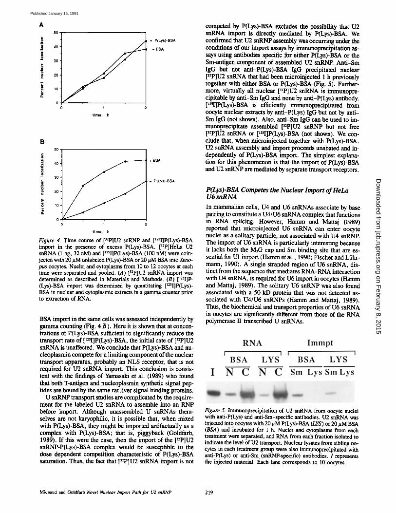
competed by P(Lys)-BSA excludes the possibility that U2 snRNA import is direcdy mediated by P(Lys)-BSA. We comSrmed that U2 snRNP assembly was occurring under the conditions of our import assays by immunoprecipitation as- says using antibodies specific for either P(Lys)-BSA or the Sin-antigen component of assembled U2 snRNP. Anti-Sin IgG but not anti-P(Lys)-BSA IgG precipitated nuclear [32P]U2 snRNA that had been microinjected 1 h previously together with either BSA or P(Lys)-BSA (Fig. 5). Further- more, virtually all nuclear p2p]u2 snRNA is immunopre- cipitable by anti-Sm IgG and none by anti-P(Lys) antibody. [mI]P(Lys)-BSA is efficiently immunoprecipitated from oocyte nuclear extracts by anti-P(Lys) IgG but not by anti- Sm IgG (not shown). Also, anti-Sm IgG can be used to im- munoprecipitate assembled p2P]U2 snRNP but not free pzP]U2 snRNA or [mI]P(Lys)-BSA (not shown). We con- clude that, when microinjected together with P(Lys)-BSA. U2 snRNA assembly and import proceeds unabated and in- dependently of PfLys)-BSA import. The simplest explana- tion for this phenomenon is that the import of P(Lys)-BSA and U2 snRNP are mediated by separate transport receptors.
P(Lys)-BSA Competes the Nuclear Import of HeLa U6 snRNA
In mammalian cells, U4 and U6 snRNAs associate by base pairing to constitute a U4/U6 snRNA complex that functions in RNA splicing. However, Hamm and Mattaj (1989) reported that microinjected U6 snRNA can enter oocyte nuclei as a solitary particle, not associated with U4 snRNP. The import of U6 snRNA is particularly interesting because it lacks both the M3G cap and Sm binding site that are es- sential for U1 import (Hamm et al., 1990; Fischer and Liihr- mann, 1990). A single stranded region of U6 snRNA, dis- tinct from the sequence that mediates RNA-RNA interaction with U4 snRNA, is required for U6 import in oocytes (Hamm and Mattaj, 1989). The solitary U6 snRNP was also found associated with a 50-kD protein that was not detected as- sociated with U4/U6 snRNPs (Hamm and Mattaj, 1989). Thus, the biochemical and transport properties of U6 snRNA in oocytes are significantly different from those of the RNA polymerase II transcribed U snRNAs.
Figure 5. Immunoprecipitation of U2 snRNA from oocyte nuclei with anti-P(Lys) and anti-Sm-spocific antibodies. U2 snRNA was injected into oocytes with 20/zM P(Lys)-BSA (LYS) or 20 #M BSA (BSA) and incubated for 1 h. Nuclei and cytoplasms from each treatment were separated, and RNA from each fraction isolated to indicate the level of U2 transport. Nuclear lysates from sibling oo- cytes in each treatment group were also immunoprecipitated with anti-P(Lys) or anti-Sm (snRNP-specific) antibodies. I represents the injected material. Each lane corresponds to 10 oocytes.
Michaud and Goldfarb Novel Nuclear Import Path for U2 snRNP 219
on February 8, 2015
jcb.rupress.orgD
ownloaded from
Published January 15, 1991

1.2 U2snRNA
1.0
O '~ 0.8
0.6 • P(Lys) -BSA
'~ 0.4 U6 snRNA m
0.2
0.0 J ' 1 '5 0 5 10 20
p.M competitor
Figure 6. Dose dependent inhibition of U2 import by P(Lys)-BSA. 32p-labeled HeLa U2 (open triangles) and U6 snRNA (open squares) and nsI-P(Lys)-BSA (closed squares) were coinjected into Xenopus oocytes with increasing concentrations of competitor P(Lys)-BSA, and the import of each karyophile determined after 1 h incubation as described in Materials and Methods. Import of each karyophile was normalized to its import in the presence of equimolar concentrations of BSA.
In our hands, U6 snRNA import into oocyte nuclei is less efficient than is U2 import. In one trial, 18% of microinjected [32p]u6 snRNA and 42 % of [32p]u2 snRNA accumulated in nuclei after 3 h. tRNA, for comparison, is virtually excluded from nuclei after this period (Fig. 1 B). Using [32p]u6 snRNA, we conducted competition experiments in Xenopus oocytes with P(Lys)-BSA. We coinjected [tESI]P(Lys) BSA, HeLa [32p]u2 snRNA, and HeLa [32p]u6 snRNA with P-(Lys)-BSA or BSA into groups of 30 oocytes and monitored the transport of each type of molecule after 1 h. Low concen- trations of P(Lys)-BSA (0.5 #M) did not significantly affect the import of [~25I]P(Lys)-BSA, U2 snRNA, or U6 snRNA compared to controls coinjected with 0.5 #M BSA (see be- low). Significantly, however, 20 #M P(Lys)-BSA decreased both U6 snRNA and P(Lys)-BSA import threefold as com- pared with the controls but, as expected, had no significant effect on U2 RNA import. This can be seen in the dose depen- dent effects of increasing concentrations of P(Lys)-BSA on the import of [32p]u2 and U6 snRNA and [~25I]-P(Lys)-BSA (Fig. 6). Because [32p]u6 snRNP is imported more slowly than [32P]U2 snRNE we incubated the oocytes for 1 h, in- stead of 45 min as in Fig. 3, to allow more transport to occur. Thus, because transport proceeds even in the presence of competitor, albeit more slowly, the apparent inhibition of [~2sI]P(Lys)-BSA as a function of P(Lys)-BSA is less in Fig. 6 than in Fig. 3.
It is possible that P(Lys)-BSA acts by inhibiting the assem- bly of the U6 snRNP rather than as a transport competitor. Because the biochemistry of the U6 snRNP is poorly under- stood, U6 snRNP-specific antibodies are not yet available. Presently, therefore, we cannot directly monitor the in situ assembly of the U6 snRNE For this reason, we restrict our conclusions to the import of the U6 snRNA and not the snRNE We have attempted to determine whether or not P(Lys)-BSA associates strongly with the U6 snRNA before injection by gel retardation assay (Konarska, 1989). Al- though a small fraction of U2 snRNA can be shown to associ- ate with P(Lys)-BSA by gel retardation, the migration of U6
snRNA in gels in unaffected by preincubating the RNA in 0.2 mM P(Lys)-BSA. Thus, it is unlikely that if P(Lys)-BSA is preventing U6 snRNP assembly it is doing so by complexing the RNA. We also asked whether the coinjection of P(Lys)- BSA with U6 snRNA has any effect on its sedimentation in glycerol gradients. In this experiment, [32p]u6 snRNA was coinjected into oocyte cytoplasms with either BSA or P(Lys)- BSA, incubated for 1 h, after which time whole oocyte ex- tracts were prepared. Glycerol gradient analysis of these extracts indicated that P(Lys)-BSA had no effect on the sed- imentation of the [32p]u6 snRNA (data not shown). Neither of these experiments is a good assay for U6 snRNP assembly, thus, we can not rigorously exclude the possibility that P(Lys)-BSA interferes with U6 snRNP assembly. But we think it is unlikely.
Discussion
In the first part of this study, we showed by kinetic criteria that two karyophilic proteins, P(Lys)-BSA and nucleoplas- min, compete for the same limiting component of the nuclear transport apparatus. We believe that these two proteins are representative of a much larger, general class of karyophiles that are imported by the cell's predominant import pathway. We have also observed the inhibition of [t25I]calf thymus histone H1 import in oocytes by P(Lys)-BSA and the inhibi- tion of [t2~I]nucleoplasmin import by H1 (Breeuwer and Goldfarb, unpublished observations). The ability of P(Lys)- BSA to compete the import of [nSI]nucleoplasmin at lower concentrations than those required to compete [~I]P(Lys)- BSA import (Fig. 3) can be explained by differences in their respective affinities for a common receptor. At 45 rain, the import of P(Lys)-BSA is reduced by half in the presence of ~10/~M P(Lys)-BSA, whereas <3 #M P(Lys)-BSA is re- quired to compete nucleoplasmin import to the same extent. Differences in the K, values of these two substrates for their common import receptor may differ by as much as an order of magnitude to account for this difference. What is the molecular basis for this difference? First, the NLS of nu- cleoplasmin is complex and contains an element(s) that ap- pears to be related to the T-antigen NLS motif (Burglin and De Robertis, 1987; Dingwall et al., 1988) but only insofar as they both contain clusters of lysine residues. It is difficult to envision a receptor recognizing such different sequences, present in the case of P(Lys)-BSA as a synthetic peptide, with identical affinities. Second, and perhaps more impor- tantly, the presence of increasing numbers of NLS on the sur- face of a protein can increase the rate and extent of its trans- port (Lanford et al., 1990; Lanford et al., 1988; Roberts et al., 1987; Dworetzky et al., 1988). Although nucleoplasmin is a pentamer, the P(Lys)-BSA used for the present study has 12-17 NLS peptides/monomer. As the local concentration of signals increases on the surface of the karyophile, a previ- ously bound receptor protein, upon dissociating, will have a statistically greater chance of reassociating with a nearby signal on the same protein resulting in higher association rate constants. The dissociation rate constant for receptor-signal complexes should not be influenced by multivalency, but the overall effect will be to increase the equilibrium binding con- stant. Thus, assuming each receptor has but one NLS- binding site, positive multivalency can be produced by signal proximity effects. Alternatively, as previously suggested
The Journal of Cell Biology, Volume 112, 1991 220
on February 8, 2015
jcb.rupress.orgD
ownloaded from
Published January 15, 1991

2
5
4
Key ~ nuclear Sm snRNP
pore O particle r"~::~ P(Ly s)_BSA ~ cytoplasmic
adaptor
Figure 7. Diagram of import models.
(Goldfarb, 1989), positive multivalency may be achieved by the simultaneous binding of more than one NLS receptor to multivalent karyophiles. This could occur in the cytoplasm or on the central transporter assembly where multiple karyophile binding sites have been demonstrated (Akey and Goldfarb, 1990).
The key finding of this study is that 132 snRNP import oc- curs independently of a limiting factor that is required for P(Lys)-BSA, nucleoplasmin and U6 snRNP import. What distinguishes the import of U2 snRNP from that of U6 snRNP? Recent evidence indicates that both the M3G cap and Sm antigen binding site of U1 are critical for nuclear im- port (Hamm et al., 1990; Fischer and Ltihrmann, 1990). U6 snRNA lacks both of these features although it is imported when artificially transcribed with a M3G cap, but not when its single stranded region is deleted (Hamm et al., 1990). Furthermore, Fischer and Ltihrmann (1990) were able to in- hibit U1 import with free M3G cap. These results together with the findings of the present study suggest that the M3G cap may have a signaling functioning distinct from the SV40 large T-antigen type of NLS. Alternatively, the M3G cap may not be a proper signal but instead binds a factor essential for the karyophilic activation of the snRNP.
How can multiple import pathways exist in a cell whose only portal to the nucleus is the NPC? Four of the more likely models are compared in Fig. 7. Based on the lack of competi- tion between 102 snRNP and P(Lys)-BSA import each model
must provide unoccupied NPCs for U2 snRNP import in the presence of saturating P(Lys)-BSA concentrations.
In model 1, which we favor, distinct cytoplasmic adaptors mediate the targeting of the two karyophiles to the nuclear envelope. Each adapter has two domains: a karyophile- specific NLS binding domain, and a NPC binding domain. Free karyophile, in excess over its adapter, is unable to directly bind the NPC. In this model, the amount of P(Lys)- BSA receptor/adapter and not the NPC is limiting. Thus in the presence of saturating P(Lys)-BSA concentrations its adaptor, but not the NPC, becomes saturated. Cytoplasmic adaptors/signal receptors are known to function in other membrane transport pathways (Bernstein et al., 1989). SRP is a well characterized adapter/cytoplasmic receptor that mediates the targeting of all start-transfer signal-sequence- containing proteins to the ER membrane. The proposition that cytoplasmic adapters act as primary NLS receptors has received support from a number of laboratories (Yamasaki et al., 1989; Breeuwer and Goldfarb, 1990; Newmeyer and Forbes, 1990; Adam et al., 1990). What would be the func- tion of cytoplasmic adaptors in nuclear transport? A major role in SRP in membrane transport is to maintain the translo- cation competence of the nascent polypeptide (Bernstein et al., 1989), which is not a requirement for nuclear import. The present study suggests that the role of putative cytoplas- mic receptors in nuclear transport may be to regulate the ac- cess of multiple karyophile classes to a relatively small num- ber of equivalent NPCs. Cytoplasmic NLS receptors may also prevent the passive diffusion of small karyophilic pro- teins through the nuclear pore (Breeuwer and Goldfarb, 1990).
In certain circumstances, translocation competent signal peptides can bypass the requirement for SRP and bind directly to an ER membrane-associated signal sequence receptor (Walter, 1987). A similar phenomenon could also occur in nuclear import. Thus, model 2 allows U2 snRNP, but not P(Lys)-BSA, to bypass the adaptor step and bind directly to the NPC. Model 2 predicts that P(Lys)-BSA im- port would be competed by saturating concentrations of U2 snRNP, but not vice versa.
In model 3, each NPC contains separate binding sites for each class of karyophile. The use of cytoplasmic NLS adap- tors is not excluded by this model; however, in this case, each adaptor would bind distinct NLSs and distinct sites at the NPC. An analogous situation occurs in mitochondrial pro- tein targeting where multiple high affinity receptors in the outer membrane mediate protein import (Hartl, 1989). The existence of multiple, spatially distinct, karyophile binding sites (peripheral binding and central docking sites) within the NPC central transporter assembly would allow for this mechanism (Akey and Goldfarb, 1989; Richardson et al., 1988; Newmeyer and Forbes, 1988).
In model 4, the nuclear envelope is studded with function- ally distinct NPCs. Each karyophile class has a cognate NPC class. Although this model is consistent with our kinetic data, binding data argue strongly against this model. By electron microscopy, all the NPCs visible in extensive fields were observed to bind nucleoplasmin-coUoidal gold or P(Lys)-BSA-colloidal gold (Feldherr et al., 1984; Richard- son et al., 1988; Newmeyer and Forbes, 1988; Akey and Goldfarb, 1989).
Michaud and Goldfarb Novel Nuclear Import Path for U2 snRNP 221
on February 8, 2015
jcb.rupress.orgD
ownloaded from
Published January 15, 1991

In conclusion, the present data suggest that U2 snRNP and P(Lys)-BSA use kinetically distinct nuclear import pathways. If P(Lys)-BSA, nucleoplasmin, and U6 snRNP belong to one class of karyophile and U2 snRNP to another, then we may ask, how many karyophilic macromolecules belong to each class and how many total classes exist? At one extreme, snRNPs that contain either Sm antigens or M3G caps, or both, may represent a unique and rather small family of karyophiles that are exceptional in that they do not use the cell's predominant import pathway. At the other extreme, the cell may have evolved a large number of independently regu- lated import pathways, each with its own characteristic NLS and receptor apparatus. The SV40 large T-antigen NLS, which can direct import in yeast and higher ceils, appears to be a member of a functionally conserved class of signals. Kinetic experiments are underway to investigate exactly how large this class is and if there are many other karyophiles like U2 snRNP that fall into other classes.
We thank Drs. Sallie Hoch and Harold Smith for providing anti-Sin anti- bodies, Dr. William Wasserman for advice regarding oocytes and their microinjection, Dr. Robert Angerer for closely reading the manuscript, Dr. lan Mattaj and Utz Fischer for sharing results before publication, and Roger Kornberg for helpful discussions.
This work was supported by U.S. Public Health Services FIRST and BRSG Awards, and Basil O'Connor Starter Scholar Research Award No. 5-730.
Received for publication 23 August 1990 and in revised form 9 October 1990.
References
Adam, S. A., R. S. Mart, and L. Gerace. 1990. Nuclear protein import in per- meabilized mammalian cells requires soluble cytoplasmic factors. J. Cell Biol. 111:807-816.
Adam, S. A., T. J. Lobl, M. A. Mitchell, and L. Gerace. 1989. Identification of specific binding proteins for a nuclear localization sequence. Nature (Lond.). 337:276-279.
Akey, C. W. 1990. Visualization of transport-related configurations of the nu- clear pore transporter. Biophys. J. 58:341-355.
Akey, C. W., end D. S. Goldfarb. 1989. Protein import through the nuclear pore complex is a multistep process. J. Cell Biol. 109:971-982.
Bach, M., G. Winkelrnann, and R. Luhrmann. 1989.20S small nucleoprotein U5 shows a surprisingly complex protein composition. Proc. Natl. Acad. Sci. USA. 86:6038-6042.
Bataill6, N., T. Helser, and H. M. Fried. 1990. Cytoplasmic transport of ribo- somal subunits microinjected into the Xenopus laevis oocyte nucleus: a gen- eralized, facilitated process. J. Cell Biol. 111:1571-1582.
Benditt, J. O., C. Meyer, H. Fasold, F. C. Barnard, and N. Reidel. 1989. Inter- action of a nuclear location signal with isolated nuclear envelopes and identification of signal-binding proteins by photoaffinity labeling. Proc. Natl. Acad. Sci. USA. 86~9327-9331.
Bernstein, H. D., T. A. Rappaport, and P. Walter. 1989. Cytosolic protein translocation factors. Is SRP still unique? Cell. 58:1017-1019.
Borer, R. A., C. F. Lehner, H. M. Eppenberger. and E. A. Nigg. 1989. Major nucleolar proteins shuttle between nucleus and cytoplasm. Cell. 56:379-390.
Breeuwer, M., and D. S. Goldfarb. 1990. Facilitated nuclear transport of his- tone H1 end other nuclcophilic proteins. Cell. 60:999-1008.
Burglin, T. R~, and E. M. De Robertis. 1987. The nuclear migration signal of Xenopus laevis nocleoplasmin. EMBO (Fur. Mol. Biol. Organ.) J. 6: 2617-2622.
Chelsky, D., R. Ralph, and G. Jonak. 1989. Sequence requirements for syn- thetic peptide-mediated translocation to the nucleus. Mol. Cell. Biol. 9: 2487-2492.
De Robertis, E. M., S. Lienhard, and R. F. Parisot. 1982. Intracellular trans- port of microinjected 5S end small nuclear RNAs. Nature (Lond.). 295:572-577.
Dingwall, C., and R. A. Laskey. 1986. Protein import into the cell nucleus. Annu. Rev. Cell Biol. 2:367-390.
Dingwall, C., J. Robbins, S. M. Dilworth, B. L. Roberts, and W. D. Richard- son. 1988. The nucleoplasmin nuclear location sequence is larger and more complex than that of SV40 large T antigen. J. Cell Biol. 107:841-849.
Dreyer, C., and P. Hausen. 1982. Two-dimensional gel analysis of the fate of oocyte nuclear proteins in the development of Xenopus laevis. Dev. Biol. 100:412-425.
Dworetzky, S. I., R. E. Lanford, and C. M. Feldherr. 1988. The effect ofvaria-
tions in the number and sequence of targeting signals on nuclear uptake. J. Cell Biol. 107:1279-1288.
Feldhetr, C. M., E. Kallenbach, and N. Sehultz. t984. Movement of a karyophilic protein through the nuclear pores of oocytes. J. Cell Biol. 99:2216-2222.
Finlay, D. R., D. D. Newmeyer, P. M. Hartl, L Horecka, and D. J. Forbes. 1989. Nuclear transport in vitro. J. Cell Sci. Suppl. 11:225-242.
Fischer, U., and R. Luhrmann. 1990. An essential signaling role for the m3G cap in the transport of U1 snRNP to the nucleus. Science (Wash. DC). 249:786-790.
Gerace, L., and B. Burke. 1988. Functional organization of the nuclear enve- lope. Annu. Rev. Cell Biol. 4:335-374.
Goldfarb, D. S. 1989. Nuclear Transport. Cart. Opin. Cell Biol. 1:441-446. Goldfarb, D. S., J. Gariepy, G. Schoolnik, and R. D. Kornberg. 1986. Syn-
thetic peptides as nuclear localization signals. Nature (Lond.). 322:641-644. Gusse, M., J. Gbysdael, G. Even, T. Soussi, and M. Mechali. 1989. Transloca-
tion of a store of maternal cytoplasmic c-myc protein into nuclei during early development. Mol. Cell. Biol. 9:5395-5403.
Hamm, J., and I. W. Mattaj. 1989. An abundant U6 snRNP found in germ cells and embryos of Xenopus laevis. EMBO (Fur. Mol. Biol. Organ.) J. 8:4179-4187.
Harem, J., E. Darzynkiewicz, S. M. Tahara, and I. W. Mattaj. 1990. The trimethylgnanosine cap structure of U1 snRNA is a component of a bipartite targeting signal. Cell. 62:569-577.
Hartl, F.-U., N. Planner, D. W. Nicholson, and W. Neupert. 1989. Mitochon- drial protein import. Biochem. Biophys. Acta. 988:1-45.
Hunt, T. 1989. Cytoplasmic anchoring proteins and the control of nuclear local- ization. Cell. 59:949-951.
Khanna-Gupta, A., and V. C. Ware. 1989. Nucleocytoplasmic transport of ribosomes in a eukaryotic system: is there a facilitated transport process? Proc. Natl. Acad. Sci. USA. 86:1791-1795.
Konarska, M. M. 1989. Analysis of splicing complexes and small nuclear ribo- nocleoprotein particles by native gel electrophoresis. Methods EnzymoL 180:442-453.
Lanford, R. E., P. Kanda, and R~ C. Kennedy. 1986. Induction of nuclear trans- port with a synthetic peptide homologous to the SV40 antigen transport sig- nal. Cell. 46:575-582.
Lanford, R. E., R. G. White, R. G. Dunham, and P. Kanda. 1988. Effect of basic and nonbasic amino acid Substitutions on transport induced by simian virus 40 T-antigen synthetic peptide nuclear transport signals. Mol. Cell. Biol. 8:2722-2729.
Lanford, R. E., C. M. Feldherr, R. G. White, R. G. Dunham, and P. Kanda. 1990. Comparison of diverse transport signals in synthetic peptide-induced nuclear transport. Exp. Cell Res. 186:32-38.
Lee, W. C., and T. Melese. 1989. Identification and characterization of a nu- clear localization sequence-binding protein in yeast. Proc. Natl. Acad. Sci. USA. 86:8808-8812.
Li, R., and J. O. Thomas. 1989. Identification of a human protein that interacts with nucle.ar localization signals. J. Cell Biol. 109:2623-2632.
Liihrmann, R. 1988. snRNP Proteins. In Structure and Function of Major and Minor Small Nuclear Ribenucleoprotein Particles. M. L. Birnstiel, editor. Springer-Verlag, Berlin. 71-99.
Mattaj, I. W. 1986. Cap trimethylation of U-snRNA is cytoplasmic and depen- dent on U-snRNP protein binding. Cell. 46:905-911.
Mattaj, L W. 1988. UsnRNF assembly and transport. In Structure and Function of Major and Minor Small Nuclear Ribonocleoprotein Particles. M. L. Birn- stiel, editor. Springer-Verlag, Berlin. 100-114.
Mattaj, I. W., and E. M. De Robertis. 1985. Nuclear segregation of U2 snRNA requires binding of specific snRNF proteins. Cell. 40:111-118.
Newmeyer, D. D., and D. J. Forbes. 1988. Nuclear import can be separated into distinct steps in vitro: nuclear pore binding and translocation. Cell. 52: 641-653.
Newmeyer, D. D., and D. J. Forbes. 1990. An N-ethylmaleimide-sensitive cy- tosolic factor necessary for nuclear protein import: requirement in signal- mediated binding to the nuclear pore. J. Cell Biol. 110:547-557.
Newport, J. W., and D. J. Forbes. 1987. The nucleus: structure, function, and dynamics. Annu. Rev. Biochem. 56:535-563.
Nigg, E. A. 1990. Mechanisms of signal transdoction to the cell nucieus. Can- cer Res. 55:271-310.
Peters, R. 1986. Fluorescence microphotolysis to measure nucleocytoplasmic transport and intracellular mobility. Biochem. Biophys. Acta. 864:305-359.
Reddy, R., and H. Busch. 1988. Small nuclear RNAs: RNA sequences, struc- ture and modifications. In Structure and Function of Major and Minor Small Nuclear Ribonuclcoprotein Particles. M. L. Birnstiel, editor. Springer- Verlag, Berlin. 1-37.
Richardson, W. D., A. D. Mills, S. M. Dilworth, R. A. Laskey, and C. Ding- wall. 1988. Nuclear protein migration involves two steps: rapid binding at the nuclear envelope, followed by slower translocation through nuclear pores. Cell. 52:655-664.
Roberts, B. L., W. D. Richardson, end J. A. Smith. 1987. The effect of protein context on nuclear location signal function. Cell. 50:465-475.
Roth, S., D. Stein, and C. Nusslein-Vollhard. 1989. A gradient of nuclear local- ization oftbe dorsal protein determines dorsoventral paRern in the Drosoph- ila embryo. Cell. 59:1189-1202.
Rushlow, C. A., K. Hart, J. L. Manley, and M. Levine. 1989. The graded dis-
The Journal of Cell Biology, Volume 112, 1991 222
on February 8, 2015
jcb.rupress.orgD
ownloaded from
Published January 15, 1991

tribution of the dorsal morphogen is initiated by selective nuclear transport in Drosophila. Cell. 59:1165-1177.
Sauterer, R. A., R. J. Feeney, and G. W. Zieve. 1988. Cytoplasmic assembly of snRNP particles from stored proteins and newly transcribed snRNAs in L929 mouse fibroblasts. Exp. Cell Res. 176:344-359.
Silver, P., L Sadler, and M. A. Osborne. 1989. Yeast proteins that recognize nuclear localization sequences. J. Cell Biol. 109:983-989.
Singh, R., and R. Reddy. 1989. -y-Monomethyl phosphate: a cap structure in splicesomal U6 small nuclear RNA. Proc. Natl. Acad. Sci. USA. 86: 8280-8283.
Steward, R. 1989. Relocalization of the dorsal protein from the cytoplasm to the nucleus correlates with its function. Cell. 59:1179-1188.
Taylor, M. V., M. Gusse, G. I. Evan, N. Dathan, andM. Mechali. 1986. Xeno- pus rayc proto-oncogene during development: expression as a stable maternal
mRNA uncoupled from ceil division. EMBO (Fur. Mol. Biol. Organ.) J. 5:3563-3570.
Walter, P. 1987. Two receptors act sequentially. Nature (Lend.). 328:763-764. White, E. M., C. D. AUis, D. S. Goldfarb, A. Srivastva, L Weir, and M. A.
Gorovsky. 1989. Nuclear specific and temporally restricted localization of proteins in Tetrahymena macronuclei and micronuclei. J. Cell Biol. 109: 1983-1992.
Yamasaki, L., P. Kanda, and R. E. Lanford. 1989. Identification of four nuclear transport signal-binding proteins that interact with diverse transport signals. Mol. Cell. Biol. 9:3028-3036.
Zasloff, M. 1983. tRNA transport from the nucleus in a eukaryotic cell: carrier- mediated translocation process. Prec. Natl. Acad. Sci. USA. 80:6436-6440.
Zieve, G. W., and R. A. Sauterer. 1990. Cell biology of the snRNP particles. CRC Crit. Rev. 25:1-46.
Michaud and Goldfarb Novel Nuclear Import Path for U2 snRNP 223
on February 8, 2015
jcb.rupress.orgD
ownloaded from
Published January 15, 1991