High resolution pHe imaging of rat glioma using pH-dependent relaxivity
Upload
independentCategory
view
0download
0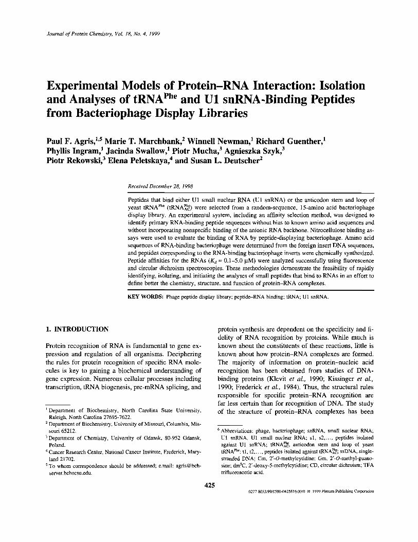
Journal of Protein Chemistry, Vol. 18, No. 4, 1999
Experimental Models of Protein-RNA Interaction: Isolationand Analyses of tRNAPhe and U1 snRNA-Binding Peptidesfrom Bacteriophage Display Libraries
Received December 28, 1998
Peptides that bind either U1 small nuclear RNA (U1 snRNA) or the anticodon stem and loop ofyeast tRNAphe (tRNAPhe AC) were selected from a random-sequence, 15-amino acid bacteriophagedisplay library. An experimental system, including an affinity selection method, was designed toidentify primary RNA-binding peptide sequences without bias to known amino acid sequences andwithout incorporating nonspecific binding of the anionic RNA backbone. Nitrocellulose binding as-says were used to evaluate the binding of RNA by peptide-displaying bacteriophage. Amino acidsequences of RNA-binding bacteriophage were determined from the foreign insert DNA sequences,and peptides corresponding to the RNA-binding bacteriophage inserts were chemically synthesized.Peptide affinities for the RNAs (Kd ~ 0.1-5.0 uM) were analyzed successfully using fluorescenceand circular dichroism spectroscopies. These methodologies demonstrate the feasibility of rapidlyidentifying, isolating, and initiating the analyses of small peptides that bind to RNAs in an effort todefine better the chemistry, structure, and function of protein-RNA complexes.
KEY WORDS: Phage peptide display library; peptide-RNA binding; tRNA; U1 snRNA.
1. INTRODUCTION
Protein recognition of RNA is fundamental to gene ex-pression and regulation of all organisms. Decipheringthe rules for protein recognition of specific RNA mole-cules is key to gaining a biochemical understanding ofgene expression. Numerous cellular processes includingtranscription, tRNA biogenesis, pre-mRNA splicing, and
1 Department of Biochemistry, North Carolina State University,Raleigh, North Carolina 27695-7622.
2 Department of Biochemistry, University of Missouri, Columbia, Mis-souri 65212.
3 Department of Chemistry, University of Gdansk, 80-952 Gdansk,Poland.
4 Cancer Research Center, National Cancer Institute, Frederick, Mary-land 21702.
5 To whom correspondence should be addressed; e.mail: [email protected].
protein synthesis are dependent on the specificity and fi-delity of RNA recognition by proteins. While much isknown about the constituents of these reactions, little isknown about how protein-RNA complexes are formed.The majority of information on protein-nucleic acidrecognition has been obtained from studies of DNA-binding proteins (Klevit et al., 1990; Kissinger et al.,1990; Frederick et al., 1984). Thus, the structural rulesresponsible for specific protein-RNA recognition arefar less certain than for recognition of DNA. The studyof the structure of protein-RNA complexes has been
6 Abbreviations: phage, bacteriophage; snRNA, small nuclear RNA;U1 snRNA, U1 small nuclear RNA; s1, s2,..., peptides isolatedagainst U1 snRNA; tRNAPheAc, anticodon stem and loop of yeasttRNAphe; t1, t2,..., peptides isolated against tRNAPheAC, ssDNA, single-stranded DNA; Cm, 2'-o-methylcytidine; Gm, 2'-O-methyl-guano-sine; dm5C, 2'-deoxy-5-methylcytidine; CD, circular dichroism; TFAtrifluoroacetic acid.
4250277-8033/99/0500-0425$16.00/0 C 1999 Plenum Publishing Corporation
Paul F. Agris,1,5 Marie T. Marchbank,2 Winnell Newman,1 Richard Guenther,1Phyllis Ingram,1 Jacinda Swallow,1 Piotr Mucha,3 Agnieszka Szyk,3Piotr Rekowski,3 Elena Peletskaya,4 and Susan L. Deutscher2

426 Agris et al.
hampered by certain inherent properties of RNA and thecorresponding RNA-binding proteins. RNA often doesnot adopt a well-defined structure in solution and is sus-ceptible to nuclease degradation. In addition, large-scalepurification of both RNA and RNA-binding proteins hasoften proven to be problematic. Nevertheless, the deter-minants of interaction between DNA-binding and RNA-binding proteins are likely to be, at least in part, distinctdue to inherent differences in DNA and RNA structure.RNA, for example, has a narrow major groove and awide minor groove (Delarue and Moras, 1989). In con-trast to DNA, RNA has many more alternative chemistriesand structures compared to the standard A-form duplex.RNA base modifications as well as single-stranded regionsand bulges are likely to be important in peptide binding ofRNA (Agris, 1996).
Some types of physicochemical interactions in-volved in protein-RNA associations have been illu-minated through well-studied protein-RNA complexes(Draper, 1995). Of particular note are the complexes ofaminoacyl-tRNA synthetase and cognate tRNA, espe-cially that of glutaminyl-tRNA synthetase and its cog-nate tRNAG/n, and the A protein with U1 small nuclearRNA (snRNA).6 X-ray crystallographic and biochemi-cal studies of the 553-amino acid glutaminyl-tRNA syn-thetase complexed with the 76-nucleotide tRNAG/n havedocumented the specific interaction of the protein withinthe minor groove of the RNA (Rould and Steitz, 1992).The importance of the RNA major groove, as well assingle-stranded regions, has been supported by studiesof the U1 small nuclear ribonucleoprotein (snRNP),which is involved in pre-mRNA splicing (Padgett et al.,1983). U1-snRNP consists of the 165-nucleotide U1snRNA, the U1-specific 70K, A, and C proteins, and theSm proteins (Steitz et al., 1988). The A and 70K pro-teins contain domains of approximately 100 amino acidsrequired for specific RNA recognition (Query et al.,1989; Mattaj, 1989; Kenan et al., 1991). Results fromthe x-ray study of crystals composed of the A protein-U1snRNA complex have demonstrated that the protein rec-ognizes a single-stranded loop of a hairpin structurewithin the RNA. A tyrosine in one peptide sequence ofthe A protein interacts with a cytosine in the hairpin'sloop and hydrogen bonding occurs between an argininein another peptide sequence of protein A and a guanine(Oubridge et al., 1994).
For the most part, however, the study of protein-RNA recognition and interaction has been hampered bythe size and complexities of the two macromolecules.The goal of the study reported here was to examine thefeasibility of isolating small peptides that bind to eitherU1 snRNA or tRNAphe in an effort to expedite structural
and functional analyses of the formed complexes. Theability to isolate small peptides that specifically bind to agiven RNA will enable us to elucidate the key amino acidsand structural determinants involved in polypeptide-RNAinteraction. Since their utility was first reported (Scottand Smith, 1990), display bacteriophage (phage) librarieshave been used often to isolate peptides and proteinsthat bind antibodies or receptors. The same librarieshave been used to study protein interactions with DNA(Calcutt et al., 1993; Krook et al., 1994), particularly forthe expression of DNA-binding zinc fingers on thesurface of the phage (Choo and Klug, 1994; Rebar andPaba, 1994;Wu et al., 1995;Rebar et al., 1996). The studyof peptide-RNA interactions has only recently been ap-proached using phage display libraries in combinationwith other methods for identifying and isolating thepeptides (Laird-Offringa and Belasco, 1996). The ap-proaches more often than not bias the selection to a posi-tively charged amino acid (Tan and Frankel, 1998) or toa particular structure, such as RNA-binding zinc fingers(Friesen and Darby, 1998).
We have identified RNA-binding peptides usingmethods designed to isolate such peptides from phagedisplay libraries without bias to amino acid sequence or aparticular protein structural motif. We also developed asensitive screening assay such that the RNA of choicecould be immobilized without destruction of secondarystructure and subsequently conducted affinity selection ofRNA-binding peptides. Peptides that bound U1 snRNAor tRNAphe were identified and physicochemical studiesbegun. Results demonstrate that bacteriophage librariesdisplaying random 15-amino acid peptide sequences willbe a good source of novel RNA-binding peptides.
2. MATERIALS AND METHODS
2.1. Materials
2.7.7. In Vitro Transcription of U1 snRNA
U1 snRNA was transcribed in vitro (Ambion'sMegaScript Sp6) and randomly biotinylated (BRL'sbiotin-14-CTP) (Marchbank and Deutscher, 1995). Theamount of biotin-14-CTP added to the transcription reac-tion was limiting in order to minimize the multiple biotinmolecule incorporation. The RNA product was purifiedby Sephadex column chromatography. The resulting U1snRNA retained binding to stem-loop-specific autoanti-bodies, as demonstrated by immunoprecipitation (March-bank and Deutscher, 1995), and therefore was likely to beof similar conformation to nonbiotinylated U1 snRNA.

Experimental Models of Protein-RNA Interaction 427
2.1.2. Synthesis of the tRNAPhe AnticodonStem-Loop Domain
The native, fully modified sequence of the tRNAphe
anticordon stem and loop (tRNAPheAc) contains 2'O-methylC32 and G34, Cm32, and Gm34 (at positions 32 and 34,respectively), a tricyclic derivative of G, wybutosine(Y37), pseudouridine (S39 and 5-methyl-C (m5C40). YeasttRNAphe was purchased from Sigma (St. Louis, MO). Forthis study an anticodon domain containing three of thefive modifications was chemically synthesized (Agriset al., 1995). The 17mer tRNAPheAC contained Cm32, Gm34,and a deoxy- instead of ribo-m5C40 (Chen et al., 1993) atlocations corresponding to positions in the native se-quence (Chen et al., 1993). This construct binds Mg2+
with high affinity, as does the native tRNA (Dao et al.,1994), and binds the ribosome in response to the propercodon (Dao et al., 1994). A biotin was attached to the 3'terminus during automated chemical synthesis.
2.1.3. Phage Libraries and Bacterial Strains
The 15-amino acid random peptide display library(fUSE 5) and the bacterial strain K91Kan were pro-vided by Dr. George P. Smith (University of Missouri-Columbia).
2.2. Affinity Selection for RNA-Binding PhageDisplaying Peptides
The random 15-amino acid phage display library wasaffinity-selected against either U1 snRNA or tRNAPheAC. Ingeneral, procedures were according to Smith and Scott(1993). Three levels of stringency were used, each levelcomprising one to three rounds of selection (Table I). Dur-ing the first level of stringency, performed in selectionrounds one to three, 35-mm Petri dishes were coatedovernight at 4°C with streptavidin (10 ug). The disheswere washed with TTBS (50 mM Tris HC1/150 mM NaCl,
pH 7.4, and 0.5% Tween-20) and blocked by filling thedish with a 3% bovine serum albumen solution (Smithand Scott, 1993). The plates were then reacted in the coldfor up to 3 hr with biotinylated U1 snRNA or tRNAPhe AC
(50 ng). The plates were then washed with TTBS and in-cubated with -1012 peptide displaying phage particles foran additional 4 hr at 4°C. Plates were washed (2-10times) and the phage retained by the RNAs were elutedwith acid (0.1 N HC1, pH 2.2) and neutralized to a pH ofapproximately 8 with base (2 M Tris), maintaining theirinfectivity (Smith and Scott, 1993). The eluted phagewere amplified by infection of E. coli K91 cells, and cul-tures were grown overnight (Smith and Scott, 1993).
In the second level of stringency, performed in thefourth round of selection, phage were reacted with bio-tinylated U1 snRNA or tRNAACPhe (5-50 ng) for 2 hr inthe cold, "captured" on the streptavidin plates for a brief10 min, washed, eluted, and amplified as above. Duringthe third level of stringency and the fifth round of se-lection, nucleic acid competitors (1 mM) consisting ofssDNA, rRNA, tRNA (for U1 snRNA selection), andU1 snRNA (for tRNA selection) were added to the phagebefore capture on the appropriate RNA. Selective en-richment of phage was monitored by calculating the per-centage yield after each round of selection (Table I).Phage were titered and yields calculated according toSmith and Scott (1993).
2.3. Filter Binding Analysis
Clonally purified phage from the more stringentlevels and fourth round of selection with U1 snRNAand with tRNAPheAC were adhered to nitrocellulose,blocked, rinsed, and incubated with biotinylated U1snRNA or tRNAPheAC. The filters were washed, incubatedwith streptavidin/alkaline phosphatase, washed, anddeveloped with the chromogenic substrate 5-bromo-4-chloro-3-indolyl phosphate /nitroblue tetrazolium(BCIP/NBT) (GIBCO, BRL).
Table I. Selective Enrichment of RNA-Binding Phage
Round
12345
Percent yielda
U1 snRNA
7.3 x ID-5
2.9 x 10⁻⁵
8.4 x 10⁻⁵
2.0 x 10⁻⁵
4.9 x 10⁻⁵
tRNAPheAC
1.9 x 10⁻⁶
6.0 x 10⁻⁵
2.0 x 10⁻⁴
4.0 x 10⁻⁵
4.9 x 10⁻⁵
Experimental variations
Two washesTen washesTen washesCapture/10 washesCapture/10 washes/competitors
aPercentage yield of phage following each round of screening, where % yield = (# ofphage eluted)/(# of input phage) x 100.

428 Agris et al.
2.4. DNA Sequencing
To determine the amino acid sequences of foreigninserts in the phage coat proteins that bound RNA, phagewere propagated on a small scale. Their DNA sequenc-ing was accomplished by a modified two-lane dideoxy-chain-termination method utilizing 32P-labeled oligo-nucleotide primer located 15 nucleotides from the phagepIII gene cloning site (Haas and Smith, 1994).
2.5. DNA Sequence Analysis
All of the DNA sequences for peptides found to bindeither U1 snRNA or yeast tRNAPheAc were compared tothe database of frequently observed false positives, cour-tesy of Dr. George P. Smith. The false positives are dueto phage directed against plastic, biotin, and other mate-rials in the protocol. Genbank and SwissProt databases,accessed through the National Center for BiotechnologyInformation, were searched for proteins with DNA se-quence homologies to the RNA-binding peptides selectedfrom the phage display libraries. The BLAST algorithmwas used in the search. Peptide sequences that bound theyeast tRNAPheAC were more rigorously analyzed againstthe yeast genome database. The alignment algorithm forthe search of the yeast genome was FASTA.
2.6. Peptide Synthesis
The putative U1 snRNA-binding peptides s1 and s2were synthesized on a Synergy Personal Peptide Synthe-sizer from Applied Biosystems (Foster City, CA). ThetRNAPheAC-binding peptides t2 and t3 and a control peptidewere synthesized on a Labortec SP60 Peptide Synthe-sizer (Labortec AG, Switzerland). Peptide synthesis andcleavage of newly synthesized peptides from the columnsupports were accomplished with standard methods(Songster and Barany, 1997). Once synthesized, the pep-tides were cleaved from the support, purified by reverse-phase HPLC on a preparative column (Vydac C18,15-20 um particle size; 25-40% acetonitrile in 0.1%TFA linear gradient, or isocratic at 22% acetonitrile,18 ml/min), and detected by UV absorbance.
2.7. Fluorescence Quenching Titrations
Fluorescence spectroscopy was based on the intrin-sic fluorescence from tryptophan residues in the se-lected peptides. The change in tryptophan fluorescence(quenching) upon ligand binding was monitored, thuspermitting a measurement of peptide-RNA interaction.
Titrations were performed using an SLM 8100 spectro-fluorimeter (Spectronic Instruments, Rochester, NY).Varying amounts of nucleic acid (deoxynucleotide, ribo-nucleotide, ssDNA, dsDNA, tRNA, U1 snRNA) wereadded to a known concentration of peptide (4-200 nM)in 2 ml of TBS (50 mM Tris HC1/150 mM NaCl, pH7.4). Peptide fluorescence was measured with 292 nmexcitation and 346 nm emission. The Kd was determinedfrom binding curves derived from the titrations using asingle-binding-site curve-fitting procedure, Kalidagraph(Komissarov et al., 1996).
2.8. Circular Dichroism Spectroscopy
Circular dichroism (CD) spectra of peptides aloneand with RNAs were obtained with a Jasco J600 spec-trapolarimeter (Jasco, Easton, MD) and at 15°C, unlessnoted otherwise (Venyaminov and Yang, 1996; Agris andBrown, 1995). The peptides had elliptic nulls between250 and 290 nm that allowed for direct observation ofchanges in the RNA spectrum with the addition of pep-tides. Spectra were collected with uM concentrations ofpeptides and RNAs in 10 mM Na2PO4 buffer (pH 7.2).tRNAPheAC with Cm32, Gm34, and dm5C40 (5-20 uM), butwithout 3'-terminal biotin, was titrated with peptide overthe range of 0-300 uM. The CD sample of tRNAPheAC wastitrated with peptide by adding the RNA solution consec-utively to larger amounts of lyophilized peptide. Thistechnique, successfully used in other binding studies(Dao et al., 1992; Agris et al. 1996), obviated the need tocorrect for effects of dilution on the CD spectra.
3. RESULTS
3.1. Design of Experimental System
Our objective was to design and test the feasibilityof an experimental system for identifying, isolating, andanalyzing small peptides that bind to RNA. One criterionwe required of the methods was that selection of peptidesfrom a phage display library should not be biased towardany particular amino acid sequence. Therefore, no pre-determined restraints, such as a selected set of polaramino acid codons for the phage coat protein insert, wereplaced on the phage display library. The only restraint onthe library was the length of the sequence, 15 amino acids.Another criterion placed on the selection system was thatit include affinity purification of the RNA-binding phage.To this end we incorporated the strong biotin-strepavidinreaction in order to attach the RNA to a solid support.

Fig. 1. Sequence and secondary structures of U1 snRNA and tRNAPheAC. Peptide libraries were probed with U1 snRNA that hadbeen transcribed in vitro and randomly biotinylated and with the anticodon domain of tRNAphe (tRNAPheAC), which was producedby automated chemical synthesis including biotin at the 3'-terminus and site-specifically placed modified nucleotides.
Experimental Models of Protein-RNA Interaction 429
The biotin-strepavidin reaction has been used success-fully in linking proteins, particularly antibodies, to solidsupports for the selection of phage that specifically bindthese proteins (Smith and Scott, 1993). U1 snRNA wasderivatized with biotin randomly throughout the nucleo-side sequence by including in its in vitro transcriptiona limited amount of biotin-14-CTP. In contrast, theanticodon stem and loop of yeast tRNAphe (tRNAPheAC)was biotinylated only at the 3'-terminus of the hepta-decamer sequence. In this manner, we were able to testboth methods of labeling RNA for biotin-strepavidinlinkages. A third criterion for the experimental systemwas that we be able to demonstrate its application tostructurally different RNAs with significantly differentbiological functions. Therefore, we chose as one ofthe two RNA targets the 165-nucleotide U1 snRNA. Be-cause U1 snRNA was produced by transcription in vitro,it was devoid of modified nucleotides (Fig. 1). The othertarget was the considerably smaller tRNAPheAC composedof only 17 nucleosides, including three site-selectivelyplaced modifications. tRNAPheAC forms the secondary struc-ture of a simple five-base paired stem and seven-memberedloop (Fig. 1; Chen et al., 1993). U1 snRNA and tRNAsdisplay many structural features distinctive of RNA, in-cluding single-strand loop and bulges, stem-loops, andbase modifications that are required for appropriate protein
recognition of the RNA. The two RNAs have been stud-ied extensively, but the physicochemical determinants ofprotein recognition have not been identified. Finally, wewanted to determine if fluorescence and CD spectro-scopies could be applied to investigating the interactionsof peptides selected from phage display libraries withtheir target RNAs.
3.2. Isolation of RNA-Binding PhageDisplaying Peptides
A phage library displaying random 15-amino acid se-quences was screened with either biotinylated U1 snRNAor tRNAPheAC (Fig. 1). Three levels of stringency were used,with each comprising one to three rounds of selection(Table I). The highest level of stringency incorporated se-lection against competing nucleic acids. Single-strandedDNA (ssDNA), rRNA, and tRNA were used in competi-tion with U1 snRNA for identifying U1 snRNA-bindingphage. U1 snRNA was used in competition with tRNAPheAC
for identifying tRNAPheAC-binding phage. Each competingnucleic acid was at a concentration of 1 mM. A dot blotanalysis of phage was performed after the fourth roundof selection with the eluted and amplified phage fromthe second level of stringency. This nitrocellulose filter

430 Agris et al.
binding analysis conducted with biotinylated U1snRNA or tRNAPheAC was a qualitative indicator of affin-ity and specificity (Fig. 2). The assay takes advantageof the biotin-labeled RNA targets for detection by useof strepavidin/alkaline phosphatase and a chromogenicsubstrate. Of all the clones analyzed with U1 snRNA,nine (s1, s2, s4, s53, s56, s58, s59, and s70) bound moststrongly to biotinylated U1 snRNA (Fig. 2A). Only fourclones (t2, t33, t52, and t72) of the tRNAPheAC selectionwere the most reactive clones with biotinylated tRNAPhe AC
(Fig. 2B). Clone t2 from the tUNAPheAC selection showedsome cross-reactivity to U1 snRNA (Fig. 2A). How-ever, clone t53, which was selected with tRNAPhe AC, butwas a relatively poor binder, bound U1 snRNA as wellas clone s1 (Fig. 2A). Clone s2, a strong binder fromthe U1 snRNA selection, did not cross-react withtRNAPheAC (Fig. 2B). Clone s60, a weak-binding clonefrom the U1 snRNA selection, also bound tRNAPheAC
weakly (Fig. 2B).
The foreign inserts of these clones as well as addi-tional clones from the fourth and fifth rounds of selectionwith U1 snRNA or tRNAPheAC were sequenced. The DNAof some clones could not be sequenced because of diffi-culty in propagating the phage. Unfortunately, this in-cluded a number of the tight-binding phage clones ana-lyzed by dot blot after the fourth round of selection. Listsof the 19 most representative amino acid sequences ofthe DNA inserts from the U1 snRNA selection and 15 ofthe most representative amino acid sequences of DNAinserts from the tRNAPheAC selection are shown in Table II.In addition, Table II contains a qualitative assessmentof the dot blot analyses from the fourth round of selectionin which these peptide-bearing phage bound either U1snRNA or tRNAPheAC. Sequences that appear in the fifthround of selection should be present also in the fourthround. However, since not all clones were sequenced, onlysome of the sequences found in the fifth round also ap-peared in the fourth. Identical sequences found between
Fig. 2. RNA binding of affinity-selected phage. (A) U1 snRNA-binding to 22 selected phage. Twenty-one of the cloned phage represent phageindependently affinity-selected against U1 snRNA. In addition, negative (N) phage were clonally purified phage before affinity selection. The chart tothe right of the dot blot identifies the individual clones by number. The dash-lined mask corresponding to this chart has been superimposed on the dotblot. Clones 1 (s1) and 2 (s2) exhibited the strongest RNA-binding signals. Clones "2 tRNA" and "53 tRNA" represent clones numbered t2 and t53,respectively, and were selected in the tRNA screening. These clones exhibited some affinity for U1 snRNA. (B) tRNAPheAC-binding to 21 tRNAPheAC-selectedphage and negative phage. Clones were probed with biotinylated tRNAPheAC. Clones 2 (t2) and 33 (t33) reacted the strongest to this RNA. Clones "2 U1RNA" and "60 U1 RNA" represent clones numbered s2 and s60, respectively, and were selected in the U1 snRNA screening. Clone s2 had no affinityfor tRNAPheAC, while s60 exhibited some affinity for tRNAPheAC. Phage used as negative controls (N) were the same as above. The binding of the biotinylatedRNAs by phage on nitrocellulose filters was assessed with strepavidin-conjugated alkaline phosphatase and enzyme substrate BCIP/NBT. Some clonesprobed in this manner were found after sequencing of the phage DNA to have identical sequences (see frequency in Table II), while others were notfurther analyzed due to difficulty in propagation.

Experimental Models of Protein-RNA Interaction 431
rounds of selection are indicated. A more diverse pool ofclones was isolated against U1 snRNA than tRNAPheAC,perhaps reflecting the smaller size and modifications oftRNAPheAC. None of the 15-amino acid sequences appearedmore than eight times in a round of selection for peptidesbinding U1 snRNA, whereas one sequence, that of t2,appeared 28 and 42 times in the fourth and fifth roundsof selection, respectively, of peptides binding tRNAPheAC.Many of the sequences share the properties of havingtryptophans toward the center and serines near the ter-mini. This was particularly true of those phage that boundU1 sRNA or tRNAPheAC, respectively, more strongly in thedot blot analysis.
3.3. Characterization of Binding Propertiesof Phage Clones
A small number of peptides corresponding to clonesfound in the fourth and fifth rounds of the selection weresynthesized and tested for binding to RNA. Peptides s1,s2, t2, and t3 were chosen because these peptides weregood binders (Fig. 2 and Table II) and their sequencesappeared among the most frequently found clones that
bound U1 snRNA and tRNAPheAC, respectively. Peptidesfrom the selection against U1 snRNA (Table II) were ex-amined for their ability to bind U1 snRNA, native tRNA,and poly(U) using fluorescence quenching. The bindingof tRNAPheAC-selected peptides to tRNAPheAC, native tRNAphe,and a DNA analog was studied using circular dichroism(CD) spectroscopy. Fluorescence quenching of trypto-phan residues is a direct method of detecting interactionof the peptide with the RNA. In contrast, monitoringchanges in the CD spectrum of the RNA with additionsof peptide is an indirect method of determining the bind-ing of peptide to the RNA. Previous studies from one ofour laboratories had demonstrated fluorescence quench-ing of tryptophan residues upon the binding of DNA(Komissarov et al., 1996). The change in fluorescence ac-curately reflected the affinity of the interaction. The otherlab had demonstrated that changes in the CD spectrum oftRNAPheAC were a measure of the RNA binding of Mg2+
(Chen et al., 1993) and of anticodon-codon interaction(Agris et al., 1996). The larger the RNA, the less likelya measurable change in CD ellipticity would be detectedduring peptide binding. Therefore, the size of the U1snRNA could preclude its use in the CD analyses. The
Table II. Peptide Sequences of Selected Phage
Clone Frequency
Fourth round
Amino acid sequence
A. Fourth and Fifth Rounds of Selection Against U 1 snRN A
s1s2s4s10s15s19s21s40s52s60
4341111118
AGLHLFWSYSSYVTPSGPFIWNFWLGARAVWVSHFSVFAFPMSSWRLLVFRYWNLFPNVSGVRVPFWEGLEFFTLGSLIVPEWHLAPPGLTNLPPIFYDLHCFAFSGWSAFYNVAFFSTRRNLPPIFNDVYWIAFGYARTFWLPGLWSVP
B. Fourth and Fifth Rounds of Selection Against tRNAPhe
t2t5t9t11t13t15t17t27t33t34
28111111111
SISPWGFSGLLRWSYTYALTSSILVYLTSSEVPRLSLLAVSLVANTVSAGIICSFLSVSCVFQFRASVGGSHTVIRLVCWRLGCVSPMGSKWPVAVTLAAGIRIPSVLSHVRTVVLFSRSLRYTLETRWAVLSVYLILFSYDFHGDYYFY
Dot blot resulta
+++++
++-
+/-
-
+
++
-
+
+++
+
+
+
-
+/-
+/-
-
++
-
Clone
s1b
s3s5s28s47s58s61s63s64s71
t2b
t3t4t8t14t59
Fifth round
Frequency
8281131635
4251111
Amino acid sequence
AGLHLFWSYSSYVTPYRVSLSVLHNSLLPSFRSWMHFVTYVSTGGVVPRFWFWLHGPGPFVVRAFLGISLGLWSVGPQSATSCCYVSTGGAHLYFDSGWASRHWTEAWARWVFAWLWGHGWAYYHAGHSSFAVWTGYARTFLWPGLWSVP
SISPWGFSGLLRWSYAAGGFFRWIFSPVYVVSVYYGSFCLHSAYGNNHPWYYGRSHHVSGVFQFRASVGGSHTVITFRLLALWRGGLYPG
a Fourth-round clones were initially screened using dot blots (Fig. 2), whereas fifth-round clones were not. Intensity of each dot was determined sub-jectively and is indicated as being either strong (+++), moderate (++), weak (+), very weak (+/-), or absent (-) relative to the negative control.
b Indicates sequence was found in both the fourth and fifth rounds of screening.

432 Agris et al.
sizes of the two RNAs and experiences of the two labswere important factors in deciding which spectroscopywas used to investigate peptide-RNA interaction.
Fluorescence spectroscopy was applicable to pep-tides s1 and s2 (Table II) because each peptide containedat least one tryptophan residue. A series of titrations wasperformed with purified nonbiotinylated U1 snRNA, andwith native yeast tRNAphe as a control. The relative flu-orescence values were used to plot a saturation curve andhence to calculate the Kd of the complex from titrationisotherms. Peptide s1 did not appreciably bind the nu-cleotides examined, i.e., Kd > 5 uM (Fig. 3). Consistentwith the dot blot analyses, peptide s2 bound U1 snRNAwith a high affinity, Kd ~ 150 nM. The peptide alsobound poly(U) with a similar affinity, Kd ~ 100 nM, butwas somewhat specific for a particular RNA since itbound tRNAphe with considerably less affinity, Kd =5 uM (data not shown).
The interactions of tRNAPheAC with selected peptidest2 and t3 were evaluated with CD spectroscopy in orderto determine the usefulness of this technique. A thirdpeptide, SWHQWIWPSGQPLSE, was designed from acomputer analysis of binding peptides as a negative con-trol for these experiments. The peptides alone exhibitedan elliptic null in the CD spectrum at the wavelengthsmost characteristic of tRNA's CD spectrum, 250-290 nm(Fig. 4A). Thus, any change in the RNA spectrum at thosewavelengths with addition of peptide could be attributedto a peptide-RNA interaction. Both native tRNAphe andtRNAPheAC were titrated with peptides t2 and t3 and the con-
Fig. 3. Fluorescence titration of U1 snRNA-binding peptides s1 and s2with U1 snRNA. The titrations were performed using an SLM 8100spectrofluorimeter with well-documented procedures (Komissarovet al., 1996). A range of concentrations of U1 snRNA (1-1000 nM)was added to 50 nM peptide in 2 ml of TBS. Peptide fluorescence wasmeasured with 292 nm excitation and 346 nm emission. The Kd wasdetermined from the binding curve. Data represented were duplicateexperiments: (S) peptides s1 (C, C) s2.
trol. The prominent positive ellipticity of the RNAs(250-290 nm) decreased with increasing concentrations ofpeptides t2 and t3, but not by the control (Figs. 4B and 4c).Kd values for the binding of peptides t2 and t3 could notbe determined from this indirect method of observing in-teractions because the amounts of free and bound peptideare not known and difficult to determine. However, we as-sume that the Kd values were in the uM range becausechanges in CD were observed with low uM concentrationsof the peptides t2 and t3 interacting with 5 uM tRNAPheAC.Specificities of peptides t2 and t3 for RNA were testedwith a DNA analog of tRNAPheAC, tDNAPheAC(Guenther et al.,1992). No interaction was observed in CD spectra (notshown).
4. DISCUSSION
Specific RNA-binding peptides were found usingphage display, filter binding, and spectroscopy methodsdesigned for the rapid identification, isolation, and analy-ses of peptides. Results of the affinity selection experi-ments indicated that RNA-binding peptides could be eas-ily identified and isolated from the phage display libraryhaving coat protein inserts of a reasonable length, 15randomized amino acid residues. The sequences of theRNA-binding peptides were distinguishable from a grow-ing data bank of sequences that present as false positivesin phage assays, i.e., they were found to bind plastic(Adey et al., 1995), biotin or strepavidin (Kay et al.,1993), or other materials in the protocols. Selection ofpeptides from a library of randomized sequences com-posed of just six amino acid residues had been shown toresult in the identification of RNA-binding peptides thatwere without specificity for the RNA (Schimmel et al.,1997). Peptides specific for the RNA were identified(Frugier and Schimmel, 1997) only when the hexapep-tide variable region of the phage coat protein was flankedwith a stable RGG motif that has been credited with non-specific RNA binding (Burd and Dreyfuss, 1994). Thelibrary and methods we used were designed to identifyprimary RNA-binding peptide sequences without bias toeither a known RNA-binding sequence (Laird-Offringaand Belasco, 1996) or to positively charged lysine andarginine residues that nonspecifically bind the anionicRNA backbone.
The RNA-binding peptides we isolated containedan array of chemistries that will be easily probed for amore mature understanding of protein recognition ofRNA. Surprisingly, the identified peptides were com-posed mostly of nonpolar aliphatic and aromatic aminoacids (Table II). Though polar amino acids were present,

Experimental Models of Protein-RNA Interaction 433
Fig. 4. Detection of peptide-tRNAPheAC binding by CD spectroscopy.(A) CD spectra (at 15°C) of tRNAPheAC-binding peptides t2 and t3, acontrol peptide, and native, full-length tRNAPhe indicated that thepeptides had an elliptic null at the major positive ellipiticity of theRNA. Peptide and RNA concentrations in 10 mM phosphate buffer(pH 7.2) were ~30 and 5 uM, respectively. (B) Native yeast tRNAPhe
was titrated with peptide t2 at molar ratios of peptide to tRNA of 3/1,6/1, 12/1, and 24/1. (C) tRNAPheAC (20 uM) was titrated with t2 (•), t3(+), and control (*) peptides at molar ratios of peptide to RNAapproximating 3/1, 7/2, and 15/1. The percent change in CD ellipticityis plotted against the molar ratio of peptide to RNA.
they were for the most part uncharged residues. Lysinedid not appear once in the most frequent 19 sequencesderived from the fourth and fifth rounds of peptide se-lection against U1 snRNA, nor the most frequent 15 se-quences derived from selection against tRNAPheAC. Usuallyone residue, and no more than three, of arginine pre-dominantly and/or histidine appeared in any one peptidesequence. RNA-binding proteins have a very commonarginine-rich motif (ARM). Short-length ARM peptidesbind RNA with high affinity, though with reduced speci-ficity compared to full-length protein. The Tat protein andmimicking peptides binding to the frans-activation re-sponse element (TAR) of human immunodeficiency virus(HTV) is a good example of the ARM (Weeks et al., 1990;Calnan et al., 1991; Tao and Frankel, 1992). The arginine-containing consensus sequence of FGRA found in pep-tides that bind ssDNA and selected from a 23-residuerandom peptide library (Chang et al., 1996) did not appear
in any of the clones selected for binding to U1 snRNAand tRNAAc6. The U1 snRNA and tRNAPheAC-binding pep-tides were composed of hydrophobic amino acids in gen-eral. Whereas the few polar residues of s1 and s3 pep-tides binding U1 snRNA and t3 peptide binding tRNAPheAC
were dispersed in the sequences, the polar residues of thet2 peptide were toward the N- and C-termini (Table II).As with RNP proteins binding single-stranded RNA(Chang and Varani, 1997), selectivity could be impartedby the center hydrophobic region and RNA affinity bythe polar tarmini. The importance of these distributionscan be explored easily by alteration of the amino acidsequences.
Higher affinity sequences for both U1 snRNA andtRNAPheAC were rich in aromatics, particularly tryptophan.Of the phage selected for U1 snRNA binding, s1, s2, s4,s21, s40, and s60 were particularly good binders of U1snRNA in the dot blot analysis. It is intriguing that three

434 Agris et al.
(s1, s2, and s60) of the six sequences have a centered FWsequence and two (s21 and s40) have an FY sequence.Tryptophan was a common residue of the phage selectedagainst tRNAPheAC, but was not as consistently located nearthe center of the sequences as found for the U1 snRNA-binding phage. Whereas aromatic residues occupied thecenter of the peptide sequences that bound U1 snRNAtightly, aliphatic residues were more common near thecenters of sequences that bound tRNAPheAC. Also, phage se-lected against tRNAPheAC often had peptide sequences withserine the most common polar residue located toward theN- or C-termini or both. Within the sequences of five ofthe six clones that bound tRNAPheAC most tightly (t2, t5, t9,t1l, and t33), a leucine or isoleucine residue was presentwith two other aliphatic amino acids, GLL, ILV, LLA,GII, AVL. Clone t2, with the GLL sequence, was the mostcommonly found clone among all the clones subjected toDNA sequencing after the fourth and fifth rounds of se-lection. Clone t3, which did not have an aliphatic aminoacid sequence near the center of the 15-amino acid se-quence, had the second most common peptide sequenceamong the clones analyzed.
Peptides that bind the RNAs may provide a basisfor examining whether similar amino acids or physico-chemical characteristics exist in specific U1 snRNA andtRNAphe-binding proteins. Sequence and structural infor-mation exists for the U1 A protein bound to U1 snRNA(Oubridge et al., 1994). Although somewhat prematurefor lack of knowledge of each peptide's critical residuesfor binding the RNAs, we attempted Genbank and Swiss-Prot database searches with DNA sequences of theRNA-binding peptides s1, s3, t2, and t3. Unfortunately,the surveys did not reveal amino acid sequence relation-ships to the U1 A protein. Though aminoacyl-tRNA syn-thetase sequence information is plentiful, structural in-formation exists for very few aminoacyl-tRNA synthetasebound to cognate tRNA (Arnez and Moras, 1997; Arnezand Steitz, 1996) and not at all for yeast phenylalanyl-tRNA synthetase and tRNAPhe. Analysis of data bank se-quences with the tRNAPheAC-binding peptides did not yieldRNA-binding proteins with similar sequences. However,some homologies were found in nucleases, methylases,and open reading frames of the yeast genome. Four of theidentifiable yeast proteins having homologies of 40-50%with the peptides were related to RNA processing andmetabolism, tRNA methyltransferases, and nucleases.
We believe the approach described here is appli-cable to RNAs of different structures and modification.Studies with antibody-U1 snRNA complexes havedemonstrated that, while proper modification is not es-sential, proper stem-loop sequence and conformation iscritical for interaction (Deutscher and Keene, 1988). It is
likely, however, that both base modification and second-ary structural elements dictate numerous interactionswithin such complexes (Rould and Steitz, 1992; Oubridgeet al., 1994; Dao et al., 1994). Therefore, methods weredeveloped to in vitro transcribe U1 snRNA and chemi-cally synthesize tRNAPheAC with biotin conjugated and withoptimal retention of secondary structure or modification.The RNAs were biotinylated in order to attach them effi-ciently, via streptavidin, to microtiter plates used in theaffinity selection procedures. We had shown previouslythat the biotinylated RNAs retained similar properties tothose exhibited in vivo (Marchbank and Deutscher, 1995).In addition, the biotinylated tRNAPheAC molecule was chem-ically synthesized with base modifications likely to be im-portant in protein interaction (Chen et al., 1993). Biotiny-lated RNA was efficiently bound to streptavidin-coatedplates such that the RNA was easily accessible to phage-displayed peptides. There was no need for the RNA tobe attached to the plates via biotinylated DNA oligonu-cleotide (Laird-Offringa and Belasco, 1996). In addition,the biotin-labeled RNA was readily applied to the filter-binding dot blot assay in which strepavidin-conjugatedphosphatase was used to assess the RNA binding by indi-vidual clones of phage.
Using fluorescence spectroscopy, the affinity of theU1 snRNA-binding peptide was found to be in the nano-molar to micromolar range. The affinity of the U1 snRNA-selected peptides does not seem to be as specific as that ofthe tRNAPheAC-selected peptides. Peptides selected for theirbinding to tRNAjIc also bound native tRNAphe, as wouldbe expected, but not the DNA analog tDNAPheAC. Thelarger size of the RNA, such as the 165-nucleotide U1snRNA, in comparison to the 17 nucleosides of tRNAPhe AC
probably increases the chance of having structural motifsin common with other RNAs and thus the chance of se-lecting for less specific RNA-binding peptides. Perhapsa set of peptides with a higher degree of specificity wereselected against tRNAPheAC because it was synthesized withsite-selected modified nucleosides that contributed to theuniqueness of both the chemistry and structure of theRNA (Agris, 1996). Fluorescence and CD spectroscopiesas well as capillary electrophoresis (Mucha et al., 1998)were demonstrably reasonable technologies to detect andassess binding specificities and affinities of peptides toRNA. Experiments with fluorescence and CD spectro-scopies are in progress to characterize more fully thespecificity, affinity, and physicochemical binding prop-erties of these peptides and the recognition elements inthe two RNAs in comparison to known RNA-bindingproteins. The methods, combined with automated chem-ical synthesis of altered peptide and RNA sequences,could lay the foundation for structure determinations that

Experimental Models of Protein-RNA Interaction 435
will define fundamental features of the peptides' recog-nition of U1 snRNA and tRNAPheAC.
ACKNOWLEDGMENTS
We thank George P. Smith (University of Missouri-Columbia) for the peptide display libraries, AndreyKomissarov for fluorescence quenching data, andThomas P. Quinn for scientific input. This work wassupported in part by grants from the NIH (GM-47979),Veteran's Administration, and Life and Health MedicalFund to S.L.D., and from the NIH (GM-23037) andNorth Carolina State University's Agricultural ResearchService to P.F.A.
REFERENCES
Adey, N. B., Mataragnon, A. H., Rider, J. E., Carter, J. M, and Kay,B. K. (1995). Gene 156, 27-31.
Agris, P. F. (1996). In Progress in Nucleic Acid Research and Molec-ular Biology, Vol. 53 (Cohn, W., and Moldave, K., eds.), Acade-mic Press, New York, pp. 79-129.
Agris, P. F., and Brown, S. C. (1995). Meth. Enzymol. 261, 277-299.Agris, P. F., Malkiewicz, A., Brown, S., Kraszewski, A., Nawrot, B.,
Sochacka, E., Everett, K., and Guenther, R. (1995). Biochimie 77,125-134.
Agris, P. F., Dao, V., Basti, M., and Guenther, R. (1996). Biospec-troscopy 2, 205-217.
Arnez, J. G., and Moras, D. (1997). Trends Biochem. Sci. 22, 211-216.Arnez, J. G., and Steitz, T. A. (1996). Biochemistry 35, 14725-14733.Burd, C. G., and Dreyfuss, G. (1994). Science 265, 615-621.Calcutt, M. J., Kremer, M. T., Giblin, M. F., Quinn, T. P., and
Deutscher, S. L. (1993). Gene 137, 77-83.Cainan, B. J., Tidor, B., Biancalana, S., Hudson, D., and Frankel, A. D.
(1991). Science 252, 1167-1171.Chang, K.-Y. and Varani, G. (1997). Nature Struct. Biol. 4, 854s-858s.Chen, Y., Sierzputowska-Gracz, H., Guenther, R., Everett, K., and
Agris, P. F. (1993). Biochemistry 32, 10249-10253.Cheng, X., Kay, B. K., and Juliano, R. L. (1996). Gene 171, 1-8.Choo, Y., and Klug, A. (1994). Proc. Natl. Acad. Sci. USA 91,
11163-11167.Dao, V., Guenther, R. H., and Agris, P. F. (1992). Biochemistry 31,
11012-11019.Dao, V. D., Guenther, R., Malkiewicz, A., Nawrot, B., Sochacka, E.,
Kraszewski, A., Jankowska, J., Everett, K., and Agris, P. F.(1994). Proc. Natl. Acad. Sci. USA 91, 2125-2129.
Delarue, M., and Moras, D. (1989). Nucleic Acids Mol. Biol. 3,182-196.
Deutscher, S. L., and Keene, J. D. (1988). Proc. Natl. Acad. Sci. USA85, 3299-3303.
Draper, D. E. (1995). Annu. Rev. Biochem. 64, 593-620.
Frederick, C. A., Grable, J., Melia, M., Samudzi, C., Jen-Jacobsen, L.,Wang, B. C., Greene, P., Boyer H. W., and Rosenberg, J. M.(1984). Nature 309, 327-331.
Friesen, W. J., and Darby, M. K. (1998). Nature Struct. Biol. 5, 543-546.Frugier, M., and Schimmel, P. (1997). Proc. Natl. Acad. Sci USA 94,
11291-11294.Guenther, R. H., Hardin, C. C., Sierzputowska-Gracz, H., and Agris,
P. F. (1992). Biochemistry 31, 11004-11011.Haas, S. J., and Smith, G. P. (1994). Biotechniques 15, 422-424.Kay, B. K., Adey, N. B., He, Y. S., Mandfredi, J. P., Mataragnon,
A. H., and Fowlkes, D. M. (1993). Gene 128, 59-65.Kenan, D. J., Query, C. C., and Keene, J. D. (1991). Trends Biol. Sci.
16, 214-220.Kissinger, C. R., Liu, B., Martin-Bianco, E., Kornberg, T. B., and
Pabo, C. O. (1990). Cell 63, 579-590.Klevit, R. E., Herriott, J. R., and Horvath, S. J. (1990). Protein Struct.
Funct. Genet. 7, 215-226.Komissarov, A. A., Calcutt, M. I, Marchbank, M. T. Peletskaya, E. N.,
and Deutscher, S. L. (1996). J. Biol. Chem. 27, 12241-12246.Krook, M., Mossbach, K., and Lindbladh, C. (1994). Biochem. Bio-
phys. Res. Commun. 204, 849-855.Laird-Offringa, I. A., and Balesco, J. G. (1996). Meth. Enzymol. 267,
149-168.Marchbank, M. T., and Deutscher, S. L. (1995). Nucleic Acids Res.
Symp. 33, 120-122.Mattaj, I. W. (1989). Cell 57, 1-3.Mucha, P., Rekowski, P., Szyk, A., Newman, W., Guenther, R., Ingram
P., Swallow, J., Agris, P. F., Marchbank, M. T., Deutscher S. L.,and Peletskaya, E. (1998). J. Peptide Sci. (in press).
Oubridge, C., Ito, N., Evans, P. R., Teo, C., and Nagai, K. (1994).Nature 372, 432-438.
Padgett, R. A., Mount, S. M., Steitz, J. A., and Sharp, P. A. (1983).Cell 35, 101-107.
Query, C. C., Bentley, R. C., and Keene, J. D. (1989). Cell 57, 89-101.Rebar, E. J., and Pabo, C. O. (1994). Science 263, 671-673.Rebar, E. J., Greisman, H. A., and Pabo, C. O. (1996). Meth. Enzymol.
267, 129-149.Rould, M. A., and Steitz, T. A. (1992). Nucleic Acids Mol. Biol. 6,
225-245.Schimmel, P., Frugier, M., and Glasfeld, E. (1997). Nucleic Acids
Symp. Ser. 36, 1-2.Scott, J. K., and Smith, G. P. (1990). Science 249, 386-390.Smith, G. P., and Scott, J. K. (1993). Meth. Enzymol. 217, 228-257.Songster, M. F., and Barany, G. (1997). Meth. Enzymol. 289, 126-174.Steitz, J. A., Black, D. L., Gerke, V., Parker, K. A., Kramer, A., Fren-
deway, A. D., and Keller, W. (1988). In Structure and Function ofMajor and Minor Small Nuclear Ribonucleoprotein Particles(Birnstiel, M. L., ed.), Springer-Verlag, New York, pp. 115-154.
Ten, R., and Frankel, A. D. (1998). Proc. Natl. Acad. Sci. USA 95,4247-4252.
Tao, J. S., and Frankel, A. D. (1992). Proc. Natl. Acad. Sci. USA 89,2723-2726.
Venyaminov, S. Y., and Yang, J. T. (1996). In Circular Dichroism andthe Conformational Analysis of Biomolecules (Fasman, G. D.,ed.), Plenum Press, New York, pp. 69-107.
Weeks, K. M., Ampe, C., Schultz, S. C., Steitz, T. A., and Crothers,D. C. (1990). Science 249, 1281-1285.
Wu, H., Yang, W. P., and Barbas 3rd, C. F. (1995). Proc. Natl. Acad.Sci. USA 92, 344-348.
Copyright © 2022 FDOKUMEN