
High -level express ion of hemoglobin A in human thalassemic erythroid progenitor cells following...
32
doi:10.1182/blood-2002-06-1869 Prepublished online August 15, 2002; Marla M Vacek, Hong Ma, Federica Gemignani, Giuseppina Lacerra, Tal Kafri and Ryszard Kole progenitor cells following lentiviral vector delivery of an antisense snRNA High-level expression of hemoglobin A in human thalassemic erythroid (523 articles) Gene Therapy Articles on similar topics can be found in the following Blood collections http://bloodjournal.hematologylibrary.org/site/misc/rights.xhtml#repub_requests Information about reproducing this article in parts or in its entirety may be found online at: http://bloodjournal.hematologylibrary.org/site/misc/rights.xhtml#reprints Information about ordering reprints may be found online at: http://bloodjournal.hematologylibrary.org/site/subscriptions/index.xhtml Information about subscriptions and ASH membership may be found online at: digital object identifier (DOIs) and date of initial publication. the indexed by PubMed from initial publication. Citations to Advance online articles must include final publication). Advance online articles are citable and establish publication priority; they are appeared in the paper journal (edited, typeset versions may be posted when available prior to Advance online articles have been peer reviewed and accepted for publication but have not yet Copyright 2011 by The American Society of Hematology; all rights reserved. 20036. the American Society of Hematology, 2021 L St, NW, Suite 900, Washington DC Blood (print ISSN 0006-4971, online ISSN 1528-0020), is published weekly by For personal use only. by guest on June 2, 2013. bloodjournal.hematologylibrary.org From
-
Upload
independent -
Category
Documents
-
view
3 -
download
0
Transcript of High -level express ion of hemoglobin A in human thalassemic erythroid progenitor cells following...

doi:10.1182/blood-2002-06-1869Prepublished online August 15, 2002;
Marla M Vacek, Hong Ma, Federica Gemignani, Giuseppina Lacerra, Tal Kafri and Ryszard Kole progenitor cells following lentiviral vector delivery of an antisense snRNAHigh-level expression of hemoglobin A in human thalassemic erythroid
(523 articles)Gene Therapy �Articles on similar topics can be found in the following Blood collections
http://bloodjournal.hematologylibrary.org/site/misc/rights.xhtml#repub_requestsInformation about reproducing this article in parts or in its entirety may be found online at:
http://bloodjournal.hematologylibrary.org/site/misc/rights.xhtml#reprintsInformation about ordering reprints may be found online at:
http://bloodjournal.hematologylibrary.org/site/subscriptions/index.xhtmlInformation about subscriptions and ASH membership may be found online at:
digital object identifier (DOIs) and date of initial publication. theindexed by PubMed from initial publication. Citations to Advance online articles must include
final publication). Advance online articles are citable and establish publication priority; they areappeared in the paper journal (edited, typeset versions may be posted when available prior to Advance online articles have been peer reviewed and accepted for publication but have not yet
Copyright 2011 by The American Society of Hematology; all rights reserved.20036.the American Society of Hematology, 2021 L St, NW, Suite 900, Washington DC Blood (print ISSN 0006-4971, online ISSN 1528-0020), is published weekly by
For personal use only. by guest on June 2, 2013. bloodjournal.hematologylibrary.orgFrom
1
Gene Therapy
High-level expression of hemoglobin A in human thalassemic erythroid progenitor
cells following lentiviral vector delivery of an antisense snRNA
Lentiviral vector transduction of thalassemic cells
Marla M. Vacek*, Hong Ma†, Federica Gemignani‡, Giuseppina Lacerra§, Tal Kafri† and
Ryszard Kole*,¶
*Curriculum in Genetics and Molecular Biology, †UNC Gene Therapy Center, ¶Department of Pharmacology and Lineberger Comprehensive Cancer Center, University
of North Carolina, Chapel Hill, NC; ‡International Agency for Research on Cancer, Lyon,
France; §Istituto di Genetica e Biofisica Adriano Buzzati Traverso-Consiglio Nazionale
delle Ricerche, Naples, Italy
All editorial correspondence and reprint requests should be addressed to:
Ryszard Kole
University of North Carolina
Lineberger Comprehensive Cancer Center, CB#7295
Chapel Hill, NC 27599-7295
Telephone: (919) 966-1143; Fax: (919) 966-3015
e-mail: [email protected]
Tal Kafri
University of North Carolina
Gene Therapy Center, Thurston-Bowles, CB#7352
Chapel Hill, NC 27599-7352
Telephone: (919) 966-7365; Fax (919) 966-0907
e-mail: [email protected]
This work was supported in part by Grant HL-51940, National Heart, Lung, and Blood
Institute, National Institutes of Health (to R.K.) and Grant DK-58702, National Institute
of Diabetes and Digestive and Kidney Diseases, National Institutes of Health (to T.K.).
Number of words: 4906
Copyright 2002 American Society of Hematology
Blood First Edition Paper, prepublished online August 15, 2002; DOI 10.1182/blood-2002-06-1869 For personal use only. by guest on June 2, 2013. bloodjournal.hematologylibrary.orgFrom

2
ABSTRACT
Mutations at nucleotides 654, 705 or 745 in intron 2 of the human β−globin gene
activate aberrant 3’ and 5’ splice sites within the intron and prevent correct splicing of β-
globin pre-mRNA, resulting in inhibition of β−globin synthesis and in consequence β-
thalassemia. Transfection of HeLa cells expressing the three thalassemic mutants with
modified U7 snRNA (U7.623), containing a sequence antisense to a region between the
aberrant splice sites, reduced the incorrect splicing of pre-mRNA and led to increased
levels of the correctly spliced β-globin mRNA and protein. A lentiviral vector carrying
the U7.623 gene was effective in restoration of correct splicing in the model cell lines for
at least six months. Importantly, the therapeutic value of this system was demonstrated in
hematopoietic stem cells and/or erythroid progenitor cells from a patient with IVS2-
745/IVS2-1 thalassemia. Twelve days after transduction of the patient cells with the
U7.623 lentiviral vector, the levels of correctly spliced β−globin mRNA and hemoglobin
A were approximately 25 fold over background. These results should be regarded as a
proof of principle for lentiviral vector based gene therapy for β-thalassemia.
For personal use only. by guest on June 2, 2013. bloodjournal.hematologylibrary.orgFrom

3
INTRODUCTION
β−thalassemia, a hereditary anemia caused by defects in the β−globin gene, is one
of the most common genetic disorders.1 Current treatment consists of regular, life-long
blood transfusions combined with iron chelation therapy.2 The only cure, bone marrow
transplantation, is limited by the scarcity of suitable donors and facilities and the high
cost of the procedure, and even when available poses significant risk.3,4 The lack of
correct β-globin expression in β-thalassemia can be partially compensated for by using
hydroxyurea or butyric acid to induce the expression of fetal hemoglobin. However, these
experimental treatments have not yet become an effective clinical treatment.5 Gene
replacement therapy for β-thalassemia is a difficult task;6 although the β-globin gene is
small, its expression is tightly controlled by a large locus control region (LCR).7 In spite
of unquestionable success with gene replacement in mouse models of β-thalassemia,8,9
alternative methods of therapy, such as gene repair, need to be explored.10 Although
defective β-globin gene expression and β-globin deficiency can be attributed to almost
200 thalassemic mutations, only 10 mutations are responsible for the majority of cases
worldwide. Among those, some of the most frequent cause aberrant splicing of intron 1
(IVS1-110, IVS1-6, IVS1-5) or intron 2 (IVS2-654, IVS2-745) of the human β-globin
gene.11 These mutations activate aberrant splice sites, leading to incorrectly spliced
mRNAs, improper translation and a deficiency in β-globin that ultimately results in β-
thalassemia. The intron 2 mutations that are the focus of this report, IVS2-654 (C>T),
IVS2-745 (C>G) and the rare IVS2-705 (T>G), create aberrant 5’ splice sites and activate
the same cryptic 3’ splice site within the intron, leading to inclusion of the intron
For personal use only. by guest on June 2, 2013. bloodjournal.hematologylibrary.orgFrom

4
FIG. 1. (A) Correction of splicing of ββββ-globin pre-mRNA by modified snRNAs. Boxes, exons; lines, introns; short bars above and below RNA, primers used in PCR and RT-PCR analysis. The dashed lines represent correct and aberrant splicing pathways. The modified U7 snRNA targeted to the 623 sequence (U7.623, see text) is depicted under the pre-mRNA. (B) Structure of modified U7 snRNA constructs.Wild-type U7 snRNA includes a stem-loop structure, the U7-specific Sm sequence, and a sequence antisense to the 3’ end of histone pre-mRNA. The promoter and 3’ terminator regions are indicated. Inmodified U7 snRNAs, the Sm and antisense sequences were replaced with the spliceosomal Sm sequence
SmOPT55 and with antisense sequences targeted to the β−globin pre-mRNA (see text). The SmOPT site is boxed and the antisense sequences are underlined. (C) Splicing Modification Antisense Assay. Cells lines were created stably expressing the enhanced green fluorescence (EGFP) gene in which the coding sequence was interrupted by the IVS2-654 or IVS2-705U β−globin intron 2 (see text). Proper splicing and translation of EGFP-654 or EGFP-705U elicited by modified antisense U7 snRNA and visualized by either fluorescence microscopy (middle panels) or flow cytometry (bottom panels) provide a positive readout for antisense activity. In this and subsequent figures histograms plot EGFP fluorescence intensity vs. cell number.
For personal use only. by guest on June 2, 2013. bloodjournal.hematologylibrary.orgFrom

5
fragment in the spliced mRNA (Fig. 1A).12-14 Work in this laboratory has shown that
blocking the aberrant splice sites with antisense oligonucleotides forces the splicing
machinery to reselect the existing correct splice sites, restoring the correct splicing
pattern.15-17 Since the oligonucleotides do not remove the mutation from the gene but
instead repair the defective pre-mRNA, they would require periodic administration
throughout the lifetime of the thalassemic patient. Long-term or permanent expression of
antisense sequences targeted to the aberrant splice sites in thalassemic pre-mRNA would
improve this approach.
Using HeLa cell lines that model expression of thalassemic β-globin genes, it was
found that U7 and U1 small nuclear RNAs (snRNAs) modified to contain sequences
antisense to the 3’ cryptic splice site in the β-thalassemic pre-mRNA led to stable
reduction of the incorrect splicing of pre-mRNA and to increased levels of the correctly
spliced mRNA and β-globin protein.18-20 The snRNAs were selected as antisense
vectors for modification of splicing because they are expressed at high levels in a cell
cycle independent fashion and are concentrated in the nucleus, the site of splicing.
Furthermore, their small size, secondary structure, and tight interactions with common
Sm and other snRNP-specific proteins render them resistant to nuclease degradation.21
To lay a foundation for in vivo studies, we chose to incorporate U7 constructs into
integrating viral vectors, which can confer permanent correction of the splicing defects in
the ultimate target, the hematopoietic stem cell (HSC). In this respect, the lentiviral
vectors appeared to be the obvious choice (see 22 for review). The ability of these vectors
to efficiently transduce human and non-human HSCs without cytokine stimulation has
been demonstrated by a number of research groups.23,24 Lentiviral vector-transduced
For personal use only. by guest on June 2, 2013. bloodjournal.hematologylibrary.orgFrom

6
HSCs engrafted efficiently into irradiated host bone marrow and maintained long-term
transgene expression in all hematopoietic lineages following primary and secondary
transplantation.25,26 More importantly, lentiviral vectors have been used successfully to
correct genetic deficiencies by gene replacement in cellular models of Fanconi anemia
group C27 and chronic granulomatous disease (CGD),28 as well as in a mouse model of
β-thalassemia.8,9
In this report, a modified U7 snRNA, either in the context of a plasmid or a
lentiviral vector, was used to correct aberrant splicing of thalassemic pre-mRNA. This U7
snRNA was targeted not to the aberrant splice sites but to a recently identified splicing
enhancer sequence located between them.29 This retargeting enhanced the antisense
effects of the snRNA. Transfection with U7 constructs as well as transduction with a U7
lentiviral vector of HeLa cells modeling IVS2-654, -705, and –745 thalassemic splicing
mutations led to effective pre-mRNA repair and restoration of expression of full-length
β-globin protein. More importantly, significant correction of β−globin pre-mRNA
splicing and restoration of expression of hemoglobin A was also effected by transduction
of erythroid progenitor cells from patients carrying the IVS2-745 mutation.
MATERIALS AND METHODS
Cell lines. HeLa cell lines IVS2-654, IVS2-705 and IVS2-745 expressed human
thalassemic β−globin genes.16,30 HeLa cell lines EGFP-654 and EGFP-705U expressed
a construct in which the coding region of EGFP was interrupted by the mutated β-globin
intron.31 All cell lines were grown at 37°C, 5% CO2 in minimal essential medium
For personal use only. by guest on June 2, 2013. bloodjournal.hematologylibrary.orgFrom

7
modified for suspension cells (S-MEM) supplemented with 5% fetal calf sera, 5% horse
sera, 50 µg/ml gentamicin, and 200 µg/ml kanamycin.
Erythroid progenitor cells. Blood samples were obtained from a patient with
IVS2-745/IVS2-1 β-thalassemia with informed consent as required by Italian and US
regulations. Following the manufacturer’s protocols, total mononuclear cells were
isolated by Ficoll gradient (lymphocyte separation medium, ICN/Cappel, Aurora, OH),
purified from the remaining red blood cells with ammonium chloride solution (Stem Cell
Technologies Inc., Vancouver BC, Canada), and washed twice with Iscove’s modified
Dulbecco’s medium (Gibco, GrandIsland NY) supplemented with 2% fetal bovine serum.
The cells were suspended in the above medium, 30% fetal bovine serum, 1% BSA (Stem
Cell Technologies), 100 µM 2-mercaptoethanol, 2 mM L-glutamine, 100 units/ml
penicillin-streptomycin, 3 units/ml of recombinant human Epoetin α (Epo, Amgen,
Thousand Oaks, CA), and 25 ng/ml recombinant mouse stem cell factor (SCF, R&D
Systems, Minneapolis, MN), and cultured at 4 x 10⁶ cells/ml in 2 cm2 wells. After
transduction with the lentiviral vector (see below), the mononuclear cells were mixed
with methylcellulose medium M4434 (Stem Cell Technologies) to 1.3 x 10⁶cells/ml and
cultured in 35-mm plates for the subsequent 10 days.
Recombinant plasmid constructs. All the modified U7 snRNA genes contained
their respective promoter and terminator sequences (Fig. 1A). The modified U7.324
construct, in which the natural 18-nucleotide sequence complementary to the 3’
processing site of histone pre-mRNA was replaced with a 24-nucleotide sequence
(AUCAUUAUUGCCCUGAAAGAAAGA) antisense to the 3’ cryptic splice site
activated in intron 2 of IVS2-654 mutant β−globin gene, was previously described.18 In
For personal use only. by guest on June 2, 2013. bloodjournal.hematologylibrary.orgFrom

8
U7.623, this antisense sequence was replaced with a 25 nucleotide sequence
(UGUUAUUCUUUAGAAUGGUGCAAAG) antisense to position 623 of intron 2 of β-
globin pre-mRNA.
pTKU7 plasmids, the lentivirus-derived constructs, carried the modified U7
inserts between the cPPT and the downstream LTR of the pTK134 plasmid (Kafri,
unpublished) (Figure 4). The forward orientation U7 lentivirus-derived plasmid
(pTKU7.324) was constructed by inserting the 605 bp Ecl136/XbaI fragment, containing
the entire modified U7 coding region of the snRNA, into Eco47/XbaI cleaved pTK134.
The reverse orientation U7 lentivirus-derived plasmids (pTKU7.324r, pTKU7.623r) were
constructed by inserting the 765 bp PvuII/BamHI fragment into BamHI/HpaI cleaved
pTK134.
Transfections. For all transfection experiments, HeLa cells were plated 24 h
before treatment at 0.8 x 10⁵ cells/ml in 2 cm2 wells. The cells were treated for 72 h with
plasmid (0.05, 0.1, 0.25, 0.5, 1 µg/ml) complexed with 2.5 µg/ml of Lipofectamine 2000,
as suggested by the supplier (Invitrogen, Carlsbad, CA).
Production and assays of viral vector. The lentiviral vector was produced by a
transient three-plasmid transfection as described.32,33 Briefly, 7.0 x 10⁶ human kidney
293T cells were transfected by calcium phosphate precipitation with 5 µg of the pMDG
envelope plasmid and 15 and 10 µg of the packaging (∆NRF)34 and vector plasmids,
respectively. After 62 h the conditioned medium was harvested, centrifuged at low speed,
and filtered through a 0.45-µm (pore size) filter. Vector titers were determined by p24gag
ELISA. Further vector concentration was achieved by ultracentrifugation at 50,000 x g
for 2 h.
For personal use only. by guest on June 2, 2013. bloodjournal.hematologylibrary.orgFrom

9
Lentiviral vector transduction. HeLa cells were transduced with lentiviral
vector particles containing 100 ng p24gag 24 h after the cells had been seeded at 2 x 10⁵
cells/2.5 ml in 9.6 cm2 wells. Cells were split on days 3, 7, 10, and 12. On the latter day a
cell sample was removed for analysis. Mononuclear cells from peripheral blood were
seeded 4.0 x 10⁶ cells/ml in 2 cm2 wells and transduced for 5 h on days 1 and 2 with
lentiviral vector particles containing 8 µg p24gag in serum free medium (SFM) composed
of Iscove’s modified Dulbecco’s medium supplemented with 10% BIT 9500 serum
substitute (Stem Cell Technologies). Following transduction, the SFM was supplemented
with the culture media described above, and replated in methylcellulose as described
above.
FACS and fluorescence microscopy. For fluorescence-activated cell sorter
analysis (FACS), transfected HeLa EGFP-654 and EGFP-705U cells were trypsinized
and resuspended in media, while transduced cells were trypsinized and fixed with 4%
paraformaldehyde, washed, and resuspended in media. Approximately 104 cells were
subjected to analysis in a Becton-Dickinson FACScan (San Jose, CA). Gating of side
versus forward scatter allowed the exclusion of dead or abnormal cells from analysis. The
remaining cells were used to generate semi-log single variety histograms (EGFP intensity
vs. cell number). Total mean fluorescence of untreated samples was set to 101, and the
brightest 2.5% of that sample constituted background fluorescence. Treated samples were
analyzed in terms of the percentage of cells fluorescing above background and mean
fluorescence intensity of cell populations. Fluorescence index (FI), i.e., mean
fluorescence intensity X percentage of cells fluorescing above background, was
calculated. For fluorescence microscopy, bright field and UV images were taken with an
For personal use only. by guest on June 2, 2013. bloodjournal.hematologylibrary.orgFrom

10
inverted Olympus microscope. Images were digitized and processed with Adobe
Photoshop software.
Isolation and analysis of ββββ-globin mRNA. Total cellular RNA was isolated
using TRI-Reagent (Molecular Research Center, Cincinnati, OH) and 10-200ng was
subjected to analysis by reverse transcription-PCR (RT-PCR) using rTth DNA
polymerase (Perkin-Elmer, Norwalk, CT) with 0.2 µCi of [α-32P]dATP per sample at 18
cycles. Correction of human IVS2-654, -705 and -745 β-globin pre-mRNA splicing was
detected with forward and reverse primers spanning positions 21-43 of exon 2 and
positions 6-28 of exon 3, respectively, in β-globin mRNA (Fig. 1B). The RT-PCR
products were separated by electrophoresis on non-denaturing 8% polyacrylamide gel
and detected by autoradiography. No product was detectable without the reverse
transcription step.
Immunoblot analysis of hemoglobin. 1.5 x 10⁶ of the cultured mononuclear
cells were washed twice with PBS, suspended in 20 µl of hemolysate reagent, and
centrifuged at 14,000 rpm to remove the cell membranes. Approximately 3 µl of the
supernatant was applied on Titan III-H cellulose acetate strips (76 x 60 mm) alongside
with standard hemoglobins. Electrophoresis was performed with Supre-Heme buffer, pH
8.2 at 350 V for 35 min. The electrophoresis protocol and the materials were from Helena
Laboratories (Beaumont, TX). Protein bands were visualized by staining the cellulose
acetate strip with 0.5% Ponceau S and de-staining with 5% acetic acid. The strip was
blocked for 1 h in 5% solution of fat-free dry milk in PBS containing 0.1% Tween 20 and
the hemoglobins were detected with polyclonal affinity-purified chicken anti-human
hemoglobin IgG as primary antibody and rabbit anti-chicken horseradish peroxidase-
For personal use only. by guest on June 2, 2013. bloodjournal.hematologylibrary.orgFrom

11
conjugated IgG as secondary antibody (Accurate, Westbury, NY), both at 1,000-fold
dilution in the blocking solution. The blots were also developed with an enhanced
chemiluminescence detection system (Amersham, Piscataway, NJ) and fluorography.
RESULTS
Modification of EGFP-654 and –705U splicing by U7.623 snRNA. A four base
pair insertion centered at position 623 of intron 2 of the β-globin gene was found to
prevent aberrant and restore correct splicing of IVS2-654 pre-mRNA. Presumably, this
insertion disrupted a sequence that in the context of the thalassemic mutation acted as a
splicing enhancer and promoted the inclusion of the exon-like sequence contained
between the aberrant 3’ and 5’ splice sites (Fig. 1A).29 Moreover, oligonucleotides
antisense to the IVS2-623 region corrected splicing of IVS2-654 pre-mRNA
approximately 50% more effectively than those targeted against the aberrant 3’ and 5’
splice sites (data not shown). These results prompted the generation of a U7 snRNA
targeted against position 623 of the β-globin pre-mRNA (U7.623, Fig. 1B). U7 snRNA
has been used previously as an effective antisense carrier by replacing the 18-nucleotide
anti-histone pre-mRNA sequence with a 24-nucleotide sequence antisense to the cryptic
3’-splice site.18
Initially, correction of aberrant splicing by U7.623 snRNA was tested in a
recently developed assay that utiliz es enhanced green fluorescence protein (EGFP) as a
read-out. In this assay the coding region of EGFP is interrupted by intron 2 of the β-
globin gene, containing either the -654 or -705 mutations (EGFP-654 and EGFP-705U,
respectively). In the latter construct, IVS2-705U, the splice site was converted to
For personal use only. by guest on June 2, 2013. bloodjournal.hematologylibrary.orgFrom
12
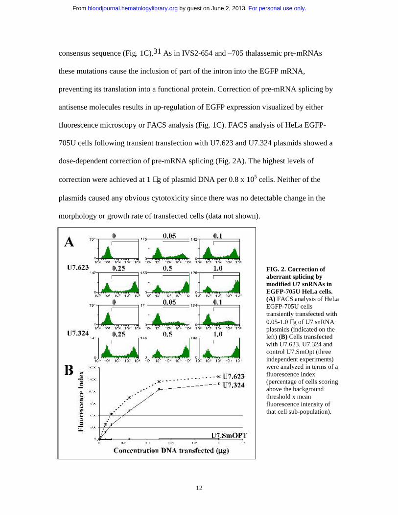
consensus sequence (Fig. 1C).31 As in IVS2-654 and –705 thalassemic pre-mRNAs
these mutations cause the inclusion of part of the intron into the EGFP mRNA,
preventing its translation into a functional protein. Correction of pre-mRNA splicing by
antisense molecules results in up-regulation of EGFP expression visualized by either
fluorescence microscopy or FACS analysis (Fig. 1C). FACS analysis of HeLa EGFP-
705U cells following transient transfection with U7.623 and U7.324 plasmids showed a
dose-dependent correction of pre-mRNA splicing (Fig. 2A). The highest levels of
correction were achieved at 1 µg of plasmid DNA per 0.8 x 10⁵ cells. Neither of the
plasmids caused any obvious cytotoxicity since there was no detectable change in the
morphology or growth rate of transfected cells (data not shown).
FIG. 2. Correction of aberrant splicing by modified U7 snRNAs in EGFP-705U HeLa cells. (A) FACS analysis of HeLaEGFP-705U cells transiently transfected with 0.05-1.0 µg of U7 snRNA plasmids (indicated on the left) (B) Cells transfected with U7.623, U7.324 and control U7.SmOpt (three independent experiments) were analyzed in terms of a fluorescence index(percentage of cells scoring above the background threshold x mean fluorescence intensity of that cell sub-population).
For personal use only. by guest on June 2, 2013. bloodjournal.hematologylibrary.orgFrom

13
Quantitative analysis of FACS data from 3 independent experiments showed that
the U7.623 construct was more effective than the previously tested U7.324. The effects of
the two constructs were sequence specific since a U7 snRNA lacking the antisense
sequence (U7SmOPT) had little effect (Fig. 2B and Table 1). The U7.623 construct was
also more effective than the U7.324 snRNA when transfected in the EGFP-654 HeLa
cells. Nevertheless, in these cells the level of correction by all three constructs was lower
than that seen in the transfected EGFP-705U cell line (Table 1), indicating that EGFP-
654 pre-mRNA might be less susceptible to modification of splicing by antisense
sequences than its 705U counterpart (See below and Discussion).30
β−β−β−β−globin IVS2-654, –705 and –745 pre-mRNAs as targets. The antisense
activity of the U7.623 snRNA was also tested in HeLa β-globin IVS2-mutant cells (Fig.
3), which more closely model the mutations found in β-thalassemic patients. In these
cells, the correction of aberrant splicing was assessed by RT-PCR of total RNA from
transfected cells, using primers that flank intron 2 of the β-globin gene (Fig. 1A). To
increase sensitivity as well as to ascertain linear response and quantifiable ratio of PCR
products obtained from aberrant and corrected β-globin mRNAs, the RT-PCR reaction
was carried out at low cycles and with [α-32P]dATP. No product was detectable without
the reverse transcription step (not shown) (See Materials and Methods and 17 for more
details).
U7.623 snRNA corrected splicing of all three thalassemic pre-mRNAs in a dose
dependent fashion. As seen in EGFP cells, the β−globin IVS2-654 pre-mRNA was more
resistant to splicing correction by antisense molecules than the remaining two mutants.
For personal use only. by guest on June 2, 2013. bloodjournal.hematologylibrary.orgFrom

14
FIG. 3. Correction of aberrant splicing by U7.623 snRNA in ββββ-globin IVS2-mutant cells. Total RNA from HeLa β-globin IVS2-mutant cells transiently transfected with 0.05-1.0 µg (indicated at the top) of U7.623snRNA plasmid were analyzed by RT-PCR.(A) β-globin IVS2-654, (B) β-globin IVS2-705 and (C) β-globin IVS2-745 HeLa cells.Lane 1, untreated cells, lanes 2-5, cells transfected with U7.623 plasmid, lane 6, (N)RNA from normal human blood. The sizes (in nucleotides) of PCR bands representingaberrantly (308, 358 and 396) and correctly(231) spliced mRNAs are indicated on the right. Similar designations were used in Figs. 5, 6C, and 7A.
Even though in IVS2-654 cells correctly spliced β−globin mRNA was detectable at 0.1
µg of transfected plasmid (Fig. 3A, lane 3), its level was only slightly increased at 1µg of
transfected DNA (lane 5). In contrast the same concentrations of U7.623 construct
elicited much higher levels of correction in IVS2-705 (nearly complete at 1 µg DNA, Fig.
3B, lane 5) and IVS2-745 cells (Fig. 3C, lane 5).
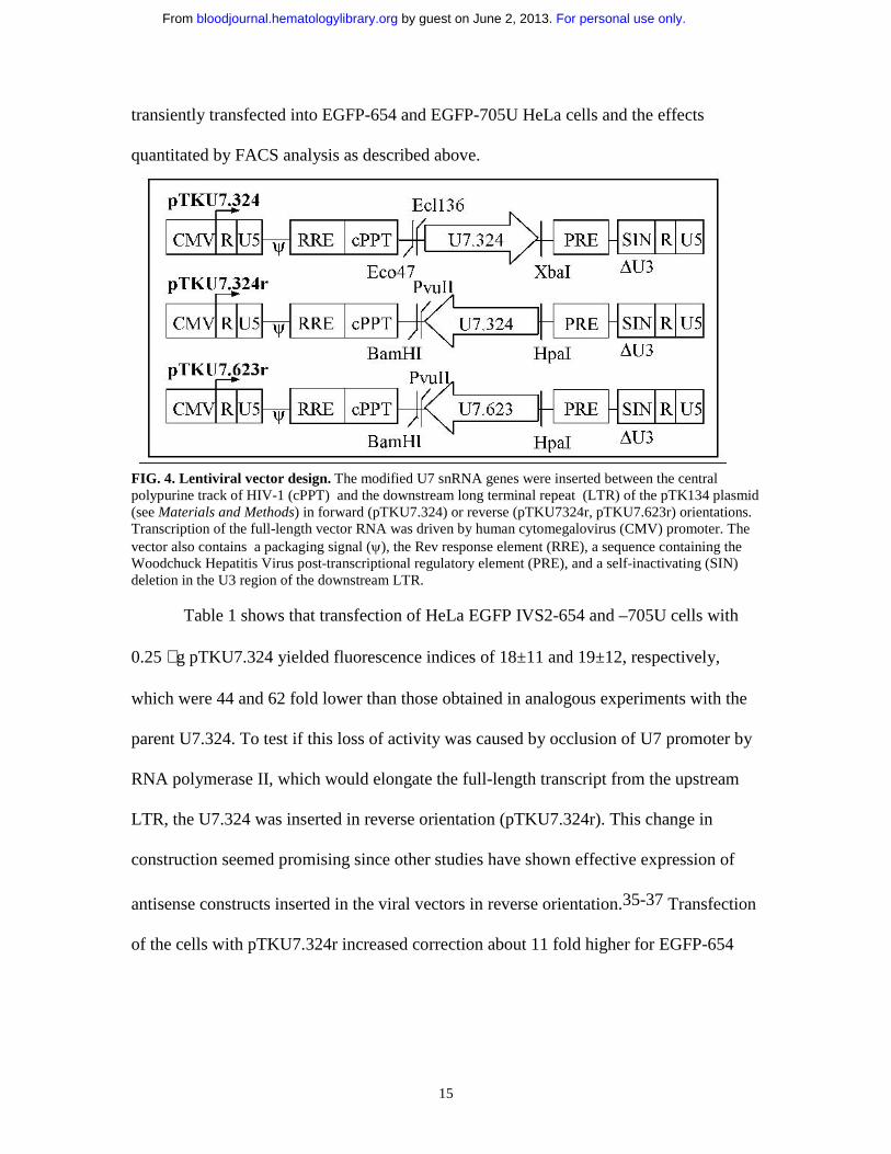
Correction of aberrant splicing by U7 lentiviral plasmids. Fig. 4 shows the
structure of three U7 antisense lentiviral constructs in which the snRNA genes were
inserted into the pTK134 plasmid (Kafri, unpublished). The inserts were comprised of
the modified U7.324 and U7.623 genes and included the flanking promoter and
terminator sequences. The U7.324 was inserted in forward and reverse orientations with
respect to the viral LTRs (pTKU7.324 and pTKU7.324r), and the U7.623 only in reverse
orientation (pTKU7.623r, see also Materials and Methods). The plasmids were
For personal use only. by guest on June 2, 2013. bloodjournal.hematologylibrary.orgFrom
15
transiently transfected into EGFP-654 and EGFP-705U HeLa cells and the effects
quantitated by FACS analysis as described above.
FIG. 4. Lentiviral vector design. The modified U7 snRNA genes were inserted between the central polypurine track of HIV-1 (cPPT) and the downstream long terminal repeat (LTR) of the pTK134 plasmid (see Materials and Methods) in forward (pTKU7.324) or reverse (pTKU7324r, pTKU7.623r) orientations. Transcription of the full-length vector RNA was driven by human cytomegalovirus (CMV) promoter. The vector also contains a packaging signal (ψ), the Rev response element (RRE), a sequence containing the Woodchuck Hepatitis Virus post-transcriptional regulatory element (PRE), and a self-inactivating (SIN) deletion in the U3 region of the downstream LTR.
Table 1 shows that transfection of HeLa EGFP IVS2-654 and –705U cells with
0.25 µg pTKU7.324 yielded fluorescence indices of 18±11 and 19±12, respectively,
which were 44 and 62 fold lower than those obtained in analogous experiments with the
parent U7.324. To test if this loss of activity was caused by occlusion of U7 promoter by
RNA polymerase II, which would elongate the full-length transcript from the upstream
LTR, the U7.324 was inserted in reverse orientation (pTKU7.324r). This change in
construction seemed promising since other studies have shown effective expression of
antisense constructs inserted in the viral vectors in reverse orientation.35-37 Transfection
of the cells with pTKU7.324r increased correction about 11 fold higher for EGFP-654
For personal use only. by guest on June 2, 2013. bloodjournal.hematologylibrary.orgFrom

16
SnRNA
Fluorescence Index
EGFP-654
Fluorescence Index
EGFP-705U
U7.623 1068 ± 52 1741 ± 59
U7.324 786 ± 38 1186 ± 43
PTKU7.324 18 ± 11 19 ±12
PTKU7.324r 194 ± 56 360 ± 58
PTKU7.623r 349 ± 58 576 ± 60
U7SmOPT 3 ± 3 7 ± 4
Table 1. Correction of aberrant splicing by modified U7 snRNAs in EGFP-mutant HeLa cells. HeLaEGFP-654 and –705U cells transiently transfected with 0.25 µg U7 snRNA plasmids (three independent experiments) were analyzed in terms of a fluorescence index (percentage of cells scoring above the background threshold x mean fluorescence intensity of that cell sub-population). Control, U7.SmOpt lacks the antisense sequence.
and 19 fold for EGFP-705U cells (fluorescent indices 194±56 and 360±58, respectively).
The U7 construct in reverse orientation targeted to the 623 sequence was even more
effective, reaching FI of 349±58 and 576±60. Note that even the latter construct was 3
times less effective than its parent plasmid in both cell lines. The most effective lentiviral
construct, pTKU7.623r, was tested in HeLa cell lines expressing IVS2-654, -705, and -
745 β−globin genes (Fig. 5). In these cells the effects of pTKU7.623r were significantly
lower than in EGFP cells; the IVS2-654 cells were unaffected by the treatment (Fig. 5A)
and only minimal correction was detectable in IVS2-705 cells transfected with 1 µg
pTKU7.623r DNA (Fig. 5B). Importantly, significant, dose dependent correction was
detected in IVS2-745 treated cells (Fig. 5C). This result indicated that pTKU7.623r was
not only expressed but also functioned properly, and underscored the difference in
susceptibility of IVS2-654, -705 and –745 pre-mRNAs.
For personal use only. by guest on June 2, 2013. bloodjournal.hematologylibrary.orgFrom

17
FIG. 5. Correction of aberrant splicing by lentiviral plasmids in ββββ-globin IVS2-mutant HeLa cells. Total RNA from HeLa β-globin IVS2-mutant cells transiently transfected with 0.05-1.0 µg of pTKU7.623r lentiviral construct were analyzed by RT-PCR. (A) β-globin IVS2-654, (B) β-globin IVS2-705, and (C) β-globin IVS2-745 cells. Lane 1, untreated cells, lanes 2-5, cells transfected with pTKU6.623r, lane 6, (N) RNA from normal human blood.
Correction of pre-mRNA splicing in HeLa cells transduced with U7.623
lentiviral vector. The pTKU7.623r construct was used to produce a lentiviral vector
(Materials and Methods), which was tested in EGFP-654, -705U, IVS2-654, -705, and –
745 cells. Note that in the context of the self-inactivating lentiviral vector, in which the
endogenous promoter is inactivated after one round of reverse transcription, the problem
of transcriptional interference should be eliminated. Of the HeLa EGFP-654 cells
transduced with the lentiviral vector, 97% fluoresced with a mean fluorescence intensity
of 1116, resulting in FI of 1086 (Fig. 6A). Transduction of EGFP-705U cells was less
efficient (76%), with a lower mean fluorescence of 914, resulting in FI= 694. For both
cell lines the FI of non-transduced cells was negligible. Fluorescence microscopy
corroborated the results of FACS analysis (Fig. 6B).
For personal use only. by guest on June 2, 2013. bloodjournal.hematologylibrary.orgFrom

18
FIG. 6. Correction of aberrant splicing of HeLa EGFP and ββββ-globin IVS2- mutants with U7 lentiviral vector. (A) FACS analysis. EGFP-654 and –705U cells were transduced with U7.623 lentiviral vector and subjected to flow cytometry analysis 2 after transduction weeks. (B) Fluorescence microscopy of identically treated cells. Phase contrast (left) and UV (right) images are shown. (C) RT-PCR of total RNA from HeLa β-globin IVS2-654 (lane 2), IVS2-705 (lane 5), and IVS2-745 (lane 8) cells transduced with U7.623 lentiviral vector. Untreated IVS2-654 (lane 1), IVS2-705 (lane 4), and IVS2-745 (lane 7) cells served as negative controls. Lanes 3, 6, 9, RNA from normal blood. Lane 10, IVS2-745 cells transduced with U7.623 lentiviral vector analyzed at 6 months. (D) Immunoblot analysis of total protein from HeLa IVS2-705 and -745 cells transduced as in (C). The blots were probed with polyclonal anti-hemoglobin antibody (see Materials and Methods). Lane 1, β−globin marker; lanes 2 and 4, untreated cells; lanes 3 and 5, lentiviral vector transduced cells.
Transduction of HeLa cell lines expressing thalassemic pre-mRNAs showed (Fig.
6C) that the U7 lentiviral vector resulted in an increase in correctly spliced products to
about 3% of total in IVS2-654 cells (lane 2), 17% in IVS2-705 (lane 5), and essentially
complete in IVS2-745 (lane 8). The IVS2-745 cell line transduced with the lentiviral
vector was maintained in continuous culture for six months. Importantly, and as expected
with the genome integrated viral sequences, the RT-PCR analysis of the RNA after this
period showed the level of correction equal to that seen 2 weeks post transduction
(compare lanes 8 and 10). Similar results were seen with lentiviral vector transduction of
For personal use only. by guest on June 2, 2013. bloodjournal.hematologylibrary.orgFrom

19
K562 cells stably expressing the β-globin IVS2-mutants (data not shown). The RT-PCR
results were also corroborated by the analysis of cellular protein by immunoblotting with
a polyclonal antibody to human hemoglobin (Fig.6D). The results show that in IVS2-705
and IVS2-745 HeLa cells transduced with the U7.623 lentiviral vector the newly
generated correctly spliced β-globin mRNA was translated into full-length, functional β-
globin protein.
Correction of IVS2-745 splicing in erythroid progenitor cells by U7.623
lentiviral vector. The U7.623 lentiviral vector was applied to a therapeutically relevant
target, the erythroid progenitor cells from a patient with IVS2-745/IVS2-1 thalassemia.
Based on the results shown in Fig. 6, this mutation was expected to be the easiest to
correct, allowing the proof of principle for antisense RNA treatment of thalassemia to be
established. Furthermore, the splicing of IVS2-745 pre-mRNA in human erythroid
progenitor cells has been repaired by treatment of the cells with oligonucleotides targeted
to the aberrant 3’ splice site.17
The mononuclear cells were isolated from the patient’s blood and cultured in the
presence of Epo and SCF, conditions that promote erythroid differentiation of stem cells
and early progenitors.17 The cells were transduced with U7.623 lentiviral vector on days
1 and 2 of culture and subsequently cultured in methylcellulose medium containing
growth factors (see M aterials and Methods) for 12 days; at this time total RNA was
analyzed by RT-PCR (Fig. 7A). As expected, in cells transduced with a control viral
vector in which the U7.623 gene was replaced with the GFP gene (Fig. 7A, lane 2) there
was no increase in the amount of correctly spliced β-globin mRNA over the existing
background in mock-transduced cells (lane 1). In contrast, in cells transduced with
For personal use only. by guest on June 2, 2013. bloodjournal.hematologylibrary.orgFrom

20
FIG. 7. U7.623 lentiviral vector-induced ββββ-globin pre-mRNA repair in erythroid progenitors from IVS2-745/IVS2-1 thalassemic patient. (A)RT-PCR. On days 1 and 2 of culture, the cells were transduced for 5 h with either no virus (lane 1), control, GFP lentiviral vector (lane 2) or U7.623 lentiviral vector (lane 3). Lane 4, RNA from normal blood. (B)Immunodetection of hemolysates separated by electrophoresis on cellulose acetate with anti-human hemoglobin antibody (see Materials and Methods). Lane 1, hemoglobin standards, lane 2, (N) normal blood, lane 3, mock transduced cells, lane 4, cells transduced with GFP lentiviral vector, lane5, cells transduced with U7.623 lentiviral vector.
U7.623 lentiviral vector (lane 3) the level of the aberrant band was diminished and that of
the correct band greatly increased, demonstrating significant correction of splicing of
IVS2-745 pre-mRNA. In addition to 396-nt and 231-nt products, representing the
aberrantly and correctly spliced β−globin mRNAs, a 278-nt product was also generated
by RT-PCR. The corresponding RNA is spliced via a cryptic splice site activated by the
IVS2-1 mutation and located 47-nt downstream.12 Quantitative analysis of the ratio of
correct to aberrant RNAs showed that lentiviral vector-mediated antisense delivery led to
a 23 fold increase in the production of correctly spliced β-globin mRNA.
To test for hemoglobin synthesis, lysates of lentiviral vector transduced cells at
day 12 of culture were separated by electrophoresis on cellulose acetate and probed with
polyclonal anti-hemoglobin antibody. This analysis showed significant levels of newly
generated hemoglobin A (Hb A) (Fig. 7B). Quantitation of the ratio of Hb A bands to Hb
F (used as an internal control) showed that the amount of Hb A in the patient cells
For personal use only. by guest on June 2, 2013. bloodjournal.hematologylibrary.orgFrom

21
transduced with the U7.623 lentiviral vector increased about 25 fold over that in cells
transduced with the control GFP viral vector. This result is consistent with the increase of
β−globin mRNA detected by RT-PCR and also shows that the antisense treatment of
progenitor cells not only repaired β−globin pre-mRNA splicing but also restored proper
expression of the Hb A protein.
DISCUSSION
Previously, viral vector transfer of antisense RNAs has been used for down-
regulation of gene expression (reviewed in 38). Here we demonstrate that this strategy
can be used to successfully restore expression of a gene inactivated by a splicing
mutation. This novel application of the lentiviral vector mediated antisense approach was
achieved not only in cell line models but also in the clinically relevant erythroid
progenitor cells from a patient with IVS2-745/IVS2-1 thalassemia.
The fact that U7.623 snRNA (targeted downstream of the cryptic 3’ splice site) is
more effective in the correction of aberrant splicing than U7.324 targeted against the
splice site itself implies the presence of a splicing enhancer at the target site.29 Splicing
enhancers function as binding sites for SR-proteins, a family of serine/arginine-rich
essential splicing factors involved in regulation of alternative splicing (see 39 for review).
Presumably, antisense RNAs targeted against the 623 region prevented the binding of
SR-proteins and induced skipping of the aberrant pseudo-exon.40
The modified U7 snRNAs were most efficient in the correction of splicing of
IVS2-745 pre-mRNA, followed by IVS2-705 and IVS2-654. Similar results have also
been observed in correction of splicing of these pre-mRNAs by antisense
For personal use only. by guest on June 2, 2013. bloodjournal.hematologylibrary.orgFrom

22
oligonucleotides targeted to the aberrant 3’ and 5’ splice sites.30 Here, these observations
are extended to antisense RNA that is targeted between the splice sites and is expressed
intracellularly. One concludes that the strength of the aberrant 5’ splice sites and the
distance between the 3’ and 5’ splice sites flanking the internal exons determine the
differences in correction achieved in the three mutants. Since the HeLa IVS2-654 cell line
was relatively resistant to correction by the lentiviral vector, these experiments were not
attempted in the IVS2-654 β-thalassemic mouse model.41 The resistance of this splice
site to correction most likely reflects the nature of the interactions of the spliceosomes
with the pre-mRNAs, including the interactions of the splicing factors that bridge the
exon.30
Previous experiments with erythroid mononuclear cultures have established that
in the presence of Epo and SCF β−globin pre- and mRNAs did not appear until day 4-5
of culture.17 Thus, the lentiviral vector added to the cultured mononuclear cells at days 1
and 2 must have transduced hematopoietic stem cells and/or very early erythroid
progenitor cells, which were not yet engaged in the expression of β−globin gene. Since
maximum correction of aberrant β−globin splicing by antisense oligonucleotides was
found at day 12-15 of culture,17,42 it is likely that the very early, multipotent erythroid
progenitor cells, which produce in about two weeks large numbers of highly multicellular
erythroid bursts (BFU-E),43 are responsible for the bulk of the repair response elicited by
the U7.623 virus. This does not exclude the possibility that a fraction of the response was
generated in the ultimate target of the antisense approach, hematopoietic stem cells.
Although these cells are found in only 1/10,000-1/100,000 bone marrow cells and
even less frequently in the peripheral blood, the latter material has been used for stem cell
For personal use only. by guest on June 2, 2013. bloodjournal.hematologylibrary.orgFrom

23
transplantation in thalassemia.44 Clearly additional studies are required to determine the
ability of thalassemic patients’ mobilized CD34+ HSC to engraft and differentiate into
Hb A producing erythroid cells following transduction with lentiviral vectors carrying the
modified U7snRNA expression cassettes.
An important advantage of our antisense approach is that the correction occurs in the
β-globin pre-mRNA transcribed from the β-globin gene in its natural chromosomal
environment, and properly controlled by the native locus control region. Therefore the
expression of β-globin cannot exceed the wild type levels, precluding the possibility of
over-expression of β-globin mRNA, and offering an attractive alternative to gene
replacement as a treatment for hemoglobinopathies. Furthermore, complete splicing
correction is not necessary. Even if only 10% levels of correction were achieved in
thalassemic patients, this would lead to a clinically relevant outcome.7
The effects of the U7.623 snRNAs expressed by the lentiviral vector are expected to
be limited only to cells that express the target sequence, that is the erythroid precursor
and progenitor cells. This is because hybridization of the U7.623 snRNA to other RNAs
will occur with a number of mismatches, rendering the molecules ineffective.16 The fact
that cell growth of cultured mononuclear cells was unaffected by lentiviral vector
transduction shows that U7.623 RNA did not effect widespread inhibition of gene
expression. Likewise, antisense oligonucleotides have been found relatively non-toxic in
clinical trials and as a marketed drug.45
It was estimated that 15% of point mutations contributing to genetic diseases cause
aberrant splicing.46 Recent results, which take into account not only genomic sequence
but also RNA expression and splicing patterns indicate that the percentage of splicing
For personal use only. by guest on June 2, 2013. bloodjournal.hematologylibrary.orgFrom

24
defects may be much higher. For example, when analyzed at the RNA level, 50% of
mutations in the ataxia-telangiectasia and neurofibromatosis type 1 genes resulted in
defective splicing.47,48 Thus, the antisense repair of defective pre-mRNAs can be
applied to other disorders in additon to thalassemia syndromes. In fact, correction of pre-
mRNA splicing by antisense oligonucleotides was investigated in the context of cystic
fibrosis,49 Duchenne muscular dystrophy,50,51Alzheimer’s-like, FTDP-17 syndrome52
and spinal muscular atrophy.53 This method was also used for modification of alternative
splicing by targeting a cancer related splice variant of bcl-x pre-mRNA.20,54 The
approach described here provides a way to effect permanent correction of the aberrant
splicing that gives rise to disease by incorporating these and other antisense sequences
into viral vectors.
Acknowledgements
We are grateful to the patients and their parents for donating blood samples. We thank
Elizabeth Smith for technical assistance and Thipparat Suwanmanee for guidance in
erythroid cell culture.
For personal use only. by guest on June 2, 2013. bloodjournal.hematologylibrary.orgFrom

25
REFERENCES
1. Schwartz E, and Benz, EJ. In: Hoffman R, Benz EJ, Shattil SJ, Furie B, Cohen HJ, ed.
Hematology: basic principles and practice. New York: Churchill Livingston, 1991:368-
392.
2. Rund D, Rachmilewitz E. New trends in the treatment of beta-thalassemia. Crit Rev
Oncol Hematol 2000;33:105-118.
3. Boulad F, Giardina P, Gillio A, et al. Bone marrow transplantation for homozygous
beta-thalassemia. The Memorial Sloan-Kettering Cancer Center experience. Ann N Y
Acad Sci 1998;850:498-502.
4. Lucarelli G, Galimberti M, Giardini C, et al. Bone marrow transplantation in
thalassemia. The experience of Pesaro. Ann N Y Acad Sci 1998;850:270-275.
5. Olivieri NF, Weatherall DJ. The therapeutic reactivation of fetal haemoglobin. Hum
Mol Genet 1998;7:1655-1658.
6. Sadelain M. Globin gene transfer for the treatment of severe hemoglobinopathies: a
paradigm for stem cell-based gene therapy. J Gene Med 2002;4:113-121.
7. Tisdale J, Sadelain M. Toward gene therapy for disorders of globin synthesis. Semin
Hematol 2001;38:382-392.
8. May C, Rivella S, Callegari J, et al. Therapeutic haemoglobin synthesis in beta-
thalassaemic mice expressing lentivirus-encoded human beta- globin. Nature
2000;406:82-86.
For personal use only. by guest on June 2, 2013. bloodjournal.hematologylibrary.orgFrom

26
9. May C, Rivella S, Chadburn A, Sadelain M. Successful treatment of murine beta-
thalassemia intermedia by transfer of the human beta-globin gene. Blood 2002;99:1902-
1908.
10. Sierakowska H, Gorman L, Kang SH, Kole R. Antisense oligonucleotides and RNAs
as modulators of pre-mRNA splicing. Methods Enzymol 2000;313:506-521.
11. Chui DH, Hardison R, Riemer C, et al. An electronic database of human hemoglobin
variants on the World Wide Web. Blood 1998;91:2643-2644.
12. Treisman R, Proudfoot NJ, Shander M, Maniatis T. A single-base change at a splice
site in a beta 0-thalassemic gene causes abnormal RNA splicing. Cell 1982;29:903-911.
13. Cheng TC, Orkin SH, Antonarakis SE, et al. beta-Thalassemia in Chinese: use of in
vivo RNA analysis and oligonucleotide hybridization in systematic characterization of
molecular defects. Proc Natl Acad Sci U S A 1984;81:2821-2825.
14. Dobkin C, Bank A. Reversibility of IVS 2 missplicing in a mutant human beta-globin
gene. J Biol Chem 1985;260:16332-16337.
15. Dominski Z, Kole R. Restoration of correct splicing in thalassemic pre-mRNA by
antisense oligonucleotides. Proc Natl Acad Sci U S A 1993;90:8673-8677.
16. Sierakowska H, Sambade MJ, Agrawal S, Kole R. Repair of thalassemic human beta-
globin mRNA in mammalian cells by antisense oligonucleotides. Proc Natl Acad Sci U S
A 1996;93:12840-12844.
17. Lacerra G, Sierakowska H, Carestia C, et al. Restoration of hemoglobin A synthesis
in erythroid cells from peripheral blood of thalassemic patients. Proc Natl Acad Sci U S
A 2000;97:9591-9596.
For personal use only. by guest on June 2, 2013. bloodjournal.hematologylibrary.orgFrom

27
18. Gorman L, Suter D, Emerick V, Schumperli D, Kole R. Stable alteration of pre-
mRNA splicing patterns by modified U7 small nuclear RNAs. Proc Natl Acad Sci U S A
1998;95:4929-4934.
19. Suter D, Tomasini R, Reber U, Gorman L, Kole R, Schumperli D. Double-target
antisense U7 snRNAs promote efficient skipping of an aberrant exon in three human
beta-thalassemic mutations. Hum Mol Genet 1999;8:2415-2423.
20. Gorman L, Mercatante DR, Kole R. Restoration of correct splicing of thalassemic
beta-globin pre-mRNA by modified U1 snRNAs. J Biol Chem 2000;275:35914-35919.
21. Baserga SJ, Steitz, J.A. The Diverse World of Small Ribonucleoproteins. In:
Gesteland RF, Atkins, J.F., ed. The RNA World. Plainview, NY: Cold Spring Harbor
Laboratory Press, 1993:359-382.
22. Kafri T. Lentivirus vectors: difficulties and hopes before clinical trials. Curr Opin
Mol Ther 2001;3:316-326.
23. Uchida N, Sutton RE, Friera AM, et al. HIV, but not murine leukemia virus, vectors
mediate high efficiency gene transfer into freshly isolated G0/G1 human hematopoietic
stem cells. Proc Natl Acad Sci U S A 1998;95:11939-11944.
24. Case SS, Price MA, Jordan CT, et al. Stable transduction of quiescent
CD34(+)CD38(-) human hematopoietic cells by HIV-1-based lentiviral vectors. Proc Natl
Acad Sci U S A 1999;96:2988-2993.
25. Chen W, Wu X, Levasseur DN, et al. Lentiviral vector transduction of hematopoietic
stem cells that mediate long-term reconstitution of lethally irradiated mice. Stem Cells
2000;18:352-359.
For personal use only. by guest on June 2, 2013. bloodjournal.hematologylibrary.orgFrom

28
26. Douglas JL, Lin WY, Panis ML, Veres G. Efficient human immunodeficiency virus-
based vector transduction of unstimulated human mobilized peripheral blood CD34+
cells in the SCID-hu Thy/Liv model of human T cell lymphopoiesis. Hum Gene Ther
2001;12:401-413.
27. Yamada K, Olsen JC, Patel M, Rao KW, Walsh CE. Functional correction of fanconi
anemia group C hematopoietic cells by the use of a novel lentiviral vector. Mol Ther
2001;3:485-490.
28. Saulnier SO, Steinhoff D, Dinauer MC, et al. Lentivirus-mediated gene transfer of
gp91phox corrects chronic granulomatous disease (CGD) phenotype in human X-CGD
cells. J Gene Med 2000;2:317-325.
29. Gemignani F, Landi S, DeMarini DM, Kole R. Spontaneous and MNNG-induced
reversion of an EGFP construct in HeLa cells: an assay for observing mutations in living
cells by fluorescent microscopy. Hum Mutat 2001;18:526-534.
30. Sierakowska H, Sambade MJ, Schumperli D, Kole R. Sensitivity of splice sites to
antisense oligonucleotides in vivo. RNA 1999;5:369-377.
31. Sazani P, Kang SH, Maier MA, et al. Nuclear antisense effects of neutral, anionic and
cationic oligonucleotide analogs. Nucleic Acids Res 2001;29:3965-3974.
32. Naldini L, Blomer U, Gallay P, et al. In vivo gene delivery and stable transduction of
nondividing cells by a lentiviral vector. Science 1996;272:263-267.
33. Kafri T, van Praag H, Ouyang L, Gage FH, Verma IM. A packaging cell line for
lentivirus vectors. J Virol 1999;73:576-584.
34. Xu K, Ma H, McCown TJ, Verma IM, Kafri T. Generation of a stable cell line
producing high-titer self-inactivating lentiviral vectors. Mol Ther 2001;3:97-104.
For personal use only. by guest on June 2, 2013. bloodjournal.hematologylibrary.orgFrom

29
35. Sun LQ, Pyati J, Smythe J, et al. Resistance to human immunodeficiency virus type 1
infection conferred by transduction of human peripheral blood lymphocytes with
ribozyme, antisense, or polymeric trans-activation response element constructs. Proc Natl
Acad Sci U S A 1995;92:7272-7276.
36. Vandendriessche T, Chuah MK, Chiang L, Chang HK, Ensoli B, Morgan RA.
Inhibition of clinical human immunodeficiency virus (HIV) type 1 isolates in primary
CD4+ T lymphocytes by retroviral vectors expressing anti-HIV genes. J Virol
1995;69:4045-4052.
37. Mautino MR, Morgan RA. Potent inhibition of human immunodeficiency virus type 1
replication by conditionally replicating human immunodeficiency virus-based lentiviral
vectors expressing envelope antisense mRNA. Hum Gene Ther 2000;11:2025-2037.
38. Sazani P, Vacek, M., and Kole, R. Modulation of gene expression by antisense
therapeutics. Current Opinion in Biotechnology;In press.
39. Blencowe BJ. Exonic splicing enhancers: mechanism of action, diversity and role in
human genetic diseases. Trends Biochem Sci 2000;25:106-110.
40. Liu HX, Chew SL, Cartegni L, Zhang MQ, Krainer AR. Exonic splicing enhancer
motif recognized by human SC35 under splicing conditions. Mol Cell Biol
2000;20:1063-1071.
41. Lewis J, Yang B, Kim R, et al. A common human beta globin splicing mutation
modeled in mice. Blood 1998;91:2152-2156.
42. Suwanmanee T, Sierakowska, H, Lacerra, G, et al. Restoration of human beta-globin
gene expression in murine and human IVS2-654 thalassemic erythroid cells by free
uptake of antisense oligonucleotides. Molecular Pharmacology;in press.
For personal use only. by guest on June 2, 2013. bloodjournal.hematologylibrary.orgFrom

30
43. Papayannoupoulou T, Abkowitz J, and D'Andrea A. Biology of erythropoiesis,
erythroid differentiation, and maturation. In: Hoffman R, Benz EJ, Shattil SJ, Furie B,
Cohen HJ, ed. Hematology, Basic Principles and Practice. New York: Churchill
Livingstone, 2000:202-219.
44. Yesilipek MA, Hazar V, Kupesiz A, Kizilors A, Uguz A, Yegin O. Peripheral blood
stem cell transplantation in children with beta-thalassemia. Bone Marrow Transplant
2001;28:1037-1040.
45. Crooke ST. Basic principles of antisense technology. In: Crooke ST, ed. Antisense
drug technology: principles, strategies and applications. New York, N.Y.: Marcel Dekker,
2001:1-28.
46. Krawczak M, Smith-Sorensen B, Schmidtke J, Kakkar VV, Cooper DN, Hovig E.
Somatic spectrum of cancer-associated single basepair substitutions in the TP53 gene is
determined mainly by endogenous mechanisms of mutation and by selection. Hum Mutat
1995;5:48-57.
47. Teraoka SN, Telatar M, Becker-Catania S, et al. Splicing defects in the ataxia-
telangiectasia gene, ATM: underlying mutations and consequences. Am J Hum Genet
1999;64:1617-1631.
48. Ars E, Serra E, Garcia J, et al. Mutations affecting mRNA splicing are the most
common molecular defects in patients with neurofibromatosis type 1. Hum Mol Genet
2000;9:237-247.
49. Friedman KJ, Kole J, Cohn JA, Knowles MR, Silverman LM, Kole R. Correction of
aberrant splicing of the cystic fibrosis transmembrane conductance regulator (CFTR)
gene by antisense oligonucleotides. J Biol Chem 1999;274:36193-36199.
For personal use only. by guest on June 2, 2013. bloodjournal.hematologylibrary.orgFrom

31
50. Wilton SD, Lloyd F, Carville K, et al. Specific removal of the nonsense mutation
from the mdx dystrophin mRNA using antisense oligonucleotides. Neuromuscul Disord
1999;9:330-338.
51. Mann CJ, Honeyman K, Cheng AJ, et al. Antisense-induced exon skipping and
synthesis of dystrophin in the mdx mouse. Proc Natl Acad Sci U S A 2001;98:42-47.
52. Kalbfuss B, Mabon SA, Misteli T. Correction of alternative splicing of tau in
frontotemporal dementia and parkinsonism linked to chromosome 17. J Biol Chem
2001;276:42986-42993.
53. Lim SR, Hertel KJ. Modulation of survival motor neuron pre-mRNA splicing by
inhibition of alternative 3' splice site pairing. J Biol Chem 2001;276:45476-45483.
54. Taylor JK, Zhang QQ, Wyatt JR, Dean NM. Induction of endogenous Bcl-xS through
the control of Bcl-x pre-mRNA splicing by antisense oligonucleotides. Nat Biotechnol
1999;17:1097-1100.
55. Grimm C, Stefanovic B, Schumperli D. The low abundance of U7 snRNA is partly
determined by its Sm binding site. EMBO J 1993;12:1229-1238.
For personal use only. by guest on June 2, 2013. bloodjournal.hematologylibrary.orgFrom