Ecto-nucleoside triphosphate diphosphohydrolase 3 in the ventral and lateral hypothalamic area of...
12
BioMed Central Page 1 of 12 (page number not for citation purposes) Reproductive Biology and Endocrinology Open Access Research Ecto-nucleoside triphosphate diphosphohydrolase 3 in the ventral and lateral hypothalamic area of female rats: morphological characterization and functional implications David S Kiss 1 , Attila Zsarnovszky* 1,2 , Krisztina Horvath 1 , Andrea Gyorffy 1 , Tibor Bartha 1 , Diana Hazai 3 , Peter Sotonyi 3 , Virag Somogyi 1 , Laszlo V Frenyo 1 and Sabrina Diano 2,4 Address: 1 Department of Physiology & Biochemistry, Szent Istvan University Faculty of Veterinary Science, Budapest, Hungary, 2 Department of Obstetrics, Gynecology & Reproductive Sciences, Yale University School of Medicine, New Haven, CT 06510, USA, 3 Department of Anatomy & Histology, Szent Istvan University Faculty of Veterinary Science, Budapest, Hungary and 4 Department of Neurobiology, Yale University School of Medicine, New Haven, CT 06510, USA Email: David S Kiss - [email protected]; Attila Zsarnovszky* - [email protected]; Krisztina Horvath - [email protected]; Andrea Gyorffy - [email protected]; Tibor Bartha - [email protected]; Diana Hazai - [email protected]; Peter Sotonyi - [email protected]; Virag Somogyi - [email protected]; Laszlo V Frenyo - [email protected]; Sabrina Diano - [email protected] * Corresponding author Abstract Background: Based on its distribution in the brain, ecto-nucleoside triphosphate diphosphohydrolase 3 (NTPDase3) may play a role in the hypothalamic regulation of homeostatic systems, including feeding, sleep-wake behavior and reproduction. To further characterize the morphological attributes of NTPDase3-immunoreactive (IR) hypothalamic structures in the rat brain, here we investigated: 1.) The cellular and subcellular localization of NTPDase3; 2.) The effects of 17β-estradiol on the expression level of hypothalamic NTPDase3; and 3.) The effects of NTPDase inhibition in hypothalamic synaptosomal preparations. Methods: Combined light- and electron microscopic analyses were carried out to characterize the cellular and subcellular localization of NTPDase3-immunoreactivity. The effects of estrogen on hypothalamic NTPDase3 expression was studied by western blot technique. Finally, the effects of NTPDase inhibition on mitochondrial respiration were investigated using a Clark-type oxygen electrode. Results: Combined light- and electron microscopic analysis of immunostained hypothalamic slices revealed that NTPDase3-IR is linked to ribosomes and mitochondria, is predominantly present in excitatory axon terminals and in distinct segments of the perikaryal plasma membrane. Immunohistochemical labeling of NTPDase3 and glutamic acid decarboxylase (GAD) indicated that γ-amino-butyric-acid- (GABA) ergic hypothalamic neurons do not express NTPDase3, further suggesting that in the hypothalamus, NTPDase3 is predominantly present in excitatory neurons. We also investigated whether estrogen influences the expression level of NTPDase3 in the ventrobasal and lateral hypothalamus. A single subcutaneous injection of estrogen differentially increased NTPDase3 expression in the medial and lateral parts of the hypothalamus, indicating that Published: 22 April 2009 Reproductive Biology and Endocrinology 2009, 7:31 doi:10.1186/1477-7827-7-31 Received: 25 February 2009 Accepted: 22 April 2009 This article is available from: http://www.rbej.com/content/7/1/31 © 2009 Kiss et al; licensee BioMed Central Ltd. This is an Open Access article distributed under the terms of the Creative Commons Attribution License (http://creativecommons.org/licenses/by/2.0 ), which permits unrestricted use, distribution, and reproduction in any medium, provided the original work is properly cited.
-
Upload
independent -
Category
Documents
-
view
2 -
download
0
Transcript of Ecto-nucleoside triphosphate diphosphohydrolase 3 in the ventral and lateral hypothalamic area of...
BioMed Central
Reproductive Biology and Endocrinology
ss
Open AcceResearchEcto-nucleoside triphosphate diphosphohydrolase 3 in the ventral and lateral hypothalamic area of female rats: morphological characterization and functional implicationsDavid S Kiss1, Attila Zsarnovszky*1,2, Krisztina Horvath1, Andrea Gyorffy1, Tibor Bartha1, Diana Hazai3, Peter Sotonyi3, Virag Somogyi1, Laszlo V Frenyo1 and Sabrina Diano2,4Address: 1Department of Physiology & Biochemistry, Szent Istvan University Faculty of Veterinary Science, Budapest, Hungary, 2Department of Obstetrics, Gynecology & Reproductive Sciences, Yale University School of Medicine, New Haven, CT 06510, USA, 3Department of Anatomy & Histology, Szent Istvan University Faculty of Veterinary Science, Budapest, Hungary and 4Department of Neurobiology, Yale University School of Medicine, New Haven, CT 06510, USA
Email: David S Kiss - [email protected]; Attila Zsarnovszky* - [email protected]; Krisztina Horvath - [email protected]; Andrea Gyorffy - [email protected]; Tibor Bartha - [email protected]; Diana Hazai - [email protected]; Peter Sotonyi - [email protected]; Virag Somogyi - [email protected]; Laszlo V Frenyo - [email protected]; Sabrina Diano - [email protected]
* Corresponding author
AbstractBackground: Based on its distribution in the brain, ecto-nucleoside triphosphatediphosphohydrolase 3 (NTPDase3) may play a role in the hypothalamic regulation of homeostaticsystems, including feeding, sleep-wake behavior and reproduction. To further characterize themorphological attributes of NTPDase3-immunoreactive (IR) hypothalamic structures in the ratbrain, here we investigated: 1.) The cellular and subcellular localization of NTPDase3; 2.) Theeffects of 17β-estradiol on the expression level of hypothalamic NTPDase3; and 3.) The effects ofNTPDase inhibition in hypothalamic synaptosomal preparations.
Methods: Combined light- and electron microscopic analyses were carried out to characterize thecellular and subcellular localization of NTPDase3-immunoreactivity. The effects of estrogen onhypothalamic NTPDase3 expression was studied by western blot technique. Finally, the effects ofNTPDase inhibition on mitochondrial respiration were investigated using a Clark-type oxygenelectrode.
Results: Combined light- and electron microscopic analysis of immunostained hypothalamic slicesrevealed that NTPDase3-IR is linked to ribosomes and mitochondria, is predominantly present inexcitatory axon terminals and in distinct segments of the perikaryal plasma membrane.Immunohistochemical labeling of NTPDase3 and glutamic acid decarboxylase (GAD) indicated thatγ-amino-butyric-acid- (GABA) ergic hypothalamic neurons do not express NTPDase3, furthersuggesting that in the hypothalamus, NTPDase3 is predominantly present in excitatory neurons.We also investigated whether estrogen influences the expression level of NTPDase3 in theventrobasal and lateral hypothalamus. A single subcutaneous injection of estrogen differentiallyincreased NTPDase3 expression in the medial and lateral parts of the hypothalamus, indicating that
Published: 22 April 2009
Reproductive Biology and Endocrinology 2009, 7:31 doi:10.1186/1477-7827-7-31
Received: 25 February 2009Accepted: 22 April 2009
This article is available from: http://www.rbej.com/content/7/1/31
© 2009 Kiss et al; licensee BioMed Central Ltd. This is an Open Access article distributed under the terms of the Creative Commons Attribution License (http://creativecommons.org/licenses/by/2.0), which permits unrestricted use, distribution, and reproduction in any medium, provided the original work is properly cited.
Page 1 of 12(page number not for citation purposes)
Reproductive Biology and Endocrinology 2009, 7:31 http://www.rbej.com/content/7/1/31
this enzyme likely plays region-specific roles in estrogen-dependent hypothalamic regulatorymechanisms. Determination of mitochondrial respiration rates with and without the inhibition ofNTPDases confirmed the presence of NTPDases, including NTPDase3 in neuronal mitochondriaand showed that blockade of mitochondrial NTPDase functions decreases state 3 mitochondrialrespiration rate and total mitochondrial respiratory capacity.
Conclusion: Altogether, these results suggest the possibility that NTPDases, among themNTPDase3, may play an estrogen-dependent modulatory role in the regulation of intracellularavailability of ATP needed for excitatory neuronal functions including neurotransmission.
BackgroundPurinergic intercellular signaling has received much atten-tion during the past decade. It has been known for sometime that nucleotide-triphosphates, such as adenosine tri-phosphate (ATP), are not only energy carriers: ATP, forexample, is a substrate for the production of its hydrolyticderivatives (ADP, AMP and adenosine) that are the spe-cific ligands of different purinergic receptors (e.g., P2X,P2Y, P1) [1]. In fact, recent data suggest that purinergic sig-naling might be one of the first biological signaling sys-tems that evolved during the phylogenesis [2]. Thespecific ligands of the relatively wide array of purinergicreceptors are provided by the ATP-hydrolyzing activity oftransmembrane ectonucleotidase enzymes (NTPDases)and 5'-ectonucleotidase. Of the known ectonucleotidases,NTPDase1-3 have been identified in the rat brain.NTPDase1 is widely expressed in neurons, glia andendothelial cells [3], while NTPDase2 was mainly foundin the germinal zones of the rat brain, and is thought toplay a role in neural development and differentiation [4].NTPDase3 was cloned in 1998 by Smith and Kirley [5]; In1998, Chadwick and Frischauf [6] demonstrated thatNTPDase3 mRNA is most abundant in the brain and pan-creas. The first description of the localization and distribu-tion of NTPDase3 in the rat brain [7] has been recentlypublished. In the latter study, NTPDase3 immunoreactiv-ity (NTPDase3-IR) was only found in neuronal structures.The vast majority of IR profiles were axon-like neuronalprocesses concentrated in midline brain regions, withhighest frequency in the hypothalamus, thalamus and themidbrain. Immunoreactive neuronal perikarya were onlyfound in the lateral hypothalamic nucleus (LHN) andarcuate nucleus (AN). Based on those results, it has beensuggested that, because of the high degree of region-spe-cific distribution of immunoreactive profiles, NTPDase3may play a role in one or more of the regulatory mecha-nisms of food-intake, sleep-wake behavior and reproduc-tive physiology. While that previous light microscopicmapping of NTPDase3-IR in the rat brain provided usefulinformation for further studies on purinergic signaling,understanding the cellular role of this enzyme warrantedfurther determination of its subcellular localization andfunction. Therefore, here we characterized the intracellu-lar localization of NTPDase3 in the hypothalamus of
adult male rats. Electron microscopic results indicated thepresence of NTPDase3-IR in neuronal perikarya and exci-tatory nerve terminals, but not in other (glial, vascular)cell types. To determine whether NTPDase3 is differen-tially or ubiquitously expressed in excitatory and/orinhibitory neuronal structures, we examined the possibleco-localization of NTPDase3 with glutamic acid decarbox-ylase (GAD, the rate-limiting enzyme of the inhibitoryneurotransmitter GABA) by means of immunohistochem-istry. Considering that the ventrobasal hypothalamus ishighly estrogen responsive, we also tested whether or not17β-estradiol (E2) influences the expression level ofNTPDase3 in hypothalamic tissue homogenates obtainedfrom ovariectomized and ovariectomized plus E2-treatedfemale rats. Finally, morphological indications of thepresence of NTPDase3 in neuronal mitochondria implieda functional role for this enzyme in mitochondrial energy(ATP) production. Therefore, we also examined the effectsof NTPDase inhibition on mitochondrial respiration inhypothalamic synaptosomal preparations.
MethodsSprague-Dawley male or female (as indicated below) ratswere used for each study. Following the guidelines laiddown by the NIH, the use of animals was approved by therespective University Committees on Animal Use at YaleUniversity and Szent Istvan University Faculty of Veteri-nary Sciences.
Animals, tissue fixation and immunolabelingMale and female Sprague-Dawley rats (body weight: 230–250 g; vendor: Charles-River Laboratories, Inc.) wereused. Animals were kept under standard laboratory condi-tions, with tap water and regular rat chow ad libitum in a12-h light, 12-h dark cycle. For histological studies, brainsof anesthetized (intramuscular injection of a mixture of200 mg/kg ketamine and 6.6 mg/kg xylazine) ovariect-omized (ovx) animals (n = 12) were fixed by transcardialperfusion of a mixture of 5% paraformaldehyde and 2%glutaraldehyde in 0.1 molar phosphate buffer and storedin 4% paraformaldehyde until tissue processing. For theelectron microscopic analysis of subcellular localizationof NTPDase3-IR, hypothalami were sectioned and 50 μmthick slices were immunostained for NTPDase3 using an
Page 2 of 12(page number not for citation purposes)
Reproductive Biology and Endocrinology 2009, 7:31 http://www.rbej.com/content/7/1/31
affinity purified rabbit anti-NTPDase3 primary antibody.Omission of the primary antibody resulted in no detecta-ble staining. (The rabbit anti-NTPDase3 [KLH14] primaryantibody was kindly provided by Dr. Terence Kirley. Test-ing the specificity of this polyclonal antibody wasdescribed in details by Belcher et al. [7]). Immunostainedsections were then processed (osmicated, dehydrated,embedded, sectioned for electron microscopy and con-trast stained) for electron microscopic analysis asdescribed in an earlier study [8]. In order to eliminatepotential pitfalls arising from the possible precipitation oflead citrate, in addition to the general protocols we alsoexamined ultra-thin sections that were only contrast-stained with uranyl-acetate during the dehydration proc-ess (1% uranyl-acetate in 70% ethanol for 60 minutes),but omitting subsequent treatment with lead citrate. Formorphological characterization of synapses we consid-ered the guidelines provided by Colonnier [9] and Palayand Chan-Palay [10].
To study the possible expression of NTPDase3 in GABAer-gic inhibitory neurons, we assessed whether NTPDase3and GAD are co-expressed in hypothalamic neurons.Adjacent hypothalamic slices were used for the compari-son of GAD (rabbit anti-GAD primary antibody, dil.:1:2000; Sigma-Aldrich Chemie GmbH, Switzerland) andNTPDase3 immunolabelings by the previously described"mirror technique" [11]. In short, adjacent sections werearranged in pairs and one section of each pair was immu-nostained for NTPDase3 as described above, whereas theircounterparts were single immunolabeled for GAD. Immu-nolabeling for GAD followed the standard immunohisto-chemistry protocol referred to above with the addition ofa negative control experiment when the primary antibodyfor GAD was omitted. Omission of the primary antibodyresulted in no detectable staining. After the visualizationof immunoreactive material by nickel-intensified diami-nobenzidine reaction, pairs of sections were thoroughlyrinsed in 0.1 molar phosphate buffer and mounted withtheir matching surfaces on the upper side. Sections werethen dehydrated through increasing ethanol concentra-tions and coverslipped.
Focusing the microscope on the upper surface of each sec-tion, digital images were captured at various magnifica-tions and corresponding areas were determined based onthe pattern of vasculature and matching cells halvesthrough the overlay of images using Adobe Photoshop v.7.0 software. After the computer-assisted reconstructionof the histological view, GAD-IR neurons were countedand potential NTPDase3-labeling of the matching cell-halves was searched.
Western blot studiesFemale animals were used for these studies. Rats were ova-riectomized (ovx) and kept under standard laboratoryconditions (as indicated above) for seven days. One weekafter ovariectomy, control animals were sacrificed (andprocessed as described below), while the rest of the ani-mals received a single subcutaneous injection of 17beta-estradiol (23 μg/100 g body weight; Sigma, water-soluble,cat. no. E4389). Estrogen-primed rats were then sacrificed2–26 hours after receiving the estrogen in two-hour inter-vals to determine the temporal changes in blood estrogenconcentrations and hypothalamic NTPDase3 expression(n = 5 for each group). After quick decapitation, a tissueblock containing the AN and LHN was excised, followingthe coordinates of the rat brain atlas [12] as follows: Acoronal slice of the entire rat brain was cut with a rostralborder at anterioposterior level 2.12 mm behind thebregma (just behind the caudal border of the optic chi-asm) and a caudal border at anterioposterior level 4.52mm behind the bregma (just before the caudal tip of themamillary body). Slices were divided into two halvesalong the midsagittal plane. The dorsal border of the tis-sue block was cut along a horizontal line dorsally tangen-tial to the third ventricle and the remaining cortical tissueand optic tract were removed. The remaining tissue blockwas further divided into two halves along a sagittal planepassing through the fornix. Thus, we obtained two tissueblocks from the medial part of the hypothalamus contain-ing the AN, and two from the lateral part containing theLHN from each animal. Tissue blocks were then homoge-nized in (in mM) 20 Tris-HCl, pH 7.5, 150 NaCl, 1 PMSF,1 EGTA, 1 EDTA, 2.5 sodium pyrophosphate, 1-beta-glicerol phosphate, and 1 Na3VO4 plus 1 mg/ml Pefabloc,10 μg/ml leupeptin 10 μg/ml pepstatin, 1 μg/ml apro-tinin, and 1% Triton X-100, 0.05% sodium deoxycholate.Homogenates were sonicated for 5 sec a total of 5 timesand cleared by centrifugation at 14,000 × g for 1 min at2°C. Protein concentrations were determined with a BCAprotein assay kit (Pierce, Rockford, IL). Western blottingand densitometric analysis were performed by standardprotocols [13,14]. Membranes were blocked with 5%nonfat dry milk for 1 hr in TBS-T and incubated withappropriate antisera (affinity purified rabbit anti-NTPDase3, KLH14, as described by Belcher et al. [7]).Immunoreactive bands were visualized onto preflashed x-ray film by enhanced chemiluminescence. Multiple expo-sures of each blot were collected, and those in the linearrange of the film were used for densitometric analysis.Optical densities were calculated as arbitrary units afterlocal area background subtraction, normalized to the pro-tein concentrations of samples and to the density of con-trols. Results are reported as fold changes relative tocontrol. All data that have been presented are representa-tive of at least three independent experiments.
Page 3 of 12(page number not for citation purposes)
Reproductive Biology and Endocrinology 2009, 7:31 http://www.rbej.com/content/7/1/31
Serum estradiol concentrations were determined fromeach animal used for the western blot studies by 3H-RIA,as described by Csernus [15].
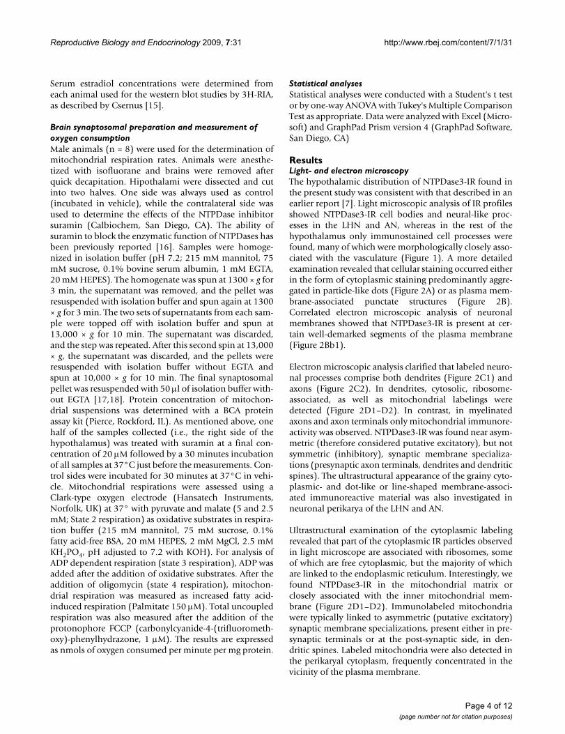
Brain synaptosomal preparation and measurement of oxygen consumptionMale animals (n = 8) were used for the determination ofmitochondrial respiration rates. Animals were anesthe-tized with isofluorane and brains were removed afterquick decapitation. Hipothalami were dissected and cutinto two halves. One side was always used as control(incubated in vehicle), while the contralateral side wasused to determine the effects of the NTPDase inhibitorsuramin (Calbiochem, San Diego, CA). The ability ofsuramin to block the enzymatic function of NTPDases hasbeen previously reported [16]. Samples were homoge-nized in isolation buffer (pH 7.2; 215 mM mannitol, 75mM sucrose, 0.1% bovine serum albumin, 1 mM EGTA,20 mM HEPES). The homogenate was spun at 1300 × g for3 min, the supernatant was removed, and the pellet wasresuspended with isolation buffer and spun again at 1300× g for 3 min. The two sets of supernatants from each sam-ple were topped off with isolation buffer and spun at13,000 × g for 10 min. The supernatant was discarded,and the step was repeated. After this second spin at 13,000× g, the supernatant was discarded, and the pellets wereresuspended with isolation buffer without EGTA andspun at 10,000 × g for 10 min. The final synaptosomalpellet was resuspended with 50 μl of isolation buffer with-out EGTA [17,18]. Protein concentration of mitochon-drial suspensions was determined with a BCA proteinassay kit (Pierce, Rockford, IL). As mentioned above, onehalf of the samples collected (i.e., the right side of thehypothalamus) was treated with suramin at a final con-centration of 20 μM followed by a 30 minutes incubationof all samples at 37°C just before the measurements. Con-trol sides were incubated for 30 minutes at 37°C in vehi-cle. Mitochondrial respirations were assessed using aClark-type oxygen electrode (Hansatech Instruments,Norfolk, UK) at 37° with pyruvate and malate (5 and 2.5mM; State 2 respiration) as oxidative substrates in respira-tion buffer (215 mM mannitol, 75 mM sucrose, 0.1%fatty acid-free BSA, 20 mM HEPES, 2 mM MgCl, 2.5 mMKH2PO4, pH adjusted to 7.2 with KOH). For analysis ofADP dependent respiration (state 3 respiration), ADP wasadded after the addition of oxidative substrates. After theaddition of oligomycin (state 4 respiration), mitochon-drial respiration was measured as increased fatty acid-induced respiration (Palmitate 150 μM). Total uncoupledrespiration was also measured after the addition of theprotonophore FCCP (carbonylcyanide-4-(trifluorometh-oxy)-phenylhydrazone, 1 μM). The results are expressedas nmols of oxygen consumed per minute per mg protein.
Statistical analysesStatistical analyses were conducted with a Student's t testor by one-way ANOVA with Tukey's Multiple ComparisonTest as appropriate. Data were analyzed with Excel (Micro-soft) and GraphPad Prism version 4 (GraphPad Software,San Diego, CA)
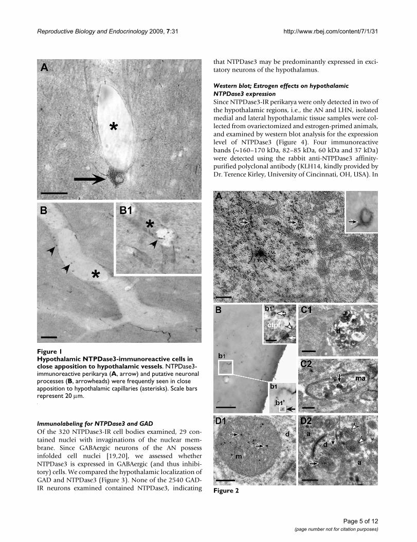
ResultsLight- and electron microscopyThe hypothalamic distribution of NTPDase3-IR found inthe present study was consistent with that described in anearlier report [7]. Light microscopic analysis of IR profilesshowed NTPDase3-IR cell bodies and neural-like proc-esses in the LHN and AN, whereas in the rest of thehypothalamus only immunostained cell processes werefound, many of which were morphologically closely asso-ciated with the vasculature (Figure 1). A more detailedexamination revealed that cellular staining occurred eitherin the form of cytoplasmic staining predominantly aggre-gated in particle-like dots (Figure 2A) or as plasma mem-brane-associated punctate structures (Figure 2B).Correlated electron microscopic analysis of neuronalmembranes showed that NTPDase3-IR is present at cer-tain well-demarked segments of the plasma membrane(Figure 2Bb1).
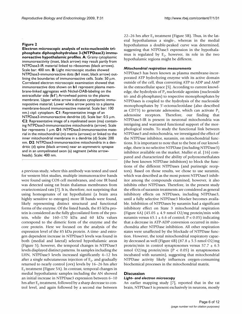
Electron microscopic analysis clarified that labeled neuro-nal processes comprise both dendrites (Figure 2C1) andaxons (Figure 2C2). In dendrites, cytosolic, ribosome-associated, as well as mitochondrial labelings weredetected (Figure 2D1–D2). In contrast, in myelinatedaxons and axon terminals only mitochondrial immunore-activity was observed. NTPDase3-IR was found near asym-metric (therefore considered putative excitatory), but notsymmetric (inhibitory), synaptic membrane specializa-tions (presynaptic axon terminals, dendrites and dendriticspines). The ultrastructural appearance of the grainy cyto-plasmic- and dot-like or line-shaped membrane-associ-ated immunoreactive material was also investigated inneuronal perikarya of the LHN and AN.
Ultrastructural examination of the cytoplasmic labelingrevealed that part of the cytoplasmic IR particles observedin light microscope are associated with ribosomes, someof which are free cytoplasmic, but the majority of whichare linked to the endoplasmic reticulum. Interestingly, wefound NTPDase3-IR in the mitochondrial matrix orclosely associated with the inner mitochondrial mem-brane (Figure 2D1–D2). Immunolabeled mitochondriawere typically linked to asymmetric (putative excitatory)synaptic membrane specializations, present either in pre-synaptic terminals or at the post-synaptic side, in den-dritic spines. Labeled mitochondria were also detected inthe perikaryal cytoplasm, frequently concentrated in thevicinity of the plasma membrane.
Page 4 of 12(page number not for citation purposes)
Reproductive Biology and Endocrinology 2009, 7:31 http://www.rbej.com/content/7/1/31
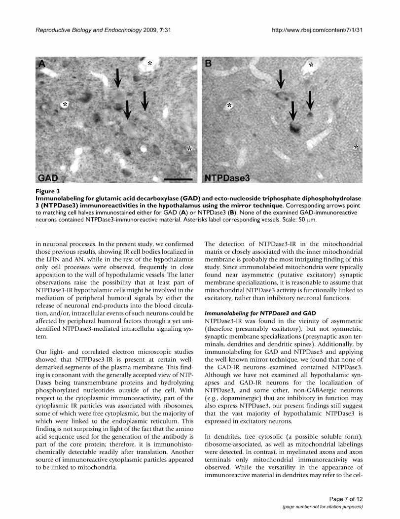
Immunolabeling for NTPDase3 and GADOf the 320 NTPDase3-IR cell bodies examined, 29 con-tained nuclei with invaginations of the nuclear mem-brane. Since GABAergic neurons of the AN possessinfolded cell nuclei [19,20], we assessed whetherNTPDase3 is expressed in GABAergic (and thus inhibi-tory) cells. We compared the hypothalamic localization ofGAD and NTPDase3 (Figure 3). None of the 2540 GAD-IR neurons examined contained NTPDase3, indicating
that NTPDase3 may be predominantly expressed in exci-tatory neurons of the hypothalamus.
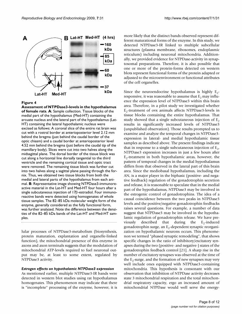
Western blot; Estrogen effects on hypothalamic NTPDase3 expressionSince NTPDase3-IR perikarya were only detected in two ofthe hypothalamic regions, i.e., the AN and LHN, isolatedmedial and lateral hypothalamic tissue samples were col-lected from ovariectomized and estrogen-primed animals,and examined by western blot analysis for the expressionlevel of NTPDase3 (Figure 4). Four immunoreactivebands (~160–170 kDa, 82–85 kDa, 60 kDa and 37 kDa)were detected using the rabbit anti-NTPDase3 affinity-purified polyclonal antibody (KLH14, kindly provided byDr. Terence Kirley, University of Cincinnati, OH, USA). In
Hypothalamic NTPDase3-immunoreactive cells in close apposition to hypothalamic vesselsFigure 1Hypothalamic NTPDase3-immunoreactive cells in close apposition to hypothalamic vessels. NTPDase3-immunoreactive perikarya (A, arrow) and putative neuronal processes (B, arrowheads) were frequently seen in close apposition to hypothalamic capillaries (asterisks). Scale bars represent 20 μm.
Figure 2
Page 5 of 12(page number not for citation purposes)
Reproductive Biology and Endocrinology 2009, 7:31 http://www.rbej.com/content/7/1/31
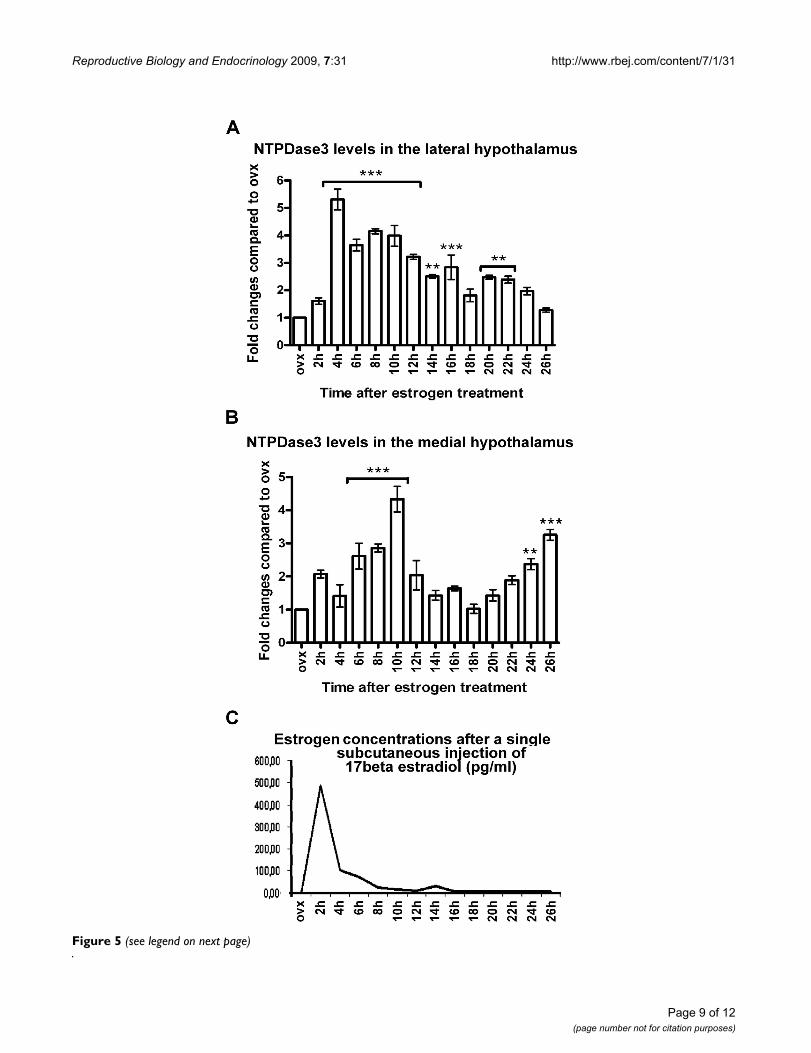
a previous study, where this antibody was tested and usedfor western blot studies, multiple immunoreactive bandswere found using COS cell membranes, and one IR bandwas detected using rat brain thalamus membranes fromovariectomized rats [7]. It is, therefore, not surprising thatusing homogenates of rat hypothalami (a brain areahighly sensitive to estrogen) more IR bands were found,likely representing distinct structural and functionalforms of the enzyme. Of the listed bands, the 85 kDa pro-tein is considered as the fully glycosilated form of the pro-tein, while the 160–170 kDa and 60 kDa valuescorrespond to the dimeric form of the enzyme and thecore protein. Here we focused on the analysis of theexpression level of the 85 kDa protein. A time- and estro-gen dependent increase in NTPDase3 levels was found inboth (medial and lateral) selected hypothalamic areas(Figure 5); however, the temporal changes in NTPDase3levels displayed distinct patterns. In samples including theLHN, NTPDase3 levels increased significantly 4–12 hrsafter a single subcutaneous injection of E2, and graduallyreturned to nearly control (ovx) levels by 16–26 hrs afterE2 treatment (Figure 5A). In contrast, temporal changes inmedial hypothalamic samples including the AN showedan initial increase in NTPDase3 expression between 6–10hrs after E2 treatment, followed by a sharp decrease to con-trol level, and again followed by a second rise between
22–26 hrs after E2 treatment (Figure 5B). Thus, in the lat-eral hypothalamus a single-, whereas in the medialhypothalamus a double-peaked curve was determined,suggesting that NTPDase3 expression in the hypothala-mus is regulated by E2, however, its role in the twohypothalamic regions might be different.
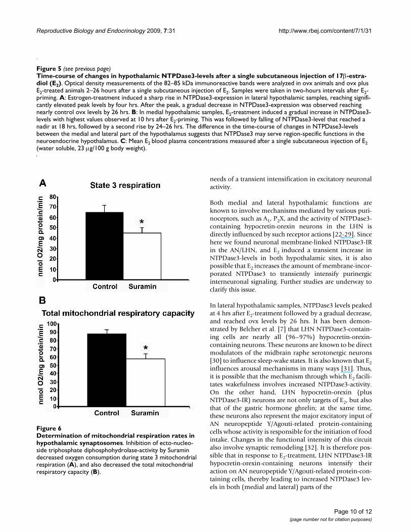
Mitochondrial respiration measurementsNTPDase3 has been known as plasma membrane-incor-porated ATP hydrolyzing enzyme with its active domainoutside of the cell, thus converting ATP to ADP and AMPin the extracellular space [5]. According to current knowl-edge, the hydrolysis of P2 nucleotide agonists (nucleosidetri- and di-phosphates) to respective monophosphates byNTPDases is coupled to the hydrolysis of the nucleosidemonophosphates by 5'-ectonucleotidase (also describedas CD73) to generate adenosine, which can activate P1adenosine receptors. Therefore, our finding thatNTPDase3-IR is present in neuronal mitochondria wasintriguing and warranted functional support of the mor-phological results. To study the functional link betweenNTPDase3 and mitochondria, we investigated the effect ofthe NTPDase inhibitor, suramin, on mitochondrial func-tions. It is important to note that to the best of our knowl-edge, there is no selective NTPDase (including NTPDase3)inhibitor available on the market. Muller et al. [16] com-pared and characterized the ability of polyoxomethalates(the best known NTPDase inhibitors) to block the func-tions of the different NTPDases (and purinergic recep-tors). Based on those results, we chose to use suramin,which was described as the most potent NTPDase3 inhib-itor among the compounds examined, however, it alsoinhibits other NTPDases. Therefore, in the present studythe effects of suramin treatments are considered as generalinhibitory effects on NTPDases, including NTPDase3,until a fully selective NTPDase3 blocker becomes availa-ble. Inhibition of NTPDases by suramin had a significantinhibitory effect on State 3 mitochondrial respiration(Figure 6A) (45.05 ± 4.9 nmol O2/mg protein/min withsuramin versus 65.1 ± 6.6 of control; P < 0.05) indicatingthat a decrease in ATP-ADP conversion occurred in mito-chondria after NTPDase inhibition. All other respirationstates were unaffected by the blockade of NTPDase func-tion. However, the total mitochondrial respiratory capac-ity decreased as well (Figure 6B) (87.8 ± 5.5 nmol O2/mgprotein/min in control synaptosomes versus 57.7 ± 6.5nmol O2/mg protein/min (P < 0.05) in synaptosomesincubated with suramin), suggesting that mitochondrialNTPDase activity likely influences oxygen-consumingbiochemical processes in the mitochondrial matrix.
DiscussionLight- and electron microscopyAn earlier mapping study [7], reported that in the ratbrain, NTPDase3 is present exclusively in neurons, mostly
Electron microscopic analysis of ecto-nucleoside triphos-phate diphosphohydrolase 3-(NTPDase3) immunoreactive hypothalamic profilesFigure 2Electron microscopic analysis of ecto-nucleoside tri-phosphate diphosphohydrolase 3-(NTPDase3) immu-noreactive hypothalamic profiles. A: Grainy cytoplasmic immunoreactivity (inset, black arrow) may result partly from NTPDase3-IR material linked to ribosomes (black arrows). Scale bar: 400 nm. B: Light microscopic studies showed NTPDase3-immunoreactive dots (b1 inset, black arrow) out-lining the boundaries of immunoreactive cells. Scale: 50 μm. Correlated electron microscopic examination showed that immunoreactive dots shown on b1 represent plasma mem-brane-linked aggregates with Nickel-DAB-labeling on the extracellular side (b1'; Arrowhead points to the plasma membrane. Upper white arrow indicates cytoplasmic immu-nopositive material; Lower white arrow points to a plasma membrane-bound immunoreactive material. Scale bar: 100 nm.) ctpl: cytoplasm. C1: Representative image of an NTPDase3-immunoreactive dendrite (d). Scale bar: 0.5 μm. C2: Representative image of a myelinated axon (ma) contain-ing NTPDase3-immunoreactive mitochondria (arrow). Scale bar represents 1 μm. D1: NTPDase3-immunoreactive mate-rial in the mitochondrial (m) matrix (arrows) or linked to the inner mitochondrial membrane in a dendrite (d) Scale: 200 nm. D2: NTPDase3-immunoreactive mitochondria in a den-dritic (d) spine (black arrows) near an asymmetric synapse and in an unmyelinated axon (a) segment (white arrow-heads). Scale: 400 nm.
Page 6 of 12(page number not for citation purposes)
Reproductive Biology and Endocrinology 2009, 7:31 http://www.rbej.com/content/7/1/31
in neuronal processes. In the present study, we confirmedthose previous results, showing IR cell bodies localized inthe LHN and AN, while in the rest of the hypothalamusonly cell processes were observed, frequently in closeapposition to the wall of hypothalamic vessels. The latterobservations raise the possibility that at least part ofNTPDase3-IR hypothalamic cells might be involved in themediation of peripheral humoral signals by either therelease of neuronal end-products into the blood circula-tion, and/or, intracellular events of such neurons could beaffected by peripheral humoral factors through a yet uni-dentified NTPDase3-mediated intracellular signaling sys-tem.
Our light- and correlated electron microscopic studiesshowed that NTPDase3-IR is present at certain well-demarked segments of the plasma membrane. This find-ing is consonant with the generally accepted view of NTP-Dases being transmembrane proteins and hydrolyzingphosphorylated nucleotides outside of the cell. Withrespect to the cytoplasmic immunoreactivity, part of thecytoplasmic IR particles was associated with ribosomes,some of which were free cytoplasmic, but the majority ofwhich were linked to the endoplasmic reticulum. Thisfinding is not surprising in light of the fact that the aminoacid sequence used for the generation of the antibody ispart of the core protein; therefore, it is immunohisto-chemically detectable readily after translation. Anothersource of immunoreactive cytoplasmic particles appearedto be linked to mitochondria.
The detection of NTPDase3-IR in the mitochondrialmatrix or closely associated with the inner mitochondrialmembrane is probably the most intriguing finding of thisstudy. Since immunolabeled mitochondria were typicallyfound near asymmetric (putative excitatory) synapticmembrane specializations, it is reasonable to assume thatmitochondrial NTPDase3 activity is functionally linked toexcitatory, rather than inhibitory neuronal functions.
Immunolabeling for NTPDase3 and GADNTPDase3-IR was found in the vicinity of asymmetric(therefore presumably excitatory), but not symmetric,synaptic membrane specializations (presynaptic axon ter-minals, dendrites and dendritic spines). Additionally, byimmunolabeling for GAD and NTPDase3 and applyingthe well-known mirror-technique, we found that none ofthe GAD-IR neurons examined contained NTPDase3.Although we have not examined all hypothalamic syn-apses and GAD-IR neurons for the localization ofNTPDase3, and some other, non-GABAergic neurons(e.g., dopaminergic) that are inhibitory in function mayalso express NTPDase3, our present findings still suggestthat the vast majority of hypothalamic NTPDase3 isexpressed in excitatory neurons.
In dendrites, free cytosolic (a possible soluble form),ribosome-associated, as well as mitochondrial labelingswere detected. In contrast, in myelinated axons and axonterminals only mitochondrial immunoreactivity wasobserved. While the versatility in the appearance ofimmunoreactive material in dendrites may refer to the cel-
Immunolabeling for glutamic acid decarboxylase (GAD) and ecto-nucleoside triphosphate diphosphohydrolase 3 (NTPDase3) immunoreactivities in the hypothalamus using the mirror techniqueFigure 3Immunolabeling for glutamic acid decarboxylase (GAD) and ecto-nucleoside triphosphate diphosphohydrolase 3 (NTPDase3) immunoreactivities in the hypothalamus using the mirror technique. Corresponding arrows point to matching cell halves immunostained either for GAD (A) or NTPDase3 (B). None of the examined GAD-immunoreactive neurons contained NTPDase3-immunoreactive material. Asterisks label corresponding vessels. Scale: 50 μm.
Page 7 of 12(page number not for citation purposes)
Reproductive Biology and Endocrinology 2009, 7:31 http://www.rbej.com/content/7/1/31
lular processes of NTPDase3-metabolism (biosynthesis,protein maturation, explantation and organelle-linkedfunction), the mitochondrial presence of this enzyme inaxons and axon terminals suggests that the modulation ofmitochondrial ATP-levels required to fuel neuronal out-put may be, at least to some extent, regulated byNTPDase3 activity.
Estrogen effects on hypothalamic NTPDase3 expressionAs mentioned earlier, multiple NTPDase3-IR bands weredetected in western blot studies using rat hypothalamushomogenates. This phenomenon may indicate that thereis "incomplete" processing of the enzyme, however, it is
more likely that the distinct bands observed represent dif-ferent maturational forms of the enzyme. In this study, wedetected NTPDase3-IR linked to multiple subcellularstructures (plasma membrane, ribosomes, endoplasmicreticulum) including neuronal mitochondria. Addition-ally, we provided evidence for NTPDase-activity in synap-tosomal preparations. Therefore, it is also possible thatone or more of the protein-forms detected on westernblots represent functional forms of the protein adapted oradjusted to the microenvironment or functional attributesof the cell organelles.
Since the neuroendocrine hypothalamus is highly E2-responsive, it was reasonable to assume that E2 may influ-ence the expression level of NTPDase3 within this brainarea. Therefore, in a pilot study we investigated whetherE2-treatment of ovx animals affects NTPDase3-levels intissue blocks containing the entire hypothalamus. Thatstudy showed that a single subcutaneous injection of E2results in significantly increased levels of NTPDase3(unpublished observation). Those results prompted us toexamine and analyze the temporal changes in NTPDase3-expression in lateral- and medial hypothalamic tissuesamples as described above. The present findings indicatethat in response to a single subcutaneous injection of E2,NTPDase3 expression increases in just a few hours afterE2-treatment in both hypothalamic areas, however, thepattern of temporal changes in the medial hypothalamusdiffers from that observed in the lateral part of this brainarea. Since the mediobasal hypothalamus, including theAN, is a major player in the biphasic (positive- and nega-tive feedback) regulation of the gonadotrophin secretionand release, it is reasonable to speculate that in the medialpart of the hypothalamus, NTPDase3 may be involved inthe estrogenic control of gonadotrophins. The idea of acausal coincidence between the two peaks in NTPDase3levels and the positive/negative gonadotrophin feedbacksraises several questions. For example, a number of datasuggest that NTPDase3 may be involved in the hypotha-lamic regulation of gonadotrophin release. We have pre-viously described that during the E2-inducedgonadotrophin surge, an E2-dependent synaptic reorgani-zation on hypothalamic neurons occurs. This phenome-non we termed "phased synaptic remodeling", that showsspecific changes in the ratio of inhibitory/excitatory syn-apses during the two (positive- and negative-) states of thegonadotrophin feedback control [21]. A sharp rise in thenumber of excitatory synapses was observed at the time ofthe E2-surge, and the formation of new synapses may verywell include ones equipped with NTPDase3-containingmitochondria. This hypothesis is consonant with ourobservation that inhibition of NTPDase activity decreasesstate 3 mitochondrial respiration and the total mitochon-drial respiratory capacity, ergo an increased amount ofmitochondrial NTPDase would well serve the energy-
Assessment of NTPDase3-levels in the hypothalamus of female ratsFigure 4Assessment of NTPDase3-levels in the hypothalamus of female rats. A: Sample collection. Tissue blocks of the medial part of the hypothalamus (Med-HT) containing the arcuate nucleus and the lateral part of the hypothalamus (Lat-HT) containing the lateral hypothalamic nucleus were excised as follows: A coronal slice of the entire rat brain was cut with a rostral border at anterioposterior level 2.12 mm behind the bregma (just behind the caudal border of the optic chiasm) and a caudal border at anterioposterior level 4.52 mm behind the bregma (just before the caudal tip of the mamillary body). Slices were cut into two halves along the midsagittal plane. The dorsal border of the tissue block was cut along a horizontal line dorsally tangential to the third ventricle and the remaining cortical tissue and optic tract were removed. The remaining tissue block was further cut into two halves along a sagittal plane passing through the for-nix. Thus, we obtained two tissue blocks from both the medial and lateral parts of the hypothalamus from each ani-mal. B: Representative image showing NTPDase3-immunore-active material in the Lat-HT and Med-HT four hours after a single subcutaneous injection of 17β-estradiol. Four immuno-reactive bands were detected using homogenates of whole tissue samples. The 82–85 kDa molecular weight form of the enzyme, generally considered as the fully functional form, was further analyzed. Note the difference between the densi-ties of the 82–85 kDa bands of the Lat-HT and Med-HT sam-ples.
Page 8 of 12(page number not for citation purposes)
Reproductive Biology and Endocrinology 2009, 7:31 http://www.rbej.com/content/7/1/31
Figure 5 (see legend on next page)
Page 9 of 12(page number not for citation purposes)
Reproductive Biology and Endocrinology 2009, 7:31 http://www.rbej.com/content/7/1/31
needs of a transient intensification in excitatory neuronalactivity.
Both medial and lateral hypothalamic functions areknown to involve mechanisms mediated by various puri-noceptors, such as A1, P2X, and the activity of NTPDase3-containing hypocretin-orexin neurons in the LHN isdirectly influenced by such receptor actions [22-29]. Sincehere we found neuronal membrane-linked NTPDase3-IRin the AN/LHN, and E2 induced a transient increase inNTPDase3-levels in both hypothalamic sites, it is alsopossible that E2 increases the amount of membrane-incor-porated NTPDase3 to transiently intensify purinergicinterneuronal signaling. Further studies are underway toclarify this issue.
In lateral hypothalamic samples, NTPDase3 levels peakedat 4 hrs after E2-treatment followed by a gradual decrease,and reached ovx levels by 26 hrs. It has been demon-strated by Belcher et al. [7] that LHN NTPDase3-contain-ing cells are nearly all (96–97%) hypocretin-orexin-containing neurons. These neurons are known to be directmodulators of the midbrain raphe serotonergic neurons[30] to influence sleep-wake states. It is also known that E2influences arousal mechanisms in many ways [31]. Thus,it is possible that the mechanism through which E2 facili-tates wakefulness involves increased NTPDase3-activity.On the other hand, LHN hypocretin-orexin (plusNTPDase3-IR) neurons are not only targets of E2, but alsothat of the gastric hormone ghrelin; at the same time,these neurons also represent the major excitatory input ofAN neuropeptide Y/Agouti-related protein-containingcells whose activity is responsible for the initiation of foodintake. Changes in the functional intensity of this circuitalso involve synaptic remodeling [32]. It is therefore pos-sible that in response to E2-treatment, LHN NTPDase3-IRhypocretin-orexin-containing neurons intensify theiraction on AN neuropeptide Y/Agouti-related protein-con-taining cells, thereby leading to increased NTPDase3 lev-els in both (medial and lateral) parts of the
Time-course of changes in hypothalamic NTPDase3-levels after a single subcutaneous injection of 17β-estradiol (E2)Figure 5 (see previous page)Time-course of changes in hypothalamic NTPDase3-levels after a single subcutaneous injection of 17β-estra-diol (E2). Optical density measurements of the 82–85 kDa immunoreactive bands were analyzed in ovx animals and ovx plus E2-treated animals 2–26 hours after a single subcutaneous injection of E2. Samples were taken in two-hours intervals after E2-priming. A: Estrogen-treatment induced a sharp rise in NTPDase3-expression in lateral hypothalamic samples, reaching signifi-cantly elevated peak levels by four hrs. After the peak, a gradual decrease in NTPDase3-expression was observed reaching nearly control ovx levels by 26 hrs. B: In medial hypothalamic samples, E2-treatment induced a gradual increase in NTPDase3-levels with highest values observed at 10 hrs after E2-priming. This was followed by falling of NTPDase3-level that reached a nadir at 18 hrs, followed by a second rise by 24–26 hrs. The difference in the time-course of changes in NTPDase3-levels between the medial and lateral part of the hypothalamus suggests that NTPDase3 may serve region-specific functions in the neuroendocrine hypothalamus. C: Mean E2 blood plasma concentrations measured after a single subcutaneous injection of E2 (water soluble, 23 μg/100 g body weight).
Determination of mitochondrial respiration rates in hypotha-lamic synaptosomesFigure 6Determination of mitochondrial respiration rates in hypothalamic synaptosomes. Inhibition of ecto-nucleo-side triphosphate diphosphohydrolase-activity by Suramin decreased oxygen consumption during state 3 mitochondrial respiration (A), and also decreased the total mitochondrial respiratory capacity (B).
Page 10 of 12(page number not for citation purposes)

Reproductive Biology and Endocrinology 2009, 7:31 http://www.rbej.com/content/7/1/31
hypothalamus. If this was the case, one could speculatethat the orexigenic effect of E2 may in some way involvethe action of NTPDase3.
Mitochondrial respiration measurementsIt has been shown that interneuronal signaling is a highlyATP-dependent, energy-demanding process [33]. To sup-ply the energy needs of neurotransmission, ATP is pro-duced and maintained in neuronal mitochondria in aregulated fashion. We have previously proposed [34] thatone potential mechanism down-regulating mitochondrialATP production may involve uncoupling proteins(UCPs), specifically UCP2, which was only found ininhibitory neurons of the hypothalamus. However, thespecific mechanism involved in the regulation of mito-chondrial ATP levels in excitatory hypothalamic neuronsis currently unknown. Therefore, the identification ofNTPDase3 in mitochondria in synaptic or perikaryal sitesof excitatory hypothalamic neurons might be the mostnovel and intriguing finding of this study, and warrantsfurther experiments to elucidate the exact functional roleof mitochondrial NTPDase3 in neurotransmission.
To confirm our morphological findings, we isolated syn-aptosomes from hypothalamic tissue homogenates andexamined mitochondrial respiration in control versussuramin (an NTPDase inhibitor) treated samples.Suramin blockade of NTPDases reduced mitochondrialoxygen consumption in state 3 mitochondrial respirationby 30%, and also decreased the total mitochondrial respi-ratory capacity by 34%. These findings imply that induc-tion of NTPDase activity in the mitochondria byhydrolyzing ATP to ADP increases mitochondrial state 3respiration and the total mitochondrial respiration capac-ity. It should be noted that the polyoxometalate suraminis not a fully selective NTPDase3-inhibitor, as such inhib-itors, to the best of our knowledge, for the 8 known NTP-Dases have not yet been found. However, suramin hasbeen shown to be a potent inhibitor of NTPDase3 andother NTPDases [16], therefore the observed reduction inoxygen consumption in hypothalamic synaptosomes canbe, at least in part, attributed to the inhibition ofNTPDase3. This idea is supported by previous results onsynaptosome fractions isolated from rat brain cortex andstriatum [35], and from hippocampal synaptosomes [36].These studies report a transient accumulation of ADP afteraddition of ATP followed by the subsequent metaboliza-tion of ADP to AMP and adenosine. As a result, the afore-mentioned studies argue against a considerablecontribution by NTPDase1 and/or NTPDase2 and suggestthat the observations would rather be compatible with aneuronal expression of NTPDase3.
Based on the present findings, it is tempting to speculatethat an increase in the activity level of NTPDases
(NTPDase3?) may result in the exhaustion of mitochon-dria (and the parent cell), whereas partial inhibition ofNTPDases may be neuroprotective. Ongoing studies inour laboratory test this hypothesis. The reported pharma-cological effects of polyoxometalates, such as suramin,seem to support this idea. For example, some data showthat NTPDase inhibition is also antidiabetic [37],although the exact mechanisms through which the bene-ficial effects of NTPDase inhibitors act are unknown.Therefore, the present results rise the possibility that in thepancreas, inhibition of NTPDases (NTPDase3) may pro-tect the insulin-producing beta cells from overt ATP con-sumption and consequential exhaustion.
ConclusionAltogether, the combined morphological and functionalexamination of neuronal NTPDase3-IR suggests a doublecellular role for this enzyme in the hypothalamus: 1. assuggested earlier, the regulation of purinergic signaling viathe production of specific ligands (ADP and AMP), and 2.the tuning of energy supply for (excitatory) neurotrans-mission by the hydrolysis of mitochondrial ATP.
Competing interestsThe authors declare that they have no competing interests.
Authors' contributionsDSK was responsible for immunohistochemical studiesincluding the "mirror" technique analysis; AZ and SD cre-ated the experimental design, supervised the experimentsand wrote this report; KH and VLF carried out the mito-chondrial respiration measurements and related analyses;AG and VS performed the western blot experiments; TBhad a substantial share in overall data analysis, discussionof data, and participated in most of these experiments;DH and PS were responsible for the electron microscopiclab work and analyses. All authors read and approved thefinal manuscript.
AcknowledgementsThis work was supported by OTKA 46914 and 49756 (to A.Z.) and NIH grant DK070039 and American Diabetes Association (to S.D.). The authors are indebted to Dr. Terence L. Kirley for generously providing the used rabbit anti-NTPDase3 polyclonal antibody. We would like to express our gratitude to Dr. Margit Kulcsar for the determination of serum estradiol concentrations.
References1. Burnstock G: Physiology and pathophysiology of purinergic
neurotransmission. Physiol Rev 2007, 87:659-797.2. Appelbaum L, Skariah G, Mourrain P, Mignot E: Comparative
expression of p2x receptors and ecto-nucleoside triphos-phate diphosphohydrolase 3 in hypocretin and sensory neu-rons in zebrafish. Brain Res 2007, 1174:66-75.
3. Wang T-F, Guidotti G: Widespread expression of ecto-apyrase(CD39) in the central nervous system. Brain Res 1998,790:318-322.
4. Braun N, Sevigny J, Mishra SK, Robson SC, Barth SW, Gerstberger R,Hammer K, Zimmermann H: Expression of the ecto-ATPase
Page 11 of 12(page number not for citation purposes)
http://www.ncbi.nlm.nih.gov/entrez/query.fcgi?cmd=Retrieve&db=PubMed&dopt=Abstract&list_uids=9593967
Reproductive Biology and Endocrinology 2009, 7:31 http://www.rbej.com/content/7/1/31
Publish with BioMed Central and every scientist can read your work free of charge
"BioMed Central will be the most significant development for disseminating the results of biomedical research in our lifetime."
Sir Paul Nurse, Cancer Research UK
Your research papers will be:
available free of charge to the entire biomedical community
peer reviewed and published immediately upon acceptance
cited in PubMed and archived on PubMed Central
yours — you keep the copyright
Submit your manuscript here:http://www.biomedcentral.com/info/publishing_adv.asp
BioMedcentral
NTPDase2 in the germinal zones of the developing and adultrat brain. Eur J Neurosci 2003, 17:1355-1364.
5. Smith TM, Kirley TL: Cloning, sequencing, and expression of ahuman brain ecto-apyrase related to both the ecto-ATPasesand CD39 ecto-apyrases. Biochim Biophys Acta 1998, 1386:65-78.
6. Chadwick BP, Frischauf AM: The CD39-like gene family: Identi-fication of three human members (CD39L2, CD39L3, andCD39L4), their murine homologues, and a member of thegene family from Drosophila melanogaster. Genomics 1998,50:357-367.
7. Belcher SM, Zsarnovszky A, Crawford PA, Hemani H, Spurling L,Kirley TL: Immunolocalization of ecto-nucleoside triphos-phate diphosphohydrolase 3 in rat brain: implications formodulation of multiple homeostatic systems including feed-ing and sleep-wake behaviors. Neuroscience 2006,137:1331-1346.
8. Zsarnovszky A, Horvath TL, Garcia-Segura LM, Horvath B, NaftolinF: Oestrogen-induced changes in the synaptology of themonkey (Cercopithecus aethiops) arcuate nucleus duringgonadotropin feedback. J Neuroendocrinol 2001, 13:22-28.
9. Colonnier M: Synaptic patterns on different cell types in thedifferent laminae of the cat visual cortex. Brain Res 1968,9:268-287.
10. Palay SL, Chan-Palay V: A guide to the synaptic analysis of theneuropil. Cold Spring Harb Symp Quant Biol 1975, 40:1-16.
11. Zsarnovszky A, Horvath TL, Naftolin F, Leranth C: AMPA recep-tors colocalize with neuropeptide-Y- and galanin-containing,but not with dopamine neurons of the female rat arcuatenucleus: a semiquantitative immunohistochemical colocali-zation study. Exp Brain Res 2000, 133:532-537.
12. Paxinos G, Watson C: The Rat Brain in Stereotaxic Coordinates 2nd edi-tion. Academic Press; 1986.
13. Jakab RL, Wong JK, Belcher SM: Estrogen receptor-β immunore-activity in differentiating cells of the developing rat cerebel-lum. J Comp Neurol 2001, 430:396-409.
14. Wong JK, Le HH, Zsarnovszky A, Belcher SM: Estrogens andICI182,780 (Faslodex) modulate mitosis and cell death inimmature cerebellar neurons via rapid activation of p44/p42mitogen-activated protein kinase. J Neurosci 2003,23:4984-4995.
15. Csernus V: Antibodies of high affinity and specificity for radio-immunological determination of progesterone, testoster-one and estradiol-17b. In Advances in steroid analysis Edited by:Gorog S. Akademiai Kiado Bp; 1982:171-179.
16. Muller CE, Iqbal J, Baqi Y, Zimmermann H, Rollich A, Stephan H:Polyoxometalates – a new class of potent ecto-nucleosidetriphosphate diphosphohydrolase (NTPDase) inhibitors.Bioorg Med Chemistry Lett 2006, 16:5943-5947.
17. Coppola A, Liu ZW, Adrews ZB, Paradis E, Roy MC, Friedman JM,Ricquier D, Richard D, Horvath TL, Gao XB, Diano S: A centralthermogenic-like mechanism in feeding regulation: an inter-play between arcuate nucleus T3 and UCP2. Cell Metab 2007,5:21-33.
18. Andrews ZB, Liu ZW, Wallingford N, Erion DM, Borok E, FriedmanJM, Tschop MH, Shanabrough M, Cline G, Shulman GI, Coppola A,Gao XB, Horvath TL, Diano S: UCP2 mediates ghrelin's actionin NPY/AgRP neurons by lowering free radicals. Nature 2008,454:846-851.
19. Leranth C, Sakamoto H, MacLusky NJ, Shanabrough M, Naftolin F:Estrogen responsive cells in the arcuate nucleus of the ratcontain glutamic acid decarboxylase (GAD): an electronmicroscopic immunocytochemical study. Brain Res 1985,331:376-381.
20. Leranth C, Shanabrough M, Naftolin F: Estrogen inducesultrastructural changes in progesterone receptor-containingGABA neurons of the primate hypothalamus. Neuroendocrinol-ogy 1991, 54:571-579.
21. Naftolin F, Garcia-Segura LM, Horvath TL, Zsarnovszky A, Demir N,Fadiel A, Leranth C, Vondracek-Klepper S, Lewis C, Chang A, ParduczA: Estrogen-induced hypothalamic synaptic plasticity andpituitary sensitization in the control of the estrogen-inducedgonadotrophin surge. Reprod Sci 2007, 14:101-116.
22. Thakkar MM, Winston S, McCarley RW: Orexin neurons of thehypothalamus express adenosine A1 receptors. Brain Res2002, 944:190-194.
23. Gordon GR, Baimoukhametova DV, Hewitt SA, Rajapaksha WR,Fisher TE, Bains JS: Norepinephrine triggers release of glialATP to increase postsynaptic efficacy. Nat Neurosci 2005,8:1078-1086.
24. Wollmann G, Acuna-Goycolea C, Pol AN van den: Direct excita-tion of hypocretin/orexin cells by extracellular ATP at P2Xreceptors. J Neurophysiol 2005, 94:2195-2206.
25. Florenzano F, Viscomi MT, Mercaldo V, Longone P, Bernardi G, BagniC, Molinari M, Carrive P: P2X2R purinergic receptor subunitmRNA and protein are expressed by all hypothalamic hypo-cretin/orexin neurons. J Comp Neurol 2006, 498:58-67.
26. Kittner H, Franke H, Harsch JI, El-Ashmawy IM, Seidel B, Krugel U,Illes P: Enhanced food intake after stimulation of hypotha-lamic P2Y1 receptors in rats: modulation of feeding behav-iour by extracellular nucleotides. Eur J Neurosci 2006,24:2049-2056.
27. Seidel B, Bigl M, Franke H, Kittner H, Kiess W, Illes P, Krugel U:Expression of purinergic receptors in the hypothalamus ofthe rat is modified by reduced food availability. Brain Res 2006,1089:143-152.
28. Knott TK, Marrero HG, Fenton RA, Custer EE, Dobson JG Jr, LemosJR: Endogenous adenosine inhibits CNS terminal Ca(2+) cur-rents and exocytosis. J Cell Physiol 2007, 210:309-314.
29. Liu ZW, Gao XB: Adenosine inhibits activity of hypocretin/orexin neurons by the A1 receptor in the lateral hypothala-mus: a possible sleep-promoting effect. J Neurophysiol 2007,97:837-848.
30. Liu RJ, Pol AN van den, Aghajanian GK: Hypocretins (orexins) reg-ulate serotonin neurons in the dorsal raphe nucleus by exci-tatory direct and inhibitory indirect actions. J Neurosci 2002,22:9453-9464.
31. Lee AW, Pfaff DW: Hormone effects on specific and globalbrain functions. J Physiol Sci 2008, 58:213-220.
32. Horvath TL: The hardship of obesity: a soft-wired hypothala-mus. Nat Neurosci 2005, 8:561-565.
33. Laughlin SB, de Ruyter van Stevenick RR, Anderson JC: The meta-bolic cost of neuronal information. Nat Neurosci 1998,1:436-441.
34. Horvath TL, Warden CH, Hajos M, Lombardi A, Goglia F, Diano S:Brain uncoupling protein 2: Uncoupled neuronal mitochon-dria predict thermal synapses in homeostatic centers. J Neu-rosci 1999, 19:10417-10427.
35. James S, Richardson PJ: Production of adenosine from extracel-lular ATP at the striatal cholinergic synapse. J Neurochem1993, 60:219-227.
36. Cunha RA: Regulation of the ecto-nucleotidase pathway in rathippocampal nerve terminals. Neurochem Res 2001, 26:979-991.
37. Hillaire-Buys D, Chapal J, Bertrand G, Petit P, Loubatieres-MarianiMM: Purinergic receptors on insulin-secreting cells. FundamClin Pharmacol 1994, 8:117-127.
Page 12 of 12(page number not for citation purposes)
http://www.ncbi.nlm.nih.gov/entrez/query.fcgi?cmd=Retrieve&db=PubMed&dopt=Abstract&list_uids=9675246
http://www.ncbi.nlm.nih.gov/entrez/query.fcgi?cmd=Retrieve&db=PubMed&dopt=Abstract&list_uids=9675246
http://www.ncbi.nlm.nih.gov/entrez/query.fcgi?cmd=Retrieve&db=PubMed&dopt=Abstract&list_uids=9675246
http://www.ncbi.nlm.nih.gov/entrez/query.fcgi?cmd=Retrieve&db=PubMed&dopt=Abstract&list_uids=9676430
http://www.ncbi.nlm.nih.gov/entrez/query.fcgi?cmd=Retrieve&db=PubMed&dopt=Abstract&list_uids=9676430
http://www.ncbi.nlm.nih.gov/entrez/query.fcgi?cmd=Retrieve&db=PubMed&dopt=Abstract&list_uids=9676430
http://www.ncbi.nlm.nih.gov/entrez/query.fcgi?cmd=Retrieve&db=PubMed&dopt=Abstract&list_uids=4175993
http://www.ncbi.nlm.nih.gov/entrez/query.fcgi?cmd=Retrieve&db=PubMed&dopt=Abstract&list_uids=4175993
http://www.ncbi.nlm.nih.gov/entrez/query.fcgi?cmd=Retrieve&db=PubMed&dopt=Abstract&list_uids=3986576
http://www.ncbi.nlm.nih.gov/entrez/query.fcgi?cmd=Retrieve&db=PubMed&dopt=Abstract&list_uids=3986576
http://www.ncbi.nlm.nih.gov/entrez/query.fcgi?cmd=Retrieve&db=PubMed&dopt=Abstract&list_uids=3986576
http://www.ncbi.nlm.nih.gov/entrez/query.fcgi?cmd=Retrieve&db=PubMed&dopt=Abstract&list_uids=1723787
http://www.ncbi.nlm.nih.gov/entrez/query.fcgi?cmd=Retrieve&db=PubMed&dopt=Abstract&list_uids=1723787
http://www.ncbi.nlm.nih.gov/entrez/query.fcgi?cmd=Retrieve&db=PubMed&dopt=Abstract&list_uids=1723787
http://www.ncbi.nlm.nih.gov/entrez/query.fcgi?cmd=Retrieve&db=PubMed&dopt=Abstract&list_uids=8417143
http://www.ncbi.nlm.nih.gov/entrez/query.fcgi?cmd=Retrieve&db=PubMed&dopt=Abstract&list_uids=8417143