Efficient Chemotherapy of Rat Glioblastoma Using Doxorubicin-Loaded PLGA Nanoparticles with...
8
Efficient Chemotherapy of Rat Glioblastoma Using Doxorubicin-Loaded PLGA Nanoparticles with Different Stabilizers Stefanie Wohlfart 1 , Alexander S. Khalansky 2 , Svetlana Gelperina 3 , Olga Maksimenko 3 , Christian Bernreuther 4 , Markus Glatzel 4 , Jo ¨ rg Kreuter 1 * 1 Institute of Pharmaceutical Technology, Goethe-University, Frankfurt, Germany, 2 Institute of Human Morphology, Moscow, Russia, 3 Nanosystem Ltd., Moscow, Russia, 4 Institute of Neuropathology, University Medical Center Hamburg-Eppendorf, Hamburg, Germany Abstract Background: Chemotherapy of glioblastoma is largely ineffective as the blood-brain barrier (BBB) prevents entry of most anticancer agents into the brain. For an efficient treatment of glioblastomas it is necessary to deliver anti-cancer drugs across the intact BBB. Poly(lactic-co-glycolic acid) (PLGA) nanoparticles coated with poloxamer 188 hold great promise as drug carriers for brain delivery after their intravenous injection. In the present study the anti-tumour efficacy of the surfactant-coated doxorubicin-loaded PLGA nanoparticles against rat glioblastoma 101/8 was investigated using histological and immunohistochemical methods. Methodology: The particles were prepared by a high-pressure solvent evaporation technique using 1% polyvinylalcohol (PLGA/PVA) or human serum albumin (PLGA/HSA) as stabilizers. Additionally, lecithin-containing PLGA/HSA particles (Dox- Lecithin-PLGA/HSA) were prepared. For evaluation of the antitumour efficacy the glioblastoma-bearing rats were treated intravenously with the doxorubicin-loaded nanoparticles coated with poloxamer 188 using the following treatment regimen: 3 6 2.5 mg/kg on day 2, 5 and 8 after tumour implantation; doxorubicin and poloxamer 188 solutions were used as controls. On day 18, the rats were sacrificed and the antitumour effect was determined by measurement of tumour size, necrotic areas, proliferation index, and expression of GFAP and VEGF as well as Isolectin B4, a marker for the vessel density. Conclusion: The results reveal a considerable anti-tumour effect of the doxorubicin-loaded nanoparticles. The overall best results were observed for Dox-Lecithin-PLGA/HSA. These data demonstrate that the poloxamer 188-coated PLGA nanoparticles enable delivery of doxorubicin across the blood-brain barrier in the therapeutically effective concentrations. Citation: Wohlfart S, Khalansky AS, Gelperina S, Maksimenko O, Bernreuther C, et al. (2011) Efficient Chemotherapy of Rat Glioblastoma Using Doxorubicin- Loaded PLGA Nanoparticles with Different Stabilizers. PLoS ONE 6(5): e19121. doi:10.1371/journal.pone.0019121 Editor: Maria G. Castro, University of California Los Angeles, and Cedars-Sinai Medical Center, United States of America Received January 24, 2011; Accepted March 16, 2011; Published May 6, 2011 Copyright: ß 2011 Wohlfart et al. This is an open-access article distributed under the terms of the Creative Commons Attribution License, which permits unrestricted use, distribution, and reproduction in any medium, provided the original author and source are credited. Funding: This study was supported in part (S.G and O.M.) by residual funds from INTAS (International Association for the promotion of cooperation with scientists from the new independent states of the former Soviet Union, Brussels, Belgium, Grant 00-838) and by travel grants for A.S.K. and S.G. by the DFG (Deutsche Forschungsgemeinschaft). All studies in Frankfurt and Hamburg including the work of A.S.K. were supported by the regular department funds of Goethe/University Frankfurt of the University Clinics Hamburg. No additional external funding was received for this study. The funders had no role in study design, data collection and analysis, decision to publish, or preparation of the manuscript. Competing Interests: Although two of the authors (S.G. and O.M.) are employed by a commercial company (Nanosystem LTD.), no patent is pending and these particles are not in a commercial development. This does not alter the authors9 adherence to all the PLoS ONE policies on sharing data and materials. * E-mail: [email protected] Introduction Glioblastoma multiforme is the most common and most aggressive type of primary brain tumours in humans accounting for 20% of all intracranial tumours [1]. Chemotherapy of glioblastoma is largely ineffective as the blood-brain barrier (BBB) prevents entry of most anticancer agents into the brain. Conventional methods for enhancing drug concentrations in the brain, such as disruption of the BBB, intraventricular drug injection or local therapy, are highly invasive and, therefore, are not applicable for long-term treatment regimens. Local drug therapy with drugs incorporated into implants or administered by local injection or implantation, on the other hand, suffers from the limited diffusional area that is accessible for the drug released after implantation [2]. One of the most promising approaches, therefore, is the intravenous injection of specially coated nanoparticles that are able to transport drugs across the BBB [3,4,5]. Due to this special coating, the nanoparticles adsorb certain blood plasma apolipo- proteins [6] that enable an interaction of the particles with the respective lipoprotein receptors located on the brain blood capillary endothelial cells followed by the internalisation of the particles [7] and drug beyond the BBB. Alternatively, other ligands for which receptors exist on these cells or antibodies against these receptors may be bound covalently to the nanoparticles to enable the passage through the BBB [8]. To date, most experiments concerning the treatment of glioblastoma with nanoparticles were performed in rats using doxorubicin-loaded poly(butyl cyanoacrylate) particles coated with polysorbate 80 (TweenH 80) using the extremely aggressive rat PLoS ONE | www.plosone.org 1 May 2011 | Volume 6 | Issue 5 | e19121
-
Upload
independent -
Category
Documents
-
view
1 -
download
0
Transcript of Efficient Chemotherapy of Rat Glioblastoma Using Doxorubicin-Loaded PLGA Nanoparticles with...

Efficient Chemotherapy of Rat Glioblastoma UsingDoxorubicin-Loaded PLGA Nanoparticles with DifferentStabilizersStefanie Wohlfart1, Alexander S. Khalansky2, Svetlana Gelperina3, Olga Maksimenko3, Christian
Bernreuther4, Markus Glatzel4, Jorg Kreuter1*
1 Institute of Pharmaceutical Technology, Goethe-University, Frankfurt, Germany, 2 Institute of Human Morphology, Moscow, Russia, 3 Nanosystem Ltd., Moscow, Russia,
4 Institute of Neuropathology, University Medical Center Hamburg-Eppendorf, Hamburg, Germany
Abstract
Background: Chemotherapy of glioblastoma is largely ineffective as the blood-brain barrier (BBB) prevents entry of mostanticancer agents into the brain. For an efficient treatment of glioblastomas it is necessary to deliver anti-cancer drugsacross the intact BBB. Poly(lactic-co-glycolic acid) (PLGA) nanoparticles coated with poloxamer 188 hold great promise asdrug carriers for brain delivery after their intravenous injection. In the present study the anti-tumour efficacy of thesurfactant-coated doxorubicin-loaded PLGA nanoparticles against rat glioblastoma 101/8 was investigated usinghistological and immunohistochemical methods.
Methodology: The particles were prepared by a high-pressure solvent evaporation technique using 1% polyvinylalcohol(PLGA/PVA) or human serum albumin (PLGA/HSA) as stabilizers. Additionally, lecithin-containing PLGA/HSA particles (Dox-Lecithin-PLGA/HSA) were prepared. For evaluation of the antitumour efficacy the glioblastoma-bearing rats were treatedintravenously with the doxorubicin-loaded nanoparticles coated with poloxamer 188 using the following treatmentregimen: 362.5 mg/kg on day 2, 5 and 8 after tumour implantation; doxorubicin and poloxamer 188 solutions were used ascontrols. On day 18, the rats were sacrificed and the antitumour effect was determined by measurement of tumour size,necrotic areas, proliferation index, and expression of GFAP and VEGF as well as Isolectin B4, a marker for the vessel density.
Conclusion: The results reveal a considerable anti-tumour effect of the doxorubicin-loaded nanoparticles. The overall bestresults were observed for Dox-Lecithin-PLGA/HSA. These data demonstrate that the poloxamer 188-coated PLGAnanoparticles enable delivery of doxorubicin across the blood-brain barrier in the therapeutically effective concentrations.
Citation: Wohlfart S, Khalansky AS, Gelperina S, Maksimenko O, Bernreuther C, et al. (2011) Efficient Chemotherapy of Rat Glioblastoma Using Doxorubicin-Loaded PLGA Nanoparticles with Different Stabilizers. PLoS ONE 6(5): e19121. doi:10.1371/journal.pone.0019121
Editor: Maria G. Castro, University of California Los Angeles, and Cedars-Sinai Medical Center, United States of America
Received January 24, 2011; Accepted March 16, 2011; Published May 6, 2011
Copyright: � 2011 Wohlfart et al. This is an open-access article distributed under the terms of the Creative Commons Attribution License, which permitsunrestricted use, distribution, and reproduction in any medium, provided the original author and source are credited.
Funding: This study was supported in part (S.G and O.M.) by residual funds from INTAS (International Association for the promotion of cooperation withscientists from the new independent states of the former Soviet Union, Brussels, Belgium, Grant 00-838) and by travel grants for A.S.K. and S.G. by the DFG(Deutsche Forschungsgemeinschaft). All studies in Frankfurt and Hamburg including the work of A.S.K. were supported by the regular department funds ofGoethe/University Frankfurt of the University Clinics Hamburg. No additional external funding was received for this study. The funders had no role in study design,data collection and analysis, decision to publish, or preparation of the manuscript.
Competing Interests: Although two of the authors (S.G. and O.M.) are employed by a commercial company (Nanosystem LTD.), no patent is pending and theseparticles are not in a commercial development. This does not alter the authors9 adherence to all the PLoS ONE policies on sharing data and materials.
* E-mail: [email protected]
Introduction
Glioblastoma multiforme is the most common and most
aggressive type of primary brain tumours in humans accounting
for 20% of all intracranial tumours [1]. Chemotherapy of
glioblastoma is largely ineffective as the blood-brain barrier
(BBB) prevents entry of most anticancer agents into the brain.
Conventional methods for enhancing drug concentrations in the
brain, such as disruption of the BBB, intraventricular drug
injection or local therapy, are highly invasive and, therefore, are
not applicable for long-term treatment regimens. Local drug
therapy with drugs incorporated into implants or administered by
local injection or implantation, on the other hand, suffers from the
limited diffusional area that is accessible for the drug released after
implantation [2].
One of the most promising approaches, therefore, is the
intravenous injection of specially coated nanoparticles that are
able to transport drugs across the BBB [3,4,5]. Due to this special
coating, the nanoparticles adsorb certain blood plasma apolipo-
proteins [6] that enable an interaction of the particles with the
respective lipoprotein receptors located on the brain blood
capillary endothelial cells followed by the internalisation of the
particles [7] and drug beyond the BBB. Alternatively, other
ligands for which receptors exist on these cells or antibodies
against these receptors may be bound covalently to the
nanoparticles to enable the passage through the BBB [8].
To date, most experiments concerning the treatment of
glioblastoma with nanoparticles were performed in rats using
doxorubicin-loaded poly(butyl cyanoacrylate) particles coated with
polysorbate 80 (TweenH 80) using the extremely aggressive rat
PLoS ONE | www.plosone.org 1 May 2011 | Volume 6 | Issue 5 | e19121

glioblastoma 101/8. This glioblastoma model is responsive to
chemotherapy and histologically closely resembles human grade
IV glioblastoma exhibiting a similarly diffuse growth pattern, high
proliferative activity, and considerable necrotization [4,9].
Other nanocarrier approaches for the treatment of glioblastoma
includes the use of nanoconjugates consisting of the biodegradable,
nontoxic b-poly(L-malic acid) with bound antisense oligonucleo-
tides and the monoclonal anti-transferrin receptor antibody OX-
26. Lee et al. [10] demonstrated the receptor-mediated uptake of
these conjugates into endothelial cells using human glioma cell
lines in-vitro. No toxicity of this bioconjugate was observed in-vivo
in a hemolysis assay. By confocal microscopy, the uptake into the
brain was demonstrated. Fujita et al. [11] later revealed a
significantly reduced tumour microvessel density and area
combined with an increased animal survival using the same
bioconjugate after intracranial administration. After intravenous
injection Ding et al. [12] evidenced the efficacy of such
bioconjugates by a cell viability assay and fluorescence imaging
analysis of the drug distribution and tumour accumulation using
the Xenogen 200 Living Image System 2.50, as well as by
measuring the tumour volume in mice using H&E stained
histological slides.
Recently, Gelperina et al. [13] have shown that PLGA
nanoparticles coated with poloxamer 188 (PluronicH F68) may
represent an even more promising alternative to poly(butyl
cyanoacrylate) nanoparticles, as, in contrast to the latter, the
PLGA nanoparticle surface properties as well as the biodegrada-
tion rate may be changed by slight alteration in the chemical
composition of the polymer. In addition, this material has for a
long time been used in clinical practice in the form of implants and
as biodegradable injectable microspheres and has a good safety
record in humans. In the above mentioned previous paper [13] the
efficacy was investigated using the analysis of the survival of rats by
Kaplan-Meier plots. This method needs a very long observation
period, requires a larger number of animals, and represents an
extreme burden on the rats. The alternative method of histological
evaluation of the treatment outcomes enables a faster and less
animal-burdening evaluation of the anti-tumour effect of the
nanoparticle preparations [9,14].
Therefore, in the present study the influence of alterations in the
composition of the PLGA nanoparticles was investigated by
determination of the anti-tumour effects of a number of PLGA-
based formulations of doxorubicin in the 101/8 rat glioblastoma
model employing histological and immunohistochemical methods
with the objective to enable a further optimization of this delivery
system.
Materials and Methods
MaterialsThe poly(lactide-co-glycolide) polymer ResomerH RG 502H
(PLGA, lactide/glycolide = 50:50, i.v. 0.16–0.24 dl/g) was ob-
tained from Boehringer Ingelheim, Germany. Doxorubicin HCl
was purchased from Yick-Vick Chemicals and Pharmaceuticals
(Hong Kong). Poloxamer 188 (PluronicH F68), poly(vinylalcohol)
(PVA, MW 30–70 kDa, 88% hydrolyzed), human serum albumin
(HSA, fraction V, purity 96–99%, 65,000 Da) and soybean
lecithin were purchased from Sigma (Steinheim, Germany). All
other chemicals and solvents were of analytical grade.
Preparation of doxorubicin-loaded PLGA nanoparticlesA 2.5% solution of doxorubicin in 2 ml of Milli-Q water was
poured into a solution of 500 mg of PLGA (or 500 mg PLGA and
35 mg lecithin) in 3 ml of dichloromethane. The mixture was
emulsified using a high shear rotor stator mixer (Ultra-Turrax T-
25, IKA, Germany). The obtained pre-emulsions were then added
to 25 ml of 1% aqueous solution of PVA or 1% HSA in phosphate
buffer saline (PBS, pH 7.2), and the mixture was passed through a
high-pressure homogenizer (Emulsiflex C-5, Avestin Inc., Canada)
at 600 bar. Then the organic solvent was removed using a rotary
evaporator. The resulting nanosuspension was filtered through a
G2 sintered glass filter and freeze-dried after addition of 5% of
mannitol used as a cryoprotector. For coating with poloxamer 188
the lyophilized nanoparticles were resuspended before injection in
a 1% aqueous solution of poloxamer 188.
Characterization of the nanoparticlesParticle size measurement. The mean particle size was
measured by dynamic light scattering using a Malvern Zetasizer
3000 HSA (Malvern Instruments Ltd., Malvern, UK). The
measurement was carried out at a cell temperature of 25uC, a
scattering angle of 90u; a He-Ne laser (633 nm) was used. The
samples were diluted 1:50 with Milli-Q water.
Evaluation of the drug content and drug encapsulation
efficiency. For the assessment of the doxorubicin content the
lyophilized doxorubicin-loaded nanoparticles were dissolved in
DMSO containing 0.004% HCl. After ultrasonication for 20 min,
the insoluble material was separated by centrifugation for 15 min
at 16,000 g. The concentration of doxorubicin in the supernatant
was measured spectrophotometrically at 480 nm with a HELIOS
ZETA Spectrometer (Thermo Scientific, UK). The encapsulation
efficiency (percentage of doxorubicin bound to nanoparticles) was
calculated as the difference between the initial drug content and
the amount of free doxorubicin in the filtrate after separation of
the nanoparticles by ultrafiltration (Ultrafree MC centrifugal filter
units, 30,000 NMWL, Millipore, USA).
Release study. The kinetics of doxorubicin release from the
different types of the PLGA nanoparticles was investigated in
aqueous milieu. The freeze-dried nanoparticles were resuspended
in Milli-Q water, and this suspension was diluted 25-fold with
water. Then the diluted suspension was incubated at 37uC under
constant stirring at 150 rpm. After predefined time intervals (1, 2,
3, 4, 6, and 24 hours) 3 ml aliquots of this suspension were taken,
and the nanoparticles were separated by centrifugation (20,000 g
for 30 minutes at ambient temperature). The concentration
of doxorubicin in the supernatant was measured by spectrop-
hotometry at lmax = 480 nm.
Differential scanning calorimetry. The influence of
doxorubicin, lecithin, and stabilizers on the thermal behaviour
of the PLGA-based formulations was analyzed by differential
scanning calorimetry (DSC). The calorimetric measurements were
performed using differential scanning calorimeters TA-4000
equipped with a DSC-30 heating cell and DSC823e (Mettler-
Toledo, Switzerland) at a heating rate of 20uC/min under argon.
The samples were measured in aluminium pans. The temperature
range was 250uC to 200uC. A blank aluminium pan was used as
reference. Glass transition temperature (Tg) was measured for the
Resomer 502H alone, Resomer 502H in combination with
doxorubicin or lecithin as well as for the tertiary mixture
Resomer 502H – lecithin – doxorubicin. All transition
temperatures were reported as the onset of the transition.
In vivo experimentsThe animal experiments were performed in accordance with
the German Tierschutzgesetz and the Allgemeine Verwaltungs-
vorschrift zur Durchfuhrung des Tierschutzgesetzes and were
authorized by the Regierungsprasidium Darmstadt (V54-19 c 20/
15-F 116/16).
Chemotherapy of Glioblastoma Using Nanoparticles
PLoS ONE | www.plosone.org 2 May 2011 | Volume 6 | Issue 5 | e19121
Animals. All experiments were carried out using adult male
Wistar rats with a body weight of 200620 g obtained from Harlan
Winkelmann GmbH, Borchen, Germany. The rats were caged in
groups of three and acclimatized for one week. They received
standard laboratory chow and water ad libitum.
Intracranial inoculation of rat glioblastoma. The
intracranial implantation of the tumour, the rat glioblastoma
101/8, was performed using fresh tumour tissue as described by
Steiniger et al. [4]. To induce the tumour, a piece of tumour tissue
(about 106 tumour cells) from the frozen stock was introduced into
the bottom of the right lateral ventricle of donor animals using a
tuberculin syringe (B. Braun, Melsungen, Germany). Animals were
deeply anaesthetized by intraperitoneal injection of 100 mg/kg
ketamine and 10 mg/kg xylazine. Through a midline sagittal
incision, a burr hole of 1.5 mm in diameter was made with a
dental drill 2 mm lateral to the sagittal midline and 2 mm
posterior to the right coronal suture. Surgical glue (Turbo 2000
Kleber Universal, Boldt Co, Wermelskirchen, Germany) was used
to close the scalp incision. The animals were sacrificed by carbon
dioxide asphyxiation after development of the pronounced clinical
signs of illness (usually day 14 to day 18), and the brains were
removed. The tumour tissue was excised and homogenized with a
scalpel. The fresh tumour tissue was implanted into the brains of
new experimental animals as described above.
Treatment regimen. Tumour-bearing animals were
randomly divided into five groups (n = 6) to receive the following
formulations by intravenous injection in the tail vein: (1) doxor-
ubicin bound to PLGA nanoparticles stabilized by PVA and coated
with 1% poloxamer 188 (Dox-PLGA/PVA); (2) doxorubicin bound
to PLGA nanoparticles stabilized by HSA and coated with 1%
poloxamer 188 (Dox-PLGA/HSA); (3) doxorubicin bound to
lecithin containing PLGA/HSA nanoparticles coated with 1%
poloxamer 188 (Dox-Lecithin-PLGA/HSA); (4) doxorubicin in 1%
aqueous solution of poloxamer 188 (Dox-sol) and (5) 1% aqueous
solution of poloxamer 188 (P188). The formulations were injected
intravenously in the dose of 362.5 mg/kg doxorubicin on days 2, 5
and 8 after tumour implantation. The injection volume of P188
solution was the same as for the Dox-sol. The animals treated with
doxorubicin formulations were sacrificed on day 18 post
implantation. The animals of the control group were sacrificed if
they exhibited pronounced signs of illness or on day 18 the latest.
The brains were carefully removed and processed for histological
analysis.
Preparation of histological tissue slides. The preparation
of the histological tissue slides was performed as described by [14].
For the histological and immunohistochemical evaluation the brains
were fixed in 3.75% zinc formalin solution (Thermo Shandon,
Pittsburgh, USA) for at least 48 hours at room temperature and
afterwards embedded in paraffin. A routine haematoxylin and eosin
(H&E) staining on 5 mm thick sections [15] was performed. Sections
were analyzed at the level where the cross-sectional area contained
the largest diameter of the tumour, if applicable. For
immunohistochemical analysis, 5 mm thick deparaffinated sections
were used. Blocking of endogenous peroxidase activity, antigen
retrieval, and counterstaining with alum-haematoxylin was
performed by a Ventana BenchMark XT automatic staining
device (Ventana, Tucson, Arizona, USA) according to
manufacturer’s instructions. Primary antibodies used were mouse
monoclonal antibodies against GFAP (M0761, 1:200, Dako,
Glostrup, Denmark), rabbit polyclonal antibodies against Ki67
(ab15580, 1:100, Abcam, Cambridge, United Kingdom), and
vascular endothelial growth factor (VEFG, sc-507, 1:1000, Santa
Cruz Biotechnologies, Santa Cruz, California, USA) in blocking
buffer (5% goat serum/45% Tris buffered saline pH 7.6 (TBS)/
0.1% Triton X-100 in antibody diluent solution, Zytomed, Berlin,
Germany). Histofine universal immunoperoxidase polymer, anti-
rabbit (Nichirei Biosciences Inc., Tokyo, Japan) was used to detect
first antibodies. For staining with Isolectin B4, biotinylated Isolectin
B4 (B-1205, 1:30, Vector Labs, Burlingame, California, USA) was
used in 100 mg/ml bovine serum albumin (BSA, Sigma-Aldrich,
Deisenhofen, Germany) in blocking buffer without goat serum.
StreptABComplex HRP duet solution (K0492, Dako) was applied
for detection of the lectin. The peroxidase reaction was detected
using diaminobenzidine (DAB, Sigma-Aldrich) as chromogen. As a
negative control, alternating sections were incubated without
primary antibodies or Isolectin B4.
Measurement of tumour size. For quantitative determination
of the tumour size in each animal the maximal tumour area was
determined in serial H&E stained tissue slides using an Axioskop
microscope (Carl Zeiss Microimaging, Gottingen, Germany) and a
Neurolucida software-controlled computer system.
Analysis of proliferation. To assess the proliferating cells,
immunohistochemical staining with antibodies against Ki67 was
performed. The Ki67- labelling index expressed as the ratio of
positively stained tumour cells of all cells was determined from at
least six random high power fields (0.19 mm2) in each animal.
Determination of necrotic areas. For evaluation of
necrosis the H&E-stained slides were investigated using a semi-
quantitative scoring system: 0, no necrotic area; 1, solitary
necroses; 2, less than 50% of the tumour area was occupied by
necroses; 3, more than 50% of the tumour cells per area are in the
necrotic state.
Analysis of blood vessel density. To determine the vessel
density, tissue sections were stained with Isolectin B4, a marker for
endothelial cells. The percentage of the area occupied by Isolectin
B4-positive vessels was quantified with the Axiovision software
(Carl Zeiss Microimaging). The areas with the highest vascular
density were selected.
Investigation of glial fibrillary acidic protein (GFAP)
expression. The extent of GFAP expression was assessed
semi-quantitatively: 0, no positive staining; 1, #20% GFAP-
positive cells; 2, $20% and #50% GFAP-positive cells; 3, $50%
of GFAP-positive cells.
Study of vascular endothelial growth factor (VEGF)
expression. The dimension of VEGF expression was
evaluated with the following semi-quantitative scoring system: 0,
no staining; 1, mild staining; 2, moderate staining; 3, strong
staining.
Statistical analysisResults are reported as mean values 6 standard deviation (SD).
Statistically significant differences were evaluated by the non-
parametric Kruskal-Wallis-test with post-hoc analysis. Probabili-
ties of p#0.05 were considered as significant.
Results
Physicochemical parameters of the nanoparticulateformulations
Doxorubicin is generally used in the form of hydrochloride,
which has relatively good solubility in water but is poorly soluble in
organic solvents suitable for PLGA nanoparticle preparation, such
as dichloromethane or ethylacetate. For this reason, the most
appropriate method for loading doxorubicin hydrochloride in the
PLGA nanoparticles is the method of double emulsions (W-O-W).
As shown by the preliminary experiments, the best results, in terms
of doxorubicin loading, were achieved when dichloromethane was
used as organic phase and 1% solution of HSA in PBS as outer
Chemotherapy of Glioblastoma Using Nanoparticles
PLoS ONE | www.plosone.org 3 May 2011 | Volume 6 | Issue 5 | e19121
aqueous phase. This method allowed ,90% loading of doxoru-
bicin at the drug-to-polymer ratio of 1:10 (w/w) (Table 1). In
contrast, the experiments using 1% HSA solution in water as the
external phase yielded only 30–40% loading (data not shown).
This phenomenon may be explained by a visibly lower solubility of
doxorubicin in PBS as compared to water, probably due to the
conversion of doxorubicin hydrochloride into a less soluble
doxorubicin phosphate. In any case, this lower solubility facilitated
the drug distribution into the organic phase, thus contributing to
the efficacy of the encapsulation. Addition of lecithin did not
influence the drug loading; however, the mean size of Dox-
Lecithin-PLGA/HSA particles was increased as compared to the
particles without lecithin: 468 nm versus 380 nm, respectively
(Table 1). The PVA-stabilized nanoparticles used in this study had
a mean size of 250 nm; doxorubicin loading was 66%.
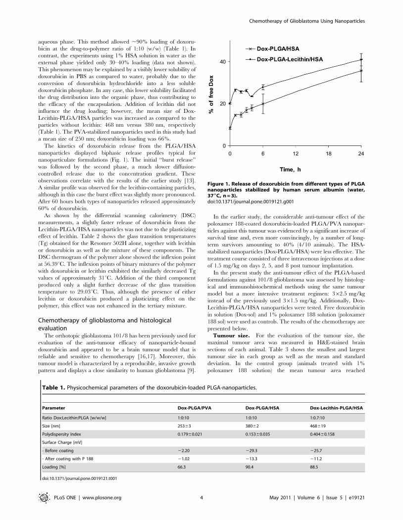
The kinetics of doxorubicin release from the PLGA/HSA
nanoparticles displayed biphasic release profiles typical for
nanoparticulate formulations (Fig. 1). The initial ‘‘burst release’’
was followed by the second phase, a much slower diffusion-
controlled release due to the concentration gradient. These
observations correlate with the results of the earlier study [13].
A similar profile was observed for the lecithin-containing particles,
although in this case the burst effect was slightly more pronounced.
After 60 hours both types of nanoparticles released approximately
60% of doxorubicin.
As shown by the differential scanning calorimetry (DSC)
measurements, a slightly faster release of doxorubicin from the
Lecithin-PLGA/HSA nanoparticles was not due to the plasticizing
effect of lecithin. Table 2 shows the glass transition temperatures
(Tg) obtained for the Resomer 502H alone, together with lecithin
or doxorubicin as well as the mixture of these components. The
DSC thermogram of the polymer alone showed the inflexion point
at 56.39uC. The inflexion points of binary mixtures of the polymer
with doxorubicin or lecithin exhibited the similarly decreased Tg
values of approximately 31uC. Addition of the third component
produced only a slight further decrease of the glass transition
temperature to 29.03uC. Thus, although the presence of either
lecithin or doxorubicin produced a plasticizing effect on the
polymer, this effect was not enhanced in the tertiary mixture.
Chemotherapy of glioblastoma and histologicalevaluation
The orthotopic glioblastoma 101/8 has been previously used for
evaluation of the anti-tumour efficacy of nanoparticle-bound
doxorubicin and appeared to be a brain tumour model that is
reliable and sensitive to chemotherapy [16,17]. Moreover, this
tumour model is characterized by a reproducible, invasive growth
pattern and displays a close similarity to human glioblastoma [9].
In the earlier study, the considerable anti-tumour effect of the
poloxamer 188-coated doxorubicin-loaded PLGA/PVA nanopar-
ticles against this tumour was evidenced by a significant increase of
survival time and, even more convincingly, by a number of long-
term survivors amounting to 40% (4/10 animals). The HSA-
stabilized nanoparticles (Dox-PLGA/HSA) were less effective. The
treatment course consisted of three intravenous injections at a dose
of 1.5 mg/kg on days 2, 5, and 8 post tumour implantation.
In the present study the anti-tumour effect of the PLGA-based
formulations against 101/8 glioblastoma was assessed by histolog-
ical and immunohistochemical methods using the same tumour
model but a more intensive treatment regimen: 362.5 mg/kg
instead of the previously used 361.5 mg/kg. Additionally, Dox-
Lecithin-PLGA/HSA nanoparticles were tested. Free doxorubicin
in solution (Dox-sol) and 1% poloxamer 188 solution (poloxamer
188 sol) were used as controls. The results of the chemotherapy are
presented below.
Tumour size. For the evaluation of the tumour size, the
maximal tumour area was measured in H&E-stained brain
sections of each animal. Table 3 shows the smallest and largest
tumour size in each group as well as the mean and standard
deviation. In the control group (animals treated with 1%
poloxamer 188 solution) the mean tumour area reached
Table 1. Physicochemical parameters of the doxorubicin-loaded PLGA-nanoparticles.
Parameter Dox-PLGA/PVA Dox-PLGA/HSA Dox-Lecithin-PLGA/HSA
Ratio Dox:Lecithin:PLGA [w/w/w] 1:0:10 1:0:10 1:0.7:10
Size [nm] 25363 38062 468619
Polydispersity Index 0.17960.021 0.15360.035 0.40460.158
Surface Charge [mV]
- Before coating 22.20 229.3 225.7
- After coating with P 188 21.02 213.3 211.2
Loading [%] 66.3 90.4 88.5
doi:10.1371/journal.pone.0019121.t001
Figure 1. Release of doxorubicin from different types of PLGAnanoparticles stabilized by human serum albumin (water,376C, n = 3).doi:10.1371/journal.pone.0019121.g001
Chemotherapy of Glioblastoma Using Nanoparticles
PLoS ONE | www.plosone.org 4 May 2011 | Volume 6 | Issue 5 | e19121

32.163.8 mm2 (Table 3). The mean tumour size in the group of
rats treated with doxorubicin solution was smaller
(21.7613.4 mm2); however, the difference between the control
and Dox-treated groups was statistically non-significant. The
groups treated with Dox-PLGA/PVA and Dox-PLGA/HSA
exhibited considerably smaller tumours (10.669.7 mm2 and
16.6612.0 mm2, respectively). These tumours were distinctively
smaller, as compared to controls; however, the difference between
these groups and the group treated with Dox-sol was not
statistically significant. Significant differences (p#0.05), as
compared to both, control and Dox-sol, were only detectable in
the group treated with Dox-Lecithin-PLGA/HSA. The animals in
this group exhibited the smallest mean tumour area of
9.6610.7 mm2. Importantly, this was the only group where 2/6
of the animals developed no observable tumour by day 18. The
high standard deviation in this group is also caused by this fact.
There were no tumour-free animals in other groups.
Proliferation index. For the evaluation of cell proliferation,
immunohistochemical staining with antibodies against Ki67 (MIB-
1) was performed. Ki67 is a nuclear antigen, which is expressed in
all phases of the cell cycle with the exception of the G0-phase.
Because of this absence of the antigen in resting cells, it is a good
marker for proliferating cells [18]. The Ki67 labelling index
expressed as the ratio of positively stained tumour cells of all cells
was determined from six random high power fields in each animal.
All animals treated with doxorubicin-coated PLGA-nanoparticles
showed a significantly lower rate of proliferation than the groups
treated with Dox-sol or surfactant solution. The overall best results
could be seen in the group treated with Dox-Lecithin-PLGA/HSA
where the percentage of proliferating cells amounted to
34.7%624.8% while the percentage of proliferating cells was
42.7%621.2% and 49.2%68.9% in the Dox-PLGA/PVA and
Dox-PLGA/HSA treated groups, respectively. The latter
difference was, however, statistically not significant. Significantly
higher growth rates were detectable in the group treated with Dox-
sol where the mean value was 63.9%66.6%. The highest
proliferation rates were found in the control group
(72.2%67.6%) (Table 3). The predominance of positively-
stained, brown-coloured cells in the control group and the
preponderance of negatively-stained, blue-coloured cells in the
successfully treated groups are presented in Fig. 2. Earlier data
indicated a significant positive correlation between high Ki67-
indexes, high proliferation rates, and a reduction of disease-free
intervals as well as shortened survival times [19]. Accordingly, the
reduced Ki67-index after treatment with the Dox-Lecithin-
PLGA/HSA nanoparticles pointed out impressively the
enhanced anti-tumour activity of doxorubicin after incorporation
in the PLGA nanoparticles coated with poloxamer 188.
Extent of necrotic areas. An additional diagnostic criterion
for high-grade glioblastoma is the presence of large necrotic areas
in the brain region occupied by tumour cells. This histological
hallmark is a predictor of poor prognosis [20]. The extent of
necrosis in different groups is shown in Fig. 2 and 3. Especially
widespread necrotic areas with more than 50% of the tumour area
occupied by necrosis in 80% of the animals (Score: 2.860.4) were
found in the group treated with surfactant solution only . An
obvious reduction was seen in the groups treated with Dox-sol
(2.060.6) and Dox-PLGA nanoparticles stabilized with HSA and
PVA (2.260.7 and 1.560.9, respectively). The group of animals
treated with Dox-Lecithin-PLGA/HSA was unique in that only
solitary necrotic foci could be found mainly in the centre of the
tumour (0.860.9). In 50% of the rats in this group no necrotic
areas were found.
GFAP expression. The glial fibrillary acidic protein is a
protein of the cytoskeleton and because of its high expression in
astroglial tumours, such as astrocytoma and glioblastoma, it is used
as a tumour marker [21]. In this study, GFAP expression was
evaluated using the following semi-quantitative scoring system: 0,
no expression; 1, #20% of tumour cells expressing GFAP; 2,
between 20% and 50% GFAP expressing cells; 3, $50% GFAP
expressing cells. The results are summarized in Fig. 2 and Table 3.
Histological examination of the control group revealed that each
animal showed an expression over 20%, and in 50% of the
animals over 50% expression was detectable. Also in the group
treated with Dox-sol, in every slide positively stained proteins were
found. The average score of the GFAP expression in this group
was 1.760.5. No significant difference, just a trend, was found
between the groups treated with Dox-sol and the particles
Table 3. Quantitative analysis of tumour incidence, tumour size, proliferation index, and vessel density as well as semiquantitativeanalysis of GFAP- and VEGF expression after chemotherapy of 101/8 rat glioblastoma with different formulations of doxorubicin.
Tumour area [mm2]
GroupIncidence oftumour Mini-mum Maxi-mum Mean Ki 67+ [%]
Vessel density[%]
GFAP(score) VEGF (score)
Poloxamer 188-sol. 5/5 28.3 38.8 32.163.8 72.267.6 3.861.0 2.660.5 2.460.8
Dox-sol. 6/6 2.7 34.1 21.7613.4 63.966.6 3.761.6 1.760.5 1.760.7
Dox-PLGA/PVA-NP 6/6 0.5 30.3 10.669.7 51.268.9 2.460.5 1.560.8 1.760.9
Dox-PLGA/HSA-NP 5/5 4.0 38.0 16.6612.0 49.268.9 2.361.0 2.460.5 1.660.5
Dox-Lecithin-PLGA/HSA-NP 4/6 0 27.4 9.6610.7 34.7624.8 1.161.2 1.060.8 0.560.5
doi:10.1371/journal.pone.0019121.t003
Table 2. Glass transition temperatures (Tg) of PLGA (Resomer502H) and its mixtures with doxorubicin and/or soybeanlecithin (differential scanning calorimetry).
Sample composition
Resomer 502H[mg]
Lecithin[mg]
Doxorubicin[mg]
Tg
[6C]
100 - - 56.39
100 - 10 30.90
100 7 - 31.13
100 7 10 29.03
doi:10.1371/journal.pone.0019121.t002
Chemotherapy of Glioblastoma Using Nanoparticles
PLoS ONE | www.plosone.org 5 May 2011 | Volume 6 | Issue 5 | e19121

Figure 2. Histological and immunohistochemical evaluation of necrotic areas, proliferation index, GFAP expression, vessel density,and VEGF expression on day 18 after chemotherapy of 101/8 rat glioblastoma with different doxorubicin-PLGA formulations,doxorubicin solution, and poloxamer 188 (P 188) solution as control.doi:10.1371/journal.pone.0019121.g002
Figure 3. Semi-quantitative analysis of the extent of necrosis after treatment with Dox-PLGA formulations, Dox-sol, and surfactantsolution.doi:10.1371/journal.pone.0019121.g003
Chemotherapy of Glioblastoma Using Nanoparticles
PLoS ONE | www.plosone.org 6 May 2011 | Volume 6 | Issue 5 | e19121

stabilized with PVA or HSA: regarding the immunoreactive cells
for GFAP, the HSA-stabilized particles were superior to the PVA-
stabilized particles. The only group exhibiting a significantly
reduced GFAP expression was the group treated with the particles
containing lecithin. There the GFAP expression did not exceed
50% expression and one third of the rats showed no expression of
this marker (1.060.8 points).
Vessel density. Glioblastomas exhibit high levels of
neovascularisation, which contribute to their aggressive behaviour
[22]. For determination of the vessel density, tissue sections were
stained with Isolectin B4, a marker for endothelial cell. The
percentage of the area occupied by Isolectin B4-positive vessels was
quantified with the Axiovision software (Carl Zeiss Microimaging).
The areas with the highest vascular density were selected. As well as
the other histological tests, the analysis of vessel density showed the
predominance of the lecithin-containing particles (Table 3).
Animals treated with these particles showed a vessel density of
only 1.1%61.1%. In the groups treated with Dox-PLGA/HSA and
Dox-PLGA/PVA, the vessel density reached 2.3%61.0% and
2.4%60.5%, respectively. A conspicuously higher vessel density
was found in the groups treated with Dox-sol and with poloxamer
188 solution: 3.7%61.6% and 3.8%61.6%, respectively.
VEGF expression. The VEGF is the one of the most
important angiogenic factors and a potential prognostic factor
for many types of cancer, including glioblastoma [23]. The range
of the VEGF expression evaluated using the previously described
semi-quantitative scoring system is demonstrated in Table 3. In
the control group a strong staining intensity was detectable
(2.460.8 points). In other doxorubicin-treated groups, the
attenuation of expression was evident but not significant. The
following scores were found: Dox-PLGA/PVA, 1.760.9 points;
Dox-PLGA/HSA, 1.660.5 points; Dox-sol, 1.760.7 points. The
lowest VEGF expression was observed in the Dox-Lecithin-
PLGA/HSA-treated group (0.560.5 points).
Discussion
As mentioned above, the anticancer antibiotic doxorubicin
bound to the PLGA nanoparticles coated with poloxamer 188
produced a considerable anti-tumour effect against intracranial
glioblastoma 101/8 in rats [13]. The study also revealed a key role
of the surfactant coating as well as the influence of the nanoparticle
composition on their biologic performance, i.e. poloxamer 188
appeared to be more effective as a coating agent than polysorbate
80, and the nanoparticles stabilized by PVA appeared to be more
effective than those stabilized by HSA.
One of the reasons for this lower efficacy of the HSA-stabilized
nanoparticles could be an insufficient attachment of poloxamer
188 to the particles’ surface occupied by HSA. It was shown that
the poloxamers – block-copolymers of poly(propylene oxide) and
poly(ethylene oxide) – interact with cell membranes and, in
particular, with lecithin, their essential component [24]. There-
fore, it was attempted to improve the efficacy of the HSA-
stabilized PLGA nanoparticles loaded with doxorubicin by
incorporating lecithin into their core.
Apart from an increase of the particle size, the presence of
lecithin did not influence the physicochemical parameters of the
HSA-stabilized nanoparticles. However, the considerable advan-
tage of the lecithin-containing particles was clearly seen in the in
vivo experiment where this formulation demonstrated a superior
anti-tumour effect. Although the histological and immunohisto-
chemical results indicated that all PLGA formulations coated with
poloxamer 188 were able to transport doxorubicin to the tumour,
the Dox-Lecithin-PLGA/HSA produced the most considerable
effect on all investigated histological parameters, i.e. the tumour
size, proliferation index, GFAP expression, extent of necrotic
areas, and vessel density, as well as VEGF expression. The
superiority of this formulation compared to the other preparations
also is exhibited by the fact that 2/6 animals were tumour-free.
This result correlates with the previous finding: considerable
inhibition of angiogenesis in 101/8 glioblastoma after chemother-
apy using doxorubicin loaded in polysorbate 80-coated poly(butyl
cyanoacrylate) nanoparticles [9].
Another outcome of the chemotherapy in this study is the
decreased necrotic area. Whereas in general oncology the increase
of necrotization indicates an increase of treatment efficacy, the
considerable necrotization of human glioma is a sign of its high
malignancy and is predictive of a bad prognosis for the patient
[20]. Accordingly, the pronounced necrotization of 101/8
glioblastoma is a permanent feature of this tumour. In this case,
the decrease of necrotization can be interpreted as a sign of the
effective chemotherapy. Indeed, in the present study the absence
of necroses or decreased necrotic areas correlated with the
decreased tumour sizes. This phenomenon was also observed in
the previous study [9].
As mentioned above, chemotherapy also led to considerably
decreased vessel density and expression of VEGF. It is known that
tumours in the early stages of development (,2 mm) do not yet
exhibit an advanced vascular system [25]. Therefore, it is possible
that the growth inhibition of small gliomas observed after
chemotherapy using doxorubicin-loaded nanoparticles is a result
of the inhibited proliferation, which, in turn, causes the delayed
formation of hypoxic areas in the tumour core, the major stimulus
for angiogenesis and that thus the effect of doxorubicin on vessel
density is an indirect rather than a direct one.
It is known that one of the major factors responsible for the
biodistribution of intravenously injected colloidal carriers are the
plasma proteins adsorbed by these carriers in the blood. In
particular, the study of Petri et al. [6] revealed a correlation
between the anti-tumour effects of doxorubicin-loaded poly(butyl
cyanoacrylate) nanoparticles coated with poloxamer 188 and
polysorbate 80 against 101/8 glioblastoma and the high amount of
a plasma protein apolipoprotein A-I found on the surface of these
particles after incubation in rat plasma. Thus, these two chemically
different surfactants obviously yielded a certain similarity of the
surface properties of their nanoparticles and mediated similar
pharmacological effects, which, remarkably, provided evidence of
the effective brain delivery of doxorubicin with these carriers.
On the contrary, in the present study the difference in the
antitumour effects between Dox-PLGA/HSA and Dox-Lecithin-
PLGA/HSA, both coated with the same surfactant – poloxamer
188, could be due to the diversity in the surface properties of the
particles. As shown before by Verrecchia et al. [26], albumin
molecules adsorbed to the PLGA nanoparticle surface during the
preparation procedure were quickly replaced by other plasma
proteins when these particles came in contact with plasma.
However, a fraction of HSA remained irreversibly bound to the
surface. It can be hypothesized that the higher efficacy of Dox-
Lecithin-PLGA/HSA nanoparticles might be due to the surface
altered by the presence of lecithin, which, in its turn, could
enhance adsorption of apolipoprotein A-I which is known to have
affinity to phospholipids [27]. This effect could be enhanced by the
aforementioned possible affinity between lecithin and poloxamer
188. The latter proved to be a key ingredient in brain delivery by
the PLGA nanoparticles [13].
Together with the data obtained previously, the results of the
present study unequivocally demonstrate that PLGA nanoparticles
coated with poloxamer 188 enable brain delivery of agents that
Chemotherapy of Glioblastoma Using Nanoparticles
PLoS ONE | www.plosone.org 7 May 2011 | Volume 6 | Issue 5 | e19121

cannot independently permeate across the blood-brain barrier in
therapeutically effective concentrations. In particular, this tech-
nology might represent a great potential for non-invasive
chemotherapy of brain tumours. The employment of lecithin as
an additional ingredient appears to further improve the anti-
glioblastoma activity, which may be an indication that further
improvements are possible.
Acknowledgments
The authors would like to thank Sandra Deutsch, Martin Haberkorn, and
Ulrike Rumpf (Institute of Neuropathology, University Medical Centre
Hamburg-Eppendorf, Germany) for the preparation of the histological
slides and G. Shandryuk (Topchiev Institute of Petrochemical Synthesis,
Moscow, Russia) for the differential scanning calorimetry measurements.
Author Contributions
Conceived and designed the experiments: SW ASK SG OM CB MG JK.
Performed the experiments: SW ASK SG OM CB. Analyzed the data: SW
ASK SG OM CB MG JK. Contributed reagents/materials/analysis tools:
SW ASK SG OM CB. Wrote the paper: SW SG CB MG JK.
References
1. Brat DJ, Castellano-Sanchez A, Kaur B, Van Meir EG (2002) Genetic andbiologic progression in astrocytomas and their relation to angiogenic
dysregulation. Adv Anat Pathol 9: 24–36.2. Roullin VG, Deverre JR, Lemaire L, Hindre F, Venier-Julienne MC, et al.
(2002) Anti-cancer drug diffusion within living rat brain tissue: an experimental
study using [3H](6)-5-fluorouracil-loaded PLGA microspheres. Eur J PharmBiopharm 53: 293–299.
3. Kreuter J (2001) Nanoparticulate systems for brain delivery of drugs. Adv DrugDeliv Rev 47: 65–81.
4. Steiniger SC, Kreuter J, Khalansky AS, Skidan IN, Bobruskin AI, et al. (2004)Chemotherapy of glioblastoma in rats using doxorubicin-loaded nanoparticles.
Int J Cancer 109: 759–767.
5. Kreuter J, Gelperina S (2008) Use of nanoparticles for cerebral cancer. Tumori94: 271–277.
6. Petri B, Bootz A, Khalansky A, Hekmatara T, Muller R, et al. (2007)Chemotherapy of brain tumour using doxorubicin bound to surfactant-coated
poly(butyl cyanoacrylate) nanoparticles: revisiting the role of surfactants.
J Control Release 117: 51–58.7. Zensi A, Begley D, Pontikis C, Legros C, Mihoreanu L, et al. (2009) Albumin
nanoparticles targeted with Apo E enter the CNS by transcytosis and aredelivered to neurones. J Control Release.
8. Ulbrich K, Knobloch T, Kreuter J (2010) Targeting the insulin receptor:nanoparticles for drug delivery across the blood-brain barrier (BBB). J Drug
Target 19: 125–132.
9. Hekmatara T, Bernreuther C, Khalansky AS, Theisen A, Weissenberger J, et al.(2009) Efficient systemic therapy of rat glioblastoma by nanoparticle-bound
doxorubicin is due to antiangiogenic effects. Clin Neuropathol 28: 153–164.10. Lee BS, Fujita M, Khazenzon NM, Wawrowsky KA, Wachsmann-Hogiu S,
et al. (2006) Polycefin, a new prototype of a multifunctional nanoconjugate based
on poly(beta-L-malic acid) for drug delivery. Bioconjug Chem 17: 317–326.11. Fujita M, Khazenzon NM, Ljubimov AV, Lee BS, Virtanen I, et al. (2006)
Inhibition of laminin-8 in vivo using a novel poly(malic acid)-based carrierreduces glioma angiogenesis. Angiogenesis 9: 183–191.
12. Ding H, Inoue S, Ljubimov AV, Patil R, Portilla-Arias J, et al. (2010) Inhibition
of brain tumor growth by intravenous poly (beta-L-malic acid) nanobioconjugatewith pH-dependent drug release [corrected]. Proc Natl Acad Sci U S A 107:
18143–18148.13. Gelperina S, Maksimenko O, Khalansky A, Vanchugova L, Shipulo E, et al.
(2010) Drug delivery to the brain using surfactant-coated poly(lactide-co-glycolide) nanoparticles: influence of the formulation parameters. Eur J Pharm
Biopharm 74: 157–163.
14. Wohlfart S, Bernreuther C, Khalansky A, Theisen A, Weissenberger J, et al.(2009) Increased Numbers of Injections of Doxorubicin Bound to Nanoparticles
Lead to Enhanced Efficacy Against Rat Glioblastoma 101/8. Journal ofNanoneuroscience 1: 144–151.
15. Schoch G, Seeger H, Bogousslavsky J, Tolnay M, Janzer RC, et al. (2006)
Analysis of prion strains by PrPSc profiling in sporadic Creutzfeldt-Jakobdisease. PLoS Med 3: e14.
16. Gelperina SE, Khalansky AS, Skidan IN, Smirnova ZS, Bobruskin AI, et al.(2002) Toxicological studies of doxorubicin bound to polysorbate 80-coated
poly(butyl cyanoacrylate) nanoparticles in healthy rats and rats with intracranialglioblastoma. Toxicol Lett 126: 131–141.
17. Ambruosi A, Khalansky AS, Yamamoto H, Gelperina SE, Begley DJ, et al.
(2006) Biodistribution of polysorbate 80-coated doxorubicin-loaded [14C]-poly(butyl cyanoacrylate) nanoparticles after intravenous administration to
glioblastoma-bearing rats. J Drug Target 14: 97–105.18. Gerdes J, Lemke H, Baisch H, Wacker HH, Schwab U, et al. (1984) Cell cycle
analysis of a cell proliferation-associated human nuclear antigen defined by the
monoclonal antibody Ki-67. J Immunol 133: 1710–1715.19. Veronese SM, Maisano C, Scibilia J (1995) Comparative prognostic value of Ki-
67 and MIB-1 proliferation indices in breast cancer. Anticancer Res 15:2717–2722.
20. Barker FGII, Davis RL, Chang SM, Prados MD (1996) Necrosis as a prognosticfactor in glioblastoma multiforme. Cancer 77: 1161–1166.
21. Hamaya K, Doi K, Tanaka T, Nishimoto A (1985) The determination of glial
fibrillary acidic protein for the diagnosis and histogenetic study of centralnervous system tumors: a study of 152 cases. Acta Med Okayama 39: 453–462.
22. Weidner N (2000) Angiogenesis as a predictor of clinical outcome in cancerpatients. Hum Pathol 31: 403–405.
23. Brem S, Cotran R, Folkman J (1972) Tumor angiogenesis: a quantitative
method for histologic grading. J Natl Cancer Inst 48: 347–356.24. Zhirnov AE, Demina TV, Krylova OO, Grozdova ID, Melik-Nubarov NS
(2005) Lipid composition determines interaction of liposome membranes withPluronic L61. Biochim Biophys Acta 1720: 73–83.
25. Folkman J (1990) What is the evidence that tumors are angiogenesis dependent?
Journal of the National Cancer Institute 82: 4–6.26. Verrecchia T, Huve P, Bazile D, Veillard M, Spenlehauer G, et al. (1993)
Adsorption/desorption of human serum albumin at the surface of poly(lacticacid) nanoparticles prepared by a solvent evaporation process. J Biomed Mater
Res 27: 1019–1028.27. Frank PG, Marcel YL (2000) Apolipoprotein A-I: structure-function relation-
ships. J Lipid Res 41: 853–872.
Chemotherapy of Glioblastoma Using Nanoparticles
PLoS ONE | www.plosone.org 8 May 2011 | Volume 6 | Issue 5 | e19121