Sensory and Proprioceptive Training Modalities for the Foot and Ankle
Upload
arteveldehsCategory
view
1download
0
Human Movement Science xxx (2014) xxx–xxx
Contents lists available at ScienceDirect
Human Movement Science
journal homepage: www.elsevier .com/locate/humov
Foot orientation affects muscle activation levelsof ankle stabilizers in a single-legged balanceboard protocol
0167-9457/$ - see front matter � 2014 Elsevier B.V. All rights reserved.http://dx.doi.org/10.1016/j.humov.2013.12.008
⇑ Corresponding author. Address: De Pintelaan 185, 3B3, 9000 Ghent, Belgium. Tel.: +32 (0)9 332 04 53; fax: +32 (0)11.
E-mail addresses: [email protected] (R. De Ridder), [email protected] (T. Willems), Sophie.DeMits@(S. De Mits), [email protected] (J. Vanrenterghem), [email protected] (P. Roosen).
Please cite this article in press as: De Ridder, R., et al. Foot orientation affects muscle activation levels ostabilizers in a single-legged balance board protocol. Human Movement Science (2014), http://dx.d10.1016/j.humov.2013.12.008
Roel De Ridder a,⇑, Tine Willems b, Sophie De Mits a, Jos Vanrenterghem c,Philip Roosen a
a Department of Rehabilitation Sciences and Physiotherapy, Ghent University, Ghent, Belgiumb Department of Physiotherapy and Orthopedics, Ghent University, Ghent, Belgiumc School of Sport and Exercise Sciences, Faculty of Science, Liverpool John Moores University, Liverpool, UK
a r t i c l e i n f o a b s t r a c t
Article history:Available online xxxx
PsycINFO classification:3380
Keywords:Balance trainingUniaxial boardFoot orientationMuscle activity
Context: The main goal of balance training is regaining a normalneuromuscular control to a functional level. Although uniaxial bal-ance boards are commonly used, no research has been done on theeffect of foot orientation on muscle activation levels.Objective: To investigate the effect of foot orientation on muscleactivation levels and modulation of the ankle stabilizing musclesin a single-legged balance protocol on a uniaxial balance board.Methods: Sixty-nine healthy subjects (age: 21.8 ± 1.7 years; mass:67.5 ± 11.9 kg; body height: 174.7 ± 8.6 cm; BMI: 21.5 ± 3.0) par-ticipated in this study. Subjects were asked to keep their balanceduring a single leg stance on a uniaxial balance board for four dif-ferent foot orientations, aligning the board’s rotation axis withfrontal, sagittal, diagonal and subtalar axes of the foot, respec-tively. Surface electromyography registered muscle activity ofperoneus longus, tibialis anterior, medial and lateral gastrocne-mius muscles.Results: Highest muscle activation levels and modulation for theperoneus longus were registered exercising along the frontal axis;for the tibialis anterior along the diagonal axis; for the medialgastrocnemius along the sagittal axis; and for the lateral gastroc-nemius along the diagonal axis.
9 332 38
ugent.be
f ankleoi.org/
2 R. De Ridder et al. / Human Movement Science xxx (2014) xxx–xxx
Please cite this article in press as: De Ridderstabilizers in a single-legged balance boar10.1016/j.humov.2013.12.008
Conclusion: Foot orientation modifications on a uniaxial balanceboard allows to differentially target specific ankle stabilizing mus-cles during balance training.
� 2014 Elsevier B.V. All rights reserved.
1. Introduction
Balance protocols are often part of the rehabilitation process of lower leg musculoskeletal injuries.Balance is defined as the capacity of a person to preserve their center of mass within their base of sup-port. The capacity to do this depends on the efficient integration of afferent visual, vestibular andsomatosensory inputs to generate an adequate efferent neuromuscular response (Winter, 1995). Mus-culoskeletal lower leg injuries such as an ankle sprain are believed to affect balance capabilities(Mitchell, Dyson, Hale, & Abraham, 2008b). As a result of an ankle sprain, damage to the mechanore-ceptors in the ankle joint has been suggested to lead to partial deafferentiation. (Freeman, Dean, &Hanham, 1965; Lephart, Pincivero, & Rozzi, 1998). Khin-Myo-Hla et al. state that this deafferentiationmay suppress gamma motorneuron activity and cause alterations in the muscle spindle sensitivity(Khin, Ishii, Sakane, & Hayashi, 1999). These deficits might lead to impaired balance, an increased riskto resprain, or evolve to chronic functional instability of the ankle joint (Hertel, 2002; Wikstrom,Tillman, Chmielewski, Cauraugh, & Borsa, 2007). Therefore, specific ankle rehabilitation programsinvolving balance training are common approaches to minimize consequences of ankle injuries andprevent recurrence.
The main goal of balance training is regaining a normal neuromuscular control around the ankle toa functional level, by maximally stimulating the muscle activity levels of the ankle stabilizing muscles.Various studies have shown improvements of postural control through balance training programs(Freeman et al., 1965; Hupperets, Verhagen, & van Mechelen, 2008; Matsusaka, Yokoyama, Tsurusaki,Inokuchi, & Okita, 2001; Michell, Ross, Blackburn, Hirth, & Guskiewicz, 2006; Verhagen et al., 2004).Not only the curative effect of balance training has been demonstrated, but also the preventative effecton overall lower limb injuries and more specifically ankle sprains (Olsen, Myklebust, Engebretsen,Holme, & Bahr, 2005; Verhagen et al., 2004). Consequently, the risk of an ankle sprain is significantlyreduced in populations who subsequently undergo balance training (Hupperets et al., 2008; Verhagenet al., 2004). As explanation, it is suggested that long-term balance training results in a higher stim-ulation of muscle spindles (Ashton-Miller, Wojtys, Huston, & Fry-Welch, 2001). This higher stimula-tion would improve neuromuscular control and, therefore, contribute to the functional stability ofthe ankle joint. However, balance protocols differ in duration, frequency, used device, and progressionof the exercises (Mattacola & Dwyer, 2002). At this moment, it is not known which exercises best servethe rehabilitation goals.
It has been suggested that higher muscle activity levels during the balance protocol are expected tospeed up the rehabilitation process and a quicker return to preinjury functional levels (Cordova, Jutte,& Hopkins, 1999). The magnitude of muscle activity depends on the stability of the used device (Wahl& Behm, 2008). For different multidirectionally unstable devices such as a trampoline, wobble board,swiss ball, dyna disc or BOSU balance trainer, muscle activity levels of ankle stabilizing muscles havebeen assessed and compared (Laudner & Koschnitzky, 2010; Wahl & Behm, 2008; Wester, Jespersen,Nielsen, & Neumann, 1996). These devices have in common that there is no control over the rotationdirection in which the ankle is challenged. Uniaxial balance boards, on the other hand, allow for uni-axial rotational instability which depends on foot orientation on the board. Whilst uniaxial balanceboards are commonly used devices for balance training in the clinical practice, to the authors’ knowl-edge no research has been done yet that identified the effect of foot orientation on muscle activationlevels of different ankle stabilizing muscles. This may be useful when rehabilitation is meant to targetresolving defects of specific ankle stabilizing muscles. Especially in the early stages of rehabilitation,this focus on a specific muscle might be desirable before using a general multiaxial device. The differ-ent foot orientations used in the clinical practice are based on the movement they create around theankle joint. It is believed that depending on foot orientation, muscle activation levels and amount of
, R., et al. Foot orientation affects muscle activation levels of ankled protocol. Human Movement Science (2014), http://dx.doi.org/
R. De Ridder et al. / Human Movement Science xxx (2014) xxx–xxx 3
modulation of the different ankle stabilizing muscles will vary according to their anatomical functions,however scientific research is lacking. The most commonly used foot orientation is with the axis of thedevice aligned with a frontal foot axis, creating a inversion–eversion instability which can be consid-ered the most common mechanism of ankle sprains. It remains, however, unclear how muscle activa-tion and modulation of the different ankle stabilizing muscles differ when working at other footorientations.
The purpose of this study was primarily to determine muscle activation levels of four ankle mus-cles, contributing to dynamic stability of the ankle joint, during a single-leg balance board protocol.The muscle activation levels of each specific muscle will be compared for four different foot orienta-tions. We hypothesized that individual ankle muscles would be maximally activated for the foot ori-entation in which they are maximally challenged with respect to their primary anatomical function.Therefore, the hypothesis was that for the peroneus longus alignment with the subtalar axis wouldprovoke highest muscle activations, for the tibialis anterior a diagonal axis, and for the medial and lat-eral gastrocnemius a sagittal axis. In addition, modulation in muscle activity as an estimate of respon-siveness to balance board motions, was compared between the different axes for each muscle.
2. Methods
2.1. Subjects
A group of 69 healthy subjects (age: 21.8 ± SD 1.7; mass: 67.5 ± 11.9 kg; body height:174.7 cm ± SD 8.6; BMI: 21.48 ± 2.99) was tested. The population consisted of 38 female and 31 maleparticipants between 18 and 25 years old. Participants were excluded from the study if they had amusculoskeletal injury to the lower limb, limiting normal activity, in the last year; if they sufferedfrom self-reported ankle instability (repetitive sprains, periods of giving way); a systemic diseasewhich could influence balance performance; or if they were pregnant at the moment of testing.Anthropometric data were collected, and the participants completed a questionnaire to establish theirmedical history. The Ghent University Hospital ethics committee approved this study and all subjectssigned the informed consent before participation.
2.2. Procedures
During the experiment, participants were asked to keep their balance on a uniaxial balance board,while their muscle activity was registered. They were instructed to perform a single-leg stance on thebalance board with the knee of the stance leg slightly flexed (knee perpendicular above the toes).Compensating movements with the arms and the contralateral free leg to keep balance were permit-ted. Every participant was tested unilaterally, and the stance leg was chosen through randomization(33 right and 36 left legs tested). Four different foot orientations were tested (Fig. 1). For every subject,the sequence of the foot orientations was randomized as a set by drawing lots. This ‘‘set’’, containingthe orientation order, was then repeated for this subject 3 times. In each set, every foot orientationwas performed during 15 s with a 30-s rest interval. Rest interval between sets was 2 min. Before eachmeasurement, the participants supported themselves with their hands on a chair for balance. Whenready, they released the chair and the measurement was started. Whenever the balance board or con-tralateral foot touched the ground or the participants searched support with their hands, they had torestore as quickly as possible their balance and continue the exercise.
The uniaxial balance board (custom made) used in this study had a length of 50 cm and a width of38 cm. Two half spheres are fixated onto the base, creating uniaxial instability. The half sphere had aheight of 4 cm and a diameter of 7.5 cm. Foot positions were marked on the balance board, as shownin Fig. 1. When the rotational axis underneath the balance board is aligned parallel with the frontalaxis of the foot (F-axis), instability occurs in the frontal plane (inversion/eversion). Alignment withthe sagittal axis (S-axis) is perpendicular to the frontal axis, leading to instability in the sagittal plane(plantar flexion/dorsiflexion). The third foot orientation has an alignment with a diagonal foot axis (D-axis), 45� externally rotated from the F-axis and creating a plantarflexion/eversion and dorsiflexion/inversion motion. The last foot orientation has an alignment with an axis that is 16� internally rotated
Please cite this article in press as: De Ridder, R., et al. Foot orientation affects muscle activation levels of anklestabilizers in a single-legged balance board protocol. Human Movement Science (2014), http://dx.doi.org/10.1016/j.humov.2013.12.008

Fig. 1. Different foot positions in relation to axis created by two spheres underneath the balance board: along A. F = Frontal axis,B. S = Sagittal axis, C. D = Diagonal axis, and D. ST = Subtalar joint axis.
4 R. De Ridder et al. / Human Movement Science xxx (2014) xxx–xxx
from the frontal axis, matching the anatomical orientation of the subtalar joint axis (ST-axis) and lead-ing to inversion/eversion instability along the subtalar axis.
2.3. EMG signal acquisition and processing
Muscle activity of the m. tibialis anterior (TA), m. peroneus longus (PL), medial (MG) and lateral(LG) m. gastrocnemius was registered using surface electromyography at 1000 Hz (Myosystem1400A, Noraxon USA Inc, Scottsdale, Arizona 85254, USA). To reduce impedance, the skin surfacewas shaved and cleaned with ether to remove dead skin and grease. Disposable bipolar Ag/AgCl sur-face electrodes with conducting gel and 2 cm diameter were placed with an inter-electrode distance of2 cm center-to-center and parallel to the muscle fibers according to SENIAM guidelines (www.sen-iam.org). The wires and electrodes were fixated with gauze to minimize the effect of motion artifacts.
For the processing of the EMG data, the MyoResearch XP Master Edition (Noraxon USA Inc, Scotts-dale, Arizona 85254, USA) was used. The raw data of these EMG signals were full-wave rectified andsmoothed using a moving average window of 100 ms. Only the intervals where the participant waseffectively balancing, as defined below, on the balance board were taken into account. To be able toomit the periods in which the participant was not balancing, a synchronized mark was placed usinga hand-held external trigger whenever the subject lost balance. This was based on real time visualdetection of the rim of the balance board touching the ground, or any attempt by the participant tofind support with the contralateral leg on the floor or with the hands on the chair. Another markwas placed as soon as unsupported balancing was restored. Subsequently, the time periods whenthe participant was balancing unsupported were assembled and the Root Mean Square (RMS), which
Please cite this article in press as: De Ridder, R., et al. Foot orientation affects muscle activation levels of anklestabilizers in a single-legged balance board protocol. Human Movement Science (2014), http://dx.doi.org/10.1016/j.humov.2013.12.008
R. De Ridder et al. / Human Movement Science xxx (2014) xxx–xxx 5
represents the mean of the varying muscle activity magnitude, was calculated to represent overallactivation levels throughout the trial. The goal of this study was to evaluate within one muscle whichfoot orientation generated highest muscle activity levels for that muscle. To emphasize this and avoidinterpretations based on absolute RMS values, all RMS values were normalized (Bellew, Frilot, Busch,Lamothe, & Ozane, 2010) to the RMS value of the foot orientation along the F-axis which is consideredthe most commonly used foot orientation in clinical practice.
In addition, variation in muscle amplitude was calculated to quantify muscle modulation duringthe balance task. Therefore, the rectified and smoothed EMG signal was exported from MyoResearchand further processed in Excel. To reflect the variation in muscle amplitude, the signal was differen-tiated representing the time dependent changes. Subsequently, the mean magnitude of these changeswas calculated as an estimate for modulation of muscle contractions. By differentiating the smoothedEMG signal, it is estimated that modulation represents neuromuscular responsiveness to balanceboard motions. Modulation is largely independent from muscle activation levels, considering that highactivation levels may be constant (e.g. during increased co-contractions) or varying (e.g. responding tobalance board motions).
2.4. Statistical analysis
Statistical analysis was performed in SPSS 20 (SPSS Inc., Chicago, Illinois 60606, USA). Differences inmuscle activity according to axis orientation, and differences in muscle modulation, were evaluatedusing repeated measures ANOVA. Post hoc multiple pairwise comparisons with a Bonferroni correc-tion were performed to identify specific differences. Significance level was set at p 6 0.05.
3. Results
The ANOVA showed significant differences in muscle activation and modulation according to thefoot orientation for all muscles tested (p < 0.001). The post hoc comparison results are presented inTable 1 for muscle activation levels, and in Table 2 for modulation in muscle activity. Overall, the sameresults were found for muscle activation magnitude and modulation. Partial eta square (gp
2), indicat-ing the effect size for the repeated measure ANOVA respectively for muscle activation level and mod-ulation, was 0.607 and 0.517 for the PL, 0.318 and 0.393 for the TA, 0.499 and 0.445 for the MG, and0.465 and 0.349 for the LG. The total durations of the assembled time periods for analysis were for theF-axis 26.12 ± 5.74 s (range 12.60–42.70 s), for the S-axis 25.04 ± 5.13 s (range 16.69–41.79s), for theD-axis 25.33 ± 5.31 s (range 12.90–38.69s), and for the ST-axis 26.58 ± 5.51 s (range 12.29–41.80s). Ageneral linear model with repeated measures showed no main effect of foot orientation on the timeincluded for analysis (p = 0.236).
3.1. Peroneus longus
The peroneus longus showed significantly higher muscle activation levels when the exercise wasperformed along the F-axis compared to the ST-, D- and the S-axis (Fig. 2). The same results werefound for the level of modulation, except for the comparison between the F-axis and the D-axis whichdid not reach statistical significance (p = 0.060). With the foot along the S-axis, muscle activation lev-els and modulation were also significantly lower than with the foot along the D- and ST-axis, whilstthere was no significant difference between the latter two.
3.2. Tibialis Anterior
Muscle activation levels and modulation of the tibialis anterior muscle were significantly higherwhen exercising along the D-axis in comparison to along the F-, S- and ST-axis (Fig. 3). No significantdifference was noted between exercising along the F-, S- and ST-axis.
Please cite this article in press as: De Ridder, R., et al. Foot orientation affects muscle activation levels of anklestabilizers in a single-legged balance board protocol. Human Movement Science (2014), http://dx.doi.org/10.1016/j.humov.2013.12.008
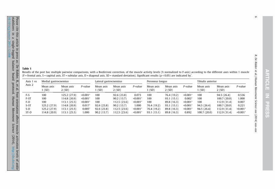
Table 1Results of the post hoc multiple pairwise comparisons, with a Bonferroni correction, of the muscle activity levels (% normalized to F-axis) according to the different axes within 1 muscle(F = frontal axis, S = sagittal axis, ST = subtalar axis, D = diagonal axis, SD = standard deviation). Significant results (p < 0.05) are indicated by*.
Axis 1 vsAxis 2
Medial gastrocnemius Lateral gastrocnemius Peroneus longus Tibialis anterior
Mean axis1 (SD)
Mean axis2 (SD)
P-value Mean axis1 (SD)
Mean axis2 (SD)
P-value Mean axis1 (SD)
Mean axis2 (SD)
P-value Mean axis1 (SD)
Mean axis2 (SD)
P-value
F-S 100 125.2 (27.9) <0.001⁄ 100 92.6 (23.8) 0.073 100 76.4 (19.2) <0.001⁄ 100 94.5 (26.4) 0.536F-ST 100 114.8 (20.9) <0.001⁄ 100 90.2 (13.7) <0.001⁄ 100 93.1 (15.1) 0.002⁄ 100 100.7 (20.0) 1.000F-D 100 113.1 (25.5) <0.001⁄ 100 112.5 (23.6) <0.001⁄ 100 89.8 (16.3) <0.001⁄ 100 112.9 (31.4) 0.007S-ST 125.2 (27.9) 114.8 (20.9) 0.017⁄ 92.6 (23.8) 90.2 (13.7) 1.000 76.4 (19.2) 93.1 (15.1) <0.001⁄ 94.5 (26.4) 100.7 (20.0) 0.221S-D 125.2 (27.9) 113.1 (25.5) 0.005⁄ 92.6 (23.8) 112.5 (23.6) <0.001⁄ 76.4 (19.2) 89.8 (16.3) <0.001⁄ 94.5 (26.4) 112.9 (31.4) <0.001⁄
ST-D 114.8 (20.9) 113.1 (25.5) 1.000 90.2 (13.7) 112.5 (23.6) <0.001⁄ 93.1 (15.1) 89.8 (16.3) 0.892 100.7 (20.0) 112.9 (31.4) <0.001⁄
6R
.De
Ridder
etal./H
uman
Movem
entScience
xxx(2014)
xxx–xxx
Pleasecite
thisarticle
inpress
as:D
eR
idder,R.,et
al.Footorientation
affectsm
uscleactivation
levelsof
anklestabilizers
ina
single-leggedbalance
boardprotocol.
Hum
anM
ovement
Science(2014),
http://dx.doi.org/10.1016/j.hum
ov.2013.12.008
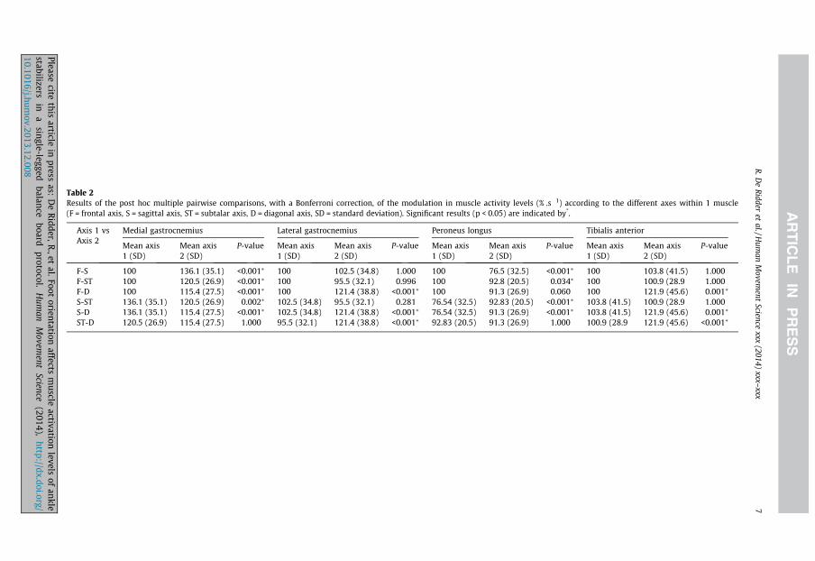
Table 2Results of the post hoc multiple pairwise comparisons, with a Bonferroni correction, of the modulation in muscle activity levels (% .s�1) according to the different axes within 1 muscle(F = frontal axis, S = sagittal axis, ST = subtalar axis, D = diagonal axis, SD = standard deviation). Significant results (p < 0.05) are indicated by*.
Axis 1 vsAxis 2
Medial gastrocnemius Lateral gastrocnemius Peroneus longus Tibialis anterior
Mean axis1 (SD)
Mean axis2 (SD)
P-value Mean axis1 (SD)
Mean axis2 (SD)
P-value Mean axis1 (SD)
Mean axis2 (SD)
P-value Mean axis1 (SD)
Mean axis2 (SD)
P-value
F-S 100 136.1 (35.1) <0.001⁄ 100 102.5 (34.8) 1.000 100 76.5 (32.5) <0.001⁄ 100 103.8 (41.5) 1.000F-ST 100 120.5 (26.9) <0.001⁄ 100 95.5 (32.1) 0.996 100 92.8 (20.5) 0.034⁄ 100 100.9 (28.9 1.000F-D 100 115.4 (27.5) <0.001⁄ 100 121.4 (38.8) <0.001⁄ 100 91.3 (26.9) 0.060 100 121.9 (45.6) 0.001⁄
S-ST 136.1 (35.1) 120.5 (26.9) 0.002⁄ 102.5 (34.8) 95.5 (32.1) 0.281 76.54 (32.5) 92.83 (20.5) <0.001⁄ 103.8 (41.5) 100.9 (28.9 1.000S-D 136.1 (35.1) 115.4 (27.5) <0.001⁄ 102.5 (34.8) 121.4 (38.8) <0.001⁄ 76.54 (32.5) 91.3 (26.9) <0.001⁄ 103.8 (41.5) 121.9 (45.6) 0.001⁄
ST-D 120.5 (26.9) 115.4 (27.5) 1.000 95.5 (32.1) 121.4 (38.8) <0.001⁄ 92.83 (20.5) 91.3 (26.9) 1.000 100.9 (28.9 121.9 (45.6) <0.001⁄
R.D
eR
idderet
al./Hum
anM
ovement
Sciencexxx
(2014)xxx–
xxx7
Pleasecite
thisarticle
inpress
as:D
eR
idder,R.,et
al.Footorientation
affectsm
uscleactivation
levelsof
anklestabilizers
ina
single-leggedbalan
ceboard
protocol.H
uman
Movem
entScience
(2014),http://dx.doi.org/
10.1016/j.humov.2013.12.008

Fig. 2. Muscle activity level (%), normalized to the F-axis, of the peroneus longus according to the different axes (F = frontal axis,S = sagittal axis, D = diagonal axis, ST = subtalar axis) (⁄p < 0.05, ⁄⁄p < 0.001).
Fig. 3. Muscle activity level (%), normalized to the F-axis, of the tibialis anterior according to the different axes (F = frontal axis,S = sagittal axis, D = diagonal axis, ST = subtalar axis) (⁄p < 0.05, ⁄⁄p < 0.001).
Fig. 4. Muscle activity level (%), normalized to the F-axis, of the medial gastrocnemius according to the different axes(F = frontal axis, S = sagittal axis, D = diagonal axis, ST = subtalar axis) (⁄p < 0.05, ⁄⁄p < 0.001).
8 R. De Ridder et al. / Human Movement Science xxx (2014) xxx–xxx
3.3. Medial gastrocnemius
Fig. 4 shows significantly higher muscle activation levels in the medial gastrocnemius when exer-cising along the S-axis in comparison to the ST-, F- and D-axis. Exercising along the F-axis generatedsignificantly lower muscle activation levels compared to all other axes. Foot orientation along the D-axis was not significantly different from ST-axis. The results for modulation were exactly the same.
Please cite this article in press as: De Ridder, R., et al. Foot orientation affects muscle activation levels of anklestabilizers in a single-legged balance board protocol. Human Movement Science (2014), http://dx.doi.org/10.1016/j.humov.2013.12.008
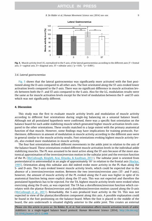
Fig. 5. Muscle activity level (%), normalized to the F-axis, of the lateral gastrocnemius according to the different axes (F = frontalaxis, S = sagittal axis, D = diagonal axis, ST = subtalar axis) (⁄p < 0.05, ⁄⁄p < 0.001).
R. De Ridder et al. / Human Movement Science xxx (2014) xxx–xxx 9
3.4. Lateral gastrocnemius
Fig. 5 shows that the lateral gastrocnemius was significantly more activated with the foot posi-tioned along the D-axis compared to all other axes. The foot orientated along the ST-axis evoked loweractivation levels compared to the F-axis. There was no significant difference in muscle activation lev-els between both the F- and ST-axis compared to the S-axis. Also for the LG, modulation results werethe same as for muscle activation levels except for the level of modulation between the F- and ST-axiswhich was not significantly different.
4. Discussion
This study was the first to evaluate muscle activity levels and modulation of muscle activityaccording to different foot orientations during single-leg balancing on a uniaxial balance board.Although not all postulated hypothesis were confirmed, there was a specific foot orientation on thebalance board for each ankle stabilizing muscle which generated higher muscle activation levels com-pared to the other orientations. These results matched to a large extent with the primary anatomicalfunction of that muscle. However, some findings may have implications for training protocols. Fur-thermore, differences in amount of modulation in muscle activity according to the different axes werein general similar to the muscle activity results. Foot orientations evoking highest muscle activity lev-els, also evoked more modulation in muscle activity.
The four foot orientations defined different movements in the ankle joint in relation to the axis ofthe balance board. These orientations evoked different muscle activation levels in the individual anklestabilizing muscles. The PL was assumed to be most active along the ST-axis because of the more ana-tomical approximation of the inversion/eversion motion in the subtalar joint and the eversion functionof the PL (McCullough, Ringleb, Arai, Kitaoka, & Kaufman, 2011). The subtalar joint is oriented fromposterolateral to anteromedial in an angle of approximately 16� in relation to the frontal axis (Rockar,1995). Orientation along this subtalar axis did indeed evoke more activity in the PL than along theS- and D-axis. The S-axis evoked lowest muscle activity levels, which could be expected due to theabsence of a inversion/eversion motion. Between the two inversion/eversion axes (ST- and F-axis),however, the amount of muscle activity of the PL evoked along the F-axis was higher in spite of itsanatomical function being more explicit along the ST-axis. This was not expected and therefore thepostulated hypothesis has to be rejected. For the TA, highest muscle activity levels were evoked whenexercising along the D-axis, as was expected. The TA has a dorsiflexion/inversion function which cor-relates with the plantar flexion/eversion and a dorsiflexion/inversion motion caused along the D-axis(McCullough et al., 2011). Remarkably, the S-axis produced least activity in the TA. This was notexpected in view of the plantar/dorsiflexion component along this axis. A possible explanation couldbe found in the foot positioning on the balance board. When the foot is placed in the middle of theboard, the axis underneath is situated slightly anterior to the ankle joint. This creates an external
Please cite this article in press as: De Ridder, R., et al. Foot orientation affects muscle activation levels of anklestabilizers in a single-legged balance board protocol. Human Movement Science (2014), http://dx.doi.org/10.1016/j.humov.2013.12.008
10 R. De Ridder et al. / Human Movement Science xxx (2014) xxx–xxx
moment pushing the ankle towards dorsiflexion which likely demands higher muscle activity of thegastrocnemius muscle to maintain the foot in a neutral position, and less activity of the TA. Finally,it was postulated that exercising along the S-axis would evoke the highest activation levels in theMG and LG. The S-axis creates a plantar/dorsiflexion motion, which is concordant with the primaryplantar flexion function of the gastrocnemius muscle (Arndt, Bruggemann, Koebke, & Segesser,1999). The MG was indeed more active along this axis compared to the ST-, F- and D-axis. This muscleis least active along the F-axis, where no plantar/dorsiflexion is created. Furthermore, the MG alsoshowed high activity along the ST-axis which fits with the inversion function of the MG on the calca-neus (Arndt et al., 1999). For the LG, however, lower muscle activity was noted when exercising alongthe S-axis. This was not expected and an immediate explanation for this result remains unclear. It isimportant to note for this muscle that highest muscle activity levels were observed when exercisingalong the D-axis. This does concur with the plantar flexion/eversion moment of the LG on the calca-neus (Arndt et al., 1999).
Remarkably, variation in the magnitude of muscle activity levels as a measure for modulationshowed almost exactly the same results as did the magnitude of the muscle activity levels itself. Onlyfor the PL, the comparison of modulation level between the F- and D-axis did not reach the signifi-cance level (p = 0.060), and for the LG between the F- and ST-axis (p = 0.996) compared to magnituderesults. In general, however, one might postulate that also modulation in muscle activity is highestmainly when the orientation of the perturbation matches the anatomical function of that muscle. Thismodulation in muscle activity, next to a sufficient amount, is considered an important mechanismcontributing to postural control (Asai et al., 2009; Vieira, Loram, Muceli, Merletti, & Farina, 2012).More modulation would entail more active influence on postural stability. A possible explanationfor differences in modulation of muscle activity might be the changes in the center of mass (CoM) dur-ing reactive balance control (Macpherson, Horak, Dunbar, & Dow, 1989). Studies have shown that tem-poral patterns of individual muscles can be modulated based on feedback resulting from CoMkinematics (Safavynia & Ting, 2012; Welch & Ting, 2008). In our study, the primary direction of theCoM excursion will normally be orthogonal to the axis of the board and is more or less controlled.These directions of CoM excursion consequently correspond to certain anatomical functions of the an-kle stabilizing muscles, apparently facilitating modulation. A high correlation between magnitude andvariation in muscle activity levels using an uniaxial balance board may therefore be rational.
The uniaxial balance board is a commonly used device in the clinical practice for balance training,but up to now scientific research on muscle activation levels depending on foot orientation was lack-ing. As mentioned before, it has been suggested that higher muscle activation levels may result in aquicker return to preinjury activity level (Cordova et al., 1999) and that the magnitude of muscle activ-ity levels depends on the stability of the used material (Wahl & Behm, 2008). Various studies havecompared muscle activity levels of the lower leg muscles during exercises using different materials.Blackburn et al. reported that using special exercise sandals during single leg stance produced equalor even higher muscle activity levels in the PL, TA and LG than single leg stance on a foam pad (respec-tively 48%, 60%, 54% higher) or T-band kicks in the frontal (respectively 32%, 32%, 22% higher) or sag-ittal plane (respectively 27%, 27%, 22% higher) (Blackburn, Hirth, & Guskiewicz, 2003). Bellew et al.found higher muscle activity levels in the PL during a ‘quarter heel raise’ (28% higher) and a ‘band heelraise’ (40.5% higher) compared to a conventional ankle eversion exercise (Bellew et al., 2010). In addi-tion, Cordova et al. found higher muscle activity levels for the PL up to 47%, for the TA up to 74%, andfor the MG up to 44%, in favor of T-band kicks compared to SLS on a firm surface. They also found smalldifferences between single leg stance, single leg stance on a trampoline, single leg stance with pertur-bations, and T-band kicks (Cordova et al., 1999). Our results are not based on different levels of insta-bility of the material used, but based on foot orientation in relation to the axis of motion. Our resultsindicated higher muscle activity levels for the PL of up to 24%, for the TA up to 18%, for the MG of up to25% and for the LG of up to 22% depending on the foot orientation on the balance board, favoring par-ticular foot orientations for targeting specific ankle stabilizing muscles.
Whilst in the above we focus on increasing muscle activations in general, one needs to be cautiouswith only targeting increased muscle activity levels. It is important always to consider a balance ofagonist–antagonist co-activations (Blackburn et al., 2003). Increased muscle activity may not be ben-eficial for every injury mechanism. For example, when using exercise sandals during functional
Please cite this article in press as: De Ridder, R., et al. Foot orientation affects muscle activation levels of anklestabilizers in a single-legged balance board protocol. Human Movement Science (2014), http://dx.doi.org/10.1016/j.humov.2013.12.008
R. De Ridder et al. / Human Movement Science xxx (2014) xxx–xxx 11
movements Blackburn et al. found higher activity levels for both the TA and PL (Blackburn et al., 2003).They however found a relative increase in TA muscle activity that was significantly higher than theincrease in PL muscle activity during single leg stance and ‘high knees’. The latter training modalitiesmay therefore result in disproportionate invertor strength gains compared to evertor strength gains,and may in fact predispose the ankle for inversion injury (Blackburn et al., 2003). The uniaxial balanceboard does not have this similar problem. Its single axis of motion allows the therapist to focus exer-cises on challenging specific agonist–antagonist muscle function during rehabilitation. This reducesthe likelihood of creating pathological imbalances between agonist and antagonist muscle groups.Therefore, using an uniaxial balance board, especially in the beginning of rehabilitation, might addressthe intended muscles better in terms of activation. Afterwards, progression can be made to multidi-rectional unstable devices, which represents more complex and functional exercises.
The usefulness of balance training in prevention of ankle sprains and treatment of for examplechronic ankle instability has been demonstrated in various studies (Hupperets et al., 2008; Matsusakaet al., 2001; Mohammadi, 2007; Osborne, Chou, Laskowski, Smith, & Kaufman, 2001; Sefton, Yarar,Hicks-Little, Berry, & Cordova, 2011; van der Wees et al., 2006; Verhagen et al., 2004). Chronic ankleinstability has been associated with a delay in peroneal reaction time and an increase in postural sway(Mitchell et al., 2008a, 2008b). Studies have tried to enhance peroneal reaction time by performingbalance training in healthy subjects and subjects with a history of an ankle sprain, but found no de-crease in the reaction time. These studies used an ankle disc for the balance training, which is a mul-tidirectionally unstable device (Osborne et al., 2001; Sheth, Yu, Laskowski, & An, 1997). It would beinteresting to investigate if the use of a uniaxial balance board could indeed induce an improvementin peroneal reaction time, when focusing on the PL by exercising with a foot orientation along the F-axis. This axis creates a similar movement in the ankle joint as the devices developed for simulating anankle sprain. Our results show that exercises along this axis evoked the highest activation levels in PL,so that specific training along these axes might therefore have improved benefits over training withmultidirectionally unstable devices. Also evident improvements in postural control through balancetraining have been demonstrated (Gauffin, Tropp, & Odenrick, 1988; Kidgell, Horvath, Jackson,& Seymour, 2007). The working mechanisms, however, remain unclear. A study by Sefton et al.postulates that single leg balance protocols on unstable surfaces such as foam, ankle discs, trampo-lines and wobble boards improve somatosensory input (Sefton et al., 2011). Other authors also foundimprovements in the non-trained contralateral side indicating an important transfer of training effectsthrough training the central nervous system (Gauffin et al., 1988). The uniaxial balance board wouldallow a more focused approach for training somatosensory mechanisms associated with specificanatomical function and aid in gaining a further understanding of transferability of training effectsin the context of improving postural control. Further research is needed.
This study focused on healthy subjects for baseline data registration. The muscle activation patternof healthy subjects is believed to react to the motion provoked by the balance board, whereas patientsmight produce higher overall cocontractions to protect the ankle joint in any unstable task, indepen-dent of the specific axis of rotation. Therefore a healthy population was chosen as it was expected toprovide clearer net effects of foot orientation on specific muscle activation levels. Further research willbe necessary on patient populations, e.g. subjects with chronic ankle instability, to be able to extrap-olate the results of this study. In addition, although we can say for each muscle which axis is the mostprovocative, our study did not allow to determine which of the muscles is the most active during exer-cise along the different axes. Normalization to a reference muscle activation level, such as MaximumVoluntary Contractions (MVC’s) would have been needed. The reliability of such reference measure-ments has, however, been questioned and requires further study (Clarys, 2000). This was beyondthe scope of our study. Furthermore, localization of active motor units during standing is divided overthe whole muscle. Electrode placement might influence activity registration (Vieira, Loram, Muceli,Merletti, & Farina, 2011). We used the universally accepted SENIAM guidelines for electrode place-ment. Visual detection was used to define the periods in which subjects were balancing. The use ofpressure switches, e.g. on the rim of the balance board, could increase the accuracy for defining theseperiods. Future work could also add further insight by including assessment of joint kinematics to beable to link muscular activation levels to the associated motion. Eventually though, our study wasmeant to provide an initial insight in the specificity of muscle activation levels for different foot
Please cite this article in press as: De Ridder, R., et al. Foot orientation affects muscle activation levels of anklestabilizers in a single-legged balance board protocol. Human Movement Science (2014), http://dx.doi.org/10.1016/j.humov.2013.12.008
12 R. De Ridder et al. / Human Movement Science xxx (2014) xxx–xxx
orientations on the uniaxial balance board, and its findings can inform the design of intervention stud-ies which for example evaluate the effects of balance training programs on neuromuscular control inpatient populations.
5. Conclusion
The main goal of balance training is regaining a normal neuromuscular control to a functional levelby maximally stimulating the muscle activity levels and modulation of the ankle stabilizing muscles.Muscle activity facilitation on a uniaxial balance board can be influenced according to the orientationof the foot in relation to the rotational axis of the device. This information can be used in the clinicalpractice where the benefits of uniaxial balance board training can be applied when exercises oftenhave to be adapted to the specific needs of the individual patient.
Acknowledgements
We would like to thank Kurt Wauman and Joshua De Vos for their help in data collection for thisstudy. No funding was received for this study.
References
Arndt, A., Bruggemann, G. P., Koebke, J., & Segesser, B. (1999). Asymmetrical loading of the human triceps surae: II. Differencesin calcaneal moments. Foot & Ankle International, 20, 450–455.
Asai, Y., Tasaka, Y., Nomura, K., Nomura, T., Casadio, M., & Morasso, P. (2009). A model of postural control in quiet standing:Robust compensation of delay-induced instability using intermittent activation of feedback control. PLoS One, 4, e6169.
Ashton-Miller, J. A., Wojtys, E. M., Huston, L. J., & Fry-Welch, D. (2001). Can proprioception really be improved by exercises?Knee Surgery, Sports Traumatology, Arthroscopy, 9, 128–136.
Bellew, J. W., Frilot, C. F., Busch, S. C., Lamothe, T. V., & Ozane, C. J. (2010). Facilitating activation of the peroneus longus:Electromyographic analysis of exercises consistent with biomechanical function. The Journal of Strength & ConditioningResearch, 24, 442–446.
Blackburn, J. T., Hirth, C. J., & Guskiewicz, K. M. (2003). Exercise sandals increase lower extremity electromyographic activityduring functional activities. Journal of Athletic Training, 38, 198–203.
Clarys, J. P. (2000). Electromyography in sports and occupational settings: An update of its limits and possibilities. Ergonomics,43, 1750–1762.
Cordova, M. L., Jutte, L. S., & Hopkins, J. T. (1999). EMG comparison of selected ankle rehabilitation exercises. Journal of SportRehabilitation, 8, 209–218.
Freeman, M. A., Dean, M. R., & Hanham, I. W. (1965). The etiology and prevention of functional instability of the foot. The Journalof Bone & Joint Surgery (British), 47, 678–685.
Gauffin, H., Tropp, H., & Odenrick, P. (1988). Effect of ankle disk training on postural control in patients with functionalinstability of the ankle joint. International Journal of Sports Medicine, 9, 141–144.
Hertel, J. (2002). Functional anatomy, pathomechanics, and pathophysiology of lateral ankle instability. Journal of AthleticTraining, 37, 364–375.
Hupperets, M. D., Verhagen, E. A., & van Mechelen, W. (2008). The 2BFit study: Is an unsupervised proprioceptive balance boardtraining programme, given in addition to usual care, effective in preventing ankle sprain recurrences? Design of arandomized controlled trial. BMC Musculoskeletal Disorders, 9, 71.
Khin, M. H., Ishii, T., Sakane, M., & Hayashi, K. (1999). Effect of anesthesia of the sinus tarsi on peroneal reaction time in patientswith functional instability of the ankle. Foot & Ankle International, 20, 554–559.
Kidgell, D. J., Horvath, D. M., Jackson, B. M., & Seymour, P. J. (2007). Effect of six weeks of dura disc and mini-trampoline balancetraining on postural sway in athletes with functional ankle instability. The Journal of Strength & Conditioning Research, 21,466–469.
Laudner, K. G., & Koschnitzky, M. M. (2010). Ankle muscle activation when using the Both Sides Utilized (BOSU) balance trainer.The Journal of Strength & Conditioning Research, 24, 218–222.
Lephart, S. M., Pincivero, D. M., & Rozzi, S. L. (1998). Proprioception of the ankle and knee. Sports Medicine, 25, 149–155.Macpherson, J. M., Horak, F. B., Dunbar, D. C., & Dow, R. S. (1989). Stance dependence of automatic postural adjustments in
humans. Experimental Brain Research, 78, 557–566.Matsusaka, N., Yokoyama, S., Tsurusaki, T., Inokuchi, S., & Okita, M. (2001). Effect of ankle disk training combined with tactile
stimulation to the leg and foot on functional instability of the ankle. The American Journal of Sports Medicine, 29, 25–30.Mattacola, C. G., & Dwyer, M. K. (2002). Rehabilitation of the ankle after acute sprain or chronic instability. Journal of Athletic
Training, 37, 413–429.McCullough, M. B., Ringleb, S. I., Arai, K., Kitaoka, H. B., & Kaufman, K. R. (2011). Moment arms of the ankle throughout the range
of motion in three planes. Foot & Ankle International, 32, 300–306.Michell, T. B., Ross, S. E., Blackburn, J. T., Hirth, C. J., & Guskiewicz, K. M. (2006). Functional balance training, with or without
exercise sandals, for subjects with stable or unstable ankles. Journal of Athletic Training, 41, 393–398.Mitchell, A., Dyson, R., Hale, T., & Abraham, C. (2008a). Biomechanics of ankle instability. Part 1: Reaction time to simulated
ankle sprain. Medicine & Science in Sports & Exercise, 40, 1515–1521.
Please cite this article in press as: De Ridder, R., et al. Foot orientation affects muscle activation levels of anklestabilizers in a single-legged balance board protocol. Human Movement Science (2014), http://dx.doi.org/10.1016/j.humov.2013.12.008
R. De Ridder et al. / Human Movement Science xxx (2014) xxx–xxx 13
Mitchell, A., Dyson, R., Hale, T., & Abraham, C. (2008b). Biomechanics of ankle instability. Part 2: Postural sway-reaction timerelationship. Medicine & Science in Sports & Exercise, 40, 1522–1528.
Mohammadi, F. (2007). Comparison of 3 preventive methods to reduce the recurrence of ankle inversion sprains in male soccerplayers. The American Journal of Sports Medicine, 35, 922–926.
Olsen, O. E., Myklebust, G., Engebretsen, L., Holme, I., & Bahr, R. (2005). Exercises to prevent lower limb injuries in youth sports:Cluster randomised controlled trial. British Medical Journal, 330, 449.
Osborne, M. D., Chou, L. S., Laskowski, E. R., Smith, J., & Kaufman, K. R. (2001). The effect of ankle disk training on muscle reactiontime in subjects with a history of ankle sprain. The American Journal of Sports Medicine, 29, 627–632.
Rockar, P. A. Jr., (1995). The subtalar joint: Anatomy and joint motion. The Journal of Orthopaedic and Sports Physical Therapy, 21,361–372.
Safavynia, S. A., & Ting, L. H. (2012). Task-level feedback can explain temporal recruitment of spatially fixed muscle synergiesthroughout postural perturbations. Journal of Neurophysiology, 107, 159–177.
Sefton, J. M., Yarar, C., Hicks-Little, C. A., Berry, J. W., & Cordova, M. L. (2011). Six weeks of balance training improvessensorimotor function in individuals with chronic ankle instability. The Journal of Orthopaedic and Sports Physical Therapy,41, 81–89.
Sheth, P., Yu, B., Laskowski, E. R., & An, K. N. (1997). Ankle disk training influences reaction times of selected muscles in asimulated ankle sprain. The American Journal of Sports Medicine, 25, 538–543.
van der Wees, P. J., Lenssen, A. F., Hendriks, E. J., Stomp, D. J., Dekker, J., & de Bie, R. A. (2006). Effectiveness of exercise therapyand manual mobilisation in ankle sprain and functional instability: A systematic review. Australian Journal of Physiotherapy,52, 27–37.
Verhagen, E., van der Beek, A., Twisk, J., Bouter, L., Bahr, R., & van Mechelen, W. (2004). The effect of a proprioceptive balanceboard training program for the prevention of ankle sprains: A prospective controlled trial. The American Journal of SportsMedicine, 32, 1385–1393.
Vieira, T. M., Loram, I. D., Muceli, S., Merletti, R., & Farina, D. (2011). Postural activation of the human medial gastrocnemiusmuscle: Are the muscle units spatially localised? The Journal of Physiology, 589, 431–443.
Vieira, T. M., Loram, I. D., Muceli, S., Merletti, R., & Farina, D. (2012). Recruitment of motor units in the medial gastrocnemiusmuscle during human quiet standing: Is recruitment intermittent? What triggers recruitment? Journal of Neurophysiology,107, 666–676.
Wahl, M. J., & Behm, D. G. (2008). Not all instability training devices enhance muscle activation in highly resistance-trainedindividuals. The Journal of Strength & Conditioning Research, 22, 1360–1370.
Welch, T. D., & Ting, L. H. (2008). A feedback model reproduces muscle activity during human postural responses to support-surface translations. Journal of Neurophysiology, 99, 1032–1038.
Wester, J. U., Jespersen, S. M., Nielsen, K. D., & Neumann, L. (1996). Wobble board training after partial sprains of the lateralligaments of the ankle: A prospective randomized study. The Journal of Orthopaedic and Sports Physical Therapy, 23, 332–336.
Wikstrom, E. A., Tillman, M. D., Chmielewski, T. L., Cauraugh, J. H., & Borsa, P. A. (2007). Dynamic postural stability deficits insubjects with self-reported ankle instability. Medicine & Science in Sports & Exercise, 39, 397–402.
Winter, D. A. (1995). Human balance and posture control during standing and walking. Gait & Posture, 3, 193–214.
Please cite this article in press as: De Ridder, R., et al. Foot orientation affects muscle activation levels of anklestabilizers in a single-legged balance board protocol. Human Movement Science (2014), http://dx.doi.org/10.1016/j.humov.2013.12.008
Copyright © 2022 FDOKUMEN