Lower Limb Interjoint Postural Coordination One Year After First-Time Ankle Sprain
30
. . . Published ahead of Print Medicine & Science in Sports & Exercise ® Published ahead of Print contains articles in unedited manuscript form that have been peer reviewed and accepted for publication. This manuscript will undergo copyediting, page composition, and review of the resulting proof before it is published in its final form. Please note that during the production process errors may be discovered that could affect the content. Copyright © 2015 American College of Sports Medicine Lower Limb Interjoint Postural Coordination One Year After First-Time Ankle Sprain Cailbhe Doherty 1 , Chris Bleakley 3 , Jay Hertel 4 , Brian Caulfield 1 , John Ryan 5 , Kevin Sweeney 6 , Matthew R Patterson 6 , and Eamonn Delahunt 1,2 1 School of Public Health, Physiotherapy and Population Science, University College Dublin, Dublin, Ireland; 2 Institute for Sport and Health, University College Dublin, Dublin, Ireland; 3 Sport and Exercise Sciences Research Institute, Ulster Sports Academy, University of Ulster, Newtownabbey, Co. Antrim, Northern Ireland; 4 Department of Kinesiology, University of Virginia, Charlottesville, VA, United States; 5 St. Vincent’s University Hospital, Dublin, Ireland; 6 Insight Centre for Data Analytics, University College Dublin, Dublin, Ireland Accepted for Publication: 23 March 2015 ACCEPTED
Transcript of Lower Limb Interjoint Postural Coordination One Year After First-Time Ankle Sprain

. . . Published ahead of Print
Medicine & Science in Sports & Exercise® Published ahead of Print contains articles in unedited manuscript form that have been peer reviewed and accepted for publication. This manuscript will undergo copyediting, page composition, and review of the resulting proof before it is published in its final form. Please note that during the production process errors may be discovered that could affect the content.
Copyright © 2015 American College of Sports Medicine
Lower Limb Interjoint Postural Coordination One Year
After First-Time Ankle Sprain
Cailbhe Doherty
1, Chris Bleakley
3, Jay Hertel
4, Brian Caulfield
1, John Ryan
5,
Kevin Sweeney6, Matthew R Patterson
6, and Eamonn Delahunt
1,2
1School of Public Health, Physiotherapy and Population Science, University College Dublin,
Dublin, Ireland; 2Institute for Sport and Health, University College Dublin, Dublin, Ireland;
3Sport and Exercise Sciences Research Institute, Ulster Sports Academy, University of Ulster,
Newtownabbey, Co. Antrim, Northern Ireland; 4Department of Kinesiology, University of
Virginia, Charlottesville, VA, United States; 5St. Vincent’s University Hospital, Dublin, Ireland;
6Insight Centre for Data Analytics, University College Dublin, Dublin, Ireland
Accepted for Publication: 23 March 2015
ACCEPTED

Copyright © 2015 by the American College of Sports Medicine. Unauthorized reproduction of this article is prohibited.
Lower Limb Interjoint Postural Coordination One Year
After First-Time Ankle Sprain
Cailbhe Doherty1, Chris Bleakley
3, Jay Hertel
4, Brian Caulfield
1, John Ryan
5,
Kevin Sweeney6, Matthew R Patterson
6, and Eamonn Delahunt
1,2
1School of Public Health, Physiotherapy and Population Science, University College Dublin,
Dublin, Ireland; 2Institute for Sport and Health, University College Dublin, Dublin, Ireland;
3Sport and Exercise Sciences Research Institute, Ulster Sports Academy, University of Ulster,
Newtownabbey, Co. Antrim, Northern Ireland; 4Department of Kinesiology, University of
Virginia, Charlottesville, VA, United States; 5St. Vincent‘s University Hospital, Dublin, Ireland;
6Insight Centre for Data Analytics, University College Dublin, Dublin, Ireland
Address for Correspondence:
Cailbhe Doherty
A101
School of Public Health, Physiotherapy and Population Science
University College Dublin
Health Sciences Centre
Belfield
Dublin 4
Ireland
Email: [email protected]
Telephone: 00 353 1 7166671
Fax: 00 353 1 716 6501
Running title: Interjoint coordination after ankle sprain
Medicine & Science in Sports & Exercise, Publish Ahead of PrintDOI: 10.1249/MSS.0000000000000673
ACCEPTED

Copyright © 2015 by the American College of Sports Medicine. Unauthorized reproduction of this article is prohibited.
Conflicts of Interest and Source of Funding:
No conflicts of interest were associated with the authors and the results of this research. This
study was supported by the Health Research Board (HRA_POR/2011/46) as follows: PI –
Eamonn Delahunt; Co-investigators – Chris Bleakley and Jay Hertel; PhD student – Cailbhe
Doherty).
ACCEPTED
Copyright © 2015 by the American College of Sports Medicine. Unauthorized reproduction of this article is prohibited.
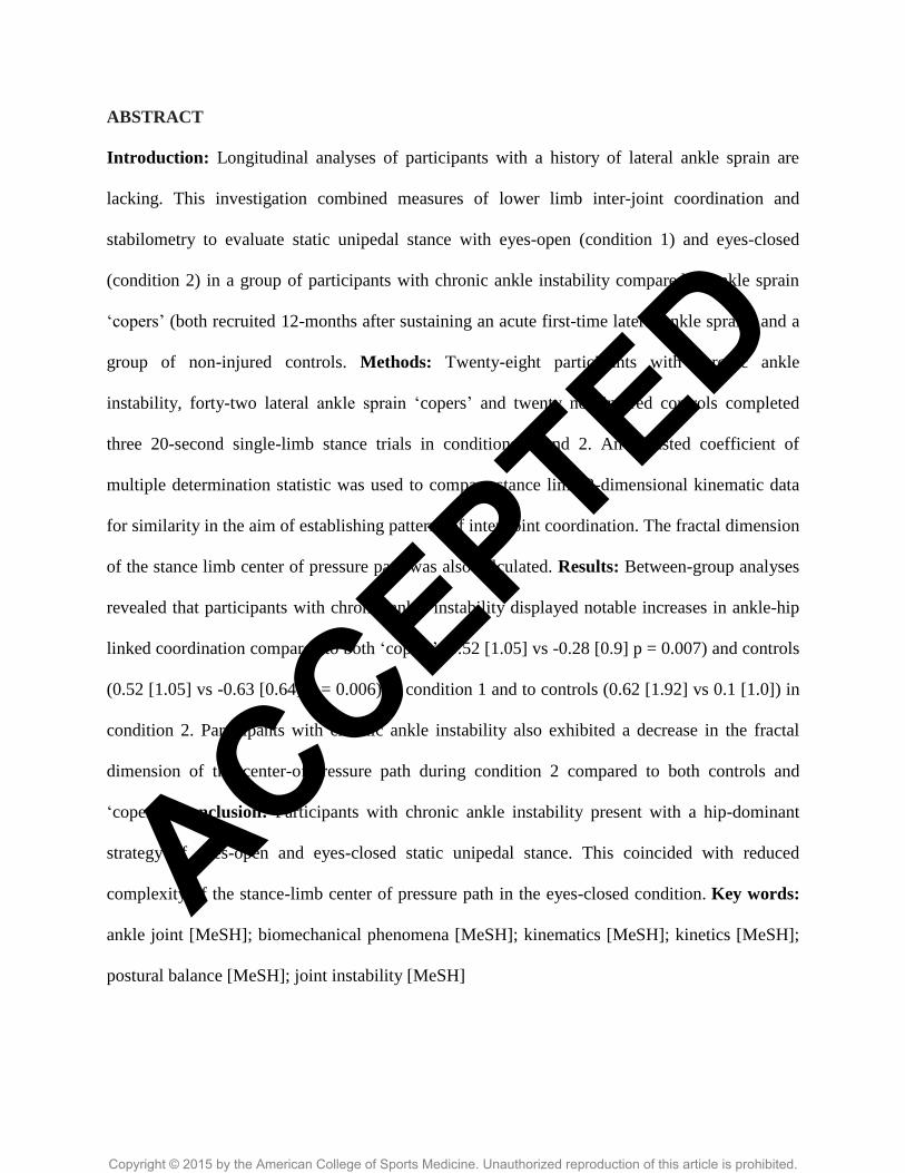
ABSTRACT
Introduction: Longitudinal analyses of participants with a history of lateral ankle sprain are
lacking. This investigation combined measures of lower limb inter-joint coordination and
stabilometry to evaluate static unipedal stance with eyes-open (condition 1) and eyes-closed
(condition 2) in a group of participants with chronic ankle instability compared to ankle sprain
‗copers‘ (both recruited 12-months after sustaining an acute first-time lateral ankle sprain) and a
group of non-injured controls. Methods: Twenty-eight participants with chronic ankle
instability, forty-two lateral ankle sprain ‗copers‘ and twenty non-injured controls completed
three 20-second single-limb stance trials in conditions 1 and 2. An adjusted coefficient of
multiple determination statistic was used to compare stance limb 3-dimensional kinematic data
for similarity in the aim of establishing patterns of inter-joint coordination. The fractal dimension
of the stance limb center of pressure path was also calculated. Results: Between-group analyses
revealed that participants with chronic ankle instability displayed notable increases in ankle-hip
linked coordination compared to both ‗copers‘ (0.52 [1.05] vs -0.28 [0.9] p = 0.007) and controls
(0.52 [1.05] vs -0.63 [0.64] p = 0.006) in condition 1 and to controls (0.62 [1.92] vs 0.1 [1.0]) in
condition 2. Participants with chronic ankle instability also exhibited a decrease in the fractal
dimension of the center-of-pressure path during condition 2 compared to both controls and
‗copers‘. Conclusion: Participants with chronic ankle instability present with a hip-dominant
strategy of eyes-open and eyes-closed static unipedal stance. This coincided with reduced
complexity of the stance-limb center of pressure path in the eyes-closed condition. Key words:
ankle joint [MeSH]; biomechanical phenomena [MeSH]; kinematics [MeSH]; kinetics [MeSH];
postural balance [MeSH]; joint instability [MeSH]
ACCEPTED

Copyright © 2015 by the American College of Sports Medicine. Unauthorized reproduction of this article is prohibited.
INTRODUCTION
Lateral ankle sprain (LAS) injury pervades a variety of activities, with between 0.88 [CI 95%:
0.73 – 1.02] and 7 [CI 95%: 6.82 – 7.18] injury events occurring per 1,000 exposures,
depending on the activity type (11). The prevalence of this injury in a wide range of sports and
activities is further complicated by its capacity to deteriorate into an array of chronic sequalae
and injury recurrence, collectively termed ―chronic ankle instability (CAI)‖(7, 15-17), which has
been linked to limitations in future physical activity participation (1).
Although CAI is considered a multifaceted condition with a range of consequences, persistent
deficits in single-limb stance (SLS) postural control strategies are well established in individuals
with CAI (18, 26, 36), and may be consequent upon a potential change in neural signalling
following the initial ankle joint trauma (14). This theory has since been tested in previous studies
comparing individuals with a history of LAS to uninjured controls (13, 37), with a new
hypothesis emerging whereby the long-term outcome following LAS is dependent upon the
success or failure of the newly adopted post-LAS postural control strategies (34, 35). This has
yet to be confirmed however, as there is currently an absence of longitudinal investigations
which prospectively track the restoration or degradation of postural control strategies after an
initial LAS.
More recently, LAS ‗copers‘, who have a history of LAS and experience a restoration of pre-
injury levels of function in the year following initial injury (15, 34), have been compared to
individuals with CAI during SLS (36); this is considered to provide a stronger, more relevant
comparison in laying the foundation for longitudinal analyses and the development of clinical
ACCEPTED

Copyright © 2015 by the American College of Sports Medicine. Unauthorized reproduction of this article is prohibited.
outcome models for the CAI paradigm (34). Recently published material from our laboratory
was developed according to this paradigm: individuals with an acute, first-time LAS were
evaluated in comparison to a non-injured control group during eyes-open and eyes-closed SLS
using kinematic and kinetic measures of joint position and platform stabilometry respectively
(9). A follow-up analysis of these same individuals 6-months following the initial assessment
revealed a hip-dominant postural control strategy prevailing during the prescribed tasks of SLS,
again in comparison to non-injured controls (10). In this latter investigation, an adjusted
coefficient of multiple determination (ACMD) statistic was utilised to evaluate waveform
similarity between lower extremity 3-D joint angular displacements in the determination of inter-
joint ‗coupling‘ strategies during 20 seconds of eyes-open and eyes-closed SLS (10). We believe
novel insight was gained by combining these laboratory measures: the increase in observed
coupling between sagittal plane hip and frontal plane ankle motion in LAS participants
underpinned a hypothesis that these individuals adopt a hip-dominant strategy in the maintenance
of single-limb postural control, perhaps to compensate for a dysfunctional ankle joint (10). This
theory is in agreement with the model of human postural control proposed by Nashner and
McCollum (29), in which an ‗ankle strategy‘ is appropriated to the fine tuning of static postural
control, and a ‗hip strategy‘ is employed to tackle more substantial postural control disturbances;
the LAS group in the aforementioned studies were considered to have reduced capacity to utilise
their ankle strategy, thus adopting the more proximal hip strategy in its place (9, 10).
The measure of platform stabilometry employed in the aforementioned investigations from our
laboratory was the fractal dimension (FD) of the center of pressure (COP) path. The FD is a unit-
less measure that conceptualises the complexity of the COP path using a value between 1 (a
ACCEPTED

Copyright © 2015 by the American College of Sports Medicine. Unauthorized reproduction of this article is prohibited.
straight line or low complexity) and 2 (a convoluted line or high complexity)(24). In addition to
a hip-dominant kinematical strategy, LAS participants were also shown to display a bilaterally
reduced FD of the COP path during eyes-closed SLS within 2-weeks of incurring their initial
LAS injury (9), and on their involved limb only 6-months following their initial sprain (10). This
was interpreted as a reduced ability to utilise the available base of support on removal of visual
afferents (8-10).
The current study is a continuation of those previously described and forms part of a larger
longitudinal analysis of the LAS cohort. Specifically, we sought to complete the 12-month
follow-up of the individuals we previously alluded to who completed the 2-week and 6-month
evaluations, thus allowing for participant segregation as CAI or LAS ‗coper‘ status. Kinematic
and stabilometric measures were combined to compare stance limb inter-joint coordination and
COP path complexity during eyes-open and eyes-closed SLS between individuals with CAI,
ankle sprain ―copers‖ and a separately recruited non-injured control group of participants. We
hypothesised that individuals with CAI would exhibit the same hip-dominant coupling strategies
for completing eyes-open and eyes-closed SLS which were documented 6-months previously,
whereas LAS ―coper‖ and control participants would not due to a superior capacity to employ an
ankle-based balance strategy in isolation. Furthermore, we hypothesised that during eyes-closed
SLS CAI participants would exhibit poorer postural control ability, as evidenced by a reduced
FD of the COP path.
ACCEPTED

Copyright © 2015 by the American College of Sports Medicine. Unauthorized reproduction of this article is prohibited.
METHODS
Participants
As part of the larger longitudinal study conducted in our laboratory, eighty-two individuals
presenting with a first-time acute LAS were recruited from a University-affiliated hospital
emergency department. All LAS participants were provided with the same basic advice on
applying ice and compression on discharge from the hospital ED: they were each encouraged to
weight-bear and walk within the limits of pain. Whether participants sought additional formal
medical healthcare services for council or rehabilitation of their LAS was recorded on arrival to
the testing laboratory but not controlled as part of the current study.
These individuals were required to attend three test sessions and complete a number of
movement tasks within 2-weeks of sustaining their initial injury, with further follow-up at 6-
months and 12-months. Testing procedures for these participants in the acute phase of their
injury has previously been reported (8). A total of seventy-one of the original eighty-two
participants returned for the third test session (i.e. 12-month follow-up); the current investigation
relates to the data collected for these individuals at this time-point. An additional convenience
group of twenty participants with no prior history of LAS were also recruited from the hospital
catchment area population using posters and flyers to act as a control group. Participant
characteristics for the individuals included in the current analysis are presented in Table 1. The
following exclusion criteria were utilised for both limbs (where applicable) at the time of
recruitment: (1) no previous history of LAS injury (excluding the initial acute LAS episode for
the CAI and coper groups); (2) no other severe lower extremity injury in the last 6 months; (3) no
history of ankle fracture; (4) no previous history of major lower limb surgery; (5) no history of
neurological disease, vestibular or visual disturbance or any other pathology that would impair
ACCEPTED

Copyright © 2015 by the American College of Sports Medicine. Unauthorized reproduction of this article is prohibited.
their motor performance. Participants provided written informed consent, and the study was
approved by the University Human Research Ethics Committee.
LAS participants‘ designation as CAI or ‗coper‘ status was completed according to recently
published guidelines (9, 10). Self-reported ankle instability was confirmed with the Cumberland
Ankle Instability Tool (19); individuals with a score of <24 were designated as having CAI while
―copers‖ were designated with a score of ≥24, to avoid the potential for false positives in this
group (39). Additionally, to be designated as a coper, participants must have returned to pre-
injury levels of activity and function (36). Finally, the activities of daily living and sports
subscales of the Foot and Ankle Ability Measure (FAAMadl and FAAMsport) were utilised as a
means to evaluate general self-reported foot and ankle function (6). All participants completed
the CAIT and subscales of the FAAM on arrival to the testing laboratory.
Based on these criteria, twenty-eight of the LAS participants were designated as having CAI, and
forty-two as ―copers‖ (Table 1). One LAS ‗coper‘ participant was excluded because he did not
return to pre-injury levels of activity participation.
Protocol
Collection methods for this study have been previously documented (10). Briefly, following the
collection of anthropometric measures required for the calculation of internal joint centres of the
lower extremity joints, each participant was instrumented with the Codamotion bilateral lower
limb gait set-up according to the manufacturer guidelines (Charnwood Dynamics Ltd,
Leicestershire, UK). A neutral stance trial was used to align the subject with the laboratory
coordinate system and to function as a reference position for subsequent kinematic analysis (40).
ACCEPTED

Copyright © 2015 by the American College of Sports Medicine. Unauthorized reproduction of this article is prohibited.
Participants then performed three, 20 second trials of quiet SLS barefoot on a force plate with
their eyes-open on both limbs, each separated by a 30 second rest period. Following another 2
minute rest period, participants then attempted to complete three 20 second SLS trials with their
eyes-closed. Participants were required to complete a minimum of three practice trials on each
limb for each condition prior to data acquisition (8, 22). Participants who were unable to
complete a full trial of unilateral stance after five attempts on the relevant limb were not included
in the analysis for that limb. The test order between legs was randomized. For both conditions of
the SLS task, participants were instructed to stand as still as possible with their hands resting on
their iliac crests while adopting a postural orientation most natural to them; the position of the
non-stance limb was not dictated in the sagittal plane as part of experimental procedures. Trials
were deemed invalid if the subject lifted their hands off their iliac crests, placed their non-stance
limb on the support surface, moved their non-stance hip into a position > 30 degrees abduction,
adducted their non-stance limb against their stance limb for support or if the foot placement
assumed by the participants relative to the support surface changed in any way over the course of
a trial. In addition a trial was deemed as failed in the eyes-closed condition if the participant
opened their eyes at any point.
Kinematic and Kinetic Data Processing
Three Codamotion cx1 units were used to acquire data on 3-D angular displacements at the hip,
knee and ankle joints for both limbs during the SLS tasks. Two AMTI (Watertown, MA)
walkway embedded force plates were used to acquire kinetic data. Kinematic and kinetic data
acquisition was made at 100 Hz. The Codamotion CX1 units were time synchronized with the
force plates. Kinematic and COP data were analysed using the Codamotion software and then
ACCEPTED
Copyright © 2015 by the American College of Sports Medicine. Unauthorized reproduction of this article is prohibited.
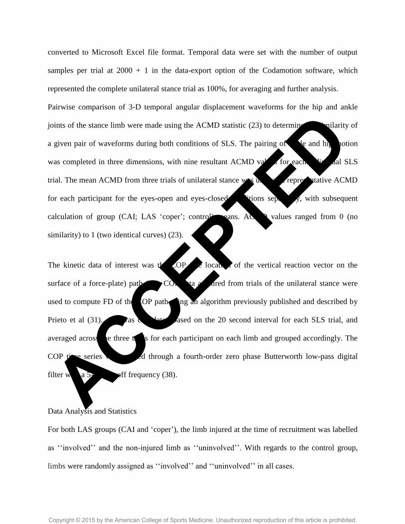
converted to Microsoft Excel file format. Temporal data were set with the number of output
samples per trial at 2000 + 1 in the data-export option of the Codamotion software, which
represented the complete unilateral stance trial as 100%, for averaging and further analysis.
Pairwise comparison of 3-D temporal angular displacement waveforms for the hip and ankle
joints of the stance limb were made using the ACMD statistic (23) to determine the similarity of
a given pair of waveforms during both conditions of SLS. The pairing of ankle and hip motion
was completed in three dimensions, with nine resultant ACMD values for each individual SLS
trial. The mean ACMD from three trials of unilateral stance was used as a representative ACMD
for each participant for the eyes-open and eyes-closed conditions separately, with subsequent
calculation of group (CAI; LAS ‗coper‘; control) means. ACMD values ranged from 0 (no
similarity) to 1 (two identical curves) (23).
The kinetic data of interest was the COP (the location of the vertical reaction vector on the
surface of a force-plate) path (31). COP data acquired from trials of the unilateral stance were
used to compute FD of the COP path using an algorithm previously published and described by
Prieto et al (31). FD was calculated based on the 20 second interval for each SLS trial, and
averaged across the three trials for each participant on each limb and grouped accordingly. The
COP time series were passed through a fourth-order zero phase Butterworth low-pass digital
filter with a 5-Hz cut-off frequency (38).
Data Analysis and Statistics
For both LAS groups (CAI and ‗coper‘), the limb injured at the time of recruitment was labelled
as ‗‗involved‘‘ and the non-injured limb as ‗‗uninvolved‘‘. With regards to the control group,
limbs were randomly assigned as ‗‗involved‘‘ and ‗‗uninvolved‘‘ in all cases.
ACCEPTED

Copyright © 2015 by the American College of Sports Medicine. Unauthorized reproduction of this article is prohibited.
For all outcomes, we calculated mean (SD) scores for the involved and uninvolved limbs of the
CAI, LAS ‗coper‘ and control groups.
A principal component analysis (PCA) was performed to reduce the dimensionality of the
kinematic data. Specifically, the nine ‗latent‘ variables of inter-joint coordination were reduced
into significant components. This was performed separately for the eyes-open and eyes-closed
conditions. Preliminary analyses (scree test and parallel analysis) informed our decision to retain
three components for the eyes-open condition and two components for the eyes-closed condition.
To test our hypothesis that the CAI group would display hip-dominant strategies of inter-joint
coordination , the components derived from the ACMD ‗latent‘ variables were compared
between groups using a 2-way MANOVA for each condition (eyes-open and eyes-closed). The
independent variables were group (CAI; LAS ‗coper‘; control) and limb (involved; uninvolved).
The dependent variables were the three extracted components for the eyes-open condition and
the two extracted components for the eyes-closed condition. Preliminary assumption testing was
conducted to check for normality, linearity, univariate and multivariate outliers, homogeneity of
variance-covariance matrices, and multicollinearity with no serious violations noted. An alpha-
level of p < 0.05 was used to determine significant differences for each analysis (20). Post-hoc
comparisons were completed using a Tukey HSD test where appropriate. The significance level
for post-hoc analyses was set with a bonferroni adjusted alpha of p < 0.017 for the eyes-open
condition (0.05/3 components) and p < 0.025for the eyes-closed condition (0.05/2 components)
(21).
ACCEPTED

Copyright © 2015 by the American College of Sports Medicine. Unauthorized reproduction of this article is prohibited.
In order to test our hypothesis that the CAI group would display reduced COP path trajectory
FD during the SLS task compared to LAS ‗copers‘ and controls, a two-way between-groups
ANOVA was conducted separately for each condition (eyes-open and eyes-closed). The
independent variables were group (CAI; LAS ‗coper‘; control) and limb (involved; uninvolved).
The dependent variable was FD of the COP path. The significance level for this analysis was set
a priori at p < 0.05. Post-hoc comparisons were completed using a Tukey HSD test where
appropriate. The significance level for post-hoc analyses was set at p < 0.05 for both conditions.
All data were analyzed using Predictive Analytics Software (Version 18, SPSS Inc., Chicago, IL,
USA).
RESULTS
All participants completed the eyes-open SLS task on both limbs. Thirty-six percent of CAI
participants (10 of 28), 76% of LAS ‗copers‘ (33 of 42) and 85% of controls (17 of 20)
completed the SLS task with their eyes-closed on both their ‗involved‘ and ‗uninvolved‘ limbs.
Regarding inter-joint coordination, there was a statistically significant main effect for group in
the eyes-open [F (3,322) = 2.585, p = 0.018; Wilks‘ Lambda = 0.91] and eyes-closed [F (3,220)
= 3.58, p = 0.008; Wilks‘ Lambda = 0.88] conditions. When the results of the dependent
variables were considered separately, the only components to reach statistical significance at the
bonferroni adjusted alpha levels were components 3 (which loaded heavily on the inter-joint
coordination between sagittal plane hip and frontal plane ankle motion, and sagittal plane hip and
transverse plane ankle motion) in the eyes-open condition [F(2,321) = 6.508, p = 0.002, p2
=
0.074] and 2 (which loaded heavily on the inter-joint coordination between sagittal plane hip
motion and ankle motion in all three dimensions, and frontal plane hip motion and sagittal plane
ACCEPTED

Copyright © 2015 by the American College of Sports Medicine. Unauthorized reproduction of this article is prohibited.
ankle motion) in the eyes-closed condition [F(2,219) = 4.125, p = 0.019, p2
= 0.069]. Post-hoc
analysis and inspection of the mean scores revealed that CAI participants exhibited lower mean
scores for component 3 in the eyes-open condition, most notably on their involved limb (M = -
0.52, SD = 1.05) compared to both LAS ‗copers‘ (M = 0.28, SD = 0.9, p = 0.007) and controls
(M = 0.63, SD = 0.64, p = 0.006). Due to the negative correlation between component 3 and its
latent variables, this represented an increase in ankle-hip linked coordination. With regards to the
eyes-closed condition, post-hoc analyses revealed that CAI participants exhibited greater mean
scores for component 2 compared to controls only (p = 0.024). This was evident on both their
involved (CAI: M = 0.62, SD = 1.92; control = 0.1, SD = 1.0) and uninvolved (CAI: M = 0.07,
SD = 1.19; control = -0.34, SD = 0.66) limbs. Due to the positive correlation between this
component and its latent variables, this too represented an increase in ankle-hip linked
coordination.
Descriptive statistics for the ‗latent‘ ACMD variables for the CAI, LAS ‗coper‘ and control
groups prior to PCA are presented in Table 2. Pattern and structure matrices for the PCA relative
to the eyes-open and eyes-closed conditions are presented in Table 3.
Regarding the kinetic variables of interest, there was a statistically significant main effect for
group in the eyes-closed condition [F (2,219) = 8.11, p = 0.001, p 2
= 0.12] only. Post-hoc
analysis and inspection of the mean scores revealed that CAI participants exhibited lower FD of
the COP path trajectory on their involved limb (M = 1.78, SD = 0.11) compared to both LAS
‗copers‘ (M = 1.90, SD = 0.1, p = 0.045) and controls (M = 1.94, SD = 0.13, p < 0.001).
In an exploratory analysis, the concurrent validity of four variables deemed ‗significantly
important‘ (eyes-closed SLS task completion, component 3 in the eyes-open condition on the
ACCEPTED

Copyright © 2015 by the American College of Sports Medicine. Unauthorized reproduction of this article is prohibited.
involved limb, and both component 2 and the FD of the COP path on the involved limb in the
eyes-closed condition) in determining the extent of disability was established by calculating their
respective Pearson correlation coefficients to CAIT score. This was performed for LAS
participants only. The ability of each of these variables to determine outcome (CAI vs LA
‗coper‘) was then tested for sensitivity and specificity. A cut-off value of 0.7 was adopted for the
C-statistic in the sensitivity and specificity analyses.
There was no correlation between CAIT score and eyes-closed SLS task completion (r = 0.004, p
= 0.97), component 3 (r = 0.109, p = 0.39), component 2 (r = 0.213, p = 0.19) or FD of the COP
path (r = 0.11, p = 0.39).
However, eyes-closed SLS task completion was moderately predictive of outcome (CAI vs LAS
‗coper‘), with a C-statistic of 0.71 (p = 0.003); the resultant prediction equation yielded a
sensitivity of 0.64 and a specificity of 0.78, with a positive likelihood ratio of 2.93.
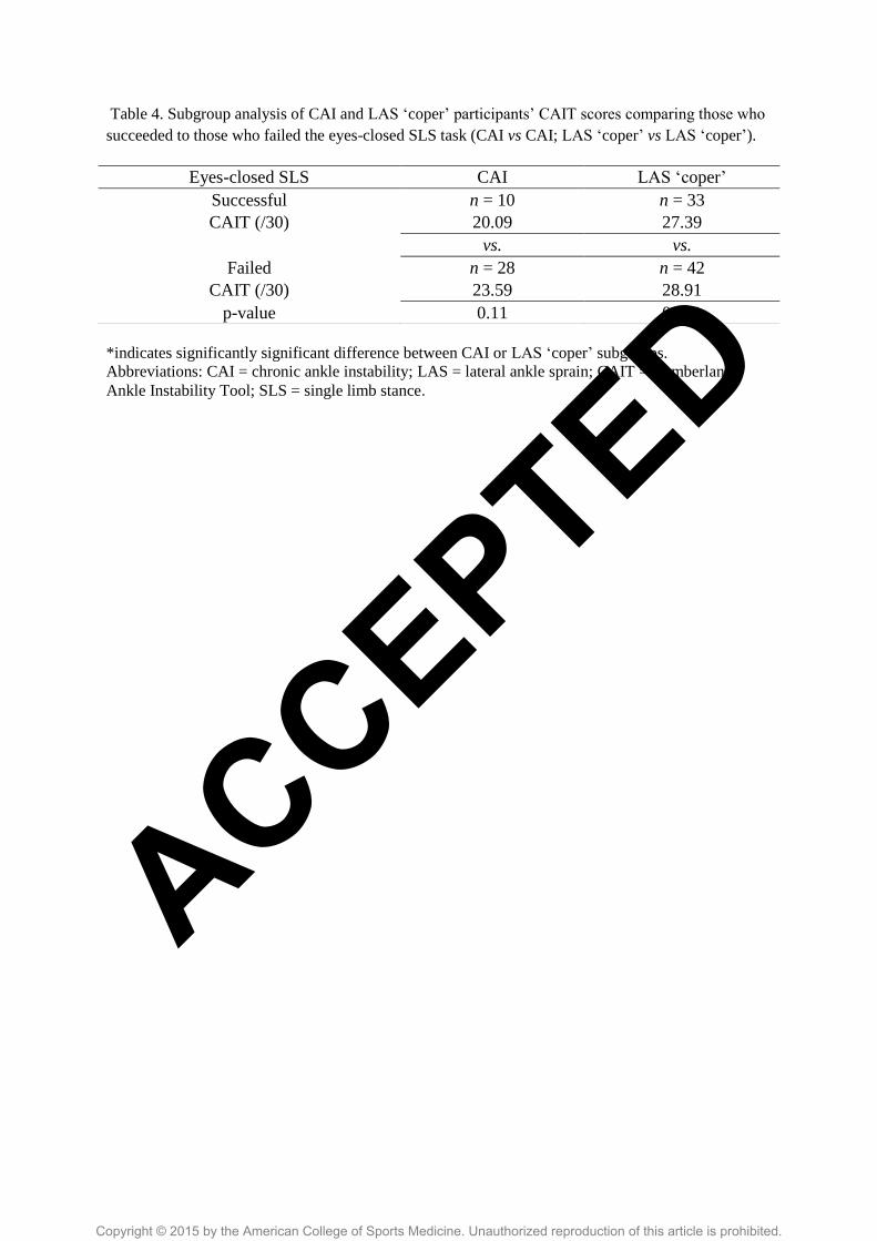
To explain these findings, post-hoc analysis using independent samples t-tests were performed to
compare the CAIT scores of the subgroups of CAI and LAS ‗coper‘ participants who succeeded
and failed at the eyes-closed SLS task. The p-value for this post-hoc analysis was set a priori
with a bonferonni adjustment at p < 0.025. This analysis revealed that LAS ‗copers‘ who were
able to complete the task actually had significantly greater disability than those who couldn‘t,
and likewise for the CAI participants, thus explaining the capacity of task completion to predict
outcome (CAI or LAS ‗coper‘), despite the absence of a correlation to CAIT score. The results of
this post-hoc analysis for both sub-groups of CAI and LAS ‗coper‘ participants are presented in
Table 4. None of the other variables (components 2 and 3, FD of the COP path) were predictive
of outcome based on the C-statistic.
ACCEPTED

Copyright © 2015 by the American College of Sports Medicine. Unauthorized reproduction of this article is prohibited.
DISCUSSION
The primary finding of this motion analysis investigation was that individuals with CAI exhibit
greater ‗coupling‘ of hip and ankle motion compared to both ankle sprain ―copers‖ and non-
injured controls during an SLS task. This increase in ankle-hip ‗coupling‘ may represent a
compensatory strategy to accommodate what is now a chronically unstable ankle in the CAI
group (as determined using the CAIT). Furthermore, the CAI group also demonstrated a reduced
FD of the COP path on their involved limb compared to both ankle sprain ―copers‖ and controls
in the eyes-closed condition of SLS. These findings are consistent with those previously
published on this group as a whole within two-weeks of their injury (9), and 6-months following
(10). Therefore, it is possible that the abatement of a hip-dominant postural control strategy may
be conducive to superior outcome. The design of the current study however means that this
cannot be confirmed.
To our knowledge, this is the first documented evaluation of postural control in a first-time LAS
population exactly 12-months following initial injury using kinematical measures of lower limb
inter-joint coordination and platform stabilometry. The advantage of the experimental design is
that all LAS participants (CAI and LAS ‗coper‘) were recruited at the time of their first ever LA
S injury, thereby securing the homogenous subgroups of ankle sprain outcome. As we have
alluded to, this study is part of a longitudinal analysis designed to develop an outcome model for
the predictors of CAI following LAS injury.
The use of ankle sprain ―copers‖ provides a superior comparison group to individuals with CAI
than non-injured controls because ankle sprain ―copers‖ have had the same exposure, but are not
characterized by the same symptom sequalae as those individuals who develop CAI (34). The
ACCEPTED

Copyright © 2015 by the American College of Sports Medicine. Unauthorized reproduction of this article is prohibited.
addition of a non-injured control group in this report has however allowed us to identify that,
based on the parameters utilised in the current investigation, ankle sprain ―copers‖ are no
different to non-injured controls in their postural control strategies for eyes-open and eyes-closed
SLS. This is evidenced by the absence of between-groups differences for copers and controls in
this analysis, which is in agreement with previous findings during a similar task protocol (32,
36). It has recently been identified that this tripartite comparison between CAI, LAS ―coper‖ and
control participants is needed in the context of LAS (34). Indeed, there are only a limited number
of previous analyses which have evaluated movement patterns in these groups (4, 5, 32, 36, 37)
with fewer still providing an analysis of SLS postural control using measures of platform
stabilometry (32, 36). Wikstrom et al. (36) identified that LAS ‗coper‘ participants‘ stance limb
COP paths exhibits a lower velocity in both the antero-posterior and the medio-lateral axes of the
foot than individuals with CAI during a similar task. Shields et al.(32), demonstrated that the
standard deviation of the COP path and it‘s range were significantly lower in ankle sprain
―copers‖ compared to subjects with CAI, a finding the authors interpreted as being demonstrative
of better postural control predictability.
The issue regarding the application of these ‗traditional measures‘ of COP excursion which
quantify the length, area and velocity of the COP path, apart from their questionable reliability
(12), is that they have previously yielded inconsistent or even contradictory findings in LAS
populations (27). By contrast, the FD measure utilised in the current analysis is a reliable
measure (12) which has previously been successful in characterising a degeneration in stability
of the postural control system in the transition from eyes-open to eyes-closed stance (3).
Furthermore, because we have adopted the FD calculation in analysing the COP paths of these
same participants during SLS within 2-weeks (9) of incurring their initial injury and 6-months
ACCEPTED

Copyright © 2015 by the American College of Sports Medicine. Unauthorized reproduction of this article is prohibited.
later (10), its use enables us to directly compare our findings across time points relevant to the
development of CAI or LAS ‗coper‘ status.
Consistent with the investigations of these participants 2-weeks and 6-months following injury
occurrence (9, 10), the findings of the current study revealed that individuals with poorer
outcome (<24 on the CAIT in this study, ‗injured‘ status in those previously described), exhibit
reduced FD of the COP path compared to individuals with superior outcome (non-injured
controls and ankle sprain ―copers‖), albeit in the eyes-closed condition only. This was previously
interpreted as a reduced ability to utilise the available base of support during SLS, isolated to
instances where the task condition dictated the removal of visual afferents (8). Similarly, the CAI
participants in the current study also exhibited greater ‗coupling‘ of hip-ankle joint coordination
in the completion of eyes-closed SLS compared to controls, a finding consistent with the acute
(2-week) and injury ―twilight‖ (6-month) data.
That a lower proportion of the CAI group were able to complete the balance task in the eyes-
closed condition prompted an exploratory analysis, whereby this dichotomous outcome and the
other group-defining variables (components 3, 2 and the FD of the COP path) were separately
correlated with CAIT score. Their capacity to predict outcome (CAI vs LAS ‗coper‘) was also
evaluated. While the group-defining variables exhibited no correlation with CAIT score, and did
not predict outcome, task completion was determined as predictive of CAI or LAS ‗coper‘ status.
The moderate specificity and sensitivity that an ability to complete eyes-closed SLS had in
predicting outcome, in the absence of a correlation to CAIT score, may be under-lied by a
disability ‗cut-off‘; the correlation between CAIT score and task ability is probably not linear,
wherein it is possible that at a certain point, an individual‘s ability to perform a difficult balance
ACCEPTED

Copyright © 2015 by the American College of Sports Medicine. Unauthorized reproduction of this article is prohibited.
task (such as eyes-closed SLS) deteriorates drastically. Individuals below this cut-off have the
potential to be equally likely to be unable to complete the task, whether they have ―more‖ or
―less‖ disability. Future analyses are required to elucidate such ‗cut-offs‘ however.
The apparent difficulty CAI participants had in completing eyes-closed SLS may represent an
impaired capacity to compensate and re-coordinate the available sensory afferents, or to rely on
the remaining somatosensory and vestibular afferents when visual ones have been removed (25).
It is generally accepted that there is redundancy of these three afferents in maintaining SLS (30),
whereby a selective priority is placed based on the availability of reliable information (28). This
allows the fully functioning somatosensory system to maintain postural control and stability in
the presence of altered afferent signals (25). However, prescribing an eyes-closed constraint
during the SLS task imposes somatosensory demands beyond the capacity of even healthy
individuals (as evidenced by the fact that 15% of controls were unable to complete our eyes-
closed task protocol), impairing their ability to exploit available redundancies in the maintenance
of static postural control (9). This impairment is seemingly magnified in individuals with
musculoskeletal injury on the basis of the current findings, and in light of the evidence
previously outlined of participants with a recent history of ankle sprain (9, 10). Thus, a decay in
somatosensory afferents, as may occur with acute LAS injury and which is considered to
contribute to instability persistence (14), combined with loss of visual input, challenged the
ability of the central nervous system to re-coordinate the available information with an
appropriated postural control response (13, 28) in individuals with CAI. This then manifested in
a deterioration of eyes-closed unilateral standing postural control and stability in the CAI group,
with less effective utilisation of the supporting base on the involved limb (9). It is also plausible
that the somatosensory deterioration associated with CAI development manifested in a ‗hip-
ACCEPTED

Copyright © 2015 by the American College of Sports Medicine. Unauthorized reproduction of this article is prohibited.
dominant‘ compensatory strategy as evidenced by the significantly greater ankle-hip coupling
compared to both ankle sprain ―copers‖ and controls in the eyes-open condition, and compared
to controls in the eyes-closed condition. Whereas the ankle strategy of human postural control is
more suited to subtle corrections, the hip strategy is considered ideal for substantial disturbances
of equilibrium (25). Tropp (33) previously utilised kinematic measures of sway amplitude at the
ankle, hip and trunk to confirm the existence of these strategies. He also identified the impaired
postural control capacity of individuals with ankle instability in utilising their ankle strategies for
SLS, based on an increased number of postural corrections at the trunk required by this group
(33). In another kinematic analysis of participants with a history of LAS during an SLS task,
Huurnink et al.(22) failed to identify differences in kinematic outcome measures (ankle and hip
angular velocities) between participants with and without a history of ankle sprain. We believe
the use of the ACMD statistic in the current study to have specifically identified an increased
reliance on the more proximal hip strategy in the CAI group, on the basis of the greater
waveform similarity between these joints. During normal control of SLS, the foot‘s narrow base
of support makes it necessary to employ the hip strategy in controlling substantial medio-lateral
disturbances of postural stability, while ankle movements may only achieve fine-tuning of
medio-lateral sway (2). The basis of CAI may be belied by an impaired capacity to fulfil this
medio-lateral fine-tuning, with subsequent transition to the more proximal hip. Herein lies a
significant limitation of the current analysis; these and any other hypotheses regarding the
neuromechanical predictors of CAI are still unclear, although the current study is part of a
project designed to investigate this issue. Another significant limitation of this analysis is that we
were unable to experimentally control whether LAS participants sought additional rehabilitation
ACCEPTED

Copyright © 2015 by the American College of Sports Medicine. Unauthorized reproduction of this article is prohibited.
for their injury. However, to do so would have been unethical, and no treatment data ‗clusters‘
were evident during data management and analysis.
The clinical implications of this study are two-fold: first, in light of the evidence presented on
these individuals during their ‗recovery‘, it would seem that the capacity to perform static
postural control tasks will challenge the individual to perform subtle corrections with ankle
movements. A SLS task and derivations of such may therefore possess value in being part of a
rehabilitation programme. Based on previous evidence, we would recommend though that the
patient only progresses to such tasks when they are sufficiently able to complete them (8).
Second, the use of eyes-closed SLS as a clinical test to quantify disability and functional capacity
should be considered. There is further potential for future research to confirm this.
In conclusion, the results of the current study suggest that participants with CAI are separated by
LAS ‗copers‘ and non-injured controls in their exhibition of a hip-dominant balance strategy
during a task of eyes-open and eyes-closed unilateral stance.
Acknowledgements
This study was supported by the Health Research Board (HRA_POR/2011/46) as follows: PI –
Eamonn Delahunt; Co-investigators – Chris Bleakley and Jay Hertel; PhD student – Cailbhe
Doherty). The results of the present study do not constitute endorsement by ACSM.
Conflict of Interest: No conflicts of interest were associated with the authors and the results of
this research.
ACCEPTED

Copyright © 2015 by the American College of Sports Medicine. Unauthorized reproduction of this article is prohibited.
REFERENCES
1. Attenborough A, Hiller C, Smith R, Stuelcken M, Greene A, Sinclair P. Chronic Ankle
Instability in Sporting Populations. Sports Med. 2014;[Epub ahead of print].
2. Baier M, Hopf T. Ankle orthoses effect on single-limb standing balance in athletes with
functional ankle instability. Arch Phys Med Rehabil. 1998;79(8):939-44.
3. Błaszczyk J, Klonowski W. Postural stability and fractal dynamics. Acta Neurobiol Exp
(Wars). 2001;61(2):105-12.
4. Brown C, Padua C, Marshall D, Guskiewicz K. Individuals with mechanical ankle
instability exhibit different motion patterns than those with functional ankle instability and ankle
sprain copers. Clin Biomech (Bristol, Avon). 2008;23(6):822-31.
5. Brown CN, Padua DA, Marshall SW, Guskiewicz KM. Variability of motion in
individuals with mechanical or functional ankle instability during a stop jump maneuver.
Clinical Biomechanics. 2009;24(9):762-8.
6. Carcia C, Martin R, Drouin J. Validity of the Foot and Ankle Ability Measure in athletes
with chronic ankle instability. J Athl Train. 2008;43(2):179-83.
7. Delahunt E, Coughlan GF, Caulfield B, Nightingale EJ, Lin CW, Hiller CE. Inclusion
criteria when investigating insufficiencies in chronic ankle instability. Med Sci Sports Exerc.
2010;42(11):2106-21.
8. Doherty C, Bleakley C, Hertel J, Caulfield B, Ryan J, Delahunt E. Balance failure in
single limb stance due to ankle sprain injury: an analysis of center of pressure using the fractal
dimension method. Gait & Posture. 2014;40(1):172-6.
ACCEPTED

Copyright © 2015 by the American College of Sports Medicine. Unauthorized reproduction of this article is prohibited.
9. Doherty C, Bleakley C, Hertel J, Caulfield B, Ryan J, Delahunt E. Postural control
strategies during single limb stance following acute lateral ankle sprain. Clinical Biomechanics.
2014;29(6):643-9.
10. Doherty C, Bleakley C, Hertel J, et al. Inter-joint coordination strategies during unilateral
stance 6-months following acute lateral ankle sprain. Clin Biomech. 2015.
11. Doherty C, Delahunt E, Caulfield B, Hertel J, Ryan J, Bleakley C. The Incidence and
Prevalence of Ankle Sprain Injury: A Systematic Review and Meta-Analysis of Prospective
Epidemiological Studies. Sports Med. 2014;44 (1) 123-140.
12. Doyle T, Newton R, Burnett A. Reliability of traditional and fractal dimension measures
of quiet stance center of pressure in young, healthy people. Arch Phys Med Rehabil
2005;86(10):2034-40.
13. Evans T, Hertel J, Sebastianelli W. Bilateral deficits in postural control following lateral
ankle sprain. Foot Ankle Int. 2004;25(11):833-9.
14. Freeman MA. Instability of the foot after injuries to the lateral ligament of the ankle. J
Bone Joint Surg Br. 1965;47(4):669-77.
15. Gribble P, Delahunt E, Bleakley C, et al. Selection criteria for patients with chronic ankle
instability in controlled research: a position statement of the International Ankle Consortium.
The Journal of orthopaedic and sports physical therapy. 2013;43(8):585-91.
16. Gribble PA, Delahunt E, Bleakley C, et al. Selection criteria for patients with chronic
ankle instability in controlled research: A position statement of the International Ankle
Consortium. British journal of sports medicine. 2014;48(13):1014-8.
ACCEPTED

Copyright © 2015 by the American College of Sports Medicine. Unauthorized reproduction of this article is prohibited.
17. Gribble PA, Delahunt E, Bleakley CM, et al. Selection criteria for patients with chronic
ankle instability in controlled research: A position statement of the international ankle
consortium. Journal of Athletic Training. 2014;49(1):121-7.
18. Hertel J, Olmsted-Kramer LC. Deficits in time-to-boundary measures of postural control
with chronic ankle instability. Gait Posture. 2007;25(1):33-9.
19. Hiller C, Refshauge K, Bundy A, Herbert R, Kilbreath S. The Cumberland ankle
instability tool: a report of validity and reliability testing. Arch Phys Med Rehabil.
2006;87(9):1235-41.
20. Hopkins W, Marshall S, Batterham A, Hanin J. Progressive statistics for studies in sports
medicine and exercise science. Med Sci Sports Exerc. 2009;41(1):3-13.
21. Huberty C, Morris J. Multivariate analysis versus multiple univariate analyses.
Psychological Bulletin. 1989;105(2):302-8.
22. Huurnink A, Fransz D, Kingma I, Verhagen E, van Dieën J. Postural stability and ankle
sprain history in athletes compared to uninjured controls. Clin Biomech (Bristol, Avon).
2014;29(2):183-8.
23. Kadaba M, Ramakrishnan H, Wootten M, Gainey J, Gorton G, Cochran G. Repeatability
of kinematic, kinetic, and electromyographic data in normal adult gait. J Orthop Res.
1989;7:849:60.
24. Katz MJ, George EB. Fractals and the analysis of growth paths. Bull Math Bio.
1985;47(2):273-86.
25. McCollum G, Shupert C, Nashner L. Organizing sensory information for postural control
in altered sensory environments. J Theor Biol. 1996;180(3):257-70.
ACCEPTED

Copyright © 2015 by the American College of Sports Medicine. Unauthorized reproduction of this article is prohibited.
26. McKeon PO, Hertel J. Chronic ankle instability affects time-to-boundary measures of
postural control in females but not males. The Journal of orthopaedic and sports physical
therapy. 2006;36(11):A-24.
27. McKeon PO, Hertel J. The dynamical-systems approach to studying athletic injury.
Athletic Therapy Today. 2006;11(1):31-3.
28. McKeon PO, Stein AJ, Ingersoll CD, Hertel J. Altered plantar-receptor stimulation
impairs postural control in those with chronic ankle instability. J Sport Rehabil. 2012;21(1):1-6.
29. Nashner L, McCollum G. The organization of human postural movements: a formal basis
and experimental synthesis. Behav Brain Sci. 1985;8:135-72.
30. Nashner LM. Organization and programming of motor activity during posture control.
Progress in Brain Research. 1979;50:177-84.
31. Prieto T, Myklebust J, Hoffmann R, Lovett E, Myklebust B. Measures of postural
steadiness: differences between healthy young and elderly adults. IEEE Trans Biomed Eng.
1996;43(9):956-66.
32. Shields C, Needle A, Rose W, Swanik C, Kaminski T. Effect of elastic taping on postural
control deficits in subjects with healthy ankles, copers, and individuals with functional ankle
instability. Foot Ankle Int. 2013;34(10):1427-35.
33. Tropp H, Odenrick P. Postural control in single-limb stance. J Orthop Res.
1988;6(6):833-9.
34. Wikstrom E, Brown C. Minimum Reporting Standards for Copers in Chronic Ankle
Instability Research. Sports Med. 2014;44(2):251-68.
35. Wikstrom E, Hubbard-Turner T, McKeon P. Understanding and treating lateral ankle
sprains and their consequences: a constraints-based approach. Sports Med. 2013;43(6):385-93.
ACCEPTED

Copyright © 2015 by the American College of Sports Medicine. Unauthorized reproduction of this article is prohibited.
36. Wikstrom EA, Fournier KA, McKeon PO. Postural control differs between those with
and without chronic ankle instability. Gait Posture. 2010;32(1):82-6.
37. Wikstrom EA, Hass CJ. Gait termination strategies differ between those with and without
ankle instability. Clin Biomech (Bristol, Avon). 2012;27(6):619-24.
38. Winter D. Biomechanics and motor control of human movement. 4th ed. Hoboken, NJ:
John Wiley and Sons; 2009. p. 14-45.
39. Wright C, Arnold B, Ross S, Linens S. Recalibration and Validation of the Cumberland
Ankle Instability Tool Cutoff Score for Individuals With Chronic Ankle Instability. Arch Phys
Med Rehabil. 2014;[Epub ahead of print].
40. Wu G, Siegler S, Allard P, et al. ISB recommendation on definitions of joint coordinate
system of various joints for the reporting of human joint motion—part I: ankle, hip, and spine.
Journal of Biomechanics. 2002;35(4):543-8.
ACCEPTED

Copyright © 2015 by the American College of Sports Medicine. Unauthorized reproduction of this article is prohibited.
Table 1. Demographics and self-reported disability/function questionnaire scores for the CAI, LAS ‘coper’ and control groups.
Abbreviations: CAI = chronic ankle instability; LAS = lateral ankle sprain; CAIT = Cumberland Ankle Instability Tool; FAAMadl = activities of
daily living subscale of the Foot and Ankle Ability Measure; FAAMsport = sport subscale of the Foot and Ankle Ability Measure; CI =
confidence interval.
Demographic: Gender Age (years) Mass (kg) Height (m)
n Male Female Mean 95% CI Mean 95% CI Mean 95% CI
CAI 28 17 11 23.21 21.62 to 24.81 75.53 70.14 to 80.91 1.72 1.69 to 1.75
LAS ‘coper’ 42 26 16 22.74 21.42 to 24.07 73.43 69.66 to 77.20 1.73 1.70 to 1.76
Control 20 15 5 22.53 21.77 to 23.28 71.55 66.46 to 76.64 1.75 1.71 to .178
Questionnaire: CAIT (/30) FAAMadl (%) FAAMsport (%)
Mean 95%CI Mean 95%CI Mean 95%CI
CAI 22.32 20.03 to 23.61 95.71 93.62 to 97.81 85.50 79.19 to 91.81
LAS ‘coper’ 27.88 27.23 to 28.52 98.01 96.85 to 99.16 90.55 85.64 to 95.45
Control 30 30 to 30 100 100 to 100 100 100 to 100
ACCEPTED

Copyright © 2015 by the American College of Sports Medicine. Unauthorized reproduction of this article is prohibited.
Table 2. Descriptive statistics for the ‘latent’ ACMD variables, specific to the limb of CAI, LAS ‘coper’ and control groups prior to principal
components analysis
‘/’ denotes comparison between two joints/planes of motion. Abbreviations: ACMD = adjusted coefficient of multiple determination; CAI =
chronic ankle instability; LAS = lateral ankle sprain; SD = standard deviation; F = frontal plane of motion; S = sagittal plane of motion; T =
transverse plane of motion.
Eyes-open Eyes-closed
CAI LAS ‘coper’ Control CAI LAS ‘coper’ Control
Hip/ankle Mean SD Mean SD Mean SD Mean SD Mean SD Mean SD
Involv
ed
F/F .27 .18 .20 .15 .15 .12 .26 .21 .32 .18 .18 .12
F/S .25 .18 .18 .18 .16 .12 .28 .23 .21 .18 .12 .12
F/T .29 .18 .27 .19 .21 .17 .29 .21 .37 .16 .23 .12
S/F .15 .09 .08 .07 .06 .04 .20 .21 .11 .10 .06 .04
S/S .25 .16 .18 .20 .18 .14 .29 .26 .25 .18 .24 .19
S/T .17 .09 .13 .12 .09 .05 .17 .18 .14 .11 .11 .08
T/F .37 .25 .34 .23 .33 .21 .40 .29 .42 .28 .36 .27
T/S .18 .16 .16 .14 .17 .13 .15 .21 .17 .17 .11 .10
T/T .47 .24 .44 .26 .41 .22 .43 .28 .50 .31 .42 .33
Unin
volv
ed
F/F .23 .17 .18 .14 .19 .15 .25 .13 .30 .16 .26 .19
F/S .19 .17 .19 .14 .25 .20 .20 .13 .19 .14 .16 .13
F/T .32 .18 .26 .16 .24 .17 .33 .14 .35 .15 .29 .16
S/F .12 .10 .12 .11 .11 .08 .11 .11 .09 .10 .08 .06
S/S .17 .14 .18 .13 .23 .12 .26 .21 .18 .18 .19 .19
S/T .15 .11 .16 .13 .13 .15 .14 .15 .15 .15 .09 .07
T/F .34 .23 .29 .21 .26 .16 .43 .25 .42 .24 .37 .23
T/S .17 .15 .15 .12 .16 .11 .12 .09 .20 .20 .16 .11
T/T .46 .24 .42 .25 .32 .23 .54 .28 .53 .24 .43 .25 ACCEPTED

Copyright © 2015 by the American College of Sports Medicine. Unauthorized reproduction of this article is prohibited.
Table 3. Pattern and structure matrices for the principal components analysis of the ACMD variables relative to the eyes-open and eyes-closed
conditions.
‘/’ denotes comparison between two joints/planes of motion. Abbreviations: ACMD = adjusted coefficient of multiple determination; CAI =
chronic ankle instability; F = frontal plane of motion; S = sagittal plane of motion; T = transverse plane of motion.
Major loadings for each variable are in bold
Pattern coefficients Structure coefficients Communalities
Hip/ankle Component 1 Component 2 Component 3 Component 1 Component 2 Component 3
Eyes
-open
T/F .838 -.107 .085 .817 -.080 -.050 .532
T/T .837 -.055 .131 .811 -.036 -.011 .657
F/F .657 .063 -.202 .699 .201 -.345 .556
F/T .656 .135 -.203 .696 .128 -.332 .661
F/S -.170 .802 .008 -.132 .792 -.095 .604
T/S .187 .797 .199 .190 .773 .033 .683
S/S -.035 .713 -.218 .040 .748 -.330 .689
S/F -.022 -.036 -.822 .248 .151 -.820 .658
S/T .102 .014 -.799 .126 .100 -.812 .679
Hip/ankle Component 1 Component 2 Component 1 Component 2
Eyes
-clo
sed
T/F .835 -.182
.835 -.185
.675
F/F .800 .192
.799 .188
.465
F/T .772 .239
.771 .235
.651
T/T .761 -.266
.762 -.269
.551
S/Z -.045 .777
-.049 .777
.516
S/F .062 .740
.059 .740
.605
S/S -.140 .704
-.143 .705
.731
F/S .116 .672
.113 .672
.652 ACCEPTED
Copyright © 2015 by the American College of Sports Medicine. Unauthorized reproduction of this article is prohibited.
Table 4. Subgroup analysis of CAI and LAS ‘coper’ participants’ CAIT scores comparing those who
succeeded to those who failed the eyes-closed SLS task (CAI vs CAI; LAS ‘coper’ vs LAS ‘coper’).
*indicates significantly significant difference between CAI or LAS ‘coper’ subgroups.
Abbreviations: CAI = chronic ankle instability; LAS = lateral ankle sprain; CAIT = Cumberland
Ankle Instability Tool; SLS = single limb stance.
Eyes-closed SLS CAI LAS ‘coper’
Successful n = 10 n = 33
CAIT (/30) 20.09 27.39
vs. vs.
Failed n = 28 n = 42
CAIT (/30) 23.59 28.91
p-value 0.11 0.02*
ACCEPTED