An incremental approach to feature aligned quad dominant remeshing
Upload
ua-birminghamCategory
view
1download
0
Available online at www.sciencedirect.com
Acta Biomaterialia 5 (2009) 305–315
www.elsevier.com/locate/actabiomat
Aligned PLGA/HA nanofibrous nanocomposite scaffoldsfor bone tissue engineering
Moncy V. Jose a, Vinoy Thomas b, Kalonda T. Johnson c, Derrick R. Dean a,*, Elijah Nyairo d
a Department of Materials Science and Engineering, University of Alabama at Birmingham, Birmingham, AL 35294, USAb Department of Physics, University of Alabama at Birmingham, Birmingham, AL 35294, USA
c Department of Biomedical Engineering, University of Alabama at Birmingham, Birmingham, AL 35294, USAd Department of Physical Sciences, Alabama State University, Montgomery, AL 36101, USA
Received 7 February 2008; received in revised form 15 July 2008; accepted 16 July 2008Available online 31 July 2008
Abstract
Aligned nanofibrous scaffolds based on poly(D,L-lactide-co-glycolide) (PLGA) and nano-hydroxyapatite (nano-HA) were synthesizedby electrospinning for bone tissue engineering. Morphological characterization using scanning electron microscopy showed that theaddition of different amounts of nano-HA (1, 5, 10 and 20 wt.%) increased the average fiber diameter from 300 nm (neat PLGA) to700 nm (20% nano-HA). At higher concentrations (P10%), agglomeration of HA was observed and this had a marked effect at 20%concentration whereby the presence of nano-HA resulted in fiber breaking. Thermal characterization showed that the fast processingof electrospinning locked in the amorphous character of PLGA; this resulted in a decrease in the glass transition temperature of the scaf-folds. Furthermore, an increase in the glass transition temperature was observed with increasing nano-HA concentration. The dynamicmechanical behavior of the scaffolds reflected the morphological observation, whereby nano-HA acted as reinforcements at lower con-centrations (1% and 5%) but acted as defects at higher concentrations (10% and 20%). The storage modulus value of the scaffoldsincreased from 441 MPa for neat PLGA to 724 MPa for 5% nano-HA; however, further increasing the concentration leads to a decreasein storage modulus, to 371 MPa for 20% nano-HA. Degradation characteristics showed that hydrophilic nano-HA influenced phosphate-buffered saline uptake and mass loss. The mechanical behavior showed a sinusoidal trend with a slight decrease in modulus by week 1 dueto the plasticizing effect of the medium followed by an increase due to shrinkage, and a subsequent drop by week 6 due to degradation.� 2008 Acta Materialia Inc. Published by Elsevier Ltd. All rights reserved.
Keywords: Electrospinning; Nanocomposite; Poly (D,L-lactide-co-glycolide); Nano-hydroxyapatite
1. Introduction
Bone is a highly complex tissue which provides mechan-ical support, acts as mineral reservoir, supports muscularcontraction resulting in motion, withstands load bearingand protects internal organs [1,2]. It is also a dynamic tis-sue since upon damage it can to a certain extent remodeland regenerate throughout life without scarring. However,when the damage is severe or disruptive, tissue engineeringis considered a promising alternative since it can compen-sate for the scarcity of donor tissue and remove the immu-
1742-7061/$ - see front matter � 2008 Acta Materialia Inc. Published by Else
doi:10.1016/j.actbio.2008.07.019
* Corresponding author. Tel.: +1 205 975 4666; fax: +1 205 934 8485.E-mail address: [email protected] (D.R. Dean).
nogenic problems and pathogenic transfer associated withallografts and xenografts [3]. For tissue-engineered boneto become a reality an important criterion is the choiceof scaffold fabrication, composition and mechanical prop-erties in order to withstand dynamic remodeling of theengineered bone in vivo. The scaffolds should mimic thenatural extracellular matrix (ECM) so that the cells canundergo proliferation, migration and differentiation to spe-cific tissue as they would have naturally. In addition, anideal scaffold should be biocompatible, biodegradable,mechanically stable, and highly porous with interconnectedpores [4-7].
Of the various techniques available for scaffold fabrica-tion, electrospinning is simple and produces scaffolds with
vier Ltd. All rights reserved.

306 M.V. Jose et al. / Acta Biomaterialia 5 (2009) 305–315
high porosity (90%) having interconnected pores and nano-scale matrix features [7,8]. Studies have shown that thenanofibers affect cellular behavior by promoting the prolif-eration and differentiation of cells because cells attach andorganize around fibers with diameters smaller than that ofcells [5]. Furthermore, it is believed that cell expression ofphenotypic markers cannot be achieved if the scaffold fiberdiameter is equivalent to, or larger than, the cell [6,9]. Useof a synthetic or natural polymer alone is limited becausedegradation products of synthetic polymers can be detri-mental to newly grown tissue, whereas the poor stabilityof natural polymers precludes their use alone. Since themajor constituent (�60%) of natural bone is the mineralhydroxyapatite (HA) embedded in a collagen matrix, it isconsidered as an essential component for bone tissue engi-neering. However, using bioceramic nano-hydroxyapatite(nano-HA) alone as scaffold material is not possiblebecause of its poor mechanical properties (i.e. high brittle-ness) [10]. Hence a combination of a synthetic polymer orbiopolymer and a bioceramic can take advantage of themechanical properties, degradation stability and cell affini-ties of the individual components. Thus incorporation ofsynthetic HA, in particular nano-HA, into a nanofibrouspolymer matrix not only mimics the natural bone structurebut also can enhance the mechanical properties and biolog-ical response of the scaffolds [11]. Moreover, incorporationof nano-HA into the polymer matrix is considered to slowdown the degradation process by neutralizing or bufferingthe pH changes caused by the typical acidic degradationproducts of polyesters [12,13]. Many researchers haveinvestigated a number of characteristics of electrospunpolymer/HA composites, using poly(3-hydroxybutyrate-co-3-hydroxyvalerate), poly(lactic acid), poly(e-carprolac-tone), collagen and gelatin [14–20]. Efforts to mimic thenatural ECM as closely as possible have lead researchersto opt for various collector designs to align the fibers sothat the cells seeded on the scaffold will have specific orien-tation and guided growth [21]. The effect of fiber orienta-tion on the cell morphology has been published byvarious researchers [21-24]. In particular, bone has signifi-cant anisotropic mechanical properties, with highly ori-ented ECM and bone cells, so alignment of the ECMmimicking the nanofibrous scaffold is greatly preferred[25,26]. The ability to orient also provides additional bene-fits, namely additional drawing of the fiber thereby furtherdecreasing the diameter and improving mechanicalstrength due to better fiber-packing over nonwoven scaf-folds. Thomas et al. [27] found that with an increase in col-lector rotation speed, the nanofibers became more alignedand oriented perpendicular to the axis of rotation. Thisalso resulted in a several-fold increase in the mechanicalproperties of the scaffold.
In the present work, poly(D,L-lactide-co-glycolide)(PLGA 85/15), which is an FDA-approved, biocompatibleand bioresorbable polymer, has been selected as the poly-mer matrix. The ester group of PLGA degrades by hydro-lysis and the byproducts are the original monomers, lactic
acid and glycolic acid, which are metabolized and subse-quently eliminated from the body as carbon dioxide andwater [28]. A random fibrous composite of PLGA (50/50)and HA has been reported for delivery of proteins by Nieand Wang [29]. However, no report on electrospun com-posite fibers of nano-HA with PLGA (85/15) for bone tis-sue scaffolds has been published thus far. Therefore,aligned nanocomposite fibers of PLGA/HA were fabri-cated using a rotating collector by electrospinning, andevaluated for mechano-morphological properties and deg-radation behavior.
2. Materials and methods
2.1. Materials
PLGA (85/15) with an inherent viscosity of 0.63 dL g�1
in CHCl3 was obtained from Lactel� Absorbable Polymers(Birmingham, AL). It has a degradation time of 5–6months, a tensile modulus of 2.0 GPa and exists as anamorphous polymer with a glass transition temperature(Tg) of 50–55 �C. 1,1,1,3,3,3-Hexafluor-2-propanol (HFP)was selected as the solvent and was purchased from SigmaAldrich. Spherical nano-HA particles with an averagediameter of 100–150 nm and surface area of 15 m2 g�1 werepurchased from Nanocerox (Ann Arbor, MI). Phosphate-buffered saline (PBS) pellets were purchased from SigmaAldrich.
2.2. Solution preparation
PLGA (85/15) was dissolved in HFP and a solution con-centration of 160 mg ml�1 was prepared. A two-stepmethod was used for the dispersion of the nano-HA inthe polymer solution. Initially the nano-HA (1.0, 5.0,10.0 and 20.0 wt.%) was dispersed in HFP in a 25 ml vialand sonicated at room temperature for 90 min to disruptpossible agglomerates. In the second step, the appropriateweight of PLGA pellets was added to the HA/HFP disper-sion. This was followed by magnetic stirring until the poly-mer dissolved completely and 90 min sonication, to obtaina uniform dispersion of the nanoparticles in the polymersolution.
2.3. Electrospinning process
The electrospinning apparatus was composed of severalcomponents: a high-voltage supply (M826, Gamma High-Voltage Research, Ormond Beach, FL), a syringe with ablunt stainless steel needle of 18 G (Small Parts, Inc.,Miami Lakes, FL), a syringe pump (KD Scientific Appa-ratus, Holliston, MA), and a grounded stainless steel col-lecting drum connected to a high-speed motor. Alignedfibers of neat PLGA as well as nanocomposites weredeposited on a collector rotating at a speed of 6000 rpm(corresponding to a linear velocity of 8 m s�1). A voltagein the range of 13–15 kV for the neat PLGA and nano-

M.V. Jose et al. / Acta Biomaterialia 5 (2009) 305–315 307
composites was applied to the needle and the syringepump was set at a flow rate of 3–5 ml h�1 depending onthe nano-HA content in solution. The collecting drumwas maintained at a distance of 20 cm from the tip ofthe needle and the electrospinning process was conductedat ambient temperature.
2.4. Structural and morphological characterization
2.4.1. Microscopy
Scanning electron microscopy (SEM) (Philips SEM 515,Holland) was used to characterize the morphology of theelectrospun nanofibers. The samples were sputter coatedwith gold–palladium and examined at an accelerating volt-age of 10 kV. The fiber diameter distributions and the fiberorientation of the scaffolds were obtained by analyzing theSEM micrographs by image-analysis software (Image-proplus, Media Cybernetics Co.). To further understand thedispersion of nano-HA particles in the matrix, thePLGA/HA composite scaffold was stained overnight withcalcein solution (5 lg ml�1). The imaging was performedon a Leica DMXRE inverted epifluorescence/Nomarskimicroscope equipped with Leica TCS SP2 Laser Confocaloptics (Leica. Inc., Exton, PA). The system uses argon ionsto image calcein (green fluorescence). Precise control ofexcitation (488 nm) and emission (500–550 nm) was affor-ded by an acousto-optical tunable filter and a TCS SP1prism spectrophotometer, respectively.
2.4.2. Fourier transform infrared (FTIR) spectroscopyFTIR spectra were recorded for PLGA and PLGA/HA
scaffolds in the attenuated total reflection (ATR) modeusing an IR spectrophotometer (Thermo Fisher Co.,USA) The spectra were obtained with 32 scans per sampleranging from 4000 to 400 cm�1.
2.4.3. Porosity
The apparent density and porosity characteristics of theelectrospun scaffolds were determined using Eqs. (1) and(2), respectively [16]
Apparent density of scaffold ðg=cm3Þ
¼ Mass of scaffoldðgÞArea of scaffoldðcm2Þ � Thickness of scaffoldðcmÞ
ð1ÞPorosity of scaffold ð%Þ
¼ 1� apparent density of scaffold ðg=cm3Þbulk density of PLGA ðg=cm3Þ
� �� 100 ð2Þ
The neat PLGA and nanocomposite scaffolds were cutinto 10 � 10 mm squares to study the initial shrinkage.A triplicate of scaffolds was placed in vials containing10 ml PBS (pH 7.4) at 37 �C in a waterbath for 1 h. Thepercentage shrinkage of the recovered scaffolds was deter-mined after drying in a vacuum oven until all the absorbed
PBS was completely removed. The initial and final dimen-sions were measured and the percentage shrinkage was cal-culated using the following equation
Shrinkage ð%Þ
¼ 1� Length after shrinakge ðmmÞInitial length ðmmÞ
� �� 100 ð3Þ
2.4.4. Differential scanning calorimetry (DSC)
A differential scanning calorimeter (TA Instrument DSCQ100) was used to study the thermal behavior of the nanof-ibers. A modulated heat/cool/heat run with a temperaturescan from �50 to 200 �C with a modulation of ±1.0 �Cevery 60 s and a scan rate of 3 �C min�1 was performed.The sample chamber was purged with nitrogen at a flowrate of 50 ml h�1.
2.4.5. Mechanical characterization
The viscoelastic behavior of the nonwoven nanofibrousmats was also measured by dynamic mechanical analysis(DMA) (DMA 2980, TA Instrument). A frequency sweepfrom 0.1 to 100 Hz at an amplitude of 15 lm and a preloadforce of 0.01 N was used at ambient conditions. A strainsweep at 1 Hz was initially carried out to determine thecritical strain (%) and the corresponding amplitude wasfound to be 15 lm; this value was used in the frequencysweep experiments.
2.4.6. In vitro biodegradation
To evaluate the physical changes that could originateunder physiological conditions, scaffolds were placed inPBS medium at 37 �C for a period of 1 h. To confirm thatthe shrinkage observed arises purely due to thermal effects(physiological temperature), rather than from the plasticiz-ing effects of water in the matrix, scaffolds were wrapped inaluminum foil and heated on a hot plate. The temperaturewas kept at the physiological temperature of 37 �C for 1 hand the changes in dimension were measured.
Rectangular specimens 2.5 � 0.8 cm were cut, weighed(Mi) and immersed in 10 ml of PBS (pH 7.4), and aged at37 �C for 6 weeks. To maintain the PBS activity, the solu-tion was changed every 4 days. Each week, a set of agedspecimens (n = 4) was weighed in wet and dry conditionsto obtain the percentage PBS uptake (%) as well as themass loss (%), respectively. To calculate the PBS absorp-tion, the PBS absorbed on the surface of the recoveredscaffolds was dabbed using a filter paper and weighedand the mass was denoted as Mw. The PBS uptake of thescaffolds was then calculated from the following equation
PBS uptake ð%Þ ¼ Mw �M i
M i
� 100 ð4Þ
To evaluate the mass loss due to degradation of the scaf-folds, the recovered samples were dried under vacuum untilall of the absorbed PBS was completely removed; the
308 M.V. Jose et al. / Acta Biomaterialia 5 (2009) 305–315
weight was then measured and denoted Md. The percent-age mass loss was then calculated from the followingequation
Mass loss ð%Þ ¼ M i �Md
M i
� 100 ð5Þ
The change in mechanical stiffness of the scaffolds wascharacterized by DMA. A frequency sweep from 0.1 to100 Hz at an amplitude of 15 lm and a preload force of0.01 N was used.
3. Results
3.1. Morphological characterization of scaffold
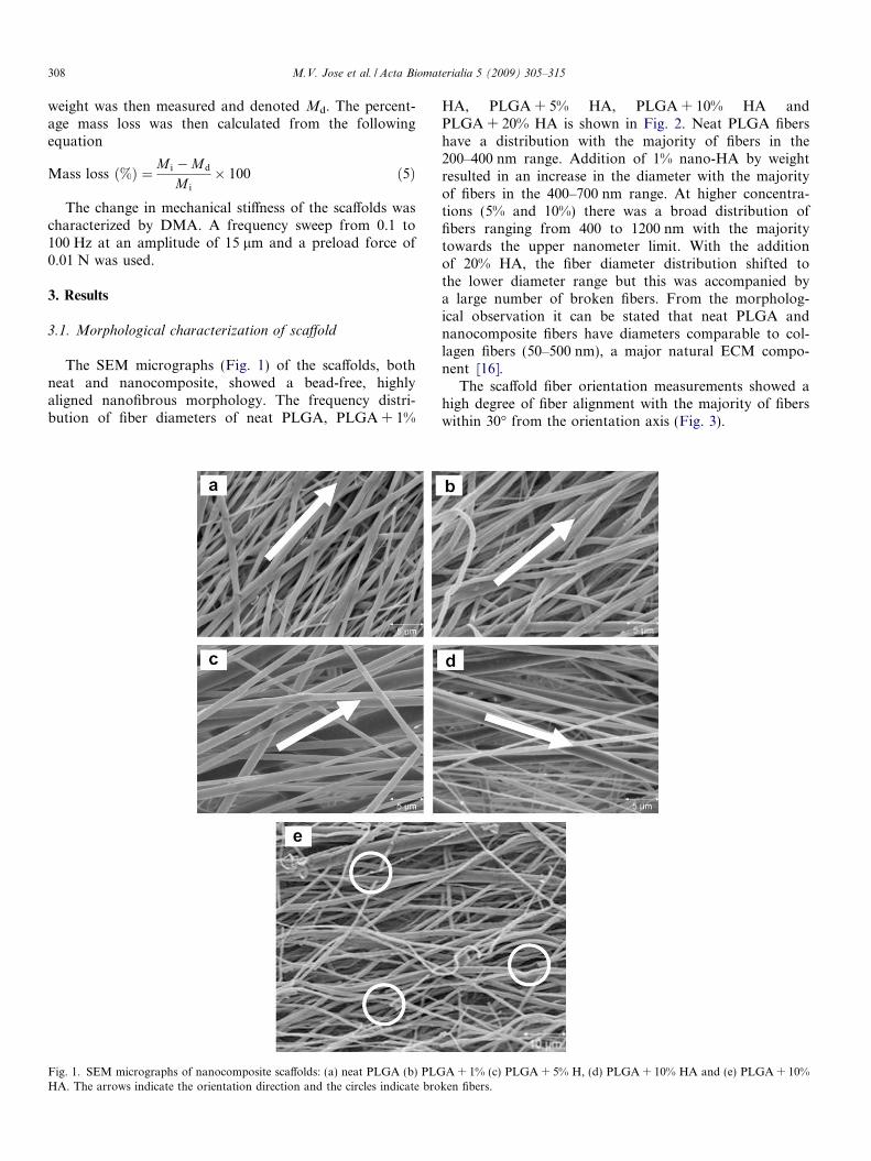
The SEM micrographs (Fig. 1) of the scaffolds, bothneat and nanocomposite, showed a bead-free, highlyaligned nanofibrous morphology. The frequency distri-bution of fiber diameters of neat PLGA, PLGA + 1%
Fig. 1. SEM micrographs of nanocomposite scaffolds: (a) neat PLGA (b) PLGHA. The arrows indicate the orientation direction and the circles indicate bro
HA, PLGA + 5% HA, PLGA + 10% HA andPLGA + 20% HA is shown in Fig. 2. Neat PLGA fibershave a distribution with the majority of fibers in the200–400 nm range. Addition of 1% nano-HA by weightresulted in an increase in the diameter with the majorityof fibers in the 400–700 nm range. At higher concentra-tions (5% and 10%) there was a broad distribution offibers ranging from 400 to 1200 nm with the majoritytowards the upper nanometer limit. With the additionof 20% HA, the fiber diameter distribution shifted tothe lower diameter range but this was accompanied bya large number of broken fibers. From the morpholog-ical observation it can be stated that neat PLGA andnanocomposite fibers have diameters comparable to col-lagen fibers (50–500 nm), a major natural ECM compo-nent [16].
The scaffold fiber orientation measurements showed ahigh degree of fiber alignment with the majority of fiberswithin 30� from the orientation axis (Fig. 3).
A + 1% (c) PLGA + 5% H, (d) PLGA + 10% HA and (e) PLGA + 10%ken fibers.

0
10
20
30
40
50
60
70
80
90
Fre
qu
ency
Dis
trib
uti
on
Diameter (nm)
Neat PLGA
PLGA + 1% HA
PLGA + 5% HA
PLGA + 10% HA
PLGA + 20% HA
Fig. 2. Frequency distribution of fiber diameters of nanofibrous scaffolds based on neat PLGA and nanocomposites (100 counts).
0
10
20
30
40
50
60
70
Fre
qu
ency
Dis
trib
uti
on
Devation from Normal (degree)
Neat PLGA
PLGA + 1% HA
PLGA + 5% HA
PLGA + 10% HA
PLGA + 20% HA
Fig. 3. Frequency distribution of fiber orientation from normal of neat and nanocomposites (100 counts).
M.V. Jose et al. / Acta Biomaterialia 5 (2009) 305–315 309
A representative confocal laser scanning microscopy(CLSM) image (Fig. 4) of calcein-stained nanocompositescaffold shows a fluorescent green color, demonstratingthat there is a good dispersion of nano-HA in the fiberwithout any visible agglomeration within the resolutionof the microscope.
3.2. FTIR spectroscopy
The FTIR spectra of neat PLGA and PLGA/HA areshown in Fig. 5. The scans showed an ester carbonylstretch from the PLGA (C@O) at 1747 cm�1 in all the scaf-folds with no significant shift due to nano-HA interaction.Other major peaks observed were the CAOAC ether groupat 1083 cm�1, CAO stretch at 1128 cm�1, A-type CAOAC
symmetric stretching at 1181 cm�1, OAH deformation at1264 cm�1, methyl group CAH stretching at 1452 cm�1
and other methylene, methyl groups at 2800–3300 cm�1
[30]. For the nanocomposite scaffolds, a PO43� stretching
(1030 cm�1) and bending (570 cm�1), typical of HA, wereobserved with intensities varying proportionally with thenano-HA concentration.
3.3. Porosity
The porosity of the scaffolds was calculated and it wasobserved that random PLGA fibers had a porosity of77%, whereas aligned fibers had 72% porosity (Table 1.).The porosity of the scaffolds decreased from 72% for neatPLGA to around 62% at nano-HA concentrations of 1%,

Fig. 4. CLSM image showing dispersion of HA particles (1%) in PLGA.(Scale �10 lm.)
Table 1Porosity and shrinkage (1 h in PBS) of PLGA and nanocomposite
Sample name % Porosity % Shrinkage
Linear Lateral
Static neat PLGA 77 37 26Neat PLGA 72 49 17PLGA + 1% HA 61 30 9PLGA + 5% HA 62 30 9PLGA + 10% HA 64 34 16PLGA + 20% HA 71 40 14
35.1
32.7
32.4
37.1 37.1
25.0
27.0
29.0
31.0
33.0
35.0
37.0
39.0
0 10 15 20
% HA
Tg
(°C
)
5
Fig. 6. Change in Tg (first heat) with increasing concentration of HA.
310 M.V. Jose et al. / Acta Biomaterialia 5 (2009) 305–315
5% and 10%. However, the porosity of the scaffoldsincreased back to that of neat PLGA at 20% HAconcentration.
3.4. Thermal characterization
The first heating of the DSC scan was used to representthe as-spun structure. The thermograms of all the speci-mens showed only a glass transition (Tg) temperature,characteristic of amorphous PLGA. The as-received PLGApellets showed an endothermic peak (enthalpic relaxation)superimposed on the Tg at 44 �C. The electrospun scaffoldsalso showed an endothermic peak; however, there was adramatic decrease in the Tg (by 14 �C) as compared tothe as-received pellets. Fig. 6 shows the Tg change as afunction of HA content for electrospun fibers. The Tg
increases from 32 �C (neat PLGA) to a maximum of37 �C for 10% HA, followed by a plateau.
Ne at PL GAPL GA + 20% HAPL GA + 1% HAPL GA + 5% HAPL GA + 10% HA
0.0
0.1
0.2
0.3
0.4
0.5
0.6
Abs
orba
nce
1500 Wavenumb
Fig. 5. FTIR spectra of neat PLGA and PLGA-HA nanocomposites. Inset shPLGA + 1% HA, PLGA + 5% HA, PLGA + 10% HA and PLGA + 20% HA
3.5. Mechanical characterization
The storage modulus obtained from DMA at a fre-quency of 1 Hz for the neat PLGA as well as the nanocom-posites is shown in Fig. 7. Neat PLGA showed an averagestorage modulus of 441 MPa. The addition of 1% nano-HA resulted in a 34% increase in storage modulus, to591 MPa. At 5% nano-HA concentration an average mod-ulus of 724 MPa was obtained, a 64% enhancement whencompared to the neat PLGA. Although the modulus
1000 ers (cm-1)
Ne at PLGAPLGA + 20% HAPLGA + 1% HAPLGA + 5% HAPLGA + 10% HA
0.04
0.06
0.08
0.10
0.12
0.14
0.16
0.18
0.20
0.22
Abs
orba
nce
550 600 650 Wavenumbers (cm-1)
AB
CDE
owing fingerprint region of HA, where A, B, C, D, and E are neat PLGA,, respectively.

441.0
591.7
724.2
557.3
371.4
0.0
100.0
200.0
300.0
400.0
500.0
600.0
700.0
800.0
900.0
0.0 1.0 5.0 10.0 20.0% HA
Stro
age
Mod
ulus
(MPa
)
Fig. 7. Change in storage modulus of PLGA with HA at 1 Hz.
M.V. Jose et al. / Acta Biomaterialia 5 (2009) 305–315 311
decreased to 557 MPa for 10% nano-HA it was still higherthan that of neat PLGA. However, at 20% nano-HA con-centration the modulus decreased further to 371 MPa, a16% decrease compared to the neat PLGA and a 49%decrease compared to 5% nano-HA.
3.6. In vitro biodegradation
In vitro biodegradation was studied by measuring theshrinkage, PBS absorption and mass loss. All the speci-mens showed shrinkage during the study (Table 1.). Theshrinkage was high along the aligned direction (up to50%) and marginal along the transverse direction. The1% and 5% nanocomposites exhibited a reduction of 39%linear shrinkage and 47% lateral shrinkage compared toneat PLGA, signifying the influence of nano-HA particlesin PLGA. However, at higher nano-HA concentrations(10% and 20%) the scaffolds showed shrinkage similar tothat of neat PLGA.
The PBS absorption measurement and the mass lossmeasurements are intertwined since both process are hap-pening simultaneously. The percentage of PBS absorbedincreased with higher HA concentrations. The maximumabsorption was observed by week 4 for neat PLGA and
0
10
20
30
40
50
60
70
0 1%
% A
bso
rpti
on
Week 1 Week 2 Week 3
Fig. 8. PBS absorption of neat PLG
by week 3 for PLGA with 1%, 5% and 10% HA concentra-tion, whereas for 20% HA, week 1 showed the highestabsorption (Fig. 8). The mass loss (%) was relatively stablewith small losses for neat as well as nanocomposites duringthe 6 weeks (Fig. 9). The mass loss of the nanocompositescaffolds was less than the neat PLGA scaffold at all timesuntil week 5. The nanocomposites with 5%, 10% and 20%nano-HA showed a steadily increasing mass loss, but thiswas still less than a total of 6% at week 6. The mechanicalcharacterization of the scaffolds showed a minor decreasein the storage modulus initially (week 1) followed by anincrease which reached a peak value at week 5, followedby a decrease (Fig. 10). The storage modulus trends of allthe samples were similar, with the 5% HA specimen show-ing the highest modulus at all times followed by 1% and10% HA; neat PLGA had the lowest modulus.
4. Discussion
4.1. Structural and morphological characterization
In electrospinning the major factors that have a directeffect on the fiber diameters are solution concentration(viscosity) and electric field. In this study the voltageapplied was in the 13–15 kV range because at higher volt-ages the needle tip produced two or more unstablesources of fiber jets. The collection of fibers on a high-speed rotating collector resulted in highly oriented fibers,and it is also assumed that a rotating collector providesadditional drawing on the fibers, which in turn reducesthe diameter. The diameters of the electrospun scaffoldswith process parameters (voltage, distance) being constantcan be affected by the choice of solvent (polarity). You etal. [31] studied the effect of solution properties on thestructure of electrospun PLGA (50/50), using two differ-ent solvents, chloroform (dielectric constant = 4.8) andHFP (dielectric constant = 16.7). The resulting fibersshowed tremendous variation in fiber diameter, with chlo-roform producing fibers with an average diameter of
5 10 20 HA
Week 4 Week 5 Week 6
A and nanocomposite scaffolds.

0
200
400
600
800
1000
1200
76543210
Sto
rag
e M
od
ulu
s (M
Pa)
Neat PLGA
PLGA+1%HA
PLGA+5%HA
PLGA+10%HA
Fig. 10. Effect of degradation on storage modulus.
0
1
2
3
4
5
6
0 1 5 10 20% HA
% M
ass
Lo
ss
Week 1 Week 2 Week 3 Week 5 Week 6
Fig. 9. Effect of degradation on mass loss of neat PLGA and nanocomposites
312 M.V. Jose et al. / Acta Biomaterialia 5 (2009) 305–315
760 nm whereas HFP produced fibers with an averagediameter of 270 nm. Furthermore, a higher diameterrange was observed in Li et al.’s [7] study of PLGA 85/15 due to the choice of solvent system (a 1:1 mixture oftetrahydrofuran and dimethylformamide) and the use ofa static collector. Thus use of HFP as solvent in our studyresulted in a fiber diameter distribution well within therange of natural ECM fibers (50–500 nm) [32]. The useof more polar and hydrophilic solvents can reduce thedegree of segregation of HA powder and can thereby givea good dispersion of particles in the polymer matrix [15].However, with increasing nano-HA content a shift to thehigher diameter range was observed, suggesting thathigher viscosity plays the major role in fiber formation.Thus the nanocomposite scaffolds have fibers showingthe upper nanoscale features of ECM. A similar trendof increasing nanofiber diameter with increasing nano-HA content was observed by Thomas et al. [16,17], on
electrospun poly(e-carprolactone) (PCL) and collagen.PCL/HA nanocomposite nanofibrous scaffolds showed athreshold limit whereby increasing the HA content ini-tially decreased the fiber diameter (due to interactions)but at higher concentrations the diameter increased. Inour study, a large number of fibers were broken at 20%nano-HA concentration and this was attributed to nano-HA agglomeration which prevents the formation of con-tinuous fibers and also results in poor polymer wetting.
The orientation of all scaffolds showed a high degree ofalignment along the rotation direction of the collector, irre-spective of the nano-HA content. The majority of fiberswas within 30� of the orientation axis, and only a veryfew showed significant angular deviation. This deviationin alignment is expected and is due to the rapid depositionof fibers on the grounded collector, which offers insufficienttime to completely release the charge resulting in repulsionof fibers depositing and being deposited [23,24].

M.V. Jose et al. / Acta Biomaterialia 5 (2009) 305–315 313
The staining agent, calcein, used to obtain CLSMimages of the nanocomposite fibers, is a calcium-ion-spe-cific staining agent and forms a calcein–calcium complexwith nano-HA. The staining produced a green color inthe CLSM image and showed a good dispersion of nano-HA particles (1%) in the polymer matrix, suggesting theroute of nanocomposite synthesis was appropriate. TheIR scans obtained did not show any interactions betweenPLGA and nano-HA; however, the absorbance due tothe presence of PO4
3� increases as nano-HA contentincreases (Fig. 4). The scaffolds produced by electrospin-ning are inherently highly porous due to their nonwovennature. However, in aligned PLGA scaffolds the chargerepulsion which causes angular deviation of fiber orienta-tion also facilitates the formation of interconnected pores[24]. The decrease in porosity of the nanocomposites com-pared to neat PLGA with the addition of nano-HA parti-cles (1%, 5% and 10%) was attributed to the effectivecharge dissipation by the particles, preventing inter-fiberrepulsion. However, the agglomeration and poor disper-sion of nano-HA particles at 20% concentration resultedin an increase in porosity. The high porosity of the alignedscaffolds (>60%) obtained herein is assumed to be sufficientfor efficient nutrient transfer as well as continuous elimina-tion of the polymer degradation products during theirapplication.
4.2. Thermal characterization
The DSC thermographs showed a glass transition tem-perature of 37 �C, and no melting, indicating that the poly-mer is essentially amorphous, even though a small degreeof chain orientation is expected using the electrospinningtechnique [33]. It is recognized that tissue engineering scaf-folds with a Tg of 37 �C (physiological temperature) orlower are desirable, since scaffolds having Tg higher thanthe physiological temperature tend to be brittle and canfracture when subjected to stress in use [34]. The modulusof the scaffolds (Section 4.3) was measured at room temper-ature and found to be at the high end of the range for can-cellous bone. Although a decrease in modulus is expectedwith increasing temperature, it is expected to remain in thatrange as the glass transition is approached. The as-spunpolymer exhibited a lower Tg, which can be attributed toeither a larger amount of free volume that is frozen in bythe rapid electrospinning process, or the presence of resid-ual solvent, which can act as a plasticizer. The latter possi-bly was eliminated by thermal gravimetric analysis, whichshowed no residual solvents after drying. A similar reduc-tion in Tg of polymer was observed by others [35,36].Endothermic (enthalpic relaxation) peaks were observedin the DSC scans for all the specimens, and typically thisis caused by a phenomenon known as physical aging [37].Physical aging is a thermally reversible event related tothe loss of free volume, which occurs when a polymer isheld isothermally (i.e. aged) at temperatures near or belowits Tg, and is most prominent in amorphous polymers [37].
As the polymer ages, free volume is lost and the chainarrangement becomes denser. This effect is manifested inphysical changes such as volumetric shrinkage, enhancedstiffness, toughness, and enthalpic relaxation in DSC scans.The enthalpic relaxation peak occurs in the DSC scanbecause more heat has to be added to the polymer toimpart mobility. Physical aging can be erased by heatingthe polymer above Tg. The glass transition, which is theonset of chain mobility, was affected by the incorporationof nano-HA into the polymer matrix which hinders chainmotion. This hindering resulted in a slight increase in theTg as the nano-HA concentration increased from 0% to10%, and thereafter a plateau was reached (Fig. 6).
4.3. Mechanical characterization
The modulus values obtained in this study (Fig. 7) are inthe range typical of cancellous bone (50.0–500 MPa) [1].The dynamic modulus of the neat and nanocomposite scaf-folds suggested that small concentrations of nano-HA hada reinforcing effect on the polymer matrix. A similar effectwas observed by Thomas et al. [16,17], whereby the addi-tion of HA resulted in an increase in mechanical propertiesof PCL and collagen. The higher modulus values obtainedherein for the neat PLGA (441 MPa) compared to thoseobtained by Li et al. [7] (323 MPa) are primarily due tothe alignment of nanofibers in the testing direction. Athigher concentrations of nano-HA (10% and 20%), themodulus decreased, suggesting a threshold limit wasreached and further addition of nano-HA particle did notreinforce the polymer matrix but instead acted as defects.This was assumed to be due to the agglomeration of nano-particles preventing good polymer–particle interaction toobtain good load transfer. When the concentration ofnano-HA was 20%, it functions more as a defect than asa reinforcement; as a result the storage modulus decreasedby 16% compared to the neat PLGA scaffolds. The mor-phology of the scaffolds observed from the SEM micro-graphs (Fig. 1e) corroborated the mechanical data,showing multiple fiber breakage which is presumably dueto agglomeration of the nano-HA particles.
4.4. In vitro biodegradation
The shrinkage perpendicular to the oriented directionwas minimal in all the scaffolds tested. This was due tothe fact that the fibers which were spread out from eachother due to the charge repulsion during electrospinningare now packed more efficiently. In the alignment directionthe amorphous chains are stretched not only by the whip-ping action during the electrospinning but also by thedrawing effect of the high-speed rotating collector. Sincethe temperature is above the Tg of the as-spun sample,mobility is imparted to the slightly oriented chains. Thechains in turn exhibit some memory of their random con-formation and recoil in an attempt to recover the confor-mation. This recoiling effect results in shrinkage of the

314 M.V. Jose et al. / Acta Biomaterialia 5 (2009) 305–315
scaffolds and compensates for any expansion due to plasti-cization by the medium. To confirm that the shrinkageobserved is thermally induced, scaffold dimension changeswere determined before and after annealing at the physio-logical temperature of 37 �C for a period of 1 h. These scaf-folds showed similar shrinkage as observed for scaffolds inPBS medium, confirming the temperature effects. The eval-uation of the physical changes in PBS medium for a periodof 1 h showed a difference in the amount of linear and lat-eral shrinkage (Table 1.). A 39% decrease in the linearshrinkage observed at 1% HA compared to neat PLGA isattributed to the hindering of chain mobility by the pres-ence of nanoscopic HA particles distributed throughoutthe polymer matrix. The 47% decrease in lateral shrinkagein the nanocomposites relative to the neat PLGA may bedue to better packing efficiency because of the efficientcharge dissipation caused by the presence of nano-HA inthe fibers. However the increased shrinkage at higher con-centrations of HA particles (10% and 20%) is assumed tobe due to the poor dispersion, resulting in agglomeration,which in turn causes incomplete charge dissipation andhence poor packing and enhanced chain mobility.
The PBS absorption values were expected to be low dueto the chemical nature of the polymer used (85% higherPLA content). However, the absorption was assumed tobe affected by the presence of hydrophilic nano-HA parti-cles in PLGA/HA nanocomposites. The similarity in PBSabsorption (up to 10%) of neat PLGA and PLGA with1% HA shows that the nanoparticles are well dispersed inthe polymer matrix and there is good wetting of HA nano-particles by the hydrophobic PLGA. At higher concentra-tions (5%, 10% and 20%) the presence of nanoparticles nearthe fiber surface and also the possibility of agglomerationresulted in higher PBS absorption (up to 60%).
The mass loss data obtained shows a constant loss(<6%) for the neat, 1% and 5% HA samples throughoutthe study. A similar trend was observed by Lu et al. [38]during a 20 week study during which there was not muchdifference in PBS absorption and mass loss for neat PLGA(85/15) foams. As the HA content increases to 10% and20%, the weight loss increases with degradation time, sug-gesting that the agglomeration or incomplete dispersion ofnano-HA particles resulted in fiber breakage due to surfaceerosion. The surface erosion may be promoted by the highsurface area of the nanofibers and may be due to the inter-connected porosity arising from the nonwoven fibrous nat-ure of the material. The storage modulus of all the scaffoldsduring the degradation study period showed a similartrend: a small decline initially, followed by an increasedue to stiffening. This is presumably due to the combinedeffect of the PBS medium and temperature. The initialslight decrease is due to the plasticizing effect of the med-ium, and as time progresses, thermally activated shrinkage(reduction in free volume) leads to increased mechanicalproperties [39]. The modulus values reach a peak at 4–5weeks for all the specimens. However, as the degradationcontinues, chain scission occurs, resulting in a drop in mod-
ulus values. The storage modulus values at week 6 was sim-ilar to that of the neat scaffolds before degradation,suggesting the properties were maintained almostunchanged for a period of 6 weeks.
5. Conclusion
Preliminary results suggest that the route selected to dis-perse the nanoparticles in the PLGA solution was appro-priate. At low concentrations the fibers had noagglomerates and good dispersion was achieved. However,higher concentrations of HA resulted in increased diameterand broken fibers due to agglomeration. The glass transi-tion temperature of the polymer was markedly reducedby the fast processing technique of electrospinning. Thisreduction brought the Tg down to be equal to or less thanphysiological temperature. In addition, the low Tg resultedin oriented amorphous chains that folded, resulting in sig-nificant shrinkage. However the presence of well-dispersednanoscopic HA particles reduced the chain mobility andhence helped to prevent shrinkage to some degree. The deg-radation characteristics of the scaffolds showed similartrends, although the neat PLGA and PLGA with 1% HAshowed lowest absorption and mass loss. The storage mod-ulus values followed similar trends at all compositions,with the 5% HA specimen having the highest modulus atall times followed by 1%, 10% and neat, respectively.
Acknowledgments
The authors acknowledge Albert Tousson for CLSMimaging at UAB Imaging Facility and partial support fromNSF CREST program (Grant HRD- 0734232).
References
[1] Murugan R, Ramakrishna S. Development of nanocomposites forbone grafting. Compos Sci Technol 2005;65:2385–406.
[2] Salgado AJ, Coutinho OP, Reis RL. Bone tissue engineering: state ofthe art and future trends. Macromol Biosci 2004;4:743–65.
[3] Sharma B, Elisseeff JH. Engineering structurally organized cartilageand bone tissues. Ann Biomed Eng 2004;V32:148–59.
[4] Mikos AG, Temenoff JS. Formation of highly porous biodegradablescaffolds for tissue engineering. Electron J Biotechnol 2000;3:114–9.
[5] Laurencin CT, Ambrosio AMA, Borden MD, Cooper JA. Tissueengineering: orthopedic applications. Annu Rev Biomed Eng1999;1:19.
[6] Murugan R, Ramakrishna S. Nano-featured scaffolds for tissueengineering: a review of spinning methodologies. Tissue Eng2006;12:435–47.
[7] Li W-J, Laurencin CT, Caterson EJ, Tuan RS, Ko FK. Electrospunnanofibrous structure: a novel scaffold for tissue engineering. JBiomed Mater Res 2002;60:613–21.
[8] Huang Z-M, Zhang YZ, Kotaki M, Ramakrishna S. A review onpolymer nanofibers by electrospinning and their applications innanocomposites. Compos Sci Technol 2003;63:2223–53.
[9] Xu C, Inai R, Kotaki M, Ramakrishna S. Electrospun nanofiberfabrication as synthetic extracellular matrix and its potential forvascular tissue engineering. Tissue Eng 2004;10:1160–8.
[10] Wang M. Developing bioactive composite materials for tissuereplacement. Biomaterials 2003;24:2133–51.

M.V. Jose et al. / Acta Biomaterialia 5 (2009) 305–315 315
[11] Webster TJ, Ergun C, Doremus RH, Siegel RW, Bizios R. Specificproteins mediate enhanced osteoblast adhesion on nanophase ceram-ics. J Biomed Mater Res 2000;51:475–83.
[12] Zhang N, Nichols HL, Tylor S, Wen X. Fabrication of nanocrystal-line hydroxyapatite doped degradable composite hollow fiber forguided and biomimetic bone tissue engineering. Mater Sci Eng C–Biomim Supramol Syst 2007;27:599–606.
[13] Ural E, Kesenci K, Fambri L, Migliaresi C, Piskin E. Poly(D,L-lactide/e-caprolactone)/hydroxyapatite composites. Biomaterials2000;21:2147–54.
[14] Ito Y, Hasuda H, Kamitakahara M, Ohtsuki C, Tanihara M, Kang I-K, et al. A composite of hydroxyapatite with electrospun biodegrad-able nanofibers as a tissue engineering material. J Biosci Bioeng2005;100:43–9.
[15] Kim H-W, Lee H-H, Knowles JC. Electrospinning biomedicalnanocomposite fibers of hydroxyapatite/poly(lactic acid) for boneregeneration. J Biomed Mater Res A 2006;79A:643–9.
[16] Thomas V, Dean DR, Jose MV, Mathew B, Chowdhury S, VohraYK. Nanostructured biocomposite scaffolds based on collagencoelectrospun with nanohydroxyapatite. Biomacromolecules 2007.
[17] Thomas V, Jagani S, Johnson K, Jose MV, Dean DR, Vohra YK,et al. Electrospun bioactive nanocomposite scaffolds of polycapro-lactone and nanohydroxyapatite for bone tissue engineering. JNanosci Nanotechnol 2006;6:487–93.
[18] Deng X-L, Sui G, Zhao M-L, Chen G-Q, Yang X-P. Poly(L-lacticacid)/hydroxyapatite hybrid nanofibrous scaffolds prepared by elec-trospinning. J Biomater Sci Polym Ed 2007;18:117–30.
[19] Kim H-W, Song JH, Kim HE. Nanofiber generation of gelatin-hydroxyapatite biomimetics for guided tissue regeneration. AdvFunct Mater 2005;15:1988–94.
[20] Bishop A, Balazsi C, Yang JHC, Gouma P-I. Biopolymer-hydroxy-apatite composite coatings prepared by electrospinning. Polym AdvTechnol 2006;17:902–6.
[21] Xu CY, Inai R, Kotaki M, Ramakrishna S. Aligned biodegradablenanofibrous structure: a potential scaffold for blood vessel engineer-ing. Biomaterials 2004;25:877–86.
[22] Lee CH, Shin HJ, Cho IH, Kang Y-M, Kim IA, Park K-D, et al.Nanofiber alignment and direction of mechanical strain affect the ECMproduction of human ACL fibroblast. Biomaterials 2005;26:1261–70.
[23] Yang F, Murugan R, Wang S, Ramakrishna S. Electrospinning ofnano/micro scale poly(L-lactic acid) aligned fibers and their potentialin neural tissue engineering. Biomaterials 2005;26:2603–10.
[24] Zhong S, Teo WE, Zhu X, Beuerman RW, Ramakrishna S, YungLYL. An aligned nanofibrous collagen scaffold by electrospinningand its effects on in vitro fibroblast culture. J Biomed Mater Res A2006;79:456–63.
[25] Li W-J, Mauck RL, Cooper JA, Yuan X, Tuan RS. Engineeringcontrollable anisotropy in electrospun biodegradable nanofibrous
scaffolds for musculoskeletal tissue engineering. J Biomech2007;40:1686–93.
[26] Zhu B, Lu Q, Yin J, Hu J, Wang Z. Alignment of osteoblast-like cellsand cell-produced collagen matrix induced by nanogrooves. TissueEng 2005;11:825–34.
[27] Thomas V, Jose MV, Chowdhury S, Sullivan JF, Dean DR, VohraYK. Mechano-morphological studies of aligned nanofibrous scaffoldsof polycaprolactone fabricated by electrospinning. J Biomater SciPolym Ed 2006;V17:969–84.
[28] Loo SCJ, Ooi CP, Wee SHE, Boey YCF. Effect of isothermalannealing on the hydrolytic degradation rate of poly(lactide-co-glycolide) (PLGA). Biomaterials 2005;26:2827–33.
[29] Nie H, Wang C-H. Fabrication and characterization of PLGA/HApcomposite scaffolds for delivery of BMP-2 plasmid DNA. J ControlRelease 2007;120:111–21.
[30] Cordewener FW, Dijkgraaf LC, Ong JL, Agrawal CM, Zardeneta G,Milam SB, et al. Particulate retrieval of hydrolytically degradedpoly(lactide-co-glycolide) polymers. J Biomed Mater Res2000;50:59–66.
[31] You Y, Lee SJ, Min B-M, Park WH. Effect of solution properties onnanofibrous structure of electrospun poly(lactic-co-glycolic acid). JAppl Polym Sci 2006;99:1214–21.
[32] Smith LA, Ma PX. Nano-fibrous scaffolds for tissue engineering.Colloids Surf B Biointerfaces 2004;39:125–31.
[33] Jose MV, Steinert BW, Thomas V, Dean DR, Abdalla MA, Price G,et al. Morphology and mechanical properties of Nylon 6/MWNTnanofibers. Polymer 2007;48:1096–104.
[34] Park PIP, Jonnalagadda S. Predictors of glass transition in thebiodegradable polylactide and poly-lactide-co-glycolide polymers. JAppl Polym Sci 2006;100:1983–7.
[35] Zong X, Ran S, Kim K-S, Fang D, Hsiao BS, Chu B. Structure andmorphology changes during in vitro degradation of electrospunpoly(glycolide-co-lactide) nanofiber membrane. Biomacromolecules2003;4:416–23.
[36] Zong X, Kim K, Fang D, Ran S, Hsiao BS, Chu B. Structure andprocess relationship of electrospun bioabsorbable nanofiber mem-branes. Polymer 2002;43:4403–12.
[37] Dean D, Husband M, Trimmer M. Time-temperature-dependentbehavior of a substituted poly(paraphenylene): tensile, creep, anddynamic mechanical properties in the glassy state. J Polym Sci PolymPhys 1998;36:2971–9.
[38] Lu L, Peter SJ, Lyman MD, Lai H-L, Leite SM, Tamada JA, et al. Invitro and in vivo degradation of porous poly(-lactic-co-glycolic acid)foams. Biomaterials 2000;21:1837–45.
[39] Yang F, Cui W, Xiong Z, Liu L, Bei J, Wang S. Poly(L,L-lactide-co-glycolide)/tricalcium phosphate composite scaffold and its variouschanges during degradation in vitro. Polym Degrad Stabil2006;91:3065–73.
Copyright © 2022 FDOKUMEN