Biodegradable Polymer (PLGA) Coatings Featuring Cinnamaldehyde and Carvacrol Mitigate Biofilm...
7
Biodegradable Polymer (PLGA) Coatings Featuring Cinnamaldehyde and Carvacrol Mitigate Biofilm Formation Katherine R. Zodrow, † Jessica D. Schiffman,* ,‡ and Menachem Elimelech † † Department of Chemical and Environmental Engineering, Yale University, New Haven, Connecticut 06520-8286, United States ‡ Department of Chemical Engineering, University of Massachusetts, Amherst, Massachusetts 01003-9303, United States ABSTRACT: Biofilm-associated infections are one of the leading causes of death in the United States. Although infections may be treated with antibiotics, the overuse of antibiotics has led to the spread of antibiotic resistance. Many natural antimicrobial compounds derived from edible plants are safe for human use and target bacteria nonspecifically. Therefore, they may impair biofilm formation with less evolutionary pressure on pathogens. Here, we explore the use of two natural antimicrobial compounds, cinnamaldehyde (CA, from cinnamon) and carvacrol (CARV, from oregano), for biofilm prevention. We have fabricated and characterized films that incorporate CA and CARV into the biodegradable, FDA- approved polymer poly(lactic-co-glycolic acid), PLGA. The addition of CA and CARV to PLGA films not only adds antimicrobial activity but also changes the surface properties of the films, making them more hydrophilic and therefore more resistant to bacterial attachment. An addition of 0.1% CA to a PLGA film significantly impairs biofilm development by Staphylococcus aureus, and 0.1% CARV in PLGA significantly decreases biofilm formation by both Escherichia coli and S. aureus. Pseudomonas aeruginosa, which is less susceptible to CA and CARV, was not affected by the addition of 0.1% CA or CARV to the PLGA coatings; however, P. aeruginosa biofilm was significantly reduced by 1.0% CA. These results indicate that both CA and CARV could potentially be used in low concentrations as natural additives in polymer coatings for indwelling devices to delay colonization by bacteria. ■ INTRODUCTION Infectious diseases are one of the leading causes of death in the United States. 1 The development of antimicrobial resistance, globalization, and a decline in antibiotic discovery exacerbate this problem. 2 In the United States, hospital-acquired infections alone cause between 60 000 and 90 000 deaths, amounting to an economic burden of $17 billion to $29 billion annually. 3 It is estimated that as many as 80% of these infections arise from bacterial biofilms. Biofilms are communities of microorganisms that colonize and grow on surfaces, such as indwelling devices or native tissues. Although many biofilms are beneficial, some biofilms cause grave health concerns. Detrimental biofilm formation is problematic because bacteria in a biofilm are more resistant to disinfectants. 4 Many harmful biofilms are associated with indwelling medical devices, and when pathogens form a biofilm on a device, the infection is difficult to eradicate. Often, the device, for example, a catheter, intubation tube, implant, or shunt, must be removed. One potential way to prevent biofilm formation on indwelling medical devices is to coat them with antimicrobial compounds. For instance, coatings with chlorhexadine and silver sulfadiazine on catheters decreased the occurrence of catheter-related bloodstream infections in intensive care units. 5 However, the use of antibiotics leads to undesired con- sequences, including the spread of antibiotic-resistant bacteria. For example, 60% of Staphylococcus aureus strains isolated from intensive care units were resistant to methicillin. 5 Vancomycin is often used to treat methicillin-resistant S. aureus (MRSA) infections; however, a recent increase in vancomycin-resistant strains underscores the need for alternative methods of microbial control. 2 This trend in antibiotic resistance contributes to the popularity of inorganic antimicrobials, including silver. However, the use of silver is also problematic, as silver resistance has been observed in bacteria, notably Pseudomonas aeruginosa. It is likely that common use of silver or other heavy metals as antimicrobial agents will lead to increased silver resistance in other microorganisms. 6 Therefore, anti- microbial compounds that will not lead to the spread of resistance genes are in high demand. The essential oil components cinnamaldehyde (CA) and carvacrol (CARV) are broad-range, natural antimicrobial compounds that can be readily isolated from cinnamon and oregano, respectively (Figure 1). Essential oils have been shown to be effective against a wide range of bacteria. In general, they act by increasing cell membrane permeability, leading to a depletion of the proton gradient and subsequent disruption of ATP synthesis. 7,8 The exact antimicrobial mechanism of essential oils is complex, making the development of resistance by bacteria more difficult. 9 Additionally, some essential oils, Received: May 11, 2012 Published: August 31, 2012 Article pubs.acs.org/Langmuir © 2012 American Chemical Society 13993 dx.doi.org/10.1021/la303286v | Langmuir 2012, 28, 13993−13999
Transcript of Biodegradable Polymer (PLGA) Coatings Featuring Cinnamaldehyde and Carvacrol Mitigate Biofilm...

Biodegradable Polymer (PLGA) Coatings Featuring Cinnamaldehydeand Carvacrol Mitigate Biofilm FormationKatherine R. Zodrow,† Jessica D. Schiffman,*,‡ and Menachem Elimelech†
†Department of Chemical and Environmental Engineering, Yale University, New Haven, Connecticut 06520-8286, United States‡Department of Chemical Engineering, University of Massachusetts, Amherst, Massachusetts 01003-9303, United States
ABSTRACT: Biofilm-associated infections are one of theleading causes of death in the United States. Althoughinfections may be treated with antibiotics, the overuse ofantibiotics has led to the spread of antibiotic resistance. Manynatural antimicrobial compounds derived from edible plantsare safe for human use and target bacteria nonspecifically.Therefore, they may impair biofilm formation with lessevolutionary pressure on pathogens. Here, we explore theuse of two natural antimicrobial compounds, cinnamaldehyde(CA, from cinnamon) and carvacrol (CARV, from oregano),for biofilm prevention. We have fabricated and characterized films that incorporate CA and CARV into the biodegradable, FDA-approved polymer poly(lactic-co-glycolic acid), PLGA. The addition of CA and CARV to PLGA films not only adds antimicrobialactivity but also changes the surface properties of the films, making them more hydrophilic and therefore more resistant tobacterial attachment. An addition of 0.1% CA to a PLGA film significantly impairs biofilm development by Staphylococcus aureus,and 0.1% CARV in PLGA significantly decreases biofilm formation by both Escherichia coli and S. aureus. Pseudomonas aeruginosa,which is less susceptible to CA and CARV, was not affected by the addition of 0.1% CA or CARV to the PLGA coatings;however, P. aeruginosa biofilm was significantly reduced by 1.0% CA. These results indicate that both CA and CARV couldpotentially be used in low concentrations as natural additives in polymer coatings for indwelling devices to delay colonization bybacteria.
■ INTRODUCTION
Infectious diseases are one of the leading causes of death in theUnited States.1 The development of antimicrobial resistance,globalization, and a decline in antibiotic discovery exacerbatethis problem.2 In the United States, hospital-acquired infectionsalone cause between 60 000 and 90 000 deaths, amounting toan economic burden of $17 billion to $29 billion annually.3 It isestimated that as many as 80% of these infections arise frombacterial biofilms. Biofilms are communities of microorganismsthat colonize and grow on surfaces, such as indwelling devicesor native tissues. Although many biofilms are beneficial, somebiofilms cause grave health concerns. Detrimental biofilmformation is problematic because bacteria in a biofilm are moreresistant to disinfectants.4 Many harmful biofilms are associatedwith indwelling medical devices, and when pathogens form abiofilm on a device, the infection is difficult to eradicate. Often,the device, for example, a catheter, intubation tube, implant, orshunt, must be removed.One potential way to prevent biofilm formation on
indwelling medical devices is to coat them with antimicrobialcompounds. For instance, coatings with chlorhexadine andsilver sulfadiazine on catheters decreased the occurrence ofcatheter-related bloodstream infections in intensive care units.5
However, the use of antibiotics leads to undesired con-sequences, including the spread of antibiotic-resistant bacteria.For example, 60% of Staphylococcus aureus strains isolated from
intensive care units were resistant to methicillin.5 Vancomycinis often used to treat methicillin-resistant S. aureus (MRSA)infections; however, a recent increase in vancomycin-resistantstrains underscores the need for alternative methods ofmicrobial control.2 This trend in antibiotic resistancecontributes to the popularity of inorganic antimicrobials,including silver. However, the use of silver is also problematic,as silver resistance has been observed in bacteria, notablyPseudomonas aeruginosa. It is likely that common use of silver orother heavy metals as antimicrobial agents will lead to increasedsilver resistance in other microorganisms.6 Therefore, anti-microbial compounds that will not lead to the spread ofresistance genes are in high demand.The essential oil components cinnamaldehyde (CA) and
carvacrol (CARV) are broad-range, natural antimicrobialcompounds that can be readily isolated from cinnamon andoregano, respectively (Figure 1). Essential oils have been shownto be effective against a wide range of bacteria. In general, theyact by increasing cell membrane permeability, leading to adepletion of the proton gradient and subsequent disruption ofATP synthesis.7,8 The exact antimicrobial mechanism ofessential oils is complex, making the development of resistanceby bacteria more difficult.9 Additionally, some essential oils,
Received: May 11, 2012Published: August 31, 2012
Article
pubs.acs.org/Langmuir
© 2012 American Chemical Society 13993 dx.doi.org/10.1021/la303286v | Langmuir 2012, 28, 13993−13999

including CA, inhibit biofilm formation at subinhibitoryconcentrations.10,11 Specifically, cinnamon essential oil (ofwhich CA is a major active constituent) decreases metabolicactivity and the replication rate of P. aeruginosa and S. aureusand causes changes in cell morphology.12 CA inhibitsswimming motility in Escherichia coli.13 In P. aeruginosa,exposure to cinnamon essential oil leads to a coagulation ofcytoplasmic material and an extrusion of intracellular material.In S. aureus, fibrous extensions are visible. CA at sublethalconcentrations acts as a quorum sensing inhibitor (QSI),interfering with the autoinducer-2 (AI-2) system, and delaysbiofilm formation by S. aureus at concentrations below theminimum inhibitory concentration (MIC).10,11,14 Therefore,the use of CA at low concentrations could prevent colonizationof surfaces by bacteria without exerting evolutionary pressure.Oregano essential oil, of which CARV is a major activecomponent, affects the membrane potential and permeability inS. aureus. In S. aureus, mesosome-like structures are observed,and in P. aeruginosa, coagulated cytoplasm and liberatedvesicles are seen.15 CARV is also known to initiate a response inE. coli similar to the response exhibited upon heat stress and toinhibit flagella synthesis.16
Many previous studies with CA and CARV focused onapplications in food preservation (reviewed by Burt17). It hasbeen demonstrated that 10 wt % CARV incorporated into low-density polyethylene films creates slow-release antimicrobialpackaging.18 Suspensions of poly(lactic-co-glycolic acid)(PLGA) nanoparticles incorporating CA inhibit the growth ofa number of organisms.19 CARV, 1.0%, has also been added toPLGA particles to treat advanced biofilms, resulting in both areduction in biofilm and changes in biofilm viscoelasticproperties.20 Coatings, in contrast to free or particle-encapsulated compounds, offer an opportunity to preemptivelydeliver bioactive compounds to a site at risk for a biofilm-related infection. The activity of CA and CARV in a PLGA filmcoating for medical devices has not yet been explored.Here, we investigate the use of natural antimicrobial
compounds isolated from plant essential oils and incorporatedinto a PLGA matrix to prevent biofilm formation on a surface.Three model organisms were testedE. coli, P. aeruginosa, and
S. aureus. These microorganisms are responsible for medicallyrelevant biofilm formation, including biofilms associated withthe gastrointestinal tract, lungs, and indwelling medical devices.PLGA coatings containing CA and CARV could potentially beapplied to a variety of indwelling medical devices to preventinfections associated with those devices.
■ MATERIALS AND METHODSMaterials and Chemicals. All compounds were used as received.
Cinnamaldehyde (CA) (≥93%, FG, Mw = 132.16 Da), carvacrol(CARV) (≥98%, FCC, Mw = 150.22 Da), poly(D,L-lactide-co-glycolide) (PLGA) (85:15, Mw = 50 000−75 000 Da), and chloroform(≥99.9%) were purchased from Sigma-Aldrich (St. Louis, MO).Paraformaldehyde was obtained from MP Biomedicals, LLC (Solon,OH). Hydrochloric acid (HCl) and Triton X-100 were purchasedfrom Fisher Scientific, sodium hydroxide (NaOH) and sodiumchloride (NaCl) from J.T. Baker (Phillipsburg, NJ), and ethanol(EtOH) (200 proof) from Decon Laboratories, Inc. (King of Prussia,PA). Deionized (DI) water was obtained from a Milli-Q ultrapurewater purification system (Millipore, Billerica, MA).
The nucleic acid stain SYTO 9 and mounting oil were purchasedfrom Invitrogen (Eugene, OR). M9 minimal salts and phosphate-buffered saline (PBS) were purchased from Sigma Life Sciences (St.Louis, MO). Difco Luria−Bertani (LB) broth was purchased from BDLife Sciences (Franklin Lakes, NJ), and Muller−Hinton Broth (MHB)from HiMedia Laboratories (Mumbai, India).
Model Bacteria Strains. Escherichia coli (E. coli), Pseudomonasaeruginosa (P. aeruginosa), and Staphylococcus aureus (S. aureus) wereused as the model microorganisms. Kanamycin-resistant E. coliBW26437 was obtained from the Yale Coli Genetic Stock Center(New Haven, CT). The P. aeruginosa PAO1 strain was described inour previous publication.21 S. aureus RN 6390B was kindly provided byDr. Naomi Balaban. Overnight cultures were grown in either LB orMHB at 37 °C. These cultures were then diluted in fresh MHB andgrown for 2 h to log phase prior to the toxicity and biofilmexperiments. Log-phase growth time was determined by a growthcurve. Cell density was determined by a plate count. Optical density(OD) was measured at 600 nm to determine cell growth. An OD600 of1.0 corresponds to 109 CFU/mL for all three strains tested.
Evaluation of the Biological Effects of Cinnamaldehyde (CA)and Carvacrol (CARV). The minimum inhibitory concentrations(MICs) of both CARV and CA were determined according to thebroth dilution method published by Wiegand et al.22 Overnightcultures of E. coli, P. aeruginosa, and S. aureus were grown in MHB.Cultures were diluted in MHB and grown to log phase (2 h). Theculture was diluted in MHB to an optical density at 600 nm (OD600)of 0.1 and subsequently diluted 100×. This corresponds to aconcentration of ∼106 CFU/mL, as determined by a plate count.Then, 50 μL of the cell suspension was diluted into 50 μL of CARV orCA in MHB in a cell-culture-treated 96-well plate (Falcon MicrotestTM
96) and pipetted up and down to mix. The 96-well plate was thenplaced in an incubator at 37 °C for 17 h. The OD600 was measured asan indication of cell growth (SPECTRA max 340PC).
The biofilm that developed on the walls of the polystyrene plate(Falcon MicrotestTM 96) was quantified according to the methoddescribed by Kiran et al.23 After the MIC assay, the suspended cellswere removed, and the remaining biofilm was gently rinsed with PBS.The biofilms were then fixed with 100% EtOH, and the EtOH wasallowed to evaporate at room temperature (25 °C). The biofilm wasthen stained with 0.1% safranin (LabChem Inc., Pittsburgh, PA) andrinsed three times with PBS. The biofilm cells containing the safraninstain were resuspended with 1% sodium dodecyl sulfate (SDS). Therelative amount of biofilm was quantified by an OD490 measurement(SPECTRA max 340PC).
Thin Film Fabrication. CA and CARV were incorporated intoPLGA thin films. PLGA is used for a variety of biomedical applicationsand has easily tunable physical properties.24 PLGA/chloroform (0.4 gin 20 mL corresponding to 2 wt %) solutions were mixed for 24 husing a rotating plate (VWR, Bridgeport, NJ). CARV or CA (0, 0.4, 4.0
Figure 1. Two naturally occurring antimicrobial compounds (A)cinnamaldehyde (CA), a phenolic aldehyde, and (B) carvacrol(CARV), a monoterpenoid phenol, are incorporated into a (C)biodegradable and biocompatible poly(lactic-co-glycolic acid) (PLGA)film.
Langmuir Article
dx.doi.org/10.1021/la303286v | Langmuir 2012, 28, 13993−1399913994
mg, corresponding to 0, 0.1, and 1.0 wt %, respectively) was dispersedinto PLGA solutions using a Vortex-Genie (Scientific Industries, Inc.,Bohemia, NY). PLGA films were spin-coated (Spin-Coater model SCSP-6708, Specialty Coating Systems Inc., Indianapolis, IN) on 22 × 22× 1.5 mm glass coverslips (Fisherbrand) with 100 μL of the solutionof interest at 200 rpm for 20 s, 300 rpm for 40 s, and 8000 rpm for 5 s;there was an additional 1 s ramp time between hold times.3 Prior tospin-coating, the coverslips were thoroughly cleaned by an overnightrinse in 2% Liquinox critical-cleaning liquid detergent solution (JerseyCity, NJ) at 75 °C, followed by repeat rinsing with DI water. Thecoverslips were then placed in a UV/ozone chamber for 30 min tooxidize any remaining organic material (UV/Ozone ProCleanerTM,BioForce Nanosciences, Ames, IA). Remnant chloroform was allowedto evaporate from the spin-coated PLGA, PLGA−CARV, and PLGA−CA films at room temperature for 3 days.Characterization of Thin Films. Film thickness was measured
using an ellipsometer (Angstrom Advanced, Inc.) positioned at 65°and equipped with a 632.8 nm laser. A Cauchy model was used with arefractive index of 1.45 to determine the film thickness.3
Contact angle measurements with DI water, diiodomethane (99+%,Acros Organics), and glycerol (99.9%, J.T. Baker) were carried outusing a Goniometer (VCA Video Contact Angle System, ASTProducts, Billerica, MA). Ten drops of 5 μL on at least two differentsamples were measured for each film and liquid combination. Surfaceenergy calculations from the averages of the measured contact anglestaken with these three liquids were carried out according to apreviously reported method.25,26
Evaluation of Biofilm Growth on PLGA−CARV and PLGA−CA Films. PLGA−CARV and PLGA−CA films were coated onto thesides and bottom of wells in a glass-coated 96-well plate (Plate+TM,SUN Sri, Rockwood, TN). A 150 μL aliquot of 2% PLGA inchloroform with 0.1 and 1.0% CARV or CA was added to the well.The solution evaporated in a chemical hood over 3 days at roomtemperature, resulting in a visible film in each of the wells. The wellplate was then sterilized during 4 h UV treatment (UVP, UVL-28ELseries, 365 nm, 8 W). Our tests indicated that 4 h is sufficient forsample sterilization with this lamp. Log-phase E. coli, P. aeruginosa, andS. aureus were added to each well in MHB (5 × 10⁵ CFU/mL), andthe bacteria were incubated at 25 °C. After 20 h incubation, the biofilmwas quantified according to the method described previously.23
Confocal laser scanning microscopy (CLSM, Zeiss LSM 510, and anAXIO Observer Z1 inverted microscope, Carl Zeiss, Inc.) was used tovisualize biofilm structure and quantify biofilm biovolume anddiffusion distance using COMSTAT2.27−29 Bacteria were stainedwith the nucleic acid stain Syto 9 (Invitrogen). Preliminaryexperiments showed no staining of biofilm by Concanavalin Aconjugated with Alexa Fluor 633 (Invitrogen), indicating thatextracellular polymeric substances (EPS) production at this stage ofdevelopment did not contribute to biofilm volume. PLGA films onglass coverslips were affixed to a microscope slide with nail polish toprevent PLGA film detachment. The nail polish was allowed toevaporate for 1 week and was then rinsed with DI water to remove anyresidual solvent. The thin films were sterilized during 4 h UVtreatment (UVP, UVL-28EL series, 365 nm, 8 W) and then placed insterile Petri dishes. Log-phase bacteria culture was diluted to an OD600of 0.1 in LB broth (108 CFU/mL), and then a 0.6 mL culture wasadded to 30 mL of M9 salts with 0.1% glucose in each of the dishes.Films were grown for 20 h at 25 °C without agitation.Prior to CLSM visualization, the biofilms were rinsed with PBS and
then fixed with 4% paraformaldehyde for 5 min. The remainingparaformaldehyde was removed, and the biofilm was rinsed twice inPBS for 5 min each. The bacteria were then permeabilized with 0.2%Triton X-100 for 5 min and subsequently stained with the nucleic acidstain Syto 9 for 10 min. The biofilms were then dehydrated with anethanol series (50, 75, and then 100% EtOH) for 5 min each. Theslides were mounted using BacLightTM mounting oil prior tovisualization with a Plan-Apochromat 20×/0.8 numerical apertureobjective. Syto 9 was excited with an argon 488 nm laser, and a BP500−530 IR emission filter was used. Data was collected with Zen(Carl Zeiss, Inc.). At least 0.45 mm2 on each sample was used for
image analysis. Subsequent image analysis was performed with ImageJ1.41 software (National Institutes of Health, Bethesda, MD) andCOMSTAT2.27−29
Statistical Analysis. The data are represented as mean ± standarddeviation of at least three samples. Significant differences betweensamples were determined with a one-way analysis of variance(ANOVA), and post hoc comparisons between groups were madewith a Tukey test (R, www.r-project.org). Significance (p ≤ 0.05) isdenoted in graphs by an asterisk.
■ RESULTS AND DISCUSSIONCA and CARV in Solution Inhibit Bacterial Growth and
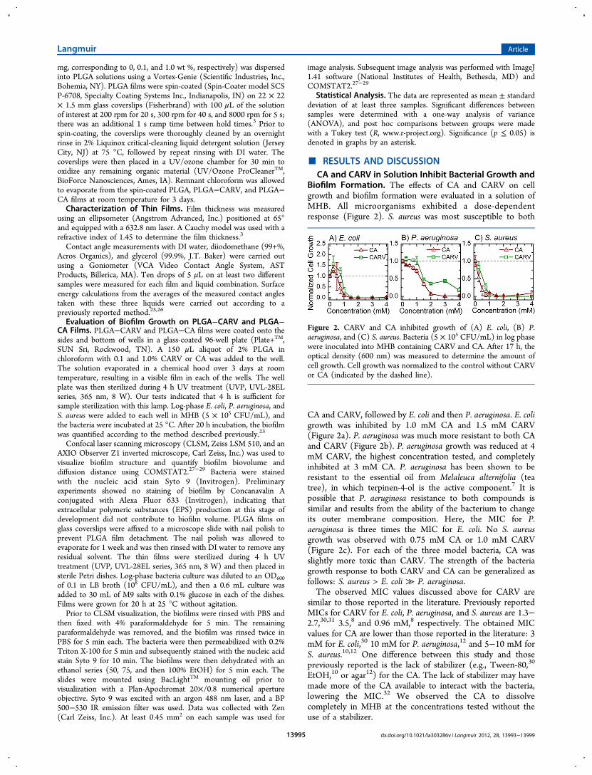
Biofilm Formation. The effects of CA and CARV on cellgrowth and biofilm formation were evaluated in a solution ofMHB. All microorganisms exhibited a dose-dependentresponse (Figure 2). S. aureus was most susceptible to both
CA and CARV, followed by E. coli and then P. aeruginosa. E. coligrowth was inhibited by 1.0 mM CA and 1.5 mM CARV(Figure 2a). P. aeruginosa was much more resistant to both CAand CARV (Figure 2b). P. aeruginosa growth was reduced at 4mM CARV, the highest concentration tested, and completelyinhibited at 3 mM CA. P. aeruginosa has been shown to beresistant to the essential oil from Melaleuca alternifolia (teatree), in which terpinen-4-ol is the active component.7 It ispossible that P. aeruginosa resistance to both compounds issimilar and results from the ability of the bacterium to changeits outer membrane composition. Here, the MIC for P.aeruginosa is three times the MIC for E. coli. No S. aureusgrowth was observed with 0.75 mM CA or 1.0 mM CARV(Figure 2c). For each of the three model bacteria, CA wasslightly more toxic than CARV. The strength of the bacteriagrowth response to both CARV and CA can be generalized asfollows: S. aureus > E. coli ≫ P. aeruginosa.The observed MIC values discussed above for CARV are
similar to those reported in the literature. Previously reportedMICs for CARV for E. coli, P. aeruginosa, and S. aureus are 1.3−2.7,30,31 3.5,8 and 0.96 mM,8 respectively. The obtained MICvalues for CA are lower than those reported in the literature: 3mM for E. coli,30 10 mM for P. aeruginosa,12 and 5−10 mM forS. aureus.10,12 One difference between this study and thosepreviously reported is the lack of stabilizer (e.g., Tween-80,30
EtOH,10 or agar12) for the CA. The lack of stabilizer may havemade more of the CA available to interact with the bacteria,lowering the MIC.32 We observed the CA to dissolvecompletely in MHB at the concentrations tested without theuse of a stabilizer.
Figure 2. CARV and CA inhibited growth of (A) E. coli, (B) P.aeruginosa, and (C) S. aureus. Bacteria (5 × 10⁵ CFU/mL) in log phasewere inoculated into MHB containing CARV and CA. After 17 h, theoptical density (600 nm) was measured to determine the amount ofcell growth. Cell growth was normalized to the control without CARVor CA (indicated by the dashed line).
Langmuir Article
dx.doi.org/10.1021/la303286v | Langmuir 2012, 28, 13993−1399913995

The biofilm formation observed (Figure 3) with CA andCARV in solution mirrored the bacterial growth (Figure 2),
indicating that a lack of bacterial growth caused a lack of biofilmformation for all the bacteria tested. CA inhibited biofilmformation slightly more than CARV. E. coli biofilms wereeliminated at 1 mM CA and 1.5 mM CARV (Figure 3a). Thesevalues directly correspond to the MIC. For P. aeruginosa,biofilms were inhibited at 3 mM CA. No inhibition of biofilmformation was observed for CARV (Figure 3b), as the MIC forCARV was above the values tested (Figure 2b). This indicatesthat with E. coli and P. aeruginosa there was no growth-independent inhibition of biofilm development. Biofilmformation by P. aeruginosa, however, was enhanced significantlyat lower concentrations of CA and CARV. The enhancement ofbiofilm formation upon exposure to subinhibitory concen-trations of antibiotics has been observed previously in P.aeruginosa.33 This enhancement does not correspond to anincrease in the bacterial growth rate (Figure 2b). A similar, butnot statistically significant, enhancement was observed with E.coli. No S. aureus biofilm formation was observed at 0.5 mM CAor 1 mM CARV (Figure 3c). Although the lack of biofilmformation at 1 mM CARV corresponds to the MIC, the MICfor S. aureus and CA is 0.75 mM. Thus, it is possible that thedecrease in biofilm formation at 0.5 mM CA is due to a directinhibition of biofilm formation, also observed by Jia et al.,10 andnot a lack of bacterial growth.Characteristics of PLGA Films Containing CA and
CARV. Spin-coating successfully produced both control PLGAfilms and films incorporating 0.1 and 1.0% CA or CARV. Theaverage thickness of the films was between 500 and 520 nm,indicating that the addition of 1.0% CA and CARV did notchange the properties of the spin-coating solution enough tosignificantly influence the thickness of the films (Table 1).Contact angle measurements were performed with three
solutionswater, glycerol, and diiodomethaneto quantifythe surface energies of the films with CA and CARV. In general,surfaces that are more hydrophilic are less susceptible tobiofouling because of lower bacterial adhesion; however,surfaces with lower surface energy show better foulant release.26
The surface tension (γ) of a film is the sum of two components:nonpolar Lifshitz−van der Waals (LW) forces and polar acid−base (AB) electron donor/acceptor forces. Each molecule ofCA adds an electron donor, and each molecule of CARV addsan electron donor and an electron acceptor. The addition ofthese electron donors and acceptors is reflected in a slightchange of the electron donor component of the surface tension(γ−) for films containing both CA and CARV (Table 1).
However, the overall surface tension of the films is dominatedby the nonpolar LW forces, leading to no great difference insurface tension of the films (γTOT = 36.5−37.7 mJ/m2).In contrast to surface tension, the free energy of cohesion
(ΔG131TOT) is quite different when CA and CARV are added to
PLGA. When ΔG131TOT < 0, the surface is hydrophobic.
Therefore, the films with CA and CARV are less hydrophobicthan films with PLGA alone. ΔG131
TOT is dominated by the ABcomponent, which decreases in magnitude from −22.9 mJ/m2
for PLGA alone to −6.90 and −6.31 mJ/m2 with the additionof CA and CARV, respectively (Table 1). This decrease inhydrophobicity may affect bacterial attachment to the CA andCARV films based on physicochemical properties alone.
PLGA Films Containing CA and CARV Inhibit BiofilmGrowth. PLGA films with 0.1 and 1.0% CA or CARV wereformed in a 96-well plate. PLGA itself is not harmful to E. coli,3
and it acted as a control for the antimicrobial CA and CARV.Bacteria in nutrient-rich MHB were incubated in the coatedwells for 20 h, and the remaining biofilm was stained withsafranin prior to quantification. No staining of the PLGA withor without CARV and CA was observed.PLGA films containing the lowest concentration of CA and
CARV tested showed promising results. CA (0.1%) lessenedbiofilm growth by S. aureus, and 0.1% CARV impaired biofilmgrowth by both E. coli and S. aureus (Figure 4). P. aeruginosawas not as susceptible to CA and CARV in the PLGA matrix.However, there was a significant decrease in P. aeruginosabiofilm formation on 1.0% CA.E. coli biofilm formation was inhibited on films with 0.1%
CARV (p = 0.01) and 1.0% CA (p = 0.02) (Figure 4a).Therefore, under these particular conditions, there is no benefitin this 10-fold increase of CA. However, under differentexperimental conditions, a benefit to higher CA concentrationsmight be observed. No significant decreases in biofilmformation were observed with P. aeruginosa on 0.1 or 1.0%CARV (Figure 5b); however, biofilm formation decreasedsignificantly in the 1.0% CA sample. S. aureus was the onlybacteria affected by both 0.1% CARV (p = 0.02) and 0.1% CA(p = 0.046) (Figure 4c). Thus, PLGA films containing CA andCARV can effectively lessen biofilms formed by differentbacterial species.
Figure 3. Biofilm formation by (A) E. coli, (B) P. aeruginosa, and (C)S. aureus was inhibited by CA and CARV in solution. Bacteria in thelog phase were added to MHB and grown for 17 h. Biofilms wererinsed, fixed, and stained with safranin. Optical density (490 nm) wasmeasured, and the total biofilm was normalized relative to the controlwithout CARV or CA (indicated by the dashed line).
Table 1. Properties of Thin Films: PLGA Control, PLGAwith 1.0% CARV, and PLGA with 1.0% CAa
PLGA PLGA−CA PLGA−CARV
thickness (nm) 510 ± 6.5 519 ± 35 502 ± 13θw (o) 81.7 ± 2.9 76.6 ± 2.9 76.8 ± 3.0γLW (mJ/m2) 36.7 34.8 37.4γ+ (mJ/m2) 0.001 0.067 0.002γ− (mJ/m2) 6.82 9.99 10.0γAB (mJ/m2) 0.166 1.64 0.28γTOT (mJ/m2) 36.8 36.5 37.7ΔG131
LW (mJ/m2) −3.85 −3.04 −4.18ΔG131
AB (mJ/m2) −22.9 −6.90 −6.31ΔG131
TOT (mJ/m2) −26.7 −9.94 −10.5aThe thickness, as measured by ellipsometry, and the contact angle ofDI water, θw, are shown as average ± standard deviation. Surfacetension, γ, was calculated from the averages of all contact anglemeasurements.26,34 The free energy of cohesion with water, ΔG131,was computed from the calculated surface tension values. (LW isLifshitz−van der Waals; + is electron acceptor; − is electron donor;AB is acid base; and TOT is the sum of LW and AB.)
Langmuir Article
dx.doi.org/10.1021/la303286v | Langmuir 2012, 28, 13993−1399913996

Structural Changes in Biofilms on PLGA FilmsContaining CA and CARV. The structure of the biofilmsformed in minimal M9 salts with 0.1% glucose was observedusing CLSM. Biofilm formation is a complex process consistingof five stages: (1) conditioning and initial attachment, (2) EPSproduction and irreversible attachment, (3) cell division andmicrocolony maturation, (4) further microcolony development,and (5) dispersion.4 After 20 h, the PLGA control biofilmsreached stage 3 (Figure 5a). On 1.0% CA films, some cells wereirreversibly attached (stage 2), but overall biofilm formationdecreased substantially (Figure 5b). E. coli microcolonies weremuch smaller on the 1.0% CARV films (Figure 5c).Biofilm biovolume indicates the volume occupied by bacteria
per unit area of the substratum (μm3/μm2). It is also an
indication of the amount of biomass observed.27 While nosignificant difference in E. coli biovolume was observed on the1.0% CARV film (p = 0.59), a significant decrease in biovolumewas observed on the 1.0% CA film (p = 0.03). This is a slightlydifferent response than was observed with the 96-well plateassay using MHB. Thus, the response of E. coli to theseadditives in the PLGA depends, in part, on solutioncomposition and/or the quantification method. The slightdecrease in P. aeruginosa biovolume calculated for both the1.0% CARV and 1.0% CA films was not statistically significant(Figure 6b). As observed in the 96-well plate assay, S. aureusbiofilm formation was inhibited on both the 1.0% CARV (p ≪0.01) and 1.0% CA (p ≪ 0.01) films (Figure 6c). These resultsemphasize the degree to which S. aureus biofilm growth isinhibited by PLGA films that contain either CA or CARV insmall amounts.Diffusion distance is the shortest distance between a small
area that contains biomass and an area that does not containbiomass. This distance could also be considered as the distancea nutrient would have to travel to reach an area within amicrocolony from the surrounding medium, and, especially inthe early stages of colony development, is related to thenumber of cells in a microcolony.27 Two diffusion distanceswere calculated for the image stacks obtainedthe maximumdiffusion distance (MDD) and the average diffusion distance(ADD) (Figure 7). Together, these distances help characterizethe diffusion state of the biofilm.Significant decreases in ADD was observed on 1.0% CA films
with E. coli (p = 0.003) (Figure 7a). An additional difference inMDD on 1.0% CARV films was observed in the P. aeruginosabiofilm (p ≪ 0.001) (Figure 7b). Significant decreases in bothADD and MDD were observed by S. aureus on both 1.0% CAand CARV films (p < 0.003 for all) (Figure 7c). We suggestthat calculated diffusion distance is related to the number ofcells contained within a microcolony, and it is likely that thesmaller diffusion distances calculated are due to an inhibition ofmicrocolony growth on the PLGA films with CA and CARV.The results discussed above emphasize that the effects of
both CARV and CA in PLGA are similar to their effects insolution. These compounds are not particularly effective againstP. aeruginosa but are quite effective against S. aureusthe mostsusceptible bacteria tested here (Figure 2). In general, PLGAfilms containing CA and CARV are most effective against S.aureus, somewhat effective against E. coli, and not very effectiveagainst P. aeruginosa.
■ CONCLUSIONThe colonization of medical devices by bacterial biofilms is aproblem of increasing concern. Biofilms are difficult toeradicate, and the increased prevalence of antibiotic-resistantorganisms increases the risk of bacterial infection. It is possiblethat the use of broad-range natural antimicrobial compounds ina clinical setting could reduce demand for conventionalantibiotics. Here, we verify that cinnamaldehyde (CA) andcarvacrol (CARV) in solution significantly impair bacterialgrowth and, therefore, biofilm formation by E. coli, P.aeruginosa, and S. aureus at millimolar concentrations. Wethen demonstrate that concentrations up to 1.0% CA andCARV can be incorporated into biocompatible PLGA films.These modified films have a similar thickness but prove to bemore hydrophilic due to additional electron donors andacceptors in the CA and CARV. The natural antimicrobialcompounds in the films delay biofilm formation by both gram-
Figure 4. Biofilm growth by (A) E. coli was inhibited on PLGA filmswith 1.0% CA and 0.1% CARV. (B) P. aeruginosa biofilms wereinhibited by 1.0% CA, and (C) S. aureus biofilms were inhibited byboth 0.1% CA and 0.1% CARV. PLGA films were formed byevaporation of 2% PLGA in chloroform (with CARV and CAadditives) in a glass-coated microtiter plate. Bacteria were incubated inMHB for 20 h. The biofilm was rinsed, fixed with ethanol, and stainedwith safranin. Absorbance was measured at 490 nm. Data werenormalized to the PLGA control for each bacterium. An asteriskdenotes that a significant (p ≤ 0.05) reduction in biofilm growth hasoccurred between the control PLGA film and a film containing CARVor CA.
Langmuir Article
dx.doi.org/10.1021/la303286v | Langmuir 2012, 28, 13993−1399913997

positive S. aureus and gram-negative E. coli on the film surface.These films could potentially be used as natural, plant-basedcoatings for a number of indwelling devices to safely impedecolonization by pathogenic bacteria.
■ AUTHOR INFORMATIONCorresponding Author*E-mail: [email protected]. Phone: (413) 545-6143.
NotesThe authors declare no competing financial interest.
■ ACKNOWLEDGMENTSThis material is based upon work supported by the NationalScience Foundation Graduate Research fellowship under GrantNo. DGE-1122492 awarded to K.R.Z. Any opinion, findings,and conclusions or recommendations expressed in this materialare those of the authors and do not necessarily reflect the viewsof the National Science Foundation. Dr. Joseph Wolenskihelped with confocal microscopy. Bacterial strain S. aureus RN6390B was kindly provided by Dr. Naomi Balaban.
■ REFERENCES(1) Peleg, A. Y.; Hooper, D. C. Hospital-acquired infections due togram-negative bacteria. New Engl. J. Med. 2010, 362 (19), 1804−1813.(2) Cohen, M. L. Changing patterns of infectious disease. Nature2000, 406 (6797), 762−767.(3) Aslan, S.; Loebick, C. Z.; Kang, S.; Elimelech, M.; Pfefferle, L. D.;Van Tassel, P. R. Antimicrobial biomaterials based on carbonnanotubes dispersed in poly(lactic-co-glycolic acid). Nanoscale 2010,2 (9), 1789−1794.(4) Davies, D. Understanding biofilm resistance to antibacterialagents. Nat. Rev. Drug Disc. 2003, 2 (2), 114−122.(5) Ramritu, P.; Halton, K.; Collignon, P.; Cook, D.; Fraenkel, D.;Battistutta, D.; Whitby, M.; Graves, N. A systematic review comparingthe relative effectiveness of antimicrobial-coated catheters in intensivecare units. Am. J. Infect. Control 2008, 36 (2), 104−117.(6) Silver, S.; Phung, L. T.; Silver, G. Silver as biocides in burn andwound dressings and bacterial resistance to silver compounds. J. Ind.Microbiol. Biotechnol. 2006, 33 (7), 627−634.(7) Bakkali, F.; Averbeck, S.; Averbeck, D.; Idaomar, M. Biologicaleffects of essential oilsA review. Food Chem. Toxicol. 2008, 46 (2),446−475.(8) Lambert, R. J. W.; Skandamis, P. N.; Coote, P. J.; Nychas, G.-J. E.A study of the minimum inhibitory concentration and mode of actionof oregano essential oil, thymol and carvacrol. J. Appl. Microbiol. 2001,91 (3), 453−462.(9) Amalaradjou, M. A. R.; Narayanan, A.; Baskaran, S. A.;Venkitanarayanan, K. Antibiofilm effect of trans-cinnamaldehyde onuropathogenic Escherichia coli. J. Urol. 2010, 184 (1), 358−363.(10) Jia, P.; Xue, Y. J.; Duan, X. J.; Shao, S. H. Effect ofcinnamaldehyde on biofilm formation and sarA expression bymethicillin-resistant Staphylococcus aureus. Lett. Appl. Microbiol. 2011,53 (4), 409−416.(11) Niu, C.; Afre, S.; Gilbert, E. S. Subinhibitory concentrations ofcinnamaldehyde interfere with quorum sensing. Lett. Appl. Microbiol.2006, 43 (5), 489−494.
Figure 5. Pictured are orthogonal sections (300 μm × 300 μm) of representative CLSM images formed after 20 h on (A) PLGA control films in M9salts with 0.1% glucose. CLSM biovolume analysis (Figure 6) showed a significant decrease in biofilm formation by E. coli on (B) PLGA with 1.0%CA compared to the (A) PLGA control. No significant difference was observed on PLGA with (C) 1.0% CARV.
Figure 6. Significantly less (A) E. coli biovolume was observed onPLGA films with 1.0% CA. (B) P. aeruginosa, the bacterium leastsusceptible to CA and CARV, was not significantly affected by 1.0%CA or 1.0% CARV. (C) S. aureus biovolume on 1.0% CA and 1.0%CARV decreased significantly compared to the PLGA control. Anasterisk denotes that a significant (p ≤ 0.05) reduction in biofilmgrowth has occurred between the control PLGA film and a filmcontaining CARV or CA. Biofilms were grown in M9 salts and 0.1%glucose for 20 h. The cells were stained with Syto 9. Biovolume wascalculated using COMSTAT2.
Figure 7. Significantly lower diffusion distances were observed in (A)E. coli biofilms grown on 1.0% CA in PLGA, (B) P. aeruginosa biofilmsgrown on 1.0% CARV, and (C) S. aureus biofilms grown on both 1.0%CA and 1.0% CARV. An asterisk denotes that a significant (p ≤ 0.05)reduction in biofilm growth has occurred between the control PLGAfilm and a film containing CARV or CA. Biofilms were grown in M9salts and 0.1% glucose for 20 h and stained with Syto 9. Maximumdiffusion distance (MDD) and average diffusion distance (ADD) werecalculated with COMSTAT2.
Langmuir Article
dx.doi.org/10.1021/la303286v | Langmuir 2012, 28, 13993−1399913998

(12) Bouhdid, S.; Abrini, J.; Amensour, M.; Zhiri, A.; Espuny, M. J.;Manresa, A. Functional and ultrastructural changes in Pseudomonasaeruginosa and Staphylococcus aureus cells induced by Cinnamomumverum essential oil. J. Appl. Microbiol. 2010, 109 (4), 1139−1149.(13) Niu, C.; Gilbert, E. S. Colorimetric method for identifying plantessential oil components that affect biofilm formation and structure.Appl. Environ. Microbiol. 2004, 70 (12), 6951−6956.(14) Brackman, G.; Defoirdt, T.; Miyamoto, C.; Bossier, P.; VanCalenbergh, S.; Nelis, H.; Coenye, T. Cinnamaldehyde andcinnamaldehyde derivatives reduce virulence in Vibrio spp. bydecreasing the DNA-binding activity of the quorum sensing responseregulator LuxR. BMC Microbiol. 2008, 8, 149.(15) Bouhdid, S.; Abrini, J.; Zhiri, A.; Espuny, M. J.; Manresa, A.Investigation of functional and morphological changes in Pseudomonasaeruginosa and Staphylococcus aureus cells induced by Origanumcompactum essential oil. J. Appl. Microbiol. 2009, 106 (5), 1558−1568.(16) Burt, S. A.; van der Zee, R.; Koets, A. P.; de Graaff, A. M.; vanKnapen, F.; Gaastra, W.; Haagsman, H. P.; Veldhuizen, E. J. A.Carvacrol induces heat shock protein 60 and inhibits synthesis offlagellin in Escherichia coli O157: H7v. Appl. Environ. Microbiol. 2007,73 (14), 4484−4490.(17) Burt, S. Essential oils: Their antibacterial properties andpotential applications in foodsA review. Int. J. Food Microbiol. 2004,94 (3), 223−253.(18) Persico, P.; Ambrogi, V.; Carfagna, C.; Cerruti, P.; Ferrocino, I.;Mauriello, G. Nanocomposite polymer films containing carvacrol forantimicrobial active packaging. Polymer Eng. Sci. 2009, 49 (7), 1447−1455.(19) Gomes, C.; Moreira, R. G.; Castell-Perez, E. Poly (DL-lactide-co-glycolide) (PLGA) nanoparticles with entrapped trans-cinnamalde-hyde and eugenol for antimicrobial delivery applications. J. Food Sci.2011, 76 (2), N16−N24.(20) Iannitelli, A.; Grande, R.; Di Stefano, A.; Di Giulio, M.; Sozio,P.; Bessa, L. J.; Laserra, S.; Paolini, C.; Protasi, F.; Cellini, L. Potentialantibacterial activity of carvacrol-loaded poly(DL-lactide-co-glycolide)(PLGA) nanoparticles against microbial biofilm. Int. J. Mol. Sci. 2011,12 (8), 5039−5051.(21) de Kerchove, A. J.; Elimelech, M. Impact of alginateconditioning film on deposition kinetics of motile and nonmotilePseudomonas aeruginosa strains. Appl. Environ. Microbiol. 2007, 73 (16),5227−5234.(22) Wiegand, I.; Hilpert, K.; Hancock, R. E. W. Agar and brothdilution methods to determine the minimal inhibitory concentration(MIC) of antimicrobial substances. Nat. Protoc. 2008, 3 (2), 163−175.(23) Kiran, M. D.; Adikesavan, N. V.; Cirioni, O.; Giacometti, A.;Silvestri, C.; Scalise, G.; Ghiselli, R.; Saba, V.; Orlando, F.; Shoham,M.; Balaban, N. Discovery of a quorum-sensing inhibitor of drug-resistant staphylococcal infections by structure-based virtual screening.Mol. Pharmacol. 2008, 73 (5), 1578−1586.(24) Omidi, Y.; Davaran, S. Impacts of biodegradable polymers:Towards biomedical applications. In Handbook of Applied BiopolymerTechnology: Synthesis, Degradation and Applications; Sharma, S. K.,Mudhoo, A., Eds.; Royal Society of Chemistry: London, U.K., 2011;pp 388−418.(25) Owens, D. K.; Wendt, R. C. Estimation of the surface freeenergy of polymers. J. Appl. Polym. Sci. 1969, 13 (8), 1741−1747.(26) Hurwitz, G.; Guillen, G. R.; Hoek, E. M. V. Probing polyamidemembrane surface charge, zeta potential, wettability, and hydro-philicity with contact angle measurements. J. Membr. Sci. 2010, 349,349−357.(27) Heydorn, A.; Nielsen, A. T.; Hentzer, M.; Sternberg, C.;Givskov, M.; Ersbøll, B. K.; Molin, S. Quantification of biofilmstructures by the novel computer program COMSTAT. Microbiology2000, 146 (10), 2395−2407.(28) Vorregaard, M.; Ersbøll, B. K.; Yang, L; Haagensen, J. A. J.;Molin, S; Sternberg, C. Personal communication.(29) COMSTAT2 Web Page; http://www.comstat.dk (March 22,2012).
(30) Pei, R.-S.; Zhou, F.; Ji, B.-P.; Xu, J. Evaluation of combinedantibacterial effects of eugenol, cinnamaldehyde, thymol, and carvacrolagainst E. coli with an improved method. J. Food Sci. 2009, 74 (7),M379−M383.(31) Xu, J.; Zhou, F.; Ji, B. P.; Pei, R. S.; Xu, N. The antibacterialmechanism of carvacrol and thymol against Escherichia coli. Lett. Appl.Microbiol. 2008, 47 (3), 174−179.(32) Si, W.; Gong, J.; Chanas, C.; Cui, S.; Yu, H.; Caballero, C.;Friendship, R. M. In vitro assessment of antimicrobial activity ofcarvacrol, thymol, and cinnamaldehyde towards Salmonella serotypeTyphimurium DT104: Effects of pig diets and emulsification inhydrocolloids. J. Appl. Microbiol. 2006, 101 (6), 1282−1291.(33) Hoffman, L. R.; D’Argenio, D. A.; MacCoss, M. J.; Zhang, Z.;Jones, R. A.; Miller, S. I. Aminoglycoside antibiotics induce bacterialbiofilm formation. Nature 2005, 436 (7054), 1171−1175.(34) Brant, J. A.; Childress, A. E. Assessing short-range membrane−colloid interactions using surface energetics. J. Membr. Sci. 2002, 203(1−2), 257−273.
Langmuir Article
dx.doi.org/10.1021/la303286v | Langmuir 2012, 28, 13993−1399913999