Study of multivesiculated polyester particles synthesis by double emulsion process
Upload
independentCategory
view
1download
0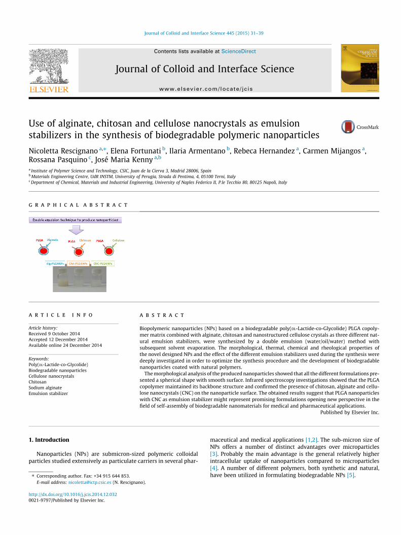
Journal of Colloid and Interface Science 445 (2015) 31–39
Contents lists available at ScienceDirect
Journal of Colloid and Interface Science
www.elsevier .com/locate / jc is
Use of alginate, chitosan and cellulose nanocrystals as emulsionstabilizers in the synthesis of biodegradable polymeric nanoparticles
http://dx.doi.org/10.1016/j.jcis.2014.12.0320021-9797/Published by Elsevier Inc.
⇑ Corresponding author. Fax: +34 915 644 853.E-mail address: [email protected] (N. Rescignano).
Nicoletta Rescignano a,⇑, Elena Fortunati b, Ilaria Armentano b, Rebeca Hernandez a, Carmen Mijangos a,Rossana Pasquino c, José Maria Kenny a,b
a Institute of Polymer Science and Technology, CSIC, Juan de la Cierva 3, Madrid 28006, Spainb Materials Engineering Centre, UdR INSTM, University of Perugia, Strada di Pentima, 4, 05100 Terni, Italyc Department of Chemical, Materials and Industrial Engineering, University of Naples Federico II, P.le Tecchio 80, 80125 Napoli, Italy
g r a p h i c a l a b s t r a c t
a r t i c l e i n f o
Article history:Received 9 October 2014Accepted 12 December 2014Available online 24 December 2014
Keywords:Poly(DL-Lactide-co-Glycolide)Biodegradable nanoparticlesCellulose nanocrystalsChitosanSodium alginateEmulsion stabilizer
a b s t r a c t
Biopolymeric nanoparticles (NPs) based on a biodegradable poly(DL-Lactide-co-Glycolide) PLGA copoly-mer matrix combined with alginate, chitosan and nanostructured cellulose crystals as three different nat-ural emulsion stabilizers, were synthesized by a double emulsion (water/oil/water) method withsubsequent solvent evaporation. The morphological, thermal, chemical and rheological properties ofthe novel designed NPs and the effect of the different emulsion stabilizers used during the synthesis weredeeply investigated in order to optimize the synthesis procedure and the development of biodegradablenanoparticles coated with natural polymers.
The morphological analysis of the produced nanoparticles showed that all the different formulations pre-sented a spherical shape with smooth surface. Infrared spectroscopy investigations showed that the PLGAcopolymer maintained its backbone structure and confirmed the presence of chitosan, alginate and cellu-lose nanocrystals (CNC) on the nanoparticle surface. The obtained results suggest that PLGA nanoparticleswith CNC as emulsion stabilizer might represent promising formulations opening new perspective in thefield of self-assembly of biodegradable nanomaterials for medical and pharmaceutical applications.
Published by Elsevier Inc.
1. Introduction maceutical and medical applications [1,2]. The sub-micron size of
Nanoparticles (NPs) are submicron-sized polymeric colloidalparticles studied extensively as particulate carriers in several phar-
NPs offers a number of distinct advantages over microparticles[3]. Probably the main advantage is the general relatively higherintracellular uptake of nanoparticles compared to microparticles[4]. A number of different polymers, both synthetic and natural,have been utilized in formulating biodegradable NPs [5].
32 N. Rescignano et al. / Journal of Colloid and Interface Science 445 (2015) 31–39
Nanoparticles based on biodegradable polyester (such as poly-lactic acid (PLA), polylactic-co-glycolic acid (PLGA) and polycapro-lactone (PCL)) represent most promising candidates for in vivodiagnosis and treatment of cancer, from preclinical developmentto clinical translation [1]. The use of these amphiphilic polymersresults in the formation of nanoparticles with a hydrophobic coreand a hydrophilic shell. The core–shell structure allows them toencapsulate and carry poorly water-soluble drugs [4] and torelease these drugs at a sustained rate in the optimal range of drugconcentration [6].
PLGA nanoparticles have been mostly prepared by emulsifica-tion–diffusion [7], solvent emulsion–evaporation [8], interfacialdeposition [9] and nanoprecipitation method [10]. The acidicnature of PLGA monomers is not suitable for drugs or bioactivemolecules [11,12]. However, few approaches to overcome theseproblems have been developed. For this, PLGA nanomedicineformulations are blended with alginate, chitosan, pectin [13],poly(propylenefumarate) [14], polyvinyalcohol (PVA) [15],poly(orthoester), etc. [16].
Emulsion solvent evaporation techniques are the most fre-quently methods used to produce NPs, wherein a significantamount of PVA is generally employed as stabilizing agent[17,18]; however, PVA is difficult to remove from the surface ofthe produced particles.
Research efforts on the development of sustainable, biodegrad-able, and nontoxic solid particle emulsion stabilizers have beencarried out since several decades due to the importance of emul-sion systems and derived particles in food, cosmetics and pharma-ceutical industries. Methods involving surface modifications or theaddition of co-surfactant compounds [19,20] have been developedto produce symmetrical or asymmetrical particles, but such meth-ods increase the use of chemicals and make the process compli-cated and expensive for large-scale production. Therefore,research efforts are being currently focused on the developmentof environmentally friendly, bio-based nanocomposites, but fewstudies, up to now, have described the use of chemicals derivedfrom renewable resources for NP stabilization [21–23].
Cellulose is one of the most widespread biopolymers and is agood candidate due to its sustainability, biodegradability, and non-toxicity. In particular, cellulose nanocrystals (CNC), extracted byhydrolysis of different natural sources, are typically rigid rod-shaped monocrystalline cellulose domains, 1–100 nm in diameterand from tens to hundreds of nanometers in length [24]. Theyare involved in different applications, as reinforcements in polymernanocomposites, in the biomedical fields and in optoelectronicdevices applications [25,26], and more recently, their potentialapplications as stabilizers in chemical reactions, were investigated[27]. Kalashnikova et al. [28] showed the possibility to use bacte-rial cellulose nanocrystals as stabilizers at the hexadecane–waterinterface, promoting monodispersed oil in water droplets around4 mm in diameter stable for several months. The authors provedthe possibility to design and produce nanosized cellulosic particles,which were directly able to stabilize irreversibly an oil-in-wateremulsion suggesting also that the nanocrystals can be used to pre-pare a monodispersed emulsion without any further modificationor wrapping process.
Furthermore, other polysaccharides have been considered asstabilizer agents in different reactions. Wang et al. prepared PLGAbased nanoparticles using chitosan and/or alginate as emulsionstabilizers [29]. The hydrophilic properties of these moleculesand their abundant functional groups for further modificationmake these natural polymers excellent candidates for biomedicalapplications.
Alginate and chitosan are two major naturally occurring poly-saccharides which have been widely used as main componentsfor drug delivery systems and tissue scaffolds. Alginate, commer-
cially available as alginate sodium salt and commonly calledsodium alginate (Alg), is a linear polysaccharide normally isolatedfrom many strains of marine brown seaweed and algae. Alginatehas the ability to bind multivalent cations, leading to the formationof covalent bonds and insoluble hydrogels. This anionic polysac-charide forms strong gels with calcium cations leading to micro-spheres with good strength and flexibility. Such crosslinkingleads to a stiffer polymer system, reducing swelling in water andorganic solvents. Alginate has carboxyl groups which may intro-duce negative charge to the polymer at appropriate pH [30].
Chitosan is a biodegradable polysaccharide extracted fromcrustacean shells. It has been shown to be non-toxic in a range oftoxicity tests, both in experimental animals [31] and humans[32]. Chitosan is a natural cationic polymer obtained by deacetylat-ing chitin comprising copolymers of b(1,4)-glucosamine andN-acetyl-D-glucosamine. Furthermore, according to the therapeuticrequirements, the NP composition (i.e., use of Alg or chitosan) canalso be considered as a useful mean to tune the drug release rateand the NP interactions with living tissues [33].
NPs are usually dispersed in water for drug delivery and theproperties of their suspensions under flow are important to deter-mine their stability and efficiency. Water has unique properties ofstabilizing particle–particle interaction, thanks to ion-relatedelectrostatic stabilization. However, other parameters, such aspH, aeration, and depletion agents, can affect the stability of thesuspensions. Rheology is a powerful method to measure the abilityof the suspension to flow under external forces [34,35].Experimental and theoretical studies have shown that morpholog-ical properties of the particles (shape, size, distribution) as well asfluid/particle and particle/particle interactions can strongly influ-ence the fluid rheology [36,37].
In the present work, poly(DL-Lactide-co-Glycolide) copolymerbased biopolymeric nanoparticles were synthesized by a doubleemulsion (water/oil/water) method comparing the effect of algi-nate, chitosan and nanostructured cellulose crystals as naturalemulsion stabilizers on the morphological (in terms of sizes andshapes), thermal, surface, chemical and rheological properties ofthe produced nanoparticles. The idea is to optimize the procedureof PLGA based NP synthesis using two natural polymers, alginateand chitosan, and a nanostructured natural system, cellulose nano-crystals, as emulsion stabilizers during the NP production. Theadvantages and drawbacks of the different formulations are deeplyinvestigated and discussed.
2. Experimental part
2.1. Materials
A poly(DL-Lactide-co-Glycolide) copolymer, (PLGA) (I.V. 0.95–1.20 dL g�1, Mw 91,600–120,000 g/mol) ether terminated, with a50/50 ratio (PLA/PGA) supplied by Absorbables Polymers/Lactel(Durect Corporation), was used as biodegradable polymer for thenanoparticle (NP) preparation.
Microcrystalline cellulose (MCC, dimensions of 10–15 lm, bulkdensity 0.5 g mL�1 at 25 �C), supplied by Sigma Aldrich, was usedas start material for cellulose nanocrystal (CNC) synthesis.
Sodium alginate (Alg) and chitosan (Chit) were purchased fromSigma Aldrich and used as received. According to the manufac-turer, the molecular weight of chitosan is approximately 50,000–190,000 Da. The molecular weight of sodium alginate was obtainedthrough viscosity measurements by applying the Mark–Houwinkequation, ½g� ¼ k �Mv
a, where ½g� is the intrinsic viscosity and kand a, are constants for a solvent-polymer pair at 25 �C. For algi-nate, k = 2�10�5 dL g�1 and a = 1.0 [38]. The molecular weight thusobtained was 165,500 Da for sodium alginate.
N. Rescignano et al. / Journal of Colloid and Interface Science 445 (2015) 31–39 33
2.2. Synthesis of cellulose nanocrystals
Cellulose nanocrystals, were obtained by a hydrolysis processstarting from the commercial microcrystalline cellulose (MCC)treated by sulfuric acid hydrolysis (64%wt/wt) at 45 �C for 30 minas previously reported [39]. After acid removal, dialysis and ultra-sonic treatment, the resultant cellulose nanocrystal aqueous sus-pension was approximately 0.5%wt/wt and the hydrolysisprocedure yield was ca. 20%. Mixed bed ion exchange resin (DowexMarathon MR-3 hydrogen and hydroxide form) was added to theCNC water solution and it was thermally stabilized by addition of1.0%v/v of 0.25 mol L�1 NaOH. The obtained CNC showed the typ-ical rod-like structure with dimensions ranging from 100 to200 nm in length and 5–10 nm in width, as previously reported[40]. The synthesized CNC were then used as one of the threeemulsion stabilizers for the polymeric nanoparticle preparation.
2.3. Biopolymeric nanoparticle preparation
Biodegradable nanoparticles were prepared by double emulsion(water/oil/water) method with subsequent solvent evaporation[41]. Briefly, 0.25 g of biodegradable polymer were dissolved in5 mL of chloroform, with 2 h magnetic stirring. This solution wasemulsified with 2 mL of bi-distilled water using a tip sonicator(VIBRA CELL Sonics mod. VC 750) for 15 min. The resulting emul-sion was mixed with 2%wt/v of emulsion stabilizers (Alg, CNCand Chit) in 20 mL of aqueous solution (or acetic acid for Chit),for 30 min with sonication, in order to form the second emulsion.For the solvent evaporation, the second emulsion was transferredin 200 mL of 0.2%wt/v emulsion stabilizer aqueous solution (oracid acetic solution in the case of Chit) and was magnetically stir-red over night at room temperature. The obtained nanosphereswere collected by centrifugation (centrifuge Sigma 2–16P) at11,000 rpm for 20 min and washed out four times with distilledwater in the case of alginate and CNC while, when Chit wasemployed as emulsion stabilizer, the washings were performedwith acetic acid solution (1%v/v in water).
PLGA particles prepared with sodium alginate, cellulose nano-crystals and chitosan as emulsion stabilizer were designated asAlg-PLGANPs, CNC-PLGANPs and Chit-PLGANPs, respectively.
2.4. Characterization methods
2.4.1. Morphological characterizationThe morphological characterization of the Alg-PLGANPs, CNC-
PLGANPs and Chit-PLGANPs was performed by a Field EmissionScanning Electron Microscope (FESEM Supra 25, Zeiss). Sampleswere deposited onto fluorinated tin oxide (FTO) substrates usinga drop-casting method, allowing them to dry at room temperaturefor 24 h and gold-coated by an Agar automatic sputter coater. NPdimensions were calculated by the analysis of the correspondingFESEM images using Image J software. The mean diameter andthe particle size distribution were obtained from the analysis ofat least 250 individual nanoparticles.
Dynamic Light Scattering (DLS) was used for the determinationof NP average diameter and zeta potential employing a MalvernNano ZS instrument with a 633 nm laser diode and a backscatter-ing detection angle of 173�. Samples were housed in quartz cuv-ettes of 1 cm optical path length. The electrophoretic mobilitywas transformed into Zeta potential (n) using the Smoluchowskiequation. All measurements were repeated three times and theaverage of three runs was computed.
2.4.2. Thermal and chemical analysisThermal properties of the Alg-PLGANPs, CNC-PLGANPs and
Chit-PLGANPs systems were assessed by differential scanning cal-
orimetry (DSC 822/e Mettler-Toledo, USA). Ten milligrams of driedsamples were placed in covered aluminum pans and then placed inthe DSC sample holder. For all samples, the measurements wereperformed in the temperature range from �25 �C to 200 �C, at10 �C min�1, performing two heating and one cooling scans. Thesame temperature range was selected for all the proposed formu-lations in order to have information about the thermal phenomenaavoiding, at the same time, any degradation mechanism. The val-ues of the glass transition temperature (Tg) were obtained fromthe dynamic thermograms, using the midpoint between the inter-sections of the two parallel baselines, before and after the Tg.
Thermogravimetric analysis (TGA) was performed using aquartz rod microbalance (Seiko Exstar 6300) in the following con-ditions: sample weight 10 mg, nitrogen flow (250 mL min�1), tem-perature range 30–600 �C, heating rate 10 �C min�1.
Fourier transform infrared (FT-IR) spectroscopy of the Alg-PLGANPs, CNC-PLGANPs and Chit-PLGANPs was carried out witha FT-IR spectrophotometer (Jasco FT-IR 615, Japan). The spectrawere performed on KBr disks in the range from 4000 to400 cm�1 with a 4 cm�1 resolution.
2.4.3. Rheological analysisNanoparticle dispersions in bidistilled water have been pre-
pared at two different solid contents, 0.5%wt/v and 1%wt/v. Thesolutions were stirred overnight to ensure homogeneous solutions.Rheological measurements were performed at room temperatureusing a stress-controlled rheometer (Bohlin CVO 120, Malverninstruments) equipped with a 60 mm smooth stainless steel diskswith cone-plate geometry (cone angle 0.0175 rad), in order tomaximize the torque value and allow good quality measurements.A steel cylindrical cage enveloping the cone-plate system has beenused to prevent sample drying. Steady tests have been performedto measure the zero shear viscosity of the solutions with differentnanoparticles content and type. In all cases, the time scale associ-ated with sedimentation was larger than the time scale associatedto the experiment [29].
2.4.4. Bulk settling experimentsSeveral milligrams of three different lioiphyilized samples were
dispersed in distilled water during 24 h with magnetic agitation inorder to obtain the uniform dispersion of NPs in the solvent. Obser-vations were made after 30 min and 24 h con concentration(C = 10 mg in 5 mL de H2O). The colloidal stability of Alg-PLGANPs,Chit-PLGANPs dispersion was determined by measuring the varia-tion of average diameter with the time, at 25 �C. Seven consecutivemeasurements were taken and then averaged and plotted in Fig. 6.
3. Results and discussion
3.1. Determination of particle size and zeta potential of the synthesizednanoparticles
The combination of natural polymers such as alginate, chitosanor nanostructured cellulose nanocrystals with biodegradable PLGAfor the formation of biopolymeric nanoparticles offer some inter-esting advantages: first of all the hydrophilic and the hydrophobicbehaviors of the polymers (emulsion stabilizers and PLGA, respec-tively) provide peculiar properties that facilitate the interactionwith the biological environment and hydrophobic drugs. Particlesize and their surface properties play a key role in determiningthe drug release and cellular uptake as well as their in vivo phar-macokinetics and biodistribution. Furthermore, the characteriza-tion of the particle size facilitates understanding of thedispersion and aggregation processes.
34 N. Rescignano et al. / Journal of Colloid and Interface Science 445 (2015) 31–39
FESEM images corresponding to the three samples under study(Fig. 1), Alg-PLGANPs, Chit-PLGANPs and CNC-PLGANPs, demon-strate the successful preparation of PLGA particles by the doubleemulsion technique. In all cases, PLGA particles are characterizedby a smooth surface and a spherical shape. In addition, it is evidentthat the particle size is related to the kind of emulsion stabilizeremployed. The histograms corresponding to each of the preparedsamples are also shown in Fig. 1. Alg-PLGANPs show a broad particlesize distribution centered at �1 lm and with particle sizes rangingfrom�250 nm to�2 lm. Some particles with diameters higher than�2 lm are also observed. In contrast, a different behavior wasdetected in the case of Chit-PLGANPs for which the particle size dis-tribution is much narrower with an average particle size of�400 nm. From these results, it is clear that the employment ofchitosan as emulsion stabilizer in the preparation of the PLGA basednanoparticles by double emulsion constitutes a more efficient strat-egy to achieve particles in the nanometer range. The results could beexplained in terms of the difference in the molecular weight of thetwo polymeric emulsion stabilizers as the chitosan employed in thiswork has a molecular weight which is �6 times lower than the cor-responding to sodium alginate as determined through viscositymeasurements. As previously reported [42], an emulsion stabilizer
2µm
2µm
a
2µm
c
b
Freq
uenc
y
1
Freq
uenc
y
1
Freq
uenc
y
Fig. 1. FESEM images of (a) Alg-PLGANPs, (b) Chit-PLGANPs, and (c
with a high molecular weight may increase the viscosity of theemulsion resulting in an increased difficulty in breaking up theemulsion into smaller droplets and yielding bigger particles inagreement with our results (see Fig. 2).
Interestingly, the employment of a polymeric nanostructuredemulsion stabilizer such as cellulose nanocrystals also yields PLGAparticles with a narrow particle size distribution centered at�560 nm. As previously reported, the rod-like shape of cellulosenanocrystals gives rise to a percolating network which yields ahigh stabilization at the oil/water interface occurring in emulsionprocesses [43]. Therefore we can assume that CNC, once confinedat the interface of the oil-in-water emulsion for the preparationof PLGA particles, might associate among them according to anaggregative process. This might increase the cohesion and stabilityof the PLGA droplets formed in the process of double emulsion thusincreasing the efficiency of CNCs as emulsion stabilizer.
The Zeta (n) potential, which is the electrostatic potential thatexists at the shear plane of a particle, is related to both surfacecharge and the local environment of the particle. Zeta potential iscommonly used as an important parameter in colloid science tounderstand the colloid electrostatic interactions [44,45]. In fact,zeta potential can greatly influence particle stability in suspension
500 1000 1500 2000 25000
20
40
60
diameter [nm]
0
20
40
60
80
00
diameter [nm]
0 500 1000 1500 2000 2500 3000
0 500 1000 1500 2000 2500 3000
0
20
40
60
80
00
) CNC-PLGANPs. The corresponding histograms are also shown.
100 200 300 400 500 600
0,00
0,05
0,10
0,15
0,20D
TG [µ
g/µg
min
]
Temperature [°C]
PLGA pellet CNC powder CNC-PLGANPs
(a)
100 200 300 400 500 600
0,00
0,05
0,10
0,15
0,20
DTG
[µg/
µg m
in]
Temperature [°C]
PLGA pellet Chit powder Chit-PLGANPs
(b)
100 200 300 400 500 600
0,00
0,05
0,10
0,15
0,20
Temperature [°C]
DTG
[µg/
µg m
in]
PLGA pellet Alg powder Alg-PLGANPs
(c)
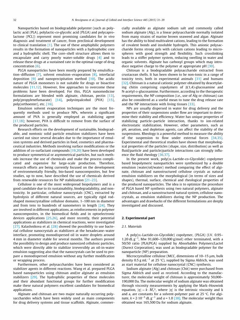
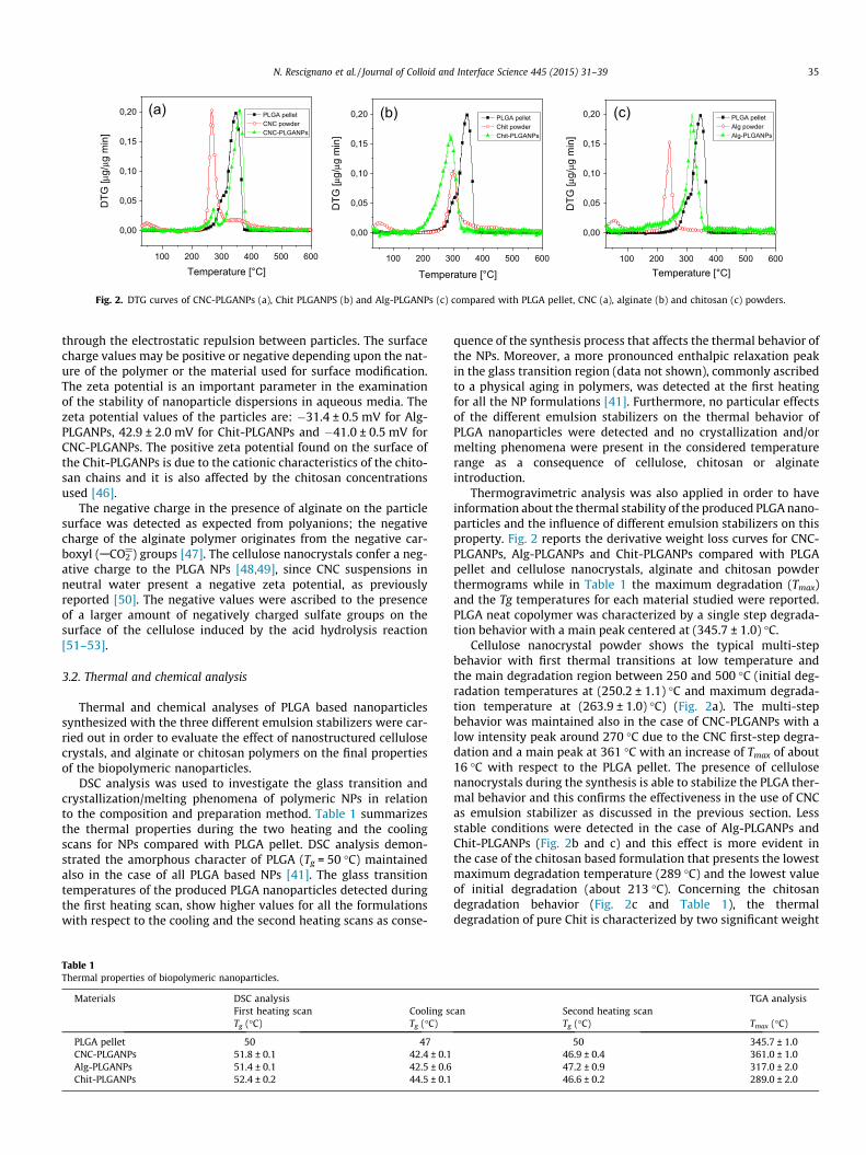
Fig. 2. DTG curves of CNC-PLGANPs (a), Chit PLGANPS (b) and Alg-PLGANPs (c) compared with PLGA pellet, CNC (a), alginate (b) and chitosan (c) powders.
N. Rescignano et al. / Journal of Colloid and Interface Science 445 (2015) 31–39 35
through the electrostatic repulsion between particles. The surfacecharge values may be positive or negative depending upon the nat-ure of the polymer or the material used for surface modification.The zeta potential is an important parameter in the examinationof the stability of nanoparticle dispersions in aqueous media. Thezeta potential values of the particles are: �31.4 ± 0.5 mV for Alg-PLGANPs, 42.9 ± 2.0 mV for Chit-PLGANPs and �41.0 ± 0.5 mV forCNC-PLGANPs. The positive zeta potential found on the surface ofthe Chit-PLGANPs is due to the cationic characteristics of the chito-san chains and it is also affected by the chitosan concentrationsused [46].
The negative charge in the presence of alginate on the particlesurface was detected as expected from polyanions; the negativecharge of the alginate polymer originates from the negative car-boxyl (ACO2
@) groups [47]. The cellulose nanocrystals confer a neg-ative charge to the PLGA NPs [48,49], since CNC suspensions inneutral water present a negative zeta potential, as previouslyreported [50]. The negative values were ascribed to the presenceof a larger amount of negatively charged sulfate groups on thesurface of the cellulose induced by the acid hydrolysis reaction[51–53].
3.2. Thermal and chemical analysis
Thermal and chemical analyses of PLGA based nanoparticlessynthesized with the three different emulsion stabilizers were car-ried out in order to evaluate the effect of nanostructured cellulosecrystals, and alginate or chitosan polymers on the final propertiesof the biopolymeric nanoparticles.
DSC analysis was used to investigate the glass transition andcrystallization/melting phenomena of polymeric NPs in relationto the composition and preparation method. Table 1 summarizesthe thermal properties during the two heating and the coolingscans for NPs compared with PLGA pellet. DSC analysis demon-strated the amorphous character of PLGA (Tg = 50 �C) maintainedalso in the case of all PLGA based NPs [41]. The glass transitiontemperatures of the produced PLGA nanoparticles detected duringthe first heating scan, show higher values for all the formulationswith respect to the cooling and the second heating scans as conse-
Table 1Thermal properties of biopolymeric nanoparticles.
Materials DSC analysisFirst heating scan Cooling sTg (�C) Tg (�C)
PLGA pellet 50 47CNC-PLGANPs 51.8 ± 0.1 42.4 ± 0.1Alg-PLGANPs 51.4 ± 0.1 42.5 ± 0.6Chit-PLGANPs 52.4 ± 0.2 44.5 ± 0.1
quence of the synthesis process that affects the thermal behavior ofthe NPs. Moreover, a more pronounced enthalpic relaxation peakin the glass transition region (data not shown), commonly ascribedto a physical aging in polymers, was detected at the first heatingfor all the NP formulations [41]. Furthermore, no particular effectsof the different emulsion stabilizers on the thermal behavior ofPLGA nanoparticles were detected and no crystallization and/ormelting phenomena were present in the considered temperaturerange as a consequence of cellulose, chitosan or alginateintroduction.
Thermogravimetric analysis was also applied in order to haveinformation about the thermal stability of the produced PLGA nano-particles and the influence of different emulsion stabilizers on thisproperty. Fig. 2 reports the derivative weight loss curves for CNC-PLGANPs, Alg-PLGANPs and Chit-PLGANPs compared with PLGApellet and cellulose nanocrystals, alginate and chitosan powderthermograms while in Table 1 the maximum degradation (Tmax)and the Tg temperatures for each material studied were reported.PLGA neat copolymer was characterized by a single step degrada-tion behavior with a main peak centered at (345.7 ± 1.0) �C.
Cellulose nanocrystal powder shows the typical multi-stepbehavior with first thermal transitions at low temperature andthe main degradation region between 250 and 500 �C (initial deg-radation temperatures at (250.2 ± 1.1) �C and maximum degrada-tion temperature at (263.9 ± 1.0) �C) (Fig. 2a). The multi-stepbehavior was maintained also in the case of CNC-PLGANPs with alow intensity peak around 270 �C due to the CNC first-step degra-dation and a main peak at 361 �C with an increase of Tmax of about16 �C with respect to the PLGA pellet. The presence of cellulosenanocrystals during the synthesis is able to stabilize the PLGA ther-mal behavior and this confirms the effectiveness in the use of CNCas emulsion stabilizer as discussed in the previous section. Lessstable conditions were detected in the case of Alg-PLGANPs andChit-PLGANPs (Fig. 2b and c) and this effect is more evident inthe case of the chitosan based formulation that presents the lowestmaximum degradation temperature (289 �C) and the lowest valueof initial degradation (about 213 �C). Concerning the chitosandegradation behavior (Fig. 2c and Table 1), the thermaldegradation of pure Chit is characterized by two significant weight
TGA analysiscan Second heating scan
Tg (�C) Tmax (�C)
50 345.7 ± 1.046.9 ± 0.4 361.0 ± 1.047.2 ± 0.9 317.0 ± 2.046.6 ± 0.2 289.0 ± 2.0
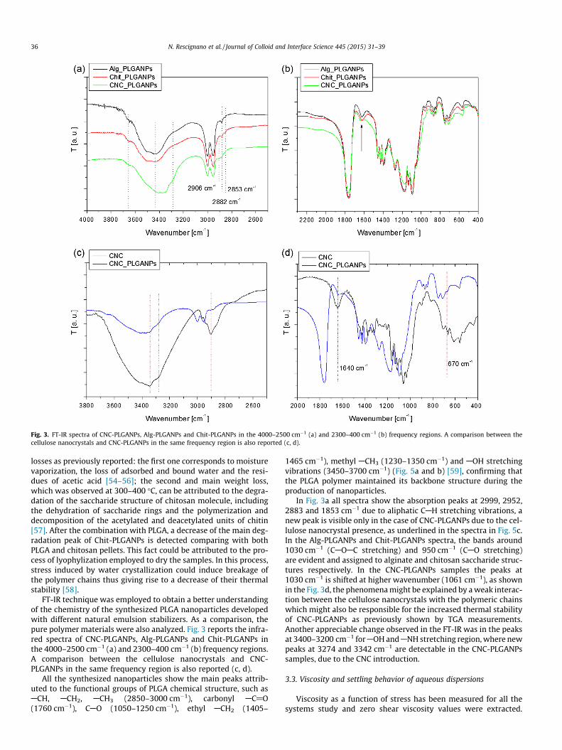
Fig. 3. FT-IR spectra of CNC-PLGANPs, Alg-PLGANPs and Chit-PLGANPs in the 4000–2500 cm�1 (a) and 2300–400 cm�1 (b) frequency regions. A comparison between thecellulose nanocrystals and CNC-PLGANPs in the same frequency region is also reported (c, d).
36 N. Rescignano et al. / Journal of Colloid and Interface Science 445 (2015) 31–39
losses as previously reported: the first one corresponds to moisturevaporization, the loss of adsorbed and bound water and the resi-dues of acetic acid [54–56]; the second and main weight loss,which was observed at 300–400 �C, can be attributed to the degra-dation of the saccharide structure of chitosan molecule, includingthe dehydration of saccharide rings and the polymerization anddecomposition of the acetylated and deacetylated units of chitin[57]. After the combination with PLGA, a decrease of the main deg-radation peak of Chit-PLGANPs is detected comparing with bothPLGA and chitosan pellets. This fact could be attributed to the pro-cess of lyophylization employed to dry the samples. In this process,stress induced by water crystallization could induce breakage ofthe polymer chains thus giving rise to a decrease of their thermalstability [58].
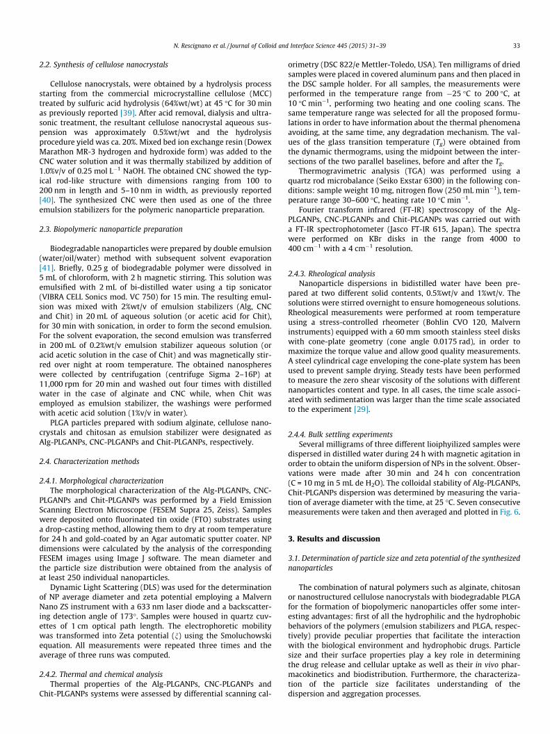
FT-IR technique was employed to obtain a better understandingof the chemistry of the synthesized PLGA nanoparticles developedwith different natural emulsion stabilizers. As a comparison, thepure polymer materials were also analyzed. Fig. 3 reports the infra-red spectra of CNC-PLGANPs, Alg-PLGANPs and Chit-PLGANPs inthe 4000–2500 cm�1 (a) and 2300–400 cm�1 (b) frequency regions.A comparison between the cellulose nanocrystals and CNC-PLGANPs in the same frequency region is also reported (c, d).
All the synthesized nanoparticles show the main peaks attrib-uted to the functional groups of PLGA chemical structure, such asACH, ACH2, ACH3 (2850–3000 cm�1), carbonyl AC@O(1760 cm�1), CAO (1050–1250 cm�1), ethyl ACH2 (1405–
1465 cm�1), methyl ACH3 (1230–1350 cm�1) and AOH stretchingvibrations (3450–3700 cm�1) (Fig. 5a and b) [59], confirming thatthe PLGA polymer maintained its backbone structure during theproduction of nanoparticles.
In Fig. 3a all spectra show the absorption peaks at 2999, 2952,2883 and 1853 cm�1 due to aliphatic CAH stretching vibrations, anew peak is visible only in the case of CNC-PLGANPs due to the cel-lulose nanocrystal presence, as underlined in the spectra in Fig. 5c.In the Alg-PLGANPs and Chit-PLGANPs spectra, the bands around1030 cm�1 (CAOAC stretching) and 950 cm�1 (CAO stretching)are evident and assigned to alginate and chitosan saccharide struc-tures respectively. In the CNC-PLGANPs samples the peaks at1030 cm�1 is shifted at higher wavenumber (1061 cm�1), as shownin the Fig. 3d, the phenomena might be explained by a weak interac-tion between the cellulose nanocrystals with the polymeric chainswhich might also be responsible for the increased thermal stabilityof CNC-PLGANPs as previously shown by TGA measurements.Another appreciable change observed in the FT-IR was in the peaksat 3400–3200 cm�1 for AOH and ANH stretching region, where newpeaks at 3274 and 3342 cm�1 are detectable in the CNC-PLGANPssamples, due to the CNC introduction.
3.3. Viscosity and settling behavior of aqueous dispersions
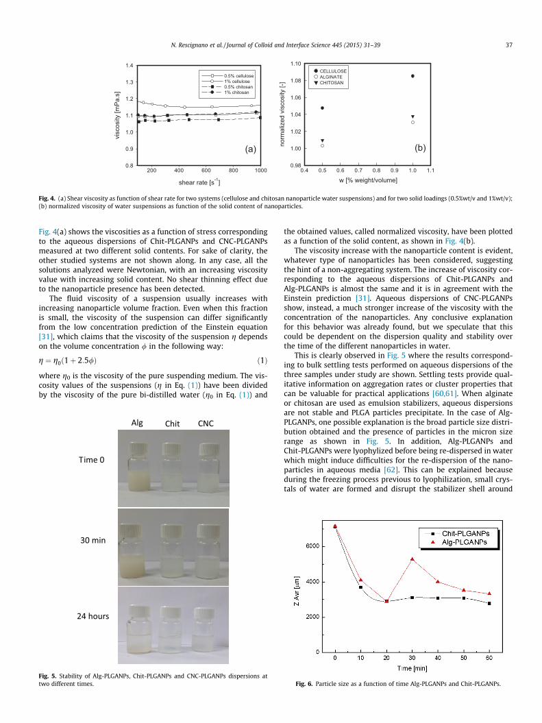
Viscosity as a function of stress has been measured for all thesystems study and zero shear viscosity values were extracted.
w [% weight/volume]0.4 0.5 0.6 0.7 0.8 0.9 1.0 1.1
norm
aliz
ed v
isco
sity
[-]
0.98
1.00
1.02
1.04
1.06
1.08
1.10CELLULOSEALGINATECHITOSAN
shear rate [s-1]
200 400 600 800 1000
visc
osity
[mP
a.s]
0.8
0.9
1.0
1.1
1.2
1.3
1.4
0.5% cellulose1% cellulose0.5% chitosan1% chitosan
(b)(a)
Fig. 4. (a) Shear viscosity as function of shear rate for two systems (cellulose and chitosan nanoparticle water suspensions) and for two solid loadings (0.5%wt/v and 1%wt/v);(b) normalized viscosity of water suspensions as function of the solid content of nanoparticles.
N. Rescignano et al. / Journal of Colloid and Interface Science 445 (2015) 31–39 37
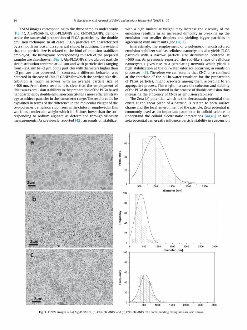
Fig. 4(a) shows the viscosities as a function of stress correspondingto the aqueous dispersions of Chit-PLGANPs and CNC-PLGANPsmeasured at two different solid contents. For sake of clarity, theother studied systems are not shown along. In any case, all thesolutions analyzed were Newtonian, with an increasing viscosityvalue with increasing solid content. No shear thinning effect dueto the nanoparticle presence has been detected.
The fluid viscosity of a suspension usually increases withincreasing nanoparticle volume fraction. Even when this fractionis small, the viscosity of the suspension can differ significantlyfrom the low concentration prediction of the Einstein equation[31], which claims that the viscosity of the suspension g dependson the volume concentration / in the following way:
g ¼ g0ð1þ 2:5/Þ ð1Þ
where g0 is the viscosity of the pure suspending medium. The vis-cosity values of the suspensions (g in Eq. (1)) have been dividedby the viscosity of the pure bi-distilled water (g0 in Eq. (1)) and
Alg Chit CNC
Time 0
30 min
24 hours
Fig. 5. Stability of Alg-PLGANPs, Chit-PLGANPs and CNC-PLGANPs dispersions attwo different times.
the obtained values, called normalized viscosity, have been plottedas a function of the solid content, as shown in Fig. 4(b).
The viscosity increase with the nanoparticle content is evident,whatever type of nanoparticles has been considered, suggestingthe hint of a non-aggregating system. The increase of viscosity cor-responding to the aqueous dispersions of Chit-PLGANPs andAlg-PLGANPs is almost the same and it is in agreement with theEinstein prediction [31]. Aqueous dispersions of CNC-PLGANPsshow, instead, a much stronger increase of the viscosity with theconcentration of the nanoparticles. Any conclusive explanationfor this behavior was already found, but we speculate that thiscould be dependent on the dispersion quality and stability overthe time of the different nanoparticles in water.
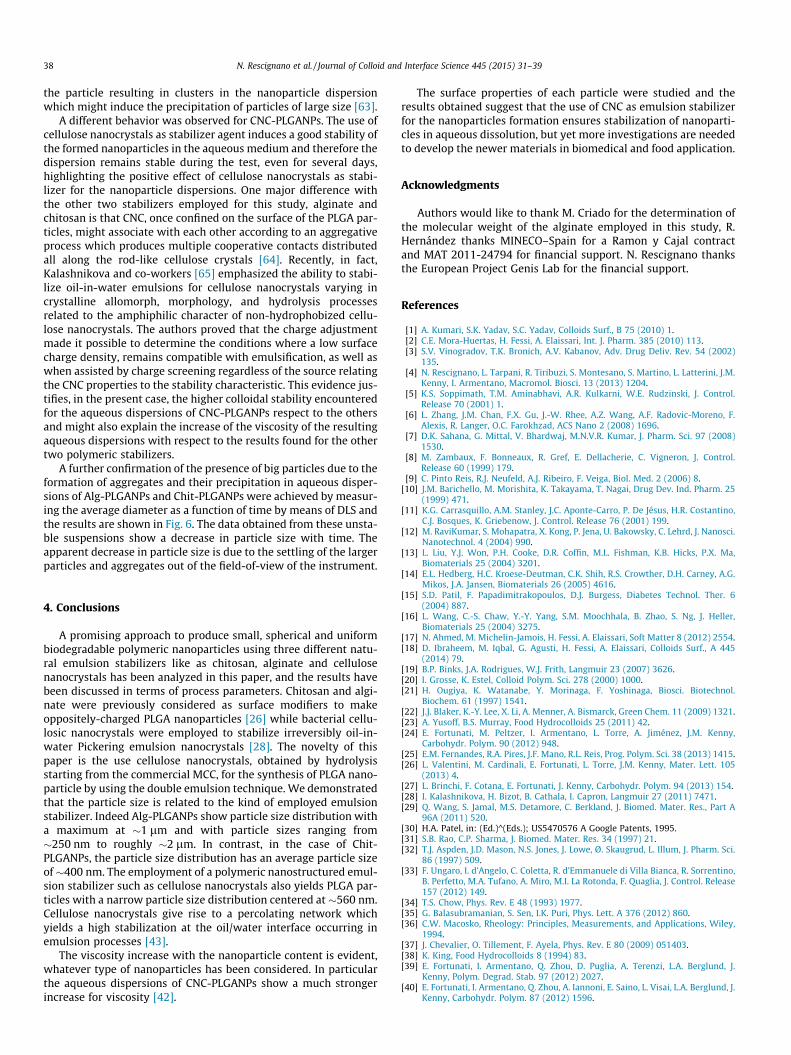
This is clearly observed in Fig. 5 where the results correspond-ing to bulk settling tests performed on aqueous dispersions of thethree samples under study are shown. Settling tests provide qual-itative information on aggregation rates or cluster properties thatcan be valuable for practical applications [60,61]. When alginateor chitosan are used as emulsion stabilizers, aqueous dispersionsare not stable and PLGA particles precipitate. In the case of Alg-PLGANPs, one possible explanation is the broad particle size distri-bution obtained and the presence of particles in the micron sizerange as shown in Fig. 5. In addition, Alg-PLGANPs andChit-PLGANPs were lyophylized before being re-dispersed in waterwhich might induce difficulties for the re-dispersion of the nano-particles in aqueous media [62]. This can be explained becauseduring the freezing process previous to lyophilization, small crys-tals of water are formed and disrupt the stabilizer shell around
Fig. 6. Particle size as a function of time Alg-PLGANPs and Chit-PLGANPs.
38 N. Rescignano et al. / Journal of Colloid and Interface Science 445 (2015) 31–39
the particle resulting in clusters in the nanoparticle dispersionwhich might induce the precipitation of particles of large size [63].
A different behavior was observed for CNC-PLGANPs. The use ofcellulose nanocrystals as stabilizer agent induces a good stability ofthe formed nanoparticles in the aqueous medium and therefore thedispersion remains stable during the test, even for several days,highlighting the positive effect of cellulose nanocrystals as stabi-lizer for the nanoparticle dispersions. One major difference withthe other two stabilizers employed for this study, alginate andchitosan is that CNC, once confined on the surface of the PLGA par-ticles, might associate with each other according to an aggregativeprocess which produces multiple cooperative contacts distributedall along the rod-like cellulose crystals [64]. Recently, in fact,Kalashnikova and co-workers [65] emphasized the ability to stabi-lize oil-in-water emulsions for cellulose nanocrystals varying incrystalline allomorph, morphology, and hydrolysis processesrelated to the amphiphilic character of non-hydrophobized cellu-lose nanocrystals. The authors proved that the charge adjustmentmade it possible to determine the conditions where a low surfacecharge density, remains compatible with emulsification, as well aswhen assisted by charge screening regardless of the source relatingthe CNC properties to the stability characteristic. This evidence jus-tifies, in the present case, the higher colloidal stability encounteredfor the aqueous dispersions of CNC-PLGANPs respect to the othersand might also explain the increase of the viscosity of the resultingaqueous dispersions with respect to the results found for the othertwo polymeric stabilizers.
A further confirmation of the presence of big particles due to theformation of aggregates and their precipitation in aqueous disper-sions of Alg-PLGANPs and Chit-PLGANPs were achieved by measur-ing the average diameter as a function of time by means of DLS andthe results are shown in Fig. 6. The data obtained from these unsta-ble suspensions show a decrease in particle size with time. Theapparent decrease in particle size is due to the settling of the largerparticles and aggregates out of the field-of-view of the instrument.
4. Conclusions
A promising approach to produce small, spherical and uniformbiodegradable polymeric nanoparticles using three different natu-ral emulsion stabilizers like as chitosan, alginate and cellulosenanocrystals has been analyzed in this paper, and the results havebeen discussed in terms of process parameters. Chitosan and algi-nate were previously considered as surface modifiers to makeoppositely-charged PLGA nanoparticles [26] while bacterial cellu-losic nanocrystals were employed to stabilize irreversibly oil-in-water Pickering emulsion nanocrystals [28]. The novelty of thispaper is the use cellulose nanocrystals, obtained by hydrolysisstarting from the commercial MCC, for the synthesis of PLGA nano-particle by using the double emulsion technique. We demonstratedthat the particle size is related to the kind of employed emulsionstabilizer. Indeed Alg-PLGANPs show particle size distribution witha maximum at �1 lm and with particle sizes ranging from�250 nm to roughly �2 lm. In contrast, in the case of Chit-PLGANPs, the particle size distribution has an average particle sizeof�400 nm. The employment of a polymeric nanostructured emul-sion stabilizer such as cellulose nanocrystals also yields PLGA par-ticles with a narrow particle size distribution centered at �560 nm.Cellulose nanocrystals give rise to a percolating network whichyields a high stabilization at the oil/water interface occurring inemulsion processes [43].
The viscosity increase with the nanoparticle content is evident,whatever type of nanoparticles has been considered. In particularthe aqueous dispersions of CNC-PLGANPs show a much strongerincrease for viscosity [42].
The surface properties of each particle were studied and theresults obtained suggest that the use of CNC as emulsion stabilizerfor the nanoparticles formation ensures stabilization of nanoparti-cles in aqueous dissolution, but yet more investigations are neededto develop the newer materials in biomedical and food application.
Acknowledgments
Authors would like to thank M. Criado for the determination ofthe molecular weight of the alginate employed in this study, R.Hernández thanks MINECO–Spain for a Ramon y Cajal contractand MAT 2011-24794 for financial support. N. Rescignano thanksthe European Project Genis Lab for the financial support.
References
[1] A. Kumari, S.K. Yadav, S.C. Yadav, Colloids Surf., B 75 (2010) 1.[2] C.E. Mora-Huertas, H. Fessi, A. Elaissari, Int. J. Pharm. 385 (2010) 113.[3] S.V. Vinogradov, T.K. Bronich, A.V. Kabanov, Adv. Drug Deliv. Rev. 54 (2002)
135.[4] N. Rescignano, L. Tarpani, R. Tiribuzi, S. Montesano, S. Martino, L. Latterini, J.M.
Kenny, I. Armentano, Macromol. Biosci. 13 (2013) 1204.[5] K.S. Soppimath, T.M. Aminabhavi, A.R. Kulkarni, W.E. Rudzinski, J. Control.
Release 70 (2001) 1.[6] L. Zhang, J.M. Chan, F.X. Gu, J.-W. Rhee, A.Z. Wang, A.F. Radovic-Moreno, F.
Alexis, R. Langer, O.C. Farokhzad, ACS Nano 2 (2008) 1696.[7] D.K. Sahana, G. Mittal, V. Bhardwaj, M.N.V.R. Kumar, J. Pharm. Sci. 97 (2008)
1530.[8] M. Zambaux, F. Bonneaux, R. Gref, E. Dellacherie, C. Vigneron, J. Control.
Release 60 (1999) 179.[9] C. Pinto Reis, R.J. Neufeld, A.J. Ribeiro, F. Veiga, Biol. Med. 2 (2006) 8.
[10] J.M. Barichello, M. Morishita, K. Takayama, T. Nagai, Drug Dev. Ind. Pharm. 25(1999) 471.
[11] K.G. Carrasquillo, A.M. Stanley, J.C. Aponte-Carro, P. De Jésus, H.R. Costantino,C.J. Bosques, K. Griebenow, J. Control. Release 76 (2001) 199.
[12] M. RaviKumar, S. Mohapatra, X. Kong, P. Jena, U. Bakowsky, C. Lehrd, J. Nanosci.Nanotechnol. 4 (2004) 990.
[13] L. Liu, Y.J. Won, P.H. Cooke, D.R. Coffin, M.L. Fishman, K.B. Hicks, P.X. Ma,Biomaterials 25 (2004) 3201.
[14] E.L. Hedberg, H.C. Kroese-Deutman, C.K. Shih, R.S. Crowther, D.H. Carney, A.G.Mikos, J.A. Jansen, Biomaterials 26 (2005) 4616.
[15] S.D. Patil, F. Papadimitrakopoulos, D.J. Burgess, Diabetes Technol. Ther. 6(2004) 887.
[16] L. Wang, C.-S. Chaw, Y.-Y. Yang, S.M. Moochhala, B. Zhao, S. Ng, J. Heller,Biomaterials 25 (2004) 3275.
[17] N. Ahmed, M. Michelin-Jamois, H. Fessi, A. Elaissari, Soft Matter 8 (2012) 2554.[18] D. Ibraheem, M. Iqbal, G. Agusti, H. Fessi, A. Elaissari, Colloids Surf., A 445
(2014) 79.[19] B.P. Binks, J.A. Rodrigues, W.J. Frith, Langmuir 23 (2007) 3626.[20] I. Grosse, K. Estel, Colloid Polym. Sci. 278 (2000) 1000.[21] H. Ougiya, K. Watanabe, Y. Morinaga, F. Yoshinaga, Biosci. Biotechnol.
Biochem. 61 (1997) 1541.[22] J.J. Blaker, K.-Y. Lee, X. Li, A. Menner, A. Bismarck, Green Chem. 11 (2009) 1321.[23] A. Yusoff, B.S. Murray, Food Hydrocolloids 25 (2011) 42.[24] E. Fortunati, M. Peltzer, I. Armentano, L. Torre, A. Jiménez, J.M. Kenny,
Carbohydr. Polym. 90 (2012) 948.[25] E.M. Fernandes, R.A. Pires, J.F. Mano, R.L. Reis, Prog. Polym. Sci. 38 (2013) 1415.[26] L. Valentini, M. Cardinali, E. Fortunati, L. Torre, J.M. Kenny, Mater. Lett. 105
(2013) 4.[27] L. Brinchi, F. Cotana, E. Fortunati, J. Kenny, Carbohydr. Polym. 94 (2013) 154.[28] I. Kalashnikova, H. Bizot, B. Cathala, I. Capron, Langmuir 27 (2011) 7471.[29] Q. Wang, S. Jamal, M.S. Detamore, C. Berkland, J. Biomed. Mater. Res., Part A
96A (2011) 520.[30] H.A. Patel, in: (Ed.)^(Eds.); US5470576 A Google Patents, 1995.[31] S.B. Rao, C.P. Sharma, J. Biomed. Mater. Res. 34 (1997) 21.[32] T.J. Aspden, J.D. Mason, N.S. Jones, J. Lowe, Ø. Skaugrud, L. Illum, J. Pharm. Sci.
86 (1997) 509.[33] F. Ungaro, I. d’Angelo, C. Coletta, R. d’Emmanuele di Villa Bianca, R. Sorrentino,
B. Perfetto, M.A. Tufano, A. Miro, M.I. La Rotonda, F. Quaglia, J. Control. Release157 (2012) 149.
[34] T.S. Chow, Phys. Rev. E 48 (1993) 1977.[35] G. Balasubramanian, S. Sen, I.K. Puri, Phys. Lett. A 376 (2012) 860.[36] C.W. Macosko, Rheology: Principles, Measurements, and Applications, Wiley,
1994.[37] J. Chevalier, O. Tillement, F. Ayela, Phys. Rev. E 80 (2009) 051403.[38] K. King, Food Hydrocolloids 8 (1994) 83.[39] E. Fortunati, I. Armentano, Q. Zhou, D. Puglia, A. Terenzi, L.A. Berglund, J.
Kenny, Polym. Degrad. Stab. 97 (2012) 2027.[40] E. Fortunati, I. Armentano, Q. Zhou, A. Iannoni, E. Saino, L. Visai, L.A. Berglund, J.
Kenny, Carbohydr. Polym. 87 (2012) 1596.
N. Rescignano et al. / Journal of Colloid and Interface Science 445 (2015) 31–39 39
[41] N. Rescignano, M. Amelia, A. Credi, J. Kenny, I. Armentano, Eur. Polymer J. 48(2012) 1152.
[42] Y.-Y. Yang, T.-S. Chung, N. Ping, Biomaterials 22 (2001) 231.[43] I. Capron, B. Cathala, Biomacromolecules 14 (2013) 291.[44] R.J. Hunter, Zeta Potential in Colloid Science. Principles and Applications,
Academic Press, 1981.[45] Y. Zhang, M. Yang, J.-H. Park, J. Singelyn, H. Ma, M.J. Sailor, E. Ruoslahti, M.
Ozkan, C. Ozkan, Small 5 (2009) 1990.[46] M.V. Lorevice, M.R.d. Moura, F.A. Aouada, L.H.C. Mattoso, J. Nanosci.
Nanotechnol. 12 (2012) 2711.[47] S.H. Ching, B. Bhandari, R. Webb, N. Bansal, Food Hydrocolloids 43 (2015) 165.[48] M. Yan, S. Li, M. Zhang, C. Li, F. Dong, W. Li, BioResources 8 (2013) 6330.[49] P.B. Filson, B.E. Dawson-Andoh, D. Schwegler-Berry, Green Chem. 11 (2009)
1808.[50] H. Kargarzadeh, I. Ahmad, I. Abdullah, A. Dufresne, S. Zainudin, R. Sheltami,
Cellulose 19 (2012) 855.[51] D. Bondeson, A. Mathew, K. Oksman, Cellulose 13 (2006) 171.[52] X. Dong, J.-F. Revol, D. Gray, Cellulose 5 (1998) 19.[53] M. Roman, W.T. Winter, Biomacromolecules 5 (2004) 1671.
[54] S. Tripathi, G. Mehrotra, P. Dutta, Int. J. Biol. Macromol. 45 (2009) 372.[55] C.-H. Chen, F.-Y. Wang, C.-F. Mao, W.-T. Liao, C.-D. Hsieh, Int. J. Biol. Macromol.
43 (2008) 37.[56] K. Lewandowska, Thermochim. Acta 493 (2009) 42.[57] A.T. Paulino, J.I. Simionato, J.C. Garcia, J. Nozaki, Carbohydr. Polym. 64 (2006)
98.[58] W. Abdelwahed, G. Degobert, H. Fessi, Int. J. Pharm. 309 (2006) 178.[59] N. Jalali, F. Moztarzadeh, M. Mozafari, S. Asgari, M. Motevalian, S.N. Alhosseini,
Colloids Surf., A 392 (2011) 335.[60] C.G. Serrano, J.J. McDermott, D. Velegol, Nat. Mater. 10 (2011) 716.[61] S. Witharana, C. Hodges, D. Xu, X. Lai, Y. Ding, J. Nanopart. Res. 14 (2012) 851.[62] A. Gursoy, L. Eroglu, S. Ulutin, M. Tasyurek, H. Fessi, F. Puisieux, J.P.
Devissaguet, Int. J. Pharm. 52 (1989) 101.[63] Y.N. Konan, R. Gurny, E. Allémann, Int. J. Pharm. 233 (2002) 239.[64] A.J. Svagan, A. Musyanovych, M. Kappl, M. Bernhardt, G. Glasser, C. Wohnhaas,
L.A. Berglund, J. Risbo, K. Landfester, Biomacromolecules 15 (2014) 1852.[65] I. Kalashnikova, H. Bizot, B. Cathala, I. Capron, Biomacromolecules 13 (2011)
267.
Copyright © 2022 FDOKUMEN