Metabolic significance of inorganic triphosphate, thiamine ...
190
Metabolic significance of inorganic triphosphate, thiamine triphosphate and their hydrolyzing enzymes Mémoire présenté par Grégory Kohn, Titulaire d’un Master en Sciences Biomédicales, en vue de l’obtention du grade de Docteur en Sciences Biomédicales et Pharmaceutiques ____________________________________________________ Année académique 2015-2016 Université de Liège Faculté de Médecine GIGA-Neurosciences Laboratoire du Vieillissement Pathologique et Epilepsies Professeur Lucien Bettendorff
-
Upload
khangminh22 -
Category
Documents
-
view
3 -
download
0
Transcript of Metabolic significance of inorganic triphosphate, thiamine ...

Metabolic significance of inorganic
triphosphate, thiamine triphosphate and their
hydrolyzing enzymes
Mémoire présenté par Grégory Kohn,
Titulaire d’un Master en Sciences Biomédicales,
en vue de l’obtention du grade de Docteur en Sciences Biomédicales et
Pharmaceutiques
____________________________________________________
Année académique 2015-2016
Université de Liège
Faculté de Médecine
GIGA-Neurosciences
Laboratoire du Vieillissement Pathologique et Epilepsies
Professeur Lucien Bettendorff


“La Science n’est plus à même de fournir aucune certitude, mais des propositions temporaires
qui se métamorphoseront aussi vite que nos certitudes d’hier.”
Ilya Prigogine
(1917-2003, physicien Belge, Prix Nobel de Chimie en 1977)
Cover picture modified from : Okuno D., Iino R., Noji H. 2011. Rotation and structure of FoF1-ATP synthase. J.
Biochem. 149 (6) 655-64. doi: 10.1093/jb/mvr049.


Abstract
Our laboratory has been interested for many years in thiamine triphosphate (ThTP), an
unusual triphosphate derivative of thiamine (vitamin B1) found in nearly all organisms. In
mammalian tissues, ThTP is hydrolyzed by a very specific cytosolic thiamine triphosphatase
(ThTPase) belonging to an ancient superfamily of proteins called CYTH. Several members of
this superfamily have been characterized and they all have in common that they act on
triphosphorylated substrates. Some bacterial members (the N. europeae specifically) hydrolyze
inorganic triphosphate (PPPi) which raises the question of the physiological significance of this
compound.
We first studied the tripolyphosphatase activity in mammals and in bacteria and we
showed that it is widely distributed in all organisms. We attempted to identify the enzymes
responsible for this activity. We showed that the E. coli CYTH enzyme, ygiF, although highly
specific for PPPi plays only a minor role in the cytosolic PPPase activity, while most of the
activity is due to inorganic pyrophosphatase. In animal tissues, most PPPase activity is due to the
short-chain exopolyphosphatase prune, which hydrolyzes PPPi with high catalytic efficiency. We
hypothesize that PPPi may be formed as a by-product of metabolism and the major role of PPPase
activities may be to keep PPPi concentrations very low to avoid toxic effects linked to
interference with Ca2+
metabolism. In order to check this hypothesis, it would be important to
have a specific and sensitive method for measuring intracellular PPPi concentrations. Such a
technique is presently not available.
In the second part of our work, we tried to manipulate intracellular ThTP concentrations
in order to get insight into the physiological role of this compound. Expression of mammalian
ThTPase in E. coli prevented ThTP accumulation, but did not affect the growth of the bacteria.
We then reduced ThTPase expression in zebrafish embryos using morpholino oligomers. This led
to severe malformations of the embryos. Finally, we attempted to produce a ThTPase knockout
mouse. However, we found that the spermatogenesis of ThTPase-null sperm cells was impaired
and the chimerae were unable to transmit. In conclusion, our results suggest that ThTPase
inactivation results in heavy developmental consequences, possibly as a result of ThTP toxicity.


List of abbreviations
List of abbreviations :
ADP Adenosine diphosphate
AMP Adenosine monophosphate
AThDP Adenosine thiamine diphosphate
AThTP Adenosine thiamine triphosphate
ATP Adenosine triphosphate
CoA Coenzyme A
CYTH CyaB-Thiamine triphosphatase
E1o 2-Oxoglutarate dehydrogense
E1p Pyruvate dehydrogenase
E2o Dihydrolipoamide succinyltransferase
E2p Dihydrolipoamide acetyltransferase
E3 Dihydrolipoamide dehydrogenase
EDTA Ethylenediaminetetraacetic acid
ES cell Embryonnic stem cell
GC Glycine cleage complex
Hpf Hour post-fertilization
IPTG Isopropyl β-D-1-thiogalactopyranoside
kcat Catalytic constant
Km Michaelis constant
KOMP Knockout Mouse Project
LC-ESI/MS/MS Liquid chromatography Electrospray ionization mass spectrometry
NAD Nicotinamide adenine dinucleotide
NADP Nicotinamide adenine dinucleotide phosphate
NCBI National Center for Biotechnology Information
NMP Nucleotide monophosphate
NMR Nuclear magnetic resonance
NTP Nucleotide triphosphate
OGDH 2-Oxoglutarate dehydrogenase complex
PCR Polymerase chain reaction
PDH Pyruvate dehydrogenase complex
Pi Inorganic phosphate
PolyP Inorganic polyphosphate
PPase Pyrophosphatase
PPi Inorganic diphosphate
PPK Polyphosphate kinase
PPPase Tripolyphosphatase
PPPi Inorganic triphosphate
PPX Exopolyphosphatase

List of abbreviations
qPCR quantitative PCR
SD Standard deviation
SDS-PAGE Sodium dodecyl sulfate Polyacrylamide gel electrophoresis
SL Standard length
SLC Solute carrier (gene)
SOC Swim-out - central part
SOP Swim-out - peripheral part
SUL Swim-up - lower part
SUS Swim-up - superior part
TCA Trichloroacetic acid
ThDP Thiamine diphosphate
ThMP Thiamine monophosphate
ThTP Thiamine triphosphate
ThTPase Thiamine triphosphatase
THTR Thiamine transporter (protein)
TTM Triphosphate tunnel metalloenzyme
WT Wild-type

Acknowledgements
Après 10 ans passés à écumer les couloirs, les amphis et les
laboratoires de l’université, il y a beaucoup de personne que je me
dois de remercier pour le soutien qu’ils m’ont apporté de différentes
manières. J’oublierai sûrement de citer certains d’entre eux mais ils
savent qu’ils sont malgré tout dans mes pensées.
Tout d’abord, bien entendu, je tiens à remercier ma famille
et surtout mes parents à qui revient tout le mérite de mon parcours,
même si je ne leur ai probablement pas souvent fait ressentir. Et un
merci tout spécial à mon grand-père qui n’est malheureusement plus
là pour assister à l’aboutissement de cette entreprise.
Il y a quelques personnes à qui mon parcours scientifique
doit énormément. Tout d’abord, le Pr Lucien Bettendorff qui m’a
conseillé et guidé au cours de toutes mes années de doctorat ; le Dr
Pierre Wins, la bibliothèque vivante perpétuellement remise à jour
que tout jeune chercheur devrait avoir la chance de rencontrer ; le
Dr Bernard Lakaye et ses précieux conseils qui m’ont permis
d’éviter nombre de pièges au cours de ces cinq années et Ilca pour
sa gentillesse.
Ensuite, bien entendu, le Dr David Delvaux pour m’avoir
appris les bases du travail scientifique et pour toujours avoir été de
bon conseil quand je débutais ma carrière. Egalement les Dr
Marjorie Gangolf et Tiziana Gigliobinaco avec qui j’ai partagé bien
plus qu’un bureau lors de mes premiers pas de chercheur et Arlette
Minet et son esprit d’aventure.
Un merci spécial va ensuite à Julie Vignisse avec qui j’ai
partagé pas mal des joies et des peines survenues aux cours de mon
master d’abord et de ma thèse ensuite. Et bien sûr, un grand merci à
Coralie Pasquet pour les pauses-café et Margaux Sambon pour ses
“Greeeeg” présageant toujours une question. Une dédicace spéciale

Acknowledgements
à mon admiratrice secrète pour avoir refait la décoration du bureau
et pour son “ théobromine-tactisme” exacerbé.
Je tiens aussi à remercier les Dr Laurence de Nijs et
Nathalie Wolkoff pour avoir contribué à la bonne atmosphère de
notre équipe ainsi que Sophie Harray qui a partagé avec moi une
première expérience d’élevage de N.A.C. et Laurie Medard pour sa
folie et sa bonne humeur.
Je tiens à remercier tous les étudiants qui ont transité pour
quelques mois par notre labo et tout spécialement Franciska, pour
m’avoir fait découvrir et repousser les limites de la patience, ainsi
que Cindy pour m’avoir rendu foi dans le travail fourni par la
nouvelle génération d’étudiant.
Je ne peux pas oublier non plus ceux avec qui j’ai collaboré,
Marc Muller et tous les membres de l’équipe du Laboratoire de
Biologie Moléculaire et Génie Génétique, surtout Marie Winandy
pour son écolage ; les membres du Laboratoire de Génétique et de
Physiologie Bactérienne de l’ULB, surtout Johan Timmermans pour
son aide, et enfin Fabien Ectors pour la production et le croisement
de nos chimères.
Enfin, merci à ceux qui étaient là avant, pendant et seront
toujours là après tout ça, Pierre et Laulau, Bruno, Jen et Benoit,
Loïc, Léon et Cath.
And last but not least, un grand merci aux membres du
GIGA-Neurosciences : tout d’abord aux techniciens Bernard,
Alexandra, Alice, Laurent, P-B, les Patricia et Guérin qui ont
toujours été là pour répondre à mes questions ; ensuite à nos
dévouées secrétaires Lari et Jess sur qui on peut toujours compter ;

Acknowledgements
Et enfin à tous les autres, doctorant, PI ou autres qui ont fait et font
du centre ce qu’il est.
I have now to switch to English to thank many people that
were or were not already cited in the first part. To all of you who
will be included in the next part, thank you for the different
experiences and opinions shared and for the free English class. As I
have always said: sorry to fail as a local for not teaching you any
French and instead using you to improve my English.
First, I want to thank all the “expats” from the GIGA-
Neurosciences, and specially the two “douche” Arash and Deb for
all the good time and Sita and Asya for all the good music, live or
on vinyl.
Also all the others members of the so called “Liege Expats
group” with a special thanks to Sophie who made me an expat in
my own city. Thank you all for the trips, parties, pic-nics, museum
visit and so on and so forth that we’ve made together.
I also want to thank my climbing partners Todd, Katya,
Rohan, Amy, Stephane, Brett and Cyrine; nothing is as good as
getting high (15 meters high) with nice people after a good day of
work.
“But standing in water up to their knees three very unimportant
little people were doing the best they could with an idea and that’s
about all they had and I don’t know maybe that night these three
people were making history but anyway what I’m telling to tell you
folks is: here we are!” J.Steinbeck. –We all had many discussions
that made me think about that sentence, sharing our experience all
around the globe.

Acknowledgements
I also want to thanks all the members of the” Institut für
Physiologie und Pathophysiologie” in Heidelberg for their warm
welcome. First, the Prof. Dr.med. Andreas Draguhn who made it
possible, then a special thinking for Azra who taught me most of
what I know about electrophysiology and Antonio because my stay
there would never had been the same without him. Of course I also
want to thanks all the others: Jana, Xiao-min, Cédric Rachel,
Pascal, Christian, Ismini, Joshua, Dimitri, Alistair, Miguel, Suse
and Pierre and all the others I forgot.
“No one looks back on their life and remembers the nights they
got plenty of sleep”

Table of content
Introduction
Chapter 1: Thiamine and its phosphorylated derivatives ................................................................ 1
Chemistry: ................................................................................................................................... 1
Physiology: .................................................................................................................................. 1
Different derivatives: ................................................................................................................... 3
Thiamine diphosphate: ...................................................................................................................... 3
Thiamine monophosphate: ................................................................................................................ 5
Thiamine triphosphate ....................................................................................................................... 5
Adenylated thiamine derivatives ....................................................................................................... 6
ThTP production in E. coli: ......................................................................................................... 6
Chapter 2: The mammalian 25-kDa ThTPase: ................................................................................ 8
Discovery and characterization ................................................................................................... 8
Phylogeny .................................................................................................................................... 8
Catalytic properties .................................................................................................................... 10
Translational control of the expression ..................................................................................... 11
Chapter 3: The inorganic polyphosphates ..................................................................................... 13
Nomenclature: ........................................................................................................................... 14
Synthesis and degradation: ........................................................................................................ 14
Function: .................................................................................................................................... 15
Energy and Pi storage: ..................................................................................................................... 15
Regulatory role in mammals ........................................................................................................... 15
Chapter 4: The zebrafish................................................................................................................ 16
Phylogeny: ................................................................................................................................. 16
Description: ............................................................................................................................... 16
Growth and mortality: ............................................................................................................... 17
Reproduction: ............................................................................................................................ 17
Genetics: .................................................................................................................................... 17

Table of content
The zebrafish as a tool for the scientist ...................................................................................... 17
CYTH in fish .............................................................................................................................. 18
Morpholino: ............................................................................................................................... 21
Anti-sense knockdown of gene expression: ................................................................................... 21
Aims of the work
Aims o the work ............................................................................................................................. 24
Material and methods
Material .......................................................................................................................................... 25
Solvents and Reagent ................................................................................................................. 25
Morpholino solution ................................................................................................................... 25
Zebrafish solution ...................................................................................................................... 26
Bacteria culture medium: ........................................................................................................... 26
Strains of E. coli used: ............................................................................................................... 26
Methods .......................................................................................................................................... 28
Determination of the tripolyphosphatase activity ...................................................................... 28
Collection of the sample ................................................................................................................. 28
Measurement of the activity ........................................................................................................... 28
Pig brain PPPase activity identification ..................................................................................... 28
E. coli PPPase activity identification ......................................................................................... 29
In-gel enzymatic activity ............................................................................................................ 30
Detection of thiamine and its derivatives in E. coli ................................................................... 30
Determination of acetyl-CoA levels .......................................................................................... 31
Polymerase chain reaction ......................................................................................................... 31
Insertion of the mammalian ThTPase coding sequence in the E.coli genome .......................... 33
Zebrafish maintenance ............................................................................................................... 34
Zebrafish breeding and eggs generation .................................................................................... 35
Morphant generation, characterization and maintenance .......................................................... 35
Zebrafish embryo RNA isolation ............................................................................................... 35
zThTPase mRNA cloning and expression ................................................................................. 36

Table of content
Mouse genotyping ..................................................................................................................... 36
Sperm isolation .......................................................................................................................... 36
Swim up .......................................................................................................................................... 37
Swim out ......................................................................................................................................... 37
Quantitative PCR: ...................................................................................................................... 38
Results
Chapter 1: Tripolyphosphatase activity ........................................................................................ 39
Tripolyphosphatase activity of mammalian tissues: .................................................................. 39
Characterization .............................................................................................................................. 39
Identification ................................................................................................................................... 39
Tripolyphosphatase activity of E. coli supernatant fraction: ..................................................... 41
ygiF is not the main tripolyphosphatase of E. coli .......................................................................... 41
Identification of the enzyme responsible for the tripolyphosphatase activity in E. coli: ................ 41
Characterization of the tripolyphosphatase activity of the EcPPase ............................................... 42
PPase, ygiF or PPX? ....................................................................................................................... 43
PPase and PPPase activities over growth of the bacteria ................................................................ 44
Conclusions: .............................................................................................................................. 44
Chapter 2: ThTP production in E. coli .......................................................................................... 47
ThTP synthesis and PDH mutants: ............................................................................................ 48
ThTP synthesis and OGDH mutant: .......................................................................................... 49
ThTP synthesis, OGDH mutant and glyoxylate cycle over-activation: .................................... 51
ThTP synthesis and the two first enzymatic activities of the Krebs cycle: ............................... 53
ThTP production and inhibition of the FoF1 ATP-synthase by octyl-α-cetoglutarate: .............. 54
Mammalian ThTPase expression in E. coli: .............................................................................. 55
ThTP synthesis and oxidative stress: ......................................................................................... 57
Conclusions ............................................................................................................................... 58
Chapter 3: Possible role of THTP and 25-kDa zThTPase on zebrafish development .................. 61
Characterization of the zTHTPA gene: ..................................................................................... 61
Morpholino-induced zThTPase expression reduction ............................................................... 63
HPLC measurement of thiamine derivatives in zebrafish embryo: ........................................... 70

Table of content
Conclusions ................................................................................................................................ 70
Chapter 4: Knock-out of the mouse ThTPase: ............................................................................... 72
Thiamine triphosphatase knock-out methods ............................................................................ 72
Results obtained from the THTPA chimerae: ............................................................................ 75
Conclusions ................................................................................................................................ 77
Discussion and conclusion
Possible biological role(s) of tripolyphosphate .............................................................................. 80
Role of ThTP and the 25-kDa ThTPase in animal tissues ............................................................. 81
Bibliography
Appendix 1
Delvaux D., Murty MR, Gabelica V., Lakaye B., Lunin VV, Skarina T.,
Onopriyenko O., Kohn G., Wins P., De Pauw E., Bettendorff L. 2011. A specific
inorganic triphosphatase from Nitrosomonas europaea: structure and catalytic
mechanism. J. Biol. Chem. 286 (39) 34023-35. doi: 10.1074/jbc.M111.233585.
Appendix 2
Kohn G., Delvaux D., Lakaye B., Servais AC, Scholer G., Fillet M., Elias B.,
Derochette JM, Crommen J., Wins P., Bettendorff L. 2012. High inorganic
triphosphatase activities in bacteria and mammalian cells: identification of
the enzymes involved. PLoS One. 7 (9) e43879. doi:
10.1371/journal.pone.0043879.
Appendix 3
Delvaux D., Kerff F., Murty MR, Lakaye B., Czerniecki J., Kohn G., Wins P.,
Herman R., Gabelica V., Heuze F., Tordoir X., Marée R., Matagne A., Charlier
P., De Pauw E., Bettendorff L., 2013. Structural determinants of specificity
and catalytic mechanism in mammalian 25-kDa thiamine triphosphatase. Biochim.
Biophys. Acta 1830 (10) 4513-23. doi: 10.1016/j.bbagen.2013.05.014.
Appendix 4
Bettendorff L., Lakaye B., Kohn G., Wins P. 2014. Thiamine triphosphate: a
ubiquitous molecule in search of a physiological role. Metab. Brain Dis. 29
(4) 1069-82. doi: 10.1007/s11011-014-9509-4.

Introduction


Introduction
1
Chapter 1: Thiamine and its phosphorylated derivatives
Thiamin (vitamin B1) was the first vitamin to be discovered and characterized as an
“antianeuric factor” more than a century ago. A vitamin is defined as an organic compound
required by humans in only small amounts and that they are unable to synthesize in sufficient
amounts. Hence, the main source of vitamins is the diet. There are a total of 13 vitamins
universally recognized, that are divided in two categories: the lipophilic vitamins (A, D, E, K)
and the water-soluble vitamins (B1, B2, B3, B5, B6, B7, B9, B12, C). All the B vitamins are
coenzymes (or coenzyme precursors) in metabolism of all living cells. For some of them, non-
coenzyme roles have also been suggested. This is the case for vitamin B1 (thiamine), a vitamin
essential for brain function. Our laboratory has been interested for many years in studying non-
cofactor roles of thiamine and its phosphorylated derivatives.
Chemistry:
Thiamine (2-{3-[(4-amino-2-methylpyrimidin-5-yl)methyl]-4-methylthiazol-5-yl}ethanol) is
composed of a pyrimidine ring linked to a thiazolium ring by a methylene bridge. The C5 of the
thiazolium ring bears an ethanol moiety that can be esterified, leading to the formation of
phosphate esters. Diphosphorylated thiamine (thiamine diphosphate, ThDP) is an important
cofactor in cell metabolism. Other phosphate esters are thiamine monophosphate (ThMP) and
thiamine triphosphate (ThTP), whose roles remain unknown. Recently, the existence of
adenylated thiamine derivatives, adenosine thiamine triphosphate (AThTP) and adenosine
thiamine diphosphate (AThDP) has been reported in our laboratory (Bettendorff et al. 2007)
(figure 1)
Physiology:
Thiamine can be synthetized by bacteria, fungi and plants (Manzetti et al. 2014) but not
by animals. Because of the positive charge in the thiazolium heterocycle and the polar character
of the molecule, thiamine does not diffuse freely through biological membranes and its transport
requires specific transporters. In mammals, thiamine is absorbed in the intestine by two specific
transporters: THTR 1 (Kd < 10 µM) and THTR 2 (Kd < 100 nm) encoded respectively by the
SLC19A2 and SLC19A3 genes. After crossing the intestinal epithelium, thiamine can reach the
blood stream and then be distributed into the whole body. In the blood, total thiamine is roughly
found for 75% in erythrocytes, 15% in leucocytes and 10% in plasma (Weber and Kewitz, 1985).

Introduction
2
R Name Short
H Thiamine Th
Pi Thiamine monophosphate ThMP
PPi Thiamine diphosphate ThDP
PPPi Thiamine triphosphate ThTP
ADP Adenosine thiamine diphosphate AThDP
ATP Adenosine thiamine triphosphate AThTP
Figure 1: thiamine and its derivative. A: thiamine and the different chemical groups naturally found attached to
the alcohol function; B: thiamine monophosphate (ThMP), thiamine diphosphate (ThDP), thiamine triphosphate
(ThTP), adenosine thiamine diphosphate (AThDP) and adenosine thiamine triphosphate (AThTP) (right panel from
Bettendorff, 2012).
In plasma, thiamine half-life is approximately one hour, then it has either been excreted by the
kidney or absorbed by the tissues (Tallaksen et al. 1993).
Once thiamine enters a mammalian cell, it is pyrophosphorylated to ThDP by a cytosolic
thiamine pyrophosphokinase (EC 2.7.6.2). ThDP is the cofactor for the cytoplasmic transketolase
and for the mitochondrial oxoglutarate and pyruvate dehydrogenases. ThDP is the precursor of
other thiamine derivative (figure 2), it can be:
hydrolyzed by a thiamine diphosphatase to thiamine monophosphate (ThMP),
which in turn can be hydrolyzed to thiamine by a thiamine monophoshatase.
Neither of these enzymes has been characterized to date.
phosphorylated in the mitochondria by the FoF1-ATP synthase (EC 3.6.3.14) to
form ThTP (Gangolf et al., 2010). ThTP in turn can be hydrolyzed to ThDP by the
25-kDa ThTPase (EC 3.6.1.28) in the cytoplasm.
converted to AThDP or AThTP by a ThDP-adenylyl transferase (EC 2.7.7.65),
depending whether the other substrate is ADP or ATP. They can be turned back
into ThDP and AMP by unknown hydrolases.
B A

Introduction
3
Figure 2: Thiamine and its derivatives in eukaryotic
cells. Thiamine diphosphate (ThDP) is formed by
pyrophosphorylation of thiamine. ThDP can bind to
cytosolic transketolase or be transported (in eukaryotic
cells) into mitochondria, where it can bind to pyruvate
dehydrogenase complex (PDH) or oxoglutarate
dehydrogenase complex (OGDH). Free ThDP is the
precursor for the synthesis of thiamine monophosphate
(ThMP), thiamine triphosphate (ThTP) and adenosine
thiamine triphosphate (AThTP). Hydrolysis of ThDP
yields ThMP, which can in turn be hydrolyzed to
thiamine. ThTP can be formed in the cytosol by
adenylate kinase and in mitochondria by a
chemiosmotic mechanism, probably involving FoF1-ATP
synthase. AThTP formation is catalyzed by a cytosolic
adenylyl thiamine diphosphate transferase, either from
ATP or from ADP. Similar reactions are involved in
thiamine metabolism in prokaryotes, except that the
mitochondrial part takes place in the cytosol.
1, thiamine pyrophosphokinase; 2, thiamine diphosphatase; 3, thiamine monophosphatase; 4, ThTP synthase
(could be identical to ATP-synthase); 5, adenylate kinase; 6, 25-kDa thiamine triphosphatase; 7, ThDP adenylyl
transferase (can use both ATP and ADP as substrate); 8, AThTP hydrolase. (from Bettendorff 2012).
Different derivatives:
Thiamine diphosphate:
Thiamine diphosphate is the main active form of the vitamin and accounts for 75-85% of
total thiamine content of most cells. After transport into the cells, thiamine is pyrophosphorylated
into ThDP according to the reaction: thiamine + ATP ThDP + AMP, further favoring thiamine
entry into the cell. The ThDP formed can then be transported into the mitochondria through the
SLC25A19 transporter which is an antiporter, probably exchanging cytoplasmic ThDP against
mitochondrial nucleotides (Lindhurst et al. 2006). In many cells, including neurons, most of the
ThDP is bound to apoenzymes. However, in erythrocytes, hepatocytes and skeletal muscle, a
larger part of ThDP is cytosolic and free (not bound to apoenzymes). Those tissues may therefore
act as storage compartments of the vitamin (in the form of ThDP) when extracellular thiamine
concentrations are high.

Introduction
4
Figure 3: catalytic mechanism of α-ketoacid decarboxylation by the cofactor ThDP. Decarboxylation is initiated by
formation of a C(2) carbanion of ThDP, that interacts with the C(2) of the substrate α-ketoacids to form a
nucleophilic adduct, followed by CO2 release and formation of the C(2α)-carbanion/enamine. Subsequent
protonation leads to the ‘active aldehyde’ intermediate whose metabolism depends on the specific enzyme
involved. Finally, decarboxylation with subsequent release of an aldehyde molecule is the main reaction of all α-
ketoacid decarboxylases. In the presence of a further aldehyde or α-ketoacid molecule acting as an acceptor, the
C(2α)-carbanion/enamine yields 2-hydroxyketones or hydroxyketoacids.
PDC: pyruvate decarboxylase, part of the ODH complex; BFD benzoylformate decarboxylase; IPDC indolpyruvate
decarboxylase (Malandrinos et al. 2006)
ThDP-dependent enzymes are found in almost every major metabolic pathway, they are
members of 9 superfamilies of enzymes. An overview of their annotated sequences can be found
in the ThDP-dependent Enzyme Engineering Database (TEED, http://www.teed.biocatnet.de).
Their main role is the decarboxylation of an α-ketoacid to an aldehyde and a CO2 molecule

Introduction
5
(RCOCOOH RCHO + CO2) (figure 3) whether by oxidative or non-oxidative mechanisms
(Malandrinos et al. 2006, Hailes et al. 2013).
Thiamine monophosphate:
Thiamine monophosphate does not have any known role in eukaryotes but in prokaryotes
it is an important intermediate in the synthesis of thiamine and ThDP. In mammalian cells it
amounts to 5 to 15% of total thiamine in the cells and it is the only phosphorylated thiamine
derivative found in plasma and in the cerebrospinal fluid.
In mammalian cells, ThMP is formed through enzymatic hydrolysis of ThDP but no
specific ThDPase has been characterized so far. Some phosphohydrolases have been shown to
hydrolyze ThDP but they are not specific for ThDP over nucleoside diphosphates. The same goes
for ThMPase, an activity is found in tissues and even used as a marker of small-diameter dorsal
root ganglia neurons, but no specific enzyme was characterized so far.
Thiamine triphosphate
Thiamine triphosphate was the first organic triphosphorylated compound other than
nucleotides to be discovered (Velluz et al. 1948). ThTP is found (generally in low amount) in all
mammals tissues but also in all major phyla tested (bacteria, fungi, plants and metazoa)
(Makarchikov et al. 2003). It generally amounts to less than a few percent of the total cellular
thiamine. In neurons, it is mainly found in mitochondria. In some animal tissues (pig and chicken
skeletal muscles or Electrophorus electricus electric organ) it accumulates in the cytoplasm as a
result of the absence of cytosolic thiamine triphosphatase (ThTPase, see below). Under specific
conditions (amino-acid starvation) it transiently accumulates in E. coli and it can reach up to 50%
of the total thiamine content.
In neurons, ThTP synthesis occurs in the mitochondria from ThDP and Pi and is linked to
the respiratory chain (Gangolf et al. 2010). As in E. coli cells, the enzyme responsible for its
synthesis seems to be the F0F1-ATP synthase (Gigliobianco et al. 2013) and the energy is
provided by the proton gradient dissipation. In skeletal muscle, where a high adenylate kinase
activity is present, ThTP synthesis can also occur in the cytoplasm according to the reaction
ThDP + ADP ThTP + AMP. ThTP is hydrolyzed by a specific 25-kDa ThTPase which was
characterized at the molecular level (Lakaye et al. 2002; Song et al. 2008; Delvaux et al. 2013). It
seems that the ThTP concentration in cells is inversely proportional to the ThTPase activity. For

Introduction
6
instance, in mice, it is very low (< 10-7
) due to the high efficiency of ThTPase while in pig tissues
it is relatively high because of the low activity of the enzyme. In humans, where the efficiency of
ThTPase is lower than in mice, it is intermediate.
Adenylated thiamine derivatives
Two new thiamine compounds, adenosine thiamine diphosphate (AThDP) and adenosine
thiamine triphosphate (AThTP) were recently discovered in our laboratory (Bettendorff et al.
2007; Frédérich et al. 2009). AThTP is found in several tissues but it is not as widespread as
ThTP. It has been shown in vitro to be an inhibitor of PARP-1 (poly(ADP-ribosyl)transferase-1)
thanks to its structural analogy with NAD+. PARP-1 is over-activated in stress condition leading
to a deleterious decrease of the NAD+ level in the cells. This suggests a potential interest in the
study of AThTP metabolism (Tanaka et al. 2011). In E. coli, AThTP accumulates during energy
stress (Gigliobianco et al., 2010). AThDP was only found in low amount in rodent liver and E.
coli.
ThTP production in E. coli:
In E. coli, we could not find any significant amounts of ThTP under normal growth
conditions but we know that the metabolic status of the bacteria is a key factor for ThTP
accumulation. The biggest production of ThTP was obtained in M9 minimal medium (which does
not contain any amino acids) in the presence of some specific carbon sources, mainly the ones
that lead to the decarboxylation of pyruvate to acetyl-CoA such as glucose or pyruvate (figure 4).
The yield was always better in the presence of oxygen but some carbon sources such as malate,
fail to induce any accumulation of ThTP (Lakaye et al. 2004c; Gigliobianco et al. 2013;
Bettendorff et al. 2014).
Figure 4: Kinetics of ThTP accumulation by E. coli in the
presence of various carbon sources (10 mM) in M9
minimum
From Lakaye et al. 2004c

Introduction
7
ThTP synthesis is specifically catalyzed by the FoF1 ATP-synthase thanks to the energy
from the proton-motive force of the electrochemical proton gradient generated by the respiratory
chain. The synthesis occurs according to the reaction: ThDP + Pi ThTP +H2O. It was
suggested that during amino-acid starvation and in the presence of an adequate carbon source, an
activator, presumably derived from acetyl-CoA, is synthesized and induces a conformational
change in the FoF1 ATP-synthase which becomes then a ThTP-synthase (Gigliobianco et al.
2013; figure 5).
Figure 5: Mechanism and regulation of ThTP synthesis in E.
coli. Under conditions of amino acid starvation and in the
presence of an energy substrate yielding pyruvate, a
hypothetical activator would be formed. This would shift F1
from the normal conformation to a ThTP synthase
conformation, binding ThDP rather than ADP. Both ATP and
ThTP synthesis are energized by the proton-motive force
generated by the respiratory chain.
From Gigliobianco et al. 2013

Introduction
8
Chapter 2: The mammalian 25-kDa ThTPase:
Discovery and characterization
The existence of a soluble thiamine triphosphatase in mammalian tissues was first
reported in 1972 (Hashitani and Cooper, 1972). It was mainly localized in the cytoplasm and
seems to be ubiquitously present in mammalian tissues. It was first purified to homogeneity from
bovine brain in 1992 and shown to migrate as a single 25-kDa band after SDS-PAGE
(Makarchikov and Chernikevich, 1992). The remarkable finding was its very high specificity for
ThTP as substrate: it had virtually no activity towards ThDP, ThMP and nucleoside tri- and di-
phosphates. Its molecular characterization was achieved in 2002 (Lakaye et al. 2002). The 219
amino acid sequence revealed that the protein contained a high proportion of charged residues
and above all that it had no homology with any other mammalian protein.
Phylogeny
Shortly after the molecular characterization of the mammalian 25-kDa ThTPase (Lakaye
et al. 2002), bioinformatics studies showed that this enzyme shared a small signature sequence
with the adenylyl cyclase 2 from Aeromonas hydrophila, CyaB (Iyer and Aravind, 2002) (figure
6). This enzyme has a sequence which is unrelated to the main prokaryote adenylate cyclase
family. Iyer and Aravind showed that proteins carrying this signature sequence were present in
nearly all organisms in the three kingdoms of life and could be traced to the last common
ancestor of all living organisms. They proposed the existence of a new protein superfamily that
they named CYTH, according to the names on its two founding members: CyaB – ThTPase (Iyer
and Aravind, 2002).
Later, it was shown that most of the CYTH family members are also members of a
superfamily carrying the CYTH signature sequence but also sharing a common structural feature
at least in the presence of the substrate. Members of this superfamily are indeed characterized by
a triphosphate-binding tunnel composed of eight antiparallel β-strand and they all require either
Mg2+
or Mn2+
to be active. Therefore this superfamily, which includes the yeast RNA-
triphosphatase Cet1, is the Triphosphate Tunnel Metalloenzyme (TTM) superfamily (Gong et al.
2006; Keppetipola et al. 2007).

Introduction
9
Figure 6: Alignment of the human thiamine triphosphatase amino-acid sequence with the Aeromonas hydrophila
adenylate cyclase, CyaB. The consensus sequence is also shown. The three red squares show the signature of the
CYTH family while the red circle shows the tryptophan 53 on the human ThTPase, the amino that confers the
specificity to ThTP and appears later in the evolution. The nonpolar amino acids are in black, the polar and neutral
are in green, the acidic are in red and the basic are in blue. The alignment was performed with CLC SEQUENCE VIEWER,
Version 7.6 (CLC bio A/S).
While the CYTH gene was initially considered to be ubiquitously present in animals, a
notable exception became apparent once several bird genomes became available: indeed, no
CYTH gene was found in the genomes of the chicken (G. gallus), the diamond mandarin (T.
guttata) and wild turkey (Meleagris gallopovo). Alongside the thtpa gene, 30 selected genes of
the same region on the human chromosome 14 have disappeared in the bird genome. The lizard
Carolina anole (A. carolinensis) genome contains 26 of those 30 selected genes on the same
chromosome 14 while in zebrafish, the thtpa and the two neighboring genes have moved as a
cluster to the chromosome 24 and the other selected genes have been scattered on many other
chromosomes. The disappearance of the thtpa gene seems restricted to a small evolutionary
branch leading to dinosaurs and birds (recent data show the presence of CYTH protein in the
american alligator : NCBI Reference Sequence: XP_006261269.1) but not to the whole phylum
of diapsids (Bettendorff and Wins, 2013; Delvaux et al. 2013) (figure 7).

Introduction
10
Figure 7: Reconstitution of the phylogenetic tree of the CYTH
superfamily. Currently, four different enzyme activities have
been identified for members of the CYTH superfamily:
tripolyphosphatase (in C. thermocellum, N. europaea, E. coli, and
A. thaliana), adenylyl cyclase (AC) (in A. hydrophila and Y. pestis),
RNA triphosphatase (in fungi and some protozoans), and
ThTPase (in some metazoans, including vertebrates). We
hypothesize that the original activity was the hydrolysis of low
molecular mass polyphosphates.
From Bettendorff and Wins, 2013
Catalytic properties
There is a high variation in 25-kDa ThTPase activity, depending on the tissues and the
species studied (Makarchikov et al. 2003). Important differences between different mammalian
species were observed concerning the catalytic properties of the purified recombinant enzyme
(table 1). However, in all cases, Mg2+
was a good activator while Ca2+
was inhibitory.
Animals Km (µM) Catalytic constant
(kcat, s-1
)
Catalytic efficiency
(kcat/Km, s-1
.M-1
) Reference
Calf 39 ± 7 240 6.106 Lakaye et al. 2002
Human 154 ± 22 140 ± 30 9,1.105 Lakaye et al. 2004b
Pig (WT) n.d. ~2,5 n.d. Szyniarowski et al. 2005
Pig (K85E) 200 ~17,5 ~87.103 Szyniarowski et al. 2005
Mouse (WT) 8 ± 2 23 ± 3 3.106 Delvaux et al. 2013
Mouse (K65A) 9 ± 2 0,019 ± 0,002 2.103 Delvaux et al. 2013
Mouse (W53A) 550 ± 90 4,4 ± 0,2 8.103 Delvaux et al. 2013
Table 1: catalytic properties of the ThTPase (WT and some mutant) in some mammals
Site-directed mutagenesis, sequence analysis, NMR and crystallography allowed the
determination of the amino acids essential for specificity and catalytic activity of the enzyme
(Figure 8). Tryptophan 53 is the amino acid responsible for the specificity for ThTP by binding to
the thiamine moiety; this explains the high increase in Km of the W53A mThTPase mutant. It was
also shown that the appearance of the W53 in the CYTH sequence is concomitant with the

Introduction
11
appearance of ThTPase activity. Lysine 65 and tyrosine 39 are essential for catalytic activity and
probably form a catalytic dyad. In particular, the K65A mutation leads to a dramatic decrease in
enzyme activity (Table 1), while the tridimensional structure is conserved (Delvaux et al. 2013).
In pig, the activity of the enzyme is very low compared to the other mammalian ThTPases
tested. There are 16 mutated amino acids compared to the conserved human or mouse sequence,
mainly between the 63rd and the 85th amino acid (Szyniarowski et al. 2005). It seems that the
most drastic change is that of glutamate 85 to a lysine in the pig enzyme. A higher activity can be
restored by replacing the lysine by a glutamate residue in the pig enzyme. The significance of
these mutations in pig is not known, but the low ThTPase activity is likely to be the main reason
for the high ThTP levels observed in pig tissues (Egi et al., 1986).
Figure 8: Docking of ThTP in the
structure of hThTPase. (A)
Representation of the three-dimensional
structure of the human ThTPase with
ThTP docked in its active site. The two ß
sheets (residues 5–12 and 78–82)
participating in the closing of the tunnel
are displayed in green. Important
ThTPase residues are shown as yellow
sticks and the ThTP molecule as magenta
sticks. Antiparallel strands ß1 and ß5 are
indicated. (B) Same as (A) rotated by 90°
with the ThTP molecule shown as
spheres. (C) Close up view of the ThTP
molecule environment with a PPPi
molecule from the crystallographic
structure superposed and displayed as
thin black lines.
From Delvaux et al. 2013
Translational control of the expression
In mouse, there is no relation between the specific activity and the mRNA level in the
different tissues tested. The activity is highest in liver > kidney > uterus, and rather low in the
testis, lung and intestine, while mRNA levels are highest in testis >> lung > skeletal muscle and

Introduction
12
lowest in spleen and intestine. In the liver, where the activity is the highest, the mRNA levels are
relatively low, a situation opposite to the one found in the testis (figure 9).
To explain those discrepancies, the mRNA was studied in different species (mouse, pig,
bovine, macaque, human) and a conserved sequence of more or less 190 nucleotides was found in
the 3’UTR (165-180pb after the STOP codon) of the ThTPase (Lakaye et al. 2004a). It is
possible that this sequence plays a role in the control of translational expression.
Figure 9: ThTPase activities (A) and mRNA expression (B) in
various mouse tissues. ThTPase mRNA expression was
determined using quantitative real time PCR and the values
were normalized relative to HPRT mRNA. All results are
expressed as mean ± S.D. (n = 3); Kruskal–Wallis test P < 0.0001
From Lakaye et al. 2004a

Introduction
13
Chapter 3: The inorganic polyphosphates
After oxygen, carbon, hydrogen and nitrogen, phosphorus is the fifth most abundant
compound in most life forms. In some animals, calcium is more important in weight but mainly
stored in bones. Phosphorus is most often bound to four oxygen atoms under the form of
orthophosphate (PO43-
), either free (Pi) or bound.
Under non aqueous conditions orthophosphoric acid spontaneously polymerize to
polyphosphate chains. The shortest polyphosphate is the inorganic diphosphate (pyrophosphate,
PPi), a compound of high physiological significance. In aqueous medium, the phosphoanhydride
bond is said to be “energy-rich” because its free enthalpy of hydrolysis is strongly negative; at 37
°C, ∆G0 = -35 kJ/mol. Though thermodynamically unstable in aqueous media, phosphoanhydride
bonds are kinetically stable except at extreme pH values. Polyphosphates can be found in living
organisms with a length of several hundreds of residues.
Most of the cellular phosphate is attached to organic compounds. The most famous one is
probably adenosine triphosphate (ATP) which is the cornerstone compound in the energy
metabolism of all organisms. ATP is used as an energy source for many reactions. But phosphate
can be found attached to sugar molecules, proteins and lipids and it is part of the backbone of
RNA and DNA.
Inorganic pyrophosphate (PPi) is released in different anabolic processes such as the
synthesis of RNA and DNA from ribo- and deoxyribonucleotides ((NMP)n + NTP (NMP)n+1 +
PPi) or the synthesis of aminoacyl-tRNA (amino acid + tRNA + ATP aminoacyl-tRNA +
AMP + PPi). In each case, PPi is rapidly hydrolyzed to 2 Pi by an inorganic PPase (EC 3.6.1.1),
rendering these reactions globally irreversible. PPases are widely distributed in all organisms and
here we show that E. coli PPase also has an important PPPase activity.
Polyphosphate chains (polyP) with three or more residue (figure 10) have been found in
every cellular type where it was searched (Kornberg et al. 1999). They are usually linear chains
of inorganic phosphate linked by phosphoanhydride bonds that can reach more than a thousand
residues. Since they naturally form, at high temperature, by dehydration from Pi, they were
thought to have had a role in the prebiotic world. Until the 90’s they were classified as fossil
molecules with no known role despite their presence in every organism. Nowadays, there is more
and more evidence that they play a role in different metabolic process.

Introduction
14
Figure 10: Inorganic polyphosphate. The value for n range from 1 to many
hundreds for long chain polyphosphates.
From Kornberg et al. 1999.
In eukaryotes, the most studied organism in the polyphosphate field is the S. cerevisae
yeast. In this organism the polyP are mainly vacuolar and represent up to 10% of the dry weight
of the cell. In rodent polyP is found at concentrations ranging from 25-120 µM (in terms of Pi
residues) in many tissues (brain, heart, kidneys, liver, and lungs) and subcellular compartments
(nuclei, mitochondria, plasma membranes, and microsomes). Their lengths are usually from 50 to
800 residues but in the brain only the longest are found.
Nomenclature:
Number of Pi Full name Short name
3 Inorganic triphosphate Tripolyphosphate PPPi or PolyP3
4 Inorganic tetraphosphate Tetrapolyphosphate PolyP4
50 PolyP50
Unknown Inorganic polyphosphate Polyphosphate PolyP
Table 2: nomenclature of the polyphosphate
Synthesis and degradation:
In E. coli there is a specific enzyme responsible for the synthesis of polyphosphate: the
polyphosphate kinase (PPK) which was described in the early 90’s and catalyzes the reaction:
nATP PolyPn + nADP (Akiyama et al. 1992; Ahn and Kornberg, 1992). A ppk homologous
gene was found in different microorganisms but not in all of them, the gene is also absent in yeast
and animal genome but polyP are still found in those organisms. PPK can also catalyze the
opposite reaction and use polyP to form ATP from ADP.
In eukaryotic cells, the polyP level is highly dynamics and can rapidly change depending
on the mitochondrial status. They seem to be constantly synthesized and used and have a rapid
turnover. PolyP production is directly linked to mitochondrial respiration and oxidative
phosphorylation. The enzyme responsible for their synthesis seems to be the FoF1 ATP-synthase
and the energy for the reaction is provided by the proton motive force (Pavlov et al. 2010).
Two categories of enzyme are directly responsible for their degradation: the
exopolyphosphatases which remove the terminal phosphate of the chain and the

Introduction
15
endopolyphosphatases which cut inside the chain forming two smaller polyP. The E. coli
exopolyphosphatase (PPX) is expressed in the same operon then ppk but it is not the case in every
microorganism with a ppk gene. Homologous enzymes with a PPX activity were also found in
yeast (Wurst and Kornberg, 1994) and mammals (Tammenkoski et al. 2008). The
endopolyphosphatases have been mainly characterized in yeast and produce two main kinds of
polyP: polyP60 and polyP3 from longer polyP but an endopolyphosphatase activity was found in
calf and rat brain and in most organisms except eubacteria (Kumble and Kornberg, 1996).
Some kinases can use the polyP to phosphorylate their substrate:
PolyPn + AMP PolyPn-1 + ADP
PolyPn + ADP PolyPn-1 + ATP
PolyPn + D-glucose PolyPn-1 + D-glucose-6-phosphate (Pepin and Wood, 1986)
PolyPn + NAD PolyPn-1 + NADP (Kulaev et al. 2004)
Function:
Energy and Pi storage:
The energy stored in the bond between the Pi residues can be used to catalyze the transfer
of the phosphate to others molecules as shown earlier. PolyP is also a way for the cells to store Pi
which is a lot more advantageous on the osmotic point of view compared to free Pi. And that
phosphate pool is readily available thanks to exopolyphosphatases (Akiyama et al. 1993).
Regulatory role in mammals
In mammals, the dependence to the environment is lower than in prokaryotes. Thus, the
phosphate storage role of the polyP is reduced but they still have a regulatory role (Kulaev et al.
1999). Among other roles, they are involved in blood coagulation (Smith and Morrissey, 2008),
they can form transmembrane voltage gated channels in association with poly-3-hydroxybutyrate
and calcium (Pavlov et al. 2005) and in mitochondria, they are involved in the maintenance of
membrane potential, NADH levels and calcium storage (Abramov et al. 2007).

Introduction
16
Chapter 4: The zebrafish
The zebrafish (Danio rerio) is a fresh water fish mainly found in north eastern India
(Bangladesh and Nepal). It was first described by Francis Hamilton in the beginning of the 19th
century. It is omnivorous.
Phylogeny:
The zebrafish is a member of the Cypriniformes, the most species-rich vertebrate family
(figure 11).
› Eukaryota
› Metazoa
› Deuterostomia
› Chordata
› Vertebrata
› Teleostomi
› Euteleostomi
› Teleostei
› Cypriniformes
› Danio
Figure 11: Simplified view of the taxonomic lineage of the
danio genus from:
http://www.uniprot.org/taxonomy/7955
Description:
The zebrafish have a fusiform and laterally compressed body. They rarely exceed 40 mm
of Standard Length (SL) i.e. from the tip of the snout to the origin of the caudal fin. They have
five to seven dark blue longitudinal stripes extending from behind the operculum to the end of the
caudal fin. The males are slightly more yellowish and females have a more rounded body shape
(figure 12) (Spence et al. 2008).
Figure 12: Male (above) and female (below) zebrafish.
From : https://devbiootago.wordpress.com/2013/02/18/female-to-male-sex-
reversal-in-adult-fish/

Introduction
17
Growth and mortality:
The zebrafish grow quickly during the first three months after hatching and then slowly
till their 18th
month of life. They generally grow faster in an aquarium (laboratory or private
aquarium) than in the wild and females always tend to be larger than males. The domesticated
zebrafish have a mean lifespan of 42 months with the oldest individual reaching 66 month while
in the wild they rarely reach more than 24 months (Spence et al. 2008).
Reproduction:
The reproductive maturity in zebrafish is size-related and starts when they reach a
standard length of 24,9 mm for the female and 23,1 mm for the male. A pair of zebrafish left
together will spawn frequently but irregularly. A single female can produce batches of eggs of
several hundred per spawning (up to 700). The batch size increases with the female size and the
inter-spawning interval.
The eggs have a diameter of approximatively 0,7 mm, are non-adhesive and sink (figure
13). The hatching takes place 48 to 72h post-fertilization (hpf) (Spence et al. 2008).
Figure 13: zebrafish egg, a few minute after spawning
From : http://www.physi.uni-heidelberg.de/~dpietra/leo_css7p1.html
Genetics:
The zebrafish genome is diploid, and organized in 25 chromosomes. It has been almost
fully sequenced with a new reference assembly (GRCz10) released on September 2014
(http://www.ncbi.nlm.nih.gov/projects/genome/assembly/grc/zebrafish/). The annotation of the
genome is still ongoing but on October 2014, 30,741 genes and 63,217 transcripts were already
identified. This includes 14,442 genes (14,019 protein-coding) with known RefSeq transcripts,
and an additional 16,158 predicted genes (12,459 protein-coding) with model RefSeqs.
The zebrafish as a tool for the scientist
The zebrafish is the most extensively used non-mammalian vertebrate in laboratories
throughout the world. It is mainly used in genetics, developmental biology, neurophysiology and
biomedicine (Spence et al. 2008). There are thousands of laboratories that use zebrafish as a
research model worldwide. It has many of the advantages of the fly (Drosophila melanogaster)

Introduction
18
and of the worm (Caenorhabditis elegans) such as a small size, a fast reproduction and the
production of a lot of embryos but, as a vertebrate, it is closer to mammals, and especially useful
for developmental studies. Its high fecundity has also allowed large-scale screening of drugs,
facilitated by the possibility to add the compound to be tested directly to the culture water. The
zebrafish development (5 days) is slower than that of the invertebrates mentioned above but still
faster than in mice (19 days). Moreover, zebrafish development is external, precluding the study
of in utero processes, but this has many advantages in terms of manipulation and observation of
the embryos. It also allows the study of the same embryo over time. The small transparent
embryo of the zebrafish has allowed the scientists to address questions in a whole living organism
about the fundamentals of cell metabolism, which before were almost only possible using
cultured cells.
Moreover, once the zebrafish model started to be commonly used in the laboratory,
forward genetics screens allowed the identification of many genes involved in its development. It
has generated a large amount of mutants carrying a specific gene disruption that became available
as a tool to study those genes. Surprisingly, the results obtained somewhat disturb the knowledge
about certain genes studied in cultured cells and that were thought to be essential and ubiquitous,
but whose expression during zebrafish development were spatially and temporally restricted
(Vacaru et al. 2014).
CYTH in fish
A member of the CYTH-TTM family is present in the genome of every species where it
has been searched until now, except for birds. When looking in more detail to the available
genomic data about the bony fishes, we could always find a member of that family described as a
ThTPase (table 3).
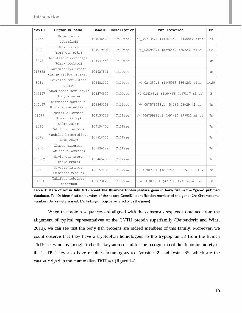
Introduction
19
TaxID Organism name GeneID Description map_location Ch
7955 Danio rerio
(zebrafish) 100048920 ThTPase NC_007135.6 (14451258 14455856 plus) 24
8010 Esox Lucius
(northern pike) 105019498 ThTPase NC_025988.1 (8296487 8302233 plus) LG21
8208 Notothenia coriiceps
(black rockcod) 104941948 ThTPase Un
215358 Larimichthys crocea
(large yellow croaker) 104927511 ThTPase Un
8081 Poecilia reticulate
(guppy) 103482317 ThTPase NC_024350.1 (4865958 4868263 plus) LG20
244447 Cynoglossus semilaevis
(tongue sole) 103376930 ThTPase NC_024309.1 (4104666 4107137 minus) 3
144197 Stegastes partitus
(bicolor damselfish) 103365356 ThTPase NW_007578563.1 (54169 58029 minus) Un
48698 Poecilia Formosa
(Amazon molly) 103135322 ThTPase NW_006799963.1 (997484 999811 minus) Un
8030 Salmo salar
(Atlantic salmon) 100194700 ThTPase Un
8078 Fundulus heteroclitus
(mummichog) 105918316 ThTPase Un
7950 Clupea harengus
(Atlantic herring) 105890182 ThTPase Un
106582 Maylandia zebra
(zebra mbuna) 101465435 ThTPase Un
8090 Oryzias latipes
(Japanese medaka) 101157256 ThTPase NC_019878.1 (10172950 10176117 plus) 20
31033 Takifugu rubripes
(torafugu) 101073828 ThTPase NC_018899.1 (471583 473914 minus) 10
Table 3: state of art in July 2015 about the thiamine triphosphatase gene in bony fish in the “gene” pubmed
database. TaxID: Identification number of the taxon; GeneID: identification number of the gene; Ch: Chromosome
number (Un: undetermined; LG: linkage group associated with the gene)
When the protein sequences are aligned with the consensus sequence obtained from the
alignment of typical representatives of the CYTH protein superfamily (Bettendorff and Wins,
2013), we can see that the bony fish proteins are indeed members of this family. Moreover, we
could observe that they have a tryptophan homologous to the tryptophan 53 from the human
ThTPase, which is thought to be the key amino-acid for the recognition of the thiamine moiety of
the ThTP. They also have residues homologous to Tyrosine 39 and lysine 65, which are the
catalytic dyad in the mammalian ThTPase (figure 14).

Figure 14: Alignment of the CYTH protein sequences available in bony fishes and of the consensus sequence for CYTH protein (Bettendorff and Wins, 2013).
The P. before most of the fish sequences means that those sequences are “Predicted” sequence. The three red squares show the signature of a member of the
CYTH family while the red circle shows the amino acid homologous to the human tyrosine 39, tryptophan 53 and lysine 65. The nonpolar amino acids are in
black, the polar and neutral are in green, the acidic are in red and the basic are in blue. The alignment was performed with CLC SEQUENCE VIEWER, Version 7.6 (CLC
bio A/S).

Introduction
21
Morpholino:
The anti-sense morpholino oligomers that are currently used in zebrafish experiments are
based on the morpholino phosphorodiamidate derivatives (figure 15). They were first developed
as suitable for in vivo use in 1996 (Hudziak et al. 1996; Taylor et al. 1996; Summerton and
Weller, 1997). They are chemically modified oligonucleotides that use a base stacking similar to
natural RNA: they have a morpholine moiety instead of the ribose, and the phosphorodiamidate
linkage results in a neutral charge instead of the negative phosphate link. This modified oligomer
is highly soluble and can hybridize with a high affinity to the single stranded nucleic acid
sequences (Ekker, 2000).
Figure 15: Model of morpholino oligomers. They are composed of a chain
of morpholino rings bearing a nucleotide on the C2 with a
dimethylaminophosphoryl intersubunit linkage.
From Hudziak et al. 1996
The morpholino oligonucleotides are the anti-sense knockdown strategy most commonly
used in zebrafish but this could change in the near future. Indeed, recent studies tend to show that
the results obtained by knocking down gene with morpholino are not always reproducible when
the target gene is deleted from the genome by knock-out methods (Kok et al. 2015), which
suggests some nonspecific effect of the morpholino.
Anti-sense knockdown of gene expression:
There are different strategies to interfere with a specific transcript or protein levels in the
zebrafish using morpholino. They can act in the cytoplasm but also in the nucleus and can either
inhibit the splicing of the pre-mRNA, the initiation of translation or the miRNA signaling.

Introduction
22
Splicing inhibition:
The morpholino can interact with the pre-mRNA splicing site and prevent its interaction
with the splicing machinery. The misspliced mRNA product will then comprise an insertion of
part or the totality of the targeted intron. It can either induce a frameshift or insert a stop codon
that will lead to a truncated protein or the insertion will produce an aberrant protein (Schmajuk et
al. 1999)
The morpholinos inhibiting the splicing site have the characteristics of targeting only the
zygotic mRNA but not the maternal one as they were already spliced in the unfertilized egg. The
efficiency of the inhibition can be evaluated by amplifying a PCR product with primers directed
at the exon sequences flanking the targeted intron (Draper et al. 2001). The spliced sequence will
be reduced or become undetectable in the morphant compared to the non-injected embryo and a
longer misspliced sequence can appear as a PCR product.
Translation initiation site inhibition:
The morpholino is designed to interact with the translation initiation site of the mRNA
and prevent its interaction with the translation machinery (Ekker, 2000). Those morpholinos will
target maternal and zygotic mRNA, which in turn can affect the very early developmental stages
and thus prevent the observation of more specific effects during the organogenesis for example.
miRNA signaling inhibition:
The micro RNAs (miRNAs) are small non-coding RNA molecules found in eukaryotes
and in some viruses. To quickly summarize their synthesis, they are first transcribed into pri-
microRNA then cleaved into pre-microRNA by the ribonuclease Drosha. They are again cleaved
by the endoribonuclease Dicer into microRNA duplexes that can be used by the RISC complex to
induce the cleavage, repress the translation or induce the deadenylation of their specific target
mRNAs.
The morpholino can block the maturation of miRNAs either at the Drosha or the Dicer
cleavage steps or inhibit their interaction with their target RNA (Choi et al. 2007; Kloosterman et
al. 2007). When the morpholinos are used to block microRNAs, they will increase the mRNA
stability or translation by preventing the miRNA to down-regulate them (figure 16). One of the
advantages of this approach is that many different morpholinos can be used to inhibit the same
microRNA and thus increase the specificity of the conclusion drawn from the morphant.

Introduction
23
Figure 16: The pathway from the production to the
action of the microRNA. The red crosses show the
processes that can be blocked by morpholinos.
Modified from: Winter et al. 2009.


Aims of
the work


Aims of the work
24
In this study we will focus our attention on two small compounds with no clearly defined
biological roles. These two molecules are the inorganic triphosphate and the thiamine
triphosphate. Both are known to be targets of different members of the CYTH superfamily of
proteins, some of which are specific inorganic tripolyphosphatase (PPPase) or thiamine
triphosphatase (ThTPase).
Inorganic triphosphate (tripolyphosphate, PPPi) is a short-chain polyphosphate known to
accumulate in protozoan acidocalcisomes. It could not be detected in other tissues because of the
lack of sensitive methods. However, recently several studies have shown that some enzymes have
a highly specific PPPase activity raising the possibility that this compound indeed exists in the
cells and may play a metabolic role. We thus decided to study the PPPase activities in mammals
and also in E. coli.
The second part of this work is focused on thiamine triphosphate (ThTP) with the aim to
gain insight into the possible physiological role of this compound. ThTP is synthesized by FoF1-
ATP synthase by a chemiosmotic mechanism similar to ATP synthesis. It is therefore impossible
to specifically inhibit ThTP synthesis and the only way we have to control intracellular levels is
to either increase or decrease its hydrolysis by up and down regulation of the specific ThTPase.
We used different organisms (E. coli, zebrafish, mice) and strategies (metabolic deregulation,
morpholino oligomers and targeted knock-out) in order to study the consequences of increased
cellular levels of this compound.
In E. coli, ThTP accumulation strongly depends on cultures condition and a transient and
important increase of the intracellular ThTP concentration can be induced under amino acid
starvation. We studied different mutant strains, commercial or mutated in our lab and the impact
of some metabolic inhibitors in order to gain insight into the physiological significance of this
compound.
In zebrafish eggs, we used morpholinos directed against ThTPase with the aim of
increasing the intracellular concentration of ThTP in order to study its consequences on the
development of the fish embryos.
In parallel, we decided to create a mouse knock-out for the gene coding for ThTPase in
order to study the effects of this deletion on the phenotype of the animal.


Material
And
Methods


Material and methods
25
Material
Solvents and Reagents
All the solutions were prepared with milli Q water (Millipore S.A. / N.V., Brussels,
Belgium).
All the compounds for the bacteria culture medium, the HPLC reagents as well as the D-
Glucose were from Merck NV/SA (Oversijse, Belgium).
Antibiotics, isopropyl β-D-1-thiogalactopyranoside (IPTG), Bovine Serum Albumin
(BSA), EDTA, sodium triphosphate (PPPi), proteinase K enzyme, E. coli pyrophosphatase,
HOCL 30%, thiamine, ThMP and ThDP were from Sigma-Aldrich NV/SA (Bornem, Belgium).
The trichloroacetic acid (TCA) and the Folin-Ciocalteu reagent were from VWR Prolabo
(VWR International NV/SA, Louvain, Belgium).
The HPLC solvents and the diethyl-ether were from Biosolve (Valkenswaard, The
Netherlands).
The octyl-α-ketoglutarate was from Cayman chemical (Ann Arbor, MI, USA)
The ThTP which is not commercially available was synthesized as described by
Bettendorff et al. in 2003.
Morpholino solution
Three different morpholinos were ordered from Gene Tools (Gene Tools, LLC 1001
Summerton Way Philomath, OR 97370 USA) and stored as stock solution at -80 °C in the
Danieau solution:
the Gene Tool standard control morpholino: 5’ CCTCTTACCTCAGTTACAATTTATA 3’
the morpholino that inhibit the splicing between the second and third exons of the
zThTPase: 5’ ATTTAAAACACACCTCCAATATCCT 3’
the morpholino that inhibits the transcription initiation site of the zThTPase-001 and 003:
5’ TCCACTTCTACAGTCATTTTGGAC 3’.
The working solutions that were injected in the eggs were always prepared with a final
volume of 10 µl and stored at -80 °C in between the experiments. They contained the
morpholino, 0.5% of rhodamine dextran and eventually the RNA coding the zThTPase or the
K61A zThTPase and were bring to 10 µl with the Danieau solution.

Material and methods
26
Zebrafish solution
E3 medium
NaCl 5 mM, KCl 0.17 mM, CaCl2 0.33 mM, MgSO4 0.33 mM, methylene blue 0,05%.
Tricaine (50X)
Final concentration of 4 g per liter of water, bring to pH 9.5 with Tris-HCl 1 M pH 9.5
Danieau
NaCl 58 mM, KCl 0.7 mM, MgSO4 0.4 mM, Ca(NO3)2 0.6 mM, HEPES 5 mM, pH 7.6
Bacteria culture medium:
LB medium:
This medium contained 1% (w/v) of peptone from casein, 0.5% (w/v) of yeast extract and
1% (w/v) of NaCl, was autoclaved and stored at 4 °C. For use as an agar plate, we added 1.5%
(w/v) of agar prior to the autoclave treatment.
M9 minimal medium:
This medium contained 47.7 mM of Na2HPO4, 22.05 mM of KH2PO4, 8.55 mM of NaCl
and 18.7 mM of NH4Cl, was autoclaved and stored at 4 °C. Right before use, MgSO4 and CaCl2
were add from a 1000x concentrated stock solution (sterilized by filtration); the final
concentrations were 1 mM and 0.1 mM respectively.
Strains of E. coli used:
Most of the experiments were carried out using the MG1655 strain (WT K12 strain)
which was generously offered to us by Dr. M. Cashel (Laboratory of Molecular Genetics,
NICHD, National Institute of Health, Bethesda, MD). He also offered us the CF5802 strain
lacking the polyP kinase (PPK) and the exopolyphosphatase (∆ppk-ppx::km) (Kuroda et al.
1997)
Different mutant strains from the E. coli Genetic Stock Center (Yale University, New
Haven CT, USA) were used (table 4).
The NM543 strain was a gift from N. Majdalani NCI, NIH. Two more strains were
produced in our lab from that strain: the ThTPase WT I and ThTPase K65A I.

Material and methods
27
Name Mutation Genotype
JW3026 ∆ygiF F-, ∆(araD-araB)567, ∆lacZ4787(::rrnB-3), l2, ∆ygiF747::kan, rph-1,
∆(rhaD-rhaB)568, hsdR514, CGSC#10312
JW0110 ∆E1p F-, Δ(araD-araB)567, ΔaceE732::kan, ΔlacZ4787(::rrnB-3), λ-, rph-1,
Δ(rhaD-rhaB)568, hsdR514, CGSC#8392
JW0111 ∆E2p F-, Δ(araD-araB)567, ΔaceF733::kan, ΔlacZ4787(::rrnB-3), λ-, rph-1,
Δ(rhaD-rhaB)568, hsdR514, CGSC#8393
JW0112 ∆E3 F-, Δ(araD-araB)567, Δlpd-734::kan, ΔlacZ4787(::rrnB-3), λ-, rph-1,
Δ(rhaD-rhaB)568, hsdR514, CGSC#8394
JWO715 ∆E1o F-, Δ(araD-araB)567, ΔlacZ4787(::rrnB-3), ΔsucA775::kan, λ
-, rph-1,
Δ(rhaD-rhaB)568, hsdR514, CGSC#8786
JRG465 ∆E1o
iclR const
F-, sucA8, gal-25, λ-, trpA9767, IN(rrnD-rrnE)1, iclR7(Const), trpR47,
CGSC#4456
JW0710 ∆CS F-, Δ(araD-araB)567, ΔlacZ4787(::rrnB-3), ΔgltA770::kan, λ-, rph-1,
Δ(rhaD-rhaB)568, hsdR514, CGSC#8784
JW1268 ∆acnA F-, Δ(araD-araB)567, ΔlacZ4787(::rrnB-3), λ-, ΔacnA785::kan, rph-1,
Δ(rhaD-rhaB)568, hsdR514, CGSC#9141
JW0114 ∆acnB F-, Δ(araD-araB)567, ΔacnB736::kan, ΔlacZ4787(::rrnB-3), λ-, rph-1,
Δ(rhaD-rhaB)568, hsdR514, CGSC#8396
NM543 MG1655, lacIQ_FLP-scar, mini-lambda::TetR , P-lacO-KanR-sacB.
(delta) lacI-lacZ PlacO-kan-sacB Table 4: different bacterial strain used, the mutation they carried and their genotypes.

Material and methods
28
Methods
Determination of the tripolyphosphatase activity
Collection of the sample
All animal experiments were made in accordance with the directives of the Committee for
Animal Care and Use of the University of Liège and in accordance with the European
Communities Council Directive of November 24, 1986 (86/609/EEC). The protocols were
approved by the Committee on the Ethics of Animal Experiments of the University of Liège (#
1253 for mice, # 823 for rats and # 727 for quails). The animals were killed by decapitation and
all efforts were made to minimize suffering. Pig brains were obtained from the local
slaughterhouse (Abattoirs Publics de Liège & Waremme SC, rue de Droixhe 15, 4020 Liège,
Belgium) with the permission to be used for experimental purposes at the University of Liège.
The bacteria were cultured overnight in LB medium at 37 °C under constant stirring (250
rpm) and centrifuged for 10 min at 8000 g at 4 °C. The pellets were solubilized in a Tris-HCl
buffer (20 mM) pH 7.4.
Measurement of the activity
The standard incubation medium contained 50 mM buffer, 5 mM MgCl2, 0.5 mM PPPi
and 20 µl of the enzyme at the adequate concentration in a total volume of 100 µl. The mixture
was incubated at 37 or 50 °C, the reaction was stopped by addition of 1 ml phosphate reagent
(Lanzetta et al. 1979) and the absorbance (UV-1800 Shimadzu UV spectrophotometer) was read
at 635 nm after 30 min and compared with a standard curve (0–0.5 mM K2HPO4) to estimate the
release of inorganic phosphate. The buffers used for incubation at different pH values were: Na-
MOPS (pH 7.0–7.5), Na-HEPES (pH 7.5–8.0), Na-TAPS (pH 8.0–9.0), Na-CHES (pH 9.0–10.0)
and Na-CAPS (pH 10.0–10.5).
Pig brain PPPase activity identification
PPPase was purified from about 500 g of pig brain. All the purification steps were carried
out at 4 °C. The tissues were homogenized in Tris-HCl buffer (30 mM Tris-HCl, 50 mM, NaCl,
0.1 mM EDTA, 1.5 mM DTT, pH 7.4). The homogenate was centrifuged at 20,000 g for 20
minutes. The supernatant was brought to 80% saturation in ammonium sulfate and kept under
constant stirring for 1 h. After centrifugation at 27,000 g for 30 minutes, the pellet was suspended

Material and methods
29
in buffer A (20 mM Tris- HCl, 1 mM DTT, pH 7.4) and 5 ml aliquots were placed on a Sephadex
G 75 column (3 x 50 cm, GE Healthcare Life Sciences) equilibrated with buffer A. The proteins
were eluted with the same buffer at a flow rate of 2 ml/min and all the fractions with high PPPase
activity were pooled. The pool was loaded on a DEAE-Sephacel anion exchange column (1.4 x
24 cm, GE Healthcare Life Sciences) equilibrated with buffer A. The proteins were eluted with
an exponential concave NaCl gradient from 0 to 0.4 M in buffer A. Fractions with highest PPPase
activity were pooled and centrifuged in Amicon ultra-15 centrifugal filter (Ultracel-10
membrane) units (Merck Millipore SA/NV, Overijse, Belgium). The retained proteins were
dissolved in buffer B containing 20 mM Tris-HCl and 50 mM NaCl. 25 mg of protein were
loaded on a Mono Q column (GE Healthcare Life Sciences) coupled to a Fast Protein Liquid
Chromatography apparatus (ÄKTApurifier, Amersham) and elution was made with buffer B
using a linear NaCl gradient from 50 to 500 mM. The fractions with the highest PPPase activity
were pooled and DTT was added to reach a concentration of 1 mM. The proteins were then
separated by non-denaturing polyacrylamide gel electrophoresis. The band with PPPase activity
was cut and analyzed by liquid chromatography-mass spectrometry as described below for E. coli
PPPase activity.
E. coli PPPase activity identification
E. coli wild type strain MG1655 was grown overnight in LB medium at 37 °C under
agitation (250 rpm). The bacteria were harvested by centrifugation for 10 minutes, at 8000 g at 4
°C. The pellet was suspended in Hepes-buffer (30 mM Hepes, 50 mM NaCl, pH 7.2–7.4) and the
cells were lysed by high pressure using a French press. The lysate was centrifuged for 30 minutes
at 40,000 g at 18 °C. The supernatant (called S1) was brought to 50% saturation with ammonium
sulfate and kept under constant stirring for one hour at room temperature. After centrifugation for
30 minutes at 20,000 g at 18 °C, the supernatant (called S50) was brought to 80% saturation with
ammonium sulfate and kept under constant stirring for one hour at room temperature. After
centrifugation 30 minutes at 20,000 g at 18 °C, the pellet was suspended in Hepes-buffer. The
preparation obtained (called P80) was loaded on a mono Q column coupled to a Fast Protein
Liquid Chromatography (FPLC) apparatus and elution was carried out with a linear NaCl
gradient from 50 to 500 mM. The fractions with the highest PPPase activity were pooled (called
F1) and placed on a Sephadex G200 column (1 x 25 cm) equilibrated with Hepes buffer. Elution
was performed in the same buffer. The fractions with the highest PPPase activity were pooled

Material and methods
30
(called F2) and used to perform a non-denaturing polyacrylamide gel separation and the PPPase
activity was determined as described below. The bands containing PPPase activity were cut and
analyzed by liquid chromatography coupled on-line to positive mode electrospray ionization
tandem mass spectrometry (UltiMate 3000 Nano LC Systems, Dionex, AmaZone, Bruker) for
protein identification.
In-gel enzymatic activity
Non-denaturating polyacrylamide gels (10%) were run in Tris-Glycine buffer (25 mM
Tris, 190 mM glycine). After incubation for 30 minutes in a bath containing PPPi (0.5 mM),
buffer (20 mM), and MgCl2 (1 mM), the gel was colored with phosphate precipitation reagent
(1% v/v ammonium heptamolybdate, 1% v/v triethylamine, 1 N HNO3) (Simonović et al. 2004).
For E. coli PPPase, the incubation temperature was 50 °C and the buffer was Na-CHES (pH 9.5)
while for pig brain PPPase incubation temperature was 37 °C and the buffer was Na-MOPS (pH
7.1).
Detection of thiamine and its derivatives in E. coli
After incubation of the bacteria in M9 minimal medium, 1 ml of the culture was
centrifuged for 1 min at max speed on a tabletop centrifuge and the pellet was dissolved in 500 µl
of TCA 12% in order to denature and precipitate the proteins. It was then centrifuged for 10 min
at 10,000 g at 4 °C. The supernatant was extracted 3 times with 2.5 ml of diethyl-ether to remove
the TCA and then analyzed by High Performance Liquid Chromatography (HPLC). The pellet
was dissolved in 500 µl of NaOH 0.8 N for the determination of protein concentrations.
The detection of thiamine and its derivative is based on their oxidation into their
fluorescent thiochrome derivatives by the potassium ferricyanide (K3[Fe(Cn)6]) in alkaline
solution prior to their injection on the HPLC column (figure 17). The oxidant solution is
composed of 500 µl of a 1% (m/v) solution of potassium ferricyanide and 2.5 ml of NaOH 15%
(m/v). 40 µl of the sample are mix with 25 µl of this oxidative solution prior to the injection in
the 20 µl injection loop of the HPLC.
Sample thiamine concentrations were calculated from peak areas by comparison with the
peak areas of an external standard solution containing known concentration of 250 µM of
thiamine, ThMP, ThDP and ThTP. The external standard solution was injected prior to each
batch of sample.

Material and methods
31
Figure 17 : Oxidation of thiamine and its derivative by potassium ferricyanide (K3[Fe(Cn)6]) in alkaline solution.
The separation of the derivatives is based on a ion-pair reversed-phase chromatography
principle using a alkaline resistant PRP-1 column. This column (150 x 4,1 mm Ø, 5 µm,
Hamilton, Reno, USA) is composed of poly(styrene-divinylbenzene) and protected by a pre-
column (25 x 2.3 mm Ø) of the same composition. The mobile phase (flow = 0.5 ml/min) is a
solution with a pH of 9.5, containing 50 mM of Na2HPO4, 25 mM of tetrabutylammonium and
4% of tetrahydrofuran. The column is linked to a fluorescence spectrometer (KONTRON, SFM
25) whith excitatory wavelength set at 365 nm and emission wavelength at 433 nm.
Determination of acetyl-CoA levels
The method was adapted from King et al. (King et al. 1988), using a BioSil C18HL
reversed phase column (Bio-Rad). The mobile phase was composed of NaH2PO4 (0.25 M)
containing 5% acetonitrile. The flow-rate was 1 ml/min and 20 µl samples were injected. Acetyl-
Coa was detected at 254 nm. The samples were prepared in trichloroacetic acid extracts as for the
determination of thiamine derivatives.
Polymerase chain reaction
Every PCR reactions were carried out using the Taq polymerase and dNTP from
Promega. All primers were ordered from Integrated DNA Technologies (IDT). The classical
protocol was: first, 2 min at 95 °C, then 35 cycles of 30 s at 95 °C, 30 s at the annealing
temperature, and an elongation step at 72 °C (time depending on the length of the target
sequence) and finally 15 min at 72 °C. The annealing temperature and the elongation time were
specific for each set of primers and depend on the length of the target to amplify. All the primers’
annealing temperatures and elongations times are listed in table 5.

Purpose Forward (5’3’)
and reverse primers (5’3’)
Annealing
temperature (
°C)
Elongation
time
(s)
Amplification of the mouse ThTPase coding
sequence (WT or K65A) for insertion in the E. coli
genome
For: GTGAGCGGATAACATTGACATTGTGAGCGGATAACAAGATA
CTGACGTCAAGGAGGAAAAAAATGGCCCAGGGCTTGATTGA
Rev: CTAGAAAGTATAGGAACTTCGAAGCAGCTCCAGCCTACACT
CAGGATGCTGAGTCCCCTG
54 50
Amplification of the chloramphenicol gene for
insertion in the E. coli genome
For: AAGCAGCCAGTTCTGGTGAGGCCACAGGGGACTCAGCATCC
TGAGTGTAGGCTGGAGCTGCTTG
Rev: CACGGTCACACTGCTTCCGGTAGTCAATATCGGCATTTTCTT
TTGCGTTTCATATGAATATCCTCCTTAG
48 50
Verification of the insertion of the ThTPase gene in
the bacteria
For: AGGTTTCCCGACTGGAAAGC
Rev: CAGCCAGTGGTCAGACAGCA 52 30
Verification of the insertion of the chloramphenicol
gene in the bacteria
For: TCAACAGGGACACCAGGAT
Rev: AGCCTGAATCAGGCATTTGA 48 30
Amplification of a region of the zebrafish thtpa
transcripts that is differentially spliced
For: TGCCCTCATTACACTGATTGA
Rev: TCTCTGCATCTCAGCCAGAAAT 53 20
Amplification of a sequence common to the 3
zebrafish thtpa transcripts
For: TGCCCTCATTACACTGATTGA
Rev: TCTCTGCATCTCAGCCAGAAAT 53 20
Rescue zebrafish: insertion of the zThTPase in the
pCS2+8
For: AGGATCCGCCACCATGACTGTAGAAG
Rev: TCTCGAGTCATAAAATATGAGCACTAA 55 50
Directed mutagenesis of the zThTPase to insert the
K61A mutation
For: TGGGAGTTGGCAAGCCCTGCAGTTT
Rev: TGCAGGGCTTGCCAACTCCCACT 62 480
Mouse genotyping
For 1-NeoFwd: TCATTCTCAGTATTGTTTTGCC
For 2-TDF: GGGTAGGCAGGGAACAGAAC
Rev-SD: ACGAGCGGAAATCAATGGAG
58 25
qPCR on the mouse sperm genomic DNA: WT
ThTPase allele amplification
For: AACAGAACCGAAGCATGTCCGA
Rev: TTCTAGCAGGCGCTGATAGTCCA 54 12
qPCR on the mouse sperm genomic DNA:
inactivated ThTPase allele amplification
For: TCATTCTCAGTATTGTTTTGCCAAGTT
Rev: AAGTGGCTCCGTTGTAAGGGAA 54 12
Table 5: List of the primer, their purposes as well as the annealing temperature and amplification time.

Material and methods
33
Insertion of the mammalian ThTPase coding sequence in the E.coli
genome
The strain used to insert the mouse thiamine triphosphatase into the E.coli genome was
the NM543. In those bacteria, the lactose operon was replaced by a sequence containing the pBR-
LacO promotor (Lutz and Bujard, 1997), the kanamycine resistance gene kan+ and the sucrose
susceptibility gene SacB. The miniphage λ was also inserted in the genome to allow the
recombination, under the dependence of a heat-inducible promotor and followed by a tetracycline
resistance gene.
The aim was to replace the kan+ - SacB sequence by our sequence of interest. We inserted
two different constructions, both containing a Shine-Dalgarno sequence, followed by either the
mouse ThTPase or the K65A mouse ThTPase and by the chloramphenicol resistance gene (Cm+).
Briefly, the general protocol was as follows: the NM543 bacteria which are resistant to
kanamycin thanks to the kan+ gene, to tetracycline thanks to the tet
+ gene of the miniphage λ but
sensitive to sucrose because of the SacB gene are grown at 30 °C. A heat choc at 42 °C induced
the expression of the miniphage λ and thus of the enzymes required for the homologous
recombination They are then electroporated with the construction containing a sequence
homologous to the end of the pBR-LacO promotor and to the sequence between SacB and Lac Z.
After electroporation, the miniphage λ enzymes will act at two different places of the bacterial
genome, first by removing the miniphage λ but also by inserting our construction at the targeted
site. If they are properly recombined, the bacteria should lose their resistance to kanamycin
together with their resistance to tetracycline and their sensitivity to sucrose but should have
gained chloramphenicol resistance as well as an inducible ThTPase (WT or K65A) (figure 18).
In practice, a NM543 clone, resistant to kanamycin and tetracycline but which does not
grow in the presence of sucrose was grown overnight at 30 °C under constant stirring (250 rpm)
in LB medium containing kanamycin and tetracycline. The culture was then diluted 100 times in
160 ml of fresh LB medium, divided in four individual cultures and grown at 30 °C under
constant stirring (250 rpm) until the optic density reached 0.4 – 0.6. Next, the four cultures were
incubated at 42 °C for 15 min then cooled down on ice for 10 min. Each culture was then
centrifuged for 15 min at 3750 g at 4 °C. The supernatants were discarded and each pellet was
suspended in 40 ml of sterile water and centrifuged again under the same conditions. The

Material and methods
34
supernatant was removed again and the bacteria were washed two more times as described above.
After the last centrifugation each pellet was suspended in 50 µl of sterile water and then pooled.
Of the pool, 40 µl were electroporated (200 Ω, 25 µF, 2500 V), then 1 ml of fresh LB medium
was added and the bacteria placed at 37 °C for three to four hour. They are then plated on LB
agar containing sucrose (20% w/v) and chloramphenicol (30 µg/ml). Some clones were then
tested for their sensitivity to kanamycin and tetracycline and those which had lost their resistance
to those antibiotics were assessed by PCR for the insertion of our transgene. The clones that pass
this test were then stored at -80 °C in LB-glycerol (1 volume of LB for 1 volume of glycerol)
until further use.
Figure 18: Different lactose operon genes in the different strain: The WT allele in the WT MG1655 strain
containing the Lac I gene which represses the operon (gene lac Z, lac Y and lac A) in the absence of lactose. In the
three next strains, the operon is rendered inactive by the insertion of different genes and in all of them, the pBR-
LacO artificial promoter is present right after the Lac I gene. In the NM543 gene, the kan+ positive selection gene
and the SacB negative selection gene were inserted. In the two next strains, those gene were replaced by a Shine-
Dalgarno sequence followed by the mouse ThTPase or the K65A mouse ThTPase both followed by the
chloramphenicol resistance gene (Cm+).
Zebrafish maintenance
The zebrafish (Danio rerio) studied were of the AB strain (ZIRC, Eugene, OR). Those
fish were kept in the aquarium at 28 °C as described by Westerfield in 2007. The light/dark cycle
was 14/10 h. Animal care and all experimentations were conducted in compliance with Belgian
and European laws (Authorization: LA1610002, Ethical commission protocols ULg1076 and
ULg624).
Wild-type fish were mated and spawning was stimulated by the onset of light. Then, eggs
were collected and placed at 28 °C in Petri dishes containing E3 medium. Embryos and larvae

Material and methods
35
were staged according to Kimmel et al. (1995). The age of the embryos and larvae is indicated as
hours post fertilization (hpf) or days post fertilization (dpf).
Zebrafish breeding and eggs generation
For the production of the eggs, fish were placed in a breeding aquarium as such: 2 male
(♂) for 1 female (♀) or 2 ♂ for 2 ♀ or 3 ♂ for 2 ♀ and males separated overnight from the
female(s) by a plastic glass. In the morning, the fish were allowed to mate and the eggs were
collected in 10 cm Petri dishes containing E3 medium.
Morphant generation, characterization and maintenance
The eggs were manually injected at the one or two cell stage under a binocular
microscope (Olympus SZX9) using the Picospritzer II (Parker Hannifin corporation, USA)
injector with needles pulled from glass capillary with the PB7 needle/electrode puller (Narishige,
Japon). The volume injected per egg was of ~1 nl of the morpholino working solution.
A few hours later, the unfertilized (undeveloped) eggs were removed from the Petri dish
and 20 - 24 h after the injection the non-injected or miss-injected embryos were also removed.
The correctly injected embryos were counted and categorized 24 h after the injection and the E3
medium was changed for an E3 medium containing 0.2 mM of PTU to prevent the melanization
of the embryo. They are counted and categorized again 24 h later.
Zebrafish embryo RNA isolation
Total RNA was isolated with Trizol (Invitrogen, Cergy Pontoise, France) using the
RNeasy extraction kit (Qiagen, Venlo, Netherlands) as in Pruvot et al. 2014. Briefly, 75 - 100
zebrafish embryos were placed in 600 µl of Trizol and passed 10 times through a G22 needle,
followed by 10 passages through a G26 needle with a 1 ml syringe until they were completely
dissolved. Then 140 µl of chloroform were added and the samples were vortexed thoroughly for
15 s and incubated under agitation for 2 - 3 min at room temperature (RT). They were centrifuged
at 12,000 g for 15 minutes at 4 °C. The upper phase was isolated and 1.5 volumes of ethanol was
added and loaded on a column from the Qiagen RNeasy extraction kit. The DNA was removed
by application of 80 µl of the RNase-Free DNase Set on the column and incubation for 15 min at
RT. The RNA is then isolated following the instruction from the RNeasy extraction kit.

Material and methods
36
zThTPase mRNA cloning and expression
Zebrafish cDNA was used to amplify the coding sequence of the zThTPase (transcript
thtpa-001/3). The forward primer contains a BamHI restriction site, then a Kozak sequence (SJ
Grzegorski et al. 2014) and the beginning of the coding sequence. The reverse primer adds a
XhoI restriction site after the stop codon. This amplicon was inserted into a pGemT vector by TA
cloning. Site directed mutagenesis allowed us to change the AAA triplet of lysine 61 into a GCA
coding an alanine instead. Both the WT and K61A zThTPase sequences were cloned into BamHI
and XhoI sites of the pCS2+8 plasmid. The two plasmids obtained were used to produce in vitro
the transcripts that will be injected into the zebrafish eggs.
The transcript expression and purification were done using the mMessage mMachine kit
from Ambion and the Nucleospin RNA Clean Up kit from Macherey-Nagel after digestion of the
two PCS2+8 - zThTPase (WT or K61A) with the NotI restriction enzyme.
Mouse genotyping
Genomic DNA was extracted from tip of the tails, tissues or sperm by overnight
incubation at 55 °C in 600 µl of TENS (Tris-HCL 100 mM pH 8,5, EDTA 5 mM, NaCl 200 mM,
SDS 0,2% (m/v)) containing 5 µl of proteinase K (20 mg/ml). At the end of the incubation, the
samples were centrifuged 10 min at 10,000 g, 500 µl of the supernatant was mixed with 500 µl of
isopropanol and centrifuged 15 min at 10,000 g. Supernatants were removed and 600 µl of 70%
(v/v) ethanol were added to the pellets before centrifugation (10 min at 10,000 g). The
supernatants were removed and the pellets were dried for 5 min at room temperature and then
homogenized in 1 ml of TE (Tris-HCl 10 mM pH 8.5, EDTA 1 mM).
The genotype was checked by PCR assay using the primer suggested by the KOMP
repository (see table 5) to assess the presence of wild-type and invalidated alleles. Depending on
the case, both types of alleles were either detected simultaneously or separately. The
amplification was done using an annealing temperature of 58 °C and an elongation time of 25 s
for a total of 40 cycles.
Sperm isolation
After the sacrifice of the animals by cervical dislocation, the epididymis and the ductus
deferens were removed and placed in cold PBS. The sperms were pushed-out of the organs which

Material and methods
37
are then removed. The PBS containing the sperm was centrifuged for 5 min at 10,000 g in 2 ml
microcentrifuge tubes. The supernatant was removed and the pellet used for genotyping.
When the sperm cells were isolated for swim up or swim out experiments, then the
epididymis and the ductus deferens were placed in pre-warmed (37 °C) DMEM culture medium
and the sperms were pushed-out of the organs which were then removed.
Swim up
500 µl of the culture medium containing the sperm are placed in the bottom of a 5 ml
sterile culture tube, then 1 ml of fresh pre-warmed (37 °C) culture medium were added without
disturbing the medium already in the tube. The tube was then incubated at 37 °C for 30 min in a
cell culture incubator (figure 19). At the end of the incubation, 150 µl are removed from the
upper part, this fraction was called Swim Up Superior part (SUS) and 150 µl are taken from the
lower part, this fraction was called Swim Up Lower part (SUL).
Figure 19: schematic view of the incubation step of the isolation of the mobile
sperms through swim-up
Swim out
A 1 ml drop of pre-warmed (37 °C) DMEM culture medium was placed in the center of a 3 cm Ø
Petri dish and 5 - 7 droplets of 50 µl of this same medium were place around it then connected
through thin medium bridge to the central drop. Then 500 µl of culture medium containing the
sperm were added to the central drop. The Petri dish was then incubated at 37 °C for 30 min in a
cell culture incubator (figure 20). At the end of the incubation, the outer droplets were pooled and
this fraction was called Swim Out Peripheral part (SOP) and 150 µl were taken from the central
drop and this fraction was called Swim Out Central part (SOC).

Material and methods
38
Figure 20: schematic view of the incubation step of the isolation of the
mobile sperms through swim-out
Quantitative PCR:
The absolute number of copies per µl of the WT and of the invalidated alleles was
measured by performing quantitative PCR (qPCR) on sperm genomic DNA samples. For the
establishment of the standard curve, the two allelic sequences amplified during the qPCR were
inserted into the pGEM-T plasmid by TA cloning. Plasmids were then purified and their
concentration was measured by spectrophotometry. A plot of the absolute number of copies per
µl versus the Ct obtained was drawn for each sequences and used to quantify the number of
copies of each allele in our samples.
Each sample was analyzed as triplicate. The qPCR mix contained for each of them 10 µl
of FastStart Universal SYBR Green Master (Rox) 2x, 0.4 µM of forward primer and 0.4 µM of
reverse primer, the DNA sample and water for a total of 20 µl. The qPCR was done in 96 well
plates in a LightCycler® 480 Multiwell Plate 96 from Roche.

Results


Results
39
Chapter 1: Tripolyphosphatase activity
Bacterial CYTH proteins are annotated as predicted adenylate cyclases as one of the
founding members of the family is the adenylate cyclase from Aeromonas hydrophila, CyaB. In
2011 it was demonstrated in our laboratory that the CYTH enzyme of the bacterium
Nitrosomonas europaea, NeuTTM, is a specific tripolyphosphatase (Delvaux et al. 2011). It was
the first time that an enzyme was found to specifically hydrolyze inorganic triphosphate (PPPi).
In 2013, it was shown by Moeder et al. that AtTTM3, one of the CYTH enzymes of the plant
Arabidopsis thaliana, is also a specific inorganic triphosphatase and that this enzyme may be
involved in root development.
Tripolyphosphatase activity of mammalian tissues:
The mammalian CYTH ThTPase has been well characterized in our laboratory (Delvaux
et al. 2013). It hydrolyzes specifically ThTP, with virtually no tripolyphosphatase (PPPase)
activity. Nonetheless, supernatant fractions from animal tissues exhibit a high PPPase activity,
suggesting that other enzymes (not belonging to the CYTH superfamily) are able to hydrolyze
PPPi.
Characterization
In order to characterize this activity, PPPi hydrolysis was measured in the supernatant
fraction of several quail and rat tissues and also in pig brain (figure 21 A). We found an activity
in all tissues tested with the highest activity in the brain. We also tested the dependence of that
activity on the pH and on the cation concentration. We found that the activity was not strongly
affected by the pH but was optimal around 7 (figure 21 B). On the other hand, the activity was
highly dependent on the cation used: Mg2+
was a good activator while Mn2+
was less effective
and no activity was observed in the presence of Ca2+
(figure 21 C).
Identification
Since the activity in pig brain was of the same order as in rat or quail brain, we decided
for technical and ethical reason to use pig brain from a local slaughterhouse as starting material to
identify the enzyme responsible for the PPPase activity in mammalian brain. We used a
combination of chromatographic techniques and native polyacrylamide gel electrophoresis, and
we carried out the identification by LC ESI/MS/MS. A major protein identified was the pig

Results
40
homologue of the Drosophila protein prune. It was shown that the human ortholog of prune
(recombinant protein) is an exopolyphosphatase with higher activity towards short chain
polyphosphates such as PPPi and polyP4 (Tammenkoski et al. 2008). It also hydrolyzes very
efficiently adenosine and guanosine tetraphosphates. For PPPi, the kcat was 13 s-1
and the Km was
2,2 µM. This suggests that most of the PPPase activity in mammalian tissues is due to prune.
Figure 21: PPPase activities in animal tissue
supernatant fractions
(A) PPPi hydrolysis in various rat and quail tissues, and in
pig brain at pH 7.0 in the presence of 5 mM Mg2+
(n = 3–
9).
(B) pH dependence of PPPase hydrolysis in rat brain in
the presence of 5 mM Mg2+
(n = 3).
(C) Activation of PPPase activity by divalent cations. The
PPPi concentration was 0.5 mM and the incubation was
carried out at 37 °C. (Means ± SD, n = 3–9).
From Kohn et al. 2012
Mammalian prune proteins (~50 kDa) are orthologs of Drosophila prune, which is so-
named for the brownish-purple eye color induced by its mutation in the fly. In Drosophila, prune
has a low phosphodiesterase activity, localizes to the mitochondrial matrix and promotes

Results
41
mitochondrial DNA replication (Zhang et al. 2015). In mammals, the human form (h-prune) also
shows low phosphodiesterase activity and its overexpression is associated with cancer
progression because it interacts with a known metastasis suppressor (D’Angelo et al. 2004). h-
Prune belongs to the so-called DHH superfamily (Aravind and Koonin, 1998) and several
members of this superfamily are known to exhibit phosphohydrolase activity such as
pyrophosphatase in B. subtilis (Shintani et al. 1998) and exopolyphosphatase in S. cerevisiae
(Wurst and Kornberg, 1994). This led Tammenkoski et al. (2008) to investigate a possible
polyphosphatase activity of h-prune. These authors indeed found that h-prune hydrolyzes short-
chain PolyP (such as PPPi, PolyP4 and adenosine tetraphosphate) with high catalytic efficiency.
Those polyphosphatase activities were much higher than the previously reported
phosphodiesterase activity.
Tripolyphosphatase activity of E. coli supernatant fraction:
ygiF is not the main tripolyphosphatase of E. coli
Our team noticed that the CYTH enzyme of E. coli, ygiF, is a specific tripolyphosphatase
like its ortholog form Nitrosomonas europaea, NeuTTM but with a lower catalytic efficiency.
We therefore compared the PPPase activity in the ygiF knockout strain JW 3026-2 and in the
wild type MG16655 crude extract but no decrease of specific PPPase activity in crude extracts
was observed (Figure 22), indicating that the contribution of ygiF to PPPase activity is, at best,
marginal.
Figure 22: PPPase activity as a function of pH in the
supernatants of MG1655 (WT) and JW3026-2 (YgiF
KO) strains at 37 °C (0.5 mM PPPi, 5 mM Mg2+
).
From Kohn et al. 2012
Identification of the enzyme responsible for the tripolyphosphatase activity in E. coli:
The PPPase activity of a crude extract from wild-type MG1655 E. coli (supernatant
obtained after sonication and centrifugation at 40,000 g) shows a relatively high Mg2+

Results
42
dependence with an alkaline pH optimum; the specific activity was about 0.2 µmol min-1
mg-1
at
37 °C (with [PPPi] = 1 mM, [Mg2+
] = 5 mM, pH 9.1). The activity was remarkably heat-stable
(up to 80 °C).
Those properties are reminiscent of E. coli inorganic pyrophosphatase (EcPPase) (Josse,
1966; Zyryanov et al. 2002). We therefore suspected that PPPi hydrolysis by E. coli extracts
might be mainly catalyzed by the PPase, which is abundant in the bacterial cytoplasm (Kukko-
Kalske et al. 1989).
We partially purified the enzyme responsible for the high PPPase activity in E. coli
supernatants using various chromatographic techniques (Table 6), followed by native
electrophoresis on agarose gels and in-gel activity determination. The band containing the
activity was then excised. Analysis by LC-ESI/MS/MS led us to identify inorganic
pyrophosphatase (PPase) as a major protein present in the fraction.
Step Fraction Protein
(mg)
Activity
(50 °C)
(µmol/min)
Specific activity
(µmol.mg-1
.min-1
)
Step
purification
factor
Total
purification
factor
Homogenate
supernatant
Ammonium
sulfate
precipitation
MonoQ
Sephadex
G200
S1
C80
F1
F2
428
163
4.69
3.09
77
160
24
3
0.18
0.98
5.1
7.5
1
5,45
5,17
1,47
1
5.5
28
41
Table 6: Purification of tripolyphosphatase activity from E. coli soluble fraction.
Characterization of the tripolyphosphatase activity of the EcPPase
We used a commercially available preparation of purified EcPPase to characterize its
PPPase activity (Figure 23). This preparation indeed hydrolyzed PPPi under the conditions
observed above. As expected, it was less efficient for the hydrolysis of PPPi than for PPi,
considered its natural substrate. With PPPi, we found a Vmax = 50 µmol min-1
mg-1
at 25 °C, about
40% of the Vmax measured with PPi under similar conditions (Vmax = 125 µmol min-1
mg-1
). The
Km for PPPi was 0.91±0.09 mM, three orders of magnitude higher than for PPi (≤10-6
M) (Avaeva
et al. 1996).

Results
43
Figure 23: Tripolyphosphatase activity of commercial E. coli
recombinant pyrophosphatase as a function of PPPi
concentration. The enzyme was incubated in the presence of
10 mM Mg2+
in 50 mM CHES buffer at pH 9.1, for 30 min at 25
°C. (Means ± SD, n = 3).
From Kohn et al. 2012
Considering a molecular mass of 20 kDa per subunit, we estimated that kcat= 16,7 s-1
at 25
°C for PPPi, compared to 42 s-1
for PPi. Using a different preparation of recombinant E. coli
pyrophosphatase, Avaeva et al. reported, in 1996, a substantially higher value for kcat (389 s-1
with PPi). In any event, due to the very large difference in Km values for PPi and PPPi, the
catalytic efficiency (kcat/Km) is much lower for PPPi (1,8.104 s M
-1) than for PPi (≥ 4,2.10
7 s M
-1).
Considering that the intracellular PPi concentration is relatively high, except under severe energy
stress (Kukko-Kalske et al. 1989), it is likely that most of the active sites of pyrophosphatase
remain saturated with PPi and unavailable for PPPi hydrolysis in vivo.
PPase, ygiF or PPX?
It may therefore be argued that, in vivo, the pyrophosphatase is ineffective as a PPPase
because of its low catalytic efficiency (see above) and ygiF, which is much more specific, might
be the main enzyme responsible for PPPi hydrolysis in vivo. However, as already shown, the
knockout of ygiF does not affect the tripolyphosphatase activity. The PPase is highly expressed in
E. coli with nearly 400 copies per cell (Taniguchi et al. 2010), whereas only 20 copies of ygiF are
expressed per cell. It is therefore not surprising that ygiF does not contribute significantly to PPPi
hydrolysis in a bacterial supernatant fraction. For ygiF to play a significant role in PPPi
hydrolysis, it would require at least a 20-fold upregulation of its expression. Also, in contrast to
inorganic pyrophosphatase, ygiF is not an essential protein.
As E. coli also contains an exopolyphosphatase which might be responsible for some PPPi
hydrolysis, we used a mutant strain devoid of both polyphosphate kinase and exopolyphosphatase
(∆ppk-ppx::km). However, the PPPase activity was the same in this strain and in the wild-type
MG1655 strain (Figure 24), suggesting that the exopolyphosphatase does not contribute
significantly to PPPi hydrolysis in an E. coli supernatant, in agreement with previous results that
showed that PPPi is not a good substrate for this enzyme (Akiyama et al. 1993).

Results
44
Figure 24: PPPase activity in the supernatants of MG1655 (WT)
and ∆PPK-PPX (CF5802) and ∆ygiF (JW3026-2) strains. The
supernatants were incubated for 20 min at 50 °C (5 mM PPPi, 5
mM Mg2+ in TAPS buffer at pH 9.5). (Means ± SD, n = 3).
From Kohn et al. 2012
PPase and PPPase activities over growth of the bacteria
We also tested whether PPase and PPPase activity changed during growth of E. coli, but
both PPase and PPPase activities remained relatively constant (Figure 25).
Figure 25: PPase (blue)
and PPPase (red)
activities as a function of
growth in E. coli. Growth
was measured by
following the absorbance
at 600 nm (black curve).
From Kohn et al. 2012
Conclusions:
In most tissues, phosphate is mainly bound to organic compounds such as nucleotides,
metabolic intermediates (such as phosphoenolpyruvate), phospholipids or, in bones, as calcium
phosphate salts. In addition to being present in organic molecules, phosphate exists in “free”
inorganic form (Pi). Hydrolysis of ATP yields Pi, whose intracellular concentration may exceed 1
mM. Pi is recycled mainly by regeneration of ATP via FoF1 ATP-synthase according to the
reaction: ADP + Pi ↔ ATP.

Results
45
Long chain polyphosphates have been discovered in many organisms (Kumble and
Kornberg, 1995; Ault-Riché et al. 1998). They are linear chains of up to 1000 Pi residues linked
by phosphoanhydride bonds. They may play multiple roles as energy sources, phosphate
reservoirs, phosphate donors and chelators of divalent cations. Short-chain polyphosphates are
difficult to detect and estimate in tissues, and their possible roles remains unclear. In E. coli,
polyP is synthesized according to the reaction (polyP)n + ATP ↔ (polyP)n+1 + ADP. A recent
study suggested that polyP could be synthesized in mammalian cells by a chemiosmotic
mechanism requiring FoF1 ATP-synthase (Pavlov et al. 2010). Polyphosphates may be
hydrolyzed by dedicated exopolyphosphatases, but less specific enzymes such as Clostridium
thermocellum CYTH protein (Jain and Shuman, 2008) or h-prune (Tammenkoski et al. 2008)
may also act as exopolyphosphatases.
PPPi has never been demonstrated to exist in living organisms with the exception of the
very specialized protozoan acidocalcisomes (Moreno et al. 2000). PPPi can be generated as the
final product of polyPglucose phosphotransferase (EC 2.7.1.63), present in various bacteria
(Mycobacterium tuberculosis, Propionobacterium shermanii…). This enzyme uses polyP to form
glucose 6-phosphate from glucose ((polyP)n + D-glucose → (polyP)n-1 + D-glucose 6-phosphate;
with n ≥ 4). In animals, an endopolyphosphatase (EC 3.6.1.10) may produce PPPi (Kornberg et
al. 1958). PPPi may also serve as a substrate for the phosphorylation of membrane proteins in rat
liver microsomes in vitro (Tsutsui, 1986). However this activity is probably not of physiological
significance as it is inhibited by micromolar concentrations of ADP and ATP.
The observation that NeuTTM is a highly specific PPPase (Delvaux et al. 2011) and that
ygiF, which is commonly annotated a predicted adenylate cyclase, is also a relatively specific
PPPase raises the question of the existence and possible roles of PPPi in living cells. This
question is even more legitimate as we show for the first time a high PPPase activity in animal
tissues, and in particular in brain. This PPPase activity is catalyzed by the homologs of the
Drosophila phosphodiesterase prune.
In conclusion, our results demonstrate the occurrence of high PPPase activities in many
cells from bacteria to birds and mammals. However, few of these activities come from specific
PPPases: the only known specific PPPases are NeuTTM and ygiF. In any event, the physiological
importance of PPPase activities remains uncertain.

Results
46
PPPi has a very strong chelating power for divalent cations and in particular Ca2+
, which
is the reason why it was extensively used in detergents to control water hardness. The
dissociation constant Kd for Ca2+
is approximately 10-7
M (Chang, 1983). This would mean that if
it accumulated in cells, it would be expected to interfere with Ca2+
signaling. It is therefore
possible that the existence of PPPase activities of widely expressed less specific
phosphohydrolases is a protective means to prevent accumulation of intracellular PPPi that may
accumulate through the degradation of long chain polyphosphates for instance. From this point of
view, specific PPPases such as NeuTTM could thus be considered as metabolite proofreading
enzymes, an expression coined by Van Schaftingen and colleagues, by analogy with the
proofreading enzymes involved in DNA repair (Linster et al. 2011; Marbaix et al. 2011), and
which protect cells from unwanted toxic metabolic side-products.
On the other hand, it cannot be excluded that PPPi, which is an energy-rich compound and
the simplest triphosphate that can be imagined, may have played a role in energy metabolism in
the earliest organisms. Therefore, we recently suggested that PPPi hydrolysis could be the
primitive activity of the CYTH protein family (Delvaux et al. 2011). But with the appearance of
PPPase side-activity in other abundant phosphohydrolases, CYTH proteins could have evolved
towards more complex activities, such as adenylate cyclase in A. hydrophila (Sismeiro et al.
1998), mRNA–triphosphatase in S. cerevisiae and some protozoans (Lima et al. 1999; Gong et
al. 2003; Gong et al. 2006) or thiamine triphosphatase in mammals (Lakaye et al. 2002; Song et
al. 2008; figure 7).

Results
47
Chapter 2: ThTP production in E. coli
Our interest in the tripolyphosphatase activity was driven, at first, by our interest in the
enzymes of the CYTH family of which the mammalian member is the soluble thiamine
triphosphatase. Our studies on the latter enzyme were done with the aim of gaining a better
insight into the physiological role of the couple ThTP-ThTPase, a topic that started to interest our
team in 1987 (Bettendorff and Schoffeniels, 1987).
Thiamine triphosphate is a minor component in most cells (generally 0.1 – 1 % of total
thiamine) yet it has been found in most organisms from prokaryotes to mammals (Makarchikov
et al. 2003). In mammalian cells, the very low ThTP content may be related to the presence of the
25-kDa ThTPase, an enzyme with high catalytic efficiency (Lakaye et al. 2002). In other animal
species, ThTP can be hydrolyzed by a membrane-bound ThTPase but the specificity of this
enzyme is uncertain.
In microorganisms, no ThTPase has been characterized so far and it might therefore be
expected that bacterial cells would accumulate relatively high amounts of ThTP. Yet, E. coli cells
grown aerobically contain no detectable amount of ThTP, but the latter will accumulate
transiently (up to 10 - 30% of total thiamine) when the bacteria are transferred to a medium
deficient in amino acids (Lakaye et al. 2002; Gigliobianco et al. 2008). It was therefore suspected
that ThTP may act as an intracellular messenger in response to specific condition of nutritional
stress (like the alarmone ppGpp). It therefore seemed interesting to study the mechanism of ThTP
synthesis and its regulation in E. coli. It was found (Gigliobianco et al. 2013) that ThTP was
produced from ThDP and Pi by a chemiosmotic mechanism: the reaction was catalyzed by FoF1
ATP-synthase and energized by respiration. It was hypothesized that, under certain conditions
(amino acid starvation and presence of a carbon source yielding pyruvate), the conformation of F1
was modified so that it catalyzed the ThDP + Pi → ThTP reaction rather than the usual ADP + Pi
→ ATP reaction. However, we cannot exclude the possibility that ThTP may be a “non-classical
metabolite formed by side activities” of the FoF1 ATP-synthase, which accumulates under those
specific conditions thanks only to a high ATP-synthase activity and to the absence of an enzyme
specifically devoted to the degradation of ThTP (Linster et al. 2011).
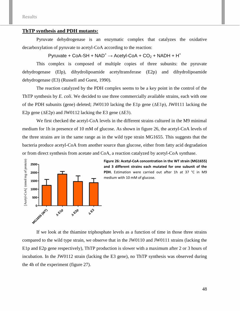
Results
48
ThTP synthesis and PDH mutants:
Pyruvate dehydrogenase is an enzymatic complex that catalyzes the oxidative
decarboxylation of pyruvate to acetyl-CoA according to the reaction:
Pyruvate + CoA-SH + NAD+ → Acetyl-CoA + CO2 + NADH + H+
This complex is composed of multiple copies of three subunits: the pyruvate
dehydrogenase (Elp), dihydrolipoamide acetyltransferase (E2p) and dihydrolipoamide
dehydrogenase (E3) (Russell and Guest, 1990).
The reaction catalyzed by the PDH complex seems to be a key point in the control of the
ThTP synthesis by E. coli. We decided to use three commercially available strains, each with one
of the PDH subunits (gene) deleted; JW0110 lacking the E1p gene (∆E1p), JW0111 lacking the
E2p gene (∆E2p) and JW0112 lacking the E3 gene (∆E3).
We first checked the acetyl-CoA levels in the different strains cultured in the M9 minimal
medium for 1h in presence of 10 mM of glucose. As shown in figure 26, the acetyl-CoA levels of
the three strains are in the same range as in the wild type strain MG1655. This suggests that the
bacteria produce acetyl-CoA from another source than glucose, either from fatty acid degradation
or from direct synthesis from acetate and CoA, a reaction catalyzed by acetyl-CoA synthase.
MG
1655
(WT)
E1p
E2p
E
3
0
500
1000
1500
2000
2500
Figure 26: Acetyl-CoA concentration in the WT strain (MG1655)
and 3 different strains each mutated for one subunit of the
PDH. Estimation were carried out after 1h at 37 °C in M9
medium with 10 mM of glucose.
If we look at the thiamine triphosphate levels as a function of time in those three strains
compared to the wild type strain, we observe that in the JW0110 and JW0111 strains (lacking the
E1p and E2p gene respectively), ThTP production is slower with a maximum after 2 or 3 hours of
incubation. In the JW0112 strain (lacking the E3 gene), no ThTP synthesis was observed during
the 4h of the experiment (figure 27).

Results
49
MG1655 (WT)
∆ E1p
∆ E2p
∆ E3
Figure 27: Kinetics of ThTP production in the WT strain (MG1655) and 3 different strains each mutated for one of
the subunits of PDH during incubation at 37 °C in M9 medium with 10 mM of glucose. From Pirson, 2011
The absence of ThTP in the JW0112 strain could be due to a general metabolic defect due
to the mutation. Indeed, we observed that the growth of that strain was very slow compared to the
WT strain. The gene that encodes the E3 subunit of the PDH complex, lpdA, also codes for the
E3 subunit of the OGDH complex and the L-protein of the glycine cleavage (GC) complex.
Moreover, this enzyme is involved in the redox status of the bacteria (Feeney et al. 2011). Those
multiple defects could provide an explanation for the absence of ThTP. Hence we decided to
study strains mutated for the other subunit of the OGDH and GC complexes to assess whether
those complexes are involved in ThTP production.
ThTP synthesis and OGDH mutant:
The α-ketoglutarate dehydrogenase is an enzymatic complex that catalyzes the oxidative
decarboxylation of α-ketoglutarate to succinyl-CoA according to the reaction:
α-ketoglutarate + CoA-SH + NAD+ → Succinyl-CoA + CO2 + NADH + H+.
In E. coli, it is composed of multiple copies of three subunits: the α-ketoglutarate
dehydrogenase (Elo), the dihydrolipoamide succinyltransferase (E2o) and the dihydrolipoamide
dehydrogenase (E3) (Derosier et al. 1971).
Since the E3 subunit of the PDH complex is also the E3 subunit of the OGDH complex,
we wondered whether the absence of ThTP in the JW0112 mutant was due to the lack of a
functional OGDH complex. We decided to study the mutant JW0715 where the E1o subunit gene
is deleted (∆E1o) and studied the time-course of the appearance of ThTP every hour for 4h
(figure 28). We could observe that the ThTP synthesis was normal.

Results
50
Figure 28: Kinetics of ThTP production in the WT strain (MG1655) and the ∆E1o strain (JW0715) during incubation
at 37 °C in M9 medium with 10 mM of glucose.
From those results, we can conclude that the mutation of E1o gene of the OGDH complex
does not affect ThTP production, while the deletion of the E1p and E2p genes of the PDH
complex only delays the production of ThTP. The mutation of the subunit specific of the GC
complex did not affect the ThTP production either (data not shown). Only the deletion of the
lpdA gene, which affects all those enzymatic complexes, totally prevents the appearance of ThTP
but also strongly impacts on the general metabolism of the bacteria.
There are two points of view to analyze those results, depending whether we consider that
ThTP accumulation is due to an activator that changes the conformation of the FoF1 ATP-
synthase, or that ThTP is a “non-classical metabolite formed by side activities” of that same
enzyme.
The delay of appearance of ThTP in the mutants E1p and E2p can be due to a delay in the
synthesis of the activator or a delay in the activation of the FoF1 ATP-synthase due to the lack of
PDH activity. When the bacteria are grown in the LB medium, rich in amino acids and in
energetic substrates, the metabolism is not much affected by the PDH mutation since the pyruvate
can easily be replaced by another source for acetyl-CoA synthesis. When they are placed in
minimum medium with glucose as the carbon source, they have to adapt their metabolism. The
glutamate necessary for the amino acid production is then derived from the α-ketoglutarate. All
those change either delay the production of the activator or delay the production of NADH and
FADH2.

Results
51
ThTP synthesis, OGDH mutant and glyoxylate cycle over-activation:
The Krebs cycle starts with one molecule of oxaloacetate and consumes one molecule of
acetyl-CoA. At the end of the cycle, oxaloacetate is restored and two molecules of CO2 are
produced. In bacteria, another metabolic pathway exists, the glyoxylate cycle which also starts
with one molecule of oxaloacetate but consumes two molecules of acetyl-CoA. At the end of that
cycle, oxaloacetate is restored but instead of releasing two molecules of CO2 it produces a second
oxaloacetate molecule (figure 29). Thanks to that metabolic pathway, the carbon atoms from the
acetyl-CoA produced from fatty acid degradation are not lost as CO2 but instead can be used for
neoglucogenesis.
Figure 29: TCA or glyoxylate cycle and gluconeogenesis. The scheme depicts the basic enzymatic steps in the TCA
cycle (black lines), which is common to all organisms, those of the glyoxylate cycle (dashed lines), which is specific
to microorganisms and plants, and those steps shared by both cycles (bold lines) and the initial reaction of
gluconeogenesis (shaded lines). PEP: phosphoenolpyruvate.
From Lorenz and Fink, 2002

Results
52
We had a mutant strain of E. coli with a deletion of the E1o gene as in the JW0715 strain
but the gene coding for the isocitrate lyase (the enzyme that directs isocitrate from the Krebs
cycle to the glyoxylate cycle) was constitutively active (∆E1o-ILconst). That strain, the JRG465,
grows at a much slower rate than the WT strain, even in LB (rich) medium, possibly because of
the lower levels of NADH produced by the glyoxylate cycle compared to the Krebs cycle.
We compared the kinetics of appearance of ThTP every hour for 4h in the WT strain and
in the JRG465 strain. We could observe that only a low amount of ThTP is produced in the
mutant, and that there is no ThTP peak (figure 30).
Figure 30: Kinetics of ThTP production in the WT strain (MG1655) and the ∆E1o-ILconst strain (JRG465) during
incubation at 37 °C in M9 medium with 10 mM of glucose.
Those results suggest that the absence of production of ThTP is due either to the
constitutive activation of the glyoxylate cycle which prevents the accumulation of the activator or
to the fact that the NADH production is too low to induce a proton-motive force (∆p) required to
energize ThTP synthesis.
If the activator is not synthesized in the ∆E1o-ILconst mutant strain but reaches normal
levels in the E1o mutant, it would mean that the activator is either isocitrate or an intermediate of
the Krebs cycle upstream of isocitrate or derived from them. Indeed, in the E1o mutant, isocitrate
can accumulate, while it is consumed by the isocitrate lyase in the JRG465 strain.

Results
53
ThTP synthesis and the two first enzymatic activities of the Krebs cycle:
In order to investigate the hypothesis of an activator that would involve either citrate or
isocitrate, we used 3 different mutant strains: One that was mutated at the level of the citrate
synthase gene (∆CS or JW0710), and two that were mutated for the aconitase A and B gene
(∆AcnA or JW1268 and ∆AcnB or JW0114 respectively). The citrate synthase catalyzes the
condensation of the acetyl-CoA with the oxaloacetate into citrate, releasing CoA-SH in the
process, according to the reaction:
oxaloacetate + acetyl-CoA + H2O → citrate + CoA-SH.
The aconitase catalyzes the stereo-specific isomerization of citrate into isocitrate
according to the reactions:
citrate ↔ cis-aconitate + H2O ↔ isocitrate.
Three different enzymes are responsible for the total aconitase activity in E. coli. Aconitase A is
responsible for ~75% of the aconitase activity while aconitase B is responsible of ~20%. In the
double knock out (∆acnAB) E. coli strain, there is still 5% of residual aconitase activity but the
enzyme responsible is still unidentified (Gruer and Guest, 1994; Blank et al. 2002).
Those three mutants had a growth rate similar to the WT strain. If we look at the ThTP
levels as a function of time in those strains compared to the wild type when they are grown in a
minimal medium with 10 mM of glucose, we observe that the ThTP production is reduced in the
citrate synthase and aconitase A mutant (figure 31). The aconitase B mutant strain had a ThTP
production that was normal but most of the aconitase activity was conserved, so the metabolism
was not much affected (data not show).
Figure 31: Kinetics of ThTP production in the WT strain (MG1655) and in A: the ∆CS strain (JW0710) or in B:
∆AcnA strain (JW1268) during incubation at 37 °C in M9 medium with 10 mM of glucose.
B A

Results
54
From those results we can conclude that the production of isocitrate is mandatory for an
optimal production of ThTP. Either because the production of the activator needs the
accumulation of isocitrate or the impairment of the Krebs cycle in those mutant strains reduces
the speed of production of ThTP by the ATP synthase.
ThTP production and inhibition of the FoF1 ATP-synthase by octyl-α-cetoglutarate:
Recently, it was shown that intermediates of the Krebs cycle can modulate the FoF1 ATP-
synthase activity and α-ketoglutarate was shown to inhibit the ATP-synthase but not the
respiratory chain (Chin et al. 2014). We used the 2-oxo-1-octyl ester pentanedioic acid or octyl-
α-ketoglutarate (figure 32), an α-ketoglutarate precursor which can enter the cell by diffusion
thanks to its hydrophobic tail. Once in the cells, it is hydrolyzed and α-ketoglutarate is released in
the cytoplasm.
Figure 32: On the left, the α-ketoglutarate and on the right, the octyl-α-ketoglutarate
When we incubated the bacteria in the M9 minimal medium with 10 mM of glucose and
increasing concentration of octyl-α-ketoglutarate, we observed a concentration-dependent
inhibition of ThTP accumulation after 20 and 60 minutes (figure 33).
0 m
M
0,1m
M
0,5m
M1m
M
0 m
M
0,1m
M
0,5m
M1m
M
0
50
100
150 20 min
1h
[octyl -ketoglutarate]
[Th
TP
] (%
of
0 m
M)

Results
55
Figure 33: Effect of increasing concentrations of octyl-α-ketoglutarate on the cell ThTP concentration in the WT
bacterial strain (MG1655) incubated at 37 °C in M9 medium with 10 mM of glucose.
If we consider those results in the perspective of the activator hypothesis, it is intriguing
that the increase of the α-ketoglutarate concentration in the bacteria has a negative impact on the
ThTP accumulation while we were expecting an increased activator synthesis. Nevertheless,
since the α-ketoglutarate inhibits the ATP-synthase, we can infer that the inhibition of the
enzyme is stronger than the increase of the activator. If we consider ThTP as a side product of the
ATP-synthase, it is consistent that the inhibition of this enzyme impacts the synthesis of ThTP.
Mammalian ThTPase expression in E. coli:
If the bacteria indeed synthesize an activator that acts on the ATP-synthase to increase the
production of ThTP, it would mean that the ThTP production helps the bacteria to adapt to a
nutritional shift (from the LB (rich) medium to the M9 minimal medium (poor)). It means that if
we can degrade the ThTP shortly after its synthesis, it would be unable to fulfill its role and thus
affect the bacterial adaptation and delay its growth.
To achieve this goal, we decided to express the specific mammalian 25-kDa ThTPase in
the bacteria. In 2004, our lab had already succeeded in overexpressing the human ThTPase in E.
coli using an expression vector. A slowing down of the growth was observed but only after 5h,
i.e. at the end of the transient ThTP peak and during the exponential phase of growth (Lakaye et
al. 2004c). We decided to include the mouse ThTPase (mThTPase) gene which has a higher
catalytic efficiency than the human enzyme, inside the genome of the bacteria. That construction
allowed us to express the ThTPase at low levels and thus prevent the strong metabolic
disturbance caused by forcing the expression of a high quantity of a heterologous protein. We
also expressed, in another clone, the K65A mutant of the mThTPase which is catalytically
inactive but where the structure of the protein is conserved (Delvaux et al. 2013). It is thus the
best control we could get to assess the disturbance due to the expression of a heterologous protein
similar to the ThTPase but without any ThTPase activity. Those two proteins were inserted into
the NM543 bacteria strain and thus we obtained two new strains where the expression of either
the WT or the K65A mThTPase is under the dependence of an inducible promoter.
The WT MG1655 strain, the “mother” strain NM543 and the two newly engineered
strains ThTPase WT I and ThTPase K65A I were grown overnight in LB medium, then the
expression of the ThTPases was induced for 3h. Afterwards, the bacteria were diluted to an
optical density (DO600) of 0,03 in the M9 minimal medium with 10 mM of glucose. They were

Results
56
incubated at 37 °C and the growth was monitored every hour by measuring the DO600. No
difference could be observed between the four strains. After 1h, we assessed that the ThTP level
was low in the ThTPase WT I strain but normal in the others (figure 34).
Figure 34: Growth and ThTP production in four bacterial strains incubated at 37 °C in M9 medium with 10 mM of
glucose. The ThTP concentration was assessed after 1h of growth.
The absence of effect is not in agreement with what was observed in 2004. However, the
expression levels of the protein from a plasmid present in the bacteria in multiple copies was
much higher than in our models with only one copy of the gene inserted in the genome. It could
be estimated that ThTPase accounted for at least 25% of the total protein in the bacteria. From the
present results (obtained without high overexpression of the 25-kDa ThTPase) it appears that
ThTP is not required for growth when the bacteria are placed in a condition of amino-acid
starvation with glucose as a carbon source. This supports the hypothesis that ThTP is a side
product of the ATP-synthase with no apparent physiological role.
Our results also indicated that ThTP is not toxic for the bacteria, as growth is not speeded
up by its hydrolysis. While the present results strongly suggest that ThTP plays no important role
in bacterial growth or survival, it remains hard to explain why ThTP is produced only under
specific conditions, i.e. amino acid deficiency and presence of a substrate yielding pyruvate. If
ThTP is a byproduct of ATP synthase activity, it should be correlated with a high proton-motive

Results
57
force, which itself would occur when the [NADH] / [NAD+] ratio is high. Further studies are
required to assess the exact role of these parameters.
ThTP synthesis and oxidative stress:
The NADH and the FADH2 produced in the Krebs cycle are used to fuel the electron
transport chain (ETC), to expel protons from the cell and create an electrochemical gradient. This
gradient is used as the energy source that allows the FoF1 ATP-synthase to catalyze the formation
of ATP or ThTP.
The ETC is also a primary source for reactive oxygen species (ROS) and thus of oxidative
stress (Wang et al. 2013). In turn, oxidative stress affects the activity of the ATP-synthase by
inducing oxidative post-translational modifications (Ox-PTMs) of the enzyme. So, under our
conditions where glucose is consumed within minutes, most of NADH and FADH2 is produced
through the Krebs cycle. In that process, oxidative stress is more likely to arise.
To test whether the oxidative stress can affect the production of ThTP, we added to the
culture medium of the WT bacterial strain increasing concentrations of HOCl ranging from 0 to
100 µM (figure 35).
0 20 40 60 80 100
0
100
200
300
400
500
600
µM HOCl
[Th
TP
] (%
of
0 µ
M)
Figure 35: Effect of increasing concentration of HOCl on ThTP concentration in the WT bacterial strain (MG1655)
incubated for 1h at 37 °C in M9 medium with 10 mM of glucose.

Results
58
We could observe that, up to 80 µM, HOCl dose-dependently increased ThTP production
but at 100 µM, ThTP production was back to normal and would probably continue to decrease as
the excess of HOCl is killing the bacteria. It thus appears that a limited oxidative stress enhances
the production of ThTP. It was observed by different authors that oxidative stress affect the FoF1
ATP-synthase activity (Hannum et al. 1995; Rexroth et al. 2012; Wang et al. 2013). We can thus
hypothesize that it could affect the affinity of the enzyme for different substrates, either by
decreasing the affinity for ADP or increasing the affinity for ThDP. Those result are also in favor
of the hypothesis that ThTP is a side product of the enzyme.
Conclusions
A summary of the results is shown in table 7, page 60.
Investigations carried out in our laboratory during the last decade have shown that, under
specific conditions of cellular stress, E. coli cells can accumulate ThTP. This production requires
a carbon source yielding pyruvate and occurs in response to a nutritional shift, i.e. amino acid
deprivation. It was thus suspected that ThTP could act as an intracellular messenger or
“alarmone” required for the adaptation of the bacteria to specific condition of stress. It was
intriguing that the energy substrate needed for ThTP synthesis was either pyruvate or yielding
pyruvate, e.g. glucose or lactate. No ThTP was produced in the presence of other substrates such
as malate (though cellular ATP level was normal). It was therefore postulated (Gigliobianco et al.
2013) that a metabolite derived from pyruvate was required to induce ThTP synthesis by FoF1
ATP-synthase. Previous results as well as our own suggest that isocitrate (or an unknow
compound derived from isocitrate) may act as a modifier of F1 so that the ATP-synthase catalyzes
the synthesis of ThTP rather than ATP.
However, the physiological relevance of the ThTP accumulation after nutritional shift was
never assessed. If ThTP is indeed an intracellular messenger produced in response to specific
conditions of cellular stress, it should exert some beneficial effects on the metabolism, growth or
survival of the bacteria when they are submitted to a nutritional shift. We attempted to answer the
question by including a mouse ThTPase in the bacterial genome. As this enzyme hydrolyzes
ThTP with high specificity and catalytic efficiency, the mutated bacteria were unable to
accumulate any significant amount of ThTP. However, there was no difference in growth rate
(after nutritional shift) between wild type bacteria and the mutant. It must be concluded that
ThTP plays no important role in the adaptation of E. coli cells to amino acid deficiency, at least

Results
59
as far as survival and growth are concerned. Apparently, the well-known “stringent response” to
nutritional shift is sufficient for adaptation of E. coli cells. It thus cannot be excluded that ThTP
may be a “non-classical metabolite formed by side activities” of the FoF1 ATP-synthase (Linster
et al. 2011). We also have results showing that the oxidative stress, which affect the ATP-
synthase activity, enhance the ThTP production after the nutritional shift.
Those results, without being a definitive proof, lead us to believe that ThTP production is
not dependent of an activator that change the conformation of the FoF1 ATP-synthase as we
previously claimed (Gigliobianco et al. 2013) but is dependent on a high activation of this
enzyme. Thus, the high levels of NADH and FADH2, achieved by the entry of high amount of
acetyl-CoA in the Krebs cycle are the key for the production of ThTP thanks to the high proton-
motive force and the concomitant oxidative stress. In addition, the accumulation of the Krebs
cycle intermediate α-ketoglutarate under certain condition may inhibit ThTP synthesis by
inhibition of fof1-ATP synthase.

Condition Effect on the bacteria ThTP production
Mutant PDH JW0110-JW0111
(E1p – E2p KO)
Loss of the PDH complex activity The ThTP peak is delayed
Mutant JW0112 (E3 KO) Loss of the PDH, OGDH and GC complex activities, strong
perturbation of the metabolism
No ThTP production
Mutant JW0715 (E1o KO) Loss of the OGDH complex activity ThTP production normal
Mutant JRG465 (E1o KO +
isocitrate lyase constitutive)
Loss of the OGDH complex activity + constitutive activation of the
glyoxylate cycle. General metabolism slightly affected.
No ThTP production
Mutant JW0710 (CS KO) Loss of the citrate synthase activity Production of ThTP reduced
Mutant JW1268 (Aconitase A
KO)
Loss of 75% of the aconitase activity Production of ThTP reduced
WT (MG1655) + octyl-α-
ketoglutrate
Increased α-ketoglutarate concentration, inhibition of FoF1 ATP-
synthase
Dose dependent inhibition of
the ThTP production
WT (NM543) strain
overexpressing the mThTPase
The mouse ThTPase hydrolyzes ThTP as soon as it is produced, no
effect on the growth
No ThTP
WT (MG1655) + HOCl Induction of an oxidative stress increasing with the concentration till
it induced strong metabolic problems and eventually the death of the
bacteria
Dose dependent enhancement
of the ThTP production
Table 7: Summary of the results obtained with the different mutant strain and with the different compound used when the E. coli bacteria are incubated at
37 °C in M9 medium with 10 mM of glucose.

Results
61
Chapter 3: Possible role of THTP and 25-kDa zThTPase on
zebrafish development
ThTP may be a side product in E. coli but what about higher organism? It was shown that
ThTP can modulate the activity of a high conductance anion channel (Bettendorff et al. 1993),
that it can phosphorylate some proteins such as the rapsyn (Nghiêm et al. 2000) and that a highly
specific ThTPase is dedicated to its degradation (Lakaye et al. 2002). Note that in E. coli, no
specific ThTP hydrolyzing enzyme has been described.
To study the role of ThTP in eukaryotic cells, there are two possible approaches: to block
its synthesis or to induce its accumulation. In mammalian cells, as in E. coli, ThTP is synthesized
by FoF1 ATP-synthase (Gangolf et al. 2010). Therefore it would be impossible to block ThTP
synthesis without also affecting ATP synthesis. In eukaryotic cells, the only known condition
under which ThTP accumulates is when 25-kDa ThTPase activity is very low (e.g. in pig tissues)
or when the gene is absent (e.g. in birds). To better understand the role of ThTP, we decided to
prevent its degradation. Two different approaches were used to achieve that goal. Both involve
interfering with the ThTPase expression. In this chapter we will discuss the use of morpholinos in
the zebrafish embryo, while in the next chapter we will discuss the knock-out approach in the
mouse.
Characterization of the zTHTPA gene:
In mammals, ThTPase is a ubiquitous enzyme and its only known role is to hydrolyze
ThTP. In zebrafish, almost nothing is known about the 25-kDa ThTPase except that the catalytic
activity of the recombinant enzyme is 10-100 fold lower than that of the mouse or human
enzymes (Delvaux et al. 2013). According to the ZFIN database (zfin.org), the ThTPase gene is
present on the 24th
chromosome and 4 different mRNA seem to exist (figure 36). The transcript
thtpa-005 lacks most of the coding sequence and does not seem relevant compared to the others.

Results
62
Figure 36: ZFIN database data for the different transcripts of zebrafish thiamine triphosphatase (zThTPase). On
the top part, we can see the whole 24th
chromosome, and below it, the enlargement of the genomic region
containing the thiamine triphosphatase gene. The thick blue lines correspond to the genes, the one highlighted in
yellow is the zThTPase gene. In red, we can see the different transcripts of the zThTPase. The thin red lines
correspond to the introns while the thick one correspond to the exons. The full part of those thick lines
corresponds to the coding sequence of the mRNA.
If we look in more detail at the 3 other transcripts, two major differences with the
mammalian gene become apparent. First, the existence of 4 exons is a notable difference with the
mammalian gene which in most cases comprises only 2 exons. Second we can see that the exon 3
and the coding sequence in exon 4 are the same for all of them. The exon 2 and the coding
sequence of the transcripts thtpa-001 and thtpa-003 are also the same, while the exon 2 of the
thtpa-002 is longer and the coding sequence is longer by 58 nucleotides, though the reading
frame is conserved (figure 37,39).
Figure 37: ZFIN database data for the thtpa-001-002-003 transcripts of the zThTPase The thin lines correspond to
the introns while the thick ones correspond to the exon. The full boxes of the thick lines correspond to the coding
sequences of the mRNA. The second frame is a higher magnification of the two first exons of the three transcripts.
We checked that the thtpa-002 transcript and at least one of the thtpa-001 or thtpa-003
transcripts (there is only a 2 nucleotides difference among them, thus they are impossible to
distinguish under our experimental conditions) were expressed in the zebrafish egg at different
time points (figure 38). We first isolated the RNA of the fish at 0, 2, 4, 6, 8, 24 and 48 hour post

Results
63
fertilization (hpf). The RNAs were retrotranscripted into cDNAs and a PCR allowed us to
observe the different transcripts. We observed that at least two of the transcripts were expressed
at every time point. The sizes of the amplified fragments correspond to the splice forms of the
transcripts and thus they are not amplified from the genomic DNA.
Figure 38: Amplification of the different thtpa transcript: 3% agarose gel; the number on the left are the size in bp
of the ladder, the numbers on top are the number of hours after fertilization at which the sample were taken. The
expected sizes for the bands were: 195 bp for thtpa-001, 197 bp for thtpa-003 and 252 bp for thtpa-002.
A careful look at the mRNA sequence tells us that in the exon 2, two (or even three for
thtpa-002) start codons are in phase. On the thtpa-001-003 transcript, there is a second potential
start codon 15 amino acid downstream of the referenced start codon. Those two potential start
codons are also included in the thtpa-002 transcript in addition to the start codon which is
referenced in the database (figure ).
Figure 39: ZFIN database for the exon 2 of the transcript thtpa-001-002-003. The selected frame was inverted to
show the complementary sequence which contains the ATG. The three red arrows show the three start codons.
Morpholino-induced zThTPase expression reduction
We decided to use antisense morpholinos directed against the zThTPase to get a better
insight in its role in general and especially during embryogenesis. Morpholinos were used
because this is a fast and efficient way to reduce the expression of a specific gene in zebrafish
embryo.
Two strategies were tested, a morpholino which inhibits the splicing and a morpholino
which inhibits the initiation of the translation. The latter was directed against the referenced start
codon of the thtpa-001-003 transcript but it blocks neither the potential start codon which is
downstream nor the one on the thtpa-002 transcript. It was thus not really efficient (data not
shown) and we used only the splicing morpholino in further investigations.

Results
64
First, a dose-response analysis was made on a total of 1890 injected embryos: the eggs
were injected right after spawning with either a control morpholino (4 ng/egg) or with the
splicing morpholino directed against the zThTPase (1 – 2 - 4 ng/egg) and observed 24 hours post
fertilization (hpf) and 48 hpf.
Although not highly specific, strong phenotypic alterations were observed: there was a
general developmental delay or a brain development delay, pericardial edema, cerebral edema,
brain necrosis, abnormal somite and abnormal hemostasis (table 8 and 9). The fish were
categorized as having a normal, light, moderate or severe phenotype, or dead. We can see that the
morpholino act in a dose-dependent manner (Figure 40). Those results are the first showing that
the absence of the THTPA transcript alters fish development.
24 hpf
Normal
Light
Developmental delay
Moderate:
malformation
+
brain necrosis (arrow)
Severe:
Strong developmental
defect
Table 8 : List of the phenotype observe in zebrafish embryos 24 hpf and the most common alteration observed.

Results
65
48 hpf
Normal
Light:
hemostasis (arrow)
Moderate:
cerebral edema (arrow)
Moderate:
pericardial edema (arrow) and
hemostasis (arrow head)
Severe:
No heart at all
+
strong malformation
Table 9: List of the phenotype observe in zebrafish embryos 48 hpf and the most common alteration observed.

Results
66
24 hpf
MO C
(n=3
67)
MO 1
ng (n
=404
)
MO 2
ng (n
=501
)
MO 4
ng (n
=618
)
0
20
40
60
80
100 Normal
light
moderte
severe
dead
nu
mb
er
of
fish
(%
)
48 hpf
MO C
(n=3
67)
MO 1
ng (n
=404
)
MO 2
ng (n
=501
)
MO 4
ng (n
=618
)
0
20
40
60
80
100
Normal
light
moderate
severe
dead
nu
mb
er
of
fish
(%
)
Figure 40: Dose response effect of the splicing morpholino directed against the zThTPase after injection into
zebrafish embryos. For each condition, phenotypes are classified as normal, light, moderate, severe or dead and
expressed as a percentage of the total number of eggs for that condition. Fish were observed at 24 hpf and 48hpf.
When we take a closer look at the results, we observe that in the fish injected with the
control morpholino, almost 80% of the embryos are normal and most of the remaining 20% are
only slightly affected, with almost no distinction between the two days of observation.
In the fish injected with the morpholino directed against the zThTPase at 24 hour post
fertilization, we can observe a decrease in the percentage of normal fish down to less than 20 %
for the 4 ng injected fish. The percentage of only lightly affected fish doubled compared to the
control for the 1 or 2 ng and almost tripled for the 4 ng injected fish. The percentages of
moderately and severely affected fish also increased from 1 to 4 ng injected fish.
The next day, 48 hpf, we can see that the percentage of normal fish remains almost
constant except for the 1 ng injected that seemed to slightly recover. We can also observe that
most of the severely affected fish were either dead or had to some extent recovered to moderately
affected. The 4 ng injected fish are the most affected with almost 10 % of dead fish, about 70% of
the surviving fish were affected, half of them lightly and the other half moderately.
In order to attribute the above-described effects to the morpholino, it is important to
demonstrate a reduction of the spliced zThTPase transcript level. We used two different primer

Results
67
sets, the first being the same as the one used for the detection of the different transcripts and the
second targeting a region common to the three transcripts to increase the sensitivity. In both
cases, there was a strong reduction of the spliced transcript level in the injected embryo compared
to the non-injected one. We can only see a remaining shadow of the spliced amplicons in the
zThTPase morphant. We could also observe that the transcript levels were similar between the
control morphant and the non-injected fish.
Figure 41: detection of the sliced transcripts of the zThTPase: 3% agarose gel; the numbers on the left of each gel
are the size in bp of the ladder; each lane is the result of the PCR on a specific sample: lane 1, cDNA from 24hpf
eggs injected with the splicing morpholino against the zThTPase; lane 2, cDNA from 24hpf eggs injected with the
control morpholino; lane 3, cDNA from 24hpf non-injected eggs. The expected sizes for the bands on the gel on the
left are: 195bp for thtpa-001, 197bp for thtpa-003 and 252bp for thtpa-002, while the expected size for the band
on the gel on the right is 140bp.
To confirm the specificity of the observed effects, we tried to rescue the phenotype by
injecting the transcript of the mature zThTPase mRNA together with the morpholinos. As the
morpholino used is impairing the splicing, our rescue mRNA will not be affected. We chose to
work with a fixed concentration of 2 ng of morpholino injected per fish in order to target an
intermediate effect, leaving room for improvement or worsening of the effect. The eggs were
injected right after spawning with either a control morpholino (2 ng/egg) or with the splicing
morpholino directed against the zThTPase (2 ng/egg), in each case, with or without the rescuing
mRNA (100 pg/egg).The fish were observed at 24 hpf and 48 hpf. (figure 42).
If we look at the rescue results at 24 hpf, the injection of the morpholino directed against
the zThTPase induces a decrease of the normal fish to 50% with again a doubling of the lightly
affected fish compared to the control morpholino. We can also observe that the injection of the
mRNA coding the zThTPase leads to a distribution of the phenotype that is similar whether it is
injected together with the zThTPA or the control morpholino and close to the distribution
observed when the control morpholino was injected alone.
1 MO THTPA
2 MO CONT
3 Non-injected

Results
68
Rescue 24 hpf
MO C
(n=3
12)
MO 2
ng (n
=324
)
MO 2
ng +
RNA W
T (100
pg) (
n=256
)
MO C
+ R
NA W
T (100
pg) (
n=155
)
0
20
40
60
80
100Normal
light
moderate
severe
dead
nu
mb
er
of
fish
(%
)
Rescue 48 hpf
MO C
(n=3
12)
MO 2
ng (n
=324
)
MO 2
ng +
RNA W
T (100
pg) (
n=258
)
MO C
+ R
NA W
T (100
pg) (
n=155
)
0
20
40
60
80
100
Normal
light
moderate
severe
dead
nu
mb
er
of
fish
(%
)
Figure 42: Effect of the rescuing mRNA coding for the zThTPase on the phenotype of zebrafish embryo injected
with control or zThTPase splicing inhibitor morpholino. For each condition, phenotypes are classified as normal,
light, moderate, severe or dead and expressed as percentage of the total number of eggs for that condition. Fish
were observed at 24 hpf and 48 hpf.
At 48 hpf, we can see that the rescued fish have a more intermediate distribution of the
phenotype, which can be explained by the degradation and the dilution of the RNA. These results
show that the fish phenotype is indeed rescued by co-injection of the zThTPase mRNA with the
morpholino suggesting that our zThTPase morpholino is indeed specific. Hence, the lack of the
ThTPase induces an unspecific but general impairment of the embryogenesis of the zebrafish.
To further confirm the specificity of the observed effects and to assess whether the
activity of the enzyme is required to rescue the phenotype, we decided to try the rescue protocol
using the coding sequence of the zThTPase with the K61A mutation. That lysine in zebrafish
ThTPase coding sequence is homologous to the lysine 65 (K65) in mouse or human. As already
mentioned, the mutation of the K65 to an alanine drastically reduces the catalytic activity but
does not affect the tridimensional structure of the enzyme (Delvaux et al. 2013).

Results
69
The transcript of the mature K61A zThTPase mRNA was injected with the morpholinos.
Once again, this transcript was not affected by the morpholino. We worked with the same
concentration of 2 ng of morpholino and 100 pg of RNA injected per fish. The eggs were injected
right after spawning with either the control morpholino (2 ng/egg) or with the splicing
morpholino directed against the zThTPase (2 ng/egg), in each case, with or without the K61A
zThTPase RNA (100 pg/egg).The fish were observed at 24 hpf and 48 hpf (figure 43).
As previously observed, injection of the morpholino (2 ng) was toxic for the embryos. In
contrast to what we observed with the WT mRNA, co-injection of the K61A mRNA appeared to
worsen the phenotype (both at 24 hpf and 48 hpf); in any event, no rescue was observed (figure
43). When the K61A RNA was co-injected with the control morpholino the phenotype
distribution was similar or slightly worsened compared to the control morpholino injected alone.
Rescue K61A 24 hpf
MO
C (n
=141
)
MO
2 n
g (n=1
96)
MO
2 n
g + R
NA K
61A (1
00 p
g) (n=1
75)
MO
C +
RNA K
61A (1
00 p
g) (n=1
68)
0
20
40
60
80
100Normal
light
moderate
severe
dead
nu
mb
er
of
fish
(%
)
Rescue K61A 48 hpf
MO C
(n=1
41)
MO 2
ng (n
=197
)
MO 2
ng +
RNA K
61A (1
00 p
g) (n=1
74)
MO C
+ R
NA K
61A (1
00 p
g) (n=1
68)
0
20
40
60
80
100
Normal
light
moderate
severe
dead
nu
mb
er
of
fish
(%
)
Figure 43: Effect of the rescuing mRNA with the coding for the K61A mutant of the zThTPase on the phenotype of
zebrafish embryos injected with control or zThTPase splicing inhibitor morpholino. For each condition,
phenotypes are classified as normal, light, moderate, severe or dead and expressed as a percentage of the total
number of eggs for that condition. Fish were observed at 24 hpf and 48hpf.

Results
70
HPLC measurement of thiamine derivatives in zebrafish embryo:
We wanted to measure ThTP and other thiamine derivatives in the embryo of zebrafish
and assess the increase of the ThTP levels in the injected fish but unfortunately, in the zebrafish
those measurements were impossible with the classical HPLC method (Bettendorff et al. 1991)
because of the presence of an auto-fluorescent compound that prevents the detection of any
thiamine derivative.
Conclusion
The organization of the ThTPase gene in the zebrafish is quite different from the one
found in its human or mouse counterparts. While the mammalian THTPA gene usually has two
exons and only one transcript, the zebrafish gene contains four exons and can be spliced in
different ways. We could observe that the different transcripts were present already in the egg
right after spawning, which means that the transcript is present before the fertilization of the eggs.
In the zebrafish, till 4 hpf, no new mRNA transcripts are synthesized. During the first hours, the
embryos start their development using only the maternal mRNA present in the egg. After those
initial 4 hours, the different transcripts were present at least till 48 hpf, which means that the
ThTPase could be involved in the development of the embryo.
When the ThTPase expression was reduced by the injection of a morpholino, we could
observe that the fish show developmental defects. The phenotype was partly restored when we
co-injected the morpholino with the mRNA coding for the ThTPase (rescue) but not when we
attempted the rescue with the K61A mutant of the ThTPase. Those results indicate that even if
the credibility of morpholinos as a tool for RNA interference has been questioned in the recent
literature (Kok et al. 2015), the effects that we observe seem specific and are likely caused by the
decrease in ThTPase activity.
To make sure that the decrease of ThTPase expression indeed increases the ThTP
concentration in embryos, it would have been interesting to measure the ThTP levels, but
unfortunately this was impossible for technical reasons.
From those results, we can wonder whether the phenotypes observed are due to the ThTP
accumulation which could be toxic for the developing embryo or if the ThTPase has some role
other than ThTP hydrolysis (as suggested by Delvaux 2012) which would be necessary for the
normal development of the fish.

Results
71
As previously discussed, both in mammalian brain and in E. coli, ThTP can be produced
either by cytoplasmic adenylate kinase or by FoF1 ATP-synthase. In both cases, ThTP production
could not be clearly related to a physiological function and accumulation of high amounts of
ThTP (e.g. in pig muscle and electrophorus electric organ) had apparently no toxic effects.
However, in the zebrafish, ThTP accumulation seems to interfere with the development of the
fish. If ThTP is indeed a side-product of enzyme producing ATP which is toxic in eukaryote
cells, the evolution of enzyme of the CYTH family towards hydrolysis of ThTP would prevent
the deleterious consequences of its accumulation on normal metabolism.

Results
72
Chapter 4: Knock-out of the mouse ThTPase:
Much information is available about mammalian ThTPase and the level of ThTP in
different organisms and tissues. Yet their metabolic function(s) or the pathways they may be
involved in, are still largely unknown.
For that reason, and in parallel to the zebrafish morphant which allows only the study of
developing animals and is based on the reduction of the protein levels by interfering with the
mRNA, we wanted to obtain a strain of mice knock-out for ThTPase.
Thiamine triphosphatase knock-out methods
In order to generate the THTPA KO transgenic line of mice, we ordered embryonic stem
(ES) cells clones from the Knockout Mouse Project (KOMP) repository at UC Davis
(www.komp.org). At the end of the validation test, they could provide us with two ES cell clones:
BC03 and BA09. In those cells, one allele of the ThTPase gene was deleted and replaced by the
“tm1(KOMP)Vlcg” cassette (figure 44). That cassette contained in the 5’ to 3’ order:
the coding sequence of the beta-galactosidase (lacZ),
the SV40 polyadenylation signal,
the first lox site,
the hUBC promoter (the promoter of the human Ubiquitin C),
the em7 promoter (a synthetic promoter based on the T7 promoter, allowing bacterial
expression of the downstream gene),
the coding sequence of the neomycin resistance gene,
the mouse phosphoglycerate kinase (mPkg) polyadenylation signal,
the second lox site.
Figure 44: Scheme of the “tm1(KOMP)Vlcg” cassette used to replace the THTPA gene in the BC03 and the BA09
clones.
We sequenced the junction site between the mouse DNA and the cassette to make sure
that it was well inserted. As we can see on figure 45, no other known gene was affected by the

Results
73
deletion and the whole coding sequence of the ThTPase was removed, while the 5’ and 3’
untranslated regions were conserved.
Figure 45: NCBI annotation of the Mus musculus genome on the surrounding region of the THTPA gene of the
14th
chromosome. The numbers on the top line correspond to the sequence of the chromosome, the green
horizontal lines represent the genes along this sequence (light green: untranslated region, dark green: coding
sequence, thin green line: intron). The green and the blue vertical lines are there to show the part of the genome
that was deleted and replaced by the tm1(KOMP)Vlcg cassette.
The ES cells were injected into blastocysts that were then implanted into the uterus of a
foster mother mouse. The surviving blastocysts developed into the mouse uterus until the
delivery. Some of the pups were chimerae which, as they grew, became black and white (figure
46) since their cells were derived either from the ES cells or the blastocysts. The genetic
background of the ES cells is C57BL/6NTac and thus black, while the blastocysts were generated
from mouse from Charles River with a CD-1® IGS (part of the International Genetic
Standardization program) white genetic background. By looking at the proportion of black fur we
could estimate the percentage of chimerism of our animals. This percentage gave us an idea of
the general chimerism but it did not reflect the percentage of chimerism among the stem cells
producing the sperm.
Figure 46: picture of a 60% chimeric mouse.
Since the ES cells have a male genotype (XY), they are unable to differentiate into mature
oocytes and thus only the male chimerae are of interest. The male chimerae obtained were
crossed with wild type (WT) C57Black female mice. While the females could only transmit a
black genetic background, the males could transmit either a white or a black genetic background.

Results
74
Hence, the descendants are either brownish or black. If the pups obtained have a brownish fur, it
means that the sperm from the chimerae that has fertilized the oocyte was derived from the
blastocyst. On the other hand, if the pups are black, it means that the sperm was derived from the
ES cells and thus has a chance to carry the mutation.
As only one of the ThTPase alleles of the ES cells is invalidated, only half of the sperms
derived from them carry the mutated allele of the ThTPase. It means that among the black pups,
half should be WT while the others half should be heterozygous with respect to the ThTPase gene
(figure 47).
Figure 47: diagram of the process to obtain heterozygous mice from the heterozygous embryonic stem cells.

Results
75
Results obtained from the THTPA chimerae:
Twelve chimerae were generated and a total of 455 pups were obtained. We genotyped all
of them and, surprisingly, they were all wild type (figure 48). Among them, 306 were black,
which means that statistically we should have had around 153 heterozygous pups.
Figure 48: detection of the WT and invalidated allele of the ThTPase: 2% agarose gel: electrophoresis of the PCR
product of the genotyping of the chimeric pups. The three first lanes correspond to three different pups, the fourth
is the size ladder (from top: 400 bp-200 bp), followed by the positive control (DNA form the mutated ES cells), and
then the negative control (DNA from a WT mouse). The two expected sizes for the amplicon were: 307 bp for the
mutated gene and 444 bp for the WT allele.
In order to check whether our chimerae were indeed carrying the mutation, we sacrificed
two of them and collected tissue samples from different organs (the brain, the lungs, the heart, the
skin and the testis). We extracted the genomic DNA from those samples and genotyped them.
Those tissues derived from the ES cells and from the blastocyst are partly THTPA+/+
and partly
THTPA+/-
. We indeed observed a signal specific of the mutated gene in every sample tested. That
signal was of variable intensity depending of the ratio between the two genetic identities in the
cells composing the sample tested (figure 49).
Figure 49: detection of the WT and invalidated allele of the ThTPase: 2% agarose gel of the PCR product of the
genotyping of different chimerae tissues: 1:brain; 2:lung; 3:heart; 4:liver; 5:skin; 6:testis; size ladder (from top: 800-
600-400-200 bp); C+:ES cells; C-:WT mouse.
Since the chimerae carried the mutation but never transmitted it, we decided to analyze
the sperm of those animals. We had two chimerae with a sex reversal. A sex reversal means that
the phenotypic sex of the chimera is different from the genotypic sex of the blastocyst. That
phenomenon is made possible by the male genotype of the mutated ES cells. When those cells are
injected in a female blastocyst, they can drive the phenotypic sex to be male (by producing the
Pups SL C+ C-

Results
76
SRY peptide). In that case, all the germ cells derive from the injected ES cells since the XX
blastocyst cells cannot differentiate into mature sperms. We could detect the chimerae with a high
probability of sex reversal because all their pups were black. In the chimerae with a sex reversal,
after meiosis, half of the immature sperm cells would be WT and the other half knock-out for the
THTPA gene. The ratio between them should thus be close to one.
We thus decided to analyze, by qPCR, the genomic DNA extracted from the sperm of two
mice with a sex reversal to obtain the ratio between the WT and the mutated allele. First, the
sperm cells were collected from the epididymis and the ductus deferens, and then we purified part
of the mobile sperm cells using two isolation methods: the swim-up and the swim-out. We thus
obtained four different fractions per chimera: the Swim Up Lower part (SUL), the Swim Up
Superior part (SUS), the Swim Out Central part (SOC) and the Swim Out Peripheral part (SOP).
After qPCR we obtained a ratio of 1 KO sperm for 100 or 1000 WT sperm. We did not see any
difference between the mobile fraction (SUS-SOP) and the mixed fraction (SUL-SOC), except
that the amount of material for the SUS fraction of the 10% chimera was too low to detect any
signal for the mutated allele (figure 50).
Ratio WT vs KO spermatozoids
10%
chim
era
60%
chim
era
ES c
ells
0.1
1
10
100
1000
ES cells
Swim Up Lower part (SUL)
Swim Out Central part (SOC)
Swim Out Peripheral part (SOP)
Swim Up Superior part (SUS)
WT
/ K
O a
llele
(ab
so
lute
co
py n
um
ber)
Figure 50: Ratio of the WT and invalidated allele of the ThTPase in the genomic sperm DNA.

Results
77
Conclusions
Even after genotyping more than 300 pups with the expected fur color, we were unable to
obtain heterozygous mice for the ThTPase and thus to obtain any KO mouse. Those results are
not very surprising if we consider the results obtained with the morphant zebrafish. A decrease in
the ThTPase expression appeared to be harmful for the developing embryos; in mice, the total
absence of the enzyme after meiosis is apparently harmful for the developing sperm cells as well.
Once again, those results are in agreement with the conclusions drawn from our results in
bacteria and in the zebrafish: ThTP appears to be a side product of the FoF1 ATP-synthase and its
accumulation might be harmful, at least in some cell types. The sperm middle piece is rich in
mitochondria and thus could produce a localized accumulation of ThTP in the KO cells. If, as
hypothesized in the zebrafish, ThTP is toxic in some way, it could prevent the development of the
sperm and thus the transmission of the THTPA invalidation to the next generation.
From those results, it would appear that, in vertebrates, the 25-kDa ThTPase can play an
essential role in some instances, but this seems to be restricted to particular cell types. ThTPase
activity may be essential for the development of fish embryos and, in mammals, the protein could
play a role in sperm differentiation. In the pig, however, the protein is largely expressed but is
nearly inactive. Therefore, pig tissues contain higher amounts of ThTP, apparently with no
harmful effects. The same is true for birds, where the ThTPase gene is lost.
It must be concluded that, although genes of the CYTH superfamily are present in most
organisms and ThTPase activity is conserved from Cnidarians (Nematostella) to vertebrates, this
enzyme may be required for the degradation of the unwanted thiamine derivative, ThTP, in some
but not all animal cells.


Discussion
And
Conclusion


Discussion and conclusion
78
When one thinks about triphosphorylated molecules, nucleoside triphosphates
immediately come to mind. Indeed they (and in particular ATP) represent the overwhelming
majority of triphosphorylated molecules in the cells. Thiamine triphosphate (ThTP) has been
shown to exist in many cells from bacteria to mammals (Makarchikov et al. 2003). It might be
the only organic molecule containing a triphosphate chain in cells that is not a nucleotide. Our
laboratory has been interested in ThTP for many years, in particular because of the existence of
an extremely specific ThTPase hydrolyzing this compound (Makarchikov et al. 1992; Lakaye et
al. 2002). This enzyme was first sequenced in our laboratory and it became rapidly evident that it
belongs to a family of proteins, the CYTH superfamily (Figure 6), with members found in nearly
all known genomes except birds (Iyer and Aravind, 2002; Delvaux et al. 2013; Bettendorff and
Wins, 2013, see Figure 7). Moreover, while members of this family with known enzyme
activities may have various substrates, these are always triphosphates and they are generally
cleaved by hydrolysis of their terminal phosphate, with the exception of the A. hydrophila and Y.
pestis members that are adenylate cyclases. While testing the hypothesis that the N. europaea
CYTH protein might be a ThTPase, we found that it had only negligible ThTPase and ATPase
activities. However, the recombinant protein hydrolyzed inorganic triphosphate (PPPi) with high
catalytic efficiency (kcat = 288 s-1
; kcat/KM = 7.2 106 s
-1/M
-1) (Delvaux et al. 2011). Moreover, it
was later shown that the A. thaliana CYTH enzyme, the only plant member of this family
characterized until now, was also a specific PPPase (Moeder et al. 2013) and its knock-down
seems to negatively impact root development. It is intriguing to find that certain organisms (N.
europaea and A. thaliana for instance) contain a highly specific PPPase. Would this imply that
the substrate may be formed in vivo? Actually, its existence in free form has never been
demonstrated in living organisms. From a chemical point of view, PPPi is extremely difficult to
determine and existing methods lack sensitivity, but it seems that it could exist at low
concentrations (< 1 µM) in E. coli (Kohn et al. 2012).
While ThTP is known to exist in most cells in low amounts (< 1% of total thiamine),
some animal cells seem to accumulate it to as much as 70-90% of total thiamine. These are for
instance E. electricus electric organ (Bettendorff et al. 1987), chicken skeletal muscle (Shioda et
al. 1991) or pig skeletal muscle (Egi et al. 1986). E. electricus and pig skeletal have an extremely
low ThTPase activity while, as mentioned above, the enzyme is absent in birds. Hence, there
seems to be a clear inverse relationship between ThTPase activity and ThTP content of the cells,

Discussion and conclusion
79
suggesting that intracellular ThTP concentrations are controlled by ThTPase. Note that in mouse
tissues ThTPase activity is very high and ThTP is nearly undetectable, while in human tissues
ThTPase activity is lower and ThTP represents from 1 % of total thiamine in the brain to 5 % in
muscles (Gangolf et al. 2010).
How about ThTP synthesis? The mechanism of ThTP synthesis has been studied for
many years and it became clear that there are at least two mechanisms (Bettendorff et al. 2014). It
was shown by Kawasaki and colleagues that adenylate kinase 1 (myokinase) is able to synthesize
ThTP according to the reaction ThDP + ADP ThTP + AMP (Shikata et al. 1989). The reaction
is very slow, but might be sufficient to accumulate ThTP in tissues such as electric organs, pig
and chicken skeletal muscles where adenylate kinase is abundant and ThTPase activity is very
low or absent. Only recently it became clear that in mammalian brain ThTP is synthesized by
FoF1 ATP-synthase by a mechanism similar to ATP (Gangolf et al. 2010). We are facing the
paradoxical condition, where an energy-rich compound is synthesized by two mechanisms
dedicated to ATP synthesis, but on the other hand there is a very specific enzymes (at least in
animals) for its hydrolysis, thereby controlling its intracellular concentration.
In E. coli, the situation is not more obvious. In contrast to mammalian cells where ThTP
concentrations seem to be in a steady state with little variations, in E. coli ThTP may accumulate
under specific conditions of amino acid starvation. While it was shown that, as in mammalian
brain, ThTP is synthesized by FoF1 ATP-synthase (Gigliobianco et al. 2013), no specific ThTPase
has been characterized in bacteria and ThTPase activities are low in E. coli extracts (Nishimune
and Hayashi, 1987).
The fact that ThTP is present is most organisms raises the question of its possible
physiological role, which is one of the issues we wanted to investigate in the present work. One
way to do this would be to manipulate intracellular ThTP concentrations by over- or down-
regulating its synthetizing or its hydrolyzing enzymes.
Adenylate kinase 1 knock-out mice have normal ThTP levels (Makarchikov et al. 2002)
and the knock-down of FoF1 ATP-synthase would be lethal because of its impact on ATP
synthesis. Therefore we decided to manipulate ThTPase expression. Before our arrival in the
laboratory, a transgenic mouse overexpressing bovine ThTPase had been produced with the aim
of reducing the cellular ThTP content. However, ThTP levels remained normal (already very low
because of the high activity of endogenous ThTPase) and no particular phenotype was observed

Discussion and conclusion
80
(Bettendorff et al. 2014). The other alternative, finally adopted was to create a transgenic mouse
invalidated in endogenous ThTPase.
Possible biological role(s) of tripolyphosphate
In the first part of our work we focused on the PPPi issue. As specific PPPases (all from
the CYTH family) have been shown to exist, we wanted to check whether animal cells contain
specific PPPases (remember that the animal CYTH protein is a specific ThTPase). Moreover, E.
coli CYTH protein, ygiF, has not been previously characterized and we wanted to check whether
it has any PPPase activity.
We were surprised to find high PPPase activities in all mouse tissues tested as well as in
E. coli supernatants. After purification and sequencing we could identify the enzyme prune (an
exopolyphosphatase targeting the short-chain polyphosphate) as the main enzyme responsible for
PPPase hydrolysis in mammalian tissues. We also showed that the bacterial ygiF is a specific
PPPase, but most of PPPase hydrolysis in E. coli supernatants is due to inorganic
pyrophosphatase (PPase). As PPPase activity is 500 times lower than its PPase activity (Yoshida
et al. 1982) it remains to be seen whether PPPi is efficiently hydrolyzed in vivo.
It is well known that long-chain polyphosphates (> 100 units) accumulate in E. coli but
also in mammalian cells where they may have various physiological roles, among which is
energy storage (Akiyama et al. 1993). It is also known how these polyphosphates can be
shortened or elongated. However it is not known if and how they are formed de novo. We can
hypothesize that phosphorylation of PPi could form PPPi which could act as a precursor for the de
novo formation of new polyphosphate molecules.
On the other hand, PPPi has a very high affinity for Ca2+
and it is well-known that calcium
phosphates are highly insoluble salts. Hence, accumulation of PPPi inside cells would chelate
Ca2+
ions and therefore interfere with its very important role as a second messenger in cell-
signaling processes. High PPPase activity would thus contribute to protect cells from the toxic
effects of PPPi formed by side metabolic reactions (possibly degradation of polyphosphates). It is
difficult to have a final answer concerning the possible physiological role of PPPi in the absence
of a clear idea of its intracellular concentrations. This would require the development of sensitive
analytical tools.

Discussion and conclusion
81
Role of ThTP and the 25-kDa ThTPase in animal tissues
The second part of our work was focused on the ThTP/ThTPase couple using three
different approaches:
Expression of mammalian ThTPase in E. coli in order to prevent ThTP accumulation
during amino acid starvation;
Inactivation of ThTPase in the zebrafish using morpholinos;
Production of a ThTPase knock-out mouse.
As E. coli cells produce ThTP only under specific conditions of amino acid starvation it
was hypothesized that this compound may be important for adaptation to stress conditions
(Lakaye et al. 2004c). Hence, if we prevent ThTP accumulation under these conditions by
expression of the ThTP-hydrolyzing mouse ThTPase, we should observe a slow-down of
bacterial growth. This was however not observed and the growth of the bacteria under these
conditions was not different from bacteria expressing a mutated ThTPase without enzymatic
activity. This could suggest that ThTP accumulation is a side-reaction of FoF1 ATP-synthase and
that the compound has no physiological role. On the other hand ThTP accumulation occurs only
under very specific conditions of amino acid starvation: it requires a carbon source leading to
pyruvate oxidation. This is understandable as FoF1 ATP-synthase requires a transmembrane
proton gradient. This condition is however not sufficient as malate oxidation by malate
dehydrogenase does not lead to ThTP production. It came as a surprise that some polycarboxylic
Krebs cycle intermediates are potent physiological inhibitors of FoF1 ATP-synthase in animals
(Chin et al. 2014). We tested octyl-α-ketoglutarate, a diffusible analog of α-ketoglutarate, on
ThTP synthesis in E. coli. This compound indeed inhibited ThTP synthesis. Therefore, ThTP
synthesis in E. coli requires an oxidizable carbon source to energize FoF1 ATP-synthase, but the
situation is more complicated and the difference between pyruvate and malate may arise from the
fact that pyruvate, a monocarboxylate, is not an inhibitor of FoF1 ATP-synthase, while malate, a
dicarboxylate, is an inhibitor of this enzyme complex. The observation that ThTP accumulation
only occurs under amino acid starvation may be due to the fact that amino acids (most bacterial
culture media contain high concentrations of amino acids) are channeled towards glutamate,
which yields -ketoglutarate inhibiting ThTP synthesis by FoF1 ATP-synthase.

Discussion and conclusion
82
In a second approach, we wanted to down-regulate the endogenous ThTPase in zebrafish
embryos using morpholinos. In the zebrafish embryo, the ThTPase transcript is present in the egg
as soon as it is laid and at least till 48 hour post-fertilization (hpf). We induced a decrease of the
spliced ThTPase transcript by the injection of morpholino oligonucleotides in the eggs. The
different developmental defects observed were rather severe but relatively unspecific. The normal
phenotype was restored by the injection of the mRNA coding for the zThTPase. Moreover, when
the K61A zThTPase mRNA, yielding a transcript without enzymatic activity, was co-injected
with the morpholino, no phenotype restoration was observed. Thus, even if morpholinos are more
and more questioned in the literature, we are quite confident in the results obtained showing that
reduction of the zThTPase expression alters the normal development of the fish. Unfortunately,
for technical reasons, it has not been possible to assess the increase of the ThTP levels.
In order to have a better insight in the role of the ThTPase in the developing embryos and
in the adult fish, a knock-out of the zThTPase would be interesting and could be achieved using
the CRISPR-Cas technology, which is now more and more routinely used in laboratories.
However, in view of the results we obtained with the mouse knock-out, it is possible that this
mutant will not be able to reproduce.
In the last part of our work, we attempted to produce a ThTPase knock-out mouse strain.
We obtained the embryonic stem (ES) cells form the KOMP repository and produced chimerae
with respect to the ThTPase gene. However, after crossing the chimerae, we were unable to
obtain a single heterozygous pup, suggesting that ThTPase-null sperm were either not viable or
unable to fertilize an oocyte. We showed that, in the sperm cells collected from the epididymis
and the ductus deferens, only 1 in 100 - 1000 sperm contained the ThTPase-null mutation. It
therefore appears that ThTPase-null sperm probably have an incomplete spermatogenesis or else
have a high death rate.
Our results show that the strategy of producing a conventional knock-out is probably
doomed for failure. This issue can be bypassed by generating a conditional knock-out. In the case
of a conditional knock-out, the gene is not deleted but a coding region of the gene is flanked by
two LoxP sequences introduced in the non-coding region of the gene. Those small sequences (34
bp) allow the excision of the sequence present between them by the Cre-recombinase (Hamilton
and Abremski, 1984).

Discussion and conclusion
83
This approach was not considered initially because (1) we did not expect such a drastic
effect of the ThTPase deletion; (2) the ThTPase gene is composed of only two exons. In a
classical conditional knock-out protocol, the coding region targeted by the deletion would be
chosen so that the LoxP sequences are not added into the 3’ or 5’ untranslated region (UTR) but
in the intron to avoid a modification of the cis regulatory elements present in those regions
(promotor, polyadenylation sequence,…). In a gene containing only two exons, this is impossible.
Moreover, in the case of the ThTPase, most of the coding sequence is in the first exon, thus one
of the LoxP sites, has to be in the promotor region and we cannot exclude that it would affect the
ThTPase expression. Nonetheless, a classical KO could not be obtained so the production of a
conditional knock-out should be considered.
As it had not been possible to separate ThTPase-null from WT sperm, we were unable to
measure intracellular ThTP levels. However, we know that mouse ThTPase is an extremely
efficient and specific enzyme, even when ThTP is present at low concentrations (kcat/KM = 7.2
106 s
-1/M
-1). Thus, we can assume that in WT sperm it is efficiently hydrolyzed.
Taken together, our results obtained on the zebrafish and mouse models suggest that
ThTPase deficiency leads to serious problems in embryo development in the first and
spermatogenesis in the second. This is probably due to decreased hydrolysis of ThTP, though we
were unable to prove it unambiguously. It is thus possible that ThTP accumulation might lead to
toxic effects. It was previously shown that ThTP can activate high conductance anion channels
present in plasma membranes (Bettendorff et al. 1993) and phosphorylate proteins (Nghiem et al.
2000). Hence, ThTP could be viewed as a toxic by-product of metabolism and CYTH enzymes
would have evolved towards ThTPase activity in animals as part of a metabolite damage control
system (Linster et al. 2013). An alternative hypothesis suggested by Linster et al. in 2013 would
be that ThTP synthesis by cytosolic adenylate kinase 1 and mitochondrial FoF1 ATP-synthase
would remove the essential cofactor ThDP and trap it in a catalytically inactive form.
This might be true in some cases but on the other hand:
ThDP is generally largely in excess compared to what is required as cofactor. During
thiamine deficiency neurological symptoms appear when ThDP levels are reduced by
≥ 70 %.
Pig ThTPase is largely inactive (Szyniarowski et al. 2005) and as a result ThTP
accounts for a much higher proportion of total thiamine than in other mammals. It

Discussion and conclusion
84
could be argued that pigs were artificially selected for generations by man to increase
profitability and we cannot exclude that, while they were becoming a fatter and faster
breeding strain, they acquired a resistance to ThTP and ThTPase mutated towards an
inefficient form. To assess this theory, it would be interesting to analyze the tissues of
wild boar for instance, and measure the ThTP levels and ThTPase activity.
Birds have no ThTPase at all, leading to high intracellular ThTP concentrations. The
whole region of the human chromosome 14 containing the ThTPase gene as well as
29 other genes has been lost. Indeed, the 30 selected genes in that region are present
in the mouse genome, while 26 are still found in the lizard A. carolinensis, but none
of them in the bird genomes available so far. Sequencing of the genome of the
American alligator showed the presence of the ThTPase gene in this species too.
Therefore, it seems that this gene was selectively lost in the branch leading to
dinosaurs and birds.
In E. coli, ThTP may accumulate under specific metabolic conditions without
apparent harm. No specific ThTPase is present in E. coli.
Considering all these data we cannot exclude the hypothesis that ThTP may be a toxic by-
product of metabolism, at least in vertebrates. The shift in the activities of the enzymes of the
CYTH superfamily throughout evolution from specific PPPases (N. europaea, E. coli, A.
thaliana) to specific ThTPases (animals) could thus be viewed as an adaptation to cope with
increasing sensitivity of the organisms to ThTP toxicity, while some organisms (e.g. birds and
domestic pigs) acquired ThTP resistance.
We cannot help wondering which processes would be so sensitive to ThTP. This first
hypothesis that comes to mind would implicate FoF1 ATP-synthase, the enzyme responsible for
mitochondrial ThTP synthesis. However, ThTPase being cytosolic will not prevent accumulation
of ThTP inside mitochondria and E. coli FoF1 ATP-synthase seems to be insensitive to ThTP. The
other ThTP-synthesizing enzyme, adenylate kinase 1, has a very low affinity for ThTP compared
to ATP and intracellular ThTP concentrations are orders of magnitude lower than ATP
concentrations suggesting that even if ThTP accumulates to some extent, it will not inhibit
adenylate kinase 1. However, the fact that ThTP and ATP metabolism are so intimately entangled
nonetheless suggests that ThTP might interfere with ATP-dependent process.


Bibliography


Bibliography
85
Abramov AY, Fraley C., Diao CT, Winkfein R., Colicos MA, Duchen MR, French RJ,
Pavlov E. 2007. Targeted polyphosphatase expression alters mitochondrial
metabolism and inhibits calcium-dependent cell death. Proc. Natl. Acad.
Sci. U.S.A. 104 (46) 18091-6.
Ahn K., Kornberg A. 1992 Polyphosphate kinase from Escherichia coli.
Purification and demonstration of a phosphoenzyme intermediate. J. Biol.
Chem. 265 (20) 11734-9.
Akiyama M., Crooke E., Kornberg A. 1992. The polyphosphate kinase gene of
Escherichia coli. Isolation and sequence of the ppk gene and membrane
location of the protein. J. Biol. Chem. 267 (31) 22556-61.
Akiyama M., Crooke E., Kornberg A. 1993. An exopolyphosphatase of Escherichia
coli. The enzyme and its ppx gene in a polyphosphate operon. J. Biol.
Chem. 268 (1) 633-9.
Aravind L., Koonin EV. 1998. A novel family of predicted phosphoesterases
includes Drosophila prune protein and bacterial RecJ exonuclease. Trends
Biochem. Sci. 23 (1) 17-9.
Ault-Riché D., Fraley CD, Tzeng CM, Kornberg A. 1998. Novel assay reveals
multiple pathways regulating stress-induced accumulations of inorganic
polyphosphate in Escherichia coli. J. Bacteriol. 180 (7) 1841-7.
Avaeva S., Ignatov P., Kurilova S., Nazarova T., Rodina E., Vorobyeva N.,
Oganessyan V., Harutyunyan E. 1996. Escherichia coli inorganic
pyrophosphatase: site-directed mutagenesis of the metal binding sites.
FEBS Lett. 399 (1-2) 99-102.
Bettendorff L., Schoffeniels E. 1987. Thiamine triphosphatase activities and
bioelectrogenesis. Bull. Mem. Acad. R. Med. Belg. 142 (2-3) 228-36.
Bettendorff L., Peeters M., Jouan C., Wins P., Schoffeniels E. 1991.
Determination of thiamin and its phosphate esters in cultured neurons and
astrocytes using an ion-pair reversed-phase high-performance liquid
chromatographic method. Anal. Biochem. 198 (1) 52-9.
Bettendorff L., Kolb HA, Schoffeniels E. 1993. Thiamine triphosphate activates
an anion channel of large unit conductance in neuroblastoma cells. J.
Membr. Biol. 136 (3) 281-8.
Bettendorff L., Nghiêm HO, Wins P., Lakaye B. 2003. A general method for the
chemical synthesis of gamma-32P-labeled or unlabeled nucleoside 5(')-
triphosphates and thiamine triphosphate. Anal. Biochem. 322 (2) 190-7.
Bettendorff L., Wirtzfeld B., Makarchikov AF, Mazzucchelli G., Frédérich M.,
Gigliobianco T., Gangolf M., De Pauw E., Angenot L., Wins P. 2007.
Discovery of a natural thiamine adenine nucleotide. Nat. Chem. Biol. 3
(4) 211-2.
Bettendorff, L. (2012). Chapter 5: The chemistry, biochemistry and metabolism
of thiamin (vitamin B1). In B Vitamins and Folate: Chemistry, Analysis,
Function and Effects (Food and Nutritional Components in Focus No 4, pp.
71-92). Cambridge: RSC Publishing.

Bibliography
86
Bettendorff L., Wins P. 2013. Thiamine triphosphatase and the CYTH superfamily
of proteins. FEBS J. 280 (24) 6443-55. doi: 10.1111/febs.12498.
Bettendorff L., Lakaye B., Kohn G., Wins P. 2014. Thiamine triphosphate: a
ubiquitous molecule in search of a physiological role. Metab. Brain Dis.
29 (4) 1069-82. doi: 10.1007/s11011-014-9509-4. (Appendix 4)
Blank L., Green J., Guest JR. 2002. AcnC of Escherichia coli is a 2-
methylcitrate dehydratase (PrpD) that can use citrate and isocitrate as
substrates. Microbiology. 148 (Pt 1) 133-46.
Chang DM. 1983. The binding of free calcium ions in aqueous solution using
chelating agents, phosphates and poly(acrylic acid). JAOCS 60: 618–622.
Chin RM, Fu X., Pai MY, Vergnes L., Hwang H., Deng G., Diep S., Lomenick B.,
Meli VS, Monsalve GC, Hu E., Whelan SA, Wang JX, Jung G., Solis GM,
Fazlollahi F., Kaweeteerawat C., Quach A., Nili M., Krall AS, Godwin HA,
Chang HR, Faull KF, Guo F., Jiang M., Trauger SA, Saghatelian A., Braas
D., Christofk HR, Clarke CF, Teitell MA, Petrascheck M., Reue K., Jung
ME, Frand AR, Huang J. 2014. The metabolite α-ketoglutarate extends
lifespan by inhibiting ATP synthase and TOR. Nature. 510 (7505) 397-401.
doi: 10.1038/nature13264.
Choi WY, Giraldez AJ, Schier AF. 2007. Target protectors reveal dampening and
balancing of Nodal agonist and antagonist by miR-430. Science. 318 (5848)
271-4.
D'Angelo A., Garzia L., André A., Carotenuto P., Aglio V., Guardiola O.,
Arrigoni G., Cossu A., Palmieri G., Aravind L., Zollo M. 2004. Prune cAMP
phosphodiesterase binds nm23-H1 and promotes cancer metastasis. Cancer
Cell. 5 (2) 137-49.
Delvaux D., Murty MR, Gabelica V., Lakaye B., Lunin VV, Skarina T.,
Onopriyenko O., Kohn G., Wins P., De Pauw E., Bettendorff L. 2011. A
specific inorganic triphosphatase from Nitrosomonas europaea: structure
and catalytic mechanism. J. Biol. Chem. 286 (39) 34023-35. doi:
10.1074/jbc.M111.233585. (Appendix 1)
Delvaux D. 2012. Thèse de doctorat. Mécanisme catalytique et structure des
enzymes de la superfamille CYTH. pp. 112
Delvaux D., Kerff F., Murty MR, Lakaye B., Czerniecki J., Kohn G., Wins P.,
Herman R., Gabelica V., Heuze F., Tordoir X., Marée R., Matagne A.,
Charlier P., De Pauw E., Bettendorff L., 2013. Structural determinants of
specificity and catalytic mechanism in mammalian 25-kDa thiamine
triphosphatase. Biochim. Biophys. Acta 1830 (10) 4513-23. doi:
10.1016/j.bbagen.2013.05.014. (Appendix 3)
Derosier DJ, Oliver RM, Reed LJ. 1971 Crystallization and preliminary
structural analysis of dihydrolipoyl transsuccinylase, the core of the 2-
oxoglutarate dehydrogenase complex. Proc. Natl. Acad. Sci. U.S.A. 68 (6)
1135-7.
Draper BW, Morcos PA, Kimmel CB. 2001. Inhibition of zebrafish fgf8 pre-mRNA
splicing with morpholino oligos: a quantifiable method for gene
knockdown. Genesis. 30 (3) 154-6.

Bibliography
87
Egi Y., Koyama S., Shikata H., Yamada K., Kawasaki T. 1986. Content of thiamin
phosphate esters in mammalian tissues--an extremely high concentration of
thiamin triphosphate in pig skeletal muscle. Biochem. Int. 12 (3) 385-90.
Ekker SC. 2000. Morphants: a new systematic vertebrate functional genomics
approach. Yeast. 17 (4) 302-306.
Feeney MA, Veeravalli K., Boyd D., Gon S., Faulkner MJ, Georgiou G., Beckwith
J. 2011. Repurposing lipoic acid changes electron flow in two important
metabolic pathways of Escherichia coli. Proc. Natl. Acad. Sci. U.S.A. 108
(19) 7991-6. doi: 10.1073/pnas.1105429108.
Frédérich M., Delvaux D., Gigliobianco T., Gangolf M., Dive G., Mazzucchelli
G., Elias B., De Pauw E., Angenot L., Wins P., Bettendorff L. 2009.
Thiaminylated adenine nucleotides. Chemical synthesis, structural
characterization and natural occurrence. FEBS J. 276 (12) 3256-68. doi:
10.1111/j.1742-4658.2009.07040.x.
Gangolf M., Wins P., Thiry M., El Moualij B., Bettendorff L. 2010. Thiamine
triphosphate synthesis in rat brain occurs in mitochondria and is coupled
to the respiratory chain. J. Biol. Chem. 285 (1) 583-94. doi:
10.1074/jbc.
Gigliobianco T., Lakaye B., Makarchikov AF, Wins P., Bettendorff L. 2008.
Adenylate kinase-independent thiamine triphosphate accumulation under
severe energy stress in Escherichia coli. BMC Microbiol. 8:16. doi:
10.1186/1471-2180-8-16.
Gigliobianco T., Lakaye B., Wins P., El Moualij B., Zorzi W., Bettendorff L.
2010. Adenosine thiamine triphosphate accumulates in Escherichia coli
cells in response to specific conditions of metabolic stress. BMC
Microbiol. 10:148. doi: 10.1186/1471-2180-10-148.
Gigliobianco T., Gangolf M., Lakaye B., Pirson B., von Ballmoos C., Wins P.,
Bettendorff L. 2013. An alternative role of FoF1-ATP synthase in
Escherichia coli: synthesis of thiamine triphosphate. Sci. Rep. 3 1071.
doi: 10.1038/srep01071.
Gong C., Martins A., Shuman S. 2003. Structure-function analysis of
Trypanosoma brucei RNA triphosphatase and evidence for a two-metal
mechanism. J. Biol. Chem. 278 (51) 50843-52.
Gong C., Smith P., Shuman S. 2006. Structure-function analysis of Plasmodium
RNA triphosphatase and description of a triphosphate tunnel metalloenzyme
superfamily that includes Cet1-like RNA triphosphatases and CYTH
proteins. RNA. 12 (8) 1468-74.
Gruer MJ, Guest JR. 1994. Two genetically-distinct and differentially-
regulated aconitases (AcnA and AcnB) in Escherichia coli. Microbiology.
140 ( Pt 10) 2531-41.
Grzegorski SJ, Chiari EF, Robbins A., Kish PE, Kahana A. 2014. Natural
variability of Kozak sequences correlates with function in a zebrafish
model. PLoS One. 9 (9) e108475.

Bibliography
88
Hailes HC., Rother D., Müller M., Westphal R., Ward JM, Pleiss J., Vogel C.,
Pohl M. 2013. Engineering stereoselectivity of ThDP-dependent enzymes.
FEBS J. 280 (24) 6374-94. doi: 10.1111/febs.12496.
Hamilton DL, Abremski K. 1984. Site-specific recombination by the
bacteriophage P1 lox-Cre system. Cre-mediated synapsis of two lox sites.
J. Mol. Biol. 178 (2) 481-6.
Hannum DM, Barrette WC Jr, Hurst JK. 1995. Subunit sites of oxidative
inactivation of Escherichia coli F1-ATPase by HOCl. Biochem. Biophys.
Res. Commun. 212 (3) 868-74.
Hashitani Y., Cooper JR. 1972. The partial purification of thiamine
triphosphatase from rat brain. J. Biol. Chem. 247 (7) 2117-9.
Hudziak RM, Barofsky E., Barofsky DF, Weller DL, Huang SB, Weller DD. 1996.
Resistance of morpholino phosphorodiamidate oligomers to enzymatic
degradation. Antisense Nucleic Acid Drug Dev. 6 (4) 267-72.
Iyer LM, Aravind L. 2002. The catalytic domains of thiamine triphosphatase and
CyaB-like adenylyl cyclase define a novel superfamily of domains that
bind organic phosphates. BMC Genomics. 3 (1) 33.
Jain R, Shuman S. 2008. Polyphosphatase activity of CthTTM, a bacterial
triphosphate tunnel metalloenzyme. J. Biol. Chem. 283: 31047–31057.
Josse J. 1966. Constitutive inorganic pyrophosphatase of Escherichia coli. 1.
Purification and catalytic properties. J. Biol. Chem. 241 (9) 1938-47.
Keppetipola N., Jain R., Shuman S. 2007. Novel triphosphate phosphohydrolase
activity of Clostridium thermocellum TTM, a member of the triphosphate
tunnel metalloenzyme superfamily. J. Biol. Chem. 282 (16) 11941-9.
Kimmel CB, Ballard WW, Kimmel SR, Ullmann B., Schilling TF. 1995. Stages of
embryonic development of the zebrafish. Dev. Dyn. 203 (3) 253-310.
King MT, Reiss PD, Cornell NW. 1988. Determination of short-chain coenzyme A
compounds by reversed-phase high-performance liquid chromatography.
Methods Enzymol. 166 70-9.
Kloosterman WP, Lagendijk AK, Ketting RF, Moulton JD, Plasterk RH. 2007.
Targeted inhibition of miRNA maturation with morpholinos reveals a role
for miR-375 in pancreatic islet development. PLoS Biol. 5 (8) e203.
Kohn G., Delvaux D., Lakaye B., Servais AC, Scholer G., Fillet M., Elias B.,
Derochette JM, Crommen J., Wins P., Bettendorff L. 2012. High inorganic
triphosphatase activities in bacteria and mammalian cells: identification
of the enzymes involved. PLoS One. 7 (9) e43879. doi:
10.1371/journal.pone.0043879. (Appendix 2)
Kok FO, Shin M., Ni CW, Gupta A., Grosse AS, van Impel A., Kirchmaier BC,
Peterson-Maduro J., Kourkoulis G., Male I., DeSantis DF., Sheppard-
Tindell S., Ebarasi L., Betsholtz C., Schulte-Merker S., Wolfe SA, Lawson
ND. 2015. Reverse genetic screening reveals poor correlation between
morpholino-induced and mutant phenotypes in zebrafish. Dev Cell. 32 (1)
97-108. doi: 10.1016/j.devcel.2014.11.018.

Bibliography
89
Kornberg SR, Lehman IR, Bessman MJ, Simms ES, Kornberg A. 1958. Enzymatic
cleavage of deoxyguanosine triphosphate to deoxyguanosine and
tripolyphosphate. J. Biol. Chem. 233 (1) 159-62.
Kornberg A., Rao NN, Ault-Riché D. 1999. Inorganic polyphosphate: a molecule
of many functions. Annu. Rev. Biochem. 68 89-125.
Kukko-Kalske E., Lintunen M., Inen MK, Lahti R., Heinonen J. 1989.
Intracellular PPi concentration is not directly dependent on amount of
inorganic pyrophosphatase in Escherichia coli K-12 cells. J. Bacteriol.
171 (8) 4498-500.
Kulaev I., Vagabov V., Kulakovskaya T. 1999. New aspects of inorganic
polyphosphate metabolism and function. J. Biosci. Bioeng. 88 (2) 111-29.
Kulaev, I., Vagabov, V. & Kulakovskaia, T. (2004). The biochemistry of
inorganic polyphosphates. Chichester, West Sussex Hoboken, NJ: J. Wiley.
Kumble KD, Kornberg A. 1995. Inorganic polyphosphate in mammalian cells and
tissues. J. Biol. Chem. 270 (11) 5818-22.
Kumble KD, Kornberg A. 1996 Endopolyphosphatases for long chain inorganic
polyphosphate in yeast and mammals. J. Biol. Chem. 271 (43) 27146-51.
Kuroda A., Murphy H., Cashel M., Kornberg A. 1997. Guanosine tetra- and
pentaphosphate promote accumulation of inorganic polyphosphate in
Escherichia coli. J Biol Chem. 272 (34) 21240-3.
Lakaye B., Makarchikov AF, Antunes AF, Zorzi W., Coumans B., De Pauw E., Wins
P., Grisar T., Bettendorff L. 2002. Molecular characterization of a
specific thiamine triphosphatase widely expressed in mammalian tissues.
J. Biol. Chem. 277 (16) 13771-7.
Lakaye B., Verlaet M., Dubail J., Czerniecki J., Bontems S., Makarchikov AF,
Wins P., Piette J., Grisar T., Bettendorff L. 2004a. Expression of 25 kDa
thiamine triphosphatase in rodent tissues using quantitative PCR and
characterization of its mRNA. Int. J. Biochem. Cell. Biol. 36 (10) 2032-
41.
Lakaye B., Makarchikov AF, Wins P., Margineanu I., Roland S., Lins L., Aichour
R., Lebeau L., El Moualij B., Zorzi W., Coumans B., Grisar T.,
Bettendorff L. 2004b. Human recombinant thiamine triphosphatase:
purification, secondary structure and catalytic properties. Int. J.
Biochem. Cell. Biol. 36 (7) 1348-64.
Lakaye B., Wirtzfeld B., Wins P., Grisar T., Bettendorff L. 2004c. Thiamine
triphosphate, a new signal required for optimal growth of Escherichia
coli during amino acid starvation. J. Biol. Chem. 279(17) 17142-7.
Lanzetta PA, Alvarez LJ, Reinach PS, Candia OA. 1979. An improved assay for
nanomole amounts of inorganic phosphate. Anal. Biochem. 100 (1) 95-7.
Lima CD, Wang LK, Shuman S. 1999. Structure and mechanism of yeast RNA
triphosphatase: an essential component of the mRNA capping apparatus.
Cell. 99 (5) 533-43.

Bibliography
90
Lindhurst MJ, Fiermonte G., Song S., Struys E., De Leonardis F., Schwartzberg
PL, Chen A., Castegna A., Verhoeven N., Mathews CK, Palmieri F.,
Biesecker LG. 2006. Knockout of Slc25a19 causes mitochondrial thiamine
pyrophosphate depletion, embryonic lethality, CNS malformations, and
anemia. Proc. Natl. Acad. Sci. U. S. A. 103 (43) 15927-32.
Linster CL, Noël G., Stroobant V., Vertommen D., Vincent MF, Bommer GT, Veiga-
da-Cunha M., Van Schaftingen E. 2011. Ethylmalonyl-CoA decarboxylase, a
new enzyme involved in metabolite proofreading. J. Biol. Chem. 286 (50)
42992-3003. doi: 10.1074/jbc.M111.281527.
Lorenz MC, Fink GR. 2002. Life and death in a macrophage: role of the
glyoxylate cycle in virulence. Eukaryot. Cell. 1 (5) 657-62.
Lutz R., Bujard H. 1997. Independent and tight regulation of transcriptional
units in Escherichia coli via the LacR/O, the TetR/O and AraC/I1-I2
regulatory elements. Nucleic Acids Res. 25 (6) 1203-10.
Makarchikov AF, Chernikevich IP. 1992. Purification and characterization of
thiamine triphosphatase from bovine brain. Biochim. Biophys. Acta. 1117
(3) 326-32.
Makarchikov AF, Wins P., Janssen E., Wieringa B., Grisar T., Bettendorff L.
2002. Adenylate kinase 1 knockout mice have normal thiamine triphosphate
levels. Biochim. Biophys. Acta. 1592 (2) 117-21.
Makarchikov AF, Lakaye B., Gulyai IE, Czerniecki J., Coumans B., Wins P.,
Grisar T., Bettendorff L. 2003. Thiamine triphosphate and thiamine
triphosphatase activities: from bacteria to mammals. Cell. Mol. Life Sci.
60 (7) 1477-88.
Malandrinos G., Louloudi M., Hadjiliadis N. 2006. Thiamine models and
perspectives on the mechanism of action of thiamine-dependent enzymes.
Chem. Soc. Rev. 35 (8) 684-92.
Manzetti S., Zhang J., van der Spoel D. 2014. Thiamin function, metabolism,
uptake, and transport. Biochemistry 53 (5) 821-35. doi:
10.1021/bi401618y.
Marbaix AY, Noël G., Detroux AM, Vertommen D., Van Schaftingen E., Linster CL.
2011. Extremely conserved ATP- or ADP-dependent enzymatic system for
nicotinamide nucleotide repair. J Biol Chem. 286 (48) 41246-52. doi:
10.1074/jbc.C111.310847.
Moeder W., Garcia-Petit C., Ung H., Fucile G., Samuel MA., Christendat D.,
Yoshioka K. 2013. Crystal structure and biochemical analyses reveal that
the Arabidopsis triphosphate tunnel metalloenzyme AtTTM3 is a
tripolyphosphatase involved in root development. Plant J. 76 (4) 615-26.
doi: 10.1111/tpj.12325.
Moreno B., Urbina JA, Oldfield E., Bailey BN, Rodrigues CO, Docampo R. 2000.
31P NMR spectroscopy of Trypanosoma brucei, Trypanosoma cruzi, and
Leishmania major. Evidence for high levels of condensed inorganic
phosphates. J. Biol. Chem. 275 (37) 28356–28362.

Bibliography
91
Nghiêm HO, Bettendorff L., Changeux JP. 2000. Specific phosphorylation of
Torpedo 43K rapsyn by endogenous kinase(s) with thiamine triphosphate as
the phosphate donor. FASEB J. 14 (3) 543-54.
Nishimune T., Hayashi R. 1987. Hydrolysis and synthesis of thiamin
triphosphate in bacteria. J Nutr. Sci. Vitaminol. (Tokyo). 33 (2) 113-27.
Pavlov E., Zakharian E., Bladen C., Diao CT, Grimbly C., Reusch RN, French RJ.
2005. A large, voltage-dependent channel, isolated from mitochondria by
water-free chloroform extraction. Biophys. J. 88 (4) 2614-25.
Pavlov E., Aschar-Sobbi R., Campanella M., Turner RJ, Gómez-García MR, Abramov
AY. 2010. Inorganic polyphosphate and energy metabolism in mammalian
cells.
J. Biol. Chem. 285 (13) 9420-8. doi: 10.1074/jbc.M109.013011.
Pepin CA, Wood HG. 1986. Polyphosphate glucokinase from Propionibacterium
shermanii. Kinetics and demonstration that the mechanism involves both
processive and nonprocessive type reactions. J. Biol. Chem. 261 (10)
4476-80.
Pirson B. 2011. Mémoire de master en science biomédicales. Etude du mécanisme
biochimique de synthèse du triphosphate de thiamine. pp. 51
Pruvot B., Curé Y., Djiotsa J., Voncken A., Muller M. 2014. Developmental
defects in zebrafish for classification of EGF pathway inhibitors.
Toxicol. Appl. Pharmacol. 274 (2) 339-49. doi:
10.1016/j.taap.2013.11.006.
Rexroth S., Poetsch A., Rögner M., Hamann A., Werner A., Osiewacz HD, Schäfer
ER, Seelert H., Dencher NA. 2012. Reactive oxygen species target specific
tryptophan site in the mitochondrial ATP synthase. Biochim. Biophys.
Acta. 1817 (2) 381-7. doi: 10.1016/j.bbabio.2011.11.006.
Russell GC, Guest JR. 1990. Overexpression of restructured pyruvate
dehydrogenase complexes and site-directed mutagenesis of a potential
active-site histidine residue. Biochem. J. 269 (2) 443-50.
Schmajuk G., Sierakowska H., Kole R. 1999. Antisense oligonucleotides with
different backbones. Modification of splicing pathways and efficacy of
uptake. J. Biol. Chem. 274 (31) 21783-9.
Shikata H., Koyama S., Egi Y., Yamada K., Kawasaki T. 1989. Cytosolic
adenylate kinase catalyzes the synthesis of thiamin triphosphate from
thiamin diphosphate. Biochem. Int. 18 (5) 933-41.
Shintani T., Uchiumi T., Yonezawa T., Salminen A., Baykov AA, Lahti R.,
Hachimori A. 1998. Cloning and expression of a unique inorganic
pyrophosphatase from Bacillus subtilis: evidence for a new family of
enzymes. FEBS Lett. 439 (3) 263-6.
Shioda T., Egi Y., Yamada K., Kawasaki T. 1991. Properties of thiamin
triphosphate-synthesizing activity of chicken cytosolic adenylate kinase
and the effect of adenine nucleotides. Biochim. Biophys. Acta. 1115 (1)
30-5.

Bibliography
92
Simonović AD, Gaddameedhi S., Anderson MD. 2004. In-gel precipitation of
enzymatically released phosphate. Anal Biochem. 334 (2) 312-7.
Sismeiro O., Trotot P., Biville F., Vivares C., Danchin A. 1998. Aeromonas
hydrophila adenylyl cyclase 2: a new class of adenylyl cyclases with
thermophilic properties and sequence similarities to proteins from
hyperthermophilic archaebacteria. J. Bacteriol. 180 (13) 3339-44.
Smith SA, Morrissey JH. 2008. Polyphosphate enhances fibrin clot structure.
Blood. 112 (7) 2810-6. doi: 10.1182/blood-2008-03-145755.
Song J., Bettendorff L., Tonelli M., Markley JL. 2008. Structural basis for
the catalytic mechanism of mammalian 25-kDa thiamine triphosphatase. J.
Biol. Chem. 283 (16) 10939-48. doi: 10.1074/jbc.M709675200.
Spence R., Gerlach G., Lawrence C., Smith C. 2008. The behaviour and ecology
of the zebrafish, Danio rerio. Biol. Rev. Camb. Philos. Soc. 83 (1) 13-
34.
Summerton J., Weller D. 1997. Morpholino antisense oligomers: design,
preparation, and properties. Antisense Nucleic Acid Drug Dev. 7 (3) 187-
95.
Szyniarowski P., Lakaye B., Czerniecki J., Makarchikov AF, Wins P., Margineanu
I., Coumans B., Grisar T., Bettendorff L. 2005. Pig tissues express a
catalytically inefficient 25-kDa thiamine triphosphatase: insight in the
catalytic mechanisms of this enzyme. Biochim. Biophys. Acta. 1725 (1) 93-
102.
Tallaksen CM, Sande A., Bøhmer T., Bell H., Karlsen J. 1993. Kinetics of
thiamin and thiamin phosphate esters in human blood, plasma and urine
after 50 mg intravenously or orally. Eur. J. Clin. Pharmacol. 44 (1) 73-
8.
Tammenkoski M., Koivula K., Cusanelli E., Zollo M., Steegborn C., Baykov AA,
Lahti R. 2008. Human metastasis regulator protein H-prune is a short-
chain exopolyphosphatase. Biochemistry. 47 (36) 9707-13. doi:
10.1021/bi8010847.
Tanaka T., Yamamoto D., Sato T., Tanaka S., Usui K., Manabe M., Aoki Y.,
Iwashima Y., Saito Y., Mino Y., Deguchi H. 2011. Adenosine thiamine
triphosphate (AThTP) inhibits poly(ADP-ribose) polymerase-1 (PARP-1)
activity. J. Nutr. Sci. Vitaminol (Tokyo). 57 (2) 192-6.
Taniguchi Y., Choi PJ, Li GW, Chen H., Babu M., Hearn J., Emili A., Xie XS.
2010. Quantifying E. coli proteome and transcriptome with single-molecule
sensitivity in single cells. Science. 329 (5991) 533-8. doi:
10.1126/science.1188308.
Taylor MF, Paulauskis JD, Weller DD, Kobzik L. 1996. In vitro efficacy of
morpholino-modified antisense oligomers directed against tumor necrosis
factor-alpha mRNA. J. Biol. Chem. 271 (29) 17445-52.
Tsutsui K. 1986. Tripolyphosphate is an alternative phosphodonor of the
selective protein phosphorylation of liver microsomal membrane. J. Biol.
Chem. 261 (6) 2645-53.

Bibliography
93
Vacaru AM, Unlu G., Spitzner M., Mione M., Knapik EW, Sadler KC. 2014. In vivo
cell biology in zebrafish - providing insights into vertebrate
development and disease. J. Cell. Sci. 127 (Pt 3) 485-95. doi:
10.1242/jcs.140194.
Velluz L., Amiard G., Bartos J. 1948. Acide thiamine-triphosphorique. C.
R. Acad. Sci. (Paris). 226 735–736
Wang SB, Murray CI, Chung HS, Van Eyk JE. 2013. Redox regulation of
mitochondrial ATP synthase. Trends Cardiovasc. Med. 23 (1) 14-8. doi:
10.1016/j.tcm.2012.08.005.
Weber W., Kewitz H. 1985. Determination of thiamine in human plasma and its
pharmacokinetics. Eur. J. Clin. Pharmacol. 28 (2) 213-9.
Westerfield, M. (2007). The zebrafish book. A guide for the laboratory use of
zebrafish (Danio rerio). 5th ed., Univ. of Oregon Press, Eugene
Winter J., Jung S., Keller S., Gregory RI, Diederichs S. 2009. Many roads to
maturity: microRNA biogenesis pathways and their regulation. Nat. Cell
Biol. 11 (3) 228-34. doi: 10.1038/ncb0309-228.
Wurst H., Kornberg A. 1994. A soluble exopolyphosphatase of Saccharomyces
cerevisiae. Purification and characterization. J. Biol. Chem. 269 (15)
10996-1001.
Yoshida C., Shah H., Weinhouse S. 1982. Purification and properties of
inorganic pyrophosphatase of rat liver and hepatoma 3924A. Cancer Res. 42
(9) 3526-31.
Zhang F., Qi Y., Zhou K., Zhang G., Linask K., Xu H. 2015. The cAMP
phosphodiesterase Prune localizes to the mitochondrial matrix and
promotes mtDNA replication by stabilizing TFAM. EMBO Rep. 16 (4) 520-7.
doi: 10.15252/embr.201439636.
Zyryanov AB, Shestakov AS, Lahti R, Baykov AA. 2002. Mechanism by which metal
cofactors control substrate specificity in pyrophosphatase. Biochem. J.
367(Pt 3) 901-6.


Appendix 1


A Specific Inorganic Triphosphatase from NitrosomonaseuropaeaSTRUCTURE AND CATALYTIC MECHANISM*□S
Received for publication, March 17, 2011, and in revised form, August 9, 2011 Published, JBC Papers in Press, August 12, 2011, DOI 10.1074/jbc.M111.233585
David Delvaux‡1, Mamidanna R. V. S. Murty§2, Valerie Gabelica§3, Bernard Lakaye‡3, Vladimir V. Lunin¶4,Tatiana Skarina¶, Olena Onopriyenko¶, Gregory Kohn‡, Pierre Wins‡, Edwin De Pauw§, and Lucien Bettendorff‡5
From the ‡GIGA-Neuroscience and the §GIGA Systems Biology and Chemical Biology, University of Liege, B-4000 Liege, Belgium andthe ¶Banting and Best Department of Medical Research, University of Toronto, Toronto, Ontario M5G 1L6, Canada
The CYTH superfamily of proteins is named after its twofounding members, the CyaB adenylyl cyclase from Aeromonashydrophila and the human 25-kDa thiamine triphosphatase.Because these proteins often form a closed �-barrel, they arealso referred to as triphosphate tunnel metalloenzymes (TTM).Functionally, they are characterized by their ability to bindtriphosphorylated substrates and divalent metal ions. Theseproteins exist inmost organisms andcatalyzedifferent reactionsdepending on their origin. Here we investigate structural andcatalytic properties of the recombinant TTM protein fromNitrosomonas europaea (NeuTTM), a 19-kDa protein. Crystal-lographic data show that it crystallizes as a dimer and that, incontrast to other TTM proteins, it has an open �-barrel struc-ture. We demonstrate that NeuTTM is a highly specific inor-ganic triphosphatase, hydrolyzing tripolyphosphate (PPPi) withhigh catalytic efficiency in the presence ofMg2�. These data aresupported by native mass spectrometry analysis showing thatthe enzyme binds PPPi (and Mg-PPPi) with high affinity (Kd <1.5 �M), whereas it has a low affinity for ATP or thiaminetriphosphate. In contrast to Aeromonas and Yersinia CyaB pro-teins, NeuTTM has no adenylyl cyclase activity, but it sharesseveral propertieswith other enzymes of theCYTHsuperfamily,e.g. heat stability, alkaline pH optimum, and inhibition by Ca2�
and Zn2� ions. We suggest a catalytic mechanism involving acatalytic dyad formed by Lys-52 and Tyr-28. The present dataprovide the first characterization of a new type of phosphohy-drolase (unrelated to pyrophosphatases or exopolyphosphata-
ses), able to hydrolyze inorganic triphosphate with highspecificity.
A large number of phosphohydrolases are able to hydrolyzetriphosphorylated compounds, generally nucleoside triphos-phates such as ATP or GTP. However, in 2002 we achieved themolecular characterization of a mammalian enzyme that spe-cifically hydrolyzed thiamine triphosphate (ThTP),6 a com-pound unrelated to nucleotides (1). This 25-kDa soluble thia-mine triphosphatase (ThTPase), which is involved in theregulation of cytosolic ThTP concentrations (2), has near abso-lute specificity forThTP (it does not hydrolyze nucleotides) anda high catalytic efficiency. No sequence homology with otherknown mammalian proteins could be found.Shortly thereafter, Iyer and Aravind (3) observed that the
catalytic domains of human 25-kDa ThTPase (1) and CyaBadenylyl cyclase (AC2) fromAeromonas hydrophila (4) define anovel superfamily of domains that, according to these authors,should bind “organic phosphates.” This superfamily was called“CYTH” (CyaB-thiamine triphosphatase), and the presence oforthologs was demonstrated in all three superkingdoms of life.This suggested that CYTH is an ancient family of proteins andthat a representative must have been present in the last univer-sal common ancestor (LUCA) of all extant life forms. It wasproposed (3) that this enzymatic domain might play a centralrole at the interface between nucleotide and polyphosphatemetabolism, but this role remains largely undefined; in fact, thetwo structurally and enzymatically characterized members ofthe superfamily, AC2 and mammalian 25-kDa ThTPase, arelikely to be secondary acquired activities, whereas other pro-karyotic CYTH proteins would have performed more ancientand fundamental roles in organic phosphate and/or polyphos-phate metabolism (3).Using multiple alignments and secondary structure predic-
tions, Iyer and Aravind (3) showed that the catalytic core ofCYTH enzymes contained a novel � � � scaffold with six con-served acidic and four basic residues. At least four of the acidic
* This work was supported, in whole or in part, by National Institutes of HealthProtein Structure Initiative Grant P50-GM62413-02 (to Alexei Savchenko(Banting and Best Department of Medical Research, University of Toronto,Toronto, Ontario, M5G 1L6, Canada). This work was also supported byGrants 2.4558.04 and 2.4508.10 from the Fonds de la Recherche Fonda-mentale Collective of the Fonds de la Recherche Scientifique (to L. B.,E. D. P., and B. L.).
□S The on-line version of this article (available at http://www.jbc.org) containssupplemental Tables S1 and S2 and Figs. S1–S6.
1 A research fellow of the Fonds pour la Formation a la Recherche dansl’Industrie et dans l’Agriculture.
2 Present address: National Centre for Mass Spectrometry, Indian Institute ofChemical Technology, Uppal Rd., Tarnaka, Hyderabad-500607, India.
3 Research Associates of the Fonds de la Recherche Scientifique.4 Present address: Biosciences Center, National Renewable Energy Labora-
tory, 1617 Cole Blvd. MS 3323 Golden, CO 80401.5 Research Director of the Fonds de la Recherche Scientifique. To whom cor-
respondence should be addressed: Unit of Bioenergetics and cerebralexcitability, GIGA-Neurosciences, University of Liege, Avenue de l’Hopital,1, 4000 Liege, Belgium. Tel.: 32-4-366-59-67; Fax: 32-4-366-59-53; E-mail:[email protected].
6 The abbreviations used are: ThTP, thiamine triphosphate; ThTPase, thiaminetriphosphatase; AC, adenylyl cyclase; Gp4, guanosine 5�-tetraphosphate;P4, inorganic tetraphosphate; PPPase, inorganic triphosphatase; PPPi, inor-ganic triphosphate; TTM, triphosphate tunnel metalloenzyme; NeuTTM,N. europaea TTM; CthTTM, C. thermocellum TTM; TAPS, 3-{[2-hydroxy-1,1-bis(hydroxymethyl)ethyl]amino}-1-propanesulfonic acid; CHES, 2-(cyclo-hexylamino)ethanesulfonic acid.
THE JOURNAL OF BIOLOGICAL CHEMISTRY VOL. 286, NO. 39, pp. 34023–34035, September 30, 2011© 2011 by The American Society for Biochemistry and Molecular Biology, Inc. Printed in the U.S.A.
SEPTEMBER 30, 2011 • VOLUME 286 • NUMBER 39 JOURNAL OF BIOLOGICAL CHEMISTRY 34023
at UN
IV D
E LIE
GE
-MM
E F
PA
SLE
, on February 21, 2013
ww
w.jbc.org
Dow
nloaded from
http://www.jbc.org/content/suppl/2011/08/12/M111.233585.DC1.html Supplemental Material can be found at:

residues (generally glutamate) are likely to chelate divalent cat-ions required for catalysis, as is the case for adenylyl cyclases,DNA polymerases, and some phosphohydrolases (3, 5, 6).More recently and independently of those bioinformatic
studies, Shuman and co-workers (7) pointed out that RNAtriphosphatases of fungi and protozoa exhibit striking similar-ities with bacterial and archaeal proteins of unknown functionthat belong to the CYTH superfamily. Although the primarystructure conservation is low, the active site folds of the Cet1RNA triphosphatase from Saccharomyces cerevisiae (8) and ofseveral prokaryotic CYTH proteins are remarkably similar; inboth cases, an eight-strand antiparallel �-barrel forms a topo-logically closed tunnel, and triphosphorylated substrates aswellas divalent cation activators bind in the hydrophilic cavity.Therefore, Shuman and co-workers (7) called these proteins“triphosphate tunnel metalloenzymes” (TTM). They assumedthat TTM was a larger superfamily including Cet1-like andCYTH proteins.However, it is not established that all members of the CYTH
superfamily exhibit the closed tunnel conformation. Forinstance, the 25-kDa mouse ThTPase has an open cleft struc-ture when free in solution, whereas the enzyme-ThTP complexhas a more closed, tunnel-like conformation (9). This suggeststhat theCYTHdomainmay be a versatile fold, both structurallyand functionally.Despite the presence of orthologs in most organisms, the
general significance ofCYTHenzymes remains amatter of con-jecture. In unicellular eukaryotes, the essential role of RNAtriphosphatases in RNA capping is clearly defined. But, likeThTPase activity in mammals, this is probably a secondaryadaptation. In bacteria, two CYTH proteins, from A. hydro-phila (4) and Yersinia pestis (10, 11) are considered as a partic-ular class of adenylyl cyclases (class IV, CyaB), but this enzymeactivity was found to be significant only under rather extremeconditions (50–60 °C and pH around 10). In addition, a moreefficient class I adenylyl cyclase is also present in A. hydrophila(4) and Y. pestis (11). Archaeal and bacterial CYTH proteinshave since been annotated as hypothetical (CyaB-like) adenylylcyclases.However, a bacterial CYTH ortholog from Clostridium ther-
mocellum was recently shown to be completely devoid ofadenylyl cyclase activity (12). The enzyme, which was charac-terized by Shuman and co-workers (12, 13) in considerabledetail had a rather broad substrate specificity. It hydrolyzedguanosine 5�-tetraphosphate (Gp4), inorganic triphosphate(tripolyphosphate (PPPi)), and to a lesser extent nucleosidetriphosphates and long-chain polyphosphates (12, 13).In 2005 we obtained the three-dimensional crystal structure
of theNitrosomonas europaea CYTH ortholog (NeuTTM), thehypothetical protein NE1496 (Protein Data Bank code 2FBL);although labeled as a TTM (12), it exhibits a C-shaped cupconformation, or incomplete �-barrel.
Because of the structural similarities between NeuTTM andmouse 25-kDa ThTPase (open cleft rather than closed tunnel),we considered the possibility that ThTP might be a good sub-strate for NeuTTM. Indeed, in Escherichia coli, ThTP may beinvolved in responses to environmental stress (15), but no spe-cific bacterial ThTPase has been characterized so far. However,
the present data show that NeuTTM is actually devoid of sig-nificant ThTPase activity (as well as of adenylyl cyclase activity)but that NeuTTM has a strong preference and high affinity forPPPi. Furthermore, we show that two residues that are highlyconserved in CYTH proteins (Lys-52 and Tyr-28) are impor-tant for catalysis. This is the first characterization of a specifictripolyphosphatase (PPPase), raising the question of the exis-tence and possible role of PPPi in living cells.
EXPERIMENTAL PROCEDURES
Materials—Sodium tripolyphosphate (PPPi), sodium cyclictriphosphate (trimetaphosphate), polyphosphate (sodium phos-phate glass, 65 � 5 residues), ATP, ITP, GTP, and guanosine5�-tetraphosphate (Gp4, Tris salt) were from Sigma. ThTP wassynthesized and purified as previously described (16). Tetra-phosphate (P4) (initially from Sigma) was a gift from Dr. EricOldfield, Dept. of Chemistry, University of Illinois, Urbana, IL.The pET15b vector for the expression ofNeuTTM as a His-tagfusion protein was a gift from Dr. A. Savchenko, University ofToronto, Banting and Best Department of Medical Research,C. H. Best Institute, Toronto, Ontario, Canada).Overexpression and Purification of NeuTTM—BL21(DE3)
E. coli cells were transformed with a pET15b plasmid contain-ing the sequence ofNeuTTMwith aHis6 sequence at theN-ter-minal side of the gene. Transformed bacteria were grown over-night at 37 °C in 2 ml of LB medium containing 500 �g/mlampicillin. Then the bacteria were cultured in 50–200ml of 2�YTmediumcontaining 250�g/ml ampicillin untilA600 reached0.6–0.8. Expression of the fusion protein was induced with iso-propyl �-D-1-thiogalactopyranoside (1 mM). After 3 h of incu-bation at 37 °C, the bacterial suspensionwas centrifuged 15minat 8000� g (4 °C). Bacteria were suspended inmodified bindingbuffer (20 mM HEPES instead of phosphate buffer, 0.5 M NaCl,30 mM imidazole, pH 7.5). After two cycles with a French pressdisruptor, the lysate was centrifuged 30 min at 50,000 � g. Thetagged protein was purified on a FPLC system (AKTATM Puri-fier, Amersham Biosciences) coupled to a 5-ml HisTrapTM FFcolumn (GE Healthcare) in the above-mentioned buffer.NeuTTM was then eluted in eluting buffer (20 mM HEPES, 0.5M NaCl, 0.5 M imidazole, pH adjusted to 7.5 with 6 N HCl).Removal of the His tag was achieved by incubating 1 mg of thepurified protein with 200 units of AcTEVTM protease (Invitro-gen). After 2 h at room temperature, the mixture was purifiedon theHisTrap column to remove the released tag as well as theprotease (also His-tagged). The purified untagged NeuTTMwas recovered in the first fractions.Crystallization, Data Collection, and Structure Solution—
For crystallographic purposes, the NeuTTM gene was ex-pressed as a selenomethionine-substituted protein using standardM9 high yield growth procedure according to the manufactur-er’s instructions (Shanghai Medicilon; catalogue numberMD045004-50) with E. coli BL21(DE3) codon plus cells.Crystals for theNeuTTMdata collection were obtained with
hanging drop vapor diffusion using a 24-well Linbro plate. 1 mlof well solution was added to the reservoir, and drops weremadewith 1�l of well solution and 1�l of protein solution. Thecrystals were grown at 20 °C using 3.5 M sodium formate as aprecipitation agent and 0.1 M Bis-Tris propane buffer, pH 7.0.
A Specific Inorganic Triphosphatase from N. europaea
34024 JOURNAL OF BIOLOGICAL CHEMISTRY VOLUME 286 • NUMBER 39 • SEPTEMBER 30, 2011
at UN
IV D
E LIE
GE
-MM
E F
PA
SLE
, on February 21, 2013
ww
w.jbc.org
Dow
nloaded from

The NeuTTM crystal was briefly dipped into a 5-�l drop ofcryoprotectant solution (3.8 M sodium formate, 0.2 M NaCl, 4%sucrose, 4% glycerol, 4% ethylene glycol, and 0.1 M Bis-Tris pro-pane buffer, pH 7.0) before flash-freezing in liquid nitrogen.Data collection was performed at the 19-ID beamline at theAdvanced Photon Source, Argonne National Laboratory. Onedataset was collected using 0.97959 Å wavelength. Data wereindexed and processed with the HKL-2000 package (17).Intensities were converted into structure factors, and 5% of
the reflections were flagged for Rfree calculations using pro-grams F2MTZ, Truncate, CAD, and Unique from the CCP4package of programs (CCP4, 1994). The program BnP (18) wasused for the determination of the selenium atom positions andphasing. ARP/wARP package (19) was used to build an initialmodel of the protein. Refinement and manual correction wasperformed using REFMAC5 (20) Version 5.5.0109 and Coot(21) Version 0.6. The final model included two protein chains(from residue 4 to residue 153 in chain A and from residue �1to residue 153 in chain B), 3 molecules of ethylene glycol, 8sodium ions, and 324 water molecules. The MOLPROBITYmethod (22) was used to analyze the Ramachandran plot androot mean square deviations of bond lengths, and angles werecalculated from ideal values of Engh and Huber stereochemicalparameters (23). Wilson B-factor was calculated usingCTRUNCATE Version 1.0.11 (CCP4, 1994). The figures werecreated using PyMOL. The data collection and refinement sta-tistics are shown in Table 1.Native Electrospray Mass Spectrometry—Electrospray mass
spectrometry experiments in native conditions (100 mM
ammonium acetate aqueous solution, pH 6.5) were carried outon a Q-TOF Ultima Global mass spectrometer (Waters, Man-chester, UK) with the electrospray source in the positive ionmode. The source tuning settings were as follows: capillary � 3kV, source pirani pressure� 3.3millibars, cone voltage� 40 V,source temperature � 40 °C, desolvation temperature � 60 °C,collision energy � 10 V. The RF lens 1 voltage was varied from50 V (soft conditions to preserve the protein dimer) to 150 V(harder conditions to achieve a better desolvation and higheraccuracy in the protein mass determination).Molecular Sieve—ATSKcolumn (G3000SW, 30� 0.75 cm, 7
mm,Tosoh, BioscienceGmbH, 70567, Stuttgart,Germany)wasused in 20 mM Hepes-Na (pH 6.8) and 200 mM NaCl at a flowrate of 0.5 ml/min.Site-directed Mutagenesis of NeuTTM—Point mutations
were introduced by the QuikChange method. The expressionplasmid containing the coding sequence of NeuTTM wasamplified with two mutagenic primers and Pfu DNA polymer-ase (Promega). Template DNA was removed by digestion withDpnI (Promega) for 2 h at 37 °C. TheDNAwas then introducedinto E. coli cells, and single clones were isolated. The presenceof the mutations was checked by DNA sequencing (Genotran-scriptomics Platform, GIGA, University of Liege). The se-quences of the oligonucleotides used are given in supplementalTable S1.Determination of Phosphohydrolase Activities—The stan-
dard incubation medium (100 �l) contained 50 mM buffer, 5mM MgCl2, 0.5 mM PPPi, and 20 �l of the enzyme at the ade-quate concentration. Themixture was incubated at 37 or 50 °C,and the reaction was stopped by the addition of 1 ml of phos-phate reagent (24). The absorbance was read at 635 nm after 30min and compared with a standard curve to estimate thereleased inorganic phosphate. When P4 was the substrate, theabsorbance was read after only 15 min, as acid hydrolysis wasrapid in the phosphate reagent. The buffers used for incubationat different pH values were: Na-MES (pH 6.0–7.0), Na-MOPS(pH7.0–7.5),Na-HEPES (pH7.5–8.0),Na-TAPS (pH8.0–9.0),Na-CHES (pH 9.0–10.0), and Na-CAPS (pH 10.0–10.5).Determination of In-gel Enzyme Activities—In-gel activities
were measured as described (25). The gel was colored in phos-phate precipitation reagent (1% w/v ammonium heptamolyb-date, 1% w/v triethylamine, 1 N HNO3).Determination of ATP, ADP, and cAMP—Purified NeuTTM
was incubated under standard assay conditions with ATP (0.1mM). The reaction was stopped by the addition of 12% trichlo-roacetic acid. After extraction of the acid with 3 � 1.5 ml ofdiethyl ether, nucleotides were determined by HPLC using UVdetection (26).Statistical Analyses—Data analysis was donewith Prism 5 for
Mac OS X (GraphPad Software, San Diego, CA) using nonlin-ear fitting to the Michaelis-Menten equation.
RESULTS
Cloning, Overexpression, and Purification of NeuTTM—TheORF for NeuTTM was subcloned in the pET15b vector, modi-fied to allow the expression of N-terminal His-tag fusion pro-teins. The latter was purified as described under “ExperimentalProcedures.” The protein obtained after elution from the His-
TABLE 1X-ray data collection and refinement statisticsStatistics for the highest resolution bin are in parentheses. r.m.s.d., rootmean squaredeviation.
Data collectionSpace group P 32 2 1Unit cell (Å, °) a � 52.28, b � 52.28, c � 252.48
� � � � 90.0 � � 120.0Wavelength (Å) 0.97959Temperature (K) 100Resolution (Å) 42.6-1.9 (1.95-1.9)Observed reflections 32,810 (2,358)Rint
a 0.057 (0.560)Average redundancy 6.7 (4.9)�I�/��(I)� 20.5 (2.3)Completeness, % 99.7 (99.0)
RefinementR/Rfree 0.213 (0.304)/0.270 (0.358)Protein atoms 2424Water molecules 324Other atoms 20r.m.s.d. from ideal bondlength (Å)b
0.02
r.m.s.d. from ideal bondanglesb
2.1°
Average B-factor for proteinatoms (Å2)
30.3
Average B-factor for watermolecules (Å2)
39.8
Ramachandran plotstatistics (%)c
Allowed 100Favored 98.3Outliers 0
a Rint � �I � �I��/�I�, where I is the intensity of an individual reflection, and �I� isthe mean intensity of a group of equivalents, and the sums are calculated over allreflections with more than one equivalent measured.
b From Ref. 23.c From Ref. 22.
A Specific Inorganic Triphosphatase from N. europaea
SEPTEMBER 30, 2011 • VOLUME 286 • NUMBER 39 JOURNAL OF BIOLOGICAL CHEMISTRY 34025
at UN
IV D
E LIE
GE
-MM
E F
PA
SLE
, on February 21, 2013
ww
w.jbc.org
Dow
nloaded from

Trap column was essentially pure as judged by SDS-PAGE(supplemental Fig. S1). This poly-His fusion protein could besubsequently cleaved by TEV protease, which considerablyincreased the triphosphatase activity of the enzyme (see below).For crystallization, NeuTTM was expressed as selenomethio-nine-substituted protein.Structural Properties of NeuTTM—The sequence and sec-
ondary structure elements are given in Fig. 1A. Crystallographicdata show thatNeuTTM, like 25-kDa ThTPase (9), has an opencleft structure in the absence of bound substrate (Fig. 1B). Sev-eral residues projecting into the cleft have been indicated. They
seem to be important for substrate and metal binding and/orcatalytic activity, as in the case of the closely related CthTTM,extensive mutational analysis (12) has revealed that most ofthese homologous residues are required for full PPPase activity.Four glutamate residues (Glu-4, -6, -114, -116, and possiblyGlu-61 and -63) are supposed to be required for Mg2� (orMn2�) binding, whereas Arg-39 and -41 (and possibly Arg-87)probably interact with substrate phosphoryl groups. Accordingto Keppetipola et al. (12), Lys-8 in CthTTM would form ahydrogen bond with a backbone carbonyl at the break of theC-terminal �4-�5 helix. However, the analysis of the crystal
FIGURE 1. Structure of NeuTTM. A, amino acid sequence and secondary structure elements are shown. The residues conserved among known CYTH proteins(9) are highlighted by red boxes. B, a schematic representation of the NeuTTM monomer is shown. Side chains of residues probably involved in substrate anddivalent cation binding and/or catalysis are rendered as sticks and are labeled. C, stabilization of the open �-barrel structure by hydrophobic interactions with�-turn 4 and �-helix 5 is shown. The NeuTTM monomer is shown in a schematic representation (gray). The residues forming a hydrophobic patch that stabilizesthe protein core are shown in magenta sticks and labeled.
A Specific Inorganic Triphosphatase from N. europaea
34026 JOURNAL OF BIOLOGICAL CHEMISTRY VOLUME 286 • NUMBER 39 • SEPTEMBER 30, 2011
at UN
IV D
E LIE
GE
-MM
E F
PA
SLE
, on February 21, 2013
ww
w.jbc.org
Dow
nloaded from

structure of NeuTTM leaves the existence of such a hydrogenbond in question. Although the distance between Lys-8NZ andTyr-142-O is 2.9Å, the angle N…O-C is 117 degrees, which israther far from the ideal geometry. Also, there is a water mole-cule (W429) located 3.5 Å away from the Tyr-142 carbonyloxygenwith the angleO…O-Cof 172 degrees, whichmakes thiswater molecule a more likely partner for a hydrogen bond withthe Tyr-142 carbonyl than Lys-8NZ.It seems that the � sheets are prevented from closing into a
complete �-barrel by the insertion of a broken C-terminal helix
(�4 and �5) into one end of the fold. Such a conformation isstabilized by hydrophobic interactions conferring a relativelyrigid structure to NeuTTM (Fig. 1C).Specificity and Kinetic Properties of Recombinant NeuTTM—
In contrast to what we expected, only a slight ThTPase activitycould be detected, indicating that, unlike its mammalian coun-terpart, NeuTTM is not a genuine ThTPase (Fig. 2).
As ATP and other NTPs were hydrolyzed by a number ofenzymes of the TTM family (7, 12), we tested these compoundsas potential substrates for NeuTTM. Nucleoside triphospha-tase activity was negligible in the presence of Mg2�, but a smallactivity was observed in the presence of Mn2�, in particularwith GTP (Fig. 2).By contrast to NTPs, PPPi was a very good substrate for
NeuTTMwith Mg2� as activator (Fig. 2, Table 2). The enzymehad a strong preference for linear PPPi compared with cyclicPPPi (cyclic trimetaphosphate) and to the linear P4. The longerchain polyphosphate (containing 65 � 5 phosphate residues)was not hydrolyzed either. Likewise, guanosine 5�-tetraphos-phate (Gpppp or Gp4), which is a good substrate for CthTTM(13), was not significantly hydrolyzed. The NeuTTM adenylylcyclase activitywas also checked by incubating the enzymewithATP plus MgCl2 or MnCl2 at different pH values, but in notcases did we detect the appearance of a cAMP peak by HPLC.Hence,NeuTTMappears to be devoid of adenylyl cyclase activ-ity. Note that the closely related C. thermocellum CYTH pro-tein (CthTTM) was also devoid of any such activity (12). So farthe only bacterial CYTH proteins with significant (albeit low)adenylyl cyclase activity are the CyaB enzyme AC2 fromA. hydrophila (4) and the homologous class IV adenylyl cyclasefrom Y. pestis (10, 11, 27).General kinetic properties ofNeuTTM PPPase are displayed
in Fig. 3. The optimal temperature was 50–55 °C (Fig. 3A), andthe optimumpHwas around 9.7 (Fig. 3B). Thermal stability andalkaline pHoptimumare characteristic features of several otherenzymes of the CYTH superfamily, notably the foundingmem-bers CyaB-AC2 (4) and human 25-kDa ThTPase (28).
FIGURE 2. Substrate specificity of recombinant His-tagged non-mutatedNeuTTM. The specificity was tested in the presence of 5 mM MgCl2 (pH 9.7) (A)or 1 mM MnCl2 (pH 10.4) (B). The incubation medium contained either 50 mM
Na-CHES (pH 9.7) or 50 mM Na-CAPS (pH 10.4), 0.5 mM substrate, and 5 mM
MgCl2 (pH 9.7) or 1 mM MnCl2 (pH 10.4). The temperature was 37 °C. Resultsare the mean � S.D., n � 2– 6. cPPPi, cyclic PPPi.
TABLE 2Kinetic parameters for non-mutated (WT) and mutated recombinant NeuTTMData are the mean � S.D.; n � 3–4.
Enzyme prepraration Substrate Conditions of experiment Km app Vmax kcat Kcat/Km
�M �mol min�1 mg�1 s�1 s�1/M�1
WT PPPi 50 °C, pH 9.7 40 � 15 910 � 70 288 7.2 106His-tagged Mg2� � 5 mM
37 °C, pH 9.7 21 � 3 240 � 30 76 3.6 106Mg2� � 5 mM37 °C, pH 7.1 58 � 5 60 � 2 19 0.33 106Mg2� � 5 mM
ATP 50 °C, pH 8.1 800 � 120 1.2 � 0.2 0.36 0.45 103Mn2� � 10 mM
WT untagged PPPi 50 °C, pH 9.7mMg2� � 5 mM 100 � 20 25,000 � 2,000 7900 79 106K8A PPPi 50 °C, pH 9.7 390 � 30 2,800 � 500 887 2.3 106His-tagged Mg2� � 5 mM
PPPi 37 °C, pH 9.7 280 � 55 235 � 30 74 0.19 106Mg2� � 5 mM
ATP 50 °C, pH 10.1 1200 � 500 12.5 � 3.2 3.96 3.3 103Mn2� � 10 mM
K85A PPPi 50 °C, pH 9.7 720 � 60 65 � 7 21 29 103His-tagged Mg2� � 5 mM
PPPi 37 °C, pH 9.7 2600 � 400 33 � 3 10 4 103Mg2� � 5 mM
K52R PPPi 37 °C, pH 9.7 191 � 8 3.1 � 0.1 0.98 5.1 103His-tagged Mg2� � 5 mM
A Specific Inorganic Triphosphatase from N. europaea
SEPTEMBER 30, 2011 • VOLUME 286 • NUMBER 39 JOURNAL OF BIOLOGICAL CHEMISTRY 34027
at UN
IV D
E LIE
GE
-MM
E F
PA
SLE
, on February 21, 2013
ww
w.jbc.org
Dow
nloaded from

As Mn2� is a good substituent for Mg2� in several TTMmembers (7, 12, 29–31), we tested the possible activatingeffects of Mn2� ions on NeuTTM PPPase activity. We foundthat it was a very poor substituent forMg2� at all temperaturestested and in the pH range 7–10 (Fig. 3, A and B). However,therewas a significantMn2�-dependent activity at very alkalinepH (10.0–10.5) (Fig. 3B).Kinetic parameters (Km, kcat, and catalytic efficiency) were
determined with different enzyme preparations and under dif-ferent experimental conditions (Table 2). Using a freshly pre-pared His-tagged purified preparation, Michaelis-Mentenkinetics were obtained (Fig. 3C). At pH 9.7 and 37 °C, Km was21 � 3 �M, and Vmax was 240 � 30 �mol min�1 mg�1 in thepresence of 5mMMg2�. Under such conditions, the substrate isessentially under the form of a Mg2�-PPPi complex, as Kd forthe dissociation of the complex is low (1.6 10�6 M). The cal-culated kcat was 76 s�1 under these conditions (assuming a
molecular mass of 19 kDa for the monomeric enzyme with onecatalytic site). The catalytic efficiency of NeuTTM triphospha-tase activity is thus very high, with kcat/Km � 3.6 106 M�1s�1. Atneutral pH, kcat/Km is about 10 times lower (3.3 105 M�1s�1),but this is still high enough to consider thatNeuTTMmay be agenuine specific inorganic triphosphatase (PPPase). This is allthe more plausible as kcat was even higher for the untaggedenzyme (up to 7900 s�1 at 50 °C and pH 9.7, see Table 2). Thisis 2 orders of magnitude higher than kcat values obtained forCthTTM with its best substrates, PPPi and Gp4 (12, 13). Thisraises the possibility that inN. europaea the physiological func-tion ofNeuTTMmight be the degradation of PPPi. It should beemphasized, however, that any firm conclusion concerning thebiological role of this enzyme awaits the demonstration of thepresence of inorganic triphosphate in Nitrosomonas or anyother cell types.Effects of Different Activators and Inhibitors on Recombinant
NeuTTM—As their name suggests, enzymes from the TTMfamily are highly dependent on divalent metal cations. In com-monwith otherCYTHenzymes,NeuTTMPPPase had an abso-lute requirement for divalent metal ions, the highest activitybeing found with Mg2� ions in the concentration range 2–10mM (Fig. 4A). As already mentioned, Mn2� was less effective asan activator (Fig. 4A), but half-maximum activation wasobtained at 0.4 mM Mn2�, which corresponds to a very lowconcentration of free Mn2�.
FIGURE 3. Kinetic properties of NeuTTM PPPase activity. The PPPi concen-tration was 0.5 mM. A, dependence on temperature in the presence of 5 mM
Mg2� at pH 9.7 (E) or 0.8 mM Mn2� at pH 10.4 (F) is shown. B, pH dependencein the presence of 10 mM Mg2� (E) or 1 mM Mn2� (F) at 37 °C is shown.C, dependence on substrate concentration at pH 7.1 (E) and 9.7 (F) at 37 °C isshown. The curves were drawn by non-linear regression of the Michaelis-Menten equation (n � 3). The buffers used for different pH values are givenunder “Experimental Procedures.” Results are the mean � S.D.
FIGURE 4. Activation of NeuTTM PPPase by divalent cations. A, shown isdependence of PPPase activity on Mg2� (E) and Mn2� (F) total concentra-tion. The incubation was carried out in the presence of 50 mM Na-CHES (Mg2�,pH 9.7) or Na-CAPS (Mn2�, pH 10.4) at 37 °C. B, shown is the effect of increas-ing concentrations of Co2� (calculated free Co2� concentration assumingKd � 8 nM for the Co-PPPi complex) on the PPPase activity. Conditions ofexperiment: PPPi, 0.5 mM; Na-CHES, 50 mM, pH 9.5, 45 °C. Results are themean � S.D, n � 3.
A Specific Inorganic Triphosphatase from N. europaea
34028 JOURNAL OF BIOLOGICAL CHEMISTRY VOLUME 286 • NUMBER 39 • SEPTEMBER 30, 2011
at UN
IV D
E LIE
GE
-MM
E F
PA
SLE
, on February 21, 2013
ww
w.jbc.org
Dow
nloaded from

Wealso tested the activating effect of Co2�, which likeMn2�
is a good activator for several TTM enzymes (30). At optimalpH (9.5–9.7), Co2� was a better activator thanMn2�, althoughthe maximum activity was less than 10% that measured in thepresence of 5 mM Mg2� (Fig. 4B). The concentration of freeCo2� corresponding to half-maximum activation was ex-tremely low (about 30–50 nM), indicating that the affinity of thebinding site for Co2� is even higher than for Mn2�.
As already reported for other CYTH enzymes such as Cet1(30) and 25-kDa ThTPase (28), Ca2� had no activating effect,but it was a potent inhibitor in the presence of Mg2� (Fig. 5A);IC50 was 50–100 �M in the presence of 5 mMMg2�. Mn2� wasalso a potent inhibitor in the presence ofMg2�, at least at pH�10 (IC50 40 �M). As Mn2� is also a weak activator, the inhi-bition byMn2� in the presence ofMg2� was not complete. Thecurves showing the activating effect of increasing concentra-
tions of Mg2� were shifted to the right in the presence of Ca2�
(Fig. 5B), suggesting that the latter ion exerts its inhibitoryeffect at least partially by competition with Mg2�. We finallytested the effect of Zn2�, which has been shown to be a potentinhibitor of mammalian 25-kDa ThTPase (28). Zn2� inhibitedNeuTTM PPPase in the lower micromolar range (IC50 1.5�M) (Fig. 5C). In contrast to Ca2�, the inhibitory effect of Zn2�
was onlymarginally affected by the concentration of the activa-tor Mg2� (data not shown), suggesting that the two cations donot compete for the same binding site. Thiswas also the case for25-kDa ThTPase (28). Those data are in agreement with theassumption that Mg2�, Mn2�, Ca2�, and Co2� may share acommon binding site, whereas Zn2� may bind to a peripheral“allosteric” site. In addition to Zn2�, Cd2� and Cu2� ions werealso inhibitory but with higher IC50 values, 10 and 20 �M,respectively.Structural and Binding Properties of the NeuTTM Protein
Analyzed by Native Mass Spectrometry—We first investigatedthe ability of the 19-kDa recombinant NeuTTM to dimerize insolution. The x-ray diffraction data already suggested that theprotein crystallized as a dimer (Protein Data Bank code 2FBL).We carried out electrospray mass spectrometry of the concen-trated His-tagged protein (total monomer concentration 10�M) in 100 mM ammonium acetate, pH 6.5 (Fig. 6), and foundthat the protein was a mixture of monomer (noted M) anddimer (noted M2) forms, the dimer being more abundant.The dimer dissociated easily in the gas phase when the RFlens 1 voltage was increased from 50 to 150 V. This suggeststhat salt bridge interactions do not prevail at the dimeriza-tion interface. Indeed, the diffraction data show that theinterface is mainly composed of hydrophobic amino acids,among which are Leu-29, -38, -40, -51, and -74 and Ile-36, -64,and -66 (supplemental Fig. S2), and this explains the lability ofthe dimers in electrospray.We then studied the complexes ofNeuTTM, with substrates
alone (PPPi, ATP, and ThTP) in the presence of Mg2�. In all
FIGURE 5. Inhibition of NeuTTM PPPase by divalent cations. A, inhibitionby Ca2� and Mn2� in the presence of Mg2� is shown. The incubation mediumcontained 0.5 mM PPPi, 5 mM MgCl2, 50 mM Na-CHES, pH 9.7, at 37 °C. B, shownis the effect of increasing concentrations of Mg2� (calculated free Mg2�) onNeuTTM PPPase activity in the presence or absence of Ca2� ions at 50 °C. Theincubation medium contained 0.5 mM PPPi, 50 mM Na-CHES, pH 9.7. C, shownis the effect of increasing concentrations of ZnSO4 on the PPPase activity ofNeuTTM. The incubation medium contained 0.5 mM PPPi, 5 mM MgCl2, 50 mM
Na-CHES, pH 9.7, at 37 °C. Results are the mean � S.D., n � 3.
FIGURE 6. Mass spectra of His-tagged NeuTTM in the absence of addedsubstrates and divalent cations. M, monomer (theoretical mass � 19,987.4Da); M2, dimer (theoretical mass � 39,974.8 Da). A, shown is a spectrumrecorded in harsher source conditions (RF lens 1 voltage � 150 V); the dimerwas partially destroyed, but the mass accuracy was higher. B, shown is a spec-trum recorded in soft source conditions (RF lens 1 voltage � 50 V), showingthat the dimer is the most abundant species in solution. The protein concen-tration was 10 �M in 100 mM ammonium acetate, pH 6.5.
A Specific Inorganic Triphosphatase from N. europaea
SEPTEMBER 30, 2011 • VOLUME 286 • NUMBER 39 JOURNAL OF BIOLOGICAL CHEMISTRY 34029
at UN
IV D
E LIE
GE
-MM
E F
PA
SLE
, on February 21, 2013
ww
w.jbc.org
Dow
nloaded from

cases the substrates remained tightly bound to NeuTTM at allvoltages, in line with ionic interactions in these complexes. Fig.7 shows themass spectra ofNeuTTM alone (Fig. 7A) and of thebinary mixture with PPPi (Fig. 7B), ATP (Fig. 7C), and ThTP(Fig. 7D). With PPPi, at the concentrations used (10 �M
NeuTTM and 25 �M PPPi), the peak of the free protein is notdistinguishable from the noise. It can, therefore, be estimatedthat the concentration of free protein is less than 8.5% that ofthe total protein concentration. This means that the dissocia-tion constant for the monomer-substrate complex under theconditions stated above must be lower than 1.5 �M. Moreover,the protein dimers contain exclusively two PPPi molecules, asseen from the uneven charge states 15� and 13�. This stronglycontrasts with ATP and ThTP, where the free protein is pre-dominant, and both the monomer and dimer bind one mole-cule of substrate on average. From the peak intensities, theKd ofthe monomer-substrate complex is estimated to be at least 90�M for ATP and ThTP. This is in agreement with the high Kmvalue (800 � 120 �M) measured for ATPase activity ofNeuTTM (Table 2).Fig. 8 shows the mass spectra of charge state 8� of the pro-
tein monomer in different mixtures with Mg2� and/or PPPi.The protein alone shows a peak atm/z � 2499.4 (Fig. 8A), thatremains predominant when the protein is mixed with a 10-foldexcess of Mg2� (Fig. 8B). We found that both monomers anddimers bindMg2�withmoderate affinity.We also tested binarymixtures of the protein with Mn2� and Zn2�, and the resultssuggest a higher affinity for Zn2� (with multiple binding sites)
than for Mg2� and Mn2� (data not shown). This is in agree-ment with the finding that the PPPase activity of NeuTTM isinhibited by micromolar concentrations of Zn2� (Fig. 5C).
A more detailed analysis of the masses of theNeuTTM-PPPicomplexes in the absence of divalent cations revealed that theyspecifically take up two monovalent cations (the peak intensi-ties of the complexes with zero and one monovalent cationsbeing insignificant). Data from Fig. 8C show the presence of acomplex with two Na� ions. The latter presumably originatesfrom the tripolyphosphate preparation, which is in the form ofa pentasodium salt. A NeuTTM-PPPi-Na�-K� complex wasalso detected, with K� possibly originating from the proteinpreparation.Finally, we investigated the properties of the ternary com-
plexes (supposed to be catalytically active) of NeuTTM, withthe substrate PPPi and the activator Mg2�. To avoid apprecia-ble hydrolysis of PPPi, the spectra were recorded immediatelyafter mixing the protein with Mg2� and PPPi. Despite the highenzyme concentration, PPPase activity is low at room temper-ature and pH 6.5 (see Fig. 3), and complete hydrolysis of PPPiunder the present conditions would probably take more than afew seconds. With MgCl2 (0.1 mM) in addition to PPPi, theenzyme-PPPi complex took up one Mg2� ion rather than twomonovalent cations (Fig. 8D). No free enzyme was observed.We can, therefore, assume that theMg-PPPi complex (whichis the true substrate) binds to the active site with high affin-ity, Kd being of the same order of magnitude than for freePPPi (�1.5 �M).
FIGURE 7. Mass spectra showing high specificity of NeuTTM for PPPi compared with ATP and ThTP. A, shown is a spectrum of 10 �M protein alone in 100mM NH4OAc, pH 6.5. B, conditions were as in A but with 25 �M PPPi. C, conditions were as in A but with 20 �M protein and 50 �M ATP. D, conditions were as inA with 25 �M ThTP instead of PPPi. M, monomer; M2, dimer.
A Specific Inorganic Triphosphatase from N. europaea
34030 JOURNAL OF BIOLOGICAL CHEMISTRY VOLUME 286 • NUMBER 39 • SEPTEMBER 30, 2011
at UN
IV D
E LIE
GE
-MM
E F
PA
SLE
, on February 21, 2013
ww
w.jbc.org
Dow
nloaded from

On the other hand, it is clear that the requirement for anactivatingMg2� ion is not simply due to the fact that it binds toPPPi. Although the true substrate is likely to be the Mg2�-PPPicomplex, the divalent metal activator must also bind to a spe-cific site on the protein, as full enzyme activity requires thepresence of millimolar free Mg2� in the incubation medium(Figs. 4A and 5B). This was also shown to be the case for RNAtriphosphatase from Trypanosoma (29), and it is probably ageneral feature of CYTH proteins (3). In the case of NeuTTM,the metal ion presumably interacts with the carboxyl groups ofGlu-4, Glu-6, Glu-114, and Glu-116 (see Fig. 1).There is no evidence from the present data that the protein
might bind more than one Mg2� ion with appreciable affinity.The same conclusion was reached by Song et al. (9) for the25-kDa mouse ThTPase. Although Iyer and Aravind (3) sug-gested that CYTH proteins may bind two divalent metal acti-vators, this is probably not a general rule for this superfamily ofproteins.In-gel Activity of NeuTTMunderNon-denaturing Conditions—
In agreement with mass spectrometric data, two peaks of 20and 40 kDa were obtained after separation on a size exclusioncolumn (supplemental Fig. S3). The specific activities of themonomer and the dimer were, respectively, 245 and 169�mol�min�1�mg�1 with 0.5 mM PPPi at 50 °C, suggesting thatthe catalytic activity was not much changed by dimerization.This was also confirmed by in-gel activity determination afterpolyacrylamide gel electrophoresis under non-denaturing con-
ditions and incubation under optimal conditions (supplemen-tal Fig. S4). Furthermore, when the monomer and the dimerwere separated on the size exclusion column and then rein-jected, both appeared pure, suggesting that no rapid conversionbetween the two forms occurs. These results suggest thatdimerization may simply be an artifact associated with enzymepurification and without physiological significance.Site-directed Mutagenesis—Alanine mutation of Lys-8 and
Lys-85 were chosen because homologous residues were re-ported to be important for catalytic activity and substrate spec-ificity of the closely related CthTTM (12). Indeed, the primarystructures of CthTTM andNeuTTM have 73 positions of side-chain identity (12).We found that in contrast to the K8A mutant of CthTTM,
the K8AmutatedNeuTTMremained highly active with PPPi assubstrate andMg2� as activator, with a catalytic efficiency closeto that of the recombinant wild-type enzyme, as both Km andkcat were increased (Table 2). This is an important differencewith CthTTM. Indeed, Keppetipola et al. (12) found that thecorresponding K8A mutation in CthTTM nearly completelyabolished the PPPase activity of the enzyme while stronglystimulating its ATPase activity. According to Keppetipola et al.(12), Lys-8 would form a hydrogen bond with a backbone car-bonyl at the break of the C-terminal helices �4-�5, stabilizingthe open structure of the eight-stranded �-barrel where thesubstrate binds. Substituting Lys-8 by alanine would preventthis specific interaction between the �1 strand and �4-�5 heli-ces, making the conformation less rigid. Our own data do notsuggest that such aH-bond is required tomaintain a rigid struc-ture. Furthermore, with the mutated K8A enzyme, ATPremains a poor substrate (Table 2), even with Mn2� as activa-tor, the catalytic efficiency being 3 orders of magnitude lowerthan with PPPi. The present results suggest that either a veryrigid conformation is not essential for a high and specificPPPase activity ofNeuTTM or that Lys-8 is not involved in thestabilization of the enzyme conformation. Note that Keppeti-pola et al. (12) made their assumption on the basis of sequencesimilarities with NeuTTM and that the three-dimensionalstructure of CthTTM is not known.
Lys-85 is located at the bottom of the cleft that is supposed tocontain the catalytic site, close to the arginine residues (Arg-39,-41, and -87) that are thought to bind the substrate phosphorylgroups electrostatically. In the case of the very similarCthTTMprotein, the Lys-87 residue, which is homologous to Lys-85 inNeuTTM, has been found to be essential for catalysis, but theauthors did not determine Km and Vmax (12).The K85Amutant ofNeuTTMwas about 10 times less active
than the non-mutated enzyme, but the optimal conditions foractivity were essentially the same, i.e. pH 9.7, temperature50 °C. The mutant was also much less active with ATP thanwith PPPi as substrate. The extrapolated Vmax value yielded akcat around 21 s1 (at 50 °C) in the presence of 5mMMg2� (Table2), which is 14 times lower than kcat measured under the sameoptimal conditions for the non-mutated enzyme. On the otherhand, the Km for PPPi was 1 or 2 orders of magnitude higherthan for the non-mutated NeuTTM. Note that due to the highKm, it was not possible to use saturating substrate concentra-tions, increasing the uncertainty on both Km and Vmax.
FIGURE 8. Detailed identification of complexes of NeuTTM (10 �M) inbinary and ternary mixtures with MgCl2 (0. 1 mM) and/or PPPi (25 �M).The spectra were all recorded in 100 mM ammonium acetate, pH 6.5, at an RFlens 1 voltage � 150 V for a more accurate mass determination of each com-plex. A, NeuTTM alone at charge state 8� is shown. The main peak corre-sponds to the uptake of eight protons, and few sodium or potassium adductsare detected. B, NeuTTM binary mixture with Mg2� is shown. The intensity ofunresolved adducts peaks increases, but the fully protonated protein remainsthe most intense peak. C, shown is a NeuTTM binary mixture with PPPi. Thefully protonated complex is minor, and the major complexes take up twomonovalent cations. D, shown is a NeuTTM ternary mixture with PPPi andMg2�. The enzyme-substrate complex with the uptake of one Mg2� is clearlydetected. M, monomer; M2, dimer.
A Specific Inorganic Triphosphatase from N. europaea
SEPTEMBER 30, 2011 • VOLUME 286 • NUMBER 39 JOURNAL OF BIOLOGICAL CHEMISTRY 34031
at UN
IV D
E LIE
GE
-MM
E F
PA
SLE
, on February 21, 2013
ww
w.jbc.org
Dow
nloaded from

Although the catalytic efficiency of the enzyme appears to berather strongly decreased by the K85A mutation, it cannot beconcluded that Lys-85 is really essential for catalysis, as kcatremains relatively high.Another important consequence of the K85A mutation is
that the effects of divalent cations are profoundly altered; incontrast to the non-mutated enzyme, the K85Amutant is morestrongly activated by Mn2� than by Mg2�, and the inhibitoryeffects of Ca2� and Zn2� are less pronounced (supplementalTable S2). Actually, Zn2� can even replace Mn2� as activator,although it is less effective. It thus appears that deleting thepositive charge of the side chain at position 85 alters the proteinconformation in such a way that cations larger than Mg2�
become better activators. This suggests that Lys-85may act as a“discriminator” residue, ensuring that the smaller and morephysiologicalMg2� ion is the specific activator. Taken togetherthe data suggest that Lys-85 is not directly involved in catalysisand is more likely to play an important (although not essential)role to properly orient the PPPi molecule in the enzyme-sub-strate complex. It is also important for the specificity of Mg2�
binding.As neither Lys-8 nor -85 appear to be essential residues for
catalysis, we suspected that Lys-52, which is highly conserved inCYTH proteins (3, 9), might play a more essential role. InCthTTM, mutation of Lys-52 resulted in the loss of 90–99% ofthe activity, depending on the substrate or activator used (12).In NeuTTM, the conservative K52R mutation resulted in adecreasedVmax by about 2 orders ofmagnitude (Table 2), and asthe apparent Km is strongly increased (supplemental Table S5),the catalytic efficiency is at least 1000 times lower than thatof the non-mutated enzyme. Mn2� did not induce a significant
activation of this mutant. Thus, in contrast to K85A, the spec-ificity for activating ions does not appear to be changed. Thefact that this conservative mutation strongly decreases the cat-alytic efficiency of the enzyme suggests that this highly con-served lysine residue plays an important role in the catalyticmechanism.A puzzling property ofNeuTTM as well as other enzymes of
the CYTH protein family is their strong activation at alkalinepH (Fig. 3B). To check that this pH profile was not due toimpaired binding of the substrate at lower pH, we plottedVmax/Km as a function of pH (Fig. 9A). Our results show that theKm does not change much with pH, and the profile obtained isquite similar to the activity versus pH profile (Fig. 3B). We usedthe Dixon-Webb plot Log Vmax versus pH to estimate the pKavalue for the main residue responsible for activity decreasebetween 9.7 and 7.0. As shown in Fig. 9B, the estimated pKawasclose to 9.3 for the non-mutated enzyme. The same value wasfound for the K52R mutant. These data are in agreement withthe proposal that the catalytic activity depends on a singledeprotonation step with pKa 9.3. This may correspond to ageneral base that removes a proton from water; the resultingOH� would attack a phosphorus atom of the PPPi substrate.
As the pH-rate profile remains largely unaffected by K52Rmutation, the decrease of activity between 9.7 and 7.0 cannot beascribed to protonation of the Lys-52 amino group. This sug-gests the involvement of another residue able to interact withLys-52 and acting as a general base in the hydrolytic mecha-nism. According to the crystal structure (Fig. 1B), only threeresidues are in a position to interact easily with Lys-52: Tyr-28,Glu-37, and Glu-61 (see also supplemental Fig. S6). Obviously,the carboxyl groups of glutamate residues could hardly exhibit
FIGURE 9. pH dependence of non-mutated NeuTTM and of the Y28F and K52R mutants. The reaction was carried out in the presence of 5 mM Mg2� at 37 °C.The control values obtained in the absence of enzyme were subtracted. A, shown is Vmax/Km as a function of pH for the non-mutated enzyme. B, shown is pHdependence of Vmax of non-mutated (F) and K52R-mutated (Œ) NeuTTM. C, activities of Y28F (E) and K52R (Œ) mutants are expressed as the percentage of therespective value obtained at pH 9.7 (100%). The PPPi concentration was 0.1 mM. D, shown is pH dependence of Y29F-mutated NeuTTM, PPPi � 0.1 mM. Resultsare the mean � S.D, n � 3– 6.
A Specific Inorganic Triphosphatase from N. europaea
34032 JOURNAL OF BIOLOGICAL CHEMISTRY VOLUME 286 • NUMBER 39 • SEPTEMBER 30, 2011
at UN
IV D
E LIE
GE
-MM
E F
PA
SLE
, on February 21, 2013
ww
w.jbc.org
Dow
nloaded from

such an alkaline pKa, and we suspected that the residue actingas the general base was Tyr-28 (pKa for the hydroxyl group offree tyrosine is 10.0) Indeed, the conservative mutation Y28Fresults in a strong loss of enzyme activity (2.6 �mol min�1
mg�1 at 100 �M PPPi, compared with 200 �mol min�1 mg�1
for the non-mutated enzyme, Fig. 3C). Surprisingly, however,the activity continuously increased when PPPi was raised to 1mM (the highest testable concentration with our method) withno tendency to saturation (supplemental Fig. S5). This lack ofsaturation was observed at all pH values tested from 7.0 to 9.0(data not shown) Thus, the kinetic parameters Km and Vmaxcould not be estimated with this mutant enzyme. The pH opti-mum of the Y28F mutant is shifted to lower values comparedwith the non-mutated (Fig. 3B) and the K52Rmutant (Fig. 9C).We plotted the log of the specific activity versus pH at 37 °C forPPPi � 100�M (Fig. 9D). It is obvious that there is an importantshift in pH-rate profile toward lower pH values, and the Dixon-Webb plot yielded a pKa value7.4. This apparent pKawas notstrongly affected by changes in PPPi concentration (not shown).This value might possibly reflect the pKa of the carboxyl
group of a neighboring glutamate (Glu-37 or -61) that mightalso act as a general base, albeit less efficiently thanTyr-28. Thiswould explain that the activity of the Y28F mutant still reachesrelatively high values when PPPi is in large excess (supplemen-tal Fig. S5). Taken together, those data suggest that Tyr-28 playsan important role inNeuTTMcatalysis (possibly forming a cat-alytic dyad with Lys-52) and is at least partially responsible forthe unusual high pH optimum of the enzyme. The relativelyslight decrease in activity at pH � 10 could result from a num-ber of possibilities, e.g. charge alterations involving unidentifiedresidues in or near the active site or destabilization of the pro-tein structure.In themodel shown in Fig. 10, we hypothesize that a catalytic
dyad is formed by the Tyr-28/Lys-52 pair, the nucleophilicnitrogen of Lys-52 being hydrogen-bonded with the phenolateoxygen of Tyr-28. The latter would act as the general base thatremoves a proton from water, and the resulting OH� attacks aphosphorus atom of the substrate PPPi (Fig. 10). Alternatively,Tyr-28might act as a nucleophile to directly attack the �-phos-phorus atom of PPPi (Fig. 10). In the latter case, we wouldexpect a covalent phosphotyrosyl intermediate to be formedthat would be unusual for a hydrolase. When the catalyticmechanism involves a phosphoenzyme intermediate, the activ-ity is generally inhibited by sodium orthovanadate acting as atransition state inhibitor. We tested the effect of sodiumorthovanadate on the non-mutated NeuTTM PPPase activity,but we found no inhibition at concentrations up to 2mM. How-ever, the activity was very sensitive to fluoride with an IC50 aslow as 30� 10�M forNaF (results not shown). Fluoride is awellknown inhibitor of acid phosphatases, but millimolar concen-trations are required. However, inorganic pyrophosphatasesare sensitive to fluoride at concentrations lower than 1 mM. Infamily I pyrophosphatases, where fluoride inhibition involvesrapid and slowphases (32), there ismuch evidence that catalysisproceeds by direct phosphoryl transfer to water rather than viaa phosphoryl enzyme intermediate (33, 34). The structure ofthe F�-inhibited complex has shown a fluoride ion replacingthe nucleophilic watermolecule (35). In family II pyrophospha-
tases, which belong to the DHH phosphoesterase superfamily(as defined by (5)), the IC50 for fluoride inhibition was evenlower than for NeuTTM (12 �M) (36). In any case fluoride isbelieved to act as an analog of hydroxide (37). The detailedmechanism of PPPi hydrolysis by NeuTTM requires furtherinvestigation, but the lack of effect of vanadate and the stronginhibitory effect of fluoride argue against the existence of acovalent phosphoenzyme intermediate. Therefore, the modelproposed in Fig. 10 seems to be the simplest and the mostplausible.
DISCUSSION
The present data demonstrate that the hypothetical proteinNE1496, present in N. europaea (NeuTTM), is a functionalphosphohydrolase with surprisingly high specificity, affinity,and catalytic efficiency for PPPi. This is also the first molecularcharacterization of a specific tripolyphosphatase. Severalenzymes hydrolyzing PPPi have been characterized previously,but they are mainly exopolyphosphatases that, in addition toPPPi, hydrolyze other substrates such as, for instance, longchain polyphosphates or Gp4 (38–40). So far, only one enzymewas reported to be strictly specific for PPPi hydrolysis; it waspurified from Neurospora crassa and found to be a dimer of40-kDa subunits (41). Its sequence remains unknown, andunlike NeuTTM, it does not appear to have a high affinity forPPPi.
NeuTTM belongs to the superfamily of CYTH proteins (3)and was first referred to as a hypothetical protein with putativephosphatase or CyaB-like adenylyl cyclase activity by analogywith the A. hydrophila CYTH protein (4). However, it hasbecome clear that the functional properties of CYTH proteins
FIGURE 10. Proposed catalytic mechanism for PPPi hydrolysis by NeuTTM.The Lys-52/Tyr-28 pair is considered to form a catalytic dyad. The most simpleand plausible mechanism is a general acid-general base catalysis with nocovalent acyl-enzyme intermediate.
A Specific Inorganic Triphosphatase from N. europaea
SEPTEMBER 30, 2011 • VOLUME 286 • NUMBER 39 JOURNAL OF BIOLOGICAL CHEMISTRY 34033
at UN
IV D
E LIE
GE
-MM
E F
PA
SLE
, on February 21, 2013
ww
w.jbc.org
Dow
nloaded from

cannot be predicted from primary or even three-dimensionalstructure. In fact, the CYTH domain is a functionally versatileprotein fold, characterized mainly by its ability to bind triphos-phate compounds and catalyze their hydrolysis or other chem-ical transformations in the presence of divalent metal ion acti-vators. Members of the CYTH superfamily exist in archaea, butnone has been functionally characterized so far. In bacteria, twoCYTH orthologs were shown to have an adenylyl cyclase activ-ity (4, 11, 27), but under physiological conditions this activity islow, and its biological significance is not established. CthTTM,the ortholog from C. thermocellum hydrolyzes PPPi with a rel-atively good turnover (kcat � 3 s�1), but it also hydrolyzes othersubstrates such as Gp4 (kcat � 96 s�1) and, to a lesser extent,nucleoside triphosphates and long chain polyphosphates (12,13). Thus, its possible physiological function remains unde-fined. In contrast, a clear biological role has been demonstratedfor the CYTH orthologs of fungi and protozoa, which catalyzethe first step of RNA capping in these organisms (7). In pluri-cellular eukaryotes, only the mammalian 25-kDa ThTPase hasbeen characterized (1, 9, 28). Like NeuTTM, the latter enzymeis characterized by a high specificity and a high catalytic effi-ciency (kcat/Km � 6 106 M�1 s�1 for the bovine ThTPase (1)).On the basis that all known CYTH proteins hydrolyze triphos-phates and that PPPi is the simplest triphosphate compoundconceivable, PPPase activity might well be the primitive enzy-matic activity in the CYTH protein family, which later evolvedto hydrolyze more complex organic substrates such as RNA orThTP in eukaryotes. It should be pointed out that a high affinityfor PPPi was already demonstrated in the case of yeast RNAtriphosphatases, where PPPi is a potent competitive inhibitor,albeit a poor substrate (29, 42, 43).The present data show that despite low sequence similarity
and profound structural differences, NeuTTM and 25-kDaThTPase have several kinetic properties in common, i.e.requirement forMg2�, inhibition by Ca2� by competition withMg2�, allosteric inhibition by Zn2�, alkaline pH optimum, andan optimal temperature around 50–60 °C. A. hydrophila andY. pestis CyaB-like adenylyl cyclases are also Mg2�-dependentand have high pH and temperature optima (4, 11, 27). Althoughmore detailed investigations are required, most of these fea-tures may possibly be functional signature properties of CYTHenzymes.Our results shed some light on the particular conformation
of the NeuTTM protein and the relationship between struc-tural and kinetic properties of the enzyme. The open cleft struc-ture (incomplete �-barrel) is in contrast to the closed tunnelconformation of other TTMproteins (7). It appears to be ratherrigid and to be stabilized by hydrophobic interactions betweenthe antiparallel � strands and the broken C-terminal �4-�5helix (Fig. 1C). Hence, in contrast to mammalian 25-kDa ThT-Pase (9), NeuTTM is probably unable to form a tunnel-likestructure evenwhen a substrate is bound. This is in linewith thevery different substrate specificities of the two enzymes.We propose a catalytic mechanism (Fig. 10) in which the
hydroxyl group of Tyr-28 hydrogen-bonded to the nucleophilicnitrogen of Lys-52 acts as a nucleophile, attacking a water mol-ecule to formOH�. Lys-52 was also considered essential in theenzymatic activity of CthTTM (12). To our knowledge, this is
the first lysine-tyrosine dyad proposed for a hydrolase. Such amechanism strongly contrasts with the classical serine-histi-dine dyad observed in many phosphatases.Catalytic dyads based on tyrosyl and lysyl residues seem to be
rare (44); nevertheless such a catalytic dyad has been suggestedfor the enol-keto tautomerization step of the NADP�-depen-dent malic enzyme (45). In that enzyme the Lys3Arg andTyr3Phemutations resulted in a decrease in kcat by 2 orders ofmagnitude as we observed here.In the crystal structure described here, the distance between
Lys-52 and Tyr-28 is 7.4 Å (supplemental Fig. S6), toomuch fora hydrogen bond between these two residues. However, asshown for ThTPase, the binding of the substrate induces animportant conformational change (9). Such a conformationalchange inNeuTTMmight bring the two residues close enoughfor an interaction.Four glutamate residues, Glu-4, -6, -114, and -116, protrude
from the bottom into the catalytic cleft. They are part of theCYTH consensus sequence and are thought to be involved inMg2� binding (3, 12). Our data show that the enzyme substratecomplex binds only one Mg2� ion (Fig. 8B). We may thus con-sider the possibility that this Mg2� as well as probably Lys-85stabilize and orient the substrate PPPi toward the catalytic dyadprojecting from the ceiling of the cleft (Fig. 1B).Concerning the quaternary structure, it is worth pointing out
that NeuTTM crystallizes as a dimer (Protein Data Bank code2FBL) and can also form dimers in solution. However, ourresults suggest that dimerization is probably not of physiologi-cal significance for NeuTTM. This is in contrast to yeast RNAtriphosphatases Cet1 from S. cerevisiae and Pet1 from Schizos-accharomyces pombe, where dimerization is important forthermal stability and in vivo function (46).The high affinity and specificity of NeuTTM for PPPi raises
the question of the possible biological roles of both PPPi and theenzymes able to synthesize it. For PPPi, there are so far very fewdata concerning its enzymatic synthesis. In E. coli, it was shownlong ago that PPPi can be produced by enzymatic cleavage ofdeoxyguanosine triphosphate (47). On the other hand, it is wellknown that PPPi is formed as an intermediate in the enzymaticsynthesis of S-adenosylmethionine, but in this case it is notreleased in the cytosol (48). It was also shown that PPPi is anintermediate in naturally occurring pterins inDrosophilamela-nogaster (49). Like ThTP (50), PPPi was shown to be an alter-native phosphate donor for protein phosphorylation in vitro(51), and therefore, like cAMP and possibly ThTP (15), PPPimight act as an intracellular signal.However, in contrast to inorganic polyphosphate of higher
molecular weight (�10 phosphoryl residues), PPPi and othervery short chain polyphosphates have never been reported toexist in any organism (except in acidocalcisomes, specificorganelles rich in calcium and polyphosphates in some proto-zoans (52)). In all likelihood this is simply due to the presentlack of sensitive and specific detection methods. For polyphos-phates of longer chain (15–750 residues), the problem wasovercome thanks to sensitive assay methods (53). In E. coli,polyphosphates play an important role in the response to vari-ous forms of environmental stress (14, 54, 55). Polyphosphatesmay also act as energy stores or divalent cation chelators (14).
A Specific Inorganic Triphosphatase from N. europaea
34034 JOURNAL OF BIOLOGICAL CHEMISTRY VOLUME 286 • NUMBER 39 • SEPTEMBER 30, 2011
at UN
IV D
E LIE
GE
-MM
E F
PA
SLE
, on February 21, 2013
ww
w.jbc.org
Dow
nloaded from

The demonstration of similar physiological roles for PPPiawaits the development of an adequate method to measure itsintracellular concentration.Hence, the present results provide the first detailed character-
ization of a specific PPPase with high affinity for PPPi and highcatalytic efficiency.Although it isnotproven that thephysiologicalfunctionof theprotein is todegradePPPi in vivo, our study is a firststep toward the understanding of the possible roles of short chainpolyphosphates, which might turn out to be just as important astheir long chain counterparts in cell biology.
Acknowledgments—Nucleic acid sequencing was performed by Vero-nique Dhennin, Genotranscriptomics Platform, GIGA, University ofLiege. We thank Dr. Eric Oldfield for the gift of tetrapolyphosphateand Dr. Ilca Margineanu for help with the manuscript and someexperiments.We are grateful to Dr. Alexei Savchenko for the gift of thepET15b vector for the expression of NeuTTM and helpful discussion.The structure of NeuTTMwas determined by the Midwest Centre forStructural Genomics as part of NIH Protein Structure InitiativeGrant GM074942.
REFERENCES1. Lakaye, B., Makarchikov, A. F., Antunes, A. F., Zorzi,W., Coumans, B., De
Pauw, E.,Wins, P., Grisar, T., and Bettendorff, L. (2002) J. Biol. Chem. 277,13771–13777
2. Gangolf,M., Czerniecki, J., Radermecker,M., Detry,O., Nisolle,M., Jouan,C., Martin, D., Chantraine, F., Lakaye, B., Wins, P., Grisar, T., and Betten-dorff, L. (2010) PLoS One 5, e13616
3. Iyer, L. M., and Aravind, L. (2002) BMC Genomics 3, 334. Sismeiro, O., Trotot, P., Biville, F., Vivares, C., and Danchin, A. (1998) J.
Bacteriol. 180, 3339–33445. Aravind, L., and Koonin, E. V. (1998) Trends Biochem. Sci. 23, 17–196. Tesmer, J. J., Sunahara, R. K., Johnson, R. A., Gosselin, G., Gilman, A. G.,
and Sprang, S. R. (1999) Science 285, 756–7607. Gong, C., Smith, P., and Shuman, S. (2006) RNA 12, 1468–14748. Lima, C. D., Wang, L. K., and Shuman, S. (1999) Cell 99, 533–5439. Song, J., Bettendorff, L., Tonelli,M., andMarkley, J. L. (2008) J. Biol. Chem.
283, 10939–1094810. Gallagher, D. T., Smith, N. N., Kim, S. K., Heroux, A., Robinson, H., and
Reddy, P. T. (2006) J. Mol. Biol. 362, 114–12211. Gallagher, D. T., Kim, S. K., Robinson, H., and Reddy, P. T. (2011) J. Mol.
Biol. 405, 787–80312. Keppetipola, N., Jain, R., and Shuman, S. (2007) J. Biol. Chem. 282,
11941–1194913. Jain, R., and Shuman, S. (2008) J. Biol. Chem. 283, 31047–3105714. Rao, N. N., Gomez-García, M. R., and Kornberg, A. (2009) Annu. Rev.
Biochem. 78, 605–64715. Lakaye, B., Wirtzfeld, B., Wins, P., Grisar, T., and Bettendorff, L. (2004)
J. Biol. Chem. 279, 17142–1714716. Bettendorff, L., Nghiem, H. O., Wins, P., and Lakaye, B. (2003) Anal.
Biochem. 322, 190–19717. Otwinowski, Z., andMinor, W. (1997) inMethods in Enzymology (Carter,
C.W., Jr., and Sweet, R. M., eds.) pp. 307–326, Academic Press, New York18. Weeks, C.M., Shah, N., Green,M. L.,Miller, R., and Furey,W. (2005)Acta
Crystallogr. Sect. A 61, C15219. Cohen, S. X., Morris, R. J., Fernandez, F. J., Ben Jelloul, M., Kakaris, M.,
Parthasarathy, V., Lamzin, V. S., Kleywegt, G. J., and Perrakis, A. (2004)Acta Crystallogr. D. Biol. Crystallogr. 60, 2222–2229
20. Murshudov, G. N., Vagin, A. A., and Dodson, E. J. (1997)Acta Crystallogr.D Biol. Crystallogr. 53, 240–255
21. Emsley, P., and Cowtan, K. (2004)Acta Crystallogr. D Biol. Crystallogr. 60,
2126–213222. Lovell, S. C., Davis, I.W., Arendall,W. B., 3rd, de Bakker, P. I.,Word, J.M.,
Prisant, M. G., Richardson, J. S., and Richardson, D. C. (2003) Proteins 50,437–450
23. Engh, R. A., and Huber, R. (1991) Acta Crystallogr. Sect. A 47, 392–40024. Lanzetta, P. A., Alvarez, L. J., Reinach, P. S., and Candia, O. A. (1979)Anal.
Biochem. 100, 95–9725. Simonovic, A. D., Gaddameedhi, S., and Anderson, M. D. (2004) Anal.
Biochem. 334, 312–31726. Hill,M., Dupaix, A., Volfin, P., Kurkdjian, A., andArrio, B. (1987)Methods
Enzymol. 148, 132–14127. Smith, N., Kim, S. K., Reddy, P. T., and Gallagher, D. T. (2006) Acta Crys-
tallogr. Sect. F. Struct. Biol. Cryst. Commun. 62, 200–20428. Lakaye, B., Makarchikov, A. F., Wins, P., Margineanu, I., Roland, S., Lins,
L., Aichour, R., Lebeau, L., El Moualij, B., Zorzi, W., Coumans, B., Grisar,T., and Bettendorff, L. (2004) Int. J. Biochem. Cell Biol. 36, 1348–1364
29. Gong, C., Martins, A., and Shuman, S. (2003) J. Biol. Chem. 278,50843–50852
30. Ho, C. K., Pei, Y., and Shuman, S. (1998) J. Biol. Chem. 273, 34151–3415631. Bisaillon, M., and Shuman, S. (2001) J. Biol. Chem. 276, 17261–1726632. Baykov, A. A., Fabrichniy, I. P., Pohjanjoki, P., Zyryanov, A. B., and Lahti,
R. (2000) Biochemistry 39, 11939–1194733. Gonzalez, M. A.,Webb,M. R.,Welsh, K. M., and Cooperman, B. S. (1984)
Biochemistry 23, 797–80134. Zyryanov, A. B., Pohjanjoki, P., Kasho, V. N., Shestakov, A. S., Goldman,
A., Lahti, R., and Baykov, A. A. (2001) J. Biol. Chem. 276, 17629–1763435. Heikinheimo, P., Tuominen, V., Ahonen, A. K., Teplyakov, A., Cooper-
man, B. S., Baykov, A. A., Lahti, R., and Goldman, A. (2001) Proc. Natl.Acad. Sci. U.S.A. 98, 3121–3126
36. Fabrichniy, I. P., Lehtio, L., Tammenkoski, M., Zyryanov, A. B., Oksanen,E., Baykov, A. A., Lahti, R., and Goldman, A. (2007) J. Biol. Chem. 282,1422–1431
37. Briley, P. A., Eisenthal, R., and Harrison, R. (1975) Biochem. J. 145,501–507
38. Wurst, H., and Kornberg, A. (1994) J. Biol. Chem. 269, 10996–1100139. Fang, J., Ruiz, F. A., Docampo, M., Luo, S., Rodrigues, J. C., Motta, L. S.,
Rohloff, P., and Docampo, R. (2007) J. Biol. Chem. 282, 32501–3251040. Tammenkoski, M., Koivula, K., Cusanelli, E., Zollo, M., Steegborn, C.,
Baykov, A. A., and Lahti, R. (2008) Biochemistry 47, 9707–971341. Egorov, S. N., and Kulaev, I. S. (1976) Biokhimiia 41, 1958–196742. Gong, C., and Shuman, S. (2002) J. Biol. Chem. 277, 15317–1532443. Issur,M., Despins, S., Bougie, I., and Bisaillon,M. (2009)Nucleic Acids Res.
37, 3714–372244. Gutteridge, A., and Thornton, J. M. (2005) Trends Biochem. Sci. 30,
622–62945. Kuo, C. C., Lin, K. Y., Hsu, Y. J., Lin, S. Y., Lin, Y. T., Chang, G. G., and
Chou, W. Y. (2008) Biochem. J. 411, 467–47346. Hausmann, S., Pei, Y., and Shuman, S. (2003) J. Biol. Chem. 278,
30487–3049647. Kornberg, S. R., Lehman, I. R., Bessman,M. J., Simms, E. S., and Kornberg,
A. (1958) J. Biol. Chem. 233, 159–16248. Perez Mato, I., Sanchez del Pino, M. M., Chamberlin, M. E., Mudd, S. H.,
Mato, J. M., and Corrales, F. J. (2001) J. Biol. Chem. 276, 13803–1380949. Switchenko, A. C., Primus, J. P., and Brown, G. M. (1984) Biochem. Bio-
phys. Res. Commun. 120, 754–76050. Nghiem, H. O., Bettendorff, L., and Changeux, J. P. (2000) FASEB J. 14,
543–55451. Tsutsui, K. (1986) J. Biol. Chem. 261, 2645–265352. Moreno, B., Urbina, J. A., Oldfield, E., Bailey, B. N., Rodrigues, C. O., and
Docampo, R. (2000) J. Biol. Chem. 275, 28356–2836253. Ault-Riche, D., Fraley, C. D., Tzeng, C. M., and Kornberg, A. (1998) J.
Bacteriol. 180, 1841–184754. Kuroda, A., Nomura, K., Ohtomo, R., Kato, J., Ikeda, T., Takiguchi, N.,
Ohtake, H., and Kornberg, A. (2001) Science 293, 705–70855. Brown, M. R., and Kornberg, A. (2008) Trends Biochem. Sci. 33, 284–290
A Specific Inorganic Triphosphatase from N. europaea
SEPTEMBER 30, 2011 • VOLUME 286 • NUMBER 39 JOURNAL OF BIOLOGICAL CHEMISTRY 34035
at UN
IV D
E LIE
GE
-MM
E F
PA
SLE
, on February 21, 2013
ww
w.jbc.org
Dow
nloaded from


Appendix 2


High Inorganic Triphosphatase Activities in Bacteria andMammalian Cells: Identification of the Enzymes InvolvedGregory Kohn1., David Delvaux1., Bernard Lakaye1, Anne-Catherine Servais2, Georges Scholer3,
Marianne Fillet2, Benjamin Elias3, Jean-Michel Derochette4, Jacques Crommen2, Pierre Wins1,
Lucien Bettendorff1*
1 GIGA-Neurosciences, University of Liege, Liege, Belgium, 2 Laboratory of Analytical Pharmaceutical Chemistry, Department of Pharmaceutical Sciences, CIRM, University
of Liege, Liege, Belgium, 3 Institute of Condensed Matter and Nanosciences, Universite catholique de Louvain, Louvain-la-Neuve, Belgium, 4 Prayon S.A., Engis, Belgium
Abstract
Background: We recently characterized a specific inorganic triphosphatase (PPPase) from Nitrosomonas europaea. Thisenzyme belongs to the CYTH superfamily of proteins. Many bacterial members of this family are annotated as predictedadenylate cyclases, because one of the founding members is CyaB adenylate cyclase from A. hydrophila. The aim of thepresent study is to determine whether other members of the CYTH protein family also have a PPPase activity, if there arePPPase activities in animal tissues and what enzymes are responsible for these activities.
Methodology/Principal Findings: Recombinant enzymes were expressed and purified as GST- or His-tagged fusion proteinsand the enzyme activities were determined by measuring the release of inorganic phosphate. We show that the hithertouncharacterized E. coli CYTH protein ygiF is a specific PPPase, but it contributes only marginally to the total PPPase activityin this organism, where the main enzyme responsible for hydrolysis of inorganic triphosphate (PPPi) is inorganicpyrophosphatase. We further show that CyaB hydrolyzes PPPi but this activity is low compared to its adenylate cyclaseactivity. Finally we demonstrate a high PPPase activity in mammalian and quail tissue, particularly in the brain. We show thatthis activity is mainly due to Prune, an exopolyphosphatase overexpressed in metastatic tumors where it promotes cellmotility.
Conclusions and General Significance: We show for the first time that PPPase activities are widespread in bacteria andanimals. We identified the enzymes responsible for these activities but we were unable to detect significant amounts of PPPi
in E. coli or brain extracts using ion chromatography and capillary electrophoresis. The role of these enzymes may be tohydrolyze PPPi, which could be cytotoxic because of its high affinity for Ca2+, thereby interfering with Ca2+ signaling.
Citation: Kohn G, Delvaux D, Lakaye B, Servais A-C, Scholer G, et al. (2012) High Inorganic Triphosphatase Activities in Bacteria and Mammalian Cells:Identification of the Enzymes Involved. PLoS ONE 7(9): e43879. doi:10.1371/journal.pone.0043879
Editor: Shuang-yong Xu, New England Biolabs, Inc., United States of America
Received June 22, 2012; Accepted July 30, 2012; Published September 12, 2012
Copyright: � 2012 Kohn et al. This is an open-access article distributed under the terms of the Creative Commons Attribution License, which permitsunrestricted use, distribution, and reproduction in any medium, provided the original author and source are credited.
Funding: LB is a Research Director and BL is a Research Associate at the Funds for Scientific Research – FNRS (Belgium). DD is a Research Fellows of the Fondspour la Formation a la Recherche dans l’Industrie et dans l’Agriculture (F.R.I.A.). This work was supported by grants of the ‘‘Fonds de la Recherche Fondamendalecollective’’ (Grants and 2.4558.04 and 2.4508.10). The funders had no role in study design, data collection and analysis, decision to publish, or preparation of themanuscript.
Competing Interests: One of the authors (Jean-Michel Derochette) is employed by Prayon S.A. This does not alter the authors’ adherence to all the PLOS ONEpolicies on sharing data and materials.
* E-mail: [email protected]
. These authors contributed equally to this work.
Introduction
Recently, we have demonstrated for the first time that a
bacterial enzyme hydrolyzed inorganic triphosphate (tripolypho-
sphate, PPPi) with high specificity and catalytic efficiency [1]. This
enzyme from Nitrosomonas europaea (referred to as NeuTTM) belongs
to the CYTH protein superfamily identified in 2002 by Iyer and
Aravind [2]. According to these authors the catalytic domains of
human 25-kDa thiamine triphosphatase (hThTPase, [3]) and
CyaB-like adenylyl cyclase from Aeromonas hydrophila (AC2, [4])
define a novel superfamily of domains supposed to bind ‘‘organic
phosphates’’. This superfamily was therefore called ‘‘CYTH’’
(CyaB-THiamine triphosphatase), and the presence of orthologs
was demonstrated in all three superkingdoms of life. Using
multiple alignments and secondary structure predictions, Iyer and
Aravind showed that the catalytic core of CYTH enzymes
contained a novel a+b scaffold with 6 conserved acidic and 4
basic residues. At least 4 of the acidic residues (generally
glutamates) are likely to chelate 1 or 2 divalent cations that are
required for catalysis, as is the case for nucleotide cyclases,
polymerases and some phosphohydrolases [2,5,6]. Independently
of those bioinformatic studies, Shuman and coworkers investigated
the first step of RNA capping in fungi and protozoa catalyzed by
RNA triphosphatases. They showed that the yeast RNA triphos-
phatase Cet1 belongs to a family of metal-dependent phosphohy-
drolases, whose active site is located within a topologically closed
hydrophilic b-barrel (composed of 8 antiparallel b strands) that
they called the ‘‘triphosphate tunnel’’ [7]. Strikingly similar
structures were found for several bacterial and archaeal proteins
PLOS ONE | www.plosone.org 1 September 2012 | Volume 7 | Issue 9 | e43879

of unknown function [7]. Since all those proteins carry the CYTH
signature, Gong et al., [7] coined the name ‘‘Triphosphate Tunnel
Metalloenzyme’’ (TTM) for a superfamily including Cet-1-like
RNA triphosphatases and all other CYTH proteins. However, the
NeuTTM PPPase does not exhibit the closed tunnel structure [1].
On the other hand, bacterial CYTH enzymes have generally been
annotated as adenylyl cyclases, but only AC2 from A. hydrophila [4]
and YpAC4 form Y. pestis [8] have been found to exhibit adenylyl
cyclase activity, and its physiological significance in these
organisms is unclear.
Only two other bacterial CYTH enzymes have been function-
ally characterized: NeuTTM [1] and CthTTM from Clostridium
thermocellum [9,10]. Both have a high PPPase activity (though
CthTM was less specific) but neither had any significant adenylyl
cyclase activity [1,9]. We thus suspect that PPPase activity might
be the ancestral role of CYTH proteins, but the significance of
such an activity is by no means clear. Indeed, in contrast to high
molecular weight polyphosphates, no data are available concern-
ing the presence or the possible role of PPPi (and other short chain
polyphosphates) in either prokaryotic or eukaryotic cells. However,
this lack of data is essentially due to the current absence of sensitive
and specific methods to detect short chain polyphosphates.
Actually, it is not excluded that these compounds might play very
important roles in cell physiology.
The aim of the present investigation was to check whether
significant PPPase activities exist in E. coli but also in mammalian
tissues and what enzymes are responsible for these activities.
Results
Characterization of recombinant E. coli ygiF proteinAs we have previously shown that the N. europaea CYTH protein
is a specific tripolyphosphatase [1], we wanted to check whether
the E. coli ortholog ygiF [2], labeled as a predicted adenylate
cyclase, had a similar activity and substrate specificity. YgiF was
overexpressed in E. coli as a GST-fusion protein and purified
(Figure S1). The purified fusion protein had indeed a high
PPPase activity (Figure 1) with a Vmax of 2762 mmoles -
min21 mg21 and the Km was 270625 mM. The optimum pH
was 8.5. The enzyme was activated by Mg2+
(EC50 = 1.360.2 mM) and inhibited by Ca2+
(IC50 = 0.660.3 mM). Among other substrates tested, only ThTP
was slowly hydrolyzed. No hydrolysis was observed with ATP,
ITP, GTP, PPi or polyphosphate (6565 residues) as substrates
(Figure 2).
These results suggest that ygiF, like NeuTTM, is a specific
tripolyphosphatase, with the characteristic properties of the
CYTH protein family, i. e. alkaline optimum pH, activation by
Mg2+ and inhibition by Ca2+.
In E. coli supernatants, hydrolysis of PPPi is mainlycatalyzed by inorganic pyrophosphatase
We first estimated the PPPase activity of a crude extract from
wild-type MG1655 E. coli (supernatant obtained after sonication
and centrifugation at 40,0006g). We found a relatively high Mg2+-
dependent PPPase activity with alkaline pH optimum: the specific
activity was about 0.2 mmol min21 mg21 at 37uC (with
[PPPi] = 1 mM, [Mg2+] = 5 mM, pH 9.1). The activity was
remarkably heat-stable (up to 80uC).
Those properties are reminiscent of E. coli inorganic pyrophos-
phatase (EcPPase) [11,12]. We therefore suspected that PPPi
hydrolysis by E. coli extracts might be mainly catalyzed by the
PPase, which is abundant in the bacterial cytoplasm [13].
We partially purified the enzyme responsible for the high
PPPase activity in E. coli supernatants using various chromato-
graphic techniques (Table S1), followed by native electrophoresis
on agarose gels and in-gel activity determination. The band
containing the activity was then excised. Analysis by LC-ESI/MS/
MS identified inorganic pyrophosphatase (PPase) as a major
protein present in the fraction.
We therefore used a commercially available preparation of
purified EcPPase to characterize its PPPase activity (Figure 3).
This preparation indeed hydrolyzed PPPi under the conditions
observed above. As expected, it was less efficient for the hydrolysis
of PPPi than for PPi, considered its natural substrate. With PPPi,
we found a Vmax = 50 mmol min21 mg21 at 25uC about 40% of
Vmax measured with PPi under similar conditions (Vmax = 125 m-mol min21 mg21). The Km for PPPi was 0.9160.09 mM, three
orders of magnitude higher than for PPi (#1023 M compared to
#1026 M) [14].
Considering a molecular mass of 20 kDa per subunit, we
estimated that kcat = 16.7 s21 at 25uC for PPPi, compared to
42 s21 for PPi. Using a different preparation of recombinant E. coli
pyrophosphatase, Avaeva et al. [14] reported a substantially higher
value for kcat (389 s21 with PPi). In any event, due to the very large
difference in Km values for PPi and PPPi, the catalytic efficiency
(kcat/Km) is much lower for PPPi (18.103 s M21) than for PPi
($42.106 s M21).Considering that the intracellular PPi concen-
tration is relatively high, except under severe energy stress [13], it
is likely that most of the active sites of pyrophosphatase remain
saturated with PPi and unavailable for PPPi hydrolysis. In
addition, the catalytic efficiency for PPPi hydrolysis is low.
Therefore, it may be argued that, in vivo, the pyrophosphatase is
uneffective as a PPPase and ygiF, which is much more specific,
might be the main enzyme responsible for PPPi hydrolysis in vivo.
We thus estimated the PPPase activity in the ygiF knockout strain
JW 3026-2. No decrease of specific PPPase activity in crude
extracts was observed (Figure S2), indicating that the contribu-
tion of ygiF to PPPase activity is, at best, marginal. This is not
surprising as PPase is highly expressed in E. coli with nearly 400
copies per cell [15]. On the other hand, ygiF is expressed at only
20 copies per cell. It is therefore not surprising that ygif does not
contribute significantly to PPPi hydrolysis in a bacterial superna-
tant fraction. For ygiF to play a significant role in PPPi hydrolysis,
it would require at least a 20-fold upregulation of its expression.
This is however not very plausible as it is constitutively
cotranscribed with glutamine synthetase adenylyl transferase
[16]. Also in contrast to inorganic pyrophosphatase, ygiF is not
an essential protein.
As E. coli also contains an exopolyphosphatase which might be
responsible for some PPPi hydrolysis, we used a mutant strain
devoid of both polyphosphate kinase and exopolyphosphatase
(Dppk-ppx::km). However, PPPase activity was the same in this
strain as in the wild-type MG1655 strain (Figure S3), suggesting
that exopolyphosphatase does not contribute significantly to PPPi
hydrolysis in an E. coli supernatant, in agreement with previous
results that showed that PPPi is not a good substrate for this
enzyme [17].
We also tested whether PPase and PPPase activity changed
during growth of E. coli, but both PPase and PPPase activities
remained relatively constant (Figure S4).
CyaB adenylate cyclase has a significant triphosphataseactivity but lacks specificity
The A. hydrophila CyaB protein, one of the founding members of
the CYTH protein superfamily has an adenylate cyclase activity,
but is different from class 1 adenylyl cyclases present in most
Triphosphatase Enzyme Activities in Living Cells
PLOS ONE | www.plosone.org 2 September 2012 | Volume 7 | Issue 9 | e43879

organisms [4]. Furthermore, CyaB was not expressed under
normal laboratory growth conditions. As for ygiF, we wanted to
test whether this enzyme is able to hydrolyze triphosphates. Our
results show that it indeed hydrolyzes substrates such as ATP,
ThTP and PPPi but at a low rate (Figure 4A). In the presence of
Mg2+ it has no preference for any of these substrates, while it has a
slight preference for ThTP over ATP and PPPi in the presence of
Mn2+. The rate of hydrolysis of these substrates is at least an order
of magnitude lower than its adenylate cyclase activity previously
reported [4] and respectively 2 and 3 orders of magnitude lower
than the PPPase activity of ygiF (this study) and NeuTTM [1].
Therefore, this activity is probably not of physiological impor-
tance. With ThTP as substrate, the optimum pH is alkaline
(Figure 4B) as is usually observed with enzymes of the CYTH
superfamily and the activity is highest at a temperature of 50uC(Figure 4C).
Figure 1. Enzymatic characterization of GST-ygiF PPPase activity. (A) Enzyme activity as a function of PPPi concentration. The curve wasobtained by non-linear fitting to the Michaelis-Menten equation. The Mg2+ concentration was 5 mM. (B) PPPase activity as a function of pH at 0.5 mMMg2+. (C) PPPase activity as a function of Mg2+ concentration. (D) Inhibition of PPPase activity by Ca2+ in the presence of Mg2+ (5 mM). If nototherwise stated, the substrate concentration was 0.5 mM and the incubation was carried out at 50uC and at pH 8.5. (Means 6 SD, n = 3).doi:10.1371/journal.pone.0043879.g001
Figure 2. Specificity of GST-ygiF. The various substrates weretested at a concentration of 0.5 mM in the presence of 5 mM Mg2+. Theincubation temperature was 50uC and the pH 8.5. (Means 6 SD, n = 3).doi:10.1371/journal.pone.0043879.g002
Figure 3. Enzyme activity of commercial E. coli recombinantpyrophosphatase as a function of PPPi concentration. Theenzyme was incubated in the presence of 10 mM Mg2+ in 50 mM CHESbuffer at pH 9.1, for 30 min at 25uC. (Means 6 SD, n = 3).doi:10.1371/journal.pone.0043879.g003
Triphosphatase Enzyme Activities in Living Cells
PLOS ONE | www.plosone.org 3 September 2012 | Volume 7 | Issue 9 | e43879

PPPase activities in mammalian tissuesAs the above results suggest that PPPase activities are rather
common in bacteria, we wanted to test whether such activities are
also present in mammalian tissues. We measured PPPi hydrolysis
in the supernatant fraction of several rat and quail tissues as well as
pig brain (Figure 5A). The specific PPPase activity was highest in
brain, only 2–3 times lower than in E. coli supernatants under
identical conditions. The activity was not strongly pH-dependent
but was highest around pH 7 (Figure 5B). PPPase activity was
activated by Mg2+, to a lesser extent by Mn2+, but was insensitive
to Ca2+ (Figure 5C).
In order to identify the enzyme responsible for PPPase
hydrolysis in mammalian brain, we used a combination of
chromatographic techniques and native polyacrylamide gel
electrophoresis, and we carried out the identification by LC-
ESI/MS/MS. A major protein identified was the pig homologue
of the Drosophila protein prune. It was recently shown that human
prune is an exopolyphosphatase with higher activity towards short
chain polyphosphates such as PPPi and P4 [18]. It also hydrolyzes
very efficiently adenosine and guanosine tetraphosphates. For
PPPi, the kcat was 13 s21 and the Km was as low as 2.2 mM. This
suggest that most of the PPPase activity in mammalian tissues is
due to h-Prune. The latter belongs to the DHH protein
superfamily named after the characteristic N-terminal Asp-His-
His motif. It has no sequence similarities with proteins of the
CYTH superfamily (Figure S5).
Figure 4. Enzymatic hydrolysis of ATP, PPPi and ThTP byrecombinant CyaB protein. (A) Specificity of CyaB in the presence of5 mM Mg2+ or Mn2+ at pH 9.1. (B) pH dependence of ThTPase activity.(C) Temperature dependence of ThTPase activity at pH 7.5 and 9.1. Ifnot otherwise stated, the substrate concentration was 0.5 mM, theMg2+ or Mn2+ concentration was 5 mM and a temperature of 50uC wasused. (Means 6 SD, n = 3).doi:10.1371/journal.pone.0043879.g004
Figure 5. PPPase activities in animal tissue supernatantfractions. (A) PPPi hydrolysis in various organs of the rat, and thequail, and in pig brain at pH 7.0 in the presence of 5 mM Mg2+ (n = 3–9).(B) pH dependence of PPPase hydrolysis in rat brain in the presence of5 mM Mg2+ (n = 3). (C) Activation of PPPase activity by divalent cations.The PPPi concentration was 0.5 mM and the incubation was carried outat 37uC. (Means 6 SD, n = 3–9).doi:10.1371/journal.pone.0043879.g005
Triphosphatase Enzyme Activities in Living Cells
PLOS ONE | www.plosone.org 4 September 2012 | Volume 7 | Issue 9 | e43879

h-Prune is often overexpressed in metastatic cancer and
interacts with the metastasis tumor suppressor nm23-H1, coding
for nucleoside diphosphate kinase A (NDPK-A). It was shown that
phosphorylation of nm23-H1 by casein kinase 1 leads to complex
formation with h-Prune, hence promoting cell motility in breast
cancer cells. The role of h-Prune in normal adult brain cells is
unknown. H-Prune and nm23-H1 are co-expressed during
embryonic development and may play a role in mammalian brain
development, while in the adult brain their role may be
predominantly linked to their respective enzyme activities [19].
Is PPPi present in living cells?Until now PPPi has not been shown to exist in living cells except
for very specialized organelles such as protozoan acidocalcisomes
[20]. One of the problems when determining PPPi is the absence
of a specific and sensitive detection method. We first tested a
chromatographic separation technique using a Dionex device
equipped with a conductivity detector. Such a method was already
used to detect exogenously added tripolyphosphate in frozen cod
and scallop adductor [21]. We were able to detect in E. coli a small
peak at the retention time of PPPi corresponding to a concentra-
tion of 1 mM, while no peak was detected in rat brain extracts
(Figure 6).
A method using capillary electrophoresis (CE) coupled to a
diode array detector was also optimized to detect PPPi with a limit
of detection estimated to 0.5 mM. After fractionation of E. coli
supernatants on G-25 column, no PPPi peak could be observed
(Figure 7), suggesting that PPPi, if it exists in living cells, is not a
very abundant compound and its concentrations are probably in
the low micromolar or even sub-micromolar range.
Discussion
Next to carbon, oxygen, nitrogen and hydrogen, phosphorus is
the most abundant element in living organisms. Phosphorus is
found mainly under the form of phosphate in many organic
compounds such as nucleotides, metabolic intermediates (inter-
mediates of glycolysis such as phosphoenolpyruvate for instance),
phospholipids or in bones as calcium phosphate salts. In addition
to be present in organic molecules, phosphates may exist in free
forms. Hydrolysis of ATP yields Pi whose intracellular concentra-
tion may exceed 1 mM. Pi is recycled mainly by regeneration of
ATP via FoF1-ATP synthase according to the reaction
ADP+PirRATP.
Inorganic pyrophosphate (PPi) is released in different anabolic
processes such as the synthesis of ribo- and deoxyribonucleotides
((NMP)n+NTPrR(NMP)n+1+PPi) or the synthesis of aminoacyl-
tRNA (amino acid+tRNA+ATPrRaminoacyl-tRNA+AMP+PPi).
Figure 6. Separation of a E. coli extract on a Dionex column. The sample was spiked with exogenous PPPi (10 mg/L). It can be estimated thatpeak 11 accounts for less than 0.5 mg/L.doi:10.1371/journal.pone.0043879.g006
Figure 7. Separation of PPPi by capillary electrophoresis.Electropherograms of (a) E. coli supernatant after fractionation on G-25 column and (b) E. coli supernatant after fractionation on G-25column spiked with 3 mM PPPi.doi:10.1371/journal.pone.0043879.g007
Triphosphatase Enzyme Activities in Living Cells
PLOS ONE | www.plosone.org 5 September 2012 | Volume 7 | Issue 9 | e43879

In each case, PPi is rapidly hydrolyzed to 2 Pi by an inorganic
PPase (EC 3.6.1.1), rendering these reactions globally irreversible.
PPases are widely distributed in all organisms and here, we show
that at least E. coli PPase also has an important PPPase activity.
Hence, PPi is constantly formed in all organisms, but it is not
synthesized de novo, at least in animal cells [22], though a
pyrophosphate synthase, related to V-type ATPases, has been
characterized from Rhodospirillum rubrum [23].
Long chain polyphosphates have been discovered in many
organisms [24,25]. These polyphosphates are linear chains of up
to 1000 Pi residues linked by phosphoanhydride bonds. They may
play multiple roles as energy sources, phosphate reservoirs,
phosphate donors and chelators of divalent cations. In E. coli,
they are synthesized according to the reaction (polyP)n+ATPrR(polyP)n+1+ADP. A recent study suggested that polyP could be
synthesized in mammalian cells by a mechanism requiring FoF1-
ATP synthase [26]. Polyphosphates may be hydrolyzed by
dedicated exopolyphosphatases, but less specific enzymes such as
Clostridium thermocellum CYTH protein [10] or h-Prune for instance
[18] may also act as exopolyphosphatases.
PPPi has never been demonstrated to exist in living organisms
with the exception of the very specialized protozoan acidocalci-
somes [20]. PPPi can be generated as the final product of polyP-
glucose phosphotransferase (EC 2.7.1.63), present in various
bacteria (Mycobacterium tuberculosis, Propionobacterium shermanii…).
This enzyme uses polyP to form glucose 6-phosphate from glucose
((polyP)n+D-glucose???(polyP)n21+D-glucose 6-phosphate). In an-
imals, an endopolyphosphatase (EC 3.6.1.10) may produce PPPi
[27]. PPPi may also serve as a substrate for the phosphorylation of
membrane proteins in rat liver microsomes in vitro [28]. This
activity is probably not of physiological significance as it is
inhibited by micromolar concentrations of ADP and ATP.
The observation that NeuTTM is a highly specific PPPase raises
the question of the existence of this compound in living cells [1].
Moreover, our present results show that ygiF, which is commonly
annotated a predicted adenylate cyclase, is also a relatively specific
PPPase. Its activity is however insignificant compared to PPPase
activity resulting from inorganic PPase in E. coli.
Figure 8 shows the comparison of the sequences of NeuTTM
and ygiF. It is obvious that ygiF is about twice as long as NeuTTM.
We previously suggested that the catalytic dyad in NeuTTM is
formed by Tyr-28 and Lys-52. Though the equivalent of Tyr-28 is
present in ygiF, the equivalent of Lys-52 seems to be absent.
Furthermore, the specific activity of ygiF is one or two orders of
magnitude lower than that of NeuTTM. These results suggest that
the physiological role of ygiF might be different from PPPi
hydrolysis, but it is probably not an adenylate cyclase, as we were
unable to detect such activity in purified ygiF.
We also studied another protein from the CYTH protein family:
the A. hydrophyla CyaB protein, which was previously shown to
have a significant adenylate cyclase activity. Our data show that
CyaB also has PPPase activity, but this activity is lower than its
adenylate cyclase activity [4]. Finally, we show for the first time a
high PPPase activity in animal tissues, and in particular in brain.
This PPPase activity is catalyzed by the homologs of the Drosophila
exopolyphosphatase prune.
Our results show a high PPPase activity in many cells from
bacteria to birds and mammals. However, few of these activities
come from specific PPPases: the only known specific PPPases are
NeuTTM and ygiF, though it is questionable that for the latter, this
activity is of physiological importance.
In view of the many and important PPPase activities present in
cells, the issue is to know whether PPPi exists as such in living cells.
This compound is difficult to detect in small amounts: there are no
specific enzymatic assays and it does not contain any chromophore
or fluorophore. We first tested an anion exchange separation
method coupled to electrochemical detection. This method is
routinely used to test the composition of industrial preparations of
small-chain polyphosphates. A small peak with a retention time
corresponding to PPPi was detected in E. coli but not in brain
extracts. We then developed a method based on a separation of
phosphates by capillary electrophoresis. Again, no definite
conclusion could be drawn as to the existence of PPPi in living
cells. However, our data allow us to draw the conclusion that if it
exists, intracellular concentrations hardly exceed 1 mM. This
would be in agreement with the high PPPase activities observed.
Furthermore, many PPPi hydrolyzing enzymes are proteins that
are constitutively expressed with a relatively constant expression
(inorganic pyrophosphatase, mammalian prune). It is therefore not
likely that intracellular PPPi concentrations undergo large
variations, as it would be the case for a signaling molecule for
instance.
PPPi has a very strong chelating power for divalent cations and
in particular Ca2+, which is the reason why it was extensively used
in detergents to control water hardness. The dissociation constant
Kd for Ca2+ is approximately 1027 M [29]. This would mean that
if it accumulated in cells, it would be expected to interfere with
Ca2+ signaling. It is therefore possible that the existence of PPPase
activities of widely expressed less specific phosphohydrolases is a
protective means to prevent accumulation of intracellular PPPi
that might accumulate from degradation of long chain polypho-
sphates for instance. From this point of view, specific PPPases such
as NeuTTM could thus be considered as metabolite proofreading
enzymes, an expression recently coined by Van Schaftingen and
colleagues, by analogy with the proofreading enzymes involved in
DNA repair for instance [30,31], and which should protect cells
from unwanted toxic metabolic side-products.
On the other hand, it cannot be excluded that PPPi, which is an
energy-rich compound and the simplest triphosphate that can be
imagined, may have played a role in energy metabolism in the
earliest organisms. Therefore, we recently suggested that PPPi
hydrolysis could be the primitive activity of the CYTH protein
family [1]. But with the appearance of PPPase side-activity in
other abundant phosphohydrolases, CYTH proteins could have
evolved towards more complex activities, such as adenylate cyclase
in A. hydrophila [4], mRNA–triphosphatase in S. cerevisiae and some
protozoans [7,32,33] or thiamine triphosphatase in mammals
[3,34].
Materials and Methods
MaterialsSodium triphosphate (PPPi), sodium trimetaphosphate (Cyclic
PPPi), polyphosphate (sodium phosphate glass, 6565 residues),
ATP, ITP, GTP, guanosine 59- tetraphosphate (Gp4, tris salt) and
E. coli pyrophosphatase were from Sigma-Aldrich NV/SA
(Bornem, Belgium). Thiamine triphosphate (ThTP) was synthe-
sized and purified as previously described [35]. The CyaB
expression plasmid pTRC99ACyaB was a gift of Drs Antoine
Danchin and Agnieszka Sekowska (AMAbiotics SAS, Genopole
Campus 1 - Genavenir 8, 5, rue Henri Desbrueres, 91030 EVRY
Cedex, France). All animal experiments were made in accordance
with the directives of the committee for animal care and use of the
University of Liege and in accordance with the European
Communities Council Directive of November 24, 1986 (86/
609/EEC). The protocols were approved by the Committee on
the Ethics of Animal Experiments of the University of Liege (#823 for rats and # 727 for quails). The animals were killed by
Triphosphatase Enzyme Activities in Living Cells
PLOS ONE | www.plosone.org 6 September 2012 | Volume 7 | Issue 9 | e43879

decapitation and all efforts were made to minimize suffering. Pig
brains were obtained from the local slaughterhouse (Abattoirs
Publics de Liege & Waremme SC, rue de Droixhe 15, 4020 Liege,
Belgium) with the permission to be used for experimental purposes
at the University of Liege.
Bacterial strainsJW3026-2 (F-, D(araD-araB)567, DlacZ4787(::rrnB-3), l2,
DygiF747::kan, rph-1, D(rhaD-rhaB)568, hsdR514,
CGSC#10312) was from the E. coli Genetic Stock Center (Yale
University, New Haven CT, USA). The CF5802 strain lacking
polyP kinase (PPK) and exopolyphosphatase (Dppk-ppx::km) [36]
was a gift from Dr. M. Cashel (Laboratory of Molecular Genetics,
NICHD, National Institutes of Health, Bethesda, MA).
Determination of phosphohydrolase activitiesThe standard incubation medium contained 50 mM buffer,
5 mM MgCl2, 0.5 mM PPPi and 20 mL of the enzyme at the
adequate concentration in a total volume of 100 mL. The mixture
was incubated at 37 or 50uC, the reaction was stopped by addition
of 1 mL phosphate reagent [37] and the absorbance (UV-1800
Shimadzu UV spectrophotometer) was read at 635 nm after
30 min and compared with a standard curve (0–0.5 mM
K2HPO4) to estimate the release of inorganic phosphate. The
buffers used for incubation at different pH values were: Na-MOPS
(pH 7.0–7.5), Na-HEPES (pH 7.5–8.0), Na-TAPS (pH 8.0–9.0),
Na-CHES (pH 9.0–10.0) and Na-CAPS (pH 10.0–10.5).
Purification of PPPase activity from E. coli supernatantsE. coli wild type strain MG1655 was grown overnight in LB
medium at 37uC under agitation (250 rpm). The bacteria were
harvested by centrifugation for 10 minutes, at 80006g at 4uC.
The pellet was suspended in Hepes-buffer (30 mM Hepes, 50 mM
NaCl, pH 7.2–7.4) and cells were lysed by high pressure using a
French press. The lysate was centrifuged for 30 minutes at
40,0006g at 18uC. The supernatant (called S1) was brought to
50% saturation with ammonium sulfate and kept under constant
stirring for one hour at room temperature. After centrifugation for
30 minutes at 20,0006g at 18uC, the supernatant (called S50) was
brought to 80% saturation with ammonium sulfate and kept under
constant stirring for one hour at room temperature. After
centrifugation 30 minutes at 20,0006g at 18uC, the pellet was
suspended in Hepes-buffer. The preparation obtained (called P80)
was loaded on a monoQ column coupled to a Fast Protein Liquid
Chromatography (FPLC) apparatus and elution was carried out
with a linear NaCl gradient from 50 to 500 mM. The fractions
with the highest PPPase activity were pooled (called F1) and placed
on a Sephadex G200 column (1625 cm) equilibrated with Hepes-
buffer. Elution was performed in the same buffer. The fractions
with the highest PPPase activity were pooled (called F2) and used
to perform a non-denaturing polyacrylamide gel separation and
the PPPase activity was determined as described below. The bands
containing PPPase activity were cut and analyzed by liquid
chromatography coupled on-line to positive mode electrospray
ionization tandem mass spectrometry (UltiMate 3000 Nano LC
Systems, Dionex, AmaZone, Bruker) for protein identification.
Figure 8. Sequence alignment of NeuTTM and ygiF. The sequence alignment was made using CLC Sequence Viewer 6 (CLC bio A/S, 8200Aarhus, Denmark).doi:10.1371/journal.pone.0043879.g008
Triphosphatase Enzyme Activities in Living Cells
PLOS ONE | www.plosone.org 7 September 2012 | Volume 7 | Issue 9 | e43879

In-gel enzyme activitiesNon-denaturating polyacrylamide gels (10%) were run in Tris-
Glycine buffer (25 mM Tris, 190 mM glycine). After incubation
for 30 minutes in a bath containing PPPi (0.5 mM), buffer
(20 mM), and MgCl2 (1 mM), the gel was colored with phosphate
precipitation reagent (1% v/v ammonium heptamolybdate, 1% v/
v triethylamine, 1 N HNO3) [38]. For E. coli PPPase, the
incubation temperature was 50uC and buffer was Na-CHES
(pH 9.5) while for pig brain PPPase incubation temperature was
37uC and buffer was Na-MOPS (pH 7.1).
Overexpression and purification of recombinant ygiFYgiF was produced in E. coli as a GST fusion protein, as
previously described [39]. Genomic DNA was isolated from E. coli
DH5a bacteria using a standard protocol (Invitrogen). YgiF
coding sequence was amplified from 1 mg of genomic DNA using
Pfu polymerase and 35 PCR cycles of denaturation (94uC, 20 s),
annealing (64uC, 30 s) and elongation (72uC, 90 s) using forward
(59-TTTGGATCCATGGCTCAGGAAATCGAATTAAAG-39)
and reverse (59-TTTGCGGCCGCTTAACGTTTTCCGCT-
GTGCAACC-39) primers. The fragment was then incubated for
15 minutes at 72uC in the presence of Taq polymerase and dATP
to add a polyA tail. The 1.3 kb PCR fragment was cloned by TA
cloning in the pGEM-T vector (Promega). The plasmid containing
the insert was sequenced as described above and then digested by
BamHI and NotI. The released fragment was purified on agarose
gel and subcloned into pGEX-4T2 (GE Healthcare) to produce
the GST-ygiF fusion protein in E. coli DH5a. The GST-fusion
protein was purified using a 1 mL GSTrap HP column (GE
Healthcare).
Cloning of the A. hydrophilia CyaB gene in the PVP16vector
The plasmid pTRC99ACyaB (gift from A. Danchin and A.
Sekowska AMAbiotics SAS, Genopole Campus 1 - Genavenir 8,
91030 EVRY Cedex, France), containing untagged CyaB, was
used as starting material. The A. hydrophila CyaB gene (Sismeiro et
al. 1998) was amplified by polymerization chain reaction and
inserted in the PVP16 vector (Song et al. 2008). To confirm the
insertion of the gene, the plasmid was sequenced. The two primers
that were used to amplify the gene are: CyaB Forward: 59-
TTGGCGCGCCCAGAAAACCTGTACTTCCAGTCCATG-
TCATCACAACACTTTCAGG-39 and CyaB Reverse: 59-
TTACTAGTTCATGAGGGCACTTTCTCTGAG-39. The
ATG underlined in the sequence of CyaB Forward corresponds
to the codon that encoded the first methionine in the protein
sequence.
Overexpression and purification of recombinant A.hydrophila CyaB
BL21DE3 were transformed with PVP16CyaB Vector and the
expression of the protein was induced by using IPTG. This vector
allows the expression of 8-histidine-tagged maltose binding protein
(8HIS-MBP) fusion protein. After a first purification on a nickel
column (1 mL HisTrapTM HP column (GE Healthcare) [1]), the
tag was removed by Tobacco Etch Virus digestion and the purity
was checked using Coomassie blue staining after SDS-PAGE.
Purification of PPPase activity from pig brainPPPase was purified from about 500 g of pig brain. All the
purification steps were carried out at 4uC. The tissues were
homogenized in Tris-HCl buffer (30 mM Tris-HCl, 50 mM,
NaCl, 0.1 mM EDTA, 1.5 mM DTT, pH 7.4). The homogenate
was centrifuged at 20,0006g for 20 minutes. The supernatant was
brought to 80% saturation in ammonium sulfate and kept under
constant stirring for 1 h. After centrifugation at 27,0006g for
30 minutes, the pellet was suspended in buffer A (20 mM Tris-
HCl, 1 mM DTT, pH 7.4) and 5 ml aliquots were placed on a
Sephadex G 75 column (3650 cm, GE Healthcare Life Sciences)
equilibrated with buffer A. The proteins were eluted with the same
buffer at a flow rate of 2 mL/min and all the fractions with high
PPPase activity were pooled. The pool was loaded on a DEAE-
Sephacel anion exchange column (1.4624 cm, GE Healthcare
Life Sciences) equilibrated with buffer A. The proteins were eluted
with an exponential concave NaCl gradient from 0 to 0.4 M in
buffer A. Fractions with highest PPPase activity were pooled and
centrifuged in Amicon ultra-15 centrifugal filter (Ultracel-10
membrane) units (Merck Millipore SA/NV, Overijse, Belgium).
The retained proteins were dissolved in buffer B containing
20 mM Tris-HCl and 50 mM NaCl. 25 mg of protein were
loaded on a Mono Q column (GE Healthcare Life Sciences)
coupled to a Fast Protein Liquid Chromatography apparatus
(AKTApurifier, Amersham) and elution was made with buffer B
using a linear NaCl gradient from 50 to 500 mM. The fraction
with the highest PPPase activity was pooled and DTT was added
to reach a concentration of 1 mM. The proteins were then
separated by non-denaturing polyacrylamide gel electrophoresis.
The band with PPPase activity was cut and analyzed by liquid
chromatography- mass spectrometry as described above for E. coli
PPPase activity.
Detection of PPPi using anion exchange column andelectrochemical detection
Ion chromatography coupled with a conductivity detector was
used to detect the tripolyphosphate anion. The chromatograph
(Dionex ICS-2000) was equipped with an Ion Pac AS 16
26250 mm column, an Ion Pac AG16 2650 mm guard column
and an ARS 300 suppressor. The KOH gradient necessary for ion
separation was generated by an EluGen EGC-KOH cartridge.
The gradient profile was the following: start at 20 mM KOH,
linear ramp to 45 mM for 4 minutes, step at this level during one
minute to fully resolve the tri-meta phosphate from pyrophosphate
followed by a second linear ramp up to 70 mM KOH during
3 minutes to elute highly charged polyphosphate ions. The ions
were detected by an integrated Dionex DS6 conductivity detector
after eluent suppression. The injection volume was 25 mL and the
flow rate was 0.36 mL/min.
Development of a method for PPPi determination bycapillary electrophoresis (CE)
CE experiments were carried out on a HP3DCE system (Agilent
Technologies, Waldbronn, Germany) equipped with an incorpo-
rated diode array detector and a temperature control system (15–
60uC60.1uC). A CE Chemstation (Hewlett-Packard, Palo Alto,
CA, USA) was used for instrument control, data acquisition and
data handling. Fused-silica capillaries were provided by Thermo-
Separation Products (San Jose, CA, USA).
Electrophoretic separations were carried out with uncoated
fused-silica capillaries having 50 mm internal diameter and 70 cm
length (61.5 cm to the detector). At the beginning of each working
day, the capillary was washed with 1 N NaOH, water and the
background electrolyte (BGE) for 10 min. Before each injection,
the capillary was washed successively with water, for 1 min, 1 N
NaOH for 3 min, water for 1 min and then equilibrated with the
BGE for 5 min. The applied voltage was 12 kV in the negative
polarity mode and UV detection was set at 260 nm. Injections
Triphosphatase Enzyme Activities in Living Cells
PLOS ONE | www.plosone.org 8 September 2012 | Volume 7 | Issue 9 | e43879

were made by applying a voltage of 210 kV for a period of 12 s
and the capillary was thermostated at 30uC. The composition of
the BGE was optimized according to sensitivity and selectivity: the
PPPi peak (N: 233.000) was well separated from PPi and Pi. The
BGE used for electrophoretic experiments was made up of 3 mM
sodium molybdate in 200 mM malonate buffer adjusted to pH 3:
acetonitrile (ACN) (80:20; v/v). The determination of PPPi is
based on the in-capillary formation of anionic polyoxomolybdate
complex in the presence of ACN, as already described for
phosphonate, Pi and PPi [40]. E. coli supernatants were injected
into the CE system after fractionation on a G-25 column.
Preparation of samples for PPPi determinationE. coli (MG1655 strain) were grown overnight in 50 ml Luria
Bertani medium and centrifuged (10 min at 80006g). The pellet
was suspended in 500 mL 20 % TCA and centrifuged for
5 minutes at 13,0006g. The TCA suspension was then extracted
10 times with 3 volumes of diethyl ether. The sample was injected
onto a 3.9 mL G-25 column and fractions of 500 ml were collected
in 10 mM Hepes pH 7. In the control samples, 83 mL of 72%
TCA were added to 417 mL 10 mM PPPi and treated as above.
Supporting Information
Table S1 Purification of tripolyphosphatase activityfrom E. coli soluble fraction.(DOCX)
Figure S1 Purification of the GST-ygiF fusion protein.(Lane 1, molecular mass weight markers; Lane 2, bacterial
supernatant; lane 3, purified protein after the GST column [2]).
(TIFF)
Figure S2 PPPase activity as a function of pH in thesupernatants of MG1655 (WT) and JW3026-2 (YgiF KO)strains at 376C (0.5 mM PPPi, 5 mM Mg2+).
(TIFF)
Figure S3 PPPase activity in the supernatants ofMG1655 (WT) and DPPK-PPX (CF5802) and DygiF(JW3026-2) strains. The supernatants were incubated for
20 min at 50uC (5 mM PPPi, 5 mM Mg2+ in TAPS buffer at
pH 9.5). (Means 6 SD, n = 3).
(TIFF)
Figure S4 PPase (blue) and PPPase (red) activities as afunction of growth in E. coli. Growth was measured by
following the absorbance at 600 nm (black curve).
(TIFF)
Figure S5 Sequence alignment of Drosophila, pig andhuman prune. The sequence alignment was made using CLC
Sequence Viewer 6 (CLC bio A/S, 8200 Aarhus, Denmark).
(TIFF)
Acknowledgments
Nucleic acid sequencing was made by the Genotranscriptomics Platform
and protein identification by the LC-ESI/MS/MS by the Proteomics
Platform, GIGA, University of Liege (http://www.giga.ulg.ac.be/).
Author Contributions
Conceived and designed the experiments: LB PW BL ACS MF JC BE.
Performed the experiments: GK DD ACS JMD GS. Analyzed the data:
GK DD BL ACS MF PW LB. Contributed reagents/materials/analysis
tools: ACS GS MF BE JC. Wrote the paper: LB PW.
References
1. Delvaux D, Murty MR, Gabelica V, Lakaye B, Lunin VV, et al. (2011) A
specific inorganic triphosphatase from Nitrosomonas europaea: structure andcatalytic mechanism. J Biol Chem 286: 34023–34035.
2. Iyer LM, Aravind L (2002) The catalytic domains of thiamine triphosphatase
and CyaB-like adenylyl cyclase define a novel superfamily of domains that bindorganic phosphates. BMC Genomics 3: 33.
3. Lakaye B, Makarchikov AF, Antunes AF, Zorzi W, Coumans B, et al. (2002)Molecular characterization of a specific thiamine triphosphatase widely
expressed in mammalian tissues. J Biol Chem 277: 13771–13777.
4. Sismeiro O, Trotot P, Biville F, Vivares C, Danchin A (1998) Aeromonashydrophila adenylyl cyclase 2: a new class of adenylyl cyclases with thermophilic
properties and sequence similarities to proteins from hyperthermophilicarchaebacteria. J Bacteriol 180: 3339–3344.
5. Aravind L, Koonin EV (1998) A novel family of predicted phosphoesterases
includes Drosophila prune protein and bacterial RecJ exonuclease. TrendsBiochem Sci 23: 17–19.
6. Tesmer JJ, Sunahara RK, Johnson RA, Gosselin G, Gilman AG, et al. (1999)
Two-metal-ion catalysis in adenylyl cyclase. Science 285: 756–760.7. Gong C, Smith P, Shuman S (2006) Structure-function analysis of Plasmodium
RNA triphosphatase and description of a triphosphate tunnel metalloenzymesuperfamily that includes Cet1-like RNA triphosphatases and CYTH proteins.
RNA 12: 1468–1474.
8. Gallagher DT, Kim SK, Robinson H, Reddy PT (2011) Active-site structure ofclass IV adenylyl cyclase and transphyletic mechanism. J Mol Biol 405: 787–803.
9. Keppetipola N, Jain R, Shuman S (2007) Novel triphosphate phosphohydrolase
activity of Clostridium thermocellum TTM, a member of the triphosphate tunnelmetalloenzyme superfamily. J Biol Chem 282: 11941–11949.
10. Jain R, Shuman S (2008) Polyphosphatase activity of CthTTM, a bacterial
triphosphate tunnel metalloenzyme. J Biol Chem 283: 31047–31057.11. Josse J (1966) Constitutive inorganic pyrophosphatase of Escherichia coli. 1.
Purification and catalytic properties. J Biol Chem 241: 1938–1947.
12. Zyryanov AB, Shestakov AS, Lahti R, Baykov AA (2002) Mechanism by whichmetal cofactors control substrate specificity in pyrophosphatase. Biochem J 367:
901–906.13. Kukko-Kalske E, Lintunen M, Inen MK, Lahti R, Heinonen J (1989)
Intracellular PPi concentration is not directly dependent on amount of inorganic
pyrophosphatase in Escherichia coli K-12 cells. J Bacteriol 171: 4498–4500.14. Avaeva S, Ignatov P, Kurilova S, Nazarova T, Rodina E, et al. (1996)
Escherichia coli inorganic pyrophosphatase: site-directed mutagenesis of themetal binding sites. FEBS Lett 399: 99–102.
15. Taniguchi Y, Choi PJ, Li GW, Chen H, Babu M, et al. (2010) Quantifying E.
coli proteome and transcriptome with single-molecule sensitivity in single cells.
Science 329: 533–538.
16. van Heeswijk WC, Rabenberg M, Westerhoff HV, Kahn D (1993) The genes of
the glutamine synthetase adenylylation cascade are not regulated by nitrogen in
Escherichia coli. Mol Microbiol 9: 443–457.
17. Akiyama M, Crooke E, Kornberg A (1993) An exopolyphosphatase of
Escherichia coli. The enzyme and its ppx gene in a polyphosphate operon.
J Biol Chem 268: 633–639.
18. Tammenkoski M, Koivula K, Cusanelli E, Zollo M, Steegborn C, et al. (2008)
Human metastasis regulator protein H-prune is a short-chain exopolypho-
sphatase. Biochemistry 47: 9707–9713.
19. Carotenuto P, Marino N, Bello AM, D’Angelo A, Di Porzio U, et al. (2006)
PRUNE and NM23-M1 expression in embryonic and adult mouse brain.
J Bioenerg Biomembr 38: 233–246.
20. Moreno B, Urbina JA, Oldfield E, Bailey BN, Rodrigues CO, et al. (2000) 31P
NMR spectroscopy of Trypanosoma brucei, Trypanosoma cruzi, and Leish-
mania major. Evidence for high levels of condensed inorganic phosphates. J Biol
Chem 275: 28356–28362.
21. Cui H, Cai F, Xu Q (2000) Determination of tripolyphosphate in frozen cod and
scallop adductor by ion chromatography. J Chromatogr A 884: 89–92.
22. Terkeltaub RA (2001) Inorganic pyrophosphate generation and disposition in
pathophysiology. Am J Physiol Cell Physiol 281: C1–C11.
23. Baltscheffsky M, Nadanaciva S, Schultz A (1998) A pyrophosphate synthase
gene: molecular cloning and sequencing of the cDNA encoding the inorganic
pyrophosphate synthase from Rhodospirillum rubrum. Biochim Biophys Acta
1364: 301–306.
24. Kumble KD, Kornberg A (1995) Inorganic polyphosphate in mammalian cells
and tissues. J Biol Chem 270: 5818–5822.
25. Ault-Riche D, Fraley CD, Tzeng CM, Kornberg A (1998) Novel assay reveals
multiple pathways regulating stress-induced accumulations of inorganic polypho-
sphate in Escherichia coli. J Bacteriol 180: 1841–1847.
26. Pavlov E, Aschar-Sobbi R, Campanella M, Turner RJ, Gomez-Garcia MR, et
al. (2010) Inorganic polyphosphate and energy metabolism in mammalian cells.
J Biol Chem 285: 9420–9428.
27. Kornberg SR, Lehman IR, Bessman MJ, Simms ES, Kornberg A (1958)
Enzymatic cleavage of deoxyguanosine triphosphate to deoxyguanosine and
tripolyphosphate. J Biol Chem 233: 159–162.
Triphosphatase Enzyme Activities in Living Cells
PLOS ONE | www.plosone.org 9 September 2012 | Volume 7 | Issue 9 | e43879

28. Tsutsui K (1986) Tripolyphosphate is an alternative phosphodonor of the
selective protein phosphorylation of liver microsomal membrane. J Biol Chem
261: 2645–2653.
29. Chang DM (1983) The binding of free calcium ions in aquous solution using
chelating agents, phosphates and poly(acrylic acid). JAOCS 60: 618–622.
30. Linster CL, Noel G, Stroobant V, Vertommen D, Vincent MF, et al. (2011)
Ethylmalonyl-CoA decarboxylase, a new enzyme involved in metabolite
proofreading. J Biol Chem 286: 42992–43003.
31. Marbaix AY, Noel G, Detroux AM, Vertommen D, Van Schaftingen E, et al.
(2011) Extremely Conserved ATP- or ADP-dependent Enzymatic System for
Nicotinamide Nucleotide Repair. J Biol Chem 286: 41246–41252.
32. Lima CD, Wang LK, Shuman S (1999) Structure and mechanism of yeast RNA
triphosphatase: an essential component of the mRNA capping apparatus. Cell
99: 533–543.
33. Gong C, Martins A, Shuman S (2003) Structure-function analysis of
Trypanosoma brucei RNA triphosphatase and evidence for a two-metal
mechanism. J Biol Chem 278: 50843–50852.
34. Song J, Bettendorff L, Tonelli M, Markley JL (2008) Structural basis for the
catalytic mechanism of mammalian 25-kDa thiamine triphosphatase. J BiolChem 283: 10939–10948.
35. Bettendorff L, Nghiem HO, Wins P, Lakaye B (2003) A general method for the
chemical synthesis of gamma-32P-labeled or unlabeled nucleoside 5(9)-triphosphates and thiamine triphosphate. Anal Biochem 322: 190–197.
36. Kuroda A, Murphy H, Cashel M, Kornberg A (1997) Guanosine tetra- andpentaphosphate promote accumulation of inorganic polyphosphate in Esche-
richia coli. J Biol Chem 272: 21240–21243.
37. Lanzetta PA, Alvarez LJ, Reinach PS, Candia OA (1979) An improved assay fornanomole amounts of inorganic phosphate. Anal Biochem 100: 95–97.
38. Simonovic AD, Gaddameedhi S, Anderson MD (2004) In-gel precipitation ofenzymatically released phosphate. Anal Biochem 334: 312–317.
39. Makarchikov AF, Lakaye B, Gulyai IE, Czerniecki J, Coumans B, et al. (2003)Thiamine triphosphate and thiamine triphosphatase activities: from bacteria to
mammals. Cell Mol Life Sci 60: 1477–1488.
40. Himeno S, Inazuma N, Kitano E (2007) Simultaneous determination ofphosphonate, phosphate, and diphosphate by capillary electrophoresis using in-
capillary complexation with Mo(VI). J Sep Sci 30: 1077–1081.
Triphosphatase Enzyme Activities in Living Cells
PLOS ONE | www.plosone.org 10 September 2012 | Volume 7 | Issue 9 | e43879

Appendix 3


Structural determinants of specificity and catalytic mechanism inmammalian 25-kDa thiamine triphosphatase
David Delvaux a,1, Frédéric Kerff b, Mamidanna R.V.S. Murty c,2, Bernard Lakaye a, Jan Czerniecki a,3,Gregory Kohn a, Pierre Wins a, Raphaël Herman b, Valérie Gabelica c,4, Fabien Heuze d, Xavier Tordoir d,Raphaël Marée d,e, André Matagne f, Paulette Charlier b, Edwin De Pauw c, Lucien Bettendorff a,⁎a GIGA-Neurosciences, Université de Liège, Avenue de l'Hôpital 1, B-4000 Liège, Belgiumb Cristallographie des Macromolécules Biologiques, Centre d'Ingénierie des Protéines, Université de Liège, Allée du 6 Août 17, B-4000 Liège, Belgiumc Physical Chemistry and Mass Spectrometry Laboratory, Department of Chemistry, Université de Liège, Allée de la Chimie 3, B-4000 Liège, Belgiumd GIGA-Bioinformatics and Modeling, GIGA Systems Biology, Avenue de l'Hôpital 1, B-4000 Liège, Belgiume GIGA-Bioinformatics Core Facility, Avenue de l'Hôpital 1, B-4000 Liège, Belgiumf Enzymologie et Repliement des Protéines, Centre d'Ingénierie des Protéines, Université de Liège, Allée du 6 Août, B-4000 Liège, Belgium
a b s t r a c ta r t i c l e i n f o
Article history:Received 19 February 2013Received in revised form 24 April 2013Accepted 9 May 2013Available online 22 May 2013
Keywords:CyaB, ThTPaseTriphosphate tunnel metalloenzymeThiamine triphosphateTripolyphosphateDivalent cation
Background: Thiamine triphosphate (ThTP) is present in most organisms and might be involved in intracellu-lar signaling. In mammalian cells, the cytosolic ThTP level is controlled by a specific thiamine triphosphatase(ThTPase), belonging to the CYTH superfamily of proteins. CYTH proteins are present in all superkingdoms oflife and act on various triphosphorylated substrates.Methods: Using crystallography, mass spectrometry and mutational analysis, we identified the key structuraldeterminants of the high specificity and catalytic efficiency of mammalian ThTPase.Results: Triphosphate binding requires three conserved arginines while the catalytic mechanism relies on anunusual lysine–tyrosine dyad. By docking of the ThTPmolecule in the active site, we found that Trp-53 shouldinteract with the thiazole part of the substrate molecule, thus playing a key role in substrate recognition andspecificity. Sea anemone and zebrafish CYTH proteins, which retain the corresponding Trp residue, are alsospecific ThTPases. Surprisingly, the whole chromosome region containing the ThTPase gene is lost in birds.Conclusions: The specificity for ThTP is linked to a stacking interaction between the thiazole heterocycle ofthiamine and a tryptophan residue. The latter likely plays a key role in the secondary acquisition of ThTPaseactivity in early metazoan CYTH enzymes, in the lineage leading from cnidarians to mammals.General significance: We show that ThTPase activity is not restricted to mammals as previously thought but isan acquisition of early metazoans. This, and the identification of critically important residues, allows us todraw an evolutionary perspective of the CYTH family of proteins.
© 2013 Elsevier B.V. All rights reserved.
1. Introduction
Thiamine triphosphate (ThTP), the triphosphorylated form of vita-min B1, has been found in all organisms investigated to date, from
bacteria to mammals[1]. In Escherichia coli cells, ThTP might be a sig-nal transiently produced in response to amino acid starvation [2],while a newly discovered thiamine derivative, adenosine thiaminetriphosphate (AThTP) accumulates in response to energy stress[3,4]. In vertebrate tissues, ThTP can phosphorylate certain proteins,especially rapsyn [5], a protein essential for the clustering of acetyl-choline receptors at neuromuscular junctions. In addition, ThTPfavors the activation of a high-conductance anion channel in excisedpatches of neuroblastoma cells [6]. Although there is no proof thatthese effects are of physiological importance, they might suggestthat, in animal tissues, ThTP is part of a new cellular signaling path-way. ThTP is synthesized by a chemiosmotic mechanism probably in-volving a conformationally modified form of FoF1-ATP-synthase [7,8].
In mammals, cytosolic ThTP concentration is kept low by a highlyspecific soluble phosphohydrolase, the 25-kDa thiamine triphosphatase(ThTPase) [9]. This 25-kDa enzyme has a relatively high catalyticefficiency, an alkaline pH optimum, which requires Mg2+ as activator
Biochimica et Biophysica Acta 1830 (2013) 4513–4523
Abbreviations: CD, circular dichroism; PPPi, inorganic tripolyphosphate; PPPase,tripolyphosphatase; ThDP, thiamine diphosphate; ThMP, thiamine monophosphate;ThTP, thiamine triphosphate; ThTPase, thiamine triphosphatase; TTM, triphosphatetunnel metalloenzyme⁎ Corresponding author. Tel.: +32 4 366 59 67; fax: +32 4 366 59 53.
E-mail address: [email protected] (L. Bettendorff).1 Present address: Angiogenesis and Cancer Research Lab, UCLouvain, 53 Avenue E.
Mounier, B-1200 Bruxelles, Belgium.2 Present address: National Centre for Mass Spectrometry, Indian Institute of Chem-
ical Technology, Uppal Road, Tarnaka, Hyderabad 500607, India.3 Present address: Department of Biology and Pathology of Human Reproduction, In-
stitute of Animal Reproduction and Food Research, Polish Academy of Sciences, Poland.4 Present address: Univ. Bordeaux, IECB, ARNA Laboratory, F-33600 Pessac, France
and INSERM, U869, ARNA Laboratory, F-33000 Bordeaux, France.
0304-4165/$ – see front matter © 2013 Elsevier B.V. All rights reserved.http://dx.doi.org/10.1016/j.bbagen.2013.05.014
Contents lists available at SciVerse ScienceDirect
Biochimica et Biophysica Acta
j ourna l homepage: www.e lsev ie r .com/ locate /bbagen

(Ca2+ is inhibitory) and its specificity for ThTP is nearly absolute [10].Its sequence has no homology with any other known mammalianproteins [11]. However, bioinformatic studies showed that humanThTPase (hThTPase) and CyaB adenylyl cyclase from Aeromonashydrophila define a new superfamily of proteins with orthologs in thethree superkingdoms of life [12]. This was called the CYTH (CyaB,ThTPase) domain and it was proposed to play several roles “at the inter-face between nucleotide and polyphosphatemetabolism” [12]. Multiplealignments and secondary structure predictions indicated that the cata-lytic core of the CYTH domain contains a novel α + β scaffold with sixconserved acidic and four basic residues. The latter are likely to bindphosphoryl groups, while four acidic residues (generally glutamate,among which the CYTH signature formed by the initial EXEXKsequence, Fig. 1) bind the divalent metal cations, generally Mg2+,required for catalysis [12].
More recently and independently of the above-mentioned bioin-formatic studies, Shuman and associates pointed out that RNAtriphosphatases of monocellular eukaryotes (yeast and protozoans)and some viruses exhibit striking similarities with bacterial andarchaeal proteins belonging to the CYTH superfamily [13]. Despitelow amino acid sequence similarity, yeast RNA triphosphatase(Cet1) and prokaryotic CYTH proteins with known crystal structurehave a remarkably similar active site fold, consisting of eight antipar-allel β strands (β-barrel). This fold forms a topologically closed tunnelwith a hydrophilic cavity, where substrates and metal activators bindto conserved charged residues. Therefore, Gong et al. called Cet1 andother CYTH proteins “triphosphate tunnel metalloenzymes” (TTM),assuming that all CYTH proteins exhibit a similar TTM conformation.
However, the closed tunnel conformation may not be a constantfeature of CYTH proteins. For instance, the solution structure ofmouse 25-kDa ThTPase (mThTPase) was determined by NMR spec-troscopy [14] and it was shown that the free protein had an opencleft structure. Yet, the ThTP-enzyme complex had a more closed con-formation. In addition, the crystal structure of human 25-kDa ThTPaseobtained in the presence of sulfate and citrate has a closed tunnel-like
structure (Protein Data Bank code 3BHD, Structural Genomics Consor-tium, Toronto, unpublished data).While the latter type of conformationmay be restricted to proteins crystallized in the presence of polyanionsand possibly enzyme–substrate complexes, free proteins may haveopen structures.
Here, we characterize inmore detail the structural determinants andspecific amino acid residues responsible for the catalytic efficiency ofmammalian 25-kDa ThTPases and for their high specificity for ThTP.We show that orthologs from other animal phyla are also specificThTPases. We discuss the implications of these findings for the molecu-lar origin and evolutionary significance of the CYTH-TTM superfamily ofprotein.
2. Experimental procedures
2.1. Materials
ThTPwas synthesized as previously described [15]. TheNematostellavectensis cDNA mixture from different embryonic, larval and polypstages was a gift from Fabian Rentzsch (Sars Centre for MarineMolecular Biology, Bergen, Norway). The zebrafish cDNA was fromthe GIGA-Zebrafish Facility, University of Liège, Belgium.
2.2. Determination of ThTPase activity
The ThTPase assay was carried out as previously described [16].The standard reaction medium contained 50 mM Na-TAPS (pH 8.5),5 mM MgCl2, variable concentrations of ThTP and 10 μl of enzymeat the adequate concentration in a total volume of 100 μl. The mixturewas incubated for 10 min at 37 °C. The reaction was stopped by addi-tion of 500 μl trichloroacetic acid (12%) which was extracted withdiethyl ether, and the ThDP formed was determined by HPLC aspreviously described [17]. When other substrates such as PPPi weretested, the reaction was stopped by addition of 1 ml phosphatereagent [18]. After 30 min the absorbance was read at 635 nm and
Fig. 1. Alignments of human, mouse, zebrafish and sea anemone ThTPase sequences. ß-Sheets (blue boxes) and α-helices (red boxes) are indicated. The CYTH signature EXEXK isevidenced and Tyr-39 and Lys-65 are indicated in yellow. The separation between exon 1 and exon 2 is drawn by a black line.
4514 D. Delvaux et al. / Biochimica et Biophysica Acta 1830 (2013) 4513–4523

compared to a standard curve to determine inorganic phosphateconcentration. For the determination of enzyme activity as a functionof pH, the following buffers were used: succinic acid (pH 5.3–5.7),MES (pH 5.8–6.4), PIPES (pH 6.5–6.9), MOPS (pH 7.0–7.3), HEPES(pH 7.4–7.9), TAPS (pH 8.0–8.8), CHES (pH 8.9–9.9), and CAPS(pH 10.0–10.5). The pH was adjusted with concentrated NaOH.
2.3. Site-directed mutagenesis
Mutants were generated by theQuickChangemethod using PfuDNApolymerase. The plasmid was transferred to competent E. coli DH5α forselection of ampicillin-resistant clones. Plasmid DNA sequences werechecked by sequencing (GIGA, University of Liège).
2.4. Circular dichroism (CD)
CD spectra were collected at 25 °C, with a Jasco J-810 spectropo-larimeter (Jasco, Japan), either in the far UV (185–260 nm) or in thenear UV (250–300 nm) regions, using protein concentrations of 0.06–0.12 or 0.6–1.2 mg/ml, and 0.1 cmor 1 cm cell path lengths respectively.Proteinswere inNaH2PO4 10 mMat pH 7.5 and spectrawere acquired ata scan speed of 20 nm/min, with a 0.2 nm data pitch, a 1 nm bandwidthand a 4 s response time. The spectra were averaged after four accumula-tions and corrected by subtraction of the buffer spectrumobtained underthe same conditions. Ellipticity measurements in mdeg were convertedin molar mean residue ellipticity, by using a mean residue weight valueof 108.7 Da, on the basis of the enzyme composition. Calculation of thesecondary structures from analysis of CD data was performed followingthe procedure of Raussens et al. [19].
2.5. Electrospray ionization mass spectrometry
Experiments were performed on a Q-TOF Ultima Global (Waters,Manchester, UK) electrospray mass spectrometer, in positive ionmode. The capillary voltage was 3.0 kV, the source and desolvationtemperatures were 40 °C and 60 °C, respectively, the cone voltagewas 40 V, the RF lens 1 voltage was 150 V, and the source pressurewas 3.29 mbar. The cone and desolvation gas (nitrogen) were keptat 40 l/h and 400 l/h. Data acquisition and processing were carriedout using MassLynx 4.1. All spectra shown are the sum of 150 scansand were smoothed (mean function, 2 × 15 channels).
2.6. Cloning, expression and purification of zebrafish (Danio rerio) andstarlet sea anemone (N. vectensis) ThTPases
The putative thtpa coding sequence of sea anemone and zebrafishwere amplified from cDNA by PCR using PfuDNA polymerase and appro-priate primer pairs (5′-TTCATATGAGTTCCATTGCAACCTCAGTTC-3′ and5′-TTGGATCCTTATAACAAATGCTTTTCCATGAGTTTCTC for sea anemoneand 5′-TTCATATGTGCCTGACTAGAAAAACTGCAGAG-3′ and 5′-TTGGATCCTCATAAAATATGAGCACTAAGAAGTTTGTTG-3′ for zebrafish). The PCRfragments were cloned into pGEM-T (Promega) and sequenced to checkfor mutation (GIGA—Genotranscriptomics Platform). Then both codingsequences were cloned into pET-15b (Novagen) to allow the productionof His6 tagged proteins in BL21λDE3 E. coli strain (Novagen). Proteinproduction and purificationwere performed as described previously [20].
2.7. Crystallization and structure determination of theThTPase–PPPi complex
The hThTPase was concentrated to 9.8 mg/ml in 40 mM Tris atpH 6.5 containing 0.2 M EDTA to inhibit the enzyme activity. Crystal-lization trials were performed at 20 °C using the hanging-drop vapordiffusion method and precipitant solutions similar to the one used forthe ThTPase structure with PDB code 3BHD. The drop producing thecrystal used for the data collection was composed of 2.5 μl of protein
solution, 2 μl of reservoir solution (0.2 M ammonium sulfate, 0.1 Msodium citrate buffer at pH 4.2), 0.5 μl ethylene glycol 50% and 1 μlof 0.3 M sodium triphosphate. The crystal was transferred into a cryo-protectant solution containing 50% glycerol before freezing in liquidnitrogen.
The diffraction data were collected at the European SynchrotronRadiation Facility FIP-BM30a beamline. The crystal belongs to themonoclinic P21 space group with unit cell parameters a = 46.38 Å,b = 63.53, Å c = 70.95 Å and β = 108.75°. The data were indexed,integrated and scaled with XDS [21]. The refinement and modelbuilding cycles were respectively performed with the CCP4 programRefmac5 [22] and Coot [23]. A summary of the relevant data collec-tion and refinement statistics is given in Table 2. The structure wasdeposited in the protein data bank and received the code 3TVL.
2.8. Docking of ThsTP in the ThTPase structures
Each monomer of the crystallographic structures of the hThTPaseavailable in the protein database (PDB code 3BHD) and of thehThTPase–PPPi complex solved in this study was used to performthe docking experiments with Autodock Vina [24]. The structures ofthe four different monomers were prepared for the docking study asfollows: water molecules were removed from the PDB files andpolar hydrogens were added; Gasteiger charges were merged to thereceptor. For the ThTP molecule, Gasteiger charges were assignedand non-polar hydrogen atoms were merged. The search space forthe docking was defined as 27 Å cube centered on the middle of thetunnel-shaped active site. The default parameters of the AutodockVina program were used except the exhaustiveness, which wasincreased from 8 to 100 to enhance the possibility of finding a deeplyembedded solution.
2.9. Search for the thtpa gene in birds using bioinformatic analysis
Cross-species analysis of the 1 Mb region surrounding the thtpagene was first performed using Synteny (http://ensembl.org release69) that relies on pairwise whole genome alignments to comparethe Homo sapiens region with Mus musculus, and with the chicken(Gallus gallus). Additionally, the 15 genes upstream and the 15genes downstream thtpa were selected in H. sapiens genome andassociated gene name searches were performed in G. gallus, zebrafinch (Taeniopygia guttata), wild turkey (Meleagris gallopavo), zebrafish(D. rerio), and the green anole (Anolis carolinensis), using EnsembleGenome Browser (http://ensembl.org release 69) and EntrezGene(http://www.ncbi.nlm.nih.gov/).
3. Results
3.1. Zebrafish and sea anemone CYTH proteins are specific ThTPases
While CYTH proteins are present in practically all organisms, aThTPase activity has been demonstrated only for some mammalianenzymes. In mammalian ThTPases, the sequence identities are nearly80% [11], a percentage too high to pinpoint those amino acids essen-tial for catalysis and substrate binding. Therefore we wanted to testThTPase activity of the CYTH proteins of other vertebrates and inver-tebrates. One of the most obvious possibilities would have been birds,but although several bird genomes are known, they do not seem tocontain a sequence corresponding to a CYTH protein (see below), inagreement with our previous studies unable to detect a significantsoluble ThTPase activity in chicken tissues [1]. A CYTH sequencewas however found in the zebrafish (D. rerio) and the starlet seaanemone (N. vectensis) (Fig. 1). Therefore, we expressed zebrafishand sea anemone CYTH proteins as His-tagged proteins and purifiedthem to test their activities. Both proteins hydrolyzed ThTP, with a
4515D. Delvaux et al. / Biochimica et Biophysica Acta 1830 (2013) 4513–4523

fairly good specificity for ThTP over other triphosphorylated sub-strates (Fig. S1).
When comparing the sequences of human,mouse, zebrafish and seaanemone (Cnidaria) ThTPases, it can be seen that 47 amino acids areconserved (Fig. 1), including the universally conserved chargedresidues of the CYTH proteins [12]. Four conserved Glu residues(Glu-7, Glu-9, Glu-157, Glu-159) are thought to be involved in metalcofactor binding. Arg-55, Arg-57 and Arg-125 are probably involved inphosphate binding.
We recently showed that the residues homologous to Tyr-39 andLys-65 most probably form the catalytic dyad in Nitrosomonaseuropaea TTM protein (NeuTTM), which is a specific inorganictripolyphosphatase [20]. Those residues are also strictly conservedin animal CYTH proteins and therefore could play a role in catalysisin these enzymes too. Other conserved residues are Lys-11, Asp-37,Glu-81 and Asp-147. Furthermore, many uncharged residues areconserved and could play a role in binding the thiamine part ofThTP to the enzyme.
3.2. Absence of the thtpa gene in birds
Bioinformatic analysis did not allow us to find a sequence homol-ogous to the mammalian thtpa gene in the chicken (G. gallus), thediamondmandarin (T. guttata) or the wild turkey (Meleagris gallopovo)genomes. Moreover, it seems that the whole region (30 selected genes)of the human chromosome 14 containing the thtpa gene has dis-appeared in birds (Table S1). This is in contrast to the mouse wherethis region is conserved, also on chromosome 14.
This also contrasts with the situation in the Carolina anole(A. carolinensis), a lizard containing 26 of the 30 selected genesconserved on the same chromosome 14. In the zebrafish, thtpa andits two direct neighbors (ngdn and zfhx2, Table S1) are localized tochromosome 24, while most other genes of the region have beenscattered over many other chromosomes. Therefore, it seems thatthe disappearance of the thtpa gene is restricted to a small evolution-ary branch that might include other archosauria (but we havepresently no data about crocodilians) but not the whole phylum ofdiapsids, as a CYTH sequence is present in the lizard A. carolinensis.
3.3. Three-dimensional structure of human ThTPase as a complex withinorganic tripolyphosphate (PPPi)
A crystal structure of human ThTPase (hThTPase), obtained in thepresence of sulfate and citrate, was first deposited in the Protein DataBank (PDB code 3BHD) by the Structural Genomic Consortium(Toronto, Canada). The asymmetric unit of the crystal containedtwo molecules and each of them presented a closed tunnel-like struc-ture, characteristic of TTM proteins. One year later, the solution struc-ture of the mouse ThTPase was determined by NMR [14]. The latterrevealed a more dynamic structure, presenting an open fold in theabsence of substrate and a more closed structure in the presence ofThTP. The protein is a monomer, as previously suggested for thesoluble ThTPase purified from bovine brain [10].
As already reported [10,11], mammalian 25-kDa ThTPases are veryspecific for ThTP. In the case of recombinant hThTPase, the rate ofhydrolysis of ATP under usual conditions ([S] = 0.5 mM, [Mg2+] =5 mM, pH 8.5) is only 0.5% of that of ThTP. However, the rate ofhydrolysis of PPPi by mThTPase reached nearly 10% of ThTPase activ-ity at Vmax (with Km = 0.5 mM) but this was observed at very alka-line pH (9.7) (Fig. S3).
We used these three substrates (ThTP, ATP and PPPi) in ourattempt to obtain crystals of the ThTPase–substrate complex withthe human and mouse enzymes. However, the only crystals obtainedwere with PPPi and the human ThTPase in the presence of EDTA toinactivate the enzyme. Crystals were difficult to reproduce and thelow availability of ThTP prevented us from using similar conditions
(0.3 M stock solution and several cocrystallization trials). The crystal-lization conditions were similar to those in the previously depositedcrystal structure (PDB code 3BHD). We obtained the structure ofhThTPase as a co-crystal with PPPi (Table 1, Fig. 2A). This structure,characterized by a 2.3 Å resolution, contains two molecules in theasymmetric unit. A few residues are not sufficiently defined at the Nand C terminal ends as well as in the loop connecting β6 and β7. Inmonomer A, residues 4 to 132 and 140 to 214 could be built in theelectronic density while residues 5 to 132 and 141 to 211 of monomerB are present in the model. No density corresponding to the citratemolecules present in the 3BHD structure could be identified in theactive site of the two monomers. Instead, in each active site, a PPPimolecule has been modeled in the observed density (Fig. 2C, D).The mean B factor calculated for all the atoms of the PPPi moleculesis, however, twice as high as the mean B factor of the protein atoms,meaning that the PPPi molecules may be characterized by an occu-pancy significantly lower than 100% and/or some significant flexibili-ty. The positions of these two PPPi molecules are not exactly identicaland the equivalent phosphorus atoms are between 1.2 Å and 1.5 Åapart (Fig. 2E).
In the structure of the hThTPase–PPPi complex, the general fold ofthe monomer A and monomer B forming the asymmetric unit is verysimilar to the equivalent monomers of the previously reported struc-ture with citrate in the active site (PDB code 3BDH). The rms devia-tion between monomers A of each structure is 0.361 Å over 172 Cαwhile it is 0.589 Å over 142 Cα for monomers B. The rms deviationbetween monomer A and monomer B of the hThTPase–PPPi complexis 0.628 Å over 192 Cα. In monomer A, the largest differences are inthe loops connecting α3 to α4 and β6 to β7 (Fig. 2A). The first oneis only defined in the hThTPase–PPPi complex while the second isonly observed in the 3BHD structure. In monomer B, as in monomerA, the β6–β7 loop is only present in the 3BHD structure but with adifferent conformation than in monomer A (Fig. 2B). A larger differ-ence, compared to hThTPase–PPPi monomer B and the two mono-mers A, is observed in the region starting from the end of the β8strand to the beginning of the α7 helix that includes α5 and α6.These helices are slightly displaced and the α6–α7 loop adopts a
Table 1Diffraction data and refinement statistics.
Diffraction data statistics
Wavelength (Å) 0.9797Space group P21a, b, c (Å) 46.378, 63.529, 70.950Β (°) 108.75Resolution range (Å) 46.2–2.3 (2.42–2.3)Unique reflections 16,547 (1969)Completeness (%) 94.8 (78.2)Redundancy 3.2 (3.1)Rmerge (%) 9.9 (45.1)Rpim (%)a 6.3 (29.3)Average I/σ 9 (2.6)
Refinement statistics
Resolution range (Å) 46.2–2.3 (2.36–2.3)Rcryst (%) 18 (18.9)Rfree (%) 26.1 (35.4)No. of non-hydrogen protein atoms 3160No. of water molecules 95Rms deviations from ideal stereochemistry
Bond length (Å) 0.014Bond angles (°) 1.574
Mean B factor (Å2)All atoms 28.3Ligand atom 61.3
Values in parentheses correspond to the highest resolution shell.aRpim: Precision-indicating merging R factor.
4516 D. Delvaux et al. / Biochimica et Biophysica Acta 1830 (2013) 4513–4523

completely different orientation in monomer B of 3BHD. This modifi-cation is also concomitant with the appearance of a short helix in theβ4–β5 loop. All these differences are mostly connected to interactionswith neighboring molecules in the crystal and do not seem to directlyresult from the presence of the PPPi molecules in the active sites.
In monomer A, the PPPi molecule is within hydrogen-bondingdistance (b3.2 Å) of five water molecules and the side chains of fiveresidues (Fig. 2C). The alpha phosphate interacts with the aminogroup of Lys-11 and four water molecules, the beta phosphate withLys-65 and the carboxyl group of Glu-157, the gamma phosphatewith one water molecule and the guanidinium group of Arg-55,Arg-57 and Arg-125. In addition, Tyr-39 is located at about 3.3 Åfrom the alpha phosphate and Lys-193 at about 3.5 Å from the betaphosphate. All but one of those residues display an orientation similarto the one observed in monomer A of the 3BHD structure whichcontains citrate in the active site. The Arg-55 side chain is indeedswitched from pointing in the solvent to a position inside the activesite tunnel because of the presence of the PPPi molecule.
In monomer B, the alpha phosphate is closer to the Lys-193 andTyr-39 side chains than to Lys-11, the ß phosphate interacts withArg-57, Arg-125 and a water molecule, and the γ-phosphate bindsto the same three arginine residues as in monomer A (Arg-55,Arg-57 and Arg-125) but also to Lys-65 (Fig. 2D). This different posi-tioning can be correlated with the slight displacement of residues 57to 62 in monomer B compared to monomer A (Fig. 2E) due to a differ-ent environment in the crystal.
In both monomers, the side chains of glutamate residues Glu-7,Glu-9, Glu-157 and Glu-159 are directed towards the active site. Theseresidues, conserved among all the proteins of the CYTH family areinvolved in Mg2+ binding [12]. No cation is observed in the structurebecause of the presence of EDTA in the crystallization condition toinhibit the enzyme activity. The space between these glutamates and
the PPPi molecule is large enough to accommodate the Mg2+ ionsrequired for the activity but the prediction of their exact positionscould not be made with enough confidence.
3.4. Docking solution of the ThTP substrate in the structure of hThTPase
In order to investigate the origin of the specificity of ThTPase forThTP and because no crystal structure could be obtained with thissubstrate, we performed docking simulations with the AutodockVinasoftware. For these calculations, we used each monomer of the twocrystallographic structures available (3BHD and the complex de-scribed above) as fixed entities and searched for the best position(lowest energy) with a flexible ThTP molecule. The solution withthe lowest energy (−8.9 kcal/mol) was obtained for monomer A ofthe ThTPase–PPPi complex. The result is shown in Fig. 3A–C. In thissolution, the ThTP molecule lies inside the active site in an extendedconformation, spanning the entire depth of the tunnel (Fig. 3B). Thethiazole ring of the thiamine forms a stacking interaction withTrp-53, and the aminopyrimidine ring lies in a pocket defined byHis-76, Pro-191 and Ile-195. The triphosphate moiety occupies asimilar position as in the ThTPase–PPPi complex, with interactionsinvolving the side chains of Lys-11, Arg-55, Arg-57, Lys-65, Arg-125and Lys-193 (Fig. 3C). The 3 phosphorus atoms of ThTP are respec-tively less than 1.3 Å and 0.8 Å away from the equivalent atoms ofthe PPPi molecule in monomer A and monomer B of the ThTPase–PPPi complex, showing a good agreement between the docking solu-tion and the structure.
This docked solution can also be compared to the CYTH proteinfrom Yersinia pestis (YpAC4 class IV adenylyl cyclase) for which thestructures of two complexes with non hydrolyzable ATP analogshave been reported [25]. The sequence identity between this adenylylcyclase and hThTPase is very low (below 15%). The comparison of
Fig. 2. Crystal structure of the hThTPase–PPPi complex. (A, B) Cartoon representation of the hThTPase–PPPi complex structure (gray) superposed to the 3BHD structure (pale cyan)with the major differences highlighted in various colors. (A) Monomer A. (B) Monomer B. (C, D) Representation of the PPPi molecule inside the active site of each monomer. Thedifference map calculated in absence of PPPi is shown in green at a cutoff of 2σ. (C) Monomer A. (D) Monomer B. (E) Superposition of monomer A (gray) and B (magenta). The57–62 region which is characterized by the largest difference is highlighted by black line.
4517D. Delvaux et al. / Biochimica et Biophysica Acta 1830 (2013) 4513–4523

these structures to the hThTPase structure was therefore performedby only superimposing the residues conserved in the active site(Fig. 3D). The result reveals a good correlation between the positionsof the triphosphate moieties of ATP and ThTP (less than 1.5 Åbetween equivalent phosphates) but their adenosine and thiamineparts are located in opposite sides of the tunnel. This apparentlysurprising observation is in line with the different activities of thetwo enzymes, as the adenylyl cyclase activity involves cleavagebetween the α and β phosphates while ThTPases cleave betweenthe β and γ phosphates. This further validates the positions obtainedfor PPPi by crystallography and for ThTP by docking.
3.5. Alanine scanning of conserved residues in the active site: implicationfor catalytic mechanism and substrate specificity
We have previously used phosphate detection for measuringThTPase activities. It appeared however that with this method, particu-larly at high substrate concentration, we often observed activation orinhibition by excess substrate [14]. Therefore, here, we used a highlysensitive HPLC method for the determination of ThDP released. Thismethod is much more sensitive than the phosphate method, leadingto a Km of mThTPase for ThTP of at least an order of magnitude lower,i.e., 8 ± 2 μM, than previously reported [14].
Based on the knowledge of conserved residues in metazoan25-kDa ThTPases (Fig. 1), as well as on our structural data (Figs. 2and 3) and on RMN data [14]. The following residues in mThTPasewere mutated to alanine: Lys-11, Asp-37, Tyr-39, Trp-53, Lys-65,
Tyr-79, Glu-81, Asp-145, Asp-147 and Lys-193. With the exceptionof D145A, all the mutations affected at least one kinetic parameter(Km or kcat) to different extents (Table 2, Fig. S2).
The most dramatic effect on kcat was observed with the K65Amutation: kcat was 103 to 104-fold lower for the mutant than for theWT mThTPase, indicating that Lys-65 is essential for catalysis. This isin agreement with our recent observation that Lys-52 (homologous toLys-65 in mThTPase) in the CYTH protein from N. europaea (NeuTTM[20]) is also essential for catalysis in this bacterial triphosphatase.
We mutated two other conserved lysine residues, 11 and 193.Lys-11 is highly conserved in CYTH proteins, being part of the EXEXKCYTH signature [12]. Surprisingly however, the K11A mutation did not
Fig. 3. Docking of ThTP in the structure of hThTPase. (A) Representation of the three-dimensional structure of the human ThTPase with ThTP docked in its active site. The twoprotein stretches (residues 5–12 and 78–82) participating in the closing of the tunnel are displayed in green. Important ThTPase residues are shown as yellow sticks and theThTP molecule as magenta sticks. Antiparallel strands ß1 and ß5 are indicated. (B) Same as (A) rotated by 90° with the ThTP molecule shown as spheres. (C) Close up view ofthe ThTP molecule environment with a PPPi molecule from the crystallographic structure superposed and displayed as thin black lines. (D) hThTPase–ThTP complex superposedto the Y. pestis adenylyl cyclase [25] (light green) with diphosphomethylphosphonic acid adenosyl ester (green) bound in the active site.
Table 2Kinetic parameters of wild-type mThTPase and some of its mutants.
Enzyme Km (μM) Vmax
(μmol min−1 mg−1)kcat (s−1) kcat/Km
(s−1 M−1)
WT 8 ± 2 59 ± 4 23 ± 3 3 × 10⁶
E81A 13 ± 3 35 ± 3 14.1 ± 1.2 1 × 10⁶
K11A 12 ± 3 15 ± 2 6 ± 1 0.5 × 10⁶
Y79A 42 ± 17 16 ± 4 6 ± 2 0.14 × 10⁶
D37A 200 ± 12 6.9 ± 0.2 2.8 ± 0.1 14 × 103
W53A 550 ± 90 11 ± 1 4.4 ± 0.2 8 × 10³
K65A 9 ± 2 0.048 ± 0.005 0.019 ± 0.002 2 × 10³
K193A 740 ± 130 3.7 ± 0.5 1.5 ± 0.2 2 × 10³
Y39A 620 ± 60 1.6 ± 0.1 0.64 ± 0.02 1 × 10³
D147A 2800 ± 500 6.5 ± 0.6 2.6 ± 0.2 1 × 10³
4518 D. Delvaux et al. / Biochimica et Biophysica Acta 1830 (2013) 4513–4523

significantly increase Km while kcat was reduced by a factor of onlyabout 4. This was unexpected as our structural data (Figs. 2C–E and 3C)suggest an interaction between Lys-11 and the α-phosphate of ThTP.Thus, wewould expect the K11Amutation to increase Km. Such an effectwas observed for the NeuTTM PPPase: the corresponding K8A mutationmarkedly increased Km without increasing kcat [20]. Therefore, our datasuggest that Lys-11 is not important for ThTP binding in the case ofmThTPase. Another possibility would be that Lys-11 interacts with theneighboring Glu-9 involved in divalent cation binding and might beimportant for the specificity of the Mg2+-binding site. However, theactivating effects of Mg2+ were the same as for the WT mThTPase(EC50 about 0.5 mM). It must be concluded that in mammalian 25 kDaThTPases, this highly conserved Lys-11 residue plays no important rolein catalysis.
In contrast to Lys-11, mutating Lys-193 (which is located in exon2) strongly decreased kcat, though the K193A mutant was still ≈80times more active than K65A. Structural data (Figs. 2 and 3) indicatean ionic interaction with the α-phosphoryl group of the substrate.This suggests that Lys-193 should not be directly involved in catalysis(as the splitting occurs between β and γ groups). Kinetic data(Table 2) show that the mutation increases the apparent Km about100-fold. Taken together, those data suggest that Lys-193 is impor-tant for binding and correct positioning of the triphosphoryl part ofthe substrate.
From the mutational analysis concerning the three conservedlysine residues, it appears therefore that only Lys-65 is likely to bedirectly involved in the catalytic mechanism. The same conclusionhad been reached concerning the homologous Lys-52 in NeuTTM[20]. In the latter case, it was suggested that Lys-52 forms a catalyticdyad with Tyr-28. We therefore mutated the homologous Tyr-39 toalanine in mThTPase and found a 40-fold decrease in kcat, while Km
increased about 80-fold (Table 2). Note that for the K65A mutant,the kcat is still two orders of magnitude lower than for Y39A. Howev-er, as Km is not significantly affected by the K65A mutation (Table 2),the catalytic efficiency is similar (and very low) for both mutants. Itthus seems plausible that Lys-65 and Tyr-39, like the correspondingresidues in NeuTTM, may form a catalytic dyad, through a mechanismas shown in Fig. 4. The hypothesis is also supported by the crystallo-graphic data: indeed, in the hThTPase–PPPi complex, Lys-65 andTyr-39 are close enough so that the nucleophilic nitrogen of Lys-65can be hydrogen-bonded with the phenolate oxygen of Tyr-39(Fig. 3C). As in NeuTTM, the phenolate oxygen would act as thegeneral base removing a proton from water, and the resulting OH−
can attack a phosphorus atom of the substrate. Note that such amechanism could hardly be operative at acid pH, as the tyrosine
phenol group would remain protonated. This is in line with the obser-vation that both NeuTTM andmammalian ThTPases have an optimumpH > 8. It is interesting to note that, in the CYTH protein from Y. pestis(YpAC4 class IV adenylyl cyclase) recently investigated [25], homolo-gous lysine and tyrosine residues are also present, but the higherdistance between Lys-76 and Tyr-45, as well as their unfavorablerespective orientations should hardly allow them to function as acatalytic dyad. This is not surprising as YpAC4 has adenylyl andguanylyl cyclase activities but, in contrast to other TTM proteins,had no phosphohydrolase activity.
Note that while mutation of Lys-65 does not affect the Km, mutationof Tyr-39 leads to a nearly 100-fold increase in the Km (Table 2). This in-crease could result from a poor positioning of the Arg-55 and/or Arg-57.Tyr-39 is indeed in the direct vicinity of Arg-55 and forms a hydrogenbond stabilizing Asp-37, which appears to be responsible for the correctorientation of Arg-57.
Surprisingly, while Glu-81 is also a highly conserved residue, fromcnidarians to mammals, the E81A mutation resulted in only a small,probably insignificant change, in kcat and Km (Table 2). The otherkinetic properties were not significantly modified. Structural datasuggest that the carboxylate of Glu-81 is oriented towards the activesite cavity, but no specific interactions with lysine or arginine resi-dues were apparent.
We then investigated the possible role of three aspartate residues,Asp-37, 145 and 147. Asp-37 is strictly conserved not only in animalspecies but also in the marine choanoflagellate Monosiga brevicollisand even in CYTH proteins from Saccharomyces cerevisiae, Y. pestisand Pyrococcus furiosus. The D37A mutation caused a ~10-fold de-crease in kcat and a ~10-fold increase in Km, suggesting that the car-boxylate is not directly involved in catalysis but is important for thebinding and correct orientation of the substrate. This view is con-firmed by structural data (Fig. 3C): the carboxylate indeed stronglystabilizes the Arg-55 side chain responsible for phosphoryl bindingby forming a salt bridge with the guanidinium group. The same struc-tural data indicate that Asp-147 plays a similar role through interac-tion with Arg-125. Indeed, the D147A mutation induced an evenstronger decrease in catalytic efficiency than the D37A mutation,mainly through a 300–400-fold increase in Km (Table 2). The impor-tance of Asp-147 for substrate binding may also be related to its inter-action with Lys-193, which binds the α-phosphate of ThTP. Asmentioned above, the K193A mutation also causes a strong increasein Km. It therefore appears that the correctly oriented D147–K193pair is very important for substrate binding in animal ThTPases.
In contrast to D147A, the D145A mutation had little effect onkinetic parameters (not shown). Like Asp-147, Asp145 is in the activesite but probably too far away on the ß7 sheet to interact with eitherArg-125 or Lys-193.
Wemutated Tyr-79, a residue well conserved fromM. brevicollis tohuman. Moreover, it seems well positioned to interact with Lys-65and be important for the correct orientation of this catalytic residue.Tyr-79 is also positioned to the side of the pocket harboring the thia-zole cycle, suggesting that it might be directly involved in substratebinding through hydrophobic interaction with the thiazole part.However, the Y79A mutation caused only a slight increase in Km (42vs 8 μM), thus disproving the above hypothesis. However, kcat wasdiminished by a factor of at least five (Table 2), suggesting thatTyr-79 may play a role in maintaining a catalytically active conforma-tion of the protein. If Tyr-79 plays no role in binding of the thiaminepart of ThTP, we must look for an alternative conserved residueresponsible for the specific binding of ThTP. After inspection of thethree-dimensional structure of hThTPase with ThTP docked in itsactive site (Fig. 3), it seems that the strictly conserved Trp-53 residueis ideally positioned to interact with the thiazole cycle through hydro-phobic interaction. We indeed found that the W53A mutation causeda 5-fold decrease in kcat while Km increased ~70-fold. Interestingly,the PPPase activity of 25-kDa ThTPase was increased in the W53AFig. 4. Proposed catalytic mechanism of 25-kDa ThTPase.
4519D. Delvaux et al. / Biochimica et Biophysica Acta 1830 (2013) 4513–4523

mutant compared to the WT enzyme but remained much lower thanits ThTPase activity (Fig. S3): Km was lower (97 ± 9 μM comparedto 290 ± 20 μM for WT) and kcat was 2.2 ± 0.1 s−1 compared to1.6 ±0.1 s−1 for WT. Note that Trp-53 is conserved in animal CYTHproteins but not in yeast or prokaryotic CYTH enzymes, which donot hydrolyze ThTP. Bacterial CthTTM and NeuTTM, however, dohydrolyze inorganic triphosphate [20,26]. Our results highlight theimportance of Trp-53 for ThTP specificity in animal (and possiblychoanoflagellate) ThTPases.
3.6. Circular dichroism (CD)
CD measurements were performed in both the near and far UVregions for K65A, D37A and D147A, the mutations that resulted in themost dramatic effect on enzyme activity. Comparison of spectrarevealed no significant conformational changes between WT andmutant enzymes; only small variations in intensities were observed,most likely resulting from inaccuracies in the determination of proteinconcentrations. As an example, the spectra of WT and K65A enzymesare shown in Fig. S4. In spite of the fact that the catalytic activity is near-ly abolished by the mutation, there is no evidence for a conformationalchange. Data collected in the peptide CDband allowed estimation of thesecondary structural content, i.e. 37 ± 4% helices, 15.4 ± 2% β-sheets,12.5 ± 0.5% turns and 31 ± 3% unordered regions. This is in goodagreement with predictions from the sequence (Fig. 1) and structuraldata (Figs. 2 and 3). Note that the different intensities observed forthe wild-type and K65A mutant in the aromatic region (Fig. S4) couldresult from a different environment of W53 which is located in the vi-cinity of K65. It is however not very likely that the position of theW53 side chain is altered by the K65Amutation, as theKm of thismutat-ed protein for ThTP is not affected.
3.7. Electrospray mass spectrometry
Structural and binding properties of human and mouse 25-kDaThTPases were analyzed by native electrospray mass spectrometry.This was carried out using concentrated purified untagged protein(5 μM) in 100 mM ammonium acetate (pH 6.5) in the presence orabsence of either ThTP or PPPi (25 μM). For mThTPase, we alsocompared the WT with the catalytically inactive K65A mutant.
Initial experiments with the human enzyme (Fig. 5A,B) showedthat, under these conditions, complexes with both ThTP and PPPiwere formed in the absence of added metal cations. In the presenceof 25 μM ThTP, about 50% of the protein was in the form of a 1:1
complex, suggesting a Kd around 20–30 μM, not very different fromthe Km value estimated from kinetic data (Table 2). Note that the lat-ter data were obtained under conditions very different from those usedfor mass spectrometry, in particular, the ThTPase activity requiresmilli-molar concentrations of the activator Mg2+ and the substrate is essen-tially a Mg–ThTP complex. We found that the apparent affinities of thesubstrateswere not significantlymodified by the presence ofMg2+ (0.1or 0.5 mM, see Fig. S5 for the spectra acquired in 0.1 mMMgCl2).
In the presence of 25 μM PPPi, a complex was also detected bymass spectrometry. The estimated Kd was somewhat higher butremained ≤0.1 mM. For comparison, the Km estimated at pH 9.5with Mg–PPPi as substrate was 0.3–0.5 mM.
The 1:1 complex between hThTPase and ThTP remained stableeven under harsh source conditions, indicating that ionic, ratherthan hydrophobic interactions play a major role in the formation ofthe enzyme–substrate complex.
Results obtained with the mouse enzyme (WT and mutantK65A-mThTPase) are shown in Fig. 5 (C–F). For the mThTPase, inthe presence of 25 μM ThTP, most of the protein was in the form ofmThTPase–ThTP-complex. As very little protein remained in the freeform, Kd is probably in the low micromolar range, one order ofmagnitude lower than for hThTPase. In contrast, the apparent affinityfor PPPi did not differ significantly between the human and mouseenzymes (around 50 μM). However, interesting differences werefound using the K65A-mThTPase mutant: while the affinity for ThTPremained relatively high (Kd around 5 μM), there was a large differ-ence in the apparent affinity for PPPi between the non mutatedmThTPase and the K65A mutant, Kd being one order of magnitudehigher for the mutant. This suggests that Lys-65 is important for thebinding of the triphosphate when it is in the free form but, whenthe substrate is ThTP, the complementarity of structure and the inter-action of Trp-53 with the thiazole ring are sufficient to maintain thehigh affinity. These data corroborate that Lys-65 is essential for catal-ysis but not for the formation of the ThTP–enzyme complex.
Taken together, the mass spectrometry data indicate that, al-though Mg2+ is required for ThTPase as well as PPPase activities, itis not required for substrate binding to ThTPases. This is in agreementwith data from NMR studies [14] showing that, even in the absence ofMg2+, ThTP binds to mThTPase with high affinity.
We finally tested the possibility that ATP could bind to theThTPases, but the amount of complex formed was very low, eitherin the presence or absence of Mg2+ (data not shown). This is in linewith the previous finding that (at least when Mg2+ is the activator),ATP is a very poor substrate for 25-kDa ThTPases [16].
Fig. 5. Electrospray mass spectra of hThTPase, mThTPase and K65A mThTPase. The mixtures contained 5 μM enzyme and 25 μM substrate in 100 mM aqueous ammonium acetate(pH 6.5). (A) hThTPase + PPPi, (B) hThTPase + ThTP, (C) mThTPase + PPPi, (D) mThTPase + ThTP, (E) K65A mThTPase + PPPi, (F) K65A mThTPase + ThTP.
4520 D. Delvaux et al. / Biochimica et Biophysica Acta 1830 (2013) 4513–4523

4. Discussion
The structure of hThTPase described in this report has commonfeatureswith that of other proteins of the CYTH superfamily crystallizedso far. Since the first characterization of Cet1 RNA triphosphatase fromS. cerevisiae [27], the crystal structures of several prokaryotic CYTHproteins have been reported [13,20,28]. In most cases, the active site islocated within a topologically closed tunnel composed of antiparallelß strandswith charged residues (involved in substrate binding or catal-ysis) pointing into the hydrophilic cavity. Although a number of crystal-lized CYTH proteins have a closed tunnel structure and are thereforereferred to as “triphosphate tunnel metalloenzymes” (TTM [13]),it was recently shown that the bacterial tripolyphosphatase fromN. europaea (NeuTTM) has an open, relatively rigid structure, formingonly an incomplete ß-barrel [20]. On the other hand, the determinationof the solution structure of mouse 25-kDa ThTPase [14] has shown thatthe free enzyme has an open cleft fold, while the ternary enzyme–ThTP–Mg2+ complex adopts a tunnel fold. This suggests that crystalstructures reported so far for CYTH enzymes are similar to those ofthe enzyme–substrate complex, rather than of the free enzymeconformation.
In the present study, crystals of human 25-kDa ThTPase wereobtained in the presence of the substrate PPPi instead of the mostcommonly used citrate and sulfate anions. The structural analysis ofthe active site using the hThTPase–PPPi complex enabled us, with areasonable degree of confidence, to identify the charged residuesinvolved in specific binding of the triphosphate part of the substrateThTP. Concerning the thiamine part, docking simulations with thewhole substrate suggested that the thiazole ring of thiamine forms astacking interaction with Trp-53. The importance of this residue forThTP binding and hydrolysis was confirmed by site-directed muta-genesis (Table 2). In animal species, hydrolysis of ThTP remains theonly well-characterized function of CYTH enzymes. Until recently,the ThTPase activity of the 25-kDa protein was considered a featureof mammalian cells only [1]. In the present work, we show thatnon-mammalian orthologs of the 25-kDa ThTPase are also specificThTPases. Remarkably, such specific ThTPase activity was found notonly in orthologs from vertebrates such as the zebrafish but even inthe cnidarian N. vectensis. Our results suggest that the specificity forThTP is linked to the presence of a conserved tryptophan residue(Trp-53 in mammalian ThTPase) interacting with the thiazole ringof ThTP. This residue, however, is not conserved in Caenorhabditiselegans or Drosophila melanogaster [12]. These observations are inagreement with the emerging picture [29] of extensive conservationin gene content, structure and organization between Nematostella andvertebrates. According to these authors, even chromosome-scale link-age has been preserved between Nematostella and vertebrates. Incontrast, nematode and insect model systems have experienced exten-sive gene and intron loss, as well as rearrangement [30,31].
In the enzyme–ThTP complex, the ThTPase adopts a tunnel-fold,formed by joining of the N-terminal ends of ß1 and ß5 and antiparallelpairing of the latter two strands (Fig. 3A). The same conclusion wasreached previously for the ternary mThTPase–Mg–ThTP complex. Inthe enzyme–substrate complexes, either PPPi–hThTPase (this study)or ThTP–Mg–mThTPase [14], the compact tunnel-like globular structureis very different from the conformation of free mThTPase in solution.Presumably, strong electrostatic interactions, occurring when thetriphosphorylated substrate is bound, are the driving force for closingthe cleft, inducing a considerable conformational change of the protein.As suggested in Fig. 3C, ThTP is bound in the extended form, aligningalong the grove. Concerning the specificity for a triphosphorylated sub-strate (which seems to be shared by all CYTH enzymes), it is notablethat three arginine residues (Arg-55, -57 and -125) are involved inphosphate binding and that their proper orientation requires theformation of ion pairs with specific acidic residues: Arg-55 and Arg-57engage in a bidentate salt bridge with Asp-37 while Arg-125 forms a
salt bridge with Asp-147. Not surprisingly, the disruption of any ofthese ion pairs strongly decreases the catalytic activity.
Such an important conformational change upon substrate bindingis in agreement with the highly negative energy (−8.9 kcal/mol) ofthe docking solution. This also suggests a high affinity for the substrate,in agreementwithNMR titration data, suggesting a Kd ≤ 1 μM[14], andour electrospray mass spectrometry data showing a tight binding ofThTP to mThTPase (Fig. 5).
Our results provide strong evidence that the highly conservedLys-65 residue is essential for catalysis in mammalian 25-kDa ThTPase.Assuming that the hydrolytic process occurs through acid–base chemis-try (which is the simplest catalyticmechanism),wemay expect that theprotonated \NH3
+ group of Lys-65 should act as a catalytic unit com-prising at least a second residue acting as general base. In the tunnelconformation as a complex with PPPi, Tyr-39 (attached to ß3) is veryclose to Lys-65 (attached to ß4) (Fig. 2C–E). The interaction with thecharged \NH3
+ group might substantially lower the pKa of the phenolgroup of tyrosine (from 11 to 7.5), so that Tyr-39 could be in the pheno-late form at optimum pH (8.5) and act as general base (Fig. 4). Weindeed found that the Y39A mutation strongly decreased the catalyticefficiency of mThTPase. It is therefore plausible that Lys-65 and Tyr-39form the catalytic dyad. It is noteworthy that the corresponding lysineand tyrosine groups of NeuTTM were also recently suggested to formthe catalytic dyad which is also a specific tripolyphosphatase [20]. Itshould also be noted that the putative catalytic dyad Y39–K65, aswell as Trp-53, are not conserved in C. elegans or D. melanogaster,suggesting in the latter two species, the CYTH proteins have a differ-ent activity.
Like several other phosphohydrolases (notably CYTH enzymes),25-kDa ThTPase has a strict requirement for Mg2+ as activator (whileCa2+ is inhibitory). As shown by mass spectrometry, the ternary ThTP–Mg–ThTPase complex contained only one Mg2+ ion, most probablycoordinated to Glu-7, -9, -157 and -159. This may seem surprising as,under the usual conditions of enzyme assay, both ThTP and PPPi aremostly in the form of the complex with Mg2+ and it might thus beexpected that the substrate would bind to the active site as a complexwith Mg2+. However, the active site contains several positively chargedresidues,which are likely to interact directlywith the phosphates, so thatthe presence of a bound Mg2+ might have unfavorable effects forsubstrate binding. We therefore hypothesize that the Mg2+ bound tothe substrate must dissociate before the triphosphate group can bebound. Thus, Mg2+ binding may simply be required for inducing afavorable conformation of the active site of the protein.
CYTH is an ancient family of proteins and a representativemust havebeen present in the Last Universal Common Ancestor (LUCA) of allextant life forms [12]. In the most primitive life forms, CYTH enzymesmay have played important metabolic roles in association with PPPiand possibly other small chain polyphosphate compounds. Enzymeactivities such as adenylyl cyclase, RNA triphosphatase and ThTPaseare likely secondarily acquired activities of the CYTH domain (Fig. 6).
Another interesting observation concerns the gene organization ofthe CYTH domain in ThTPases from Nematostella, Danio and othervertebrate species: in all instances, an intron is present at equivalentpositions in the coding region, i.e. between Leu-182 and Gly-183 ofthe mouse sequence (Fig. 1). The significance of this feature is notclear, however. Furthermore, both in mammals and in fish, theThTPase gene has the same genes (zfhx2 and ngdn) as neighbors,while all three have disappeared in birds.
In conclusion, here we identified key residues for catalysis (K65)and substrate recognition (W53) of mammalian 25-kDa ThTPase.We propose an unusual catalytic dyad composed of a tyrosine (Y39)and lysine (K65) dyad. We identified three arginine residues involvedin phosphate binding. Our results suggest that ThTPase activity is asecondary acquisition of CYTH proteins in early metazoans and wasconserved in the lineage leading from cnidarians to mammals. Theloss of Y39, K65 and W53 in insects and nematodes suggests that
4521D. Delvaux et al. / Biochimica et Biophysica Acta 1830 (2013) 4513–4523
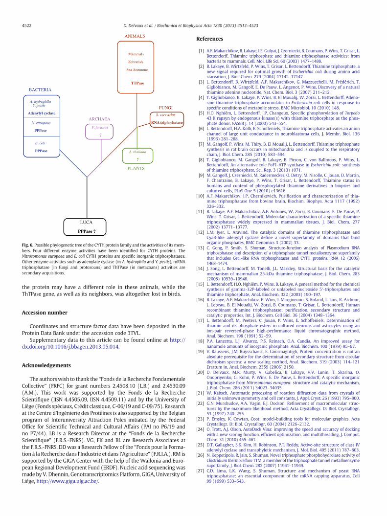
the protein may have a different role in these animals, while theThTPase gene, as well as its neighbors, was altogether lost in birds.
Accession number
Coordinates and structure factor data have been deposited in theProtein Data Bank under the accession code 3TVL.
Supplementary data to this article can be found online at http://dx.doi.org/10.1016/j.bbagen.2013.05.014.
Acknowledgements
The authorswish to thank the “Fonds de la Recherche FondamentaleCollective” (FRFC) for grant numbers 2.4508.10 (L.B.) and 2.4530.09(A.M.). This work was supported by the Fonds de la RechercheScientifique (IISN 4.4505.09, IISN 4.4509.11) and by the University ofLiège (Fonds spéciaux, Crédit classique, C-06/19 and C-09/75). Researchat the Centre d'Ingénierie des Protéines is also supported by the Belgianprogram of Interuniversity Attraction Poles initiated by the FederalOffice for Scientific Technical and Cultural Affairs (PAI no P6/19 andno P7/44). LB is a Research Director at the “Fonds de la RechercheScientifique” (F.R.S.-FNRS). VG, FK and BL are Research Associates atthe F.R.S.-FNRS. DDwas a Research Fellow of the “Fonds pour la Forma-tion à la Recherche dans l'Industrie et dans l'Agriculture” (F.R.I.A.). RM issupported by the GIGA Center with the help of the Wallonia and Euro-pean Regional Development Fund (ERDF). Nucleic acid sequencing wasmade by V. Dhennin, Genotranscriptomics Platform, GIGA, University ofLiège, http://www.giga.ulg.ac.be/.
References
[1] A.F.Makarchikov, B. Lakaye, I.E. Gulyai, J. Czerniecki, B. Coumans, P.Wins, T. Grisar, L.Bettendorff, Thiamine triphosphate and thiamine triphosphatase activities: frombacteria to mammals, Cell. Mol. Life Sci. 60 (2003) 1477–1488.
[2] B. Lakaye, B. Wirtzfeld, P. Wins, T. Grisar, L. Bettendorff, Thiamine triphosphate, anew signal required for optimal growth of Escherichia coli during amino acidstarvation, J. Biol. Chem. 279 (2004) 17142–17147.
[3] L. Bettendorff, B. Wirtzfeld, A.F. Makarchikov, G. Mazzucchelli, M. Frédérich, T.Gigliobianco, M. Gangolf, E. De Pauw, L. Angenot, P. Wins, Discovery of a naturalthiamine adenine nucleotide, Nat. Chem. Biol. 3 (2007) 211–212.
[4] T. Gigliobianco, B. Lakaye, P. Wins, B. El Moualij, W. Zorzi, L. Bettendorff, Adeno-sine thiamine triphosphate accumulates in Escherichia coli cells in response tospecific conditions of metabolic stress, BMC Microbiol. 10 (2010) 148.
[5] H.O. Nghiêm, L. Bettendorff, J.P. Changeux, Specific phosphorylation of Torpedo43 K rapsyn by endogenous kinase(s) with thiamine triphosphate as the phos-phate donor, FASEB J. 14 (2000) 543–554.
[6] L. Bettendorff, H.A. Kolb, E. Schoffeniels, Thiamine triphosphate activates an anionchannel of large unit conductance in neuroblastoma cells, J. Membr. Biol. 136(1993) 281–288.
[7] M. Gangolf, P. Wins, M. Thiry, B. El Moualij, L. Bettendorff, Thiamine triphosphatesynthesis in rat brain occurs in mitochondria and is coupled to the respiratorychain, J. Biol. Chem. 285 (2010) 583–594.
[8] T. Gigliobianco, M. Gangolf, B. Lakaye, B. Pirson, C. von Ballmoos, P. Wins, L.Bettendorff, An alternative role FoF1-ATP synthase in Escherichia coli: synthesisof thiamine triphosphate, Sci. Rep. 3 (2013) 1071.
[9] M. Gangolf, J. Czerniecki, M. Radermecker, O. Detry, M. Nisolle, C. Jouan, D. Martin,F. Chantraine, B. Lakaye, P. Wins, T. Grisar, L. Bettendorff, Thiamine status inhumans and content of phosphorylated thiamine derivatives in biopsies andcultured cells, PLoS One 5 (2010) e13616.
[10] A.F. Makarchikov, I.P. Chernikevich, Purification and characterization of thia-mine triphosphatase from bovine brain, Biochim. Biophys. Acta 1117 (1992)326–332.
[11] B. Lakaye, A.F. Makarchikov, A.F. Antunes, W. Zorzi, B. Coumans, E. De Pauw, P.Wins, T. Grisar, L. Bettendorff, Molecular characterization of a specific thiaminetriphosphatase widely expressed in mammalian tissues, J. Biol. Chem. 277(2002) 13771–13777.
[12] L.M. Iyer, L. Aravind, The catalytic domains of thiamine triphosphatase andCyaB-like adenylyl cyclase define a novel superfamily of domains that bindorganic phosphates, BMC Genomics 3 (2002) 33.
[13] C. Gong, P. Smith, S. Shuman, Structure-function analysis of Plasmodium RNAtriphosphatase and description of a triphosphate tunnel metalloenzyme superfamilythat includes Cet1-like RNA triphosphatases and CYTH proteins, RNA 12 (2006)1468–1474.
[14] J. Song, L. Bettendorff, M. Tonelli, J.L. Markley, Structural basis for the catalyticmechanism of mammalian 25-kDa thiamine triphosphatase, J. Biol. Chem. 283(2008) 10939–10948.
[15] L. Bettendorff, H.O. Nghiêm, P. Wins, B. Lakaye, A general method for the chemicalsynthesis of gamma-32P-labeled or unlabeled nucleoside 5′-triphosphates andthiamine triphosphate, Anal. Biochem. 322 (2003) 190–197.
[16] B. Lakaye, A.F. Makarchikov, P. Wins, I. Margineanu, S. Roland, L. Lins, R. Aichour,L. Lebeau, B. El Moualij, W. Zorzi, B. Coumans, T. Grisar, L. Bettendorff, Humanrecombinant thiamine triphosphatase: purification, secondary structure andcatalytic properties, Int. J. Biochem. Cell Biol. 36 (2004) 1348–1364.
[17] L. Bettendorff, M. Peeters, C. Jouan, P. Wins, E. Schoffeniels, Determination ofthiamin and its phosphate esters in cultured neurons and astrocytes using anion-pair reversed-phase high-performance liquid chromatographic method,Anal. Biochem. 198 (1991) 52–59.
[18] P.A. Lanzetta, L.J. Alvarez, P.S. Reinach, O.A. Candia, An improved assay fornanomole amounts of inorganic phosphate, Anal. Biochem. 100 (1979) 95–97.
[19] V. Raussens, J.M. Ruysschaert, E. Goormaghtigh, Protein concentration is not anabsolute prerequisite for the determination of secondary structure from circulardichroism spectra: a new scaling method, Anal. Biochem. 319 (2003) 114–121Erratum in, Anal. Biochem. 2359 (2006) 2150.
[20] D. Delvaux, M.R. Murty, V. Gabelica, B. Lakaye, V.V. Lunin, T. Skarina, O.Onopriyenko, G. Kohn, P. Wins, E. De Pauw, L. Bettendorff, A specific inorganictriphosphatase from Nitrosomonas europaea: structure and catalytic mechanism,J. Biol. Chem. 286 (2011) 34023–34035.
[21] W. Kabsch, Automatic processing of rotation diffraction data from crystals ofinitially unknown symmetry and cell constants, J. Appl. Cryst. 26 (1993) 795–800.
[22] G.N. Murshudov, A.A. Vagin, E.J. Dodson, Refinement of macromolecular struc-tures by the maximum-likelihood method, Acta Crystallogr. D: Biol. Crystallogr.53 (1997) 240–255.
[23] P. Emsley, K. Cowtan, Coot: model-building tools for molecular graphics, ActaCrystallogr. D: Biol. Crystallogr. 60 (2004) 2126–2132.
[24] O. Trott, A.J. Olson, AutoDock Vina: improving the speed and accuracy of dockingwith a new scoring function, efficient optimization, and multithreading, J. Comput.Chem. 31 (2010) 455–461.
[25] D.T. Gallagher, S.K. Kim, H. Robinson, P.T. Reddy, Active-site structure of class IVadenylyl cyclase and transphyletic mechanism, J. Mol. Biol. 405 (2011) 787–803.
[26] N. Keppetipola, R. Jain, S. Shuman, Novel triphosphate phosphohydrolase activity ofClostridium thermocellum TTM, amember of the triphosphate tunnelmetalloenzymesuperfamily, J. Biol. Chem. 282 (2007) 11941–11949.
[27] C.D. Lima, L.K. Wang, S. Shuman, Structure and mechanism of yeast RNAtriphosphatase: an essential component of the mRNA capping apparatus, Cell99 (1999) 533–543.
Fig. 6. Possible phylogenetic tree of the CYTH protein family and the activities of its mem-bers. Four different enzyme activities have been identified for CYTH proteins. TheNitrosomonas europaea and E. coli CYTH proteins are specific inorganic triphosphatases.Other enzyme activities such as adenylate cyclase (in A. hydrophila and Y. pestis), mRNAtriphosphatase (in fungi and protozoans) and ThTPase (in metazoans) activities aresecondary acquisitions.
4522 D. Delvaux et al. / Biochimica et Biophysica Acta 1830 (2013) 4513–4523

[28] D.T. Gallagher, N.N. Smith, S.K. Kim, A. Heroux, H. Robinson, P.T. Reddy, Structure of theclass IV adenylyl cyclase reveals a novel fold, J. Mol. Biol. 362 (2006) 114–122.
[29] N.H. Putnam, M. Srivastava, U. Hellsten, B. Dirks, J. Chapman, A. Salamov, A. Terry,H. Shapiro, E. Lindquist, V.V. Kapitonov, J. Jurka, G. Genikhovich, I.V. Grigoriev,S.M. Lucas, R.E. Steele, J.R. Finnerty, U. Technau, M.Q. Martindale, D.S. Rokhsar,Sea anemone genome reveals ancestral eumetazoan gene repertoire and genomicorganization, Science 317 (2007) 86–94.
[30] U. Technau, S. Rudd, P. Maxwell, P.M. Gordon, M. Saina, L.C. Grasso, D.C. Hayward,C.W. Sensen, R. Saint, T.W. Holstein, E.E. Ball, D.J. Miller, Maintenance of ancestralcomplexity and non-metazoan genes in two basal cnidarians, Trends Genet. 21(2005) 633–639.
[31] F. Raible, K. Tessmar-Raible, K. Osoegawa, P.Wincker, C. Jubin, G. Balavoine, D. Ferrier,V. Benes, P. de Jong, J. Weissenbach, P. Bork, D. Arendt, Vertebrate-type intron-richgenes in the marine annelid Platynereis dumerilii, Science 310 (2005) 1325–1326.
4523D. Delvaux et al. / Biochimica et Biophysica Acta 1830 (2013) 4513–4523


Appendix 4


RESEARCH ARTICLE
Thiamine triphosphate: a ubiquitous molecule in searchof a physiological role
Lucien Bettendorff & Bernard Lakaye & Gregory Kohn &
Pierre Wins
Received: 16 December 2013 /Accepted: 12 February 2014 /Published online: 4 March 2014# Springer Science+Business Media New York 2014
Abstract Thiamine triphosphate (ThTP) was discovered over60 years ago and it was long thought to be a specificallyneuroactive compound. Its presence in most cell types, frombacteria to mammals, would suggest a more general role butthis remains undefined. In contrast to thiamine diphosphate(ThDP), ThTP is not a coenzyme. In E. coli cells, ThTP istransiently produced in response to amino acid starvation,while in mammalian cells, it is constitutively produced at alow rate. Though it was long thought that ThTP was synthe-sized by a ThDP:ATP phosphotransferase, more recent studiesindicate that it can be synthesized by two different enzymes:(1) adenylate kinase 1 in the cytosol and (2) FoF1-ATP syn-thase in brain mitochondria. Both mechanisms are conservedfrom bacteria to mammals. Thus ThTP synthesis does notseem to require a specific enzyme. In contrast, its hydrolysisis catalyzed, at least in mammalian tissues, by a very specificcytosolic thiamine triphosphatase (ThTPase), controlling thesteady-state cellular concentration of ThTP. In some tissueswhere adenylate kinase activity is high and ThTPase is absent,ThTP accumulates, reaching ≥ 70% of total thiamine, with noobvious physiological consequences. In some animal tissues,ThTP was able to phosphorylate proteins, and activate a high-conductance anion channel in vitro. These observations raisethe possibility that ThTP is part of a still uncharacterizedcellular signaling pathway. On the other hand, its synthesisby a chemiosmotic mechanism in mitochondria and respiringbacteria might suggest a role in cellular energetics.
Keywords Leigh’s syndrome . Adenosine thiaminetriphosphate .Mitochondria . Adenylate kinase . Brain .
Skeletal muscle
Introduction
Thiamine (vitamin B1) is essential for energy metabolism andthe nervous system is particularly sensitive to its deficiency,which causes specific brain lesions (Hazell and Butterworth2009). It is thought that this vulnerability results from de-creased levels of thiamine diphosphate (ThDP), an indispens-able cofactor for several key enzymes such as pyruvate andoxoglutarate dehydrogenases and transketolase.
Indeed, in brain, as inmost other mammalian tissues, ThDPis the most abundant thiamine derivative, amounting to 80–90 % of total thiamine. However, five other forms may bepresent in various proportions: free thiamine and thiaminemonophosphate generally account for 5–15 % of total thia-mine and have no known physiological function. The threeremaining derivatives are thiamine triphosphate (ThTP) andthe recently discovered adenosine thiamine triphosphate(AThTP) (Bettendorff et al. 2007) and adenosine thiaminediphosphate (AThDP) (Frédérich et al. 2009). They are minorcomponents, generally amounting to less than 1 % of totalthiamine under normal physiological conditions, but theyseem more likely to play specific roles as intracellular mes-sengers or metabolic regulators. In particular, we have recent-ly shown (Gigliobianco et al. 2008, 2010; Lakaye et al. 2004c)that, in E. coli, either ThTP or AThTP may accumulate(reaching 10–50 % of total thiamine) under different condi-tions of cellular stress or nutritional downshifts.
ThTP was chemically synthesized in 1948 at a time whenthe only organic triphosphate compounds known were nucle-oside triphosphates (Velluz et al. 1948). The first claims forthe existence of ThTP in living organisms were made in ratliver (Rossi-Fanelli et al. 1952) and in baker’s yeast (Kiessling1953), though, ironically, what was measured was probablynot ThTP (see below). The presence of ThTP was later sug-gested in several rat tissues (Greiling and Kiesow 1958) and inplant germs (Yusa 1961). In these studies and until 1979,
L. Bettendorff (*) :B. Lakaye :G. Kohn : P. WinsGIGA-Neurosciences, University of Liège, Avenue de l’Hôpital, 1,4000 Liège, Belgiume-mail: [email protected]
Metab Brain Dis (2014) 29:1069–1082DOI 10.1007/s11011-014-9509-4

ThTP was separated from other thiamine derivatives using apaper chromatographic method, followed by oxidation tofluorescent thiochromes with ferricyanide in alkaline solution(Itokawa and Cooper 1970). This method, however, proved tobe not very reliable. In the studies mentioned above, ThTPwas reported to be a major thiamine compound in mammaliantissues (about 10 % of total thiamine), much more than sug-gested by modern HPLC methods.
The development of HPLC techniques in the late 1970’sand 1980’s by Kawasaki and colleagues in Hiroshima (Ishiiet al. 1979a, b; Sanemori et al. 1980) and our own group(Bontemps et al. 1984; Bettendorff et al. 1986, 1991) led to animportant simplification, acceleration and increased sensitivi-ty in the determination of ThTP in biological samples (forreview see: (Kawasaki 1992b; Lynch and Young 2000)).HPLC methods generally give ThTP levels much lower thanprevious methods (paper chromatography, paper electropho-resis, low pressure liquid chromatography), casting doubt onat least the quantitative dimension of the earlier findings.Indeed, as we previously showed, under some conditions,especially when poorly selective filter fluorometers are usedinstead of spectrofluorometers and thiochrome triphosphate iseluted early, chemically unrelated fluorescent compoundspresent in cells may be detected and mistakenly consideredto be ThTP (Bontemps et al. 1984).
The reliability of ThTP determination was an important issuein a particularly controversial case 40 years ago. In 1969, Cooperand associates reported that ThTP levels were strongly decreasedin post-mortem brains from patients having died from Leigh’sdisease, a fatal necrotizing myeloencephalopathy (Cooper et al.1969, 1970; Pincus et al. 1976). This seemed to substantiate theidea that ThTP plays an important role in brain function. Thiswas however unfounded, as ThTP is always undetectable inpost-mortem brains (see below).
The role of thiamine diphosphate (ThDP or cocarboxylase)in the decarboxylation of pyruvate by yeast had been eluci-dated 15 years before the discovery of ThTP (Lohmann andSchuster 1937) and so it was quite straightforward to testwhether ThTP had any cocarboxylase activity in yeast ex-tracts. Although several studies claimed that ThTP could, atleast partially, replace ThDP in pyruvate decarboxylation inyeast extracts (Velluz et al. 1949; De La Fuente and Diaz-Cadavieci 1954; Rossi-Fanelli et al. 1955), the use of crystal-line ThTP and apo-carboxylase free of phosphatase activitydisproved this possibility (Rossi-Fanelli et al. 1955). An initialcommunication suggested that ThTP, just like ATP, couldphosphorylate hexoses (Greiling and Kiesow 1956), but thiswas later attributed to impurities in the enzyme and/or sub-strate preparations (Yusa 1962). The hypothesis that ThTPcould act as a reservoir for ThDP is also unlikely because inmost tissues ThTP represents only a small fraction of theamount of ThDP. The fact that ThTP is present only in smallamounts in most cells (<1 nmol/g of fresh weight) also
excludes that ThTP could be an energy storage compoundfor the regeneration of ATP, like phosphocreatine for instance.
Here we shall critically review existing data, especially inrelation to Leigh’s disease, and examine possible cellular rolesfor ThTP in light of recent data obtained on ThTP synthesiz-ing and hydrolyzing enzymes.
A neurophysiological role for ThTP?
A specific non-cofactor role for thiamine and its phosphory-lated derivatives has been suggested for a long time (Cooperand Pincus 1979). The first observation in favor of a specific“neuroactive” role of thiamine was that the electrical stimula-tion of vagus nerves in vitro induced a release of thiamine intothe incubation medium (Minz 1938; Von Muralt 1942, 1947;Von Muralt and Zemp 1943). It was later suggested (Kunz1956; Cooper et al. 1963; Gurtner 1961) that thiamine releasewas linked to the dephosphorylation of phosphorylated deriv-atives such as ThMP; ThDP or ThTP. This led Cooper andassociates to suggest that ThTP was the neuroactive form ofthiamine (Cooper and Pincus 1979), but no subsequent studycould substantiate the idea that ThTP plays any significantrole in the regulation of nerve excitability.
ThTP is consistently found in the nervous system of allanimal species tested so far, ranging from 0.1 % (mouse brain)to 10 % (Slug cerebral ganglion) of total thiamine(Makarchikov et al. 2003; Frédérich et al. 2009). There areonly a few data on the regional distribution of ThTP in brain.In rat brain, it was found to be somewhat more abundant in thestem than in the cerebral hemispheres or the cerebellum(Gangolf et al. 2010a). On the other hand, neurons in primaryculture were found to contain more ThTP than astrocytes(Bettendorff et al. 1991).
We studied the effect of intermittent light stimulation on thecontent of thiamine derivatives in the brain of the photosensi-tive baboon Papio papio (Bettendorff et al. 1989b). Some ofthese animals are naturally photosensitive and, after intermit-tent light stimulation, develop paroxysmal discharges in themotor and premotor regions of the brain, responsible forgeneralized seizures. After intermittent light stimulation, therewas a decrease in ThTP levels in the occipital area (containingthe visual cortex) of both photosensitive and non-photosensitive animals; however no decrease in ThTP wasobserved in the motor and premotor areas. This might suggestthat the observed decrease is linked to increased stimulation ofthe visual cortex, but that it is not related to photosensitivityand epileptic discharges. From these experiments, it cannot beinferred whether decreased ThTP levels are a direct conse-quence of nerve excitability or increased metabolic activity ofthe neurons during light stimulation. In view of the recentobservation that the molecular mechanisms for ThTP and ATP
1070 Metab Brain Dis (2014) 29:1069–1082

synthesis (see below) are the same, the latter possibility is themost plausible.
When synaptoneurosomes prepared from rat brain mem-branes were incubated in the presence of ThDP, internal ThTPconcentrations increased. Under these conditions, membranechloride permeability was also increased, suggesting a rela-tionship between ThTP and chloride permeability(Bettendorff et al. 1990b, 1993a, c, 1994a). Indeed, usingthe patch-clamp technique, we showed that addition ofThTP (1 or 10 μM) to inside-out patches of mouse neuroblas-toma cells activated an anion channel of large unitary conduc-tance (Bettendorff et al. 1993b). No effect on cation channelswas observed. Maxi anion channels seem to be involved inswelling-inducedATP release, but their molecular identity stillremains unknown (Dubyak 2012).
More recently, it was found that in Torpedo marmorataelectric organ, ThTP specifically phosphorylates rapsyn(Nghiêm et al. 2000), a protein required for the clustering ofacetylcholine receptors at the neuromuscular junction(Gautam et al. 1995). Interestingly, ThTP phosphorylatedrapsyn on histidyl residues while phosphorylation by ATPoccurred on seryl or threonyl residues. ThTP was also shownto phosphorylate some as yet unidentified proteins in rodentbrain (Nghiêm et al. 2000). It was therefore suggested thatprotein phosphorylation by ThTP is part of a new cellularsignaling pathway. Until now, the only compound known tophosphorylate proteins in eukaryotic cells was ATP.
Intracellular localization of ThTP
A recent study has shown that ThTP exists not only in mam-mals, but in all major phyla tested: bacteria, fungi, plants,invertebrates and vertebrates (Makarchikov et al. 2003). Itseems that the intracellular localization of ThTP may changefrom one tissue to another. Thus in Electrophorus electricuselectric organ, 96 % of ThTP was found in the cytosolicfraction and was not protein-bound (Bettendorff et al. 1987).A similar observation was made concerning ThTP in pigskeletal muscle (Egi et al. 1986). In T. marmorata electricorgan, ThTP was mainly found in the synaptosomal fraction,either cytoplasmic or associated with membrane structures(Eder and Dunant 1980), but it was not detected inside syn-aptic vesicles. In this study, the authors report an extremelyhigh total thiamine content of 120±24 nmol / g of freshweight, only an order of magnitude less than ATP (Eder andDunant 1980). However, according to our data, the totalthiamine content is three orders of magnitude lower in theelectric organ of T. marmorata (Table 1).
In primary cell cultures, ThTP levels were five times higherin cerebellar granule cells compared to astrocytes (Bettendorffet al. 1991), but there is presently no experimental evidence
that ThTP is also preferentially localized in neurons in theintact adult brain.
In rat brain, the highest content of ThTP was found insynaptosomes. A significant amount was present in the mito-chondrial fraction (Bettendorff et al. 1994b; Gangolf et al.2010b) but only a small percentage was cytosolic. At least partof this ThTP seems to be protein-bound (Bettendorff et al.1993c). Matsuda et al. homogenized rat tissues in 20 mMTris–HCl (pH 7.4) – 1 mM EDTA, centrifuged the homoge-nate at 279,000×g and assayed ThTP in the particulate andsupernatant fractions (Matsuda et al. 1989c): in brain, heartand kidney, ThTP was mainly associated with the particulatefraction, while in white and red skeletal muscle it was presentin the supernatant fraction. In liver, it was evenly distributedbetween both fractions. Matsuda and Cooper (1981a, b) re-ported the association of thiamine phosphate esters and inparticular ThTP with brain synaptosomal membranes. Onthe other hand, Laforenza et al. (1990) could not find asignificant enrichment of synaptosomes compared with totalrat brain homogenate. Our recent data is suggested that, in themammalian brain, ThTP is mainly localized in mitochondria(Gangolf et al. 2010b).
Cytosolic ThTP levels are controlled by a 25-kDa ThTPase
In mammalian cells, concentrations of triphosphorylated thia-mine derivatives are generally low, less than 1 % of totalthiamine. ThTP levels are kept relatively constant (≤ 1 μM)because the compound is continuously hydrolyzed by a spe-cific 25-kDa cytosolic thiamine triphosphatase (ThTPase, EC3.6.1.28). This enzyme, first described in 1972 by Hashitaniand Cooper (1972), was purified in 1992 and shown to havean absolute specificity for ThTP (Makarchikov andChernikevich 1992). The molecular characterization revealedthat the sequence had no homology with any known mamma-lian protein (Lakaye et al. 2004a). It is widely distributed inhuman (Gangolf et al. 2010a) and rodent tissues (Lakaye et al.2002, 2004b), though it is not a very abundant protein. Thecatalytic efficiency was high (kcat/Km is typically in the 106–107 s−1 M−1 range) (Makarchikov and Chernikevich 1992;Lakaye et al. 2002, 2004a; Song et al. 2008; Delvaux et al.2013). In rodent brain, 25-kDa ThTPase seems to be essen-tially neuronal, with a high expression in cerebellar Purkinjecells and pyramidal hippocampal neurons (Czerniecki et al.2004). Using an antibody, staining was observed in cell bodiesand in dendrites. In cultured cells, a high labeling was alsoobserved in the nucleus.
Human 25-kDa ThTPase and the so-called cyaB (class IV,(Sismeiro et al. 1998)) adenylyl cyclase from Aeromonashydrophila are the founding members of a novel superfamilyof domains that bind triphosphorylated compounds and diva-lent metal cations (Bettendorff and Wins 2013; Iyer and
Metab Brain Dis (2014) 29:1069–1082 1071

Aravind 2002). This domain was called CYTH (CyaBand ThTPase) and members of this family, can be tracedback to the Last Universal Common Ancestor of allliving organisms. ThTPase may represent a relativelydivergent acquisition of a new catalytic activity which,so far, appears to be restricted to metazoans (Delvauxet al. 2013; Bettendorff and Wins 2013). The NMRsolution structure (Song et al. 2008) and the crystalstructure (Delvaux et al. 2013) were obtained for re-spectively the mouse and the human enzymes. The latterstudy suggests that the specificity for ThTP is linked toa Trp residue interacting with the thiazolium heterocycleof thiamine.
No sequence homologous to 25-kDa ThTPase and nospecific soluble ThTPase activity have been found in birdsso far (Makarchikov et al. 2003; Bettendorff and Wins 2013).It seems that the thtpa gene has been lost in the reptile branchleading to birds. The fish enzyme seems to have a much lowercatalytic activity than its mammalian counterpart (Delvauxet al. 2013). This could be the reason why no solubleThTPase activity was observed in fish tissues (Makarchikov
et al. 2003). Surprisingly, and due a single amino acid substi-tution, the pig has a low catalytic activity, though it seems tobe expressed according to a similar pattern than in rodenttissues (Szyniarowski et al. 2005). This means that in pig, birdand fish tissues, 25-kDa ThTPase is either absent or its cata-lytic activity is very low, allowing ThTP to accumulate(Table 1). This suggests that the enzyme indeed plays a majorrole in the control of cytosolic ThTP concentration. Also,human tissues generally have a much higher proportion ofThTP compared to total thiamine than rodent tissue. This mayprobably be correlated to the higher abundance and catalyticefficiency of rodent ThTPase compared to the human enzyme(Gangolf et al. 2010a).
In mammalian cells, ThTP seems to be more constitutivelysynthesized, but in much lower amounts, than in E. coli.Nevertheless, we produced a transgenic mouse expressingbovine ThTPase in addition to the endogenous mouseThTPase. Total ThTPase activity was increased by approxi-mately a factor of two, but ThTP levels were not significantlydecreased (Fig. 1). Mouse ThTPase is a very efficient enzyme,and ThTP levels are much lower in this species than in most
Table 1 Thiamine derivatives in various tissues
nmol/g fresh weight Thiamine ThMP ThDP ThTP Total
Electric organs
T. marmorataa 38±3 26±3 9.6±2.3 45±4 118.6
T. marmoratab 0.03±0.01 0.04±0.02 0.37±0.06 0.024±0.003 0.46
E. electricusc 0.06±0.01 0.09±0.01 0.46±0.08 3.9±0.5 4.5
Skeletal muscle
Pigd (n=1) 0.014 0.047 11 20 31.1
Pige 0.38±0.22 0.56±0.17 7.17±1.90 18.24±5.83 26.35
Ratb (n=5) 0.10±0.06 0.54±0.16 6.2±1.0 0.39±0.09 7.2
Mousef 0.16±0.02 0.49±0.05 6.7±0.7 0.026±0.006 7.38
Rabbitb (n=1) 0.14 0.26 2.0 0. 08 2.48
Chickend (n=1) – 0.33 1.2 3.7 5.23
Quaild (n=3) – 1.3±0.1 16.7±0.1 0.34±0.02 18.3
Humang, h (n=11) 0.08±0.09 0.09±0.05 2.2±1.6 0.13±0.13 2.5
Liver
Ratb (n=3) 0.61±0.04 1.8±0.3 13.9±1.3 <0.1 16.3
Humang, h (n=3) 0.03±0.01 0.39±0.26 5.9±3.8 0.022±0.023 6.34
Brain
Human cerebral cortexg, h (n=5) 0.03±0.04 0.05±0.03 2.7±0.07 0.05±0.04 2.83
a (Eder and Dunant 1980)b Bettendorff L., unpublished resultsc (Bettendorff et al. 1987)d (Makarchikov et al. 2003)e (Egi et al. 1986)f (Makarchikov et al. 2002)g (Gangolf et al. 2010a)h Considering that proteins account for about 13 % of wet weight
1072 Metab Brain Dis (2014) 29:1069–1082

other species tested. Therefore ThTP levels are hardly affectedafter increasing cytosolic ThTPase activity by overexpressionof the bovine enzyme.
Though ThDP is the major thiamine compound in mostliving organisms (Makarchikov et al. 2003), a few exceptionshave been noted: in pig (Egi et al. 1986) and chicken (Miyoshiet al. 1990) skeletal muscle and the muscle-derivedE. electricus electric organ (Bettendorff et al. 1987), ThTP isthe major thiamine compound accounting for 70–80% of totalthiamine (Table 1). However, such a high ThTP content is bynomeans a general characteristic of skeletal muscle or skeletalmuscle-derived tissues (Table 1). Rat and rabbit skeletal mus-cle have a much lower ThTP content. In trout, quail andguinea pig skeletal muscle (Makarchikov et al. 2003; Egiet al. 1986), ThTP accounts for less than 10 % of totalthiamine. White muscle fibers have a lower ThDP contentthan red muscle fibers in chicken (Miyoshi et al. 1990), rat,guinea pig and rabbit (Matsuda et al. 1989b) but a higherThTP content. However, ThTP levels are very low in mouse
white and red skeletal muscle (0.1 % of total thiamine)(Matsuda et al. 1989b; Makarchikov et al. 2002) (Table 2).Thus, there is apparently no obvious relationship betweenThTP levels and functional characteristics of the tissueinvestigated.
In addition to the soluble 25-kDa ThTPase, a membrane-associated ThTPase has also been reported in most animalspecies (Barchi and Braun 1972; Bettendorff et al. 1987). Thisenzyme is strongly activated by anions in E. electricus electricorgan (Bettendorff et al. 1988, 1989a) and mammalian skele-tal muscle (Matsuda et al. 1991) but this was not the case forthe brain enzyme. The latter is inhibited by ATP but seems tobe distinct from membrane-bound ATPases. The membrane-associated ThTPase has not been characterized at the molec-ular levels and its contribution in the control of intracellularThTP levels is unknown, but is was recently speculated that itcould be identical to a synaptosomal thiamine-binding protein(Sidorova et al. 2009). Indeed, a thiamine-binding protein hasbeen isolated from rat brain synaptosomes (Postoenko et al.1988). This protein of 107 kDa also binds thiamine phosphateesters with the sequence ThMP > ThTP > ThDP, but itsmolecular identity is unknown.
ThTP is synthesized in vivo
Despite ThTP being known for nearly 60 years, the mecha-nism of its biosynthesis remains controversial.
After subcutaneous (Gaitonde and Evans 1983),intracerebro-ventricular (Iwata et al. 1985) or intraperitoneal(Bettendorff et al. 1994b) administration of radiolabeled
Fig. 1 Thiamine triphosphatase activities and thiamine triphosphate inseveral tissues of a transgenic mouse overexpressing bovine 25-kDaThTPase (bThTPase). The bThTPase coding sequence was placed bytargeted insertion in the ROSA26 locus using the method described bySrinivas et al. (2001). The mice generated were then mated with EIIaCrefemale mice (kindly provided by Dr E. Weinsphal) to remove the PGK-Neo cassette and place the bThTPase coding sequence under the controlof the ubiquitous ROSA26 promoter. ThTPase activities (a) and ThTPcontent (b) were measured as previously described (Gangolf et al. 2010a).The results are expressed as means ± SD for 5 mice. ThTPase activitieswere significantly increased in all tissues (Two-wayANOVA: p≤0.0001),but no significant decrease was observed for ThTP (p=0.2)
Table 2 Soluble 25-kDa ThTPase activities
ThTPase activities(nmol. mg−1. min−1)
Mammals
Rat brain 3.4±0.5a
Rat skeletal muscle 0.40±0.1a
Mouse skeletal muscle 2.0±0.2
Pig skeletal muscle 0.095a
Birds
Chicken skeletal muscle (G. gallus) < 0.01a
Quail skeletal muscle (C. coturnix japonica) < 0.01a
Fish
Electrophorus electric organ (E. electricus) < 0.01b
Torpedo electric organ (T. marmorata) < 0.01b
Trout skeletal muscle (S. trutta) < 0.01a
ThTPase activities were determined in the supernatants from varioustissue homogenates centrifuged at 100,000×ga Adapted from (Makarchikov et al. 2003)b Bettendorff L., unpublished results
Metab Brain Dis (2014) 29:1069–1082 1073

thiamine to rats, radioactivity was rapidly incorporated intoThTP, demonstrating that this compound is indeed synthe-sized in vivo in the brain. A net synthesis of ThTP wasobtained after administration of thiamine disulfide derivativesin rat liver (Sanemori and Kawasaki 1982) and brain(Bettendorff et al. 1990a), though at least in the later case,administration of equivalent doses of thiamine was ineffectivein raising brain ThTP content.
It came as a surprise that administration of radiolabeledthiamine in rats (Iwata et al. 1985; Bettendorff et al. 1994b;Gaitonde and Evans 1983) or incubation of mouse neuroblas-toma cells with radiolabeled thiamine (Bettendorff 1994a) ledto a more rapid increase in specific radioactivity in intracellu-lar ThTP and ThMP than in ThDP. Similarly, incubation ofbrain slices with [32P]Pi led to a much more rapid incorpora-tion of radioactivity in ThTP than in ThDP (Berman andFishman 1975). In animal cells, ThDP is directly formed fromthiamine by thiamine pyrophosphokinase (thiamine + ATP ⇆ThDP + AMP) and ThMP can only be formed from ThDP byenzymatic hydrolysis. The fact that, the precursor ThDP has aconsiderably lower specific radioactivity than its products,suggests that there are two ThDP pools: one small pool ofhigh turnover (which is the precursor of ThTP and ThMP) anda larger pool of lower turnover, probably corresponding toThDP bound to apoenzymes (Berman and Fishman 1975;Bettendorff 1994a). The high turnover pool probably corre-sponds to free cytosolic ThDP (≤ 10 % of total thiamine)whereas the low turnover pool consists of ThDP bound totransketolase in the cytoplasm and PDH and OGDH in mito-chondria (Bettendorff 1994a). The turnover of ThTP was ~1–2 h, while the turnover of the slow ThDP pool was ~17 h.
A key observation was that in some species cellular ThTPconcentrations are not constant. They are generally very lowbut, under conditions of cellular stress, the intracellular ThTPcan increase considerably. This phenomenon was observed inplants during withering (Makarchikov et al. 2003) and inbacteria during amino acid starvation (Lakaye et al. 2004c).The intracellular ThTP concentration is highly regulated inE. coli, strongly depending on physiological conditions: inbacteria growing in aerated rich medium, ThTP is barelydetectable, but it quickly accumulates (reaching cellular con-centrations ≥ 10 μM) when the bacteria are transferred tominimum medium devoid of amino acids but containing acarbon source such as glucose (Lakaye et al. 2004c).Interestingly, this accumulation is transient, reaching a maxi-mum after 1–2 h, then decreasing eventually to zero. Howthose regulatory mechanisms respond to the nutritional statusremains an open question.
When the specific human 25-kDa ThTPase wasoverexpressed as a GST-fusion protein in E. coli, ThTP didnot accumulate and the bacterial growth curves displayed anintermediate plateau, suggesting that ThTP is involved in therapid adaptation of the bacteria to amino acid starvation. As
the expression level of the fusion protein was very high (>10 % of total protein), toxic effects can however not beexcluded, though overexpression of a protein unrelated tothiamine metabolism did not produce this intermediate pla-teau. This led us to propose that ThTP (as well as AThTP)could act as “alarmones” or signals responding to specificconditions of nutritional downshift or metabolic stress(Lakaye et al. 2004c; Gigliobianco et al. 2008, 2010, 2013).The biochemical mechanisms underlying the production ofthese signals remain to be clarified.
Mechanisms of ThTP synthesis
ThTP synthesis by a ThDP:ATP phosphotransferaseand Leigh’s disease
The most obvious mechanism for the synthesis of a triphos-phate compound would be the transfer of a phosphate groupfrom ATP to the diphosphorylated precursor. Such a mecha-nism, involving a cytosolic ThDP:ATP phosphotransferase(ATP + ThDP ⇆ ADP + ThTP), was already proposed in1964 (Eckert and Möbus 1964) in acetone powder extractsof supernatants of pig spinal cord, but the enzyme was notcharacterized.
Years later, an enzyme thought to catalyze such a reactionwas partially purified from rat liver hyaloplasm (Voskoboevand Luchko 1980; Voskoboev and Chernikevich 1985). Theauthors measured the formation of labeled ThTP from[14C]ThDP in the presence of ATP and Mg2+. After a 100-fold purification, the specific activity was still low and two pHoptima were found (5.3 and 7.8). In 1984, the same groupreported the complete purification of a soluble ThDP kinasefrom brewer’s yeast (Chernikevich et al. 1984; Voskoboyevet al. 1987). Though the authors give the amino acid compo-sition of their preparation, which seemed to be a multi-subunitcomplex, we have been unable to identify a possible candidatefrom current databases. However, the product ThTP was notunambiguously identified. Indeed, it could have been therecently discovered adenosine thiamine triphosphate(AThTP, (Bettendorff et al. 2007)), which is synthesized inthe cytosol according to the reaction ThDP + ATP (ADP) ⇆AThTP + PPi (Pi) (Makarchikov et al. 2007). Using, superna-tants from E. electricus electric organs or pig brain, whichcontain no significant soluble ThTPase activity, we wereunable to demonstrate any ThTP synthesis by such a mecha-nism. Likewise, no such activity could be detected in cell-freeextracts from E. coli.
J. R. Cooper and colleagues at Yale University reported thesynthesis of ThTP in a mitochondrial fraction from rat brainby a ThDP:ATP phosphotransferase (Cooper et al. 1969;Itokawa and Cooper 1968). The reaction did not seem to occurin the mitochondrial matrix and no functional mitochondria
1074 Metab Brain Dis (2014) 29:1069–1082

were required. Amost puzzling observation was that the urine,spinal fluid and deproteinized blood from patients withLeigh’s syndrome contained an inhibitor of this reaction.Moreover, no ThTP was detected in the post-mortem brainsamples from patients with Leigh’s syndrome (Cooper et al.1969; Pincus et al. 1976; Murphy 1976), while in brainsamples from control patients ThTP accounted for ~10 % oftotal thiamine. This result could not be confirmed, however.Indeed, we were unable to detect any ThTP in post-mortemhuman brains (Bettendorff et al. 1996). This is probablydue to hydrolysis by 25-kDa ThTPase during the post-mortem delay, which is at least 2–3 h, and 25-kDaThTPase remains highly active in post-mortem human brain(Bettendorff et al. 1996). In cultured mouse neuroblastomacells (which have a low ThTPase activity, Bettendorff,unpublished results), the half-life of ThTP was ~1 h(Bettendorff 1994a). However, we found significantamounts of ThTP in human brain biopsies (Bettendorffet al. 1996; Gangolf et al. 2010a), but it accounted foronly ~1 % of total thiamine, far less than the 10 % report-ed by Cooper et al. in post-mortem human brains.
L e i g h ’ s s y n d r om e ( s u b a c u t e n e c r o t i z i n gmyeloencephalopathy), first described by Denis Leigh in1951 (Leigh 1951), is a rare heterogeneous neurodegenerativedisorder of early childhood, characterized by focal, symmetricnecrotic brain lesions, leading to death before the age of five(Finsterer 2008). It is now recognized that Leigh’s syndromeresults from deficits in mitochondrial respiratory chain com-plexes (I, II, III, IV), FoF1-ATP synthase or pyruvate dehydro-genase. In a small percentage of patients (<10 %), particularlywhen pyruvate dehydrogenase is affected, high-dose thiaminetreatment may have beneficial effects (Di Rocco et al. 2000;Naito et al. 1997), but there is probably no involvement of aThTP deficiency in this disease.
In 1977, Ruenwongsa and Cooper (1977) reported that, inthe supernatant fraction from rat liver, labeled ThTP wasformed from [γ-32P]ATP. No addition of ThDP to the prepa-ration was required and the authors concluded that the sub-strate was ThDP bound to a protein. Schrijver and colleaguesreinvestigated this assertion (Schrijver et al. 1978), but theywere unable to reproduce any ThTP synthesis by the mecha-nism described. They suggested a chromatographic artifactresulting from formation of complexes between thiamine andphosphates behaving chromatographically like ThTP(Schrijver et al. 1978). Moreover, Ruenwongsa and Cooperreport a synthesis of 1.28 nmol of ThTP per mg of protein in arat liver homogenate from endogenous ThDP, while totalThDP in rat liver was only about 0.1 nmol/mg of protein(Table 1, see also (Schrijver et al. 1978)). As radioactive[32P]ATP was used as substrate, it was suggested that the[32P]ThTP formed (if any) was contaminated by radioactiveadenine nucleotides and inorganic phosphate (Cooper et al.1982).
Finally, in 1983, Cooper and coworkers reported the puri-fication of a 103 000 kDa protein from an acetone powderprepared from what appears to be a bovine brain mitochon-drial fraction. They used a purified ThDP-binding proteinfrom liver as substrate (Nishino et al. 1983). Hence, thereaction studied was: ThDP-protein + ATP ⇆ ThTP-protein+ ADP. The enzyme was unstable and inactivated by sulfhy-dryl reagents. The activity of the purified enzyme was67 nmol. mg−1. h−1, which would correspond to a kcat of0.11 min−1. The authors attributed this low turnover to thefact that the substrate was protein-bound and its concentrationcould therefore not be raised to attain the maximum velocity.There was a controversy about the nature of the ThDP-proteincomplex used as substrate. According to Nishino et al. (1983),this soluble protein (purified from a liver supernatant fraction)was distinct from transketolase, which is a well-known cyto-solic protein binding ThDP with high affinity. Voskoboev andChernikevich (1985) reinvestigated the purification procedureand concluded that the only ThDP-binding protein in the ratliver supernatant fraction was transketolase and, at least in ratliver, the ThDP-transketolase complex was not a substrate forThTP synthesis. Shortly thereafter, Yoshioka et al. reportedthe complete purification of liver ThDP-binding proteins(Yoshioka et al. 1987). They found a single band of 68 kDacorresponding to a transketolase monomer. Using immunocy-tochemistry, these authors also found that the ThDP-bindingprotein is localized in the cytoplasm in neurons and in nucleiin glial cells. Such differential localizations between neuronsand glial cells were later established for transketolase(Calingasan et al. 1995). Therefore, it is very likely that theThDP-binding protein used by Cooper and colleagues. wastransketolase.
How about the enzyme catalyzing the synthesis of ThTP ?We checked whether the amino acid composition, as well asthe molecular mass of 103 000 of the enzyme purified byNishino et al. (1983) could match any known protein(Table 3). The only protein with a molecular mass close to103 000 that gave a significant correlation was hexokinasetype I (R2=0.80, p<0.0001, molecular mass of 103,064; 918amino acids) (Fig. 2).
Hexokinase type I is highly abundant in brain, and most ofit is associated with mitochondria (Sui and Wilson 2002;Wilson 2003), while type II and III isoforms are expressedonly at low levels in the brain. Though Nishino et al. claimedthat their preparation had no hexokinase activity, this wasprobably destroyed by the acetone treatment. Furthermore, itis well known that hexokinase activity is quickly lost in theabsence of glucose and EDTA (Redkar and Kenkare 1972)and both compounds were absent during the purificationprocedure described by Nishino et al. In this respect, it maybe significant that Nishino et al. report that ThTP-synthesizingactivity required a cofactor that they suggest to be glucose(Nishino et al. 1983). This cofactor was supposed to be
Metab Brain Dis (2014) 29:1069–1082 1075

characterized separately and, though they refer to a manuscriptin preparation, this was never published. Therefore, it remainsa mystery as to whet the authors really measured.
Therefore, the mechanisms described by Nishino et al. inbovine brain and by Chernikevich and colleagues in yeasthave not been confirmed in other laboratories and there is nocompelling evidence that a ThDP:ATP phosphotransferasebearing the enzyme nomenclature number EC 2.7.4.15 reallyexists.
ThTP synthesis by adenylate kinases
Although ThTP generally accounts for less than 1 % of totalthiamine, in a few tissues such as pig skeletal muscle (Egiet al. 1986), E. electricus electric organ (Bettendorff et al.1987) and chicken white muscle (Miyoshi et al. 1990),ThTP is the major thiamine compound, accounting for>70 % of total thiamine (Table 2). Kawasaki and coworkershave suggested that this high ThTP content was correlatedwith the high amount of adenylate kinase 1 (AK1, myokinase,EC 2.7.4.3) found in skeletal muscle and electric organ. Theyindeed showed that, in vitro, the adenylate kinase isoform 1(AK1) may synthesize ThTP according to the reaction : ThDP+ ADP ⇆ ThTP + AMP (Shikata et al. 1989b).
The same authors suggested that this enzyme was alsoresponsible for ThTP synthesis in chicken white musclein vivo (Miyoshi et al. 1990; Shioda et al. 1991). However,the significance of this reaction in vivo can be questioned asAK has a sharp pH profile with an optimum at 10 with ThTPas substrate (Shikata et al. 1989). Moreover, physiologicalconcentrations of free ThDP in the cytosol are at best 10 μM(Bettendorff et al. 1994b), much lower than the Km for ThDPreported for AK1 (830 μM). Thus, the rate of ThTP synthesisby AK1 is about 4 orders of magnitude lower than ATPsynthesis under physiological conditions.
In rat skeletal muscle (quadriceps), ThTP content varied asa function of age: ThTP content increased until the age of 6–12 months and then decreased (Fig. 3a). This is in agreementwith results fromMatsuda et al. who showed an increase in ratskeletal muscle ThTP content between birth and 3months, butthey did not test older mice (Matsuda et al. 1989a). Weobserved a similar relationship between AK activity and agein rat skeletal muscle and a significant correlation betweenThTP content and AK activity was observed (R2=0.68; p=0.0009, Fig. 3b). No significant correlation was observed forthe other thiamine derivatives (not shown). We observed asignificant relationship between AK activity and lactate dehy-drogenase (R2=0.38, p=0.032). These results suggest thatanaerobic metabolism increases until about the age of12 months and then decreases. These results strongly suggestthat, in rat skeletal muscle, ThTP is mainly synthesized byAK1 as was previously suggested by Kawasaki (1992a).
The accumulation of large amounts of cytosolic ThTP maybe interpreted as follows: the equilibrium of the reaction ADP+ ThDP ⇆ AMP + ThTP will be shifted to the right becauseAMP reacts with ATP (which is in excess) to form 2 ADP.Both reactions are catalyzed by AK and the global reactionwill be ThDP + ATP ⇆ ThTP + ADP. This equilibrium willalso be shifted to the right because the muscle contains highamounts of phosphocreatine and creatine-kinase: ADP +phosphocreatine ⇆ ATP + creatine. The global process canthus be described by the reaction ThDP + phosphocreatine ⇆ThTP + creatine.
Table 3 Comparison of the amino acid content of the protein purified byNishino et al. from bovine brain and bovine hexokinase HK1
Nishino el al.a
(mol/103,000 g protein)(%)b Bovine
HK1c (%)
Lys 72.8 7.1 5.7
His 19.4 1.9 2.2
Arg 53.4 5.2 5.7
Asp + Asn 101 9.8 9.7
Thr 58.4 5.7 5.8
Ser 51 5 6.9
Glu + Gln 107.2 10.4 10.2
Gly 88.5 8.6 7.2
Ala 54.2 5.3 8.3
Val 64 6.2 6.6
Met 36.4 3.5 2.4
Ile 56.7 5.5 5.2
Leu 92.5 9 9
Tyr 18.5 1.8 3.2
Phe 40.3 3.9 3.9
a From (Nishino et al. 1983)b Calculated from column 1c Obtained from the protein sequence [UniProt identifier P27595(HXK1_BOVIN)], using the Expasy AACompIdent tool (http://www.expasy.ch/tools/aacomp/)
Fig. 2 Correlation between the amino acid composition of bovine hexo-kinase 1 and the enzyme purified by Nishino et al. (1983). The relativeabundance of each amino acid of bovine hexokinase 1 is plotted againstthe relative amino acid abundance of the bovine brain protein isolated byNishino et al., taking into account that the values for Asp and Glurepresent the sum of respectively Asp + Asn and Glu + Gln. Cys, Trpand Pro were not determined by Nishino et al. [F(1,13)=53.06,P<0.0001, R2=0.8032)]
1076 Metab Brain Dis (2014) 29:1069–1082

Therefore, it is likely that in skeletal muscle (and mostprobably electric organs) the main reaction responsible forThTP synthesis is the one catalyzed by AK1. The role ofAK1, which is a cytosolic enzyme, is in agreement with theobservation that ThTP is essentially found in the cytosol ofskeletal muscle (Egi et al. 1986; Matsuda et al. 1989c) andelectric organs (Bettendorff et al. 1987; Eder and Dunant1980).
However, it may not be the only ThTP synthesizing reac-tion. Indeed, in a previous study we showed that ThTP levelsin adenylate kinase knock-out mice are very low, but the sameas in wild-type animals (Makarchikov et al. 2002). Otherisoforms of mammalian AK may also be responsible forThTP synthesis. Hence, bacterial AKs are also able to synthe-size ThTP at a low rate (Gigliobianco et al. 2008), but mostprobably another enzyme is involved.
ThTP synthesis by a chemiosmotic mechanism
In the 1990s, we reported a net synthesis of ThTP in aparticulate fraction (synaptoneurosomes) isolated from ratbrain (Bettendorff et al. 1993a, c, 1994a) with either thiamineor ThDP as substrate. ThTP was formed inside closed com-partments that we thought to be synaptosomes. Recently, wereinvestigated this issue and found that ThTP synthesis oc-curred inmitochondria fromThDP and inorganic phosphate inthe presence of substrates of the respiratory chain (Gangolf
et al. 2010b). Addition of external phosphate and ThDP toisolated rat brain mitochondria lead to an increase in ThTPsynthesis. Indeed, ThDP can enter mitochondria via a specifictransporter located in the internal membrane of the organelle(Marobbio et al. 2002; Lindhurst et al. 2006). Moreover,ThTP synthesis requires intact mitochondria and a functionalrespiratory chain. It is inhibited by uncouplers and by inhib-itors of FoF1-ATP synthase, suggesting that it might be syn-thesized by a chemiosmotic mechanism similar to ATP syn-thesis by oxidative phosphorylation: ThDP + Pi ⇆ ThTP. Inorder to test whether such a mechanism may be of physiolog-ical importance, we incubated brain slices with the uncouplerCCCP: ThTP levels decreased, suggesting that such a chemi-osmotic mechanism is indeed operative in vivo.
As ThTP has been found in most types of organisms, it wasof interest to investigate whether it could be produced througha similar mechanism in tissues other than the mammalianbrain, and also in organisms that are phylogenetically distantfrom mammals, e.g. bacteria. Indeed, E. coli cells having acatalytically inactive ATP synthase or lacking the enzyme areunable to synthesize ThTP. In the latter case, ThTP synthesiscan be restored by transformation of the bacteria with aplasmid containing the whole operon for coding for E. coliFoF1-ATPase subunits (Gigliobianco et al. 2013).
The occurrence of this chemiosmotic mechanism wouldaccount for the fact that in the mammalian brain, in contrast toskeletal muscle (see above), most ThTP is localized insidemitochondria. External phosphate increased the release ofThTP from the mitochondria (Gangolf et al. 2010b). It is notknownwhether this release is of physiological importance, butit might suggest that ThTP produced inside mitochondriacould have a role either in the intermembrane space or in thecytosol (Fig. 4).
It could be argued that ThTP is merely a byproduct of FoF1-ATPase without physiological significance. In this case, wewould expect that ThTP would be synthesized in all mito-chondrial preparations, but no such synthesis was observed inrat liver mitochondria for instance. These results suggest atight, organ-specific, regulation of mitochondrial ThTP syn-thesis, possibly depending on metabolic differences betweencells. This is in agreement with results obtained in E. coli,where ThTP is synthesized only in the absence of amino acidsin the incubation medium, provided that a suitable carbonsource is present (Lakaye et al. 2004c); in the absence of acarbon source, they synthesize AThTP (Bettendorff et al.2007; Gigliobianco et al. 2010).
Conclusion
The aim of the present review was to carefully examineexisting data on the mechanism of ThTP synthesis, its hydro-lysis and its role. The data reviewed here summarize over
Fig. 3 ThTP content in rat skeletal muscle (quadriceps) as a function ofage (a) and AK1 activity (b). ThTP and AK1 activity were determined inmuscle cytosolic fractions as described (Makarchikov et al. 2002).[F(1,10)=21.39, P=0.0009, R2=0.6814]
Metab Brain Dis (2014) 29:1069–1082 1077

60 years of research on ThTP and especially the mechanism ofits synthesis. It now appears that, because of the low concen-tration of this compound in most tissues, real progress wasmade only after the development of reliable and sensitiveHPLC methods.
The situation was even more frustratingly complicated asfour different mechanisms for the synthesis of ThTP had beenproposed: ThDP:ATP phosphotransferase, a ThDP:ATP phos-photransferase using bound ThDP as substrate, AK1 andFoF1-ATP synthase.
It is our opinion that there is no compelling evidence for amechanism involving a ThDP:ATP phosphotransferase asproposed by several authors with various variations usingeither free or bound ThDP as substrate. Indeed, some of thesestudies are flawed by chromatographic artifacts and reliabledetermination of the small amounts of ThTP present in bio-logical samples became only possible with the advent of
sensitive HPLC methods, beginning of the 1980s. In otherstudies, the compound detected was probably not ThTP, butthe recently discovered AThTP. On the other hand, it is mostprobable that the only cytosolic and soluble ThDP-bindingprotein is transketolase, not involved in ThTP synthesis.
It has been convincingly demonstrated that AK1 mayconstitutively synthesize ThTP at a very slow rate. This reac-tion is probably relevant only in tissues with a high AK1content such as skeletal muscle and electric organs. In thesetissues, which have a low 25-kDa ThTPase activity, ThTP canaccumulate to high levels and even become the major thia-mine compound. This is the case in pig and chicken skeletalmuscle and E. electricus electric organ. In view of the obser-vation that ThTP is by far the major thiamine compound inelectric organs of E. electricus, we, probably erroneously,postulated that ThTP might be involved in bioelectrogenesis(Bettendorff et al. 1987; Bettendorff 1994b), a hypothesis that
Fig. 4 Synthesis and hydrolysisof ThTP in a mammalian cell.ThTP may be synthesized in thecytosol by AK1. This reaction isonly significant in tissues withhigh AK1 activity such as skeletalmuscle and possibly erythrocytes.ThTP accumulates in the cytosolonly when 25-kDa activity is low.In brain cells, ThTP may besynthesized in the mitochondrialmatrix by a chemiosmoticmechanism and it may beexchanged with cytosolicinorganic phosphate (Gangolfet al. 2010b). ThDP may entermitochondria by a specifictransporter, probably by exchangeagainst nucleotides (Lindhurstet al. 2006). I to IV, respiratorycomplexes; F1
*: hypotheticalmodified conformation of F1 thatwould bind ThDP rather thanADP
1078 Metab Brain Dis (2014) 29:1069–1082

would have been in accord with previous studies suggesting arole of ThTP in membrane excitability (Cooper and Pincus1979). However, the fact that ThTP accumulation in thesetissues is the result of two unrelated factors, which was notknown at that time, is not in favor of a physiological role inthese tissues.
Recently we demonstrated a third mechanism for ThTPsynthesis (Gangolf et al. 2010b; Gigliobianco et al. 2013).According to these data, ThTP is synthesized insidemitochon-dria following the reaction ThDP + Pi ⇆ ThTP by a chemios-motic mechanism similar to oxidative phosphorylation(Fig. 3). ThTP can be released from mitochondria in thepresence of external Pi. Therefore, cytosolic ThTP may havetwo different origins, either by exit from mitochondria orsynthesis by cytosolic AK. In most mammalian tissues, withthe exception of those mentioned above, 25-kDa ThTPaserapidly hydrolyzes cytosolic ThTP. This enzyme, especiallyin rodents, has a very high catalytic efficiency (kcat/Km=10
6–107 s−1 M−1). Therefore, ThTP is efficiently hydrolyzed, evenwhen its cytoplasmic concentrations are very low, i. e. in thesubmicromolar range, which is generally the case. It has beenproposed that ThTP results from metabolite (ThDP) damageand that 25-kDa ThTPase would be a kind of repair enzyme,converting ThTP to ThDP (Linster et al. 2013). Though thishypothesis cannot be excluded, there are two argumentsagainst it: (1) as stated above, in skeletal muscle and electricorgans ThTP accumulates in the absence of any apparentdamage; (2) synthesis of ThTP by the chemiosmotic mecha-nism is tissue-specific.
Moreover, ThTP synthesis by FoF1-ATP synthase in bac-teria is highly dependent on metabolic conditions and occursonly transiently at the onset of amino acid starvation (Lakayeet al. 2004c). In this organism, no specific ThTPase is presentand the control of ThTP concentration occurs at the level ofsynthesis involving FoF1-ATP synthase.
As mitochondria have probably evolved from endosymbi-otic bacteria, this raises the interesting hypothesis that inanimal cells the role of ThTP may be mitochondrial ratherthan cytoplasmic. Hence, a cytoplasmic accumulation ofThTP as a result of high AK1 activity, as observed in someskeletal muscles and electric organs, would therefore not bephysiologically relevant.
The data reviewed here also emphasize the uniqueness ofthiamine, which, to our knowledge, is the only moleculeexisting in its mono-, di-, and triphosphate forms that is nota nucleotide. Therefore, the metabolism of thiamine is morereminiscent of a nucleotide such as adenosine, than that ofother vitamins. The parallelism between ThTP and ATP isfurther strengthened by the observation that they are synthe-sized by the same enzymes: adenylate kinase and FoF1-ATPsynthase. Moreover, like ATP, ThTP may phosphorylaterapsyn as well as several other brain proteins, though this isprobably by autophosphorylation (Nghiêm et al. 2000).
Finally, the discovery of AThTP, another thiaminetriphosphorylated derivative is not meant to simplify thepicture.
Now that the mechanisms of ThTP synthesis have beenelucidated and that a specific ThTP-hydrolyzing enzyme hasbeen identified, future work should focus on its targets. Inthis respect, the study of protein phosphorylation by ThTPshould be helpful. Organisms such as E. coli and higherplants, in which ThTP appears under very specific stresssituations should give interesting insights into the physio-logical role of this compound. Finally, in mammalian cells,strategies aiming at silencing ThTPase expression (knockoutmice, siRNA) should represent a promising approaches.
Acknowledgments This work was funded by the “Fonds de laRecherche Scientifique Collective” (grant 2.4558.04). L. Bettendorff isResearch Director and B. Lakaye Reserch Associate at the Funds forScientific Research (F.R.S.-FNRS, Belgium). The data from Fig. 2 wereobtained with the technical assistance of Olivier Hennen.
Conflict of interest The authors declare that they have no competinginterests.
References
Barchi RL, Braun PE (1972) A membrane-associated thiaminetriphosphatase from rat brain. Properties of the enzyme. J BiolChem 247:7668–7673
Berman K, Fishman RA (1975) Thiamine phosphate metabolism andpossible coenzyme-independent functions of thiamine in brain. JNeurochem 24:457–465
Bettendorff L (1994a) The compartmentation of phosphorylated thiaminederivatives in cultured neuroblastoma cells. Biochim Biophys Acta1222:7–14
Bettendorff L (1994b) Thiamine in excitable tissues: reflections on a non-cofactor role. Metab Brain Dis 9:183–209
Bettendorff L, Wins P (2013) Thiamine triphosphatase and the CYTHfamily of proteins. FEBS J 280:6443–6455
Bettendorff L, Grandfils C, De Rycker C, Schoffeniels E (1986)Determination of thiamine and its phosphate esters in human bloodserum at femtomole levels. J Chromatogr 382:297–302
Bettendorff L, Michel-Cahay C, Grandfils C, De Rycker C, SchoffenielsE (1987) Thiamine triphosphate and membrane-associated thiaminephosphatases in the electric organ of Electrophorus electricus. JNeurochem 49:495–502
Bettendorff L, Wins P, Schoffeniels E (1988) Thiamine triphosphatasefrom Electrophorus electric organ is anion-dependent and irrevers-ibly inhibited by 4,4′-diisothiocyanostilbene-2,2′disulfonic acid.Biochem Biophys Res Commun 154:942–947
Bettendorff L, Grandfils C, Wins P, Schoffeniels E (1989a) Thiaminetriphosphatase in the membranes of the main electric organ ofElectrophorus electricus: substrate-enzyme interactions. JNeurochem 53:738–746
Bettendorff L, Schoffeniels E, Naquet R, Silva-Barrat C, Riche D,MeniniC (1989b) Phosphorylated thiamine derivatives and cortical activityin the baboon Papio papio: effect of intermittent light stimulation. JNeurochem 53:80–87
Metab Brain Dis (2014) 29:1069–1082 1079

Bettendorff L, Weekers L, Wins P, Schoffeniels E (1990a) Injection ofsulbutiamine induces an increase in thiamine triphosphate in rattissues. Biochem Pharmacol 40:2557–2560
Bettendorff L,Wins P, Schoffeniels E (1990b) Regulation of ion uptake inmembrane vesicles from rat brain by thiamine compounds. BiochemBiophys Res Commun 171:1137–1144
Bettendorff L, Peeters M, Jouan C, Wins P, Schoffeniels E (1991)Determination of thiamin and its phosphate esters in cultured neu-rons and astrocytes using an ion-pair reversed-phase high-performance liquid chromatographic method. Anal Biochem 198:52–59
Bettendorff L, Hennuy B, Wins P, Schoffeniels E (1993a) Thiamin andderivatives as modulators of rat brain chloride channels.Neuroscience 52:1009–1017
Bettendorff L, Kolb HA, Schoffeniels E (1993b) Thiamine triphosphateactivates an anion channel of large unit conductance in neuroblas-toma cells. J Membr Biol 136:281–288
Bettendorff L, Peeters M, Wins P, Schoffeniels E (1993c) Metabolism ofthiamine triphosphate in rat brain: correlation with chloride perme-ability. J Neurochem 60:423–434
Bettendorff L, Hennuy B, De Clerck A, Wins P (1994a) Chloride per-meability of rat brain membrane vesicles correlates with thiaminetriphosphate content. Brain Res 652:157–160
Bettendorff L, Wins P, Lesourd M (1994b) Subcellular localization andcompartmentation of thiamine derivatives in rat brain. BiochimBiophys Acta 1222:1–6
Bettendorff L, Mastrogiacomo F, Kish SJ, Grisar T (1996) Thiamine,thiamine phosphates, and their metabolizing enzymes in humanbrain. J Neurochem 66:250–258
Bettendorff L, Wirtzfeld B, Makarchikov AF, Mazzucchelli G, FrédérichM, Gigliobianco T, Gangolf M, De Pauw E, Angenot L, Wins P(2007) Discovery of a natural thiamine adenine nucleotide. NatChem Biol 3:211–212
Bontemps J, Philippe P, Bettendorff L, Lombet J, Dandrifosse G,Schoffeniels E, Crommen J (1984) Determination of thiamine andthiamine phosphates in excitable tissues as thiochrome derivativesby reversed-phase high-performance liquid chromatography onoctadecyl silica. J Chromatogr 307:283–294
Calingasan NY, Sheu KF, Baker H, Jung EH, Paoletti F, Gibson GE(1995) Heterogeneous expression of transketolase in rat brain. JNeurochem 64:1034–1044
Chernikevich IP, Luchko V, Voskoboev AI, Ostrovsky YM (1984)Purification and properties of ATP:thiamine diphosphate phospho-transferase from brewer’s yeast. Biokhimiya 49:899–907
Cooper JR, Pincus JH (1979) The role of thiamine in nervous tissue.Neurochem Res 4:223–239
Cooper JR, Roth RH, Kini MM (1963) Biochemical and physiologicalfunction of thiamine in nervous tissue. Nature 199:609–610
Cooper JR, Itokawa Y, Pincus JH (1969) Thiamine triphosphate deficiencyin subacute necrotizing encephalomyelopathy. Science 164:74–75
Cooper JR, Pincus JH, Itokawa Y, Piros K (1970) Experience withphosphoryl transferase inhibition in subacute necrotizing encepha-lomyelopathy. N Engl J Med 283:793–795
Cooper JR, Nishino K, Nishino N, Piros K (1982) The enzymatic syn-thesis of thiamin triphosphate. Ann N YAcad Sci 378:177–187
Czerniecki J, Chanas G, Verlaet M, Bettendorff L, Makarchikov AF,Leprince P,Wins P, Grisar T, Lakaye B (2004) Neuronal localizationof the 25-kDa specific thiamine triphosphatase in rodent brain.Neuroscience 125:833–840
De La Fuente G, Diaz-Cadavieci R (1954) Cocarboxylase activity ofthiamine triphosphoric esters. Nature 174:1014
DelvauxD, Kerff F, MurtyMR, Lakaye B, Czerniecki J, Kohn G,Wins P,Herman R, Gabelica V, Heuze F, Tordoir X, Maree R, Matagne A,Charlier P, De Pauw E, Bettendorff L (2013) Structural determinantsof specificity and catalytic mechanism in mammalian 25-kDa thia-mine triphosphatase. Biochim Biophys Acta 1830:4513–4523
Di Rocco M, Lamba LD, Minniti G, Caruso U, Naito E (2000) Outcomeof thiamine treatment in a child with Leigh disease due to thiamine-responsive pyruvate dehydrogenase deficiency. Eur J PaediatrNeurol 4:115–117
Dubyak GR (2012) Function without form: an ongoing search for maxi-anion channel proteins. Focus on “Maxi-anion channel andpannexin 1 hemichannel constitute separate pathways for swelling-induced ATP release in murine L929 fibrosarcoma cells”. Am JPhysiol Cell Physiol 303:C913–C915
Eckert T, MöbusW (1964) Uber eine ATP:thiaminediphosphat-phospho-transferase—Aktivität imNervengewebe. H-S Z Physiol Chem 338:286–288
Eder L, Dunant Y (1980) Thiamine and cholinergic transmission in theelectric organ of Torpedo. I. Cellular localization and functionalchanges of thiamine and thiamine phosphate esters. J Neurochem35:1278–1286
Egi Y, Koyama S, Shikata H, Yamada K, Kawasaki T (1986) Content ofthiamin phosphate esters in mammalian tissues—an extremely highconcentration of thiamin triphosphate in pig skeletal muscle.Biochem Int 12:385–390
Finsterer J (2008) Leigh and Leigh-like syndrome in children and adults.Pediatr Neurol 39:223–235
Frédérich M, Delvaux D, Gigliobianco T, Gangolf M, Dive G,Mazzucchelli G, Elias B, De Pauw E, Angenot L, Wins P,Bettendorff L (2009) Thiaminylated adenine nucleotides.Chemical synthesis, structural characterization and natural occur-rence. FEBS J 276:3256–3268
Gaitonde MK, Evans GM (1983) Metabolism of thiamin in rat brain.Biochem Soc Trans 11:695–696
Gangolf M, Czerniecki J, Radermecker M, Detry O, Nisolle M, Jouan C,Martin D, Chantraine F, Lakaye B, Wins P, Grisar T, Bettendorff L(2010a) Thiamine status in humans and content of phosphorylatedthiamine derivatives in biopsies and cultured cells. PLoS One 5:e13616
Gangolf M, Wins P, Thiry M, El Moualij B, Bettendorff L (2010b)Thiamine triphosphate synthesis in rat brain occurs in mitochondriaand is coupled to the respiratory chain. J Biol Chem 285:583–594
GautamM,Noakes PG,Mudd J, NicholM, Chu GC, Sanes JR,Merlie JP(1995) Failure of postsynaptic specialization to develop at neuro-muscular junctions of rapsyn-deficient mice. Nature 377:232–236
Gigliobianco T, Lakaye B, Makarchikov AF, Wins P, Bettendorff L(2008) Adenylate kinase-independent thiamine triphosphate accu-mulation under severe energy stress in Escherichia coli. BMCMicrobiol 8:16
Gigliobianco T, Lakaye B, Wins P, El Moualij B, Zorzi W, Bettendorff L(2010) Adenosine thiamine triphosphate accumulates in Escherichiacoli cells in response to specific conditions of metabolic stress. BMCMicrobiol 10:148
Gigliobianco T, Gangolf M, Lakaye B, Pirson B, von Ballmoos C, WinsP, Bettendorff L (2013) An alternative role of FoF1-ATP synthase inEscherichia coli: synthesis of thiamine triphosphate. Sci Rep 3:1071
Greiling H, Kiesow L (1956) Die enzymatische Phosphorylierung vonGlucose mit Thiamintriphosphorsäureester. Z Naturforsch 11b:491
Greiling H, Kiesow L (1958) Zur Biochemie des ThiamintriphosphorsäureIV. Mitt.: Das Vorkommen von thiamintriphosphate im tierischenOrganismus. Z Naturforsch 13b:251–252
Gurtner HP (1961) Aneurin und Nervenerregung. Versuche mit 35Smarkiertem Aneurin and Aneurinantimetaboliten. Helv PhysiolActa Suppl.XI:1–47
Hashitani Y, Cooper JR (1972) The partial purification of thiaminetriphosphatase from rat brain. J Biol Chem 247:2117–2119
Hazell AS, Butterworth RF (2009) Update of cell damage mechanisms inthiamine deficiency: focus on oxidative stress, excitotoxicity andinflammation. Alcohol Alcohol 44:141–147
Ishii K, Sarai K, Sanemori H, Kawasaki T (1979a) Analysis of thiamineand its phosphate esters by high-performance liquid chromatogra-phy. Anal Biochem 97:191–195
1080 Metab Brain Dis (2014) 29:1069–1082

Ishii K, Sarai K, Sanemori H, Kawasaki T (1979b) Concentrations ofthiamine and its phosphate esters in rat tissues determined by high-performance liquid chromatography. J Nutr Sci Vitaminol (Tokyo)25:517–523
Itokawa Y, Cooper JR (1968) The enzymatic synthesis oftriphosphothiamin. Biochim Biophys Acta 158:180–182
Itokawa Y, Cooper JR (1970) Electrophoretic separation andflurorometric determination of thiamine and its phosphate esters.Methods Enzymol 18A:91–92
Iwata H, Yabushita Y, Doi T, Matsuda T (1985) Synthesis of thiaminetriphosphate in rat brain in vivo. Neurochem Res 10:779–787
Iyer LM, Aravind L (2002) The catalytic domains of thiamine triphosphataseand CyaB-like adenylyl cyclase define a novel superfamily of domainsthat bind organic phosphates. BMC Genomics 3:33
Kawasaki T (1992a) Thiamin triphosphate synthesis in animals. J NutrSci Vitaminol (Tokyo) Spec No :383–386
Kawasaki T (1992b) Vitamin B1: thiamine. In: De Leenheer AP, LambertWE, Nelis HJ (eds) Modern chromatographic analysis of vitamins,vol 60. Chromatographic science series, 2nd edn. Marcel Dekker,Inc, New York, pp 319–354
Kiessling K-H (1953) Thiamine triphosphate in bakers’ yeast. Nature172:1187–1188
KunzHA (1956)Uber dieWirkung vonAntimetaboliten deAneurins auf dieeinzelne markhaltige Nervenfaser. Helv Physiol Acta 14:411–423
Laforenza U, Patrini C, Mazzarello P, Poloni M, Rindi G (1990)Thiamine, thiamine phosphates and thiamine metabolizing enzymesin synaptosomes of rat brain. Basic Appl Histochem 34:249–257
LakayeB,MakarchikovAF, Antunes AF, ZorziW, Coumans B, De PauwE,Wins P, Grisar T, Bettendorff L (2002)Molecular characterizationof a specific thiamine triphosphatase widely expressed in mamma-lian tissues. J Biol Chem 277:13771–13777
Lakaye B, Makarchikov AF, Wins P, Margineanu I, Roland S, Lins L,Aichour R, Lebeau L, El Moualij B, Zorzi W, Coumans B, Grisar T,Bettendorff L (2004a) Human recombinant thiamine triphosphatase:purification, secondary structure and catalytic properties. Int JBiochem Cell Biol 36:1348–1364
Lakaye B, Verlaet M, Dubail J, Czerniecki J, Bontems S, MakarchikovAF, Wins P, Piette J, Grisar T, Bettendorff L (2004b) Expression of25 kDa thiamine triphosphatase in rodent tissues using quantitativePCR and characterization of its mRNA. Int J Biochem Cell Biol 36:2032–2041
Lakaye B,Wirtzfeld B,Wins P, Grisar T, Bettendorff L (2004c) Thiaminetriphosphate, a new signal required for optimal growth ofEscherichia coli during amino acid starvation. J Biol Chem 279:17142–17147
LeighD (1951) Subacute necrotizing encephalomyelopathy in an infant. JNeurol Neurosurg Psychiatry 14:216–221
Lindhurst MJ, Fiermonte G, Song S, Struys E, De Leonardis F,Schwartzberg PL, Chen A, Castegna A, Verhoeven N, MathewsCK, Palmieri F, Biesecker LG (2006) Knockout of Slc25a19 causesmitochondrial thiamine pyrophosphate depletion, embryonic lethal-ity, CNSmalformations, and anemia. Proc Natl Acad Sci U SA 103:15927–15932
Linster CL, Van Schaftingen E, Hanson AD (2013) Metabolite damageand its repair or pre-emption. Nat Chem Biol 9:72–80
Lohmann K, Schuster P (1937) Untersuchungen über die Cocarboxylase.Biochem Z 294:188
Lynch PL, Young IS (2000) Determination of thiamine by high-performance liquid chromatography [in process citation]. JChromatogr A 881:267–284
Makarchikov AF, Chernikevich IP (1992) Purification and characteriza-tion of thiamine triphosphatase from bovine brain. Biochim BiophysActa 1117:326–332
Makarchikov AF, Wins P, Janssen E, Wieringa B, Grisar T, Bettendorff L(2002) Adenylate kinase 1 knockout mice have normal thiaminetriphosphate levels. Biochim Biophys Acta 1592:117–121
Makarchikov AF, Lakaye B, Gulyai IE, Czerniecki J, Coumans B, WinsP, Grisar T, Bettendorff L (2003) Thiamine triphosphate and thia-mine triphosphatase activities: from bacteria to mammals. Cell MolLife Sci 60:1477–1488
Makarchikov AF, Brans A, Bettendorff L (2007) Thiamine diphosphateadenylyl transferase from E. coli: functional characterization of theenzyme synthesizing adenosine thiamine triphosphate. BMCBiochem 8:17
Marobbio CM, Vozza A, Harding M, Bisaccia F, Palmieri F, Walker JE(2002) Identification and reconstitution of the yeast mitochondrialtransporter for thiamine pyrophosphate. EMBO J 21:5653–5661
Matsuda T, Cooper JR (1981a) The separation and determination ofthiamine and its phosphate esters in brain. Anal Biochem 117:203–207
Matsuda T, Cooper JR (1981b) Thiamine as an integral component of brainsynaptosomal membranes. Proc Natl Acad Sci U S A 78:5886–5889
Matsuda T, Doi T, Tonomura H, Baba A, Iwata H (1989a) Postnataldevelopment of thiamine metabolism in rat brain. J Neurochem 52:842–846
Matsuda T, Tonomura H, Baba A, Iwata H (1989b) Difference in thia-mine metabolism between extensor digitorum longus and soleusmuscles. Comp Biochem Physiol B 94:399–403
Matsuda T, Tonomura H, Baba A, Iwata H (1989c) Tissue difference incellular localization of thiamine phosphate esters. Comp BiochemPhysiol B 94:405–409
Matsuda T, Tonomura H, Baba A, Iwata H (1991) Membrane-associatedthiamine triphosphatase in rat skeletal muscle. Int J Biochem 23:1111–1114
Minz B (1938) Sur la libération de la vitamine B1 par le tronc isolé dunerf pneumogastrique soumis à l’excitation électrique. C R Soc Biol(Paris) 127:1251–1253
Miyoshi K, Egi Y, Shioda T, Kawasaki T (1990) Evidence for in vivosynthesis of thiamin triphosphate by cytosolic adenylate kinase inchicken skeletal muscle. J Biochem (Tokyo) 108:267–270
Murphy JV (1976) Neurochemical changes in Leigh’s disease. J Nutr SciVitaminol (Tokyo) 22 SUPPL:69–73
Naito E, Ito M, Yokota I, Saijo T, Matsuda J, Osaka H, Kimura S, KurodaY (1997) Biochemical and molecular analysis of an X-linked case ofLeigh syndrome associated with thiamin-responsive pyruvate dehy-drogenase deficiency. J Inherit Metab Dis 20:539–548
Nghiêm HO, Bettendorff L, Changeux JP (2000) Specific phosphoryla-tion of Torpedo 43K rapsyn by endogenous kinase(s) with thiaminetriphosphate as the phosphate donor. FASEB J 14:543–554
Nishino K, Itokawa Y, Nishino N, Piros K, Cooper JR (1983) Enzymesystem involved in the synthesis of thiamin triphosphate. I.Purification and characterization of protein-bound thiamin diphos-phate: ATP phosphoryltransferase. J Biol Chem 258:11871–11878
Pincus JH, Solitare GB, Cooper JR (1976) Thiamine triphosphate levelsand histopathology. Correlation in Leigh disease. Arch Neurol 33:759–763
Postoenko VA, Parkhomenko YM, Vovk AI, Khalmuradov AG,Donchenko GV (1988) Isolation and certain properties of thethiamine-binding protein of rat brain synaptosomes. Biokhimiya52:1792–1797
Redkar VD, Kenkare UW (1972) Bovine brain mitochondrial hexoki-nase. Solubilization, purification, and role of sulfhydryl residues. JBiol Chem 247:7576–7584
Rossi-Fanelli A, Siliprandi N, Fasella P (1952) On the presence of thetriphosphothiamine (TPT) in the liver. Science 116:711–713
Rossi-Fanelli A, Siliprandi N, Siliprandi P, Ciccarone P (1955)Triphosphothiamine. I. Preparation and crystallization of the purecompound, some chemical and enzymatic properties. Arch BiochemBiophys 58:237–243
Ruenwongsa P, Cooper JR (1977) The role of bound thiamine pyrophos-phate in the synthesis of thiamine triphosphate in rat liver. BiochimBiophys Acta 482:64–70
Metab Brain Dis (2014) 29:1069–1082 1081

Sanemori H, Kawasaki T (1982) Thiamine triphosphate metabolism andits turnover in the rat liver. Experientia 38:1044–1045
Sanemori H, Ueki H, Kawasaki T (1980) Reversed-phase high-performance liquid chromatographic analysis of thiamine phosphateesters at subpicomole levels. Anal Biochem 107:451–455
Schrijver J, Dias T, Hommes FA (1978) Studies on ATP: thiaminediphosphate phosphotransferase activity in rat brain. NeurochemRes 3:699–709
Shikata H, Egi Y, Koyama S, YamadaK,Kawasaki T (1989) Properties ofthe thiamin triphosphate-synthesizing activity catalyzed by adenyl-ate kinase (isoenzyme 1). Biochem Int 18:943–949
Shioda T, Egi Y, Yamada K, Kawasaki T (1991) Properties of thiamintriphosphate-synthesizing activity of chicken cytosolic adenylatekinase and the effect of adenine nucleotides. Biochim BiophysActa 1115:30–35
Sidorova AA, Stepanenko SP, Parkhomenko Iu M (2009) Characteristicsof thiamine triphosphatase from neural cells plasma membranes.Ukr Biokhim Zh 81:57–65
Sismeiro O, Trotot P, Biville F, Vivares C, Danchin A (1998) Aeromonashydrophila adenylyl cyclase 2: a new class of adenylyl cyclases withthermophilic properties and sequence similarities to proteins fromhyperthermophilic archaebacteria. J Bacteriol 180:3339–3344
Song J, Bettendorff L, Tonelli M, Markley JL (2008) Structural basis forthe catalytic mechanism of mammalian 25-kDa thiaminetriphosphatase. J Biol Chem 283:10939–10948
Srinivas S, Watanabe T, Lin CS, William CM, Tanabe Y, Jessell TM,Costantini F (2001) Cre reporter strains produced by targeted inser-tion of EYFP and ECFP into the ROSA26 locus. BMCDev Biol 1:4
Sui D,Wilson JE (2002) Purification of the Type II and Type III isozymesof rat hexokinase, expressed in yeast. Protein Expr Purif 24:83–89
Szyniarowski P, Lakaye B, Czerniecki J, Makarchikov AF, Wins P,Margineanu I, Coumans B, Grisar T, Bettendorff L (2005) Pigtissues express a catalytically inefficient 25-kDa thiamine
triphosphatase: insight in the catalytic mechanisms of this enzyme.Biochim Biophys Acta 1725:93–102
Velluz L, Amiard G, Bartos J (1948) Acide thiamine-triphosphorique. CR Acad Sci (Paris) 226:735–736
Velluz L, Amiard G, Bartos J (1949) Thiamine triphosphoric acid, itsrelation to the thiamine pyrophosphate (cocarboxylase) of Lohmannand Schuster. J Biol Chem 180:1137–1145
Von Muralt A (1942) Uber den Nachweis von Aktionssubstanzen desNervenerregung. Pflugers Arch 245:604–632
Von Muralt A (1947) Thiamine and peripheral neurophysiology. In:Harris RS, Thimann KV (eds) Vitamins and hormones, vol 5.Academic, New York, pp 93–118
Von Muralt A, Zemp J (1943) Uber die Freisetzung von Aneurin bei derNervenerregung. Pflugers Arch 246:746–748
Voskoboev AI, Chernikevich IP (1985) Biosynthesis of thiamine triphos-phate and identification of thiamine diphosphate-binding protein ofrat liver hyaloplasm. Biokhimiya 50:1421–1427
Voskoboev AI, Luchko VS (1980) Isolation and radiometric determina-tion of rat liver ATP: thiamine diphosphate phosphotransferaseactivity. Vopr Med Khim 26:564–568
Voskoboyev AI, Chernikevich IP, Luchko VS (1987) Studies on thiaminediphosphate kinase (EC 2.7.4.15) from brewer’s yeast: purificationand some properties. Biomed Biochim Acta 46:3–13
Wilson JE (2003) Isozymes of mammalian hexokinase: structure, subcel-lular localization andmetabolic function. J Exp Biol 206:2049–2057
Yoshioka H, Nishino K, Miyake T, Ohshio G, Kimura T, Hamashima Y(1987) Immunohistochemical localization of a new thiaminediphosphate-binding protein in the rat nervous system. NeurosciLett 77:10–14
Yusa T (1961) Thiamine triphosphate in yeasts and some plant materials.Plant Cell Physiol 2:471–474
Yusa T (1962) Studies on thiamine triphosphate II. Thiamine triphosphateas phosphate donor. Plant Cell Physiol 3:95–103
1082 Metab Brain Dis (2014) 29:1069–1082