Drug-inorganic-polymer nanohybrid for transdermal delivery
8
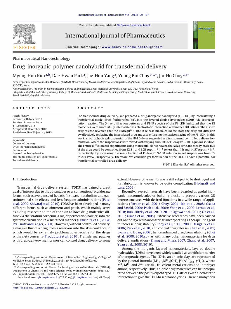
International Journal of Pharmaceutics 444 (2013) 120–127 Contents lists available at SciVerse ScienceDirect International Journal of Pharmaceutics jo ur n al homep age: www.elsevier.com/locate/ijpharm Pharmaceutical Nanotechnology Drug-inorganic-polymer nanohybrid for transdermal delivery Myung Hun Kim a,b , Dae-Hwan Park a , Jae-Hun Yang a , Young Bin Choy b,c,∗ , Jin-Ho Choy a,∗∗ a Center for Intelligent Nano-Bio Materials (CINBM), Department of Bioinspired Science and Department of Chemistry and Nano Science, Ewha Womans University, Seoul, 120-750, Korea b Interdisciplinary Program in Bioengineering, College of Engineering, Seoul National University, Seoul 152-742, Republic of Korea c Department of Biomedical Engineering, College of Medicine and Institute of Medical & Biological Engineering, Medical Research Center, Seoul National University, Seoul 110-799, Republic of Korea a r t i c l e i n f o Article history: Received 2 October 2012 Received in revised form 11 December 2012 Accepted 31 December 2012 Available online 26 January 2013 Keywords: Controlled delivery Drug-inorganic nanohybrid Formulation Layered double hydroxide The Frantz diffusion cell experiments Transdermal delivery a b s t r a c t For transdermal drug delivery, we prepared a drug-inorganic nanohybrid (FB-LDH) by intercalating a transdermal model drug, flurbiprofen (FB), into the layered double hydroxides (LDHs) via coprecipi- tation reaction. The X-ray diffraction patterns and FT-IR spectra of the FB-LDH indicated that the FB molecules were successfully intercalated via electrostatic interaction within the LDH lattices. The in vitro drug release revealed that the Eudragit ® S-100 in release media could facilitate the drug out-diffusion by effectively replacing the intercalated drug and also enlarging the lattice spacing of the FB-LDH. In this work, a hydrophobic gel suspension of the FB-LDH was suggested as a transdermal controlled delivery for- mulation, where the suspensions were mixed with varying amounts of Eudragit ® S-100 aqueous solution. The Frantz diffusion cell experiments using mouse full-skins showed that a lag time and steady-state flux of the drug could be controlled from 12.8 h and 3.28 g cm −2 h −1 to less than 1 h and 14.57 g cm −2 h −1 , respectively, by increasing the mass fraction of Eudragit ® S-100 solution in gel suspensions from 0% to 20% (w/w), respectively. Therefore, we conclude gel formulation of the FB-LDH have a potential for transdermal controlled drug delivery. © 2013 Elsevier B.V. All rights reserved. 1. Introduction Transdermal drug delivery system (TDDS) has gained a great deal of interest due to the advantages over conventional oral dosage forms, such as avoidance of hepatic first pass metabolism and gas- trointestinal side effects, and less frequent administrations (Patel et al., 2009; Shivaraj et al., 2010). TDDS has been developed in many different forms, such as ointment and patch, which mainly serve as a drug reservoir on top of the skin to have drug molecules dif- fuse via the stratum corneum, a major permeation barrier, into the systemic circulation in a sustained manner (Prausnitz et al., 2004; Prausnitz and Langer, 2008). However, without controlled delivery, a massive flux of a drug from a reservoir into the skin could occur, which would be extremely problematic especially for the drugs with safety concerns (Prodduturi et al., 2010). Transdermal patches with drug-delivery membranes can control drug delivery to some ∗ Corresponding author at: Department of Biomedical Engineering, College of Medicine, Seoul National University, Seoul, 110-799, Republic of Korea. Tel.: +82 2 740 8592; fax: +82 2 741 6303. ∗∗ Corresponding author at: Center for Intelligent Nano-Bio Materials (CINBM), Department of Chemistry and Nano Science, Ewha Womans University, Seoul 120- 750, Republic of Korea. Tel.: +82 2 3277 4135; fax: +82 2 3277 4340. E-mail addresses: [email protected] (Y.B. Choy), [email protected] (J.-H. Choy). extent. However, the membrane is still subject to be destroyed and its fabrication is known to be quite complicating (Hadgraft and Lane, 2006). Recently, layered materials have been regarded as useful inor- ganic macromolecules or building blocks to prepare various 2D heterostructures with desired functions in a wide range of appli- cations (Portier et al., 2001; Choy, 2004; Ida et al., 2008; Osada and Sasaki, 2009; Paek et al., 2009; Yoon et al., 2009; Leroux et al., 2010; Ruiz-Hitzky et al., 2010, 2011; Ogawa et al., 2011; Oh et al., 2011; Okada et al., 2005). Extensive researches have been carried out on the layered nanomaterials incorporating a therapeutic agent to increase drug stability (Choy et al., 1999, 2000; Desigaux et al., 2006; Park et al., 2010) and control drug release (Khan et al., 2001; Evans and Duan, 2006), hence enhanced drug bioavailability (Choi et al., 2008, 2010a,b), as with many other nanomaterials for drug delivery applications (Zhang and Misra, 2007; Zhang et al., 2007; Yuan et al., 2008, 2010). Among the inorganic layered nanomaterials, layered double hydroxides (LDHs) have been widely studied as an efficient carrier of therapeutic agents. The LDHs, an anionic clay, are represented by the general formula [M II 1−x M III x (OH) 2 ] x+ (A n− ) x/n · yH 2 O, where M II , M III , and A n− are di-, tri-valent metal cations and interlayer anions, respectively. Thus, anionic drug molecules can be incorpo- rated between the positively charged LDH lattices with electrostatic interaction to give the LDH-based nanohybrids. These nanohybrids 0378-5173/$ – see front matter © 2013 Elsevier B.V. All rights reserved. http://dx.doi.org/10.1016/j.ijpharm.2012.12.043
Transcript of Drug-inorganic-polymer nanohybrid for transdermal delivery
P
D
Ma
1b
c
S
a
ARR1AA
KCDFLTT
1
dftedafsPawww
MT
D7
0h
International Journal of Pharmaceutics 444 (2013) 120– 127
Contents lists available at SciVerse ScienceDirect
International Journal of Pharmaceutics
jo ur n al homep age: www.elsev ier .com/ locate / i jpharm
harmaceutical Nanotechnology
rug-inorganic-polymer nanohybrid for transdermal delivery
yung Hun Kima,b, Dae-Hwan Parka, Jae-Hun Yanga, Young Bin Choyb,c,∗, Jin-Ho Choya,∗∗
Center for Intelligent Nano-Bio Materials (CINBM), Department of Bioinspired Science and Department of Chemistry and Nano Science, Ewha Womans University, Seoul,20-750, KoreaInterdisciplinary Program in Bioengineering, College of Engineering, Seoul National University, Seoul 152-742, Republic of KoreaDepartment of Biomedical Engineering, College of Medicine and Institute of Medical & Biological Engineering, Medical Research Center, Seoul National University,eoul 110-799, Republic of Korea
r t i c l e i n f o
rticle history:eceived 2 October 2012eceived in revised form1 December 2012ccepted 31 December 2012vailable online 26 January 2013
eywords:
a b s t r a c t
For transdermal drug delivery, we prepared a drug-inorganic nanohybrid (FB-LDH) by intercalating atransdermal model drug, flurbiprofen (FB), into the layered double hydroxides (LDHs) via coprecipi-tation reaction. The X-ray diffraction patterns and FT-IR spectra of the FB-LDH indicated that the FBmolecules were successfully intercalated via electrostatic interaction within the LDH lattices. The in vitrodrug release revealed that the Eudragit® S-100 in release media could facilitate the drug out-diffusionby effectively replacing the intercalated drug and also enlarging the lattice spacing of the FB-LDH. In thiswork, a hydrophobic gel suspension of the FB-LDH was suggested as a transdermal controlled delivery for-
®
ontrolled deliveryrug-inorganic nanohybridormulationayered double hydroxidehe Frantz diffusion cell experimentsransdermal deliverymulation, where the suspensions were mixed with varying amounts of Eudragit S-100 aqueous solution.The Frantz diffusion cell experiments using mouse full-skins showed that a lag time and steady-state fluxof the drug could be controlled from 12.8 h and 3.28 �g cm−2 h−1 to less than 1 h and 14.57 �g cm−2 h−1,respectively, by increasing the mass fraction of Eudragit® S-100 solution in gel suspensions from 0%to 20% (w/w), respectively. Therefore, we conclude gel formulation of the FB-LDH have a potential fortransdermal controlled drug delivery.
. Introduction
Transdermal drug delivery system (TDDS) has gained a greateal of interest due to the advantages over conventional oral dosageorms, such as avoidance of hepatic first pass metabolism and gas-rointestinal side effects, and less frequent administrations (Patelt al., 2009; Shivaraj et al., 2010). TDDS has been developed in manyifferent forms, such as ointment and patch, which mainly serves a drug reservoir on top of the skin to have drug molecules dif-use via the stratum corneum, a major permeation barrier, into theystemic circulation in a sustained manner (Prausnitz et al., 2004;rausnitz and Langer, 2008). However, without controlled delivery,
massive flux of a drug from a reservoir into the skin could occur,
hich would be extremely problematic especially for the drugsith safety concerns (Prodduturi et al., 2010). Transdermal patchesith drug-delivery membranes can control drug delivery to some∗ Corresponding author at: Department of Biomedical Engineering, College ofedicine, Seoul National University, Seoul, 110-799, Republic of Korea.
el.: +82 2 740 8592; fax: +82 2 741 6303.∗∗ Corresponding author at: Center for Intelligent Nano-Bio Materials (CINBM),epartment of Chemistry and Nano Science, Ewha Womans University, Seoul 120-50, Republic of Korea. Tel.: +82 2 3277 4135; fax: +82 2 3277 4340.
E-mail addresses: [email protected] (Y.B. Choy), [email protected] (J.-H. Choy).
378-5173/$ – see front matter © 2013 Elsevier B.V. All rights reserved.ttp://dx.doi.org/10.1016/j.ijpharm.2012.12.043
© 2013 Elsevier B.V. All rights reserved.
extent. However, the membrane is still subject to be destroyed andits fabrication is known to be quite complicating (Hadgraft andLane, 2006).
Recently, layered materials have been regarded as useful inor-ganic macromolecules or building blocks to prepare various 2Dheterostructures with desired functions in a wide range of appli-cations (Portier et al., 2001; Choy, 2004; Ida et al., 2008; Osadaand Sasaki, 2009; Paek et al., 2009; Yoon et al., 2009; Leroux et al.,2010; Ruiz-Hitzky et al., 2010, 2011; Ogawa et al., 2011; Oh et al.,2011; Okada et al., 2005). Extensive researches have been carriedout on the layered nanomaterials incorporating a therapeutic agentto increase drug stability (Choy et al., 1999, 2000; Desigaux et al.,2006; Park et al., 2010) and control drug release (Khan et al., 2001;Evans and Duan, 2006), hence enhanced drug bioavailability (Choiet al., 2008, 2010a,b), as with many other nanomaterials for drugdelivery applications (Zhang and Misra, 2007; Zhang et al., 2007;Yuan et al., 2008, 2010).
Among the inorganic layered nanomaterials, layered doublehydroxides (LDHs) have been widely studied as an efficient carrierof therapeutic agents. The LDHs, an anionic clay, are representedby the general formula [MII
1−xMIIIx(OH)2]x+(An−)x/n · yH2O, where
MII, MIII, and An− are di-, tri-valent metal cations and interlayeranions, respectively. Thus, anionic drug molecules can be incorpo-rated between the positively charged LDH lattices with electrostaticinteraction to give the LDH-based nanohybrids. These nanohybrids
al of P
a(oie
fbwiniLbH
sad0(mcl(igbiTdr
rtrbsm
2
2
pMftELfm
2
ZoscrnTa
M.H. Kim et al. / International Journ
re known to be fairly stable in suspension and also biocompatibleChoy et al., 2004; Choi and Choy, 2011). A narrow size distributionf the LDH-based nanohybrids also allows controlled drug deliveryn a reproducible manner (Choy and Son, 2004; Oh et al., 2009; Bullt al., 2011).
Therefore, in this work, we propose the LDH-based nanohybridor transdermal delivery formulation. We prepared the nanohy-rids, employing flurbiprofen (FB) as a model drug (i.e., FB-LDH),here the anionic FB molecules were intercalated into the pos-
tively charged LDH layers by electrostatic interaction. FB, aon-steroidal anti-inflammatory drug, has been already approved
n transdermal dosage forms (Poul et al., 1993; Gao et al., 1998;awrencea and Reesb, 2000). The LDHs were reported to be highlyiocompatible and also safe for dermatological application (Deloyo, 2007; Blasi et al., 2011).
For controlled delivery, we prepared a nonaqueous gel suspen-ion of the FB-LDH, where a varying amount of the water includingn anionic macromolecule, Eudragit® S-100, was added. Thus, fourifferent gel suspensions were prepared in this work by mixing, 50, 100 and 200 �l of an aqueous solution of Eudragit® S-1002.5%, w/v) with 1 g of the FB-LDH gel suspension, respectively. The
acromolecules, possessing the same polarity as the intercalatedompounds, are known to facilitate the out-diffusion of interca-ated compounds by enlarging the lattice spacing of nanohybridChoi et al., 2010a,b). Therefore, the strategy is following: (1) drugs released from the FB-LDH into a gel medium and (2) drug in ael medium is then permeated via the skin. The former is tailoredy the amount of an aqueous solution of Eudragit® S-100 present
n a gel and the latter by the drug concentration in a gel medium.he FB is a poorly water-soluble (<8 �g/ml) and thus, most of therug is expected to distribute in a hydrophobic gel medium wheneleased from the FB-LDH.
In this study, we characterized the FB-LDH with the powder X-ay diffraction, FT-IR analysis and TGA–DTA analysis to investigatehe drug property in the LDH after intercalation. The in vitro drugelease profiles of the FB-LDH were examined using the phosphateuffered saline (PBS; pH 7.4). To investigate drug delivery via thekin, we performed the Franz diffusion cell experiments, using theouse full skin.
. Materials and methods
.1. Materials
ZnCl2, AlCl3·6H2O, flurbiprofen (C15H13FO2), monopotassiumhosphate and ethylcellulose were obtained from Sigma (St. Louis,O, USA). Sodium hydroxide and ethanol (EtOH) were purchased
rom Daejung (Siheung, Korea). Anhydride acetic acid and acetoni-rile, all in HPLC grade, were obtained from JT Baker (NJ, USA).udragit® S-100 was purchased from Degussa (Essen, Germany).abrafac® CC, Labrafil® M 1944 CS, and Transcutol® P purchasedrom Gattefosse (Lyon, France) were utilized to prepare the gel
edium (Table S1).
.2. Preparation of FB-LDH nanohybrid
A solution of 0.04 M FB in 15 ml EtOH and a solution of 0.08 MnCl2 and 0.04 M AlCl3·6H2O (molar ratio of M2+/M3+ = 2) in 15 mlf deionized water were vigorously mixed for 30 min until theolution became transparent. After this, the solution was copre-ipitated by titrating with 0.5 M NaOH solution to pH 8, and aged at
oom temperature for 12 h. All the reaction was carried out underitrogen atmosphere to prevent carbonate contamination from air.he resulting FB-LDH precipitate was separated by centrifugationnd washed three times with the mixed solution of EtOH andharmaceutics 444 (2013) 120– 127 121
deionized water (1:1 volume ratio) to remove the unreacted saltions and excess FB. A dry powder of the FB-LDH nanohybrid wasobtained by freeze-drying
2.3. Preparation of FB-LDH gel formulations
To prepared the gel formulation of the FB-LDH, the nanohy-brids were suspended in a nonaqueous gel medium, whichis already widely accepted for transdermal drug-delivery for-mulations (Kogan and Garti, 2006; Balakrishnan et al., 2009).The gel medium was prepared by mixing Labrafac® CC andethyl cellulose (49:6, w/w) with agitation at 90 ◦C until themixture became transparent. After cooling down the resultingmixture to room temperature, Labrafil® M 1944, Transcutol®
and the FB-LDH were added (Labrafil:Transcutol:FB-LDH:the mix-ture = 15:20:10:55, weight ratio) and stirred at 90 ◦C to give a gelsuspension of the FB-LDH. Thus, all gel formulations prepared inthis work contained 45 mg FB-LDH per g gel suspension.
In this work, we also added a varying amount of Eudragit® S-100aqueous solution to the FB-LDH gel suspensions for controlled drugdelivery: 0, 50, 100 and 200 �l of Eudragit® S-100 aqueous solution(2.5 wt%) into 1 g of the FB-LDH gel suspension to give G-FN-0, G-FN-50, G-FN-100 and G-FN-200, respectively.
2.4. Characterizations of FB-LDH nanohybrid
2.4.1. Powder X-ray diffraction (XRD)The crystallinity of the FB-LDH was examined by an X-ray
diffractometer (D/MAXRINT 2200-Ultima, Rigaku, Japan) equippedwith Ni-filtered Cu K� radiation (� = 1.5418 A). The samples wereloaded on a glass substrate holder and continuously scanned at thetube voltage and the current of 40 kV and 30 mA, respectively. Topredict the interlayer structure of guest species in layered inorganiccompounds, one-dimensional (1D) electron density was also cal-culated by eliminating certain conformational possibilities of guestspecies that are incompatible with the experimental X-ray diffrac-tion data. The 1D electron densities along the z axis (�(z)) werecalculated by using the following equation (Cunha et al., 2012).
�(z) =∞∑
l=0
F00l cos(
2�lz
c
)
where c is the unit cell parameter of FB-LDH and F00l is the struc-ture factors for the (0 0 l) reflections, derived from their intensitiescorrected by Lorentz polarization factor. The signs of the structurefactors were directly obtained from the scattering contributions ofoctahedral Zn2Al hydroxide layers, assuming that the contributionof incorporated molecule is relatively small. For the FB-LDH, six(0 0 l) reflections were used.
2.4.2. Fourier transform infrared spectroscopy (FT-IR)FT-IR spectra of the intact FB, pristine LDH, and FB-LDH were
collected with a JASCO 6100 spectrophotometer (JASCO, Inc., Japan)in a range of 400–4000 cm−1 with 4 cm−1 resolution, using the KBrdisk method.
2.4.3. Scanning electron microscopy (SEM)The field emission scanning electron microscopic image of the
FB-LDH was monitored with JEOL JSM-6700F (JEOL Ltd., Japan). Thesample was coated with platinum before imaging.
2.4.4. Transmission electron microscopy (TEM)
The transmission electron microscopic image of the FB-LDH wasobtained on JEOL JEM-2000EXII (JEOL Ltd., Japan) at 200 kV of anaccelerating voltage. To prepare a TEM sample, the FB-LDH wassuspended in deionized water, a drop of which was placed on a Cu

1 al of Pharmaceutics 444 (2013) 120– 127
giuo
2
ts
2
wiO
2(
mi1
2
Fa3s0o(U(piit
2
tUtmoosos
2
iAhsswibE
a
22 M.H. Kim et al. / International Journ
rid coated with carbon and dried. To obtain the cross-sectionalmage, the FB-LDH was embedded in an epoxy resin, which wasltramicrotomed (RMC, Tucson, USA) with 50 nm thickness with-ut staining and placed on a Cu grid coated with carbon.
.4.5. Particle size distribution and zeta potential measurementsThe size distribution and surface charge of the pristine LDH and
he FB-LDH in distilled water were measured with Malvern Zeta-izer Nano ZS (Malvern, UK).
.4.6. Elemental analysisThe chemical compositions of the pristine LDH and FB-LDH
ere determined by the elemental CHN analyses with EA1110 (CEnstrument, Italy) and inductive coupled plasma (ICP) analyses withptima-4300 DV (PerkinElmer, USA).
.4.7. Thermogravimetry and differential thermal analysisTGA–DTA)
The TGA–DTA curve of the FB-LDH was recorded on TA instru-ent SDT Q600 (TA instrument, USA). The temperature was
ncreased from ambient to 800 ◦C at a heating rate of 2 ◦C/min under00 ml/min airflow.
.4.8. High performance liquid chromatography (HPLC) analysisTo evaluate the amount of FB incorporated in the FB-LDH, 45 mg
B-LDH was dispersed into 100 ml of the solution mixed with EtOHnd pH 7.4 PBS (1:4, volume ratio), which was then sonicated for0 min to completely extract the FB from the FB-LDH. The suspen-ion was then filtered by a Nylon syringe filter with a pore size of.45 �m (Millipore, USA). The chromatogram of the solution wasbtained with the high performance liquid chromatography (HPLC)Agilent 1100 series, Agilent Technologies, USA) equipped with aV detector (�max = 254) and reverse phase Zorbax® C18 column
25 cm × 0.45 cm, 5 �m, Agilent Technologies, USA). The mobilehase composed of anhydride acetic acid, acetonitrile and deion-
zed water (1:7:12, volume ratio) was used with a flow rate andnjection volume of 1.5 ml/min and 10 �l, respectively. The columnemperature was maintained at 25 ◦C.
.5. In vitro drug release test
In vitro release experiments were carried out using a dissolu-ion apparatus 2, Vision® G2 Elite 8TM Dissolution Tester (Hanson,SA) with the paddle-type stirrer (50 rpm) based on a USP dissolu-
ion test method (USP, 2002, pp. 2011–2012). Two distinct releaseedia were employed, which were the pH 7.4 PBS solutions with-
ut and with Eudragit® S-100 (2.5%, w/v), to examine the effectf anionic macromolecules on FB release. The FB-LDH (45 mg) wasuspended in 900 ml of the release media at 37 ◦C and the amountf released FB was determined by monitoring the HPLC spectra atcheduled intervals.
.6. In vitro skin permeability test
The in vitro passive skin permeability of FB from the FB-LDH wasnvestigated by Franz diffusion cell diffusion method (Franz, 1975).t first, fresh full skins were obtained from 6-week-old femaleairless nude mice (Orientbio, Korea). Only the mice without theigns of overt toxicity and readily apparent clinical symptoms wereelected and sacrificed by cervival dislocation, from which the skinsithout any defect were collected. All experiments were conducted
n compliance with the experimental protocol, which was approved
y the Institutional Animal Care and Use Committee (IACUC) at thewha Womans University (IACUC No.: 2010-9-3).The full skins thus obtained were each placed between the donornd acceptor chambers, where the stratum corneum and dermal
Fig. 1. Powder XRD patterns of the (a) intact FB, (b) pristine LDH and (c) FB-LDH.
side were exposed to the donor and receptor chambers, respec-tively. The receptor chamber was filled with pH 7.4 PBS, wherethe temperature was maintained at 37 ± 1 ◦C by the thermostati-cally controlled circulating water jacket. The exposed area of theskin membrane available for drug permeation was 1.76 cm2 andthe capacity of each receptor chamber was 12.5 ml. The gel formu-lations prepared in this work was each applied on top of the skin viathe donor chamber. At scheduled intervals, the media in the recep-tor chamber was drawn out and the receptor was re-filled with anequal amount of fresh PBS. The sampled media were each assayedwith the HPLC as described above. Each of the gel formulations wastested at least five times for statistics.
3. Results and discussion
3.1. Powder X-ray diffraction analysis
To assess the formation of the nanohybrid prepared by the con-ventional coprecipitation method, we examined the powder XRDpattern of the FB-LDH, together with the ones of intact FB and pris-tine Zn2Al LDH, as shown in Fig. 1. The pristine LDH shows a seriesof well ordered (0 0 l) reflections corresponding to the basal spacingof 7.5 A, indicating the formation of the Cl− intercalated Zn2Al LDH.On the other hand, the FB-LDH shows the sharp and well devel-oped (0 0 l) reflections with a marked increase in basal spacing to24.4 A. This clearly demonstrated that the FB molecules were wellintercalated into the interlayer space of LDH lattice. Notably, nocrystalline peaks of intact FB were observed with the FB-LDH, noti-fying that FB molecules were well ordered and distributed in themolecular level in the interlayer of LDH lattices. From the thicknessof the Zn2Al LDH layer (4.8 A) and the basal spacing of the FB-LDH(24.4 A), the gallery height of the FB-LDH could be determined to be19.6 A, which was larger than the longitudinal and lateral molecu-lar dimensions of FB (i.e., 6.2 A and 11.5 A, respectively, as shownin Fig. S1). Therefore, it was expected that the FB molecules werelikely to be stabilized in a tilted double layer arrangement.
To assess this configuration, we first examined the steric limi-tation between the intercalated FB molecules and charged sites ofLDH layers (Yang et al., 2007). The steric limitation in the inter-layer of layered materials can be explained in the viewpoint of the
equivalent area (Ae) (area per positive unit charge (Å2/e−) of a lay-ered double hydroxide) and the area demand (Ac) (area demand perunit charge (Å2/e−) for intercalated single molecules). The equiva-lent area (Ae) of the layered host lattices can be estimated from
M.H. Kim et al. / International Journal of Pharmaceutics 444 (2013) 120– 127 123
F nd tho
tp1cZata
mamdqefao0tcBtGwt
3
Lio(rtwtiaTca
corresponding calculated cell parameter (a = 0.30 nm) were, withinthe error limit, identical to those obtained from the XRD analy-sis. The cross-sectional TEM image revealed that the FB-LDH was
ig. 2. One-dimensional electron density projection along c-axis for the FB-LDH abserved and calculated electron density projection, respectively.
he equation, Ae = a × b × sin �/2�, where a and b are the latticearameters and � is the layer charge per formula unit (Meyn et al.,990). Based on the lattice parameters of the FB-LDH (a = b = 3.07 A,
= 73.04 A, � = 120◦ and � = 1/3e−), the equivalent area (Ae) of then2Al host layer and the area demand (Ac) of a single FB moleculere about 24.4 A2 and 71.3 A2, respectively. As Ac is larger than 2Ae,he intercalated FB molecules were likely to have a tilted bilayerrrangement to avoid steric hindrance.
To get further insight into the interlayer arrangement of FBolecules, the 1D electron density map of the FB-LDH structure
long the c axis was constructed. Among various different structureodels, the FB-LDH structure model in Fig. 2, which has confi-
ence with value of difference Fourier map (��(z) = 0.98), wasuite well consistent with the results of observed and calculatedlectron density projection compared to different models tiltedrom 0◦ to 31.55◦ (Fig. S2). The 1D plot can be used to support
structural model for the interlayer space. The �(z) values werebtained by Fourier transform of the intensities for well-developed
0 3/0 0 6/0 0 9/0 0 1 2/0 0 1 5/0 0 1 8 reflection peaks. The 1D elec-ron density map displayed two strong peaks at z = 0 and 1,orresponding to the metals present in the hydroxide layers.etween these two strong peaks, six additional peaks of lower elec-ron densities were observed due to the intercalated FB molecules.iven this and upon comparison with the length of the FB molecule,e could again propose that FB in the LDH layer has an interdigi-
ated bilayer arrangement with a tilt angle of ≈20.96◦.
.2. FT-IR analysis
To examine the chemical interaction between FB molecules andDH lattices, we measured and compared the FT-IR spectra of thentact FB, pristine LDH and FB-LDH, as shown in Fig. 3. In the spectraf intact FB, the characteristic peaks were observed at 1195 cm−1
C F) and 1624 cm−1 (C C) due to stretching mode of aromaticing (Fig. 3(a)). These characteristic peaks were superimposed onhe spectra of the FB-LDH, as shown in Fig. 3(c), indicating that FBas intercalated into the LDH matrix without any noticeable struc-
ural changes. The notable feature upon the intercalation of the FBnto LDH was disappearance of the absorption peak at 1698 cm−1,
nd appearance of the new bands at 1550 cm−1 and 1403 cm−1.he former was ascribed to the carbonyl group (C O) of the freearboxylic acid group in intercalated FB and the latter was due tosymmetric and symmetric modes of (COO−), implying that thee corresponding structural model. The solid and dashed lines show the curves of
deprotonated carboxylate form of FB molecules were bound to theinterlayer of LDH lattice by electrostatic interaction (Li et al., 2004).
The pristine LDH exhibited the broad absorption band centeredat 3452 cm−1, which is assigned to OH vibration and interlayerwater, as shown in Fig. 3(b). This broad band was also seen with theFB-LDH, which was centered at 3396 cm−1 (Fig. 3(c)). The peak seenaround at 1629 cm−1 was assigned to the O H stretching modesof interlayer water molecules of the pristine LDH. The absorptionbands observed below 800 cm−1 were associated with lattice vibra-tion modes attributed to M O and M O M (M: metals, such as Znand Al) vibration in the brucite layer, which were also seen withboth pristine LDH and FB-LDH (Fig. 3(b) and (c)).
3.3. Particle analysis
The FB-LDH was observed with both SEM and TEM to assess theirmorphology and crystal structure. Fig. 4(a) and (b) shows that theFB-LDH possessed plate-like morphology with primary particle sizeof 105 nm. The selected-area electron diffraction (SAED) patternsof the FB-LDH displayed hexagonally arranged spots (Fig. 4(c)). The
Fig. 3. FT-IR spectra of the (a) intact FB, (b) pristine LDH and (c) FB-LDH.

124 M.H. Kim et al. / International Journal of Pharmaceutics 444 (2013) 120– 127
F electem ohybr
c(e
tpLt(plwZ
3
t
ig. 4. Particle analyses of the FB-LDH, showing (a) SEM image, (b) TEM image, (c) sagnification, (e) particle size distribution and (e) zeta potential of the FB-LDH nan
omposed of the layered matrices with the basal spacing of 24.4 AFig. 4(d)), which was also in good agreement with the dimensionstimated from the XRD data.
We also examined the size distribution and surface charge ofhe FB-LDH based on the dynamic light scattering (DLS) and zetaotential methods, respectively. As shown in Fig. 4(e), the FB-DH exhibited an average size of 105 ± 13 nm, as obtained withhe SEM and TEM data. The FB-LDH had a positive surface chargezeta potential: +5.25 ± 0.17 mV at pH 7.4) in Fig. 4(f) due to theositively-charged nature of Zn2Al hydroxide layer. After interca-
ation of negatively-charged FB, the surface charge of the FB-LDHas lowered by charge compensation, as compared to the pristine
n2Al LDH (zeta potential: +34.1 ± 1.4 mV at pH 7.4, Fig. S3).
.4. Thermal analysis
We performed the TGA–DTA on the dry powder of the pris-ine LDH and FB-LDH. In the TGA–DTA curve for the pristine LDH
d-area electron diffraction pattern, (d) cross-section TEM image with inlet of higherid (+5.25 mV).
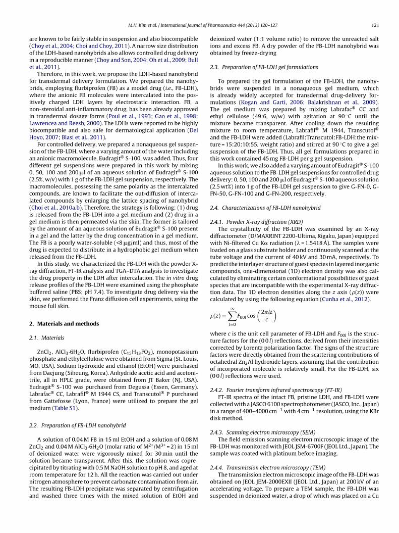
(Fig. 5(a)), the first weight loss (ca. 13.1%) with an endothermicpeak at 135.9 ◦C was attributed to the dehydration of physicallyabsorbed water molecules located both at the exterior surfaceand in the interlayer space of the LDH particles (30.0–232.8 ◦C).A second mass loss (ca. 24.1%) with an endothermic peak centeredat 363.19 ◦C is related to partial dehydroxylation of the brucite-type layers (232.8–493.4 ◦C). Still, the third weight loss (ca. 4.8%)was observed above 493.4 ◦C, which is attributed to the carbon-ate decomposition and elimination of chloride species (Rives et al.,1998).
For the FB-LDH, the TGA–DTA curves show the thermalevolution with three consecutive stages due to dehydration,decomposition, and dehydroxylation, respectively (Fig. 5(b)). Thefirst weight loss (ca. 6.51%) with a weak endothermic response
around at 71.9 ◦C was ascribed to removal of absorbed water on thesurface of the FB-LDH, which was smaller than that with the pristineLDH because of the presence of intercalated hydrophobic FB. Thesecond weight loss (ca. 8.03%) with an endothermic peak around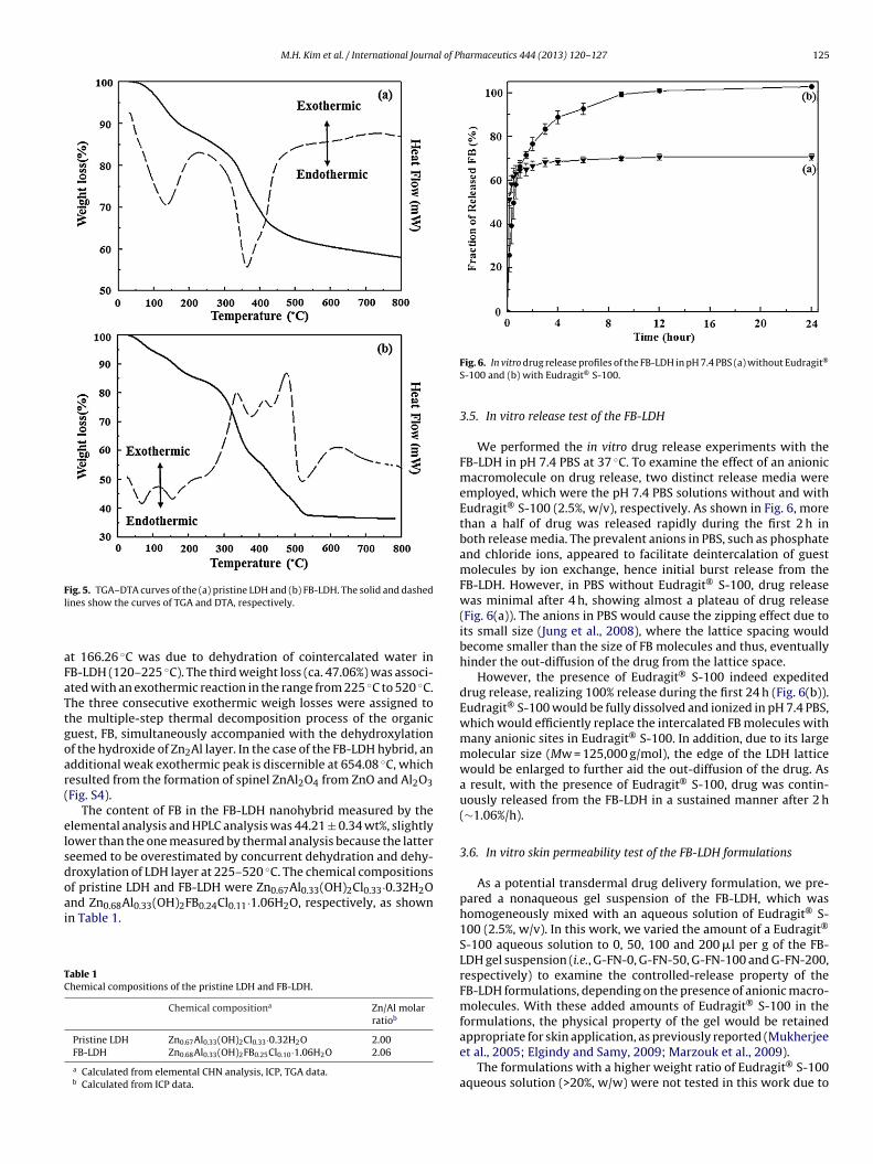
M.H. Kim et al. / International Journal of Pharmaceutics 444 (2013) 120– 127 125
Fig. 5. TGA–DTA curves of the (a) pristine LDH and (b) FB-LDH. The solid and dashedl
aFaTtgoar(
elsdoai
TC
ines show the curves of TGA and DTA, respectively.
t 166.26 ◦C was due to dehydration of cointercalated water inB-LDH (120–225 ◦C). The third weight loss (ca. 47.06%) was associ-ted with an exothermic reaction in the range from 225 ◦C to 520 ◦C.he three consecutive exothermic weigh losses were assigned tohe multiple-step thermal decomposition process of the organicuest, FB, simultaneously accompanied with the dehydroxylationf the hydroxide of Zn2Al layer. In the case of the FB-LDH hybrid, andditional weak exothermic peak is discernible at 654.08 ◦C, whichesulted from the formation of spinel ZnAl2O4 from ZnO and Al2O3Fig. S4).
The content of FB in the FB-LDH nanohybrid measured by thelemental analysis and HPLC analysis was 44.21 ± 0.34 wt%, slightlyower than the one measured by thermal analysis because the lattereemed to be overestimated by concurrent dehydration and dehy-roxylation of LDH layer at 225–520 ◦C. The chemical compositionsf pristine LDH and FB-LDH were Zn0.67Al0.33(OH)2Cl0.33·0.32H2Ond Zn0.68Al0.33(OH)2FB0.24Cl0.11·1.06H2O, respectively, as shown
n Table 1.able 1hemical compositions of the pristine LDH and FB-LDH.
Chemical compositiona Zn/Al molarratiob
Pristine LDH Zn0.67Al0.33(OH)2Cl0.33·0.32H2O 2.00FB-LDH Zn0.68Al0.33(OH)2FB0.25Cl0.10·1.06H2O 2.06
a Calculated from elemental CHN analysis, ICP, TGA data.b Calculated from ICP data.
Fig. 6. In vitro drug release profiles of the FB-LDH in pH 7.4 PBS (a) without Eudragit®
S-100 and (b) with Eudragit® S-100.
3.5. In vitro release test of the FB-LDH
We performed the in vitro drug release experiments with theFB-LDH in pH 7.4 PBS at 37 ◦C. To examine the effect of an anionicmacromolecule on drug release, two distinct release media wereemployed, which were the pH 7.4 PBS solutions without and withEudragit® S-100 (2.5%, w/v), respectively. As shown in Fig. 6, morethan a half of drug was released rapidly during the first 2 h inboth release media. The prevalent anions in PBS, such as phosphateand chloride ions, appeared to facilitate deintercalation of guestmolecules by ion exchange, hence initial burst release from theFB-LDH. However, in PBS without Eudragit® S-100, drug releasewas minimal after 4 h, showing almost a plateau of drug release(Fig. 6(a)). The anions in PBS would cause the zipping effect due toits small size (Jung et al., 2008), where the lattice spacing wouldbecome smaller than the size of FB molecules and thus, eventuallyhinder the out-diffusion of the drug from the lattice space.
However, the presence of Eudragit® S-100 indeed expediteddrug release, realizing 100% release during the first 24 h (Fig. 6(b)).Eudragit® S-100 would be fully dissolved and ionized in pH 7.4 PBS,which would efficiently replace the intercalated FB molecules withmany anionic sites in Eudragit® S-100. In addition, due to its largemolecular size (Mw = 125,000 g/mol), the edge of the LDH latticewould be enlarged to further aid the out-diffusion of the drug. Asa result, with the presence of Eudragit® S-100, drug was contin-uously released from the FB-LDH in a sustained manner after 2 h(∼1.06%/h).
3.6. In vitro skin permeability test of the FB-LDH formulations
As a potential transdermal drug delivery formulation, we pre-pared a nonaqueous gel suspension of the FB-LDH, which washomogeneously mixed with an aqueous solution of Eudragit® S-100 (2.5%, w/v). In this work, we varied the amount of a Eudragit®
S-100 aqueous solution to 0, 50, 100 and 200 �l per g of the FB-LDH gel suspension (i.e., G-FN-0, G-FN-50, G-FN-100 and G-FN-200,respectively) to examine the controlled-release property of theFB-LDH formulations, depending on the presence of anionic macro-molecules. With these added amounts of Eudragit® S-100 in theformulations, the physical property of the gel would be retained
appropriate for skin application, as previously reported (Mukherjeeet al., 2005; Elgindy and Samy, 2009; Marzouk et al., 2009).The formulations with a higher weight ratio of Eudragit® S-100aqueous solution (>20%, w/w) were not tested in this work due to

126 M.H. Kim et al. / International Journal of P
Fa
d(
aowpElotoa
dfllEtrwtmrp
tftrabAfl
4
ttdiom
ig. 7. In vitro skin permeability profiles of (a) G-FN-0, (b) G-FN-50, (c) G-FN-100nd (d) G-FN-200.
iscernible phase separation between a gel and aqueous solutionFig. S5).
As shown in Fig. 7, the cumulative amounts of FB deliveredcross a full thickness nude mouse skin were obtained from eachf the gel formulations prepared in this work. The dose of FBas 20 mg for all gel formulations tested in this in vitro skinermeation study. For G-FN-0 (i.e., a gel suspension without audragit® S-100 solution), drug permeation via the skin was veryow, showing an evident lag time of 12.8 h and a steady state fluxf ≈3.28 �g cm−2 h−1. The ionic exchange of the intercalated FB inhe LDH lattices appeared to occur inefficiently without an aque-us phase, hence minimal drug release and thus, almost no drugvailable in the gel medium.
However, with the presence of a Eudragit® S-100 solution, drugelivery across the skin became more efficient. The steady stateux of FB increased to ≈5.79 �g cm−2 h−1 for G-FN-50 with the
ag time of less than 1 h (Fig. 7(b)). Anionic macromolecules (i.e.,udragit® S-100) ionized in an aqueous phase would allow effec-ive deintercalation of FB from the FB-LDH, as depicted in Fig. 6. Theeleased drug would be distributed in a hydrophobic gel medium,hich would then be delivered across the skin. As stated above,
he hydrophobic FB would be likely to distribute mostly in a geledium when released from the FB-LDH. The concentration of FB
eleased into a gel medium is a critical determinant of the drugermeability via the skin.
Therefore, to control transdermal drug delivery, we increasedhe added amount of a Eudragit® S-100 solution, which wouldurther facilitate the deintercalation of FB (i.e., FB release) fromhe FB-LDH and increase the drug concentration in the gel. As aesult, the steady state flux of FB increased to ≈8.42 �g cm−2 h−1
nd ≈14.57 �g cm−2 h−1 for G-FN-100 and G-FN-200, respectively,oth also showing the lag times of less than 1 h (Fig. 7(c) and (d)).s compared to G-FN-50, about a three-fold increase in steady stateux of FB could be obtained with G-FN-200 in this work.
. Conclusion
For transdermal controlled drug delivery, we suggest a formula-ion of the drug-inorganic nanohybrid in this work. As we preparedhe nanohybrid (i.e., FB-LDH) by intercalating a transdermal model
rug, FB, into the LDH, the drug molecules could be well stabilizedn the LDH layers, as proven by the PXRD patterns and FT-IR spectraf the FB-LDH. The nanohybrid can release the drug in a sustainedanner, which can be better controlled with the presence of a
harmaceutics 444 (2013) 120– 127
macromolecule, possessing the same polarity comparable to theincorporated drug. According to the in vitro release experimentswith the FB-LDH, the drug release could be facilitated with the pres-ence of an anionic macromolecule, Eudragit® S-100, in the releasemedia by expediting deintercalation of FB in the LDH lattices.
The gel suspension of the nanohybrid can deliver the drug acrossthe skin in a controlled manner, depending on the amount of anaqueous solution of an anionic macromolecule mixed with the gelsuspension. In this work, we prepared the gel suspensions of the FB-LDH with a varying amount of an aqueous solution of Eudragit® S-100 and performed the diffusion cell experiments using the mouseskin. As a result, an average lag time and steady state flux of the drugcould be varied from 12.8 h and ≈3.28 �g cm−2 h−1 to less than 1 hand ≈14.57 �g cm−2 h−1, respectively. Therefore, we conclude thata gel suspension of the nanohybrid containing a tailored amount ofan aqueous solution of macromolecules is a novel formulation forcontrolled delivery of a transdermal drug.
Acknowledgements
This work was supported by the National Research Foundationof Korea (NRF) (SRC program: 2012-0000650, and WCU program:R31-2008-000-10010-0) and partly by the Ewha Womans Univer-sity through Ewha Global Top 5 Grant in 2011.
Appendix A. Supplementary data
Supplementary data associated with this article can befound, in the online version, at http://dx.doi.org/10.1016/j.ijpharm.2012.12.043.
References
Balakrishnan, P., Lee, B.J., Oh, D.H., Kim, J.O., Lee, Y.I., Kim, D.D., Jee, J.P., Lee, Y.B.,Woo, J.S., Yong, C.S., Choi, H.G., 2009. Enhanced oral bioavailability of CoenzymeQ10 by self-emulsifying drug delivery systems. Int. J. Pharm. 374, 66–72.
Blasi, P., Schoubben, A., Giovagnoli, S., Rossi, C., Ricci, M., 2011. The real value ofnovel particulate carriers for sunscreen formulation. Expert Rev. Dermatol. 6,509–517.
Bull, R.M.R., Markland, C., Williams, G.R., O’Hare, D., 2011. Hydroxy double salts asversatile storage and delivery matrices. J. Mater. Chem. 21, 1822–1828.
Choi, G.E., Lee, J.H., Oh, Y.J., Choy, Y.B., Park, M.C., Chang, H.C., Choy, J.H., 2010a.Inorganic-polymer nanohybrid carrier for delivery of a poorly-soluble drug,ursodeoxycholic acid. Int. J. Pharm. 402, 117–122.
Choi, S.J., Oh, J.M., Choy, J.H., 2008. Human-related application and nanotoxicologyof inorganic particles: complementary aspects. J. Mater. Chem. 18, 615–620.
Choi, S.J., Choi, G.E., Oh, J.M., Oh, Y.J., Park, M.C., Choy, J.H., 2010b. Anticancer drugencapsulated in inorganic lattice can overcome drug resistance. J. Mater. Chem.20, 9463–9469.
Choi, S.J., Choy, J.H., 2011. Effect of physico-chemical parameters on the toxicity ofinorganic nanoparticles. J. Mater. Chem. 21, 5547–5554.
Choy, J.H., Kwak, S.Y., Park, J.S., Jeong, Y.J., Portier, J., 1999. Intercalative nanohy-brids of nucleoside monophosphates and DNA in layered metal hydroxide. J.Am. Chem. Soc. 121, 1399–1400.
Choy, J.H., Kwak, S.Y., Jeong, Y.J., Park, J.S., 2000. Inorganic layered double hydroxidesas nonviral vectors. Angew. Chem. Int. Ed. 39, 4041–4045.
Choy, J.H., 2004. Intercalative route to heterostructured nanohybrid. J. Phys. Chem.Solids 65, 373–383.
Choy, J.H., Jung, J.S., Oh, J.M., Park, M., Jeong, J., Kang, Y.K., Han, O.J., 2004. Layered dou-ble hydroxide as an efficient drug reservoir for folate derivatives. Biomaterials25, 3059–3064.
Choy, J.H., Son, Y.H., 2004. Intercalation of vitamer into LDH and their controlledrelease properties. Bull. Korean Chem. Soc. 25, 122–126.
Cunha, V.R.R., Petersen, P.A.D., Gonc alves, M.B., Petrilli, H.M., Taviot-Gueho, C., Ler-oux, F., Temperini, M.L.A., Constantino, V.R.L., 2012. Structural, spectroscopic(NMR, IR, and Raman), and DFT investigation of the self-assembled nanostruc-ture of pravastatin-LDH (Layered Double Hydroxides) systems. Chem. Mater. 24,1415–1425.
Del Hoyo, C., 2007. Layered double hydroxides and human health: an overview. Appl.Clay Sci. 36, 103–121.
Desigaux, L., Belkacem, M.B., Richard, P., Cellier, J., Léone, P., Cario, L., Leroux, F.,Taviot-Guého, C., Pitard, B., 2006. Self-assembly and characterization of layereddouble hydroxide/DNA hybrids. Nano Lett. 6, 199–204.
Elgindy, N., Samy, W., 2009. Evaluation of the mechanical properties and drug releaseof cross linked eudragit films containing metronidazole. Int. J. Pharm. 376, 1–6.
al of P
E
F
G
H
I
J
K
K
L
L
L
M
M
M
O
O
O
O
O
M.H. Kim et al. / International Journ
vans, D.G., Duan, X., 2006. Preparation of layered double hydroxides andtheir applications as additives in polymers, as precursors to mag-netic materials and in biology and medicine. Chem. Commun. 11,485–496.
ranz, T.J., 1975. Percutaneous absorption: on the relevance of in vitro data. J. Invest.Dermatol. 64, 190–195.
ao, Z.G., Choi, H.G., Shin, H.J., Park, K.M., Lim, S.J., Hwang, K.J., Kim, C.K., 1998.Physicochemical characterization and evaluation of a microemulsion systemfor oral delivery of cyclosporin A. Int. J. Pharm. 161, 75–86.
adgraft, J., Lane, M.E., 2006. Passive transdermal drug delivery systems: recentconsiderations and advances. Am. J. Drug Deliv. 4, 153–160.
da, S., Ogata, C., Shiga, D., Izawa, K., Ikeue, K., Matsumoto, Y., 2008. Dynamic con-trol of photoluminescence for self-assembled nanosheet films intercalated withlanthanide ions by using a photoelectrochemical reaction. Angew. Chem. Int. Ed.47, 2480–2483.
ung, H., Kim, H., Choy, Y.B., Hwang, S., Choy, J., 2008. Itraconazole–Laponite: kineticsand mechanism of drug release. Appl. Clay Sci. 40, 99–107.
han, A.I., Lei, L., Norquist, A.J., O’Hare, D., 2001. Intercalation and controlled releaseof pharmaceutically active compounds from a layered double hydroxide. Chem.Commun. 22, 2342–2343.
ogan, A., Garti, N., 2006. Microemulsions as transdermal drug delivery vehicles.Adv. Colloid Interface Sci. 369, 123–126.
awrencea, M.J., Reesb, G.D., 2000. Microemulsion-based media as novel drug deliv-ery systems. Adv. Drug Deliv. Rev. 45, 89–121.
eroux, F., Illaik, A., Stimpfling, T., Troutier-Thuilliez, A., Fleutot, S., Martinez, H.,Cellier, J., Verney, V., 2010. Percolation network of organo-modified layereddouble hydroxide platelets into polystyrene showing enhanced rheological anddielectric behavior. J. Mater. Chem. 20, 9484–9494.
i, B., He, J., Evans, D.G., Duan, X., 2004. Inorganic layered double hydroxides as adrug delivery system-intercalation and in vitro release of fenbufen. Appl. ClaySci. 27, 199–207.
arzouk, M.A.E., Kassem, A.E.D., Samy, A.M., Amer, R.I., 2009. In vitro release,thermodynamics and pharmacodynamic studies of aceclofenac transdermaleudragit patches. Drug Invent. Today 1, 16–22.
eyn, M., Beneke, K., Lagaly, G., 1990. Anion-exchange reactions of layered doublehydroxides. Inorg. Chem. 29, 5201–5207.
ukherjee, B., Mahapatra, S., Gupta, R., Patra, B., Tiwari, A., Arora, P., 2005. A com-parison between povidone ethyl cellulose and povidone eudragit transdermaldexamethasone matrix patches based on in vitro skin permeation. Eur. J. Pharm.Biopharm. 59, 475–483.
gawa, M., Sohmiya, M., Watase, Y., 2011. Stabilization of photosensitizing dyes bycomplexation with clay. Chem. Commun. 47, 8602–8604.
h, J.M., Biswick, T.T., Choy, J.H., 2009. Layered nanomaterials for green materials. J.Mater. Chem. 19, 2553–2563.
h, J.M., Park, D.H., Choy, J.H., 2011. Integrated bio-inorganic hybrid systems fornano-forensics. Chem. Soc. Rev. 40, 583–595.
kada, T., Watanabe, Y., Ogawa, M., 2005. Photoregulation of the intercalationbehavior of phenol for azobenzene–clay intercalation compounds. J. Mater.Chem. 15, 987–992.
sada, M., Sasaki, T., 2009. Exfoliated oxide nanosheets: new solution to nanoelec-tronics. J. Mater. Chem. 19, 2503–2511.
harmaceutics 444 (2013) 120– 127 127
Paek, S.M., Kang, J.H., Jung, H., Hwang, S.J., Choy, J.H., 2009. Enhanced lithium storagecapacity and cyclic performance of nanostructured TiO2–MoO3 hybrid electrode.Chem. Commun., 7536–7538.
Park, D.H., Kim, J.E., Oh, J.M., Shul, Y.G., Choy, J.H., 2010. DNA core@inorganic shell.J. Am. Chem. Soc. 132, 16735–16736.
Patel, R.P., Patel, G., Baria, A., 2009. Formulation and evaluation of transdermal patchof Aceclofenac. Int. J. Drug Deliv. 1, 41–51.
Portier, J., Choy, J.H., Subramanian, M.A., 2001. Inoganic-organic-hybrids as precur-sors to functional materials. Int. J. Inorg. Mater. 3, 581–592.
Poul, J., Buchanan, N., Grahame, R., 1993. Local action transcutaneous flurbiprofenin the treatment of soft tissue rheumatism. Br. J. Rheumatol. 32, 1000–1003.
Prausnitz, M.R., Mitragotri, S., Langer, R., 2004. Current status and future potentialof transdermal drug delivery. Nat. Rev. Drug Discov. 3, 115–124.
Prausnitz, M.R., Langer, R., 2008. Transdermal drug delivery. Nat. Biotechnol. 26,1261–1268.
Prodduturi, S., Sadrieh, N., Wokovich, A.M., Doub, W.H., Westenberger, B.J., Buhse,L., 2010. Transdermal delivery of fentanyl from matrix and reservoir systems:effect of heat and compromised skin. J. Pharm. Sci. 99, 2357–2366.
Rives, V., Labajos, R.M., Trujillano, R., Romeo, E., Royo, C., Monzo’n, A., 1998. Acetylenehydrogenation on Ni–Al–Cr oxide catalysts: the role of added Zn. Appl. Clay Sci.13, 363–379.
Ruiz-Hitzky, E., Darder, M., Aranda, P., Ariga, K., 2010. Advances in biomimetic andnanostructured biohybrid materials. Adv. Mater. 22, 323–336.
Ruiz-Hitzky, E., Aranda, P., Darder, M., Ogawa, M., 2011. Hybrid and biohybrid silicatebased materials: molecular vs. block-assembling bottom-up processes. Chem.Soc. Rev. 40, 801–828.
Shivaraj, A., Selvam, R.P., Mani, T.T., Sivakumar, T., 2010. Design and evaluation oftransdermal drug delivery of ketotifen fumarate. Int. J. Pharm. Biomed. Res. 1,42–47.
2002. The United States Pharmacopoeia, 25th ed. United States Pharmacopeial Con-vention, Rockville, MD, pp. 2011–2012.
Yang, J.H., Han, Y.S., Park, M., Park, T., Hwang, S.J., Choy, J.H., 2007. New inorganic-based drug delivery system of indole-3-acetic acid-layered metal hydroxidenanohybrids with controlled release rate. Chem. Mater. 19, 2679–2685.
Yoon, Y.S., Lee, B.I., Lee, K.S., Im, G.H., Byeon, S.H., Lee, J.H., Lee, I.S., 2009. Surfacemodification of exfoliated layered gadolinium hydroxide for the developmentof multimodal contrast agents for MRI and fluorescence imaging. Adv. Funct.Mater. 19, 3375–3380.
Yuan, Q., Subramanian, R., Hein, S., Misra, R.D.K., 2008. Stimuli-responsive magneticnanoparticle drug carrier: magnetite encapsulated with chitosan grafted-copolymer. Acta Biomater. 4, 1024–1037.
Yuan, Q., Shah, J., Hein, S., Misra, R.D.K., 2010. Controlled and extended drugrelease behavior of chitosan-based nanoparticle carrier. Acta Biomater. 6,1140–1148.
Zhang, J., Misra, R.D.K., 2007. Magnetic drug targeting carrier encapsulated withthermosensitive smart polymer: core–shell nanoparticle carrier and drug
release response. Acta Biomater. 3, 838–850.Zhang, J., Srivastava, R.S., Misra, R.D.K., 2007. Core–shell magnetite nanoparticlessurface encapsulated with smart stimuli-responsive polymer: synthesis, char-acterization and LCST of viable drug-targeting delivery system. Langmuir 23,6342–6351.