Transdermal delivery of propranolol hydrochloride through ...
Photoinduced Disaggregation of TiO2 NanoparticlesEnables Transdermal PenetrationSamuel W. Bennett, Dongxu Zhou, Randall Mielke, Arturo A. Keller*
University of California Center on the Environmental Implications of Nanotechnology and Bren School of Environmental Science and Management, University of California
Santa Barbara, Santa Barbara, California, United States of America
Abstract
Under many aqueous conditions, metal oxide nanoparticles attract other nanoparticles and grow into fractal aggregates asthe result of a balance between electrostatic and Van Der Waals interactions. Although particle coagulation has beenstudied for over a century, the effect of light on the state of aggregation is not well understood. Since nanoparticle mobilityand toxicity have been shown to be a function of aggregate size, and generally increase as size decreases, photo-induceddisaggregation may have significant effects. We show that ambient light and other light sources can partially disaggregatenanoparticles from the aggregates and increase the dermal transport of nanoparticles, such that small nanoparticle clusterscan readily diffuse into and through the dermal profile, likely via the interstitial spaces. The discovery of photoinduceddisaggregation presents a new phenomenon that has not been previously reported or considered in coagulation theory ortransdermal toxicological paradigms. Our results show that after just a few minutes of light, the hydrodynamic diameter ofTiO2 aggregates is reduced from ,280 nm to ,230 nm. We exposed pigskin to the nanoparticle suspension and found200 mg kg21 of TiO2 for skin that was exposed to nanoparticles in the presence of natural sunlight and only 75 mg kg21 forskin exposed to dark conditions, indicating the influence of light on NP penetration. These results suggest thatphotoinduced disaggregation may have important health implications.
Citation: Bennett SW, Zhou D, Mielke R, Keller AA (2012) Photoinduced Disaggregation of TiO2 Nanoparticles Enables Transdermal Penetration. PLoS ONE 7(11):e48719. doi:10.1371/journal.pone.0048719
Editor: Vipul Bansal, RMIT University, Australia
Received July 20, 2012; Accepted September 28, 2012; Published November 14, 2012
Copyright: � 2012 Bennett et al. This is an open-access article distributed under the terms of the Creative Commons Attribution License, which permitsunrestricted use, distribution, and reproduction in any medium, provided the original author and source are credited.
Funding: This material is based upon work supported by the National Science Foundation and the U.S. Environmental Protection Agency under CooperativeAgreement Number DBI-0830117. Any opinions, findings, and conclusions or recommendations expressed in this material are those of the author(s) and do notnecessarily reflect the views of the National Science Foundation or the U.S. Environmental Protection Agency. This work has not been subjected to USEPA reviewand no official endorsement should be inferred.
Competing Interests: The authors have declared that no competing interests exist.
* E-mail: [email protected]
Introduction
Nanoparticles (NPs) are used increasingly in many industrial,
commercial and personal care products to replace bulk size
materials [1,2]. Based largely on scale, NPs exhibit unique
physicochemical properties that require a better understanding
of the biological and environmental behavior and implications
[3,4]. Preliminary research suggests that NPs may be more
reactive and toxic than their bulk sized counterparts [4]. The
bioavailability and toxicity of NPs in environmental and biological
systems is influenced by the degree of particle aggregation, with
smaller more dispersed particle generally more bioavailable and
toxic [4–6]. Bare NPs without a stabilizing coating or cap will
rapidly aggregate in most aqueous systems to well over 100 nm
[7–11]. Classical colloid theory has been the basis for predicting
the balance of forces that control NP aggregation, including van
der Waals; electrostatic and acid base interactions, as well as steric
repulsion and hydrophobic hindrance [8–11]. Our results indicate
that a key phenomenon has been overlooked by previous research,
namely the effect of sunlight, a common environmental condition,
on NP aggregation state. Since NP size and degree of aggregation
are critical properties in numerous applications, e.g., material
synthesis, biomedical imaging; food-product coloration and
stabilization; paint stabilization; sunscreen and cosmetics; and
environmental remediation, fate and transport, this phenomenon
can be important and useful for many disciplines [3,12]. Although
sunlight may also have an effect on larger colloids, it is likely to be
minimal; it is most relevant at the nanoscale.
Much of the preliminary work associated with the environmen-
tal implications of NPs has pointed to the importance of NP
aggregation, where the degree of aggregation can serve to estimate
key environmental and ecologically important processes, e.g.,
transport, photoactivity, and bioavailability [8,13–18]. NP trans-
port in environmental media is a strong function of aggregate size,
which influences their Brownian motion, sedimentation, deposi-
tion, filtration and straining [3,11–18]. Animal toxicity studies
have also shown a relationship between particle size and
toxicological outcomes, where smaller particles can more readily
transport in vivo and may lead to increased toxicity [19–21].
Since TiO2 photocatalytic reactions can produce free radicals,
there have been a number of studies that investigated whether
TiO2 can penetrate human skin [19–20,22]. In an analysis of four
sunscreen formulations, researchers found that the particle size of
the raw materials were not changed, i.e., the initial nanoparticle
materials remained as nanoparticles in the sunscreen formulations
[23]. Most investigations have shown that TiO2 NPs remain in the
stratum corneum, the outermost layer, although some researchers
have shown TiO2 NPs can penetrate deeper via the hair follicles
[19–20,22]. Although smaller particles have a greater ability to
transport in vivo, convincing evidence that TiO2 NPs transport past
PLOS ONE | www.plosone.org 1 November 2012 | Volume 7 | Issue 11 | e48719

or through the stratum corneum is lacking [20]. Tinkle et al [2003]
have shown size dependent penetration of 0.5, 1, 2 and 4 mm BeO
spherical particles through the stratum corneum into the epidermis
of flexed human skin [21]. Wu and coworkers [2009] exposed
TiO2 NPs, with sizes ranging from 4 to 60 nm, in vivo and in vitro to
porcine skin and to hairless mice, and found that TiO2 did not
diffuse beyond the stratum corneum in the isolated porcine skin
[20]. However, after sub chronic dermal exposure of TiO2 NPs to
live pig ears, particles were able to cross the stratum corneum, with
transport dependent on particle size, with the smallest particles
penetrating into the deepest layer of the epidermis [20]. After a 60-
day dermal exposure of either 4, 10, 25 or 60 nm TiO2 particles to
hairless mice, TiO2 was found in the skin, subcutaneous muscle,
liver, heart, lungs, spleen and brain. The distribution of particles in
the body was strongly dependent on particle size, where the
smaller particles distributed more widely and had much greater
toxicity, resulting in reduced body weight, keratinized skin, thinner
dermis, focal necrosis in the liver and minor lung lesions; no
pathologies were associated with bulk TiO2 [20]. Although Wu
and coworkers showed TiO2 transport through the stratum
corneum in live pigs, similar to other researchers, they did not
observe NP penetration into the deeper layers of the isolated skin
sections [19–22,24].
In this work, we demonstrate that the absorption of light
provides enough energy to partially disaggregate TiO2 NPs in
aqueous media, releasing small particles from the larger aggregate.
We find that light provides NPs with enough energy to shift the
secondary minimum and subsequently release a few particles from
the larger aggregate. Our work shows that photoinduced
disaggregation has the potential to increase NP transport in vivo
and possibly within the environment. We used porcine skin as a
model biological tissue and show that sunlight facilitates NP
penetration into the viable layers of the skin.
Materials and Methods
MaterialsP25 TiO2 was obtained from Evonik Degussa Corporation
(USA). The primary particle diameter was 27 (64) nm, as
measured by transmission electron microscopy (TEM) (Figure S1).
Porcine skin was obtained from a local abattoir (Albertson’s, Inc
and used with permission). Adipose and connective tissues were
removed from the skin by blunt dissection. Filters (1000 K and
100 K Microsep) with 100 and 10 nm nominal pore sizes were
obtained from Pall Life Sciences (Pall Corporation, USA).
Irradiation ExperimentsLight exposures were either conducted with natural sunlight
(Santa Barbara, CA, 34.4125uN, 119.8481uW) or a 75 W xenon-
arc lamp (Optical Building Blocks, Inc., USA) powered by a
regulated power supply ( Xe spectrum in Figure S2). The spectrum
and absolute irradiance of the Xe lamp were measured using a
spectrometer (USB 4000, Ocean Optics, USA) with a calibrated
cosine corrector that allows light collection from a 180u field of
view. A power meter (842-PE, Newport Portable Optical Power
Meter) outfitted with a silicon diode sensor was used as an
additional instrumental method to verify light intensity. The total
irradiance of the 75 W Xe arc lamp was 300 W m22. Mid-day
sunlight intensities in Santa Barbara, CA, range between 500 and
800 W m22 [25], KG1, UG3 and 1000 nm bandpass filters were
obtained from Schott Glass (USA) and used to filter the Xe light in
specific experiments. Size and f-potential were measured with a
Malvern Zetasizer Nano-ZS90 (Malvern, Inc., UK), except the
real-time measurements (Figure 1), which were performed using
Dynamic Light Scattering (DLS) with a BI-200SM (Brookhaven
Instruments, USA). Sonication was done with a Sonicator 4000
(Misonix Ultrasonic, USA) fitted with a microtip.
Photoinduced disaggregationTo prepare the NP stock suspension, the NPs were weighed,
placed in NanoPure water (NanoPure Diamond, Barnstead, MA)
and sonicated (Misonix Ultrasonic, USA) for 1.5 minutes, with ca.
10 W. To prepare the initial 100 mg l21 NP dispersions for
experimental disaggregation trials, 100 ml of 1000 mg l21 stock
were diluted in 900 ml of unbuffered deionized water (DI). The pH
of the unbuffered solutions was generally 5.5 (60.3). The 1000 ml
samples for disaggregation were placed into 1 cm plastic cuvettes.
The samples were not stirred, and to ensure settling of NPs during
exposure did not influence the DLS size measurements several
trials were well mixed after exposure and then measured.
Immediately after light exposure, 1000 ml samples of the NP
dispersions were placed in the Zetasizer for size and f-potential
measurements; the samples were measured within one minute. For
the real-time measurements reflected in Figure 1, 1000 ml samples
were dispensed in a glass cuvette and placed inside the
Brookhaven DLS that is open to the air and allows laser
measurement through the side of the cuvette with simultaneous
irradiation by the Xenon lamp from 20 cm above the sample.
Real-time measurements were kept isothermal at 24.1uC with a
recirculating water bath. The sunlight and xenon lamp exposed
samples were measured at 25uC, although the sunlight irradiated
samples heated to ca. 30uC after exposure.
For the experiments using the 100 nm pore filters, a 200 mL
dose of 1000 mg L21 TiO2 was dispensed directly onto the filter
paper inside the receptor vial. The xenon lamp was positioned
25 cm above the filter. The permeate was acid digested, with four
parts permeate added to 6 parts 60% concentrated H2SO4 and
40% saturated ammonium sulfate solution. The final permeate-
acid solution was heated at 90uC for 1.5 hours, diluted 10 times
Figure 1. Real-time TiO2 NP disaggregation and re-aggregationin deionized water with a Xe lamp. The reaction was isothermal at24.1uC. The X series represent size measurements of the dispersion for10 minutes before light exposure. The x series represent sizemeasurements during exposure to the full spectrum of the Xe lamp.The # series represent size measurements at the conclusion of lightexposure. The rates of disaggregation and aggregation are also shown.doi:10.1371/journal.pone.0048719.g001
Disaggregation Enables Nanoparticle Penetration
PLOS ONE | www.plosone.org 2 November 2012 | Volume 7 | Issue 11 | e48719

with Nanopure water and then analyzed by inductively coupled
plasma (ICP) using a Thermo iCap 6300 (Thermo Scientific,
USA). NIST traceable titanium standards were obtained from
High Purity Standards (South Carolina, USA) and used to
produce a calibration curve ranging from 1 part per billion to
10 parts per million Ti. All disaggregation experiments were
conducted at least ten times, except the Pall filter experiments
which were conducted in triplicate.
DLVO and force-energy calculationsThe attraction force at the secondary minimum was obtained by
evaluating the force-separation distance profile, constructed by
summing the attractive van der Waals force and the repulsive
electrostatic force based on DLVO theory, the parameters used
can be seen in Table 1 (see also Supporting Information S1 for
more details on DLVO).
Transdermal penetrationPorcine skin was cut into circular sections, 3 cm in diameter,
positioned over a 40 ml EPA vial filled with 0.9% saline and held
in place with a cap modified to hold the skin sample. The vial was
filled such that the skin was in contact with the physiological saline
and thus partially hydrated and isotonic. For both the light and
dark trials, a dose of 200 ml of 1000 mg l21 TiO2 was applied
directly to the stratum corneum for the thin section and the
permeate experiments, while the dose was applied every 30 min
for the skin samples analyzed via ICP and the tape stripping
experiments. Control experiments were conducted under dark
conditions, i.e., no light and in an oven (Yamato DK-3, Japan) to
ensure isothermal conditions. Like the light-exposed samples, the
control experiments were conducted at 25uC. Although some signs
of drying were observed in sunlight exposed skin samples, sunlight
exposure to skin is a common environmental condition. Never-
theless, control experiments were conducted to ascertain the
influence of potential sunlight damage to skin. The porcine skin
was first exposed to sunlight for 180 min, with 200 ml of Nanopure
water applied every 30 min and then subjected to the aforemen-
tioned TiO2 dosing regiment.
After exposure, the skin samples were rinsed several times with
NanoPure water and subsequently with 5% HNO3. The permeate
was also collected and digested for ICP elemental analysis, to
determine the mass of titanium within the skin. To confirm
transport into and through the dermis, the skin samples were
embedded in resin and the top ,500 mm were removed before
ultramicrotomy was used to prepare 60 nm thin sections. Since the
stratum corneum comprises the uppermost 10–40 mm, the thin
sections were well within the dermis.
Triplicate skin samples used for sectioning via tape stripping
were exposed to one 200 mL dose of 1000 mg L21 TiO2 and
either light or dark conditions for 90 min. After exposure, one tape
strip was firmly adhered to the skin and then pulled from the skin
following established methods [21,22,26], removing corneocytes
and TiO2. Tape stripping was repeated, one strip at a time, for 40
times, yielding 40 tape strips. The absorbance was measured at
UV254 to determine TiO2 content and Vis430 used to monitor the
amount of corneocytes removed (Shimadzu Biospec 1601, Japan).
The tape (Model 371, 3M, USA) was cut to 1.563.5 cm strips and
adhered directly onto the cuvette holder in the spectrophotometer.
Experiments were conducted in triplicate to produce samples for
the ICP analysis
Tissue embedding, sectioning and microscopyAt the conclusion of an experiment the tissues were initially
rinsed several times with NanoPure water and 5% HNO3. Several
skin samples were cut to yield roughly 1 mm by 1 mm by 10 mm
vertical skin profiles. The embedding procedure is described in
detail in the Supporting Information S1. A Leica 2065 ultrami-
crotome, equipped with automatic rotary sample advancement,
was used to cut 60 nm thin sections from the skin sections. The
thin sections were placed on TEM grids and analyzed on an
STEM stage with an FEI - Nano600 SEM with a STEM
attachment using an EDAX energy dispersive X-ray spectrometer.
Results and Discussion
Photons appear to provide sufficient energy to dislodge loosely
bound nanoparticles or small clusters of nanoparticles from the
secondary minimum and induce partial nanoparticle disaggrega-
tion. To evaluate this hypothesis, the force-separation distance
profile for two 27 nm TiO2 nanoparticles was calculated using
DLVO theory [27,28]. The calculation indicates that 3.9610221 J
are needed to release a particle bound in the secondary minimum.
When irradiated with UVA light, each primary particle can
absorb up to 1.4610215 J s21, which is much greater than the
energy required to overcome the secondary minimum energy
(calculation details in Supplementary Information). Given the
energy requirement needed to disaggregate particles form the
secondary minimum, even infrared light provides enough energy
to release NPs from the secondary minimum. Since photons below
the band-gap energy can induce disaggregation, it is unlikely that
the UV conversion of TiO2 to a hydrophilic state, with trapped
surface charges, is responsible for the observed photodisaggrega-
tion phenomenon [29]. In addition to thermally induced
molecular vibrations, these NPs are infrared active; direct
absorption of IR photons can induce vibrational modes. [30,31].
The photons thus provide more than sufficient thermal energy to
induce disaggregation of NPs, which can diffuse away from the
secondary minimum. However, the nanoparticles in the core of
the NP clusters are held at the primary minimum, which explains
why the NP clusters are not fully disaggregated. Once the NPs
dissipate the excess thermal energy, they can be recaptured in the
secondary minimum.
Photoinduced disaggregationReal-time measurements of the hydrodynamic diameter during
irradiation of TiO2 NPs in deionized (DI) water with a Xenon (Xe)
lamp shows rapid disaggregation from 282.9 nm (7.3 std. error)
before light to 246.2 nm (2.7 std. error), based on the average of
the first 4 measurements after 10 min irradiation with light
(Figure 1). The average hydrodynamic diameter after light
exposure, also computed as the average of four measurements,
was 230.6 nm (std. error 2.5). Although there is measurement
scattering, as is typical with DLS, the differences in treatments are
statistically significant; comparing the ‘before light’ and ‘light’
Table 1. Parameters used in the DLVO calculation.
Parameters TiO2
Hamaker Constant (J) 9.10610220 a
Zeta-potential (mV) 30.97
Primary particle radius (nm) 27.0
Ionic strength (mM) 1.0
Temperature (uC) 25.0
aZhang, et al., 2010.doi:10.1371/journal.pone.0048719.t001
Disaggregation Enables Nanoparticle Penetration
PLOS ONE | www.plosone.org 3 November 2012 | Volume 7 | Issue 11 | e48719
treatment sizes yields a 1.161025 p-value, a p-value of 2.7610224
for the ‘light’ and ‘after light’ treatments and a p-value of 0.006 for
the ‘before light’ and ‘after light’ treatments. The mean primary
TiO2 NP diameter is 27 nm, indicating that the initial aggregates
are clusters of dozens of NPs, as has been observed by other
researchers [9,11]. The initial rate of disaggregation is rapid but
quickly decelerates; aggregate size increases again upon extinction
of the light source (after 180 min in Fig. 1). Prolonged light
exposure does not lead to full disaggregation since the core cluster
of nanoparticles are bound by solid-state necks [13]. We found
that light effectively disaggregates the agglomerated particles held
by weak DLVO forces, but not those particles held by irreversible
chemical bonds. Analysis of the first 10 min. of disaggregation or
re-aggregation shown in Figure 1 indicates that the rates of
disaggregation (22.27 min21), and re-aggregation (2.01 min21)
are similar; these kinetics suggest that the dislodged nanoparticles
migrate away from the core and back at a rate, seemingly
controlled by Brownian motion.
As an independent confirmation of DLS, we tracked disaggre-
gation with 100 nm pore size filters. To perform the experiments
we dispensed TiO2 NPs onto the filter, irradiated the dispersion,
and analyzed the permeate for titanium. Since the pore size of the
membrane (100 nm) is much smaller than the hydrodynamic
diameter of the dispersed NPs (,270 nm) the observation of
titanium in the permeate confirms the influence of light on
aggregation state. We found that after 120 and 180 min of
exposure, 0.006% and 0.025% of the total TiO2 dose applied was
found in the permeate. This corresponds to Ti concentrations in
the 120 and 180 min treatments of 0.67 mg L21 (std. error 0.04)
and 2.9 mg L21 (std. error 0.48) and approximately 361011 and
161012 particles, respectively, considering the mass of primary
TiO2 nanoparticles for the calculation. Permeate from control
experiments conducted at 25uC in the dark had only traces of
titanium near the detection limit (ca. 5 ppb) after 300 minutes. We
also conducted similar experiments with 10 nm filters and did not
detect any titanium in the permeate, which was expected for
27 nm primary particles; ICP analysis confirmed that the there
was no Ti in the permeate. As a percentage, the quantity of TiO2
that penetrated the filters was low since only a few primary
particles were dislodged from the secondary minima of the
aggregates. However, the importance of this experiment was to
show that not only can small aggregates or primary particles pass
through small pores, but also that light induces the transport of
NPs through the filter.
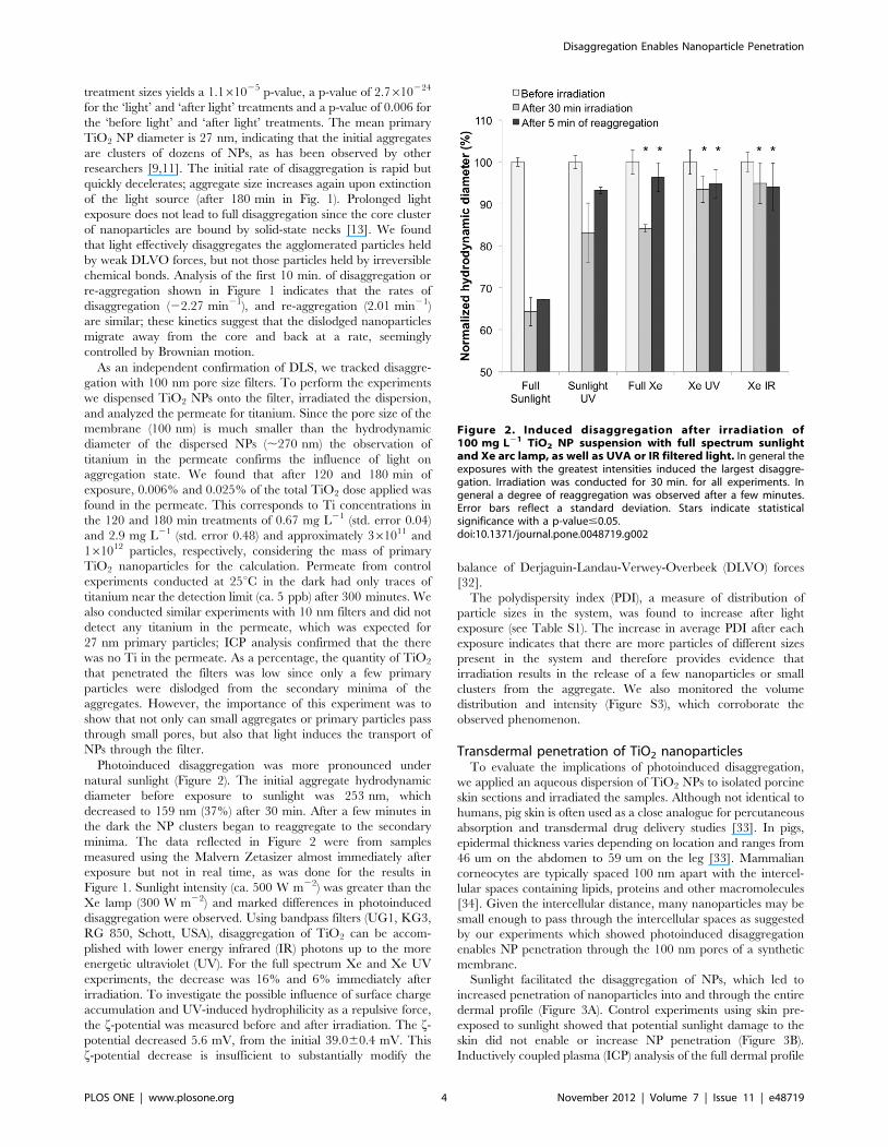
Photoinduced disaggregation was more pronounced under
natural sunlight (Figure 2). The initial aggregate hydrodynamic
diameter before exposure to sunlight was 253 nm, which
decreased to 159 nm (37%) after 30 min. After a few minutes in
the dark the NP clusters began to reaggregate to the secondary
minima. The data reflected in Figure 2 were from samples
measured using the Malvern Zetasizer almost immediately after
exposure but not in real time, as was done for the results in
Figure 1. Sunlight intensity (ca. 500 W m22) was greater than the
Xe lamp (300 W m22) and marked differences in photoinduced
disaggregation were observed. Using bandpass filters (UG1, KG3,
RG 850, Schott, USA), disaggregation of TiO2 can be accom-
plished with lower energy infrared (IR) photons up to the more
energetic ultraviolet (UV). For the full spectrum Xe and Xe UV
experiments, the decrease was 16% and 6% immediately after
irradiation. To investigate the possible influence of surface charge
accumulation and UV-induced hydrophilicity as a repulsive force,
the f-potential was measured before and after irradiation. The f-potential decreased 5.6 mV, from the initial 39.060.4 mV. This
f-potential decrease is insufficient to substantially modify the
balance of Derjaguin-Landau-Verwey-Overbeek (DLVO) forces
[32].
The polydispersity index (PDI), a measure of distribution of
particle sizes in the system, was found to increase after light
exposure (see Table S1). The increase in average PDI after each
exposure indicates that there are more particles of different sizes
present in the system and therefore provides evidence that
irradiation results in the release of a few nanoparticles or small
clusters from the aggregate. We also monitored the volume
distribution and intensity (Figure S3), which corroborate the
observed phenomenon.
Transdermal penetration of TiO2 nanoparticlesTo evaluate the implications of photoinduced disaggregation,
we applied an aqueous dispersion of TiO2 NPs to isolated porcine
skin sections and irradiated the samples. Although not identical to
humans, pig skin is often used as a close analogue for percutaneous
absorption and transdermal drug delivery studies [33]. In pigs,
epidermal thickness varies depending on location and ranges from
46 um on the abdomen to 59 um on the leg [33]. Mammalian
corneocytes are typically spaced 100 nm apart with the intercel-
lular spaces containing lipids, proteins and other macromolecules
[34]. Given the intercellular distance, many nanoparticles may be
small enough to pass through the intercellular spaces as suggested
by our experiments which showed photoinduced disaggregation
enables NP penetration through the 100 nm pores of a synthetic
membrane.
Sunlight facilitated the disaggregation of NPs, which led to
increased penetration of nanoparticles into and through the entire
dermal profile (Figure 3A). Control experiments using skin pre-
exposed to sunlight showed that potential sunlight damage to the
skin did not enable or increase NP penetration (Figure 3B).
Inductively coupled plasma (ICP) analysis of the full dermal profile
Figure 2. Induced disaggregation after irradiation of100 mg L21 TiO2 NP suspension with full spectrum sunlightand Xe arc lamp, as well as UVA or IR filtered light. In general theexposures with the greatest intensities induced the largest disaggre-gation. Irradiation was conducted for 30 min. for all experiments. Ingeneral a degree of reaggregation was observed after a few minutes.Error bars reflect a standard deviation. Stars indicate statisticalsignificance with a p-value#0.05.doi:10.1371/journal.pone.0048719.g002
Disaggregation Enables Nanoparticle Penetration
PLOS ONE | www.plosone.org 4 November 2012 | Volume 7 | Issue 11 | e48719

showed that irradiated TiO2 was able to penetrate the skin and
yielded concentrations as high as 370 mg TiO2 kg21 in the skin
profile after a 5 hr. sunlight exposure. The amount of irradiated
titanium found within the skin increased with increasing exposure
time (Figure 3A). By contrast, only 130 mg TiO2 kg21 were
present in unirradiated samples, and did not increase with time.
Other studies have shown that a simple soap wash is suitable for
removing TiO2 NPs from the surface of pig skin [24]. Since our
washing method is more robust, with multiple washes of the skin
surface with Nanopure water and 5% HNO3, we expect to have
washed off almost all the TiO2 from the skin surface. Hence, these
ICP results reflect titanium concentrations inside the skin. Since
5% HNO3 cannot dissolve TiO2 NPs, it is unlikely to increase
their dermal transport, or that of dissolved titanium.
The permeate through the skin was collected from several
samples to determine the mass of TiO2 that travelled through the
entire dermal profile. After a 90 min exposure, the mass of
titanium that permeated through the irradiated skin was 0.324 mg
or 0.1% of the applied amount. In a similar experiment using
0.22 um filters as a membrane and an equivalent dose of titanium
Figure 3. Mass of titanium in sunlight exposed skin grafts increasing with exposure to sunlight, indicating higher accumulation ofTiO2 NPs within the skin (Panel A). The mass of titanium in the dark treatments is much lower compared to the mass found in the irradiatedtreatment, and does not increase significantly with time. Panel B: Skin pre-exposed to 180 min of sunlight and then subjected to 180 min of sunlightand TiO2 doesn’t accumulate more TiO2 than skin not pre-exposed to sunlight. Control samples in Panel B were skin samples not pre-exposed tosunlight but subjected to the TiO2 dosing. Skin sections show that little to no titanium is found in the dark treatment, while it is found in all 40sections for the irradiated sample (panel C). Stars indicate statistical significance with a p-value#0.05. Molar exctinction coefficient was2,169 cm21 M21.doi:10.1371/journal.pone.0048719.g003
Disaggregation Enables Nanoparticle Penetration
PLOS ONE | www.plosone.org 5 November 2012 | Volume 7 | Issue 11 | e48719
and exposure time, 1.2% of titanium passed through the filters.
The small amount of TiO2 found in the permeate represents a
small fraction of the total dose, since only a small fraction of the
NPs can disaggregate and migrate through the skin. Most of the
TiO2 applied remains in the large clusters.
Thin skin sections also showed increased titanium within the
skin of irradiated samples. After 90 min exposure to light, TiO2
NPs were observed in detectable amounts on 40 successive sections
(Figure 3C, see also Figure S4). In unirradiated samples, there was
either no TiO2 or the signal was very small after the first section.
This provides evidence of transdermal penetration facilitated by
photo-induced disaggregation.
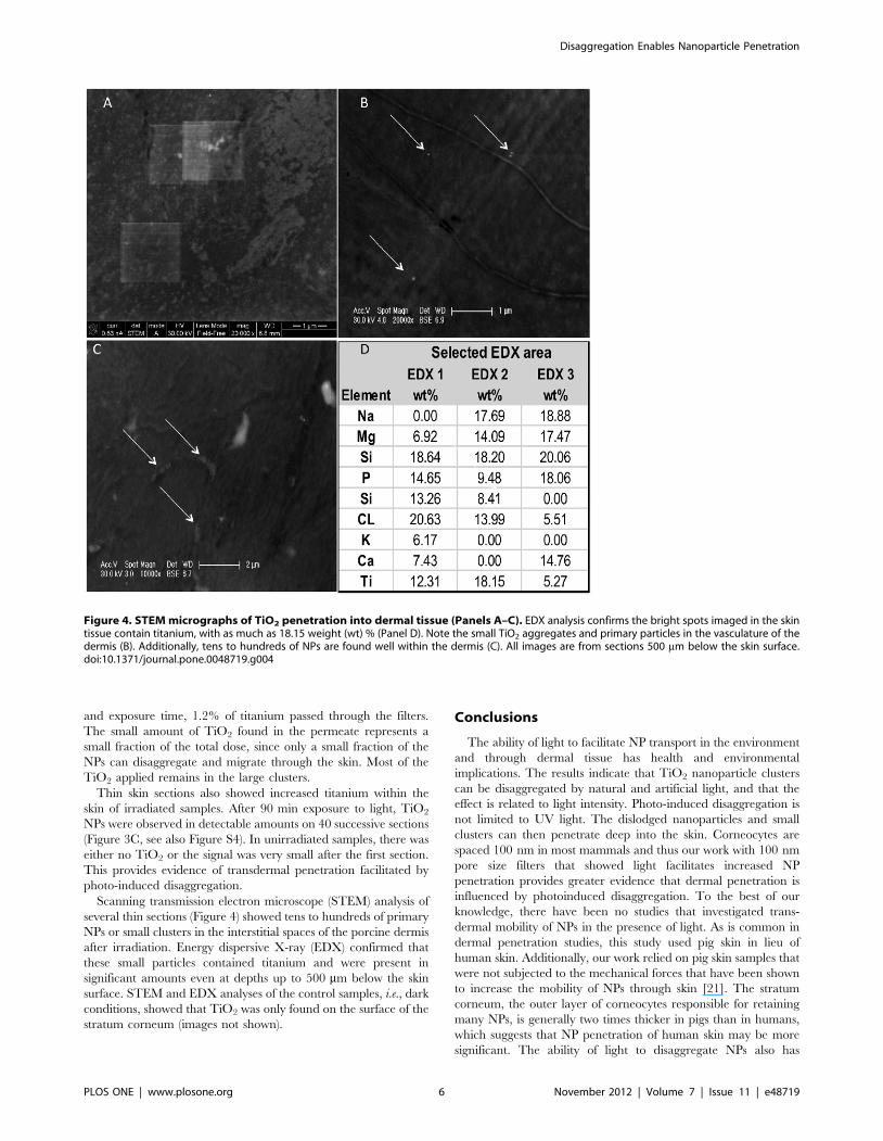
Scanning transmission electron microscope (STEM) analysis of
several thin sections (Figure 4) showed tens to hundreds of primary
NPs or small clusters in the interstitial spaces of the porcine dermis
after irradiation. Energy dispersive X-ray (EDX) confirmed that
these small particles contained titanium and were present in
significant amounts even at depths up to 500 mm below the skin
surface. STEM and EDX analyses of the control samples, i.e., dark
conditions, showed that TiO2 was only found on the surface of the
stratum corneum (images not shown).
Conclusions
The ability of light to facilitate NP transport in the environment
and through dermal tissue has health and environmental
implications. The results indicate that TiO2 nanoparticle clusters
can be disaggregated by natural and artificial light, and that the
effect is related to light intensity. Photo-induced disaggregation is
not limited to UV light. The dislodged nanoparticles and small
clusters can then penetrate deep into the skin. Corneocytes are
spaced 100 nm in most mammals and thus our work with 100 nm
pore size filters that showed light facilitates increased NP
penetration provides greater evidence that dermal penetration is
influenced by photoinduced disaggregation. To the best of our
knowledge, there have been no studies that investigated trans-
dermal mobility of NPs in the presence of light. As is common in
dermal penetration studies, this study used pig skin in lieu of
human skin. Additionally, our work relied on pig skin samples that
were not subjected to the mechanical forces that have been shown
to increase the mobility of NPs through skin [21]. The stratum
corneum, the outer layer of corneocytes responsible for retaining
many NPs, is generally two times thicker in pigs than in humans,
which suggests that NP penetration of human skin may be more
significant. The ability of light to disaggregate NPs also has
Figure 4. STEM micrographs of TiO2 penetration into dermal tissue (Panels A–C). EDX analysis confirms the bright spots imaged in the skintissue contain titanium, with as much as 18.15 weight (wt) % (Panel D). Note the small TiO2 aggregates and primary particles in the vasculature of thedermis (B). Additionally, tens to hundreds of NPs are found well within the dermis (C). All images are from sections 500 mm below the skin surface.doi:10.1371/journal.pone.0048719.g004
Disaggregation Enables Nanoparticle Penetration
PLOS ONE | www.plosone.org 6 November 2012 | Volume 7 | Issue 11 | e48719
potential application in industries which have a need to
manipulate aggregate size in aqueous media, e.g., pharmaceutics,
paint, metallurgy, rubber manufacturing.
Supporting Information
Supporting Information S1 The calculation protocol needed
to estimate the amount of light energy absorbed per particle is
included in Supporting Information S1, as well as the theory
behind our DLVO calculations and the parameters used to make
our DLVO calculations. Additional DLS data, size as volume and
intensity, are also included. We have also included additional
TiO2 penetration data. Finally, TEM images of the TiO2
materials used for this work appear in SI.
(DOCX)
Figure S1 Transmission electron micrographs of TiO2 used for
our work. Micrograph courtesy of Ivy Ji at UCLA.
(TIFF)
Figure S2 Spectrum of the Xenon arc lamp.
(TIFF)
Figure S3 Representative volume and intensity distribution
results for a 100 mg L-1 TiO2 solution before and after
irradiation. A shift in volume distribution of smaller particles is
clearly present after 30 min irradiation. Similarly, the intensity of
smaller particles also increases after irradiation.
(TIFF)
Figure S4 The mass (absorbance) of corneocytes removed with
each section via tape stripping from both the light and dark
exposed skin grafts (Panel A). Panel B presents the amount TiO2
found per section normalized by the mass of corneocytes removed
with each section.
(TIFF)
Table S1 The polydispersity index (PDI) results for sunlight
experiments are presented in Table S1 for unirradiated and
irradiated 100 mg L21 TiO2 samples. The samples were irradi-
ated by the UVA fraction of natural sunlight for 30 min.
(TIFF)
Author Contributions
Conceived and designed the experiments: SWB DZ AAK. Performed the
experiments: SWB DZ RM. Analyzed the data: SWB DZ AAK.
Contributed reagents/materials/analysis tools: AAK. Wrote the paper:
SWB DZ AAK.
References
1. Landsiedel R, Ma-Hock L, Kroll A, Hahn D, Schnekenburger J, et al. (2010)
Testing Metal-Oxide Nanomaterials for Human Safety. Advanced Materials 22:
2601–2627.
2. Peralta-Videa JR, Zhao LJ, Lopez-Moreno ML, de la Rosa G, Hong J, et al.
(2011) Nanomaterials and the environment: A review for the biennium 2008–
2010. Journal of Hazardous Materials 186: 1–15.
3. Godwin HA, Chopra K, Bradley KA, Cohen Y, Harthorn BH, et al. (2009) The
University of California Center for the Environmental Implications of
Nanotechnology. Environmental Science & Technology 43: 6453–6457.
4. Nel A, Xia T, Madler L, Li N (2006) Toxic potential of materials at the
nanolevel. Science 311: 622–627.
5. Wang X, Xia T, Ntim SA, Ji Z, George S, et al. (2010) Quantitative Techniques
for Assessing and Controlling the Dispersion and Biological Effects of
Multiwalled Carbon Nanotubes in Mammalian Tissue Culture Cells. ACS
Nano 4: 7241–7252.
6. Albanese A, Chan WCW (2011) Effect of Gold Nanoparticle Aggregation on
Cell Uptake and Toxicity. ACS Nano 5: 5478–5489.
7. Domingos RF, Tufenkji N, Wilkinson KJ (2009) Aggregation of Titanium
Dioxide Nanoparticles: Role of a Fulvic Acid. Environmental Science &
Technology 43: 1282–1286.
8. French RA, Jacobson AR, Kim B, Isley SL, Penn RL, et al. (2009) Influence of
Ionic Strength, pH, and Cation Valence on Aggregation Kinetics of Titanium
Dioxide Nanoparticles. Environmental Science & Technology 43: 1354–1359.
9. Zhang Y, Chen Y, Westerhoff P, Hristovski K, Crittenden JC (2008) Stability of
commercial metal oxide nanoparticles in water. Water Research 42: 2204–2212.
10. Chen KL, Elimelech M (2009) Relating colloidal stability of fullerene (C60)
nanoparticles to nanoparticle charge and electrokinetic properties. Environ-
mental Science & Technology 43:7270–7276.
11. Thio BJR, Zhou DX, Keller AA (2011) Influence of natural organic matter on
the aggregation and deposition of titanium dioxide nanoparticles. Journal of
Hazardous Materials 189: 556–563.
12. Colvin VL (2003) The potential environmental impact of engineered
nanomaterials. Nat Biotechnol 21: 1166–1170.
13. Zhou D, Bennett SW, Keller AA (2012) Increased Mobility of Metal Oxide
Nanoparticles Due to Photo and Thermal Induced Disagglomeration. PLoS
ONE 7:e37363.
14. Keller AA, Wang H, Zhou D, Lenihan HS, Cherr G, et al. (2010) Stability and
Aggregation of Metal Oxide Nanoparticles in Natural Aqueous Matrices.
Environmental Science & Technology 44:1962–1967.
15. Battin TJ, Kammer Fvd, Weilhartner A, Ottofuelling S, Hofmann T (2009)
Nanostructured TiO2: Transport behavior and effects on aquatic microbial
communities under environmentalconditions. Environmental Science & Tech-
nology 43: 8093–8104.
16. Baun A, Hartmann NB, Grieger K, Kusk KO (2008) Ecotoxicity of engineered
nanoparticles to aquatic invertebrates: A brief review and recommendations for
future toxicity testing. Ecotoxicology 17: 387–395.
17. Navarro E, Baun A, Behra R, Hartmann NB, Filser J, et al. (2008)
Environmental behavior and ecotoxicity of engineered nanoparticles to algae,
plants, and fungi. Ecotoxicology 17: 372–386.
18. Addamo M, Del Arco M, Bellardita M, Carriazo D, Di Paola A, et al. (2007)
Photoactivity of nanostructured TiO2; catalysts in aqueous system and theirsurface acid-base, bulk and textural properties. Research on Chemical
Intermediates 33: 465–479-479.
19. Sadrieh N, Wokovich AM, Gopee NV, Zheng J, Haines D, et al. (2003) Lack ofSignificant Dermal Penetration of Titanium Dioxide (TiO2) from Sunscreen
Formulations containing Nano- and Sub-Micron-Size TiO2 Particles. Toxico-logical Sciences 115:156–166.
20. Wu J, Liu W, Xue C, Zhou S, Lan F, et al. (2009) Toxicity and penetration ofTiO2 nanoparticles in hairless mice and porcine skin after subchronic dermal
exposure. Toxicology Letters 191: 1–8.
21. Tinkle SS, Antonini JM, Rich BA, Roberts JR, Salmen R, et al. (2003) Skin as aRoute of Exposure and Sensitization in Chronic Beryllium Disease. Environ
Health Perspect 111:1202–8.22. Lademann J, Weigmann HJ, Rickmeyer C, Barthelmes H, Schaefer H, et al.
(1999) Penetration of Titanium Dioxide Microparticles in a Sunscreen
Formulation into the Horny Layer and the Follicular Orifice. Skin Pharmacol-ogy and Physiology 12: 247–256.
23. Wokovich A, Tyner K, Doub W, Sadrieh N, Buhse LF (2009) Particle sizedetermination of sunscreens formulated with various forms of titanium dioxide.
Drug Development and Industrial Pharmacy 35:1180–1189.24. Gamer AO, Leibold E, van Ravenzwaay B (2006) The in vitro absorption of
microfine zinc oxide and titanium dioxide through porcine skin. Toxicology in
Vitro 20:301–307.25. National Solar Radiation Database, National Renewable Energy Laboratory,
http://rredc.nrel.gov/solar/old_data/nsrdb/ Last Accessed on 10/05/2012.26. Mattin E N-SM, De-Haan FH, Bodde HE (1996) Critical Comparison of
Methods to Quantify Stratum Corneum Removed by Tape Stripping. Skin
Pharmacology 9:69–77.27. Derjaguin B.V. (1954) A theory of the heterocoagulation, interaction and adhesion
of dissimilar particles in solutions of electrolytes. Discuss Faraday Soc 18:85–98.28. Verwey EJW, Overbeek JTh (1948) Theory of Stability of Lyophobic Colloids:
Elsevier, Amsterdam.
29. Hashimoto K, Irie H, Fujishima A (2005) TiO2 Photocatalysis: A HistoricalOverview and Future Prospects. Japanese Journal of Applied Physics 44: 8269–
8285.30. Garkusha I, Nagy A, Guennoun Z, Maier JP (2008) Electronic Absorption
Spectrum of Titanium Dioxide in Neon Matrices. Chemical Physics 353:115–118.31. Wang H ST, Apetrei C, Maier JP (2009) Characterization of the 1A1 and 1B2
Electronic States of Titanium Dioxide, TiO2. Phys Chem Chem Phys 11: 2649–
2656.32. Elimelech M, Gregory J, Jia X, Williams R (1998) Particle Deposition and
Aggregation Measurement, Modeling and Simulation. Butterworth-Heinemann:UK.
33. Monteiro-Riviere NA, Banks YB, Birnbaum LS (1991) Laser Doppler
measurements of cutaneous blood flow in ageing mice and rats. ToxicologyLetters 57: 329–338.
34. Cevc G, Vierl U (2010) Nanotechnology and the transdermal route: A state ofthe art review and critical appraisal. Journal of Controlled Release 141: 277–
299.
Disaggregation Enables Nanoparticle Penetration
PLOS ONE | www.plosone.org 7 November 2012 | Volume 7 | Issue 11 | e48719
Copyright © 2022 FDOKUMEN