Discovery of transdermal penetration enhancers by high-throughput screening
Upload
khangminh22Category
view
0download
0
Al-Kassas, R, Wen, J, Cheng, AE-M, Kim, AM-J, Liu, SSM and Yu, J
Transdermal delivery of propranolol hydrochloride through chitosan nanoparticles dispersed in mucoadhesive gel
http://researchonline.ljmu.ac.uk/id/eprint/12325/
Article
LJMU has developed LJMU Research Online for users to access the research output of the University more effectively. Copyright © and Moral Rights for the papers on this site are retained by the individual authors and/or other copyright owners. Users may download and/or print one copy of any article(s) in LJMU Research Online to facilitate their private study or for non-commercial research.You may not engage in further distribution of the material or use it for any profit-making activities or any commercial gain.
The version presented here may differ from the published version or from the version of the record. Please see the repository URL above for details on accessing the published version and note that access may require a subscription.
For more information please contact [email protected]
http://researchonline.ljmu.ac.uk/
Citation (please note it is advisable to refer to the publisher’s version if you intend to cite from this work)
Al-Kassas, R, Wen, J, Cheng, AE-M, Kim, AM-J, Liu, SSM and Yu, J (2016) Transdermal delivery of propranolol hydrochloride through chitosan nanoparticles dispersed in mucoadhesive gel. Carbohydrate Polymers, 153.pp. 176-186. ISSN 0144-8617
LJMU Research Online
1
Transdermal delivery of propranolol hydrochloride through chitosan 1
nanoparticles dispersed in mucoadhesive gel 2
3
*Raida Al-Kassas, Jingyuan Wen, Angel En-Miao Cheng, Amy Moon-Jung Kim, 4
Stephanie Sze Mei Liu, Joohee Yu 5
6
School of Pharmacy, Faculty of Medical and Health Sciences, University of Auckland, 7
Auckland, New Zealand. 8
9
10
11
Corresponding Author: 12
*Dr Raida Al-Kassas 13
School of Pharmacy 14
Faculty of Medical and Health Sciences 15
The University of Auckland 16
Private Bag 92019 17
Auckland 18
New Zealand 19
Email: [email protected] 20
21
22
23
24
25
26
27
28
29
30
31
32
33
2
Abstract 34
This study aimed at improving the systemic bioavailability of propranolol-HCl by the design 35
of transdermal drug delivery system based on chitosan nanoparticles dispersed into gels. 36
Chitosan nanoparticles were prepared by ionic gelation technique using tripolyphosphate 37
(TPP) as a cross-linking agent. Characterization of the nanoparticles was focused on particle 38
size, zeta potential, surface texture and morphology, and drug encapsulation efficiency. The 39
prepared freeze dried chitosan nanoparticles were dispersed into gels made of poloxamer and 40
carbopol and the rheological behaviour and the adhesiveness of the gels were investigated. 41
The results showed that smallest propranolol loaded chitosan nanoparticles were achieved 42
with 0.2% chitosan and 0.05% TPP. Nanoparticles were stable in suspension with a zeta 43
potential (ZP) above ± 30 mV to prevent aggregation of the colloid. Zeta potential was found 44
to increase with increasing chitosan concentration due to its cationic nature. At least 70% of 45
entrapment efficiency and drug loading were achieved for all prepared nanoparticles. When 46
chitosan nanoparticles dispersed into gel consisting of poloxamer and carbopol, the resultant 47
formulation exhibited thixotropic behaviour with a prolonged drug release properties as 48
shown by the permeation studies through pig ear skin. Our study demonstrated that the 49
designed nanoparticles-gel transdermal delivery system has a potential to improve the 50
systemic bioavailability and the therapeutic efficacy of propranolol-HCl. 51
52
Keywords: Propranolol-HCl; Chitosan nanoparticles; gels; transdermal drug delivery. 53
54
55
56
57
58
59
60
61
62
63
64
65
66
3
1. Introduction 67
The transdermal route has been a topic of interest for many years and is generally regarded as 68
a “patient friendly” option due to the avoidance of gastrointestinal side effects which usually 69
entail many oral preparations. Not only it avoids first pass metabolism and varying acidic 70
conditions of the gastrointestinal tract, it can also be used to maintain a constant, prolonged 71
and therapeutically effective drug concentration in the body. Transdermal drug delivery also 72
avoids fluctuations in plasma drug concentration, which helps minimising adverse effects and 73
therapeutic failure (Tanner and Marks, 2008). The main challenge in transdermal drug 74
delivery however, is to overcome the inherent barrier of the skin. It has been reported that 75
the rate limiting step in transdermal delivery is the ~ 30 µm thick stratum corneum which acts 76
as the primary barrier for the diffusion and drug penetration ( Cevc and Vierl, 2009). Various 77
strategies have been followed to improve delivery of drugs through skin among these is the 78
use of nanoparticulate carriers based on polymers (Prow et al., 2011). 79
80
Chitosan is a cationic polysaccharide made of 2-acetamido-2-deoxy-d-glucose (N-acetyl 81
glucosamine, GlcNAc), and 2-amino-2-deoxy-d-glucose (glucosamine, GlcNH2) with β-d-(1 82
→ 4) glycoside linkages as shown in Figure 1.a. Types of chitosan are differentiated by the 83
degree of N-acetylation (DA). Chitosan contains free amino groups which render it insoluble 84
in water. However, the amino groups undergo protonation in acid and therefore it becomes 85
soluble in aqueous solution. It has very low toxicity and breaks down slowly to harmless 86
products (amino sugars) which are absorbed by the body (Arai et al., 1968). Chitosan is also 87
recognized as a permeation enhancer due to its mucoadhesive properties. It binds to the 88
epithelial cell membrane and the positive charges result in F-actin depolymerisation and 89
disbandment of the tight junction protein ZO-1, leading to opening the tight junctions. With 90
all these attributes, chitosan is a desirable polymer and therefore has been widely used in 91
preparation of micro- and nanoparticles (Agnihotri et al., 2004). 92
93
Nanoparticles are characterised by a mean particle diameter of ≤ 1 µm (Gan et al., 2005). 94
These colloidal polymeric drug carriers are used to protect drugs from premature degradation 95
and prevent interaction with the biological environment. Furthermore, they enhance 96
bioavailability, absorption and penetration into the specific target tissues ( Budhian et al., 97
2007). Since drug uptake at the cellular level is size-dependent, smaller particles are taken up 98

4
to a higher extent ( Ubrich et al., 2004). It has been reported that a particle size of less than 99
500 nm is crucial for transdermal delivery (Kholi and Alpar, 2004). 100
For topical application, nanoparticulate systems are needed to be dispersed into suitable 101
semisolid vehicle such as hydrogels to maintain adherence on the skin. However, when 102
dispersed, the characteristics of the dispersed systems as well as the vehicle may be affected. 103
104
105
106
Figure 1.a. Structure of chitosan 107
108
Propranolol (PLN) (Figure 1.b), a non-selective β-blocking agent is commonly used for 109
cardiovascular conditions such as hypertension, angina pectoris and cardiac arrhythmia. 110
Propranolol has only an approximate half-life of 4 hours which requires frequent dosing to 111
maintain a therapeutic effect. Although PNL is rapidly absorbed from the gastrointestinal 112
tract, high oral doses are necessary due to an oral bioavailability of less than 23%, extensive 113
first-pass metabolism and susceptibility to enzymatic degradation. It is currently available as 114
an oral preparation and an intravenous formulation which is usually exclusive for hospital 115
use. 116
117
118
Figure 1.b: Structure of PNL and PNL-HCl 119
120

5
The aim of this study was to develop a transdermal delivery system for propranolol based on 121
chitosan nanoparticles dispersed into gels in attempt to improve the systemic bioavailability of 122
the drug. The properties of the nanoparticles as well the gels before and after dispersion of 123
nanoparticles into gels were evaluated. 124
125
2. MATERIALS AND METHODS 126
2.1 Materials 127
Propranolol-HCl, medium molecular weight chitosan and pentasodium tripolyphosphate (TPP) 128
were purchased from Sigma-Aldrich Chemical Co. Ltd (New Zealand). Carbopol 940 129
(carbopol) was purchased from Lubrizol Advance Materials, Inc (USA) and Poloxamer 407 130
was purchased from BASF (Germany). 131
132
2.2 Formulation and characterisation of nanoparticles 133
2.2.1 Preparation of nanoparticles 134
Nanoparticles were prepared by the ionic gelation method (Gan et al., 2005) at room 135
temperature with combinations of chitosan (0.1%, 0.2%, 0.3%) and TPP (0.02%, 0.05%, 136
0.08%) in triplicates. Chitosan was dissolved in acetic acid solution adjusted to pH 4.5 and 137
TPP was dissolved in Milli-Q water. TPP solution was added dropwise to an equal volume of 138
chitosan solution under magnetic stirring at 650 rpm over 60 minutes. The formed 139
nanoparticles were immediately analyzed for particle size and zeta potential in order to obtain 140
appropriate polymer concentrations for further investigation of propranolol-loaded chitosan 141
nanoparticles. 142
Propranolol-loaded chitosan nanoparticles were prepared by the same method mentioned 143
above, but that the appropriate amount of PNL was dissolved in chitosan solution before the 144
dropwise addition of TPP solution. To study the effect of propranolol concentration on the 145
physicochemical properties of nanoparticles, chitosan 0.3% was used in the ratio of 146
propranolol to chitosan at 1:1, 2:1 and 3:1. Whereas to study the effect of chitosan 147
concentration, 20 mg of propranolol was used in the ratio of propranolol to chitosan at 1:0.5, 148
1:1, and 1:1.5. 149
2.2.2 Particle size and zeta potential measurements: 150
Particle size, zeta potential and polydispersity index (PDI) of nanoparticle formulations were 151
measured using a Zetasizer Nano Series Nano-NS (Malvern Instruments, UK). Each sample 152
was measured in triplicate and the average results were calculated. 153

6
Total drug added
2.2.3 Determination of entrapment efficiency: 154
The entrapment efficiency of propranolol was calculated by measuring the amount of free 155
drug left in supernatant after centrifugation. Briefly nanoparticle suspensions were 156
centrifuged (Eppendorf MiniSpin Plus Microcentrifuge) at 13,000 rpm for 30 min. The 157
supernatants were diluted and amount of recovered propranolol was determined 158
spectrophotometric ally at 280 nm. The entrapment efficiency was calculated using the 159
following equation: 160
161
% EE = (Total drug added – free drug) x 100 162
163
2.2.4 Morphology of propranolol loaded chitosan nanoparticles: 164
The morphology of freshly prepared and freeze dried propranolol-loaded chitosan 165
nanoparticles were examined using scanning electron microscopy. Samples were coated in a 166
polaron Sc7640 sputter coater and analyzed by a Phillips x L30SFG with SiLi (Lithium 167
drifted) super ultra Thin Window EDS detector. 168
2.3 Formulation and characterisation of gels 169
2.3.1 Formulation of gels 170
Poloxamer gels (15% w/v) were prepared using the cold technique. Poloxamer was slowly 171
added to certain volume of cold Milli-Q water (5-10 ºC) with constant stirring for 60 min at 172
650 rpm. Additional amount of Cold Milli-Q water was added to the solution at 30 min to 173
make up to the volume. Poloxamer solutions were kept in the refrigerator (4-5 ºC) overnight 174
then kept at room temperature for a further 24 hrs ( Singh et al., 2009). 175
Carbopol 940 (1% and 2% w/v) gels were prepared by dispersing appropriate amount of 176
carbopol into certain volume of Milli-Q water at room temperature with constant stirring for 177
60 min at 650 rpm (9). Milli-Q water was added to the solution at 30 min to make up the 178
volume to the total amount. Carbopol gels were kept at room temperature for 24 h. 179
15% poloxamer / 1% carbopol combination gels were prepared with similar methods as 180
above. Poloxamer was dissolved in cold Milli-Q water and carbopol was separately dissolved 181
at room temperature in Milli-Q water of the same volume. Both were stirred at 650 rpm for 182
60 min. At 30 min, the two were mixed and Milli-Q water was added to the mixture with 183
stirring to make up the volumes to the total amount. These gels were kept at room 184
temperature for 24 h. 185
7
Each of the above gels containing 0.6% (w/v) propranolol-HCl was prepared separately. 186
Similar steps as above were followed except propranolol-HCl was dissolved in a small 187
volume of Milli-Q water before the final gels were made up to the volume. pH of all of the 188
gels were adjusted to 5.5. 189
2.4 Formulation of nanoparticles/gels transdermal delivery systems: 190
The nanoparticulate system and gel were selected after studying different parameters 191
affecting the formulations. The selected freeze dried nanoparticulate system was incorporated 192
into the gel, and the characteristics of resultant transdermal delivery systems was evaluated 193
and compared with gels containing drug. 194
2.4.1 Rheological measurements: 195
The rheological behaviours of the gel and transdermal delivery system formulations 196
consisting of nanoparticles dispersed into gel were measured at 25 ºC and 33 ºC (Digital 197
Viscometer Brookfield DV-III). Measurement at 33 ºC was required to represent the skin 198
temperature. Spindle sizes (CP40 and CP52) were used depending on the thickness of the gel 199
and shear rate adjustments for a torque between 5-100%. 200
2.4.2 Texture analysis 201
Adhesive capacity of gels and transdermal delivery systems were measured using (Stable 202
Micro System Texture Analyser TA. XT. Plus). The adhesive test settings were as follows; 203
test speed=0.5 mm/sec, force applied=100 g, contact time=5 seconds, trigger force=5 g. 204
Maximum force (N) was recorded from the texture analysis graph. 205
2.4.3 Preparation of pig ear skin: 206
Fresh male pig ears were obtained from the abattoir (Auckland Meat Processors, Auckland, 207
New Zealand). Ears were washed with water and dried. The subcutaneous tissue of the skin 208
was carefully removed using a scalpel to retain the stratum corneum of the skin. The skin 209
specimen was cut into appropriate sizes and washed with normal saline. 210
2.4.4 In vitro and ex vivo drug permeation studies 211
In vitro and ex vivo drug permeation studies were conducted with Franz diffusion cells (FDC- 212
6 Logan Instruments Corp). Cellulose membrane or freshly excised pig ear skin were 213
mounted between the donor and receptor cell (stratum corneum side facing the donor). For 214
the ex vivo permeation study, pig stratum corneum was equilibrated in Franz cells overnight 215
for hydration (El Maghraby, 2009). The receptor compartment was filled with pH 6.8 PBS 216
and its temperature was maintained at 37 1 °C by circulating water bath in order to ensure 217
that the surface membrane temperature was 32 1 °C (Wissing and Müller, 2002). The 218

8
donor compartment contained the following samples; propranolol loaded chitosan 219
nanoparticle suspension, gels and nanoparticles in gels. 0.5 mL samples were withdrawn at 220
different time intervals and replaced with an equal quantity of PBS into the receptor 221
compartment (Parsaee et al., 2002). All samples were analysed for propranolol content by 222
spectrophotometer at 280 nm (UV/Visible Spectrophotometer Libra S32PC). 223
SEM imaging was taken for pig stratum corneum used in ex vivo release study and was 224
compared with untreated pig stratum corneum (Herkenne et al., 2006). 225
226
3. Results and discussion: 227
3.1 Formation and physicochemical properties of chitosan nanoparticles: 228
Particle size is one of the most important factors in the development of nanoparticles, 229
especially for transdermal delivery as there are evidences of skin penetration of very small 230
nanoparticles into viable tissues (Ryman-Rasmussen et al., 2006; Zhang et al., 2008). 231
Table 1 shows the effect of polymer and TPP concentration on nanoparticle parameters 232
prepared without drug. The smallest drug free nanoparticles were achieved with 0.2% 233
chitosan and 0.05% TPP as they were in the size range of 160-210 nm. This was found to be 234
the optimum polymer to TPP ratio for favourable electrostatic interactions to yield small 235
nanoparticles. 236
The polydispersity index (PDI) which describes the size distribution was also the least for the 237
combination of 0.2% chitosan and 0.05% TPP. Nanoparticles formulated with 0.1% chitosan 238
were significantly larger than those obtained using 0.2% and 0.3% of chitosan with minimal 239
variation between the latter two. de Moura et al. (2009) reported an opposite trend where an 240
increase in particle size was observed with increasing chitosan concentration. They explained 241
the reason for their findings as the solubility of chitosan becomes less at increasing 242
concentrations leaving free particles to aggregate. However the trend observed in this study 243
can be a consequence of the CS/TPP ratio as the ratio is another significant factor that can 244
impact the particle size (Ricci et al., 2005). Table 1 shows that 0.02% and 0.08% 245
concentrations of TPP generally produced larger nanoparticles than 0.05% TPP. Wide PDI 246
was achieved with 0.1% and 0.3% chitosan whereas 0.2% chitosan showed a narrow PDI 247
indicating that nanoparticles with this polymer concentration possessed more uniform 248
nanoparticle size. 249
All nanoparticle formulations reported in the table are suitable for transdermal delivery as 250
they are less than 500 nm in size. Zeta potential reflects the density of the surface charge and 251

9
is influenced by the composition of the particles and the medium in which they are dispersed 252
(Mohanraj and Chen, 2007). In aqueous solution, chitosan changes its conformation and 253
becomes more flexible even with the presence of TPP, and the overall surface charge 254
becomes positive. Nanoparticles with zeta potential above ± 30mV are stable in suspension 255
due to the repulsion of surface charge preventing aggregation of nanoparticles ( Mohanraj and 256
Chen, 2007). Table 1 shows the zeta potential increased with ascending chitosan 257
concentration due to its cationic nature. However the zeta potential of nanoparticles prepared 258
with 0.08% TPP was less compared to those prepared with other concentrations which may 259
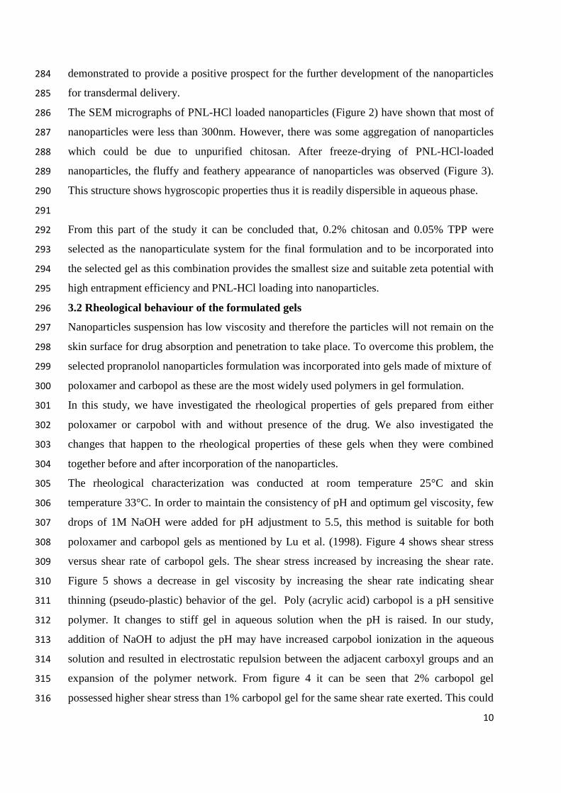
be due to polyanionic nature of TPP. 260
261
The concentrations of propranolol-HCl have been varied in our research in order to study its 262
effect on the properties of the nanoparticles (Table 2). Nanoparticles containing 0.2% 263
chitosan and 0.05% TPP were used for this study. Generally, there was an increase in size 264
and zeta potential of the nanoparticles with the addition of propranolol-HCl. However, 265
smaller nanoparticles were achieved with 1:2 chitosan to propranolol-HCl ratio at 266.47 nm. 266
The table shows that the zeta potential decreases as the concentration of propranolol increases 267
in the formulation which was possibly because of interactions between the positively charged 268
chitosan and negatively charged propranolol-HCl. 269
Table 3 illustrates the effect of increasing chitosan content in propranolol loaded chitosan 270
nanoparticles. Nanoparticles consisting of 0.2% chitosan and 0.05% TPP were used for this 271
experiment. The table shows that 1:1 chitosan to propranolol-HCl ratio produced particles 272
with average size of 166.53 nm which is comparable to the size of drug free nanoparticles 273
prepared at the same chitosan and TPP content. This may be due to the favourable ratio of 274
cationic and anionic charges between the polymers, TPP and propranolol-HCl. The zeta 275
potential of this formulation was found to be > ±30 mV indicating good physical stability of 276
nanoparticles. PDI has increased with drug incorporation. This may be again due to the 277
aggregation of nanoparticles as explained above. When propranolol-HCl was increased (table 278
2), an increase in entrapment efficiency and drug loading was observed due to the increased 279
drug available for incorporation into the nanoparticles. However at constant propranolol-HCl 280
concentration (table 3) a decrease in entrapment efficiency and drug loading occurred with 281
increasing chitosan concentrations possibly due to the increase of electrostatic repulsion 282
between chitosan polymers. The high drug loading ability and small nanoparticles 283
10
demonstrated to provide a positive prospect for the further development of the nanoparticles 284
for transdermal delivery. 285
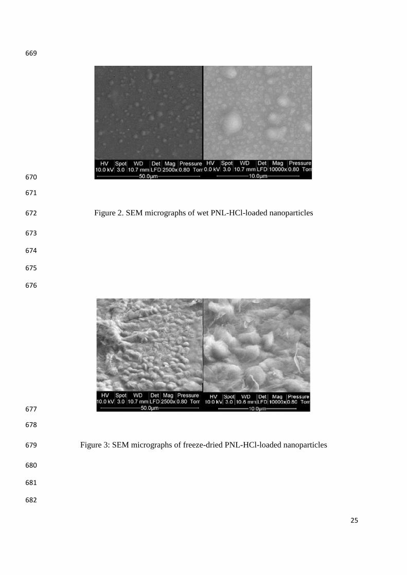
The SEM micrographs of PNL-HCl loaded nanoparticles (Figure 2) have shown that most of 286
nanoparticles were less than 300nm. However, there was some aggregation of nanoparticles 287
which could be due to unpurified chitosan. After freeze-drying of PNL-HCl-loaded 288
nanoparticles, the fluffy and feathery appearance of nanoparticles was observed (Figure 3). 289
This structure shows hygroscopic properties thus it is readily dispersible in aqueous phase. 290
291
From this part of the study it can be concluded that, 0.2% chitosan and 0.05% TPP were 292
selected as the nanoparticulate system for the final formulation and to be incorporated into 293
the selected gel as this combination provides the smallest size and suitable zeta potential with 294
high entrapment efficiency and PNL-HCl loading into nanoparticles. 295
3.2 Rheological behaviour of the formulated gels 296
Nanoparticles suspension has low viscosity and therefore the particles will not remain on the 297
skin surface for drug absorption and penetration to take place. To overcome this problem, the 298
selected propranolol nanoparticles formulation was incorporated into gels made of mixture of 299
poloxamer and carbopol as these are the most widely used polymers in gel formulation. 300
In this study, we have investigated the rheological properties of gels prepared from either 301
poloxamer or carpobol with and without presence of the drug. We also investigated the 302
changes that happen to the rheological properties of these gels when they were combined 303
together before and after incorporation of the nanoparticles. 304
The rheological characterization was conducted at room temperature 25°C and skin 305
temperature 33°C. In order to maintain the consistency of pH and optimum gel viscosity, few 306
drops of 1M NaOH were added for pH adjustment to 5.5, this method is suitable for both 307
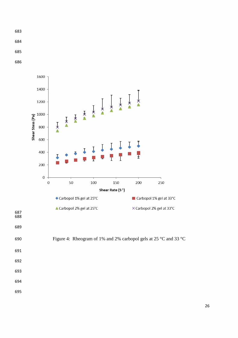
poloxamer and carbopol gels as mentioned by Lu et al. (1998). Figure 4 shows shear stress 308
versus shear rate of carbopol gels. The shear stress increased by increasing the shear rate. 309
Figure 5 shows a decrease in gel viscosity by increasing the shear rate indicating shear 310
thinning (pseudo-plastic) behavior of the gel. Poly (acrylic acid) carbopol is a pH sensitive 311
polymer. It changes to stiff gel in aqueous solution when the pH is raised. In our study, 312
addition of NaOH to adjust the pH may have increased carpobol ionization in the aqueous 313
solution and resulted in electrostatic repulsion between the adjacent carboxyl groups and an 314
expansion of the polymer network. From figure 4 it can be seen that 2% carbopol gel 315
possessed higher shear stress than 1% carbopol gel for the same shear rate exerted. This could 316

11
be due to the increased amount of polymer available leading to increased electrostatic 317
repulsion, polymer swelling and consequently increased elastic solid behavior. No marked 318
difference in the shear stress was observed when the temperature was increased from 25 to 319
33°C. This indicates that pH and polymer concentration are the major factors contributing to 320
gelling properties of carbopol. 321
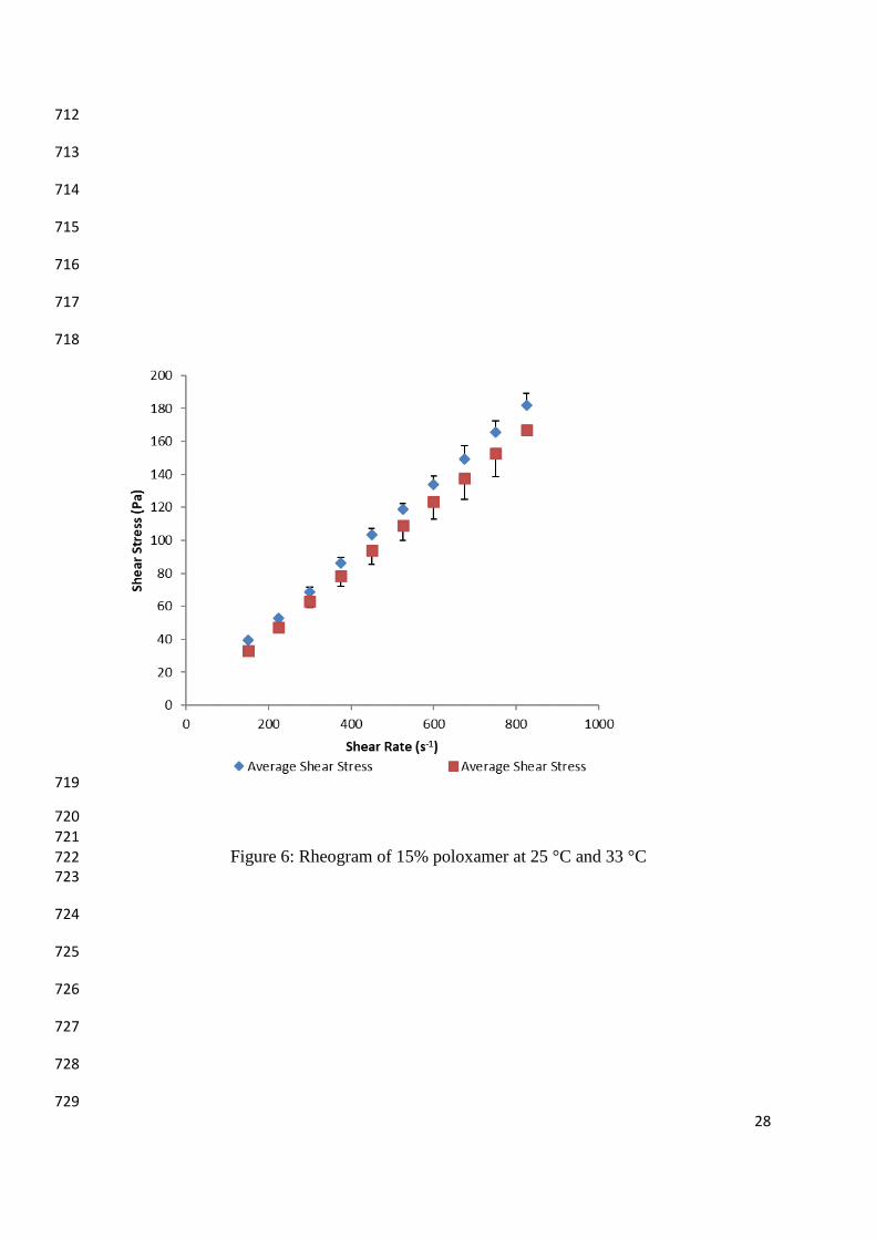
The flow curves of poloxamer formulations (Figure 6) at the experimental conditions 322
investigated exhibited a Newtonian flow as demonstrated by a linear increase in shear stress 323
with increasing shear rate. Poloxamers are in situ gelling polymers as they perform sol to gel 324
transition by enhancement of the elasticity network when the temperature increases (Santos et 325
al., 2015). It has been reported that when the concentration and temperature of the polymer 326
are above a critical value, poloxamer molecules in aqueous solution will self assemble to 327
form spherical micelles with a dehydrated PPO core surrounded by hydrated swollen PEO 328
chain (Dholakia et al., 2012). Therefore gelation in this case, is the result of micelles 329
entanglement and packing. Thus the results presented in Figure 6 suggest that the poloxamer 330
solutions didn’t undergo phase transition to turn into gels and remained as free flowing 331
liquids. This could be due to that the experiments were conducted at conditions below the 332
gelation temperature of poloxamer which is 36°C therefore the molecular structure of the 333
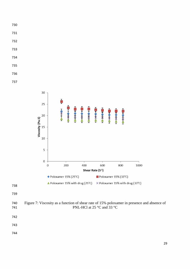
polymer solution didn’t change. The flow curves presented in figure 7 confirm these findings 334
and reveal that the viscosity remained constant by increasing the shear rate. 335
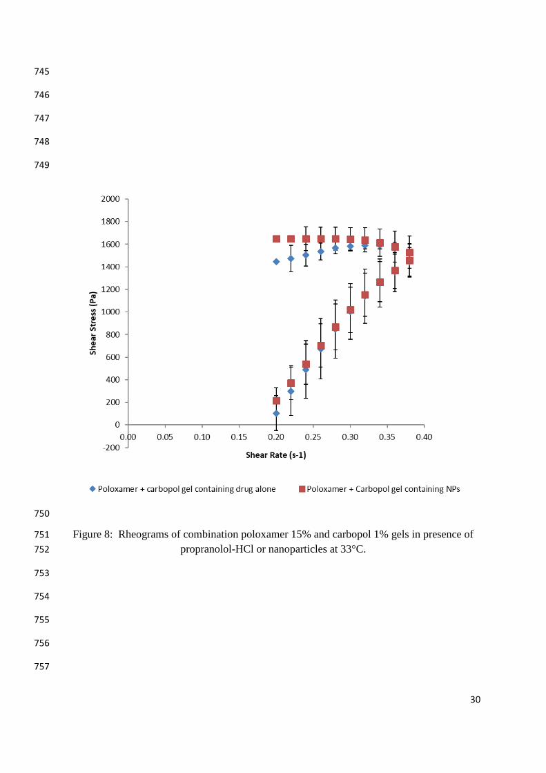
When carpobol and poloxamer gel were mixed the rheological properties of the resultant 336
system have changed significantly. The rheograms presented in Figure (8) show thixotropic 337
behaviour of gels consisting of combination of carbopol and poloxamer and containing either 338
propranolol-HCl or propranolol loaded nanoparticles as the downward curve was displaced 339
with regards to upward curve. Thixotropy can be defined as isothermal and a slow recovery 340
upon standing of a material of a consistency lost through shearing. These systems are 341
characterized by a decrease in viscosity when they are subjected to shear stress due to the 342
time dependant reformation of the secondary structure. Figure 8 also shows that hysteresis 343
loop formed by the up and down curves of the rheogram is bigger for combination gel 344
containing propranolol nanoparticles than that for gel containing drug alone indicating greater 345
magnitude of structural breakdown and thixotropy of this formulation. This is a desirable 346
property for a topical formulation as the greater the thixotropy, the lower is the settling and 347
sedimentation rate of the nanoparticles in the system. The viscograms presented in Figure 9 348
12
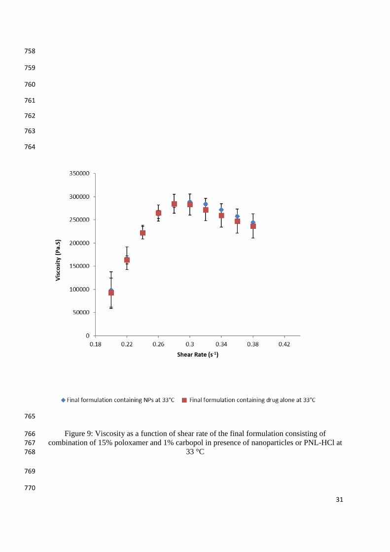
show that all combination gels possessed non-Newtonian, pseudoplastic (shear thinning) 349
behaviour. 350
The complex rheological properties of systems consisting of nanoparticles dispersed in gels 351
was also reported by others (Chawla and Saraf, 2012). 352
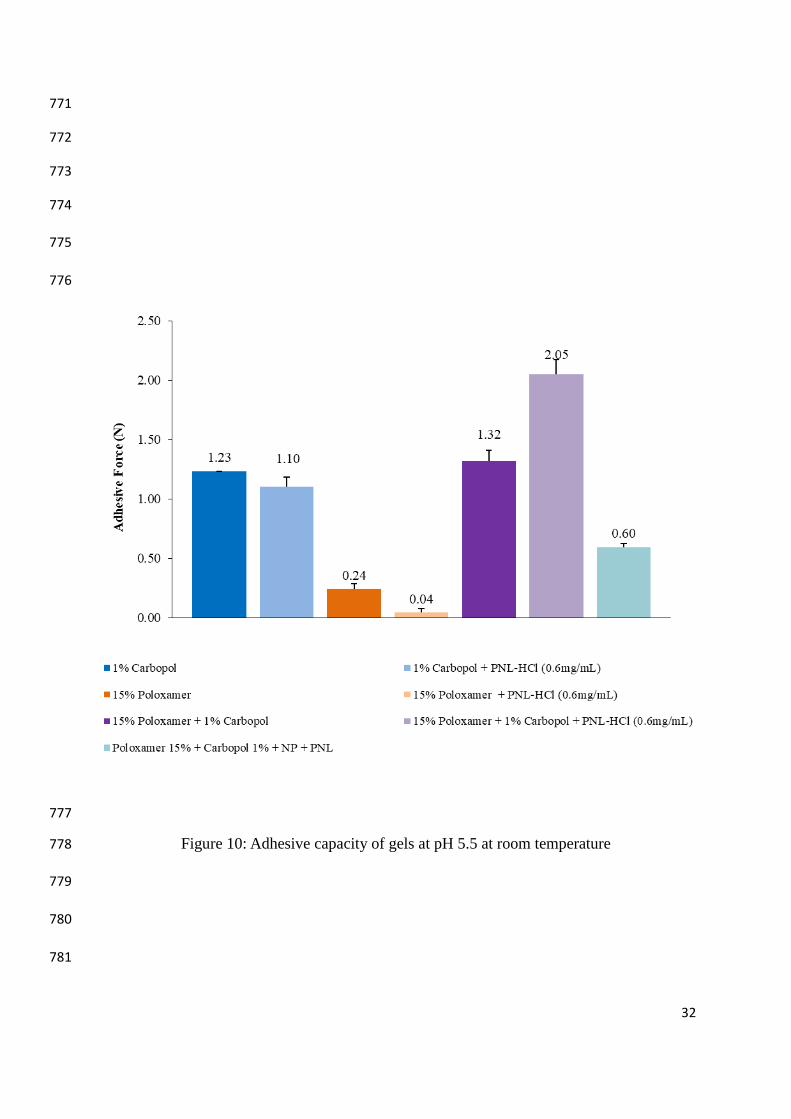
3.3 Adhesive capacity of the gels 353
Transdermal delivery system should possess desirable adhesiveness as weak adhesion may 354
results in incomplete absorption of drug through skin. In this study, the adhesiveness of the 355
designed transdermal delivery systems was investigated and the results are presented in 356
Figure 10. The adhesive capacity is dependent on the type and concentration of bioadhesive 357
polymer used in the formulation. In formulation based on Carpobol, the adhesiveness of the 358
gel increased as the concentration of the polymer increased. This may be attributed to the 359
increased number of the hydrophilic carboxyl functional groups available for binding, but 360
may also be a function of increased tack of the gel. Choi et al. (1998) have reported an 361
increase in the adhesive forces of gels by increasing carbopol concentration. From the figure, 362
it can be seen that poloxamer solution possessed significantly lower adhesiveness than 363
Carbopol gel. However, combining carbopol to poloxamer has increased the adhesive 364
properties of both polymers. These results are in agreement with Qi et al., 2007 who 365
demonstrated an increase in mucoadhesive force of ophthalmic gels when Carbopol was 366
incorporated into poloxamer solution. The possible explanation for these finding is the 367
combined effects of hydrophilic oxide groups of poloxamer and the carboxyl group of 368
carbopol which has improved the binding capacity of the formulation to the underlying 369
surface through electrostatic and hydrophobic interaction. 370
With the addition of propranolol-HCl, the adhesive force of carbopol gels decreased. The 371
effect on poloxamer gels was more substantial with an approximate 75- 80% drop in the 372
adhesive force. Propranolol-HCl causes a decrease in adhesive due to its positive charge 373
which can interact with the negatively charged of carbopol to form a complex. This 374
decreases the negative charge repulsions between carbopol polymers which uncoil and 375
expand, leading to reduction in polymer swelling and gel formation. On the other hand, the 376
hydroxyl group of propranolol molecules can form hydrogen bond with the PEO block of 377
poloxamer molecules (Kim et al., 2002) which may have been responsible for the adhesive 378
force becoming reduced. Interestingly, the contrary was observed for poloxamer and carbopol 379
combination gels. An increase in adhesive force was observed for the combination gels from 380
1.35 to 1.76 N after incorporation of propranolol-HCl. The observed increase can be 381
13
explained by the steric stabilisation properties of poloxamer which prevents the interaction 382
between propranolol-HCl and carbopol so that the cross-linking, viscoelastic properties of 383
carbopol can be potentiated. 384
The formulation of poloxamer 15% and carbopol 1% containing nanoparticles loaded with 385
propranolol-HCl had an adhesive force of 0.60 N. The chitosan component of the 386
nanoparticles carries a positive charge which can also interact with carbopol. It is observed to 387
cause a significant difference in the adhesion of gels compared with the addition of 388
propranolol-HCl due to the increased positive charge preventing the electrostatic repulsions 389
between carbopol. 390
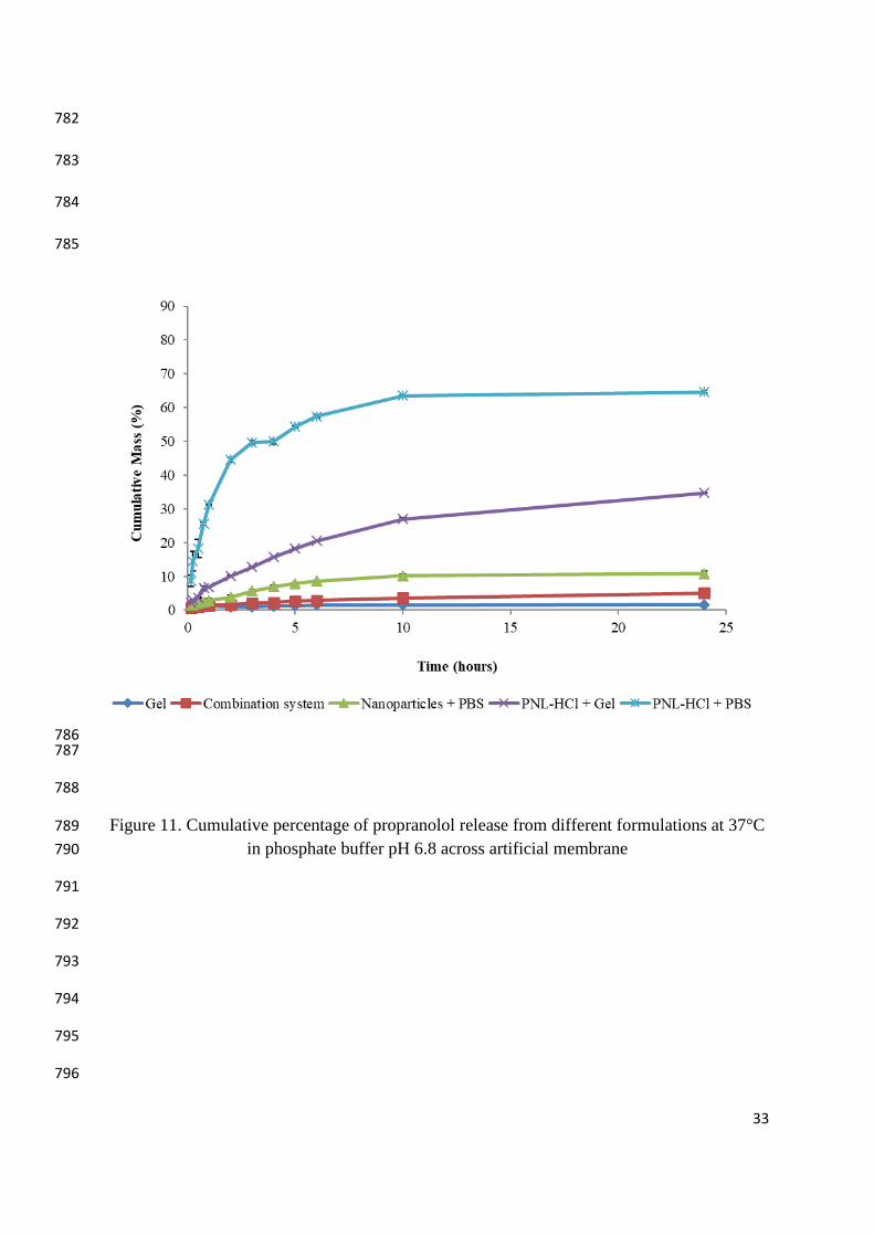
3.4 In vitro drug release study 391
The effect of the type of transdermal formulation on the release of propranolol through 392
cellophane membrane was investigated and the results are illustrated in Figure 11. The 393
release profiles followed predictable trends in relation to each other. For propranolol 394
containing buffer solution, the release of propranolol was very rapid and approximately 65% 395
of drug was released in 24 h. However, when propranolol-HCl was dispersed in combination 396
gel system, the release rate has reduced significantly. It was thought that this effect was the 397
result of combining the swollen carbopol with poloxamer solution which has increased the 398
density of the chain structure of the gel and reduced the diffusion of propranolol through the 399
formulation. Figure 11 shows chitosan nanoparticles yielded lowest cumulative mass of drug 400
released. Only 7% and 11 % of propranolol was released in 24 hours from the nanoparticle 401
suspension and nanoparticle/gel. This can be explained by the sustained release properties of 402
cross-linked chitosan and hydrophobic interactions with propranolol-HCl has led to a delayed 403
and an incomplete release of drug from the nanoparticles ( Ubrich et al., 2004). It was noticed 404
that the burst effect from these systems was negligible and release profiles were almost 405
linear. Generally, the release of propranolol from each formulation remained steady after 10 h 406
except for propranolol in gel which was shown to increase until at least 24 h. 407
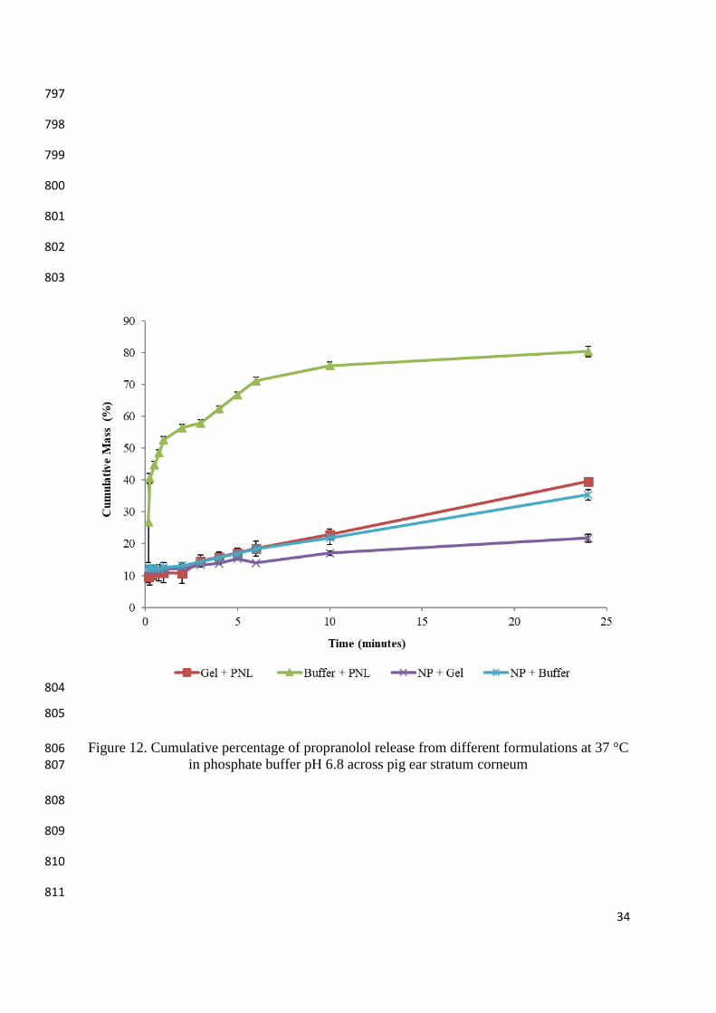
3.5 Ex vivo drug release study 408
Permeation studies were conducted in an attempt to assess the effect of the nanoparticles-Gel 409
transdermal system on the skin uptake and permeation properties of propranolol. The studies 410
were performed across pig ear skin since it can be considered as a reasonable model for 411
human barrier (Testa et al., 2001). The % cumulative mass of propranolol permeated across 412
the skin of different transdermal formulations over 24 hrs is shown in Figure 12. An initial 413
burst of drug permeation was noticed from the formulations in the first 5hrs, after which the 414
14
drug continued to permeate slowly and steady. The permeation profiles have exhibited zero 415
order kinetics with r2 values of 0.9911, 0.9973 and 0.9622 for gel, nanoparticles suspension 416
and nanoparticles in gel respectively. Of all formulations investigated, gel showed the highest 417
permeation rate. This can be explained by the high drug release properties of the gel system 418
which resulted in an increase in drug concentration in the donor compartment and an increase 419
of the concentration gradient towards the skin. Figure 12 shows that the permeation rate of 420
propranolol from nanoparticles in gel was the lowest. It has been reported that both high and 421
low permeation rates are of interest in skin application. Enhanced permeation rate can 422
improve drug permeability through skin whereas; sustained release can provide the skin with 423
drug over long period of time. It was noticed that The Papp values (Table 4) have confirmed 424
the permeation profiles results and they have followed this order which is gel > nanoparticle 425
suspension> nanoparticles in gel. The same trend was also observed from release studies. The 426
fact that trend of Papp values is the same as the release rate from formulations suggests that 427
the mechanism of propranolol permeation through the skin is formulation controlled rather 428
than skin controlled. In an attempt to support this finding, the skin uptake effect was followed 429
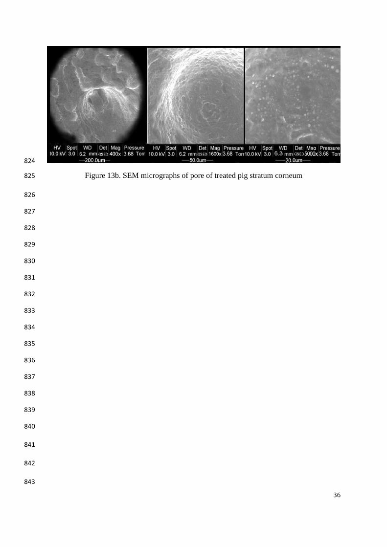
using scanning electron microscopy. The SEM micrographs of untreated and treated pig 430
stratum conium with nanoparticles in gel formulation are shown in Figure 13 (a&b). Figure 431
(a) indicates that there are unblocked and clear pores before treatment. However, after 432
treatment the micrograph reveals no clear pore since the nanoparticles have penetrated 433
through the stratum cornium and blocked all the pores. One of the interesting properties of 434
chitosan is that it can widen the tight junctions between the mucoepithelial cells reversibly by 435
interaction of the protonated CS with anionic components of glycoprotein on the surface of 436
the epithelial cells and with fixed negative charges in the interior of the tight junction, which 437
lead to absorption enhancement of the drug (Yeh et al., 2011). Therefore, presence of 438
nanoparticles in gel have increased their contact time with the skin and the properties of 439
chitosan might have affected the stratum cornuim nature and widened the tight junctions and 440
pores in the skin and allowed the particles to be up taken. Although the Ex vivo studies 441
showed slow permeation rate from nanoparticles in gel over 24 hours, however the results of 442
the SEM suggest that nanoparticles uptaken will create a reservoir of drug within the skin 443
where it provide the system over long period of time with small doses of propranolol to 444
control the systemic blood pressure. From these results it can be concluded that the type of 445
formulation and its unique properties have affected and both the permeation rate of drug and 446
its concentration within the skin. 447
15
4. Conclusion 448
The present work showed that transdermal delivery system for propranolol based on chitosan 449
nanoparticles dispersed into gel was successfully prepared and characterized. The novel gel 450
formulation exhibited thixotropic behaviour with a prolonged drug release properties as shown 451
by the permeation studies through pig ear skin. Furthermore, the SEM images showed that 452
the chitosan nanoparticles were uptaken by the skin which may create a drug reservoir to 453
provide the system with propranolol over long period of time to control the blood pressure. 454
Thus, the nanoparticles gel could be a promising transdermal delivery system for propranolol 455
however, in vivo studies are necessary to confirm this conclusion. 456
457
5. Acknowledgment: 458
459
The authors would like to thank the School of Pharmacy at the University of Auckland for 460
funding the study. 461
462
463
464
465
466
467
468
469
16
6. References: 470
1. Arai K, Kinumaki T, Fujita T. Toxicity of chitosan. Bull Tokai Reg Fish Lab. 1968;56:89-471
94. 472
2. Agnihotri SA, Mallikarjuna NN, Aminabhavi TM. Recent advances on chitosan-based 473
micro- and nanoparticles in drug delivery. Journal of Controlled Release. 2004;100(1):5-28. 474
1. Budhian, A., Siegel, S., Winey, K. (2007). Haloperidol-loaded PLGA nanoparticles: 475
Systematic study of particle size and drug content. International journal of pharmaceutics, 476
336(2), 367-75. 477
2. Cevc, G., Vierl, U. (2009). Nanotechnology and the transdermal route: A state of the art 478
review and critical appraisal. Journal of controlled Release, 479
3. de Moura, M., Aouada, F., Avena-Bustillos, R., McHugh, T., Krochta, J., Mattoso, L. 480
(2009). Improved barrier and mechanical properties of novel hydroxypropyl methylcellulose 481
edible films with chitosan/tripolyphosphate nanoparticles. Journal of Food Engineering. 482
92(4), 448-53. 483
4. Dholakia, M., Thakkar, V., Patel, N., Gandhi, N. (2012). Development and 484
characterisation of thermo reversible mucoadhesive moxifloxacin hydrochloride in situ 485
ophthalmic gel. Journal of Pharmacy and Bioallied Science, 4(1), S42–S45. 486
5. El Maghraby, G. (2009). Self-microemulsifying and microemulsion systems for 487
transdermal delivery of indomethacin: Effect of phase transition. Colloids and Surfaces B: 488
Biointerfaces. 489
6. Gan, Q., Wang, T., Cochrane, C., McCarron, P. (2005). Modulation of surface charge, 490
particle size and morphological properties of chitosan-TPP nanoparticles intended for gene 491
delivery. Colloids and Surfaces B: Biointerfaces, 44(2-3), 65-73. 492
7. Herkenne, C., Naik, A., Kalia, Y., Hadgraft, J., Guy, R. (2006). Pig ear skin ex vivo as a 493
model for in vivo dermatopharmacokinetic studies in man. Pharmaceutical research. 23(8), 494
1850-6. 495
8. Kohli A., Alpar, H. (2004). Potential use of nanoparticles for transcutaneous vaccine 496
delivery: effect of particle size and charge. Int J Pharmceutics, 4, 275(1-2), 13-7. 497
9. Lu, G., Jun, H. (1998). Diffusion studies of methotrexate in Carbopol and Poloxamer gels. 498
International journal of pharmaceutics, 160(1), 1-9. 499
10. Mohanraj, V., Chen, Y. (2007). Nanoparticles-A review. Tropical Journal of 500
Pharmaceutical Research, 5(1), 561. 501
17
11. Parsaee, S., Sarbolouki, M., Parnianpour, M. (2002). In-vitro release of diclofenac 502
diethylammonium from lipid-based formulations. International journal of pharmaceutics, 503
241(1):185-90. 504
12. Prow, T., Grice, J., Lin, L., Faye, R., Butler, M., Becker, W., Wurm, E., Yoong, C., 505
Robertson, T., Soyer, H., Roberts, M. Nanoparticles and microparticles for skin drug 506
delivery, Adv. Drug Deliv. Rev. 63 (2011) 470–491. 507
13. Ricci, E., Lunardi, L., Nanclares, D., Marchetti, J. (2005). Sustained release of lidocaine 508
from Poloxamer 407 gels. International journal of pharmaceutics, 288(2), 235-44. 509
14. Ryman-Rasmussen, J., Riviere, J., Monteiro-Riviere, N. (2006). Penetration of intact skin 510
by quantum dots with diverse physicochemical properties, Toxicological Sciences, 91, 159–511
165. 512
15. Singh, S., Gajra, B., Rawat, M., Muthu, M. (2009). Enhanced transdermal delivery of 513
ketoprofen from bioadhesive gels. Pakistan journal of pharmaceutical sciences, 22(2):193. 514
16. Santos, A., Akkari, A., Ferreira, I., Maruyama, C., Pascoli, M., Guilherme, V., Paula, E., 515
Fraceto, L., de Lima, R., Melo, P., de Araujo, D. (2015). Poloxamer-based binary hydrogels 516
for delivering tramadol hydrochloride: sol-gel transition studies, dissolution-release kinetics, 517
in vitro toxicity, and pharmacological evaluation. International Journal of Nanomedicine, 10, 518
2391–2401 519
17. Shin, S., Kim, J., Oh, I. (2000). Mucoadhesive and physicochemical characterization of 520
carbopol-poloxamer gels containing triamcinolone acetonide. Drug Development and 521
Industrial Pharmacy, 26(3), 307-12. 522
18. Tanner T., Marks R. (2008). Delivering drugs by the transdermal route: review and 523
comment. Skin Research and Technology, 14(3), 249-60. 524
19. Testa, B., Waterbeemd, H., Folkers, G., Guy, R. (2001). Pharmacokinetic Optimization in 525
Drug Research: Biological, Physicochemical, and Computational Strategies. John Wiley & 526
Sons. P167. 527
19. Ubrich, N., Bouillot, P., Pellerin, C., Hoffman, M., Maincent, P. (2004). Preparation and 528
characterization of propranolol hydrochloride nanoparticles: a comparative study. Journal of 529
controlled Release, 97(2), 291-300. 530
20. Wissing, S., Müller, R. (2002). Solid lipid nanoparticles as carrier for sunscreens: in vitro 531
release and in vivo skin penetration. Journal of controlled Release, 81(3), 225-33. 532
533
18
21. Yeh, T., , Hsu, L., Tseng, M., Lee, P., Sonjae, K., Ho, Y., Sung, H. (2011). Mechanism 534
and consequence of chitosan-mediated reversible epithelial tight junction opening. 535
Biomaterials, 32(26):6164-73. doi: 10.1016/j.biomaterials.2011. 536
22. Zhang, L., Yu, W., Colvin, V., Monteiro-Riviere, N. (2008). Biological interactions of 537
quantum dot nanoparticles in skin and in human epidermal keratinocytes, Toxicol. Appl. 538
Pharmacol, 228, 200–211. 539
19
540
541
542
543
544
545
546
547
548
549
550
551
552
553
554
555 556
557
558
559
560
561
562
563
564

20
565
566
567
568
569
570
571 572
573
574 575
576
577
578
579
580
581
582
583
584
585
586
587
588
589
590
591

21
Table 1: 592
Effect of chitosan and TPP concentrations on the physical properties of the nanoparticles 593
594
Particle size (nm) ±
SD
Average PdI
± SD
Average ZP
(mV) ± SD % Chitosan % TPP
0.1
0.02 462.59 ± 212.26 0.49 ± 0.27 15.97 ± 4.67
0.05 311.72 ± 111.70 0.34 ± 0.13 17.91 ± 2.23
0.2
0.08 421.56 ± 102.71 0.43 ± 0.16 3.09 ± 8.15
0.02 254.60 ± 25.91 0.26 ± 0.10 53.91 ± 3.49
0.05 191.30 ± 18.33 0.19 ± 26.40 35.48 ± 26.36
0.3
0.08 253.10 ± 16.06 0.28 ± 16.06 7.12 ± 10.07
0.02 215.26 ± 19.08 0.18 ± 0.08 63.58 ± 10.83
0.05 270.03 ± 141.66 0.40 ± 0.16 62.76 ± 2.55
0.08 247.22 ±14.91 0.30 ± 0.13 57.08 ± 2.39
595
596
597
598
599
600
601
602
603
604
605
22
606
607
Table 2 608 609
Effect of chitosan to propranolol HCl ratio on the physical properties of the nanoparticles. 610 611
Chitosan :
PNL-HCl
ratio
Particle size
(nm) ±SD
Average PdI
± SD
Average ZP
(mV)
Amount
(mg) EE (%)
Drug loading
(%)
Drug free
particles 191.30 ± 18.33 0.19 ± 26.40 35.48 ± 26.36 0 0 0
1:1 310.63 ± 69.78 0.41 ± 0.01 55.53 ± 2.20 28.31 ± 0.24 94.38 80.18
1:2 266.47 ± 14.81 0.21 ± 0.05 51.30 ± 1.74 57.32 ± 0.33 95.53 89.12
1:3 291.43 ± 22.38 0.29 ± 0.06 48.40 ± 2.43 87.40 ± 0.17 97.12 92.58
612
613
614
615
616
617
618
619
620
621
622
623
624
625
626
627
628
629
23
630
631
Table 3 632
633
Effect of changing chitosan concentration on the physical properties of nanoparticles 634 635
PNL-HCl :
chitosan ratio
Particle size
(nm)±SD
Average
PdI±SD
Average ZP
(mV) ±SD Amount (mg) EE (%)
Drug
loading (%)
Drug free
particles 191.30 ± 18.33 0.19 ± 26.40
35.48 ±
26.36 0 0 0
1:0.5
644.70 ± 31.24
0.76 ± 0.04
17.77 ± 1.07
17.72 ± 0.09
88.60
85.52
1:1 166.53 ± 5.55 0.58 ± 0.03 41.90 ± 1.15 17.56 ± 0.14 87.78 77.83
1:1.5 311.63 ± 26.52 0.73 ± 0.21 49.43 ± 0.50 17.45 ± 0.11 87.26 71.37
636
637
638
639
640
641
642
643
644
645
646
647
648
649
24
650
651
652
Table 4 653
Apparent permeability coefficient of formulations investigated 654
Formulations Papp (cm/s)
Propranolol-HCL solution 7.702 x 10⁻⁷
Propranolol-HCl gel 1.844 x 10⁻⁷
Propranolol nanoparticle suspension 0.363 x 10⁻⁷
Propranolol HCl nanoparticles gel 0.167 x 10⁻⁷
655
656
657
658
659
660
661
662
663
664
665
666
667
668
25
669
670
671
Figure 2. SEM micrographs of wet PNL-HCl-loaded nanoparticles 672
673
674
675
676
677
678
Figure 3: SEM micrographs of freeze-dried PNL-HCl-loaded nanoparticles 679
680
681
682
26
683
684
685
686
687 688
689
Figure 4: Rheogram of 1% and 2% carbopol gels at 25 °C and 33 °C 690
691
692
693
694
695

27
696
697
698
699
700
701
702
703
Figure 5: Viscosity as a function of shear rate of 1% and 2% carbopol polymer at 25 °C and 704
33 °C 705
706
707
708
709
710
711
28
712
713
714
715
716
717
718
719
720
721 Figure 6: Rheogram of 15% poloxamer at 25 °C and 33 °C 722
723
724
725
726
727
728
729
29
730
731
732
733
734
735
736
737
738
739
Figure 7: Viscosity as a function of shear rate of 15% poloxamer in presence and absence of 740
PNL-HCl at 25 °C and 33 °C 741
742
743
744
30
745
746
747
748
749
750
Figure 8: Rheograms of combination poloxamer 15% and carbopol 1% gels in presence of 751
propranolol-HCl or nanoparticles at 33°C. 752
753
754
755
756
757
31
758
759
760
761
762
763
764
765
Figure 9: Viscosity as a function of shear rate of the final formulation consisting of 766 combination of 15% poloxamer and 1% carbopol in presence of nanoparticles or PNL-HCl at 767
33 °C 768
769
770
32
771
772
773
774
775
776
777
Figure 10: Adhesive capacity of gels at pH 5.5 at room temperature 778
779
780
781
33
782
783
784
785
786 787
788
Figure 11. Cumulative percentage of propranolol release from different formulations at 37°C 789
in phosphate buffer pH 6.8 across artificial membrane 790
791
792
793
794
795
796
34
797
798
799
800
801
802
803
804
805
Figure 12. Cumulative percentage of propranolol release from different formulations at 37 °C 806 in phosphate buffer pH 6.8 across pig ear stratum corneum 807
808
809
810
811

35
812
813
814
815
816
817
818
819
820
Figure 13a. SEM micrographs of pore of untreated pig stratum corneum 821
822
823
36
824
Figure 13b. SEM micrographs of pore of treated pig stratum corneum 825
826
827
828
829
830
831
832
833
834
835
836
837
838
839
840
841
842
843

37
844
Copyright © 2022 FDOKUMEN