Novel gel-niosomes formulations as multicomponent systems for transdermal drug delivery
8
Colloids and Surfaces B: Biointerfaces 110 (2013) 281–288 Contents lists available at SciVerse ScienceDirect Colloids and Surfaces B: Biointerfaces jou rn al hom epage: www.elsevier.com/locate/colsurfb Novel gel-niosomes formulations as multicomponent systems for transdermal drug delivery Lorena Tavano a,b , Luigi Gentile c , Cesare Oliviero Rossi c , Rita Muzzalupo a,∗ a Dipartimento di Farmacia e Scienze della Salute e della Nutrizione, Università della Calabria, Ed. Polifunzionale, 87036 Arcavacata di Rende (CS), Italy b Dipartimento di Ingegneria Informatica, Modellistica, Elettronica e Sistemistica, Università della Calabria, Via P. Bucci Cubo 39/C, 87036 Arcavacata di Rende (CS), Italy c Dipartimento di Chimica e Tecnologie Chimiche, Università della Calabria, Ponte P. Bucci, Cubo 14/D, 87036 Arcavacata di Rende (CS), Italy a r t i c l e i n f o Article history: Received 12 February 2013 Received in revised form 27 March 2013 Accepted 18 April 2013 Available online 6 May 2013 Keywords: Pluronic AOT Liquid crystals Niosomes Transdermal delivery a b s t r a c t The percutaneous permeation profiles of sulfadiazine sodium salt, propranolol hydrochloride and tyrosol from novel liquid crystal-niosomes formulations as multicomponent systems, were investigated. The new carriers were prepared from mixture of water/surfactant, AOT or Pluronic L64 as anionic and nonionic surfactants, respectively, in order to obtain lamellar LLC phases. The same surfactants were used to prepare also the vesicular systems (niosomes) that were added to the corresponding gel. The obtained multicomponent drug carrier was characterized by deuterium nuclear magnetic resonance spectroscopy, in order to understand if the introduction of the drug or drug-loaded niosomal suspension, as third component in the formulations, could influence the microstructure of the system and then the drug delivery across the skin. Simple AOT and L64-based niosomal formulations and LLCs phases were then prepared and used as control. Different drugs percutaneous availability was achieved, and the results revealed that the obtained gel-niosomes carriers were affected by the chemical structure of the drugs and by their affinity for the components. As a consequence these systems could be proposed as novel transdermal drug delivery systems, since they were found able to control the percutaneous permeation of small drugs across the skin. © 2013 Elsevier B.V. All rights reserved. 1. Introduction In recent years, the transdermal route of administration has found wide applications and gained considerable commercial success, winning the competition with oral, intravenous or intra- muscular routes [1]. A problem that has been faced is the low penetration of most compounds through the outermost layer of the skin, the stratum corneum (SC). The SC consists of terminally differ- entiated keratinocytes, referred to as corneocytes, embedded in a lipid-rich intercellular matrix. The main constituents of this matrix are ceramides (CER), cholesterol (CHOL) and long-chain free fatty acids. These lipids are organized in crystalline lamellae at room temperature and play a key role in the barrier function of human skin. Absorption of substances into and across skin takes place by passive diffusion through the lipid domains of SC, since the driv- ing force is the difference in the drug concentration between drug carriers and blood. In order to reduce temporary the SC barrier and improve drug vehiculation, penetration enhancers are developed to alter the SC lipid structural organization, avoiding side effects and ∗ Corresponding author. E-mail address: [email protected] (R. Muzzalupo). allowing a better control than other conventional delivery methods [2]. Among the several approaches, one possibility to increase the amount of drug vehiculated across the SC, is the use of lyotropic liquid crystals (LLCs), due to their stability, low interfacial tension arising at the oil/water interface and capability to increase the sol- ubility of drugs, which are either insoluble or slightly soluble in water [3]. The LLC are excellent examples of self-assembling nanoma- terials. They are usually formed from water and one or more surfactants at very definite proportions. Their phase sequence (cubic, hexagonal, lamellar) depends on both the different com- ponents concentration and temperature. Among the different mesophases, the lamellar ones represent the best approaches to be used as transdermal drug delivery systems, due to their special skin similarly structure. In fact, the structural units for the lamellar phase are double layers formed by surfactant molecules disposed in a bidimensional stacking of infinite layers, delimited by water. The polar heads of the molecules are in contact with the aqueous medium, while the hydrocarbon chains are interdigitating in order to avoid water. This phase is rather fluid and the bilayers can slip easily one on the other. This structure is very similar to that occurring in living organisms 0927-7765/$ – see front matter © 2013 Elsevier B.V. All rights reserved. http://dx.doi.org/10.1016/j.colsurfb.2013.04.017
Transcript of Novel gel-niosomes formulations as multicomponent systems for transdermal drug delivery

Nt
La
b
8c
ARRAA
KPALNT
1
fsmpselaatspicia
0h
Colloids and Surfaces B: Biointerfaces 110 (2013) 281– 288
Contents lists available at SciVerse ScienceDirect
Colloids and Surfaces B: Biointerfaces
jou rn al hom epage: www.elsev ier .com/ locate /co lsur fb
ovel gel-niosomes formulations as multicomponent systems forransdermal drug delivery
orena Tavanoa,b, Luigi Gentilec, Cesare Oliviero Rossi c, Rita Muzzalupoa,∗
Dipartimento di Farmacia e Scienze della Salute e della Nutrizione, Università della Calabria, Ed. Polifunzionale, 87036 Arcavacata di Rende (CS), ItalyDipartimento di Ingegneria Informatica, Modellistica, Elettronica e Sistemistica, Università della Calabria, Via P. Bucci Cubo 39/C,7036 Arcavacata di Rende (CS), ItalyDipartimento di Chimica e Tecnologie Chimiche, Università della Calabria, Ponte P. Bucci, Cubo 14/D, 87036 Arcavacata di Rende (CS), Italy
a r t i c l e i n f o
rticle history:eceived 12 February 2013eceived in revised form 27 March 2013ccepted 18 April 2013vailable online 6 May 2013
eywords:luronicOT
a b s t r a c t
The percutaneous permeation profiles of sulfadiazine sodium salt, propranolol hydrochloride and tyrosolfrom novel liquid crystal-niosomes formulations as multicomponent systems, were investigated. The newcarriers were prepared from mixture of water/surfactant, AOT or Pluronic L64 as anionic and nonionicsurfactants, respectively, in order to obtain lamellar LLC phases. The same surfactants were used toprepare also the vesicular systems (niosomes) that were added to the corresponding gel. The obtainedmulticomponent drug carrier was characterized by deuterium nuclear magnetic resonance spectroscopy,in order to understand if the introduction of the drug or drug-loaded niosomal suspension, as thirdcomponent in the formulations, could influence the microstructure of the system and then the drug
iquid crystalsiosomesransdermal delivery
delivery across the skin. Simple AOT and L64-based niosomal formulations and LLCs phases were thenprepared and used as control. Different drugs percutaneous availability was achieved, and the resultsrevealed that the obtained gel-niosomes carriers were affected by the chemical structure of the drugsand by their affinity for the components. As a consequence these systems could be proposed as noveltransdermal drug delivery systems, since they were found able to control the percutaneous permeationof small drugs across the skin.
. Introduction
In recent years, the transdermal route of administration hasound wide applications and gained considerable commercialuccess, winning the competition with oral, intravenous or intra-uscular routes [1]. A problem that has been faced is the low
enetration of most compounds through the outermost layer of thekin, the stratum corneum (SC). The SC consists of terminally differ-ntiated keratinocytes, referred to as corneocytes, embedded in aipid-rich intercellular matrix. The main constituents of this matrixre ceramides (CER), cholesterol (CHOL) and long-chain free fattycids. These lipids are organized in crystalline lamellae at roomemperature and play a key role in the barrier function of humankin. Absorption of substances into and across skin takes place byassive diffusion through the lipid domains of SC, since the driv-
ng force is the difference in the drug concentration between drug
arriers and blood. In order to reduce temporary the SC barrier andmprove drug vehiculation, penetration enhancers are developed tolter the SC lipid structural organization, avoiding side effects and∗ Corresponding author.E-mail address: [email protected] (R. Muzzalupo).
927-7765/$ – see front matter © 2013 Elsevier B.V. All rights reserved.ttp://dx.doi.org/10.1016/j.colsurfb.2013.04.017
© 2013 Elsevier B.V. All rights reserved.
allowing a better control than other conventional delivery methods[2].
Among the several approaches, one possibility to increase theamount of drug vehiculated across the SC, is the use of lyotropicliquid crystals (LLCs), due to their stability, low interfacial tensionarising at the oil/water interface and capability to increase the sol-ubility of drugs, which are either insoluble or slightly soluble inwater [3].
The LLC are excellent examples of self-assembling nanoma-terials. They are usually formed from water and one or moresurfactants at very definite proportions. Their phase sequence(cubic, hexagonal, lamellar) depends on both the different com-ponents concentration and temperature. Among the differentmesophases, the lamellar ones represent the best approaches tobe used as transdermal drug delivery systems, due to their specialskin similarly structure.
In fact, the structural units for the lamellar phase are doublelayers formed by surfactant molecules disposed in a bidimensionalstacking of infinite layers, delimited by water. The polar heads of
the molecules are in contact with the aqueous medium, while thehydrocarbon chains are interdigitating in order to avoid water. Thisphase is rather fluid and the bilayers can slip easily one on the other.This structure is very similar to that occurring in living organisms
2 es B: Biointerfaces 110 (2013) 281– 288
atpoSavptitcostefad
cppwftrlpt(tlneebcotAeitactLl(tiafdd
2
2
oBfa
Table 1Details on the preparation of drug-loaded gels.
Samples Surfactantname
Surfactant(g)
D2O (g) Drug Drug (g)
G-AOT-Sul AOT 0.90 2.07 Sulfadiazinesodium salt
0.03
G-AOT-Pro AOT 0.90 2.07 Propranololhydrochloride
0.03
G-AOT-Tyr AOT 0.90 2.07 Tyrosol 0.03G-L64-Sul L64 2.25 0.72 Sulfadiazine
sodium salt0.03
82 L. Tavano et al. / Colloids and Surfac
nd for this reason lamellar LLC represents optimal candidate forhe transdermal vehiculation of drugs [4]. An improvement of theharmaceutical properties of these drug delivery systems can bebtained by the addition of vesicular systems in the LLCs lamellae.everal works, in fact, reported that the LLC networks can serve asn encapsulating matrix, escorting and protecting the embeddedesicles entrapping active entities and can enhance their stabilityrolonging the circulation time without losing drug [5]. In additionhese multicomponent systems have attracted particular interestn the design of dermal and transdermal delivery systems due tohe possibility to achieve prolonged and programmed drug per-utaneous permeation through the skin [6]. In a previous work,ur research group has carried out investigation on the complexystems based on Pluronic F127 and Tween 60 vesicles, to studyhe rheological interaction between polymer and vesicles and tovaluate the potential use of the systems as efficient drug deliveryormulation [7]. Results demonstrated that the binary systems canct as transdermal controlled and prolonged delivery systems oficlofenac sodium salt.
In this light, we decide to evaluate the effect of the chemi-al structure of different surfactants and vehiculated drug on thehysico-chemical properties and drug percutaneous permeationrofiles of novel multicomponent formulations. For these reasonse used Pluronic L64 or Aerosol OT (AOT) as surfactants and sul-
adiazine sodium salt (Sul), propranolol hydrochloride (Pro) andyrosol (Tyr) as model drugs. LLCs samples were prepared at fixedatio between Pluronic L64 or AOT and water, in order to obtainamellar LLC phases and the corresponding surfactant was used torepare also the vesicular systems (niosomes) that were added tohe gel. Pluronic L64 is a non-ionic copolymer of ethylene oxideEO) and propylene oxide (PO) blocks arranged in a triblock struc-ure, in which the hydrophobic polypropylene oxide (PPO) groupinks two hydrophilic polyethylene oxide (PEO). The amphiphilicature of Pluronic makes it extremely useful in various fields asmulsifier, stabilizer and pharmaceutical additive. The presence ofthylene oxide moieties may reduce opsonisation and clearancey the reticuloendothelial system, leading to improved pharma-okinetic properties of the carriers. Several works reported the usef L64 as surfactant to obtain niosomes useful as parenteral andransdermal delivery systems vehiculation of different drugs [8].OT is a versatile double-tailed anionic surfactant whose phasequilibria with water and organic solvent has been extensivelynvestigated, because its non toxicity, spontaneously formed andhermodynamically stable long lived micellar forms and it is knowns a transdermal drug delivery vehicle [9]. Drugs with anionic,ationic and nonionic chemical structures and different proper-ies such as Sul, Pro and Tyr, respectively, were incorporated into64 and AOT lamellar LLC phases as free drug solutions or as drug-oaded niosomal suspension. Deuterium resonance spectroscopy2H NMR) were used to evaluate the occurred structural modifica-ions caused by the incorporation of drugs as a third componentn the surfactant–water systems. Finally the percutaneous perme-tion profile of all the drugs from the lamellar LLC phases obtainedrom L64 and AOT were performed, in order to understand how theifferent formulations could give rise to a different transdermalelivery of drugs.
. Materials and methods
.1. Materials
Pluronic L64, poly(ethylene oxide)-block-poly(propylene
xide)-block-poly(ethylene oxide) copolymer, was provided fromASF (Mount Olive, NJ, USA). AOT (sodium bis(2-ethylhexyl) sul-osuccinate), sulfadiazine sodium salt, propranolol hydrochloridend tyrosol were supplied by Sigma–Aldrich, Milan, Italy. The drug
G-L64-Pro L64 2.25 0.72 Propranololhydrochloride
0.03
G-L64-Tyr L64 2.25 0.72 Tyrosol 0.03
content in the permeation studies was analyzed by UV-VIS JASCOV-530 spectrometer using 1 cm quartz cells at the pertinent wave-lengths. Ultrapure water from a Millipore Synergy® purificationunit was used. Deuterium oxide (Sigma–Aldrich, Milan, Italy) wasused in order to perform 2H NMR measurements.
2.2. Preparation of drug-loaded LLC gels
LLC gel samples were prepared using a fixed ratio between sur-factants and water, in order to obtain lamellar LLC phases. Thepercentage of drug added to each formulation was 1% in weight.Details on the preparation were reported in Table 1. Briefly, thepreparation is as follows: 0.03 g of hydrophilic drug was dissolvedin the appropriate amount of water and mixed with each surfac-tant. Samples were mixed several times and heated at 40 ◦C, untila homogeneous mixture was obtained and stored at room temper-ature. The samples were analyzed only after one week. Details onthe samples preparation are reported in Table 1.
2.3. Preparation of niosomes
Multilamellar niosomal vesicles (MLVs) were prepared vortex-ing and subsequent sonication. Accurately weighed amounts of L64or AOT were putted in a round-bottom flask in the presence of 10 mLof mixture of distilled and deuterated water (empty niosomes)or 10 mL of mixture of distilled and deuterated water containing0.03 g of drug (drug-loaded niosomes) at 25 and 60 ◦C for AOT andL64, respectively, for 30 min, to form large multilamellar vesicles.After preparation, the dispersion was left to equilibrate at 25 ◦Covernight, to allow complete annealing and partitioning of the drugbetween the lipid bilayer and the aqueous phase. Small unilamellarvesicles (SUV) were prepared starting from MLV by sonication in anultrasonic bath for 30 min at 25 and 60 ◦C for AOT and L64, respec-tively. The niosomes purification was also carried out by exhaustivedialysis for 6 h, using Visking tubing (20/30), manipulated beforeuse in according to Fenton’s method [10]. Final formulations werestored at 4 ◦C until used in subsequent experiments (Table 2).
2.3.1. Characterization of niosomes2.3.1.1. Morphology. The morphology of hydrated niosome disper-sions was examined by transmission electron microscopy (TEM). Adrop of dispersion was stratified onto a carbon-coated copper gridand left to adhere on the carbon substrate for about 1 min. The dis-persion in excess was removed by a piece of filter paper. A drop of 2%phosphotungstic acid solution was stratified and, again, the solu-tion in excess was removed by a tip of filter paper. The sample wasair-dried and observed under a ZEISS EM 10 electron microscopeat an accelerating voltage of 80 kV.
2.3.1.2. Size and distribution. The niosomes size and standard devi-ation were determined by dynamic light scattering (DLS) analysisusing 90 Plus Particle Size Analyzer (Brookhaven Instruments
L. Tavano et al. / Colloids and Surfaces B: Biointerfaces 110 (2013) 281– 288 283
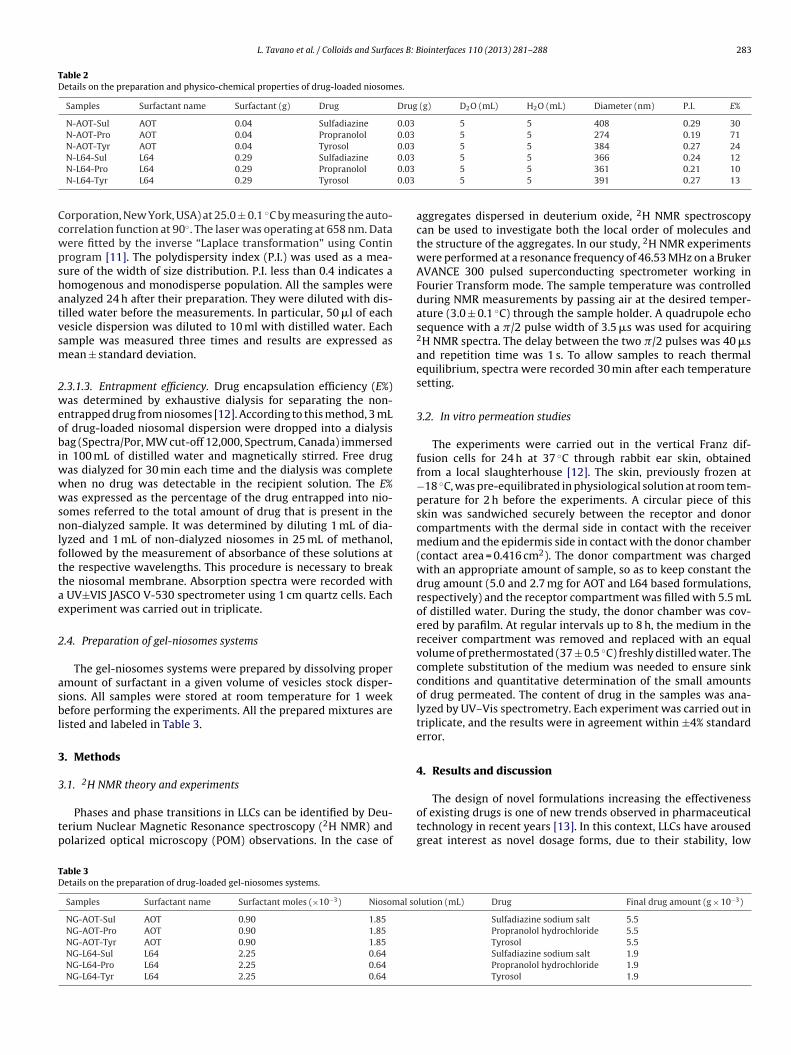
Table 2Details on the preparation and physico-chemical properties of drug-loaded niosomes.
Samples Surfactant name Surfactant (g) Drug Drug (g) D2O (mL) H2O (mL) Diameter (nm) P.I. E%
N-AOT-Sul AOT 0.04 Sulfadiazine 0.03 5 5 408 0.29 30N-AOT-Pro AOT 0.04 Propranolol 0.03 5 5 274 0.19 71N-AOT-Tyr AOT 0.04 Tyrosol 0.03 5 5 384 0.27 24
0.03
0.03
0.03
Ccwpshatvsm
2weobiwwwsnlfttae
2
asbl
3
3
tp
TD
N-L64-Sul L64 0.29 Sulfadiazine
N-L64-Pro L64 0.29 Propranolol
N-L64-Tyr L64 0.29 Tyrosol
orporation, New York, USA) at 25.0 ± 0.1 ◦C by measuring the auto-orrelation function at 90◦. The laser was operating at 658 nm. Dataere fitted by the inverse “Laplace transformation” using Continrogram [11]. The polydispersity index (P.I.) was used as a mea-ure of the width of size distribution. P.I. less than 0.4 indicates aomogenous and monodisperse population. All the samples werenalyzed 24 h after their preparation. They were diluted with dis-illed water before the measurements. In particular, 50 �l of eachesicle dispersion was diluted to 10 ml with distilled water. Eachample was measured three times and results are expressed asean ± standard deviation.
.3.1.3. Entrapment efficiency. Drug encapsulation efficiency (E%)as determined by exhaustive dialysis for separating the non-
ntrapped drug from niosomes [12]. According to this method, 3 mLf drug-loaded niosomal dispersion were dropped into a dialysisag (Spectra/Por, MW cut-off 12,000, Spectrum, Canada) immersed
n 100 mL of distilled water and magnetically stirred. Free drugas dialyzed for 30 min each time and the dialysis was completehen no drug was detectable in the recipient solution. The E%as expressed as the percentage of the drug entrapped into nio-
omes referred to the total amount of drug that is present in theon-dialyzed sample. It was determined by diluting 1 mL of dia-
yzed and 1 mL of non-dialyzed niosomes in 25 mL of methanol,ollowed by the measurement of absorbance of these solutions athe respective wavelengths. This procedure is necessary to breakhe niosomal membrane. Absorption spectra were recorded with
UV±VIS JASCO V-530 spectrometer using 1 cm quartz cells. Eachxperiment was carried out in triplicate.
.4. Preparation of gel-niosomes systems
The gel-niosomes systems were prepared by dissolving propermount of surfactant in a given volume of vesicles stock disper-ions. All samples were stored at room temperature for 1 weekefore performing the experiments. All the prepared mixtures are
isted and labeled in Table 3.
. Methods
.1. 2H NMR theory and experiments
Phases and phase transitions in LLCs can be identified by Deu-erium Nuclear Magnetic Resonance spectroscopy (2H NMR) andolarized optical microscopy (POM) observations. In the case of
able 3etails on the preparation of drug-loaded gel-niosomes systems.
Samples Surfactant name Surfactant moles (×10−3) Niosomal so
NG-AOT-Sul AOT 0.90 1.85
NG-AOT-Pro AOT 0.90 1.85
NG-AOT-Tyr AOT 0.90 1.85
NG-L64-Sul L64 2.25 0.64
NG-L64-Pro L64 2.25 0.64
NG-L64-Tyr L64 2.25 0.64
5 5 366 0.24 125 5 361 0.21 105 5 391 0.27 13
aggregates dispersed in deuterium oxide, 2H NMR spectroscopycan be used to investigate both the local order of molecules andthe structure of the aggregates. In our study, 2H NMR experimentswere performed at a resonance frequency of 46.53 MHz on a BrukerAVANCE 300 pulsed superconducting spectrometer working inFourier Transform mode. The sample temperature was controlledduring NMR measurements by passing air at the desired temper-ature (3.0 ± 0.1 ◦C) through the sample holder. A quadrupole echosequence with a �/2 pulse width of 3.5 �s was used for acquiring2H NMR spectra. The delay between the two �/2 pulses was 40 �sand repetition time was 1 s. To allow samples to reach thermalequilibrium, spectra were recorded 30 min after each temperaturesetting.
3.2. In vitro permeation studies
The experiments were carried out in the vertical Franz dif-fusion cells for 24 h at 37 ◦C through rabbit ear skin, obtainedfrom a local slaughterhouse [12]. The skin, previously frozen at−18 ◦C, was pre-equilibrated in physiological solution at room tem-perature for 2 h before the experiments. A circular piece of thisskin was sandwiched securely between the receptor and donorcompartments with the dermal side in contact with the receivermedium and the epidermis side in contact with the donor chamber(contact area = 0.416 cm2). The donor compartment was chargedwith an appropriate amount of sample, so as to keep constant thedrug amount (5.0 and 2.7 mg for AOT and L64 based formulations,respectively) and the receptor compartment was filled with 5.5 mLof distilled water. During the study, the donor chamber was cov-ered by parafilm. At regular intervals up to 8 h, the medium in thereceiver compartment was removed and replaced with an equalvolume of prethermostated (37 ± 0.5 ◦C) freshly distilled water. Thecomplete substitution of the medium was needed to ensure sinkconditions and quantitative determination of the small amountsof drug permeated. The content of drug in the samples was ana-lyzed by UV–Vis spectrometry. Each experiment was carried out intriplicate, and the results were in agreement within ±4% standarderror.
4. Results and discussion
The design of novel formulations increasing the effectivenessof existing drugs is one of new trends observed in pharmaceuticaltechnology in recent years [13]. In this context, LLCs have arousedgreat interest as novel dosage forms, due to their stability, low
lution (mL) Drug Final drug amount (g × 10−3)
Sulfadiazine sodium salt 5.5Propranolol hydrochloride 5.5Tyrosol 5.5Sulfadiazine sodium salt 1.9Propranolol hydrochloride 1.9Tyrosol 1.9

2 es B: B
iopo
bpeirtapfiloaotpmt
cuirmhigoaiadoiosa
4
lpihrtdtesimctdlvddvt
84 L. Tavano et al. / Colloids and Surfac
nterfacial tension, and capability to increase the solubility of bothil and water soluble compounds. In addition, they exhibit goodenetration across the skin, and show extensive similarity to livingrganisms biological membranes and chromosomes [14].
The LLCs samples were prepared using an accurate ratioetween surfactant and water, in order to obtain lamellar LLChases, for both L64 and AOT. In the case of the Pluronic L64, X-rayxperiments reported in literature reveal a large lamellar LLC phasen the surfactant-rich region of the phase diagram [15] and for thiseason we decided to prepare the sample at 75 wt% of the surfac-ant. For the AOT, since the published AOT–water diagram showed
lamellar phase extends from ca. 5 to ca. 70 wt%, the systems wererepared starting from 30 wt% of AOT, in order to minimize thenal amount of surfactant present in the formulations [16]. The
oaded-drugs formulations were prepared by adding drug solutionr drug-loaded niosomal suspension in a fixed wt percentage, sos to keep constant the amount of drug in all the formulations. Thebtained mesophases, were characterized by 2H NMR to evaluatehe structural modifications. In fact, it is known that the incor-oration of drugs as a third component could induce structuralodifications and these influence the in vitro penetration of the
herapeutic agents in the skin.In the present study, drugs with anionic, cationic and nonionic
hemical structures such as Sul, Pro and Tyr, respectively, weresed as model drugs. Sul is an topical antibiotic used to protect
nfected burns; it prevents the growth of a wide array of bacte-ia, as well as yeast, on the damaged skin. Propranolol, one of theost widely prescribed �-blockers in the long-term treatment of
ypertension and in psychotherapy is usually taken orally. Follow-ng oral administration, it is rapidly and completely absorbed fromastrointestinal tract, still the oral bioavailability is low becausef significant first pass hepatic metabolism. For this reason topicalpplication is ideal for propranolol, but the transdermal absorptions poor, whereby different approaches to increase the permeationre needed. Tyr is a phenolic compound present in two of the tra-itional components of the Mediterranean diet: wine and virginlive oil. It was shown to be able to carry out antioxidant activityn vitro studies and for these reasons it could be used to preventr reduce erythema, photoaging, photocarcinogenesis, edema, andkin hypersensitivity associated with exposure to ultraviolet B radi-tion [17].
.1. AOT and L64-based niosomes
The AOT/water binary system is dominated by a large lamel-ar LLC phase and at high surfactant content, a discontinuous cubichase and a reversed hexagonal phase are formed, as well reported
n literature. The lamellar microstructure of the system AOT/wateras been subjected to different analysis in the past years: unusualesults were observed, like the modification of the optical signal ofhe birefringence in the range between 30 and 40 wt% of AOT, theiscontinuity of the electrical conductivity and the anomalies fromhe X-ray data that divided the lamellar mesophase in three differ-nt ranges. Two of them show an ideal swelling behavior and areeparated by a no-swelling one [18]. Many hypothesis were stud-ed and was observed that the systems were strongly influenced by
echanical stresses which force the lamellae to assume defectiveonfigurations: Coppola et al. showed that the lamellar phase ofhe AOT/water system is constituted by small non-interconnectedomains [19]. AOT is well known to form monodispersed, unilamel-
ar and stable vesicles in aqueous solution. The efficacy of theseesicles to host both charged and hydrophobic drugs has also been
emonstrated in several works and makes them useful as drugelivery systems [20]. In our case the vesicles were prepared byortex followed by sonication method, in order to obtain a con-rol on the physico-chemical properties of the niosomes. Dynamiciointerfaces 110 (2013) 281– 288
light scattering measurements of the obtained empty vesicles werecarried out in dilute aqueous solution at room temperature andindicated a diameter of 372 nm. The distribution profile was foundto be unimodal with relatively low P.I., suggesting the existence ofvesicles of uniform sizes. The introduction of equal amount of drugswas found to affect the vesicles size, depending on the molecularstructure of the encapsulated molecules (Table 2).
Hydrodynamic diameter of 274, 408 and 384 nm were found forthe cationic, anionic and non-ionic drugs, respectively. Since AOT isan anionic surfactant, it is clear that these results are strongly con-nected with the charge of the drugs: in fact, ionic repulsions amonganionic groups of AOT and sulfadiazine result in the higher increaseof vesicles dimensions; a decrease of the hydrodynamic diameterwas achieved in the presence of propranolol, since ionic attractionoccurred, while in the case of tyrosol as non-ionic molecule, anintermediate value of diameter was found, probably due to theabsence of net charge. The P.I. was between 0.19 and 0.29: thesevalues were considered as evidence of homogeneous distributionof colloidal vesicles. Drug vesicle encapsulation efficiency is a prod-uct of different factors, the most important are: the ionization stateof the drugs and the charges of the niosomal matrix. As reportedbefore concerning the niosomal size, the chemical structure of theencapsulated drug and the affinity for the niosomal bilayer stronglyaffect the partitioning and location of drug molecules into nio-somes. Sul, Pro and Tyr are hydrophilic drugs and they are locatedin the aqueous core of the vesicles. As reported in Table 2, E% werefound to be 30, 71 and 24% for Sul, Pro and Tyr, respectively, andare dependent on the ionic and electrostatic interactions betweenthe ionic groups of the carrier and the drugs.
Pluronic L64 is a water-soluble triblock copolymer forming vesi-cles. These drug carriers were demonstrated to have a sphericalstructure, with the hydrophobic block forming the inner part of thebilayer, while the hydrophilic PEO blocks are withdrawing to theinside aqueous core of the vesicles or outside. These block copoly-mer vesicles showed properties as drug carriers for the transdermaland parenteral vehiculations of small molecules of pharmacologicalinterest.
As reported in Table 2, empty vesicles of L64 were prepared fol-lowing the thin layer evaporation method, and the diameter wasfound to be 396 nm. This value was found to be similar to thatreported in our previous work [8] and it was also similar to thatobtained from AOT, even if, in this case no repulsions betweensimilar charged groups resulting in an increase of vesicles diam-eter occurred. The introduction of drugs determined a decrease ofvesicles size: this is very common when hydrophilic drugs wereintroduced into the niosomes. The chemical structure of the drugsdid not affect in a relevant manner the hydrodynamic diameter ofthe carriers: in the case of Sul, this value was found to be 356 nm,while in the case of Pro and Tyr the dimensions increased up to 361and 391 nm, respectively. The P.I. was between 0.21 and 0.27. TheE% values of 12, 10 and 13% were found in the case of Sul, Pro andTyr, respectively. These values are comparable and reflect the trendfound in the case of vesicles size, in which the chemical structureof the drugs did not influence the dimensions. In fact, it seems that,regardless of the charge of the drug, the L64 based niosomes wereable to encapsulate only a small amount of hydrophilic molecules.
In the transmission electron micrograph, all niosomal formu-lations were spherical and homogeneous in shape and the sizewas well correlated with the results of the laser diffraction particlesize (DLS). No sedimentation or flocculation were observed and thesamples were found to be stable for 9 months.
4.2. AOT based liquid crystal and liquid crystal-niosomes systems
Generally the 2H NMR spectra obtained by the binary sys-tem AOT/water, at the concentration used in this study, did not

es B: B
smts
pvtot(ms
Ptmasaaewhd
4
asltSl
4
tmctrgAtstsptrtauc
4
AF
ro
L. Tavano et al. / Colloids and Surfac
how a classical lamellar structure, because of the presence oficrodomains that gives rise to biaxial lineshapes. Previous inves-
igations demonstrated that the structural organization of theystems can be attributed to a “defective” lamellar phase [19].
In our case, the addition of drugs to the liquid crystals sam-les, both as drug solution or niosomal suspension, resulted in aiscous, transparent and birefringent gel. Collected 2H NMR spec-ra showed the presence of lamellar mesophases similar to thosebtained without the drug: the third component did not changehe phase diagram of the sample, at this surfactant concentrationFig. 1). In fact, the drugs induced a weak size modifications of
icrodomains consequently, the systems keep the typical biaxialhape.
Only when the sample was prepared in the presence ofro loaded vesicular suspension, the recorded 2H NMR spec-rum showed significant modifications because of lamellar
acrodomains presence (Fig. 1f). All systems were investigatedfter 6 months, the spectra were not reported because theyhowed similar lineshape. Just the systems containing Pro gave
lamellar uniaxial phase, due to the interaction between drugnd surfactant, giving the formation in situ of propranolol-bis(2-thylhexyl)sulfosuccinate salt (Fig. 1g and h). In literature, itas reported that the system sodium dodecyl sulfate-propranololydrochloride (as surfactant and drug, respectively) affected therug release in tablet [21].
.3. L64 based liquid crystal and liquid crystal-niosomes systems
The G-L64 and NG-L64-Pro samples appeared as transparentnd birefrangent lamellar gels, but less viscous than the corre-ponding ones based on AOT. The 2H NMR spectra showed a pureamellar mesophase and this means that the introduction of Pro ashird component did not affect its microstructures (Fig. 2a and b).imilar trends were found in the case of Sul and Tyr-based formu-ations (data not shown).
.4. Percutaneous permeation studies
The success of a transdermal drug delivery system depends onhe drug ability to permeate the skin in a sufficient amount to
aintain its therapeutic levels. Drug compounds can be classifiedhemically as ionic, zwitterionic or neutral. It is well establishedhat small, neutral compounds will permeate the skin barrier moreeadily than charged species, while charged drug molecules are, ineneral, considered to be unsuitable for transdermal drug delivery.n ideal drug candidate would have sufficient lipophilicity to par-
ition into the SC, but also sufficient hydrophilicity to enable theecond partitioning step into the viable epidermis and, eventually,he systemic circulation. However, research continues into pos-ible methods of enhancing the transdermal permeation of thesearticular compounds: among these, LLC represents a valid alterna-ive [22]. A good drug nanocarrier should prerequisitely be able toelease a drug in a controlled manner and for this reason the percu-aneous permeation profiles of the three different model drugs fromll obtained formulations were evaluated. Results showed partic-lar trends because the formation of ion pair between AOT andationic drugs and repulsions between charged drugs and skin.
.5. G-AOT and G-L64 systems percutaneuos permeation profiles
The cumulative quantities of Prop, Sul and Tyr permeated fromOT and L64 gel systems across the skin are plotted versus time in
ig. 3a and b, respectively.In the case of AOT-based formulations, the permeation profilesanged from 0.300 to 0.025 mg/cm2 within 8 h: the higher amountf permeated drug was obtained in the case of Tyr, the non-ionic
iointerfaces 110 (2013) 281– 288 285
molecule, while lower values were obtained by G-AOT-Sul; inter-mediate values were collected with G-AOT-Pro (0.139 mg/cm2).When L64 was used as surfactant, some differences in the perme-ation profiles occurred: in fact, the higher cumulative amount ofdrug permeated across the skin was obtained for G-L64-Pro, fol-lowed by G-L64-Tyr and G-L64-Sul samples. This trend could beascribed to the synergistic action between the positive charge ofthe propranolol and the cell membrane negative potential of thestratum corneum, increasing the drug permeation. In both cases,the cumulative sulfadiazine percutaneous permeations achievedfrom G-AOT-Sul and G-L64-Sul were found to be very low and thesebehaviors appeared to confirm the repulsions between the nega-tive charged drug and the cell membrane negative potential of thestratum corneum. In addition, as showed in Fig. 3a and b, the cumu-lative drugs permeations from AOT-based gel systems were foundto be higher than those obtained from L64-based ones. An addi-tional mechanism for the skin penetration enhancement by AOTcould involve the hydrophobic interaction of its alkyl chains withthe skin structure which leaves the end sulfonate group of the sur-factant exposed, creating additional sites in the membrane whichleads to permit an increase in skin hydration [23].
4.6. N-AOT and N-L64 systems percutaneuos permeation profiles
The drugs permeation profiles from AOT and L64 niosomal sam-ples were reported in Fig. 4a and b. As showed, the trends found forthese formulations were similar to those obtained in the case of gelsystems. In fact the higher cumulative amount of permeated drugwas obtained by N-AOT-Tyr, followed by N-AOT-Pro and then byN-AOT-Sul samples, while in the case of L64, the order is as follows:N-L64-Pro, N-L64-Tyr and N-L64-Sul.
These results confirmed the hypothesis that the percutaneouspermeations of the model drugs are strongly dependent on thephysico-chemical nature of the surfactants and not by the kind ofcarrier used in the experimental (gel or niosomes). In fact, the drugspermeation order from niosomal systems was found to be the sameobtained by the gel systems.
4.7. NG-AOT and NG-L64 systems percutaneous permeationprofiles
Generally, in order to make them suitable for transdermaladministration, niosomes were usually incorporated in a gel. Onceincorporated they showed slightly lower drug permeation, ascribedto the slow diffusion of drug through the gel network. Percutaneouspermeation profiles of Pro, Sul and Tyr from niosomes-gel systemsare reported in Fig. 5a and b.
The presence of intact niosomes into the multicomponentsystems was confirmed by diluting the formulation in aqueousmedium and performing light scattering experiments. In fact, inthe case of sulfadiazine, vesicles diameter after dilution was foundto be 415 nm for AOT based-niosomes and 350 nm for L64 ones.Similar trends have been obtained for the other drugs.
In niosome-gel samples, the drug is distributed inside and out-side the vesicles: however, unlike simple niosomal systems, thedrug outside the vesicles is not quickly available for the permeation,because it is embedded into the matrix and must diffuse throughit to reach the skin and to permeate it. In this light the capabilityof the drug to interact with the gel matrix could affect in a relevantmanner its diffusion through the network. As shown in Fig. 4b, thehigher cumulative permeated drug amount was achieved by NG-AOT-Pro, followed by NG-AOT-Tyr and NG-AOT-Sul. This behavior
differs from those obtained in the case of corresponding gel or nio-somes systems, in which the samples containing tyrosol showedthe best permeation performance. This could be due to the forma-tion of the lipophilic propranolol-bis(2-ethylhexyl)sulfosuccinate
286 L. Tavano et al. / Colloids and Surfaces B: Biointerfaces 110 (2013) 281– 288
Fig. 1. 2H NMR spectra of AOT lamellar mesophases: (a) G-AOT-Sul; (b) NG-AOT-Sul; (c) G-AOT-Tyr; (d) NG-AOT-Tyr; (e) G-AOT-Pro; (f) NG-AOT-Pro; (g) G-AOT-Pro after 6months; (h) NG-AOT-Pro after 6 months.
Fig. 2. 2H NMR spectra of L64 lamellar mesophases: (a) G-L64-Pro and (b) NG-L64-Pro.

L. Tavano et al. / Colloids and Surfaces B: Biointerfaces 110 (2013) 281– 288 287
Fig. 3. Permeated drug cumulative amounts as a function of time from: (a) G-AOT formulations: (triangle) tyrosol; (square) propranolol hydrochloride; (diamond) sulfadiazinesodium salt. (b) G-L64 formulations: (triangle) tyrosol; (square) propranolol hydrochloride; (diamond) sulfadiazine sodium salt (mean ± SD; n = 3).
F mulas hlorid
sraltomNdtfwta
4
si
Fa
ig. 4. Permeated drug cumulative amounts as a function of time from: (a) N-AOT forodium salt. (b) N-L64 formulations: (triangle) tyrosol; (square) propranolol hydroc
alt that possesses an higher ability to diffuse into the networkespect to the Pro temporarily altering the membrane permeabilitynd enhancing the drug skin permeation. The formation of a moreipophilic salt is the reason because the propranolol, despite attrac-ive interactions, permeated faster than the sulfadiazine. In the casef L64-based multicomponent systems, the higher cumulative per-eated drug amount was achieved by NG-L64-Tyr, followed byG-L64-Pro and NG-L64-Sul. Also in this case the trend is partiallyifferent from those obtained by the simple gel or niosomal sys-ems. As reported above, since drugs entrapment efficiencies wereound to be similar, the amount of drugs embedded into the net-ork and then immediately available were comparable, whereby
he only explanation could be related to the different interactionsnd affinity between each drug and its diffusion through the matrix.
.8. General remarks
The results obtained in this work are in line with recent findingsuggesting that factors such as vehicle composition, drug solubilityn the matrix and its viscosity, play an important role in the skin
ig. 5. Permeated drug cumulative amounts as a function of time from: (a) NG-AOT formzine sodium salt. (b) NG-L64 formulations: (triangle) tyrosol; (square) propranolol hydr
tions (triangle) tyrosol; (square) propranolol hydrochloride; (diamond) sulfadiazinee; (diamond) sulfadiazine sodium salt (mean ± SD; n = 3).
permeation. In all cases it is important to note that both AOT andL64-based formulations act as percutaneous permeation enhancersof Sul, Pro and Tyr. In fact, all cumulative permeated drug amountswere always higher than those obtained from the correspondingfree solutions. As consequence, these systems could be proposedas novel transdermal drug delivery systems. The diffusion mech-anisms involved in the drugs permeation are probably differentfor the kind of systems (gel, niosomes and gel-niosomes formula-tions) and also for the different structures of the drugs. Clearly AOTand L64 both in the form of niosomes and as LLCs, interact withthe lipid structure of the SC and also change its hydration level insuch a way that facilitates the permeation of drugs across the skin.Several works reported that the hydrophobic interactions of thealkyl chain of an anionic surfactant (AOT) with the keratinous pro-teins, leaves the negative end group of the surfactant exposed andinduces additional anionic sites in the membrane cells. This results
in a greater repulsive force that separates the protein matrix andresults in the softening of the SC. On the other hand, L64, as pluroniccopolymer surfactant of ethylene oxide (EO) and propylene oxide(PO) blocks arranged in a triblock structure, is claimed to attractulations (triangle) tyrosol; (square) propranolol hydrochloride; (diamond) sulfadi-ochloride; (diamond) sulfadiazine sodium salt (mean ± SD; n = 3).

2 es B: B
wl
5
mtacaebsmueo
A
fipE
R
[
[[
[
[
[[
[
[[
[
[
88 L. Tavano et al. / Colloids and Surfac
ater molecules and this could also increase skin hydration andipid fluidization.
. Conclusions
In this paper we have developed novel multicomponent for-ulations based on liquid crystal-niosomes systems, for the
ransdermal drugs delivery. AOT and L64 were used as surfactantsnd Sul, Pro and Tyr were used as model drugs. Clearly, the vehi-le composition, drug solubility and its chemical structure playn important role in skin permeation. In fact, it is not possible tostablish a definite, common trend, since several variables coulde considered. All the obtained formulations, such as gel-niosomesystems, niosomal suspensions and gels, act as percutaneous per-eation enhancer respect to the corresponding free drug solutions
sed as control. Finally these formulations could be considered asffective functional materials for controlled transdermal deliveryf small hydrophilic molecules.
cknowledgements
MIUR, the Italian Ministry for University, is acknowledged fornancial support (Grants # EX-60%, PRIN 2010/11). Moreover, theroject has been co-funded with support from the Commissionuropean Social Fund and Region of Calabria (Italy).
eferences
[1] H.Y. Thong, H. Zhai, H.I. Maibach, Skin Pharmacol. Physiol. 20 (2007) 272.[2] B.W. Barry, Dermatological Formulation: Percutaneous Absorption, Marcel
Dekker, New York, 1983.
[
[
iointerfaces 110 (2013) 281– 288
[3] G.A. Kossena, W.N. Charman, B.J. Boyd, C.J. Porter, J. Control. Release 99 (2004)217.
[4] D. Chapman, Lyotropic mesophase in biological systems, in: B. Bahadur(Ed.), Liquid Crystals Applications and Uses, World Scientific, Singapore,1991.
[5] G. Grassi, A. Crevatin, R. Farra, G. Guarnieri, A. Pascotto, B. Rehimers, R. Lapasin,M. Grassi, J. Coll. Interf. Sci. 301 (2006) 282.
[6] A. Manosroi, P. Jantrawuta, J. Manosroi, Int. J. Pharm. 360 (2008) 156.[7] F.E. Antunes, L. Gentile, C. Oliviero Rossi, L. Tavano, G.A. Ranieri, Coll. Surf. B:
Biointerfaces 1 (2011) 42.[8] L. Tavano, R. Muzzalupo, S. Trombino, R. Cassano, A. Pingitore, N. Picci, Coll.
Surf. B: Biointerfaces 79 (2010) 227.[9] R. Muzzalupo, L. Tavano, R. Cassano, S. Trombino, T. Ferrarelli, N. Picci, Eur. J.
Pharm. Biopharm. 79 (2011) 28.10] R.R. Fenton, W.J. Easdable, H. Meng, E.S.M. Omara, M.J. Mckeage, P.J. Russel,
T.W. Hambley, J. Med. Chem. 40 (1997) 1090.11] S.W. Provencher, Comput. Phys. Commun. 27 (1982) 213.12] L. Tavano, R. Muzzalupo, R. Cassano, S. Trombino, T. Ferrarelli, N. Picci, Coll.
Surf. B: Biointerfaces 75 (2010) 319.13] L. Tavano, P. Alfano, R. Muzzalupo, B. De Cindio, Coll. Surf. B: Biointerfaces 87
(2011) 333.14] R. Muzzalupo, L. Tavano, F.P. Nicoletta, S. Trombino, R. Cassano, N. Picci, J. Drug
Targets 8 (2010) 404.15] P. Alexandridis, D. Zhou, A. Khan, Langmuir 12 (1996) 2690.16] O. Robles-Vàsqez, S. Corona-Galvàn, J.F.A. Soltero, J.E. Puig, J. Coll. Interf. Sci.
160 (1993) 65.17] N. Vassallo, Polyphenols and Health: New and Recent Advances, Nova Publish-
ers, New York, 2008.18] Park, J. Rogers, R.W. Toft, P.A. Winsor, Coll. Surf. B: Bionterfaces 32 (1970) 81.19] L. Coppola, R. Muzzalupo, G.A. Ranieri, M. Terenzi, Langmuir 11 (1995)
1116.20] R. Saha, P.K. Verma, R.K. Mitra, S.K. Pal, Coll. Surf. B: Biointerfaces 88 (2011)
345.21] J.F. Ford, K. Mitchell, D. Sawh, S. Ramdour, D.J. Armstrong, P.N.C. Elliott, C.
Rostron, J.E. Hogan, Int. J. Pharm. 71 (1991) 213.22] Y.B. Bannon, J. Corish, O.I. Corrigan, J.G. Devane, M. Kavanagh, S. Mulligan, Eur.
J. Clin. Pharmacol. 37 (1989) 285.23] L.D. Rhein, C.R. Robbins, K. Fernee, R. Cantore, J. Soc. Cosmet. Chem. 37 (1986)
125.