Transdermal delivery of Chinese herbal medicine ... - CityU Scholars
9
Transdermal delivery of Chinese herbal medicine extract using dissolvable microneedles for hypertrophic scar treatment Ning, Xiaoyu; Wiraja, Christian; Chew, Wan Ting Sharon; Fan, Chen; Xu, Chenjie Published in: Acta Pharmaceutica Sinica B Published: 01/09/2021 Document Version: Final Published version, also known as Publisher’s PDF, Publisher’s Final version or Version of Record License: CC BY-NC-ND Publication record in CityU Scholars: Go to record Published version (DOI): 10.1016/j.apsb.2021.03.016 Publication details: Ning, X., Wiraja, C., Chew, W. T. S., Fan, C., & Xu, C. (2021). Transdermal delivery of Chinese herbal medicine extract using dissolvable microneedles for hypertrophic scar treatment. Acta Pharmaceutica Sinica B, 11(9), 2937-2944. https://doi.org/10.1016/j.apsb.2021.03.016 Citing this paper Please note that where the full-text provided on CityU Scholars is the Post-print version (also known as Accepted Author Manuscript, Peer-reviewed or Author Final version), it may differ from the Final Published version. When citing, ensure that you check and use the publisher's definitive version for pagination and other details. General rights Copyright for the publications made accessible via the CityU Scholars portal is retained by the author(s) and/or other copyright owners and it is a condition of accessing these publications that users recognise and abide by the legal requirements associated with these rights. Users may not further distribute the material or use it for any profit-making activity or commercial gain. Publisher permission Permission for previously published items are in accordance with publisher's copyright policies sourced from the SHERPA RoMEO database. Links to full text versions (either Published or Post-print) are only available if corresponding publishers allow open access. Take down policy Contact [email protected] if you believe that this document breaches copyright and provide us with details. We will remove access to the work immediately and investigate your claim. Download date: 24/08/2022
-
Upload
khangminh22 -
Category
Documents
-
view
0 -
download
0
Transcript of Transdermal delivery of Chinese herbal medicine ... - CityU Scholars
Transdermal delivery of Chinese herbal medicine extract using dissolvable microneedles forhypertrophic scar treatment
Ning, Xiaoyu; Wiraja, Christian; Chew, Wan Ting Sharon; Fan, Chen; Xu, Chenjie
Published in:Acta Pharmaceutica Sinica B
Published: 01/09/2021
Document Version:Final Published version, also known as Publisher’s PDF, Publisher’s Final version or Version of Record
License:CC BY-NC-ND
Publication record in CityU Scholars:Go to record
Published version (DOI):10.1016/j.apsb.2021.03.016
Publication details:Ning, X., Wiraja, C., Chew, W. T. S., Fan, C., & Xu, C. (2021). Transdermal delivery of Chinese herbal medicineextract using dissolvable microneedles for hypertrophic scar treatment. Acta Pharmaceutica Sinica B, 11(9),2937-2944. https://doi.org/10.1016/j.apsb.2021.03.016
Citing this paperPlease note that where the full-text provided on CityU Scholars is the Post-print version (also known as Accepted AuthorManuscript, Peer-reviewed or Author Final version), it may differ from the Final Published version. When citing, ensure thatyou check and use the publisher's definitive version for pagination and other details.
General rightsCopyright for the publications made accessible via the CityU Scholars portal is retained by the author(s) and/or othercopyright owners and it is a condition of accessing these publications that users recognise and abide by the legalrequirements associated with these rights. Users may not further distribute the material or use it for any profit-making activityor commercial gain.Publisher permissionPermission for previously published items are in accordance with publisher's copyright policies sourced from the SHERPARoMEO database. Links to full text versions (either Published or Post-print) are only available if corresponding publishersallow open access.
Take down policyContact [email protected] if you believe that this document breaches copyright and provide us with details. We willremove access to the work immediately and investigate your claim.
Download date: 24/08/2022
Acta Pharmaceutica Sinica B 2021;11(9):2937e2944
Chinese Pharmaceutical Association
Institute of Materia Medica, Chinese Academy of Medical Sciences
Acta Pharmaceutica Sinica B
www.elsevier.com/ loca te /apsbwww.sc iencedi rec t .com
ORIGINAL ARTICLE
Transdermal delivery of Chinese herbalmedicine extract using dissolvable microneedlesfor hypertrophic scar treatment
Xiaoyu Ninga,b, Christian Wirajab, Wan Ting Sharon Chewa,b,Chen Fanc, Chenjie Xua,b,d,*
aNTU Institute for Health Technologies, Interdisciplinary Graduate Group, Nanyang Technological University,Singapore 639798, SingaporebSchool of Chemical and Biomedical Engineering, Nanyang Technological University, Singapore 637459,SingaporecSkin Research Institute of Singapore, 8A Biomedical Grove, Singapore 138648, SingaporedDepartment of Biomedical Engineering, City University of Hong Kong, Hong Kong SAR 999077, China
Received 26 November 2020; received in revised form 15 January 2021; accepted 6 February 2021
KEY WORDS
Traditional Chinese
*
Pee
http
221
by
medicine;
Shikonin;
Hyaluronic acid;
Microneedle;
Hypertrophic scarring;
Drug delivery;
Transdermal delivery;
Skin
Corresponding author.
E-mail address: [email protected]
r review under responsibility of Chine
s://doi.org/10.1016/j.apsb.2021.03.016
1-3835 ª 2021 Chinese Pharmaceutic
Elsevier B.V. This is an open access a
Abstract Hypertrophic scars are unfavorable skin diseases characterized by excessive collagen depo-
sition. Although systemic treatments exist in clinic to manage hypertrophic scars, they pose significant
side effects and tend to lose efficacy over prolonged applications. Traditional Chinese medicine
(TCM) offers as a promising candidate to treat pathological scars. A large number of TCMs have been
studied to show anti-scarring effect, however, the natural barrier of the skin impedes their penetration,
lowering its therapeutic efficacy. Herein, we reported the use of dissolvable hyaluronic acid (HA) micro-
needles (MNs) as a vehicle to aid the transdermal delivery of therapeutic agent, a model TCM called shi-
konin for the treatment of hypertrophic scars. Here, shikonin was mixed with HA to make MNs with
adequate mechanical strength for skin penetration, making its dosage controllable during the fabrication
process. The therapeutic effect of the shikonin HA MNs was studied in vitro using HSFs and then further
verified with quantitative reverse transcriptase polymerase chain reaction. Our data suggest that the shi-
konin HA MNs significantly reduce the viability and proliferation of the HSFs and downregulate the
fibrotic-related genes (i.e., TGFb1, FAP-a and COL1A1). Furthermore, we observed a localized therapeu-
tic effect of the shikonin HA MNs that is beneficial for site-specific treatment.
.hk (Chenjie Xu).
se Pharmaceutical Association and Institute of Materia Medica, Chinese Academy of Medical Sciences.
al Association and Institute of Materia Medica, Chinese Academy of Medical Sciences. Production and hosting
rticle under the CC BY-NC-ND license (http://creativecommons.org/licenses/by-nc-nd/4.0/).
2938 Xiaoyu Ning et al.
ª 2021 Chinese Pharmaceutical Association and Institute of Materia Medica, Chinese Academy of Medical
Sciences. Production and hosting by Elsevier B.V. This is an open access article under the CC BY-NC-ND license
(http://creativecommons.org/licenses/by-nc-nd/4.0/).
1. Introduction
Hypertrophic scars are characterized by excessive collagendeposition by dermal fibroblasts. The underlying causes remainelusive, although ethnicity and genetic predisposition play a role.Current treatments, including corticosteroids, pressure therapy,laser therapy and surgical operation, are not satisfactory and maycause significant side effects over prolonged use1. For instance,intralesional corticosteroid injection is the first-line approach forphysicians to prevent and treat the keloids and hypertrophic scars2.While it provides a 50%e100% flattening of the keloids, it re-quires multiple painful injections, and may cause hypo-pigmentation and skin atrophy with 9%e50% recurrence rate3.Surgical removal of scars could be temporarily gratifying; how-ever, it is normally followed by more aggressive regrowth of thescar unless other therapies were performed post-surgery. Thiscomplicates the treatment and requires exemplary persistencefrom the patients.
With rich resource and relatively low cost, traditional Chinesemedicines (TCMs) have been widely used for thousands of yearsin prevention and treatment of many diseases including scarring4.Sourced from the nature and typically possessed little side effects,TCMs emphasize on improving the general health of patients. Alarge amount of TCMs have been studied to display anti-scarringeffect. However, the natural skin barrier such as stratum corneumand the compact structure of scar tissues limit the permeation andefficiency of TCM on cutaneous diseases5. Although reformula-tion through microemulsion or nanoparticles has been found toimprove the penetration of TCM, they introduce additional com-ponents which alter the original TCM structure and interfere withits functionality and toxicity6. For instance, the microemulsionproduct of oxymatrineephospholipid complex (OMT-PLC) con-sists of not only 10.0% OMT-PLC, but also 8.0% isopropylmyristate, 30.0% cremophor RH40/polyethyleneglycol 400 (1:1)and 52.0% water7. In addition, when TCMs are nanomaterialized,the physicochemical properties of the active ingredient might bealtered, diminishing its therapeutic efficacy6.
Microneedle (MN) technology is effective in delivering variousdrugs intradermally without affecting blood vessels and nerves. Itovercomes the protective stratum corneum of the epidermis layerand creates transient micro holes on the skin to aid the permeationof the drugs. Many studies have shown MN’s potential to suc-cessfully deliver therapeutic agents including oligonucleotide,vaccine, peptide and so on8,9,10.
Thus, we believe the transdermal delivery of TCM formula-tions utilizing MNs would address the limitations met currently inclinics. As a proof-of-concept, we chose shikonin as a model TCMdrug. Shikonin is an active component extracted from ArnebiaeRadix (ZiCao) and has been demonstrated to inhibit cell viability,cell proliferation and collagen production in scar-derived fibro-blasts, thus offering a promising alternative for the treatment ofscars11. It is generally mixed in oil-based creams or ointments
before application to human skin, however, achieving highbioavailability is challenging due to its insolubility in water12. Thetransdermal delivery of shikonin with MN platform offers apromising route to enhance its bioavailability.
Herein, we incorporate shikonin into MN made from dissolv-able hyaluronic acid (HA) to aid the delivery. HA is widelydistributed throughout our tissues, offering excellent biocompati-bility with minimal side effects13. Shikonin was pre-dissolved inDimethyl sulfoxide (DMSO) before mixing with HA solution andthe mixture solution was then used to make MNs by micromolding (Scheme 1). The amount of shikonin encapsulated in theMN could be easily controlled during the fabrication process byadjusting the pre-dissolved shikonin concentrations. Our previousstudy indicated that shikonin could inhibit cell proliferation,induce apoptosis of hypertrophic scar derived fibroblast (HSF),and attenuate transdermal growth factor beta 1 (TGFb1)-inducedcollagen production14,15. In this study, the efficiency of shikonin inkilling the HSF and attenuating scar-related gene expressions wasmaintained after incorporating into HA MNs. Moreover, shikonin-MNs offers a localized therapeutic effect that is beneficial for site-specific scar treatment.
2. Methods
2.1. Materials
All chemicals and reagents were obtained from SigmaeAldrich(Singapore) except mentioned otherwise. The stainless-steelmaster mold of MN (300 mm base diameter, 5 mm tip radius,and 1000 mm height) was purchased from Micropoint Technolo-gies Pte Ltd. (Singapore). Hyaluronic acid (HA, 300 kDa) waspurchased from Freda Biochem Co., Ltd. (China). Phosphatebuffered saline (1 � , PBS), high glucose (4.5 g/L) Dulbecco’smodified Eagle’s medium (DMEM) were obtained from Lonza.Fetal bovine serum (FBS), Trypsin�EDTA (0.05%), andpenicillin�streptomycin (10,000 U/mL) were purchased fromGibco (Singapore). AlarmarBlue cell viability reagent was pur-chased from Thermofisher (Singapore). TRIzol reagent was ob-tained from Invitrogen (Singapore). iScript Adv cDNA kit &Universal SYBR� Green Supermix were purchased from Biorad(Singapore).
2.2. Fabrication of the PDMS mold
The fabrication of the PDMS mold followed our previous meth-odology9. Briefly, the PDMS daughter mold was produced bypouring PDMS into a Petri dish containing stainless steel mothermolds. Degassing was performed by vacuum oven and subse-quently, curing was performed at 70 �C for 1 h. PDMS molds thatreversely replicate the mother molds were obtained by detachingthe cured PDMS from the Petri dish.

Scheme 1 Shikonin hyaluronic acid microneedle for the facile treatment of abnormal wound scarring.
Microneedle assisted delivery of Chinese herbal medicine 2939
2.3. Fabrication of the shikonin HA MN
Shikonin was first dissolved in DMSO with a concentration of50 mg/mL as stock solution for subsequent experiment. HApowder was dissolved in deionized (DI) water and mixed homo-geneously before different amounts of shikonin stock were addedto a final concentration of 0, 4, 20, 100 and 200 mg/mL. Theshikonin‒HA mixtures were then poured into PDMS molds andfollowed by centrifugation (3200�g, 5 min) to fill the tips of theMN completely and remove air bubbles. More mixtures were thentopped up into the PDMS mold to form the MN base. The dryingprocess was conducted in fume hood under room temperature.
2.4. Loading efficiency of shikonin HA MN
Shikonin HA MNs (made from different shikonin mixture con-centrations) were thoroughly dissolved in PBS at 37 �C. Subse-quently, the solution was mixed gently to ensure homogeneoussolution. Shikonin distribution was read under a microplate reader(SpectraMax M5, Molecular Devices) at l Z 520 nm. Solutionabsorbance was fitted against a standard curve to calculate theconcentration of shikonin.
2.5. Compression test of shikonin HA MN
The compression test was done with a Tensile Meter (Instron5543). HA MNs containing varying amount ofshikonin wereplaced on the specimen plate with tips facing upwards. Thedisplacement versus the load were measured and recorded until apreset maximum loading force of 80 N was reached.
2.6. Ex vivo penetration test
HA MNs containing the highest shikonin (made from 200 mg/mLshikonin HA solution) was thumb-pressed into an excised porcineskin and hold steadily for 3 min. Subsequently, the treated skinwas cryo-sectioned and hematoxylin & eosin (H&E)-stained forhistological analysis.
2.7. Fibroblast cell culture
Fibroblasts derived from hypertrophic scar tissue (HSFs) werecultured in high-glucose DMEM with 10% FBS and 1%penicillin�streptomycin in a humidified condition of 37 �C with
5% CO2. Culture medium was replaced every 2 days and cellswere sub-cultured when they reached 95% confluency.
2.8. Biocompatibility assessment of HA
The biocompatibility of blank HA MN was investigated usinglive/dead staining kit which contains calcein-AM and propidiumiodide (PI) to stain viable and dead cells, respectively. HA MNswere placed and incubated for 24 h on HSFs when the cellsreached 80% confluency in the well plate. The live/dead stainingsolution were then added as per the manufacturer’s instructionsand incubated in 37 �C for 15 min. Fluorescence images weretaken by a digital microscope (Leica DVM6) at lex Z 490 nm andlem Z 515 nm for live signal and lex Z 535 nm, lem Z 617 nmfor dead signal.
The biocompatibility of HA solutions (0.125, 0.5, 2 and4 mg/mL) were investigated using the AlamarBlue assay. Inaccordance with the manufacturer’s protocols, the AlamarBluereagent was added to cells following 24 h treatment of HA solu-tions (initial confluency of 60%) and left incubated for 2 h beforemicroplate reading (lex Z 560 nm, lem Z 590 nm). Thenormalized metabolism rate was determined by normalizing thefluorescence intensities with that of untreated group.
2.9. Efficacy ofshikonin HA MNs on HSFs
Four groups were investigated in the test including an untreatedgroup, TGFb1 treated group, TGFb1 & shikonin solution treatedgroup and TGFb1 & shikonin HA MN-treated group. The MNswere segmented to ensure the same amount of shikonin wereadded in the cell culture plate as the shikonin solution group.Above-mentioned treatments were applied to HSFs (80% con-fluency) for 24 and 48 h. Subsequently, live/dead staining andfluorescence imaging follows as described above. Cell viabilityrate was then calculated by counting the numbers of live and deadcells in ImageJ. Moreover, cell metabolism rate following 24 h oftreatment were assessed with AlamarBlue assay as describedabove.
2.10. Quantitative reverse transcriptase polymerase chainreaction (qRT-PCR)
To assess the modulation of fibrotic genes on HSFs undergoingshikonin HA MN treatment, cells underwent the treatments

2940 Xiaoyu Ning et al.
mentioned above for 24 h and suspended and lysed with TRIzol.Following RNA isolation through ethanol precipitation, cDNAconversion was performed using the iScript Adv cDNA kit ac-cording to the manufacturer’s protocol. Primer sequences forTGFb1, FAP-a, and COL1A1 mRNA were obtained and verifiedfrom previous studies16,17 (Supporting Information Table S1). CTvalues were obtained through qPCR with CFX Connect PCRSystem (Biorad) and 2�DDCT expression was utilized to calculatethe change in fold expression.
2.11. Preparation of 3D in vitro cell model
HSF cell suspension (5 � 106 cells/mL) in culture media wasmixed gently with 2.8% of warm agarose solution at 1:1 volumeratio and settled to solidify for 5 min inside the well plate beforetopping up additional culture media. Cells in agarose hydrogelwere incubated for 24 h prior to shikonin HA MN application. TheMNs were gently inserted into the hydrogel and the live/deadstaining were performed after 24 h of treatment. Following which,3D fluorescence images were obtained using the Z-stack functionof a confocal microscope (Zeiss LSM 800). Viability of the 3Dcultured cells was then assessed by quantifying the live/deadsignals at 3 different Z-positions.
Figure 1 Characterization of the Shikonin HA MN patches. (A) Photog
Shikonin. From left to right: 0, 4, 20, 100, and 200 mg/mL Shikonin s
Quantification of Shikonin loading in each MN patch in (A). (C) Represe
Scale bar: 100 mm. (D) Scanning electron microscopic image of the MN i
displacement curve of the MNs made from different Shikonin HA soluti
treatment showing micro hole being created by MN. Scale Bar: 0.5 mm.
2.12. Statistical analysis
Data normalization (when applicable) is presented individually inassociated figure legend. One-way ANOVA was employed tocalculate the significant P values. A suitable post-hoc test waschosen using the IBM SPSS Statistics 22 software (Tukey). Ineach experiment, values were reported as mean � standard devi-ation of 4 samples. Unless stated otherwise, N.S. represents non-significance, *P < 0.05, **P < 0.01, and ***P < 0.001.
3. Results and discussion
HA (300 KDa) was chosen to make the dissolvable MNs. shikoninwas pre-dissolved in DMSO to make a stock solution beforemixed with HA solution (50 mg/mL). With this mixture, shikoninHA MNs were made in polydimethylsiloxane (PDMS) moldthrough a two-step process, where the tips and base of MNs weremade consecutively (Scheme 1). The dosage of shikonin loaded inthe MNs were reflected by the color of the MN patches (Fig. 1A).While blank MNs are transparent, darker color of the MNs in-dicates higher dosage of shikonin. The exact dosage of shikonin inthe shikonin HA MNs was quantified through MN dissolution andmeasurement of shikonin absorbance at l Z 520 nm (Fig. 1B). Atthe highest loading concentration, 30.76 � 0.98 mg shikonin was
raphic images of the HA MN patches loaded with various amount of
olution was mixed with HA during molding. Scale bar: 2 mm. (B)
ntative microscopic image of a single tip of Shikonin HA MN patch.
n (C). Scale bar: top: 100 mm, bottom: 10 mm. (E) Load/needle versus
on. (F) H&E staining image of the pig skin after MN (SHI-200-HA)

Microneedle assisted delivery of Chinese herbal medicine 2941
carried per MN patch (100 MN tips). The delivery of 1 mg ofshikonin every 2 days could effectively treat a burn wound createdon porcine with a diameter of 5 cm to remediate hypertrophicscar18. Therefore, we achieved a significantly greater loading ifone MN patch is used per dosage. Furthermore, considering thepatch size (w1 cm2) is considerably smaller than a 5 cm wound(area w20 cm2), we could simply enlarge the MN size or applyseveral MN (3e4) to achieve greater shikonin delivery dosage. Inoverall, we believe that the proposed shikonin MN can meet thedosage requirement for clinical applications.
The shikonin HA MN has a pyramidal shape with the heightand base dimension of 1000 and 300 mm, respectively (Fig. 1Cand D). Shikonin HA MNs showed the typical elastic deformationunder the compression test (Fig. 1E). The minimum force requiredfor MN to penetrate the human skin was proven to be 0.058 N/needle19. As these shikonin HA MNs were strong enough towithstand a force of more than 0.7 N/needle without fracture, wewere able to puncture ex vivo porcine skin without any difficulty(Fig. 1F).
HA is well known for its biocompatibility. Nevertheless, wehad to ascertain the overall effect of the blank HA MN (beyondstrictly physical or chemical properties20) to distinguish andevaluate the therapeutic effects exerted by the loaded shikonin. Inthis case, the toxicity of the HA MNs on HSFs were first examinedto distinguish the therapeutic efficacy of shikonin. The HA MNsshow negligible toxicity to the HSFs as very few dead cells wereobserved from the live/dead staining (Fig. 2A). HSFs treated with0.125 mg/mL HA showed slight increase in metabolism rate whilethe other groups (0.5, 2 and 4 mg/mL HA) showed lower meta-bolism rate as compared to the untreated group. Nevertheless, thealtered metabolism rate on HSFs is minimal, with w90% retainedeven at the highest 4 mg/mL treatment (Fig. 2B), indicating thesafety and biocompatibility of HA as an excipient.
To assess the apoptosis-inducing ability of shikonin HA MN onHSFs, the viability of TGFb1-stimulated HSFs following shikoninHA MN treatment (TGFb1 þ Shi HA MN) was compared withuntreated group (UT), TGFb1-activated group (TGFb1) and shi-konin solution treated group (TGFb1 þ shikonin). According toour previous study, a concentration of 1 mg/mL shikonin is suffi-cient for inducing apoptosis on HSFs. Thus, we performed thesubsequent cell studies with shikonin HA MN made from20 mg/mL shikonin solution. Live/dead staining was performed
Figure 2 Biocompatibility assessment of blank HA MN. (A) Represen
green; dead: red) of HSFs treated with blank HA MN. Scale bar: 100 mm.
even up to 4 mg/mL concentration. Error bars indicate the mean � SD (n
performed using one-way ANOVA test.
after 24 and 48 h of treatment (Fig. 3A and SupportingInformation Fig. S1). After 24 h of treatment, HSFs treated withMNs started to show cell death while those treated by shikoninsolution showed significantly altered cell morphology, where thecells partially shrank and change from the original spindle shapeto round shape. After 48 h, the viability rate in shikonin solutiongroup is comparable with that in shikonin HA MN group. Notably,compared to the homogenous killing effect observed for the shi-konin solution group, shikonin HA MN-treated group showedlocalized killing of cells in close proximity with the MN. Mean-while, cells further away from the MNs have good viability evenafter 48 h of treatment (Supporting Information Fig. S2). Releaseof shikonin from the MN is associated with the HA tips dissolu-tion. Therefore, cells located in the vicinity of the MNs areexposed to higher amount of shikonin. This is corroborated withmore effective killing of cells after 24 h of treatment for shikoninHA MN group, relative to the shikonin solution group in whichshikonin was homogeneously distributed throughout the wellplate. Such localized cytotoxicity is beneficial for scar treatment,to minimize undesirable side-effects on healthy skin in its vicinity.
To examine the effect of shikonin HA MNs on the proliferationof TGFb1 stimulated HSFs, the same groups were tested byAlamarBlue proliferation assay and the results were standardizedby the untreated group (UT, Fig. 4A). The addition of TGFb1activated and enhanced the proliferation rate of HSFs to108.27 � 0.93%. Meanwhile, shikonin solution and shikonin HAMNs significantly reduced the proliferation rate of the TGFb1-stimulated HSFs to 34.14 � 0.66% and 35.58 � 5.57%, respec-tively. This data indicates that shikonin HA MNs could overridethe TGFb1-mediated HSF activation.
TGFb1, fibroblast activation protein, alpha (FAP-a) andcollagen, type I, alpha 1 (COL1A1) are genes closely related toscar formation. In detail, TGFb1 improves myofibroblasts differ-entiation and stimulates the collagen synthesis21, FAP-a is upre-gulated in scar tissue and most intensively expressed in keloid22,while COL1A1 is the gene encoding collagen type I and found topromote hypertrophic scar formation23. Thus, inhibition of thescar-related gene expression is a potential way to mitigate tissuescarring24. Therefore, we evaluate the modulation of these genesafter 24 h of shikonin or shikonin HA MN treatment (Fig. 4B).The results show that TGFb1 stimulation upregulated theexpression of the above-mentioned genes and that shikonin
tative images showing cell morphology and Live/Dead staining (live:
(B) Alamarblue metabolism assay showcase minimal toxicity of HA
Z 4). *P < 0.05 versus the untreated control. Statistical analysis was
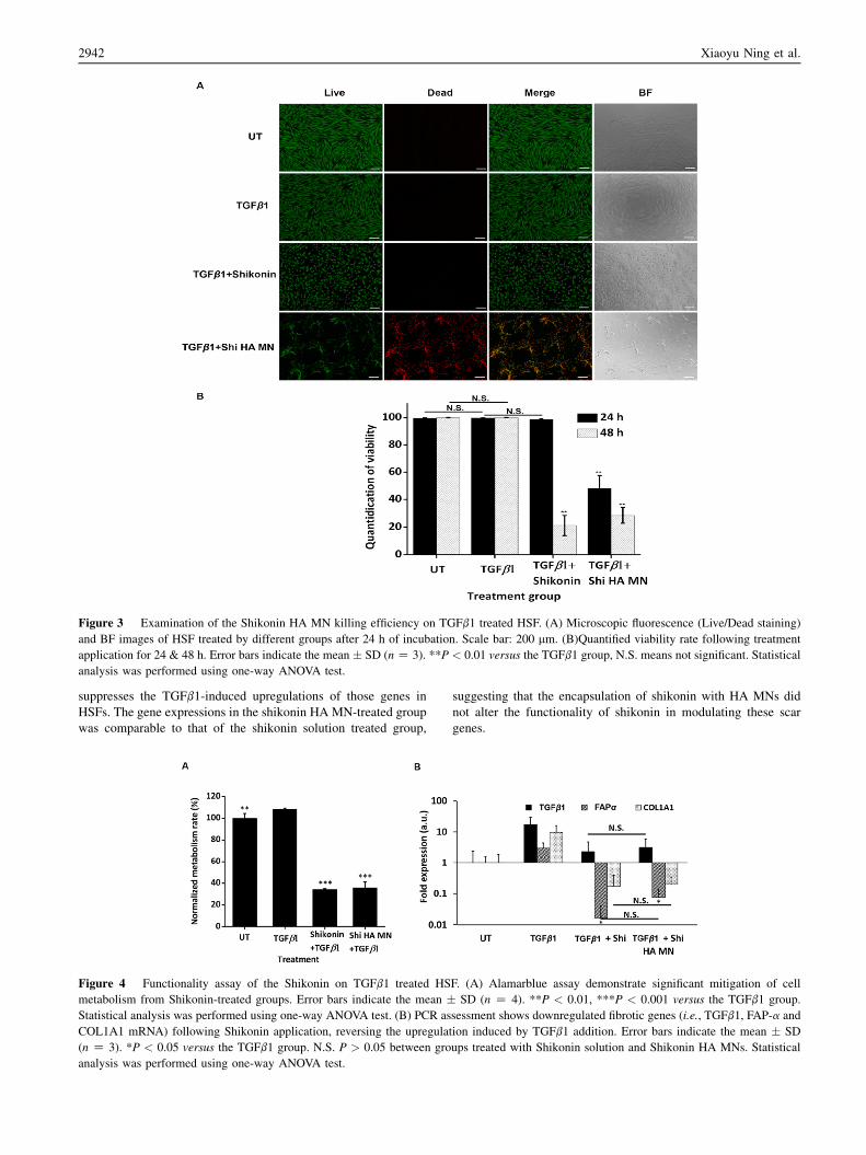
Figure 3 Examination of the Shikonin HA MN killing efficiency on TGFb1 treated HSF. (A) Microscopic fluorescence (Live/Dead staining)
and BF images of HSF treated by different groups after 24 h of incubation. Scale bar: 200 mm. (B)Quantified viability rate following treatment
application for 24 & 48 h. Error bars indicate the mean � SD (nZ 3). **P < 0.01 versus the TGFb1 group, N.S. means not significant. Statistical
analysis was performed using one-way ANOVA test.
2942 Xiaoyu Ning et al.
suppresses the TGFb1-induced upregulations of those genes inHSFs. The gene expressions in the shikonin HA MN-treated groupwas comparable to that of the shikonin solution treated group,
Figure 4 Functionality assay of the Shikonin on TGFb1 treated HS
metabolism from Shikonin-treated groups. Error bars indicate the mean �Statistical analysis was performed using one-way ANOVA test. (B) PCR as
COL1A1 mRNA) following Shikonin application, reversing the upregula
(n Z 3). *P < 0.05 versus the TGFb1 group. N.S. P > 0.05 between gro
analysis was performed using one-way ANOVA test.
suggesting that the encapsulation of shikonin with HA MNs didnot alter the functionality of shikonin in modulating these scargenes.
F. (A) Alamarblue assay demonstrate significant mitigation of cell
SD (n Z 4). **P < 0.01, ***P < 0.001 versus the TGFb1 group.
sessment shows downregulated fibrotic genes (i.e., TGFb1, FAP-a and
tion induced by TGFb1 addition. Error bars indicate the mean � SD
ups treated with Shikonin solution and Shikonin HA MNs. Statistical

Figure 5 Examination of the localized killing effect of Shikonin HA MN in 3D HSF culture. (A) Confocal fluorescence (live/dead staining;
top) and 2D BF images (bottom) of 3D HSF agarose following Shikonin HA MN treatment. White dashed lines indicate MN tips area. Scale bar:
200 mm. (B) Quantification of the viability rate of 3D HSF culture in (A). Error bars indicate the mean � SD (n Z 3). ***P < 0.001. Statistical
analysis was performed using one-way ANOVA test.
Microneedle assisted delivery of Chinese herbal medicine 2943
To further clarify the localized efficacy that the MNs endow toshikonin, a 3D cell culture model was established by incorporatingHSFs in 1.4% agarose followed by the treatment of shikonin HAMNs. The 3D live/dead staining images show a significantlyhigher dead signal in area in vicinity of MN as compared to theedge area (Fig. 5A). Further quantification of the fluorescencesignals show a significantly lower viability of HSFs in MN tipsarea than that of the edge area (w50%; Fig. 5B). This data isconsistent with our 2D culture tests and confirm the localizedkilling effect of shikonin HA MN.
4. Conclusions
This study aimed to incorporate shikonin into dissolvable HA MNto aid its intradermal delivery for scar treatment. The HA MNshowed negligible toxicity to HSFs and different amount of shi-konin could be loaded into HA MNs by simply altering the shi-konin concentration in the mixture during fabrication. Themechanical performance and surface morphology of the MNswere minimally affected by the addition of shikonin, and thefabricated shikonin HA MNs can withstand a force higher than0.7 N/needle without fracture and successfully puncture theporcine skin. The fabricated shikonin HA MN can modulate theviability, and metabolism rate of TGFb1-treated HSFs. Moreover,a localized treatment effect was observed from the shikonin HAMN which is beneficial for the site-specific scar treatment inclinic. In addition, the expression of scar-related genes (TGFb1,FAP-a and COL1A1) were also attenuated by the treatment ofshikonin HA MNs. Taken together, shikonin HA MN possessesgreat potential for topical scar treatment.
Acknowledgments
Chenjie Xu acknowledges the funding support from SingaporeAgency for Science, Technology and Research (A)STAR) Sci-ence and Engineering Research Council Additive Manufacturingfor Biological Materials (AMBM) program (A18A8b0059,Singapore), City University of Hong Kong (#9610472, China),General Research Fund (GRF) from University Grant Committeeof Hong Kong (UGC) Research Grant Council (RGC) (#9042951,China), and NSFC/RGC Joint Research Scheme (N_CityU118/20,China).
Author contributions
Chenjie Xu and Xiaoyu Ning designed the research. Xiaoyu Ningcarried out the experiments and performed data analysis. ChristianWiraja and Sharon Chew Wan Ting participated part of the ex-periments. Xiaoyu Ning wrote the manuscript. Fan Chen andChristian Wiraja gave comments on the manuscript. Chenjie Xurevised the manuscript. All of the authors have read and approvedthe final manuscript.
Conflicts of interest
The authors have no conflicts of interest to declare.
Appendix A. Supporting information
Supporting data to this article can be found online at https://doi.org/10.1016/j.apsb.2021.03.016.
References
1. Rabello FB, Souza CD, Farina Junior JA. Update on hypertrophic scar
treatment. Clinics 2014;69:565e73.
2. Juckett G, Hartman-Adams H. Management of keloids and hypertro-
phic scars. Am Fam Physician 2009;80:253e60.
3. Niessen FB, Spauwen PH, Schalkwijk J, Kon M. On the nature of
hypertrophic scars and keloids: a review. Plast Reconstr Surg 1999;
104:1435e58.4. Hou Q, He WJ, Hao HJ, Han QW, Chen L, Dong L, et al. The four-
herb Chinese medicine ANBP enhances wound healing and inhibits
scar formation via bidirectional regulation of transformation growth
factor pathway. PLoS One 2014;9:e112274.
5. Ye Q, Wang SJ, Chen JY, Rahman K, Xin HL, Zhang H. Medicinal
plants for the treatment of hypertrophic scars. Evid Based Complement
Alternat Med 2015;2015:101340.
6. Wu Y, Yan G, Cai B. Advances in studies on nano-technology applied
in Chinese Materia Medica. Chin Tradit Herb Drugs 2011;42:403e8.
7. Cao FH, OuYang WQ, Wang YP, Yue PF, Li SP. A combination of a
microemulsion and a phospholipid complex for topical delivery of
oxymatrine. Arch Pharm Res (Seoul) 2011;34:551.
8. Waghule T, Singhvi G, Dubey SK, Pandey MM, Gupta G, Singh M,
et al. Microneedles: a smart approach and increasing potential for
transdermal drug delivery system. Biomed Pharmacother 2019;109:
1249e58.
2944 Xiaoyu Ning et al.
9. Ning X, Wiraja C, Lio DCS, Xu C. A Double-layered microneedle
platform fabricated through frozen spray-coating. Adv Healthc Mater
2020;9:2000147.
10. Zheng M, Wiraja C, Yeo DC, Chang H, Lio DCS, Shi W, et al.
Oligonucleotide molecular sprinkler for intracellular detection and
spontaneous regulation of mrna for theranostics of scar fibroblasts.
Small 2018;14:1802546.
11. Xie Y, Fan C, Dong Y, Lynam E, Leavesley DI, Li K, et al. Functional
and mechanistic investigation of Shikonin in scarring. Chem Biol
Interact 2015;228:18e27.
12. Albreht A, Vovk I, Simonovska B. Addition of beta-lactoglobulin pro-
duces water-soluble shikonin. J Agric Food Chem 2012;60:10834e43.
13. Gupta RC, Lall R, Srivastava A, Sinha A. Hyaluronic acid: molecular
mechanisms and therapeutic trajectory. Front Vet Sci 2019;6:192.
14. Fan C, Xie Y, Dong Y, Su Y, Upton Z. Investigating the potential of
Shikonin as a novel hypertrophic scar treatment. J Biomed Sci 2015;
22:70.
15. FanC,DongY,XieY, SuY,ZhangX,LeavesleyD, et al. Shikonin reduces
TGF-beta 1-induced collagen production and contraction in hypertrophic
scar-derived human skin fibroblasts. Int J Mol Med 2015;36:985e91.
16. Yeo D, Wiraja C, Miao Q, Ning X, Pu K, Xu C. Anti-scarring drug
screening with near-infrared molecular probes targeting fibroblast
activation protein-a. ACS Appl Bio Mater 2018;1:2054e61.17. Dabiri G, Tumbarello DA, Turner CE, Van de Water L. Hic-5
promotes the hypertrophic scar myofibroblast phenotype by
regulating the TGF-beta 1 autocrine loop. J Invest Dermatol 2008;
128:2518e25.
18. Deng X, Chen Q, Qiang L, Chi M, Xie N, Wu Y, et al. Development of
a porcine full-thickness burn hypertrophic scar model and investiga-
tion of the effects of shikonin on hypertrophic scar remediation. Front
Pharmacol 2018;9:590.
19. Lee C, Yang H, Kim S, Kim M, Kang H, Kim N, et al. Evaluation of
the anti-wrinkle effect of an ascorbic acid-loaded dissolving micro-
needle patch via a double-blind, placebo-controlled clinical study. Int
J Cosmet Sci 2016;38:375e81.
20. de Moraes Porto I. Polymer biocompatibility, polymerization. London:
IntechOpen Ltd.; 2012.
21. Penn JW, Grobbelaar AO, Rolfe KJ. The role of the TGF-b family in
wound healing, burns and scarring: a review. Int J Burns Trauma 2012;
2:18e28.
22. Dienus K, Bayat A, Gilmore BF, Seifert O. Increased expression of
fibroblast activation protein-alpha in keloid fibroblasts: implications
for development of a novel treatment option. Arch Dermatol Res 2010;
302:725e31.23. Bi S, Chai L, Yuan X, Cao C, Li S. MicroRNA-98 inhibits the cell
proliferation of human hypertrophic scar fibroblasts via targeting
Col1A1. Biol Res 2017;50:22.
24. Fan C, El Andaloussi S, Lehto T, Kong KW, Seow Y. Smad-binding
decoy reduces extracellular matrix expression in human hypertrophic
scar fibroblasts. Mol Med Rep 2020;6:4589e600.