Transdermal Delivery of Ketoprofen: The Influence of Drug–Dioleylphosphatidylcholine Interactions
10
Research Paper Transdermal Delivery of Ketoprofen: The Influence of Drug–Dioleylphosphatidylcholine Interactions Maria Teresa Junqueira Garcia, 1 Carlos Henrique Tomich de Paula da Silva, 1 Dione ´ ia C. R. de Oliveira, 1 Eliane Candiane Arantes Braga, 1 Jose ´ Anto ˆ nio Thomazini, 2 and Maria Vito ´ ria Lopes Badra Bentley 1,3 Received November 6, 2005; accepted April 18, 2006 Purpose. Considering that most inflammatory diseases occur locally and near the body surface, transdermal delivery of non-steroidal anti-inflammatory drugs (NSAIDs) may be an interesting strategy for delivering these drugs directly to the diseased site. To optimize ketoprofen (KP) transdermal delivery we investigated the influence of dioleylphosphatidylcholine (DOPC) on skin permeation. Materials and Methods. The formulations studied were: i) a physical mixture of KP and DOPC and ii) DOPC and KP complex, in a molar ratio of 1:3, obtained by dissolution of the components in chloroform followed by drying under a N 2 atmosphere. Both systems were dispersed in mineral oil and the in vitro percutaneous was assayed by absorption using a flow through diffusion cell. Differential Scanning Calorimetry (DSC) and 1 H NMR studies were carried out to characterize KP and DOPC interactions. Geometry optimizations using Density Functional Theory and semiempirical methods, as well as a flexible docking procedure were carried out to obtain a binding model for KP with DOPC. KP solubility and partition studies in the formulations, as well as skin irritation and hypersensitivity assays were also carried out. Results. DSC determinations in the complex showed enthalpy and temperature depressions, indicating KP and DOPC interaction. In addition, dipoleYdipole interactions between the KP carboxylic acid and OH groups in phospholipids were shown by 1 H NMR studies. Based on the NMR studies, a KPYDOPC binding model is proposed, in which KP is involved by the two long aliphatic chains of the phospholipid. Solubility studies indicated that DOPC improved drug solubility. KP permeation was enhanced by both formulations tested, but the complex also increased its skin uptake. Such behavior could be attributed to the solubilizing, melting and enhancing effects of DOPC. Skin irritation and hypersensitivity were not significantly changed compared to control, suggesting that the formulation may be therapeutically explored for KP transdermal delivery. KEY WORDS: transdermal delivery; dioleylphosphatidylcholine; drug complex; ketoprofen; skin permeation; skin toxicity. INTRODUCTION Drug delivery through the transdermal route is limited by low skin permeability. The stratum corneum, the outer- most layer of the skin, acts as a major barrier and is often rate limiting. Considerable research work has been focused on discovering methods to increase stratum corneum permeabil- ity. One approach was to employ chemical penetration enhancers, which may increase the permeability of stratum corneum (SC) by increasing drug diffusivity within the mem- brane and/or by increasing drug partition from the applied formulation into the skin and/or by increasing the effective concentration of drug in the vehicle (1). The effect of these compounds on the drug cutaneous absorption profile has been quantified by permeation parameters, such as drug flux across the skin and skin drug uptake. Phospholipids have received attention as penetration enhancers (2Y9). This class of enhancers has the advantage of containing endogenous components of the human skin, representing 50% of basal layer and 25% of granulosum stratum lipids (10). Phospholipids molecules differ in several features such as polar group contents (choline, serine, glycerol), non-polar chain lengths, characteristic double bonds (in position, number and/or configuration) (11), and these structural variations can influence their effects as skin enhancers (4Y9). For example, some phospholipids are capable of insertion among the hydrophobic tails of SC lipids bilayer, decreasing its diffusion resistance to permeants by disturbing packing and increasing fluidity. In addition, some phospholipids are capable to form complexes with drugs, enhancing their thermodynamic activity in the formulation and increasing skin permeation (2,3,12). 0724-8741/06/0800-1776/0 # 2006 Springer Science + Business Media, Inc. 1776 Pharmaceutical Research, Vol. 23, No. 8, August 2006 ( # 2006) DOI: 10.1007/s11095-006-9040-3 1 Faculdade de Cie ˆ ncias Farmace ˆ uticas de Ribeira ˜ o Preto, Universi- dade de Sa ˜o Paulo, Av. do Cafe ´ s/n, 14040-903, Ribeira ˜ o Preto, Sa ˜o Paulo, Brazil. 2 Departamento de Anatomia da Faculdade de Medicina de Ribeira ˜o Preto, Universidade de Sa ˜ o Paulo, Ribeira ˜o Preto, Sa ˜ o Paulo, Brazil. 3 To whom correspondence should be addressed. (e-mail: vbentley@ usp.br)
Transcript of Transdermal Delivery of Ketoprofen: The Influence of Drug–Dioleylphosphatidylcholine Interactions

Research Paper
Transdermal Delivery of Ketoprofen: The Influenceof Drug–Dioleylphosphatidylcholine Interactions
Maria Teresa Junqueira Garcia,1 Carlos Henrique Tomich de Paula da Silva,1 Dioneia C. R. de Oliveira,1
Eliane Candiane Arantes Braga,1 Jose Antonio Thomazini,2 and Maria Vitoria Lopes Badra Bentley1,3
Received November 6, 2005; accepted April 18, 2006
Purpose. Considering that most inflammatory diseases occur locally and near the body surface,
transdermal delivery of non-steroidal anti-inflammatory drugs (NSAIDs) may be an interesting strategy
for delivering these drugs directly to the diseased site. To optimize ketoprofen (KP) transdermal
delivery we investigated the influence of dioleylphosphatidylcholine (DOPC) on skin permeation.
Materials and Methods. The formulations studied were: i) a physical mixture of KP and DOPC and ii)
DOPC and KP complex, in a molar ratio of 1:3, obtained by dissolution of the components in chloroform
followed by drying under a N2 atmosphere. Both systems were dispersed in mineral oil and the in vitro
percutaneous was assayed by absorption using a flow through diffusion cell. Differential Scanning
Calorimetry (DSC) and 1H NMR studies were carried out to characterize KP and DOPC interactions.
Geometry optimizations using Density Functional Theory and semiempirical methods, as well as a
flexible docking procedure were carried out to obtain a binding model for KP with DOPC. KP solubility
and partition studies in the formulations, as well as skin irritation and hypersensitivity assays were also
carried out.
Results. DSC determinations in the complex showed enthalpy and temperature depressions, indicating
KP and DOPC interaction. In addition, dipoleYdipole interactions between the KP carboxylic acid and
OH groups in phospholipids were shown by 1H NMR studies. Based on the NMR studies, a KPYDOPC
binding model is proposed, in which KP is involved by the two long aliphatic chains of the phospholipid.
Solubility studies indicated that DOPC improved drug solubility. KP permeation was enhanced by both
formulations tested, but the complex also increased its skin uptake. Such behavior could be attributed to
the solubilizing, melting and enhancing effects of DOPC. Skin irritation and hypersensitivity were not
significantly changed compared to control, suggesting that the formulation may be therapeutically
explored for KP transdermal delivery.
KEY WORDS: transdermal delivery; dioleylphosphatidylcholine; drug complex; ketoprofen; skinpermeation; skin toxicity.
INTRODUCTION
Drug delivery through the transdermal route is limitedby low skin permeability. The stratum corneum, the outer-most layer of the skin, acts as a major barrier and is often ratelimiting. Considerable research work has been focused ondiscovering methods to increase stratum corneum permeabil-ity. One approach was to employ chemical penetrationenhancers, which may increase the permeability of stratumcorneum (SC) by increasing drug diffusivity within the mem-brane and/or by increasing drug partition from the appliedformulation into the skin and/or by increasing the effective
concentration of drug in the vehicle (1). The effect of thesecompounds on the drug cutaneous absorption profile hasbeen quantified by permeation parameters, such as drug fluxacross the skin and skin drug uptake.
Phospholipids have received attention as penetrationenhancers (2Y9). This class of enhancers has the advantage ofcontaining endogenous components of the human skin,representing 50% of basal layer and 25% of granulosumstratum lipids (10). Phospholipids molecules differ in severalfeatures such as polar group contents (choline, serine,glycerol), non-polar chain lengths, characteristic doublebonds (in position, number and/or configuration) (11), andthese structural variations can influence their effects as skinenhancers (4Y9). For example, some phospholipids arecapable of insertion among the hydrophobic tails of SC lipidsbilayer, decreasing its diffusion resistance to permeants bydisturbing packing and increasing fluidity. In addition, somephospholipids are capable to form complexes with drugs,enhancing their thermodynamic activity in the formulationand increasing skin permeation (2,3,12).
0724-8741/06/0800-1776/0 # 2006 Springer Science + Business Media, Inc. 1776
Pharmaceutical Research, Vol. 23, No. 8, August 2006 (# 2006)DOI: 10.1007/s11095-006-9040-3
1 Faculdade de Ciencias Farmaceuticas de Ribeirao Preto, Universi-
dade de Sao Paulo, Av. do Cafe s/n, 14040-903, Ribeirao Preto, Sao
Paulo, Brazil.2 Departamento de Anatomia da Faculdade de Medicina de Ribeirao
Preto, Universidade de Sao Paulo, Ribeirao Preto, Sao Paulo,
Brazil.3 To whom correspondence should be addressed. (e-mail: vbentley@
usp.br)
Ketoprofen (KP), a potent non-steroidal anti-inflamma-tory drug (NSAID) inhibits arachidonic acid metabolism bycyclo-oxigenase and lipoxygenase. Also, it appears to stabilizelysosomal membranes and antagonize the action of bradyki-nin. The potent inhibitory action on human lipoxigenasedistinguishes KP from most other NSAIDs. The compoundhas been widely used in the treatment of rheumatoid arthritis,osteoarthritis, as well as a mild to moderate painkiller.However, its use by oral administration has been associatedwith a number of gastrointestinal disorders (13).
Considering that most inflammatory diseases occurlocally and near the body surface, transdermal delivery ofNSAID may turn out to be an interesting strategy to deliverthese drugs directly to the diseased site and to increase localconcentration (14,15). However, poor skin penetration limitsthe efficacy of topically applied KP. Methods trying toimprove its cutaneous delivery rely either on chemicalpenetration enhancers (16,17), novel vehicle systems (18,19),or more complex physical enhancement strategies (20).
The purpose of this study was to investigate the effect ofphospholipids, used as penetration enhancers or componentsof lipid/drug complexes, on in vitro percutaneous absorptionof KP. The potential of KP transdermal delivery was alsoassessed by toxicity studies.
MATERIALS AND METHODS
Materials
Dioleylphosphatidylcholine (DOPC) and ketoprofen(KP) were from Sigma (St. Louis, USA). All other chemicalswere of analytical or HPLC reagent grade and purchasedfrom Merck (Darmstadt, Germany).
Methods
HPLC Assay for KP
Sample analysis was performed by high pressure liquidchromatography (HPLC) as previously described (21,22).The HPLC system consisted of a model SPD 10A VPShimadzu Liquid Chromatograph (Kyoto, Japan), fitted witha variable wavelength UV detector operating at 251 nm, aLC-10 ADVP pump, a Rheodyne injector and a Model CR6-A integrator. Separations were performed in a C18 reverse-phase column LichroCART\ (Merck, Darmstadt, Germany)(125 � 4 mm i.d.) (5 mm) and a C18 pre-column (4 � 4 mmi.d.) (5 mm) kept at room temperature. Acetonitrile: 0.02 Mphosphate buffer (pH 3.0) (45:55) mixture was used as themobile phase, at a flow rate of 0.8 ml/min. The injectedvolume was 20 ml and KP retention time 6.103 min. The assaywas linear over the range of 0.0125Y200.0 mg/ml (r = 0.999)with an injection variability of <4.2% for inter-days variation,and <3.2% for intra-day variation.
Formulations
The DOPC and KP complex was obtained by adding thecomponents (molar ratio, 1:3) to chloroform followed by
drying under N2 atmosphere. The dried residue was dispersedin mineral oil. The physical mixture of DOPC and KP (molarratio, 1:3) was also dispersed in mineral oil. Both dispersioncontained KP at 2% (w/w). A formulation containing 2% w/w KP in mineral oil was used as a control.
Solubility Studies
KP solubility, both in the physical mixture and in thecomplex, was investigated by adding an excess of each tomineral oil and submitting the suspension to constantstirring (300 rpm) at room temperature for 24 h. Thedispersion obtained was then centrifuged at 450 � g for 10min. A 1 ml aliquot of the supernatant and 3 ml of methanolwere mixed in a vortex for 6 min and centrifuged at 450 g for10 min. KP present in the methanol phase was assayed byHPLC.
Stratum Corneum/Formulation PartitionCoefficients (KSC/formulation)
Porcine skins were floated (dermal side facing solution)for 12 h in a 0.1% trypsin solution in phosphate buffer (pH7.2) at 37-C. The transparent SC sheets obtained were brieflywashed in water and dried in desiccators containing silica gelfor use in partition studies.
The KP partition coefficient from vehicle into powderedSC was determined following the method of Zhao and Sing(22). Briefly, 10 mg of dried SC pulverized in a mortar weremixed with 1 ml of formulation and vortexed for 6 min,ensuring the partition equilibrium. After centrifugation for10 min at 450 � g, 0.5 ml of the supernatant was added to 1ml of methanol, the mixture vortexed for 6 min andcentrifuged at 450 g for 10 min. KP present in the methanolphase was determined by HPLC. KSC/formulation was calculat-ed as (Co j C)/C, where Co and C are KP concentrationsprior and after partition, respectively.
Characterization of the Complex
DOPC:KP (1:3 molar ratio) complexes were analyzedby Differential Scanning Calorimetry (DSC-200, NETZSCHGeratebau GmBH, Selb, Germany) and by Nuclear Mag-netic Resonance (NMR) Spectroscopy (BRUKER DPX,Billerica, MA, USA). For DSC analysis, the samples (KP,DOPC and DOPC:KP complex) were sealed in an alumi-num pan and scanned between j50 and 260-C at a heatingrate of 5-C/min under N2 atmosphere. Temperature cali-bration was made using indium as standard. For NMRanalysis, samples (KP, DOPC, DOPC:KP complex andphysical mixture) were dissolved in deuterated chloroform(CDCl3) containing a small amount of tetramethysilane(TMS) as internal standard, and the spectra obtained at400 MHz. The conditions for Fourier Transform measure-ments were: acquisition time, 3 s; pulse angle, 30-; delaytime, 2 s, number of spectra, 32. In addition, NuclearOverhouse Enhancement (NOE) experiments were carriedout on the complex to get spatial information on KP andDOPC groups and to propose a corresponding molecularmodel. Thus, some hydrogens of DOPC were selected andirradiated.
1777Transdermal Delivery of Ketoprofen: The Influence of Drug–DOPC
Molecular Modeling Studies
We have used GOLD 3.0 (23), Insight II (24) andSPARTAN 4.0.3 (25) softwares to calculate structures and tosuggest a binding model for the KPYDOPC complex. GOLDuses genetic algorithm to perform flexible docking in ligands,and each docking result is slightly different from the other.Flexible docking was parameterized for zero ligand bumps, apopulation size of 100, five islands, 100,000 operations, 95mutations, and 95 crossovers, adjusted for ten dockings.When the superpositions of the top three solutions (ligandorientations) are within 1.5 A mean square root deviation(RMSD), the calculation is finished. The fitness function(GoldScore) is evaluated in six stages: (a) a conformation ofthe ligand inside the receptor binding site is generated; (b)the ligand is placed within the binding site using a leastsquare fitting procedure; (c) a hydrogen bonding energy isobtained for the complex; (d) as well as a steric energy ofinteraction between the receptor and the ligand; (e) molec-ular mechanics expressions are used to generate a measure ofthe ligand internal energy; and (f) the energy terms aresummed to give a final fitness.
The docking parameters used have been optimized forsingle docking calculations, and the program fully validatedagainst 221 diverse and extensively checked proteinYligandcomplexes with high resolution registered in the PDB (23). Inorder to consider the DOPC large flexibility, this moleculewas used as the ligand in the docking procedure. KP, thereceptor had a 20 A radius sphere centered in the middlecarbonyl of the molecule. Docking calculations were per-formed inside the sphere. Natural Bond Order (NBO) partialatomic charges were obtained for KP from the densityfunctional geometry optimization (at B3LYP/6Y31G* levelof calculation) and used in the docking procedure. Atomiccharges for DOPC were obtained using the AM1 Hamilto-nian. Previous to this last calculation, a conformationalsearch on DOPC was performed using the MONTE CARLOmethod with the Merck Pharmaceuticals force field (25).NMR experiments with the DOPCYKP complex suggestedthe hydrogen bonding and distance restraints employed inthe docking procedure. The hydrogen bond between thephosphate group hydroxyl in choline and the KP carboxylgroup as well as the distance between the following atompairs of KP and DOPC, i.e. C1 and N3, C2 and N3, C4 andN3 have been considered. After docking calculations, thetop-ranked GOLD solution for the complex was optimizedfor implicit solvation conditions, using chloroform as solvent(dielectric constant of 4.8). Initial restrained optimizationswere performed for the complex using 1,000 steps of SteepestDescent followed by 5,000 steps of Conjugate Gradient asminimization algorithms. Restraints were released step bystep until the convergence of the calculation. An interval of3Y6 A has been used for distance restraints, in order to satisfythe NOEs observed.
In Vitro Permeation and Retention Studies
Porcine ears obtained from a local slaughterhouse hadthe dorsal skin removed from the underlying cartilage with ascalpel. Skins were dermatomized to a thickness of approx-imately 500 mm and stored wrapped in aluminum foil at
j20-C for a maximum of 4 weeks before use, as alreadydescribed (26).
Flow-through diffusion cells with a diffusion area of1 cm2 were used in the in vitro percutaneous absorptionstudies. The skin was mounted between the donor andreceptor compartments the SC facing the donorcompartment and dermis the receptor compartment. Thereceiving solution (phosphate buffer, pH 7.2) was pumpedthrough the diffusion cell at a flow rate of 3.0 ml/h; a magneticbar at 300 rpm stirred the contents of the receivercompartment. The whole cell system was maintained at37-C with a circulating water bath. Samples were collectedwith a fraction collector at 1 h intervals and the amount of KPpermeated was assayed by HPLC. At the end of permeationstudies, the skin was removed from the diffusion cell and thesurface gently cleaned to eliminate formulation residue. Theimprints of the diffusion cell flanges clearly marked theexposed diffusion area. The skin was dabbed dry with cotton,and submitted to the tape stripping method in order to removeSC from the remaining skin, i.e. epidermis (E) without SC +dermis (D). The skins were fixed on a flat surface and thediffusion area of SC was removed by ten pieces of adhesivetape (Scotch Book Tape, n. 845, 3M, St. Paul, MN, USA). KPcontained in the strips was extracted with 3 ml of methanol,left overnight and shaken for 1 min before filtering. After thetape stripping procedure, the remaining tissue (E without SC +D) was cut with scissors and placed in a tube. The KP was,then, extracted with 5 ml of methanol under agitation for 6 minin a vortex, sonicated in an ultra-sound bath (40 KHz,continuous mode) for 20 min at 25-C and filtered through a0.45 mm polytetrafluoroethylene (PTFE) membrane (CorningIncorporated, Corning, NY). The filtrates were then analyzedby HPLC.
The cumulative amount of KP permeated through theskin was plotted as a function of time. The flux was cal-culated as the slope of the linear portion of the plot (mg/cm2/h).The KP skin retention (mg/cm2) was plotted as a function offormulation.
Animals
The skin toxicity studies were carried out in femalehairless mice, 5Y6 weeks old (strain HRS/J Jackson Labora-tories, Bar Harbor, ME) and female Balb/c mice (5Y6 weeksold). The animals were housed at 24Y26-C, exposed to a daily12:12 h light: dark cycle (lights on at 6 A.M.), and had freeaccess to standard mice chow and tap water. To reduce thestress associated with the experimental procedure, mice werehandled daily for 1 week before experimentation. Theprotocols were in accordance with guidelines of the Univer-sity of Sao Paulo Animal Care and Use Committee.
Skin Irritation Test
The skin irritation test was based on a methodologydescribed by Sintov et al. (27). The animals were divided intofour groups containing three animals each. About 100 ml ofeach formulation (saline, KP and DOPC:KP complex inmineral oil) were applied non-occlusively over the dorsum ofeach animal for two consecutive days. On the third day, theanimals were euthanized by carbon dioxide vapor and
1778 Junqueira Garcia et al.
samples of the applied skin area were excised and fixed forhistological studies by immersion in Bouin solution for 24 h.
After removal of picric acid with alcohol 70-GL, thesamples were processed according to the histological tech-nique of inclusion in paraffin and observed by light micros-copy. Each tissue sample was cut in 6 mm sections in amicrotome. Sections were stained with Masson tricromic andhematoxylin and eosin for histological examination using a 20fold augmentation objective.
Hypersensitivity Response Test
KP hypersensitivity responses were determined in Balb/cmice using the ear swelling test (MEST) (28), with slightmodifications. Mice were divided into three groups contain-ing six animals each. A selected area on the back of the earwas treated with 100 ml of test formulation for threeconsecutive days. 2,4-dinitrochlorobenzene (DNCB) wasused as positive control and saline as negative control. Onthe eighth day, the ear thicknesses were measured using anengineering micrometer. The animals were then challengedon both sides of each ear with 50 ml of the respectiveformulation or control substance. The post challenge earthicknesses were measured 24 h after treatment to determinethe percent swelling at 24 h. The percentage of ear swellingwas calculated as the ([mean 24 h post treatment earthickness/mean pretreatment ear thickness] � 100)j100.
Statistical Analysis
The results are reported as means T S.E.M. Data weresubjected to one-way analysis of variance (ANOVA) fol-lowed by the Kruskal<Wallis test or to t-test analysis followedby the Mann Whitney test. The level of significance was set atp < 0.05.
RESULTS AND DISCUSSION
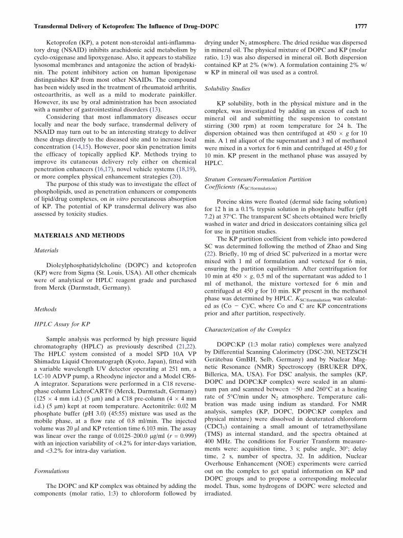
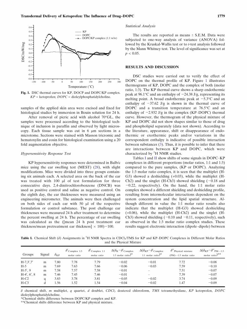
DSC studies were carried out to verify the effect ofDOPC on the thermal profile of KP. Figure 1 illustratesthermograms of KP, DOPC and the complex of both (molarratio, 1:3). The KP thermal curve shows a sharp endothermicpeak at 96.1-C and an enthalpy of j24.38 J/g, representing itsmelting point. A broad endothermic peak at j5.3-C and anenthalpy of j37.62 J/g is shown in the thermal curve ofDOPC and a transition temperature at 76.5-C and anenthalpy of j2.932 J/g in the complex (KPYDOPC) thermalcurve. However, the thermogram of the physical mixture ofKP and DOPC did not show shapes similar to those of drugand phospholipid separately (data not shown). According tothe literature, appearance, shift or disappearance of endo-thermic or exothermic peaks and/or variations in thecorrespondent enthalpy is indicative of possible interactionbetween substances (3). Thus, it is possible to infer that thereare interactions between KP and DOPC, which werecharacterized by 1H NMR studies.
Tables I and II show shifts of some signals in DOPCYKPcomplexes in different proportions (molar ratios, 1:1 and 1:3)compared to the pure samples, (KP or DOPC). Analyzingthe 1:3 molar ratio complex, it is seen that the multiplet (H-G3) showed a deshielding (+0.03), while the multiplet (H-Ch2) and the singlet (H-Ch3) showed shielding (j0.18 andj0.22, respectively). On the hand, the 1:1 molar ratiocomplex showed a different shielding and deshielding profile,resulting from intermolecular interactions dependent on thesystem concentration and the lipid spatial structure. Al-though different in value the 1:1 molar ratio results alsoindicate that the multiplet (H-G3) showed deshielding(+0.06), while the multiplet (H-Ch2) and the singlet (H-Ch3) showed shielding (j0.10 and j0.11, respectively), suchas observed in the 1:3 molar ratio complex studies. Theseresults suggest electronic interaction (dipoleYdipole) between
DOPC
-150 -100 -50 0 50 100 150 200 250 300-0.6 -0.5 -0.4 -0.3 -0.2 -0.1 0.0 0.1 0.2 0.3 0.4 0.5 0.6
KP
DOPC/KP complex (1:1 w/w)
DSC
/mW
/mg
Temperature (˚C)
Fig. 1. DSC thermal curves for KP, DOCP and DOPC/KP complex.
KP = ketoprofen; DOPC = dioleylphosphatidylcholine.
Table I. Chemical Shift (d) Assignments in 1H NMR Spectra in CDCl3/TMS for KP and KPYDOPC Complexes in Different Molar Ratios
and the Physical Mixture
Groups Signal dKP
d0Complex 1:3
molar ratio
d0Complex 1:1
molar ratio
D(dKjd0Complex
1:3 molar ratio)*
D(dKPjd0Complex
1:1 molar ratio)*
d0 0Physical mixture
(PM)V1:3 molar ratio
D(dKPjd0 0PMV1:3
molar ratio)**
H-7,30,70 m 7.80 7.78 7.79 j0.02 j0.01 7.72 j0.08
H-5 m 7.69 7.63 7.66 j0.06 j0.03 7.59 j0.10
H-50, 9 m 7.58 7.57 7.58 j0.01 Y 7.51 j0.07
H-40, 60, 8 m 7.46 7.45 7.46 j0.01 Y 7.39 j0.07
H-C2 q 3.83 3.78 3.81 j0.05 j0.02 3.74 j0.09
H-C3 d 1.56 1.52 1.54 j0.04 j0.02 1.47 j0.09
d chemical shift, m multiplet, q quartet, d doublet, CDCl3 deutered chloroform, TMS tetramethysilane, KP ketoprofen, DOPC
dioleylphosphatidylcholine.*Chemical shifts difference between DOPC/KP complex and KP.**Chemical shifts difference between KP and physical mixture.
1779Transdermal Delivery of Ketoprofen: The Influence of Drug–DOPC

DOPC and KP in both systems (1:1 and 1:3, molar ratio),where choline group hydrogens (H-Ch2 e H-Ch3) areshielding because the group is located near the center ofthe aromatic ring, and the glycerol group hydrogens (H-G3)
are deshielding due to hydrogen bonding between thehydroxyl and carboxylic groups of DOPC and KP, respec-tively. In addition, the spectra of both complex and physicalmixture (1:3 molar ratio of DOPC to KP) are different
Table II. Chemical Shift (d) Assignments in 1H NMR Spectra in CDCl3/TMS for DOPC and KPYDOPC Complexes in Different Molar
Ratios and the Physical Mixture
Groups Signal dDOPC
d0Complex 1:3
molar ratio
d0Complex 1:1
molar ratio
D(dDOPCjd0Complex
1:3 molar ratio)*
D(dDOPCjd0Complex
1:1 molar ratio)*
d0 0Physical mixture
(PM)V1:3 molar ratio
D(dDOPCjd0 0PMV1:3
molar ratio)**
H-AG 9 e 10 m 5.33 5.34 5.33 +0.01 Y 5.26 j0.07
H-G2 m 5.19 5.18 5.20 j0.01 +0.01 5.12 j0.07
H-G1a dd 4.38 4.33 4.36 j0.05 j0.02 4.28 j0.10
H-Ch1 m 4.29 4.24 4.30 j0.05 +0.01 4.22 j0.07
H-G1b dd 4.11 4.08 4.12 j0.04 +0.01 4.03 j0.08
H-G3 m 3.92 3.95 3.98 +0.03 +0.06 3.90 j0.02
H-Ch2 m 3.77 3.59 3.67 j0.18 j0.10 3.57 j0.20
H-Ch3 s 3.33 3.12 3.22 j0.22 j0.11 3.11 j0.22
H-AG2 m 2.27 2.26 2.28 j0.10 +0.01 2.20 j0.07
H-AG8 m 2.00 2.00 2.00 Y Y 1.93 j0.08
H-AG3 m 1.57 1.56 1.57 j0.01 Y 1.49 j0.08
H-AG (CH2) wl 1.29 1.27 1.27 j0.02 j0.02 1.19 j0.07
wl 1.26 1.26 1.26 Y Y 1.20 j0.09
H-AG18 t 0.87 0.87 0.87 Y Y 0.80 j0.07
d chemical shift, m multiplet, dd double doublet, s singlet, wl width singlet, t triplet, CDCl3 deutered chloroform, TMS tetramethysilane, KP
ketoprofen, DOPC dioleylphosphatidylcholine.*Chemical shifts difference between DOPC/KP complex and DOPC.**Chemical shifts difference between DOPC and physical mixture
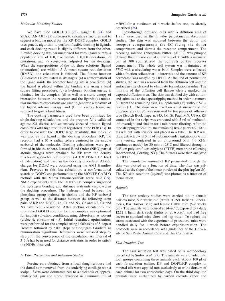
Fig. 2. (A) KP structure fully optimized at B3LYP/6Y31G* level of calculation. (B) DOPC
structure optimized at AM1 level of calculation. No restraints were applied. Labeled atoms
are the hydrogens investigated by NOE. (C) The DOPC and KP complex structure, after
docking procedure and subsequent optimization. Hydrogen bond restraint was applied
between the hydroxyl of the choline phosphate group and the carboxyl group of KP,
whereas distance restraints were applied to the following atom pairs: C1 and N3, C2 and N3,
C4 and N3. Hydrogens were omitted for clarity. (D) The complex structure with a density
surface built for DOPC and showing the KP short area accessible to the solvent. Hydrogens
were omitted for clarity. KP = ketoprofen; DOPC = dioleylphosphatidylcholine.
1780 Junqueira Garcia et al.
(Tables I and II). By comparing the spectra of the physicalmixture and of its components separately it is shown that themultiplets (H-G3 and H-Ch2) and the singlet (H-Ch3) areshielding (j0.02, j0.20 and j0.22, respectively), suggestingthat there is no hydrogen bonding between the DOPCYOHgroup and the carboxylic acid of KP in this system.
The nature of KP and DOPC interactions was elucidatedby molecular modeling studies. 1H NMR and NOE studies ofthe isolated components and the complex were used for thisapproach. Due to the large flexibility of DOPC and thenumber of possibilities for the complex model, theseexperimental biases were important to propose the morereliable model shown in Fig. 2. It suggests a lipophilicenvelope partially shielding KP from the apolar solvent, asobserved by other authors (29) for phospholipidYpolyphenolcomplexes in low polarity solvents.
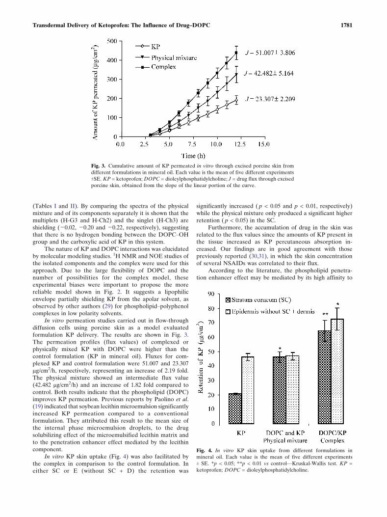
In vitro permeation studies carried out in flow-throughdiffusion cells using porcine skin as a model evaluatedformulation KP delivery. The results are shown in Fig. 3.The permeation profiles (flux values) of complexed orphysically mixed KP with DOPC were higher than thecontrol formulation (KP in mineral oil). Fluxes for com-plexed KP and control formulation were 51.007 and 23.307mg/cm2/h, respectively, representing an increase of 2.19 fold.The physical mixture showed an intermediate flux value(42.482 mg/cm2/h) and an increase of 1.82 fold compared tocontrol. Both results indicate that the phospholipid (DOPC)improves KP permeation. Previous reports by Paolino et al.(19) indicated that soybean lecithin microemulsion significantlyincreased KP permeation compared to a conventionalformulation. They attributed this result to the mean size ofthe internal phase microemulsion droplets, to the drugsolubilizing effect of the microemulsified lecithin matrix andto the penetration enhancer effect mediated by the lecithincomponent.
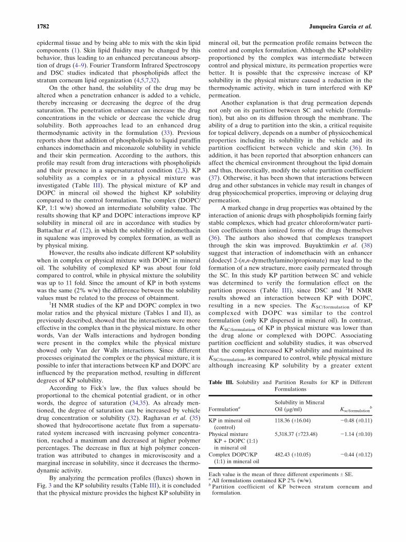
In vitro KP skin uptake (Fig. 4) was also facilitated bythe complex in comparison to the control formulation. Ineither SC or E (without SC + D) the retention was
significantly increased ( p < 0.05 and p < 0.01, respectively)while the physical mixture only produced a significant higherretention ( p < 0.05) in the SC.
Furthermore, the accumulation of drug in the skin wasrelated to the flux values since the amounts of KP present inthe tissue increased as KP percutaneous absorption in-creased. Our findings are in good agreement with thosepreviously reported (30,31), in which the skin concentrationof several NSAIDs was correlated to their flux.
According to the literature, the phospholipid penetra-tion enhancer effect may be mediated by its high affinity to
Fig. 3. Cumulative amount of KP permeated in vitro through excised porcine skin from
different formulations in mineral oil. Each value is the mean of five different experiments
TSE. KP = ketoprofen; DOPC = dioleylphosphatidylcholine; J = drug flux through excised
porcine skin, obtained from the slope of the linear portion of the curve.
Fig. 4. In vitro KP skin uptake from different formulations in
mineral oil. Each value is the mean of five different experiments
T SE. *p < 0.05; **p < 0.01 vs controlVKruskal<Wallis test. KP =
ketoprofen; DOPC = dioleylphosphatidylcholine.
1781Transdermal Delivery of Ketoprofen: The Influence of Drug–DOPC
epidermal tissue and by being able to mix with the skin lipidcomponents (1). Skin lipid fluidity may be changed by thisbehavior, thus leading to an enhanced percutaneous absorp-tion of drugs (4Y9). Fourier Transform Infrared Spectroscopyand DSC studies indicated that phospholipids affect thestratum corneum lipid organization (4,5,7,32).
On the other hand, the solubility of the drug may bealtered when a penetration enhancer is added to a vehicle,thereby increasing or decreasing the degree of the drugsaturation. The penetration enhancer can increase the drugconcentrations in the vehicle or decrease the vehicle drugsolubility. Both approaches lead to an enhanced drugthermodynamic activity in the formulation (33). Previousreports show that addition of phospholipids to liquid paraffinenhances indomethacin and miconazole solubility in vehicleand their skin permeation. According to the authors, thisprofile may result from drug interactions with phospholipidsand their presence in a supersaturated condition (2,3). KPsolubility as a complex or in a physical mixture wasinvestigated (Table III). The physical mixture of KP andDOPC in mineral oil showed the highest KP solubilitycompared to the control formulation. The complex (DOPC/KP, 1:1 w/w) showed an intermediate solubility value. Theresults showing that KP and DOPC interactions improve KPsolubility in mineral oil are in accordance with studies byBattachar et al. (12), in which the solubility of indomethacinin squalene was improved by complex formation, as well asby physical mixing.
However, the results also indicate different KP solubilitywhen in complex or physical mixture with DOPC in mineraloil. The solubility of complexed KP was about four foldcompared to control, while in physical mixture the solubilitywas up to 11 fold. Since the amount of KP in both systemswas the same (2% w/w) the difference between the solubilityvalues must be related to the process of obtainment.
1H NMR studies of the KP and DOPC complex in twomolar ratios and the physical mixture (Tables I and II), aspreviously described, showed that the interactions were moreeffective in the complex than in the physical mixture. In otherwords, Van der Walls interactions and hydrogen bondingwere present in the complex while the physical mixtureshowed only Van der Walls interactions. Since differentprocesses originated the complex or the physical mixture, it ispossible to infer that interactions between KP and DOPC areinfluenced by the preparation method, resulting in differentdegrees of KP solubility.
According to Fick’s law, the flux values should beproportional to the chemical potential gradient, or in otherwords, the degree of saturation (34,35). As already men-tioned, the degree of saturation can be increased by vehicledrug concentration or solubility (32). Raghavan et al. (35)showed that hydrocortisone acetate flux from a supersatu-rated system increased with increasing polymer concentra-tion, reached a maximum and decreased at higher polymerpercentages. The decrease in flux at high polymer concen-tration was attributed to changes in microviscosity and amarginal increase in solubility, since it decreases the thermo-dynamic activity.
By analyzing the permeation profiles (fluxes) shown inFig. 3 and the KP solubility results (Table III), it is concludedthat the physical mixture provides the highest KP solubility in
mineral oil, but the permeation profile remains between thecontrol and complex formulation. Although the KP solubilityproportioned by the complex was intermediate betweencontrol and physical mixture, its permeation properties werebetter. It is possible that the expressive increase of KPsolubility in the physical mixture caused a reduction in thethermodynamic activity, which in turn interfered with KPpermeation.
Another explanation is that drug permeation dependsnot only on its partition between SC and vehicle (formula-tion), but also on its diffusion through the membrane. Theability of a drug to partition into the skin, a critical requisitefor topical delivery, depends on a number of physicochemicalproperties including its solubility in the vehicle and itspartition coefficient between vehicle and skin (36). Inaddition, it has been reported that absorption enhancers canaffect the chemical environment throughout the lipid domainand thus, theoretically, modify the solute partition coefficient(37). Otherwise, it has been shown that interactions betweendrug and other substances in vehicle may result in changes ofdrug physicochemical properties, improving or delaying drugpermeation.
A marked change in drug properties was obtained by theinteraction of anionic drugs with phospholipids forming fairlystable complexes, which had greater chloroform/water parti-tion coefficients than ionized forms of the drugs themselves(36). The authors also showed that complexes transportthrough the skin was improved. Buyuktimkin et al. (38)suggest that interaction of indomethacin with an enhancer(dodecyl 2-(n,n-dymethylamino)propionate) may lead to theformation of a new structure, more easily permeated throughthe SC. In this study KP partition between SC and vehiclewas determined to verify the formulation effect on thepartition process (Table III), since DSC and 1H NMRresults showed an interaction between KP with DOPC,resulting in a new species. The KSC/formulation of KPcomplexed with DOPC was similar to the controlformulation (only KP dispersed in mineral oil). In contrast,the KSC/formulation of KP in physical mixture was lower thanthe drug alone or complexed with DOPC. Associatingpartition coefficient and solubility studies, it was observedthat the complex increased KP solubility and maintained itsKSC/formulation, as compared to control, while physical mixturealthough increasing KP solubility by a greater extent
Table III. Solubility and Partition Results for KP in Different
Formulations
FormulationaSolubility in Mineral
Oil (2g/ml) Ksc/formulationb
KP in mineral oil
(control)
118.36 (T16.04) j0.48 (T0.11)
Physical mixture
KP + DOPC (1:1)
in mineral oil
5,318.37 (T723.48) j1.14 (T0.10)
Complex DOPC/KP
(1:1) in mineral oil
482.43 (T10.05) j0.44 (T0.12)
Each value is the mean of three different experiments T SE.a All formulations contained KP 2% (w/w).b Partition coefficient of KP between stratum corneum and
formulation.
1782 Junqueira Garcia et al.
decreased its partition coefficient. Again, it suggests that theprocess of obtainment interferes in these parameters,resulting in different degrees of KP solubilization andpartition, which in turn influence drug permeation and skinuptake.
It has been shown that a reduction in the melting pointof a permeant has a direct effect on its solubility in skinlipids. It follows that, if the melting point of a drug can bereduced without causing unfavorable changes to otherphysicochemical parameters, its transdermal flux should beenhanced (39). Eutectic systems have been suggested toreduce the melting point of delivery systems (40). Analyzingthe present DSC studies (Fig. 1), a temperature and enthalpydepression is observed in the complex formulation, suggest-ing that complexation of KP and DOPC has changed(increased) the solubility of the drug in skin lipids andinterfered in its permeation profile.
It may be concluded, that the optimization degree in KPpermeation may be due to changes in drug thermodynamicactivity by interaction with DOPC, modifying KP solubilityand partition coefficient. These interactions may also con-tribute to KP melting point reduction, resulting in a favorablesolubility of drug in stratum corneum skin lipids in additionto the DOPC mediated penetration enhancer effect.
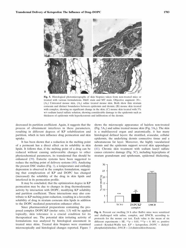
Since pharmaceutical preparations containing the pro-posed complex DOPC:KP (molar ratio, 1:3) will be appliedtopically, skin tolerance is a crucial condition for itstherapeutical use. The potential skin irritating activity offormulations was analyzed by histological assessment intreated mice skins. Treated skin biopsies were examinedmicroscopically and histological changes reported. Figure 5
shows the microscopic appearance of hairless non-treated(Fig. 5A1) and saline treated mouse skin (Fig. 5A2). The skinis a multilayered organ and anatomically, it has manyhistological defined layers: the stratified, avascular, cellularepidermis, the underlying dermis connective tissue and asubcutaneous fat layer. Moreover, the highly vascularizeddermis and the epidermis support several skin appendages(41). Chronic skin treatment with sodium lauryl sulfatecauses extensive damage (Fig. 5C), including hyperplasia ofstratum granulosum and spinhosum, epidermal thickening,
Fig. 5. Histological photomicrographs of skin biopsies taken from non-treated mice or
treated with various formulations. H&E stain and MT stain. Objective augment: 20�.
(A1) Untreated mouse skin, (A2) saline treated mouse skin. Both show thin stratum
corneum and distinct boundaries between epidermis and dermis; (B) mouse skin treated
with complex, showing no significant change in the skin; (C) mouse skin treated with 5%
w/v sodium lauryl sulfate solution, showing considerable damage to the epidermis such as
thickness of epidermis with hyperkeratosis and infiltration of the dermis.
Fig. 6. Percent ear swelling 24 h after Balb/c mice were sensitized
and challenged with saline, complex, and DNCB, according to
protocols for the mouse ear test. Each value is the mean of six
different experiments T SE. **p < 0.01; ***p < 0. 001 vs. positive
controlVKruskal<Wallis test. KP = ketoprofen; DOPC = dioleyl-
phosphatidylcholine; DNCB = 2,4-dinitrochlorobenzene.
1783Transdermal Delivery of Ketoprofen: The Influence of Drug–DOPC
hyperkeratosis, increase in blood vessel and inflammatorycell infiltration. Our results are in accordance with Sintov etal. (26), who submitted the animals to acute treatment with5.0% sodium lauryl sulfate.
In comparison, Fig. 5B shows skin submitted to acutetreatment with the drugYphospholipid complex in mineral oil.The system caused a slight increase in the stratum spinhosum,resulting in a little epidermal thickening, hyperkeratosis andslight inflammatory cell infiltration.
Hence, the mild skin irritation caused by the DOPC/KPcomplex system in the present study may be regarded asacceptable in order to achieve some degree of drug perme-ation through the skin barrier. According to the literature,penetration enhancing effects are caused by structuralalteration of the SC, the main skin barrier (1). However,the stronger the effect, the greater the changes in the deepercutaneous layers. Although it is difficult to quantify the effectof accelerating agents on drug penetration by histologicalobservations, histopathology is still an effective tool forevaluating optimal enhancer concentrations to producedesired permeation defects in the SC with minimal damageto the epidermal and dermal layers (27).
The delayed hypersensitivity response to the KP systemwas evaluated using the mouse ear swelling test (MEST).MEST requires the use of induction and elicitation phases forevaluation but relies on the measurement of ear swelling asan endpoint (28). Ear swelling responses at 24 h afterchallenge are shown in Fig. 6. DNCB (1%), the positivecontrol, led to an increase in ear swelling of 23% after 24 h,while saline and DOPC/KP complex increased ear swellingby 5 and 7%, respectively, in the same conditions. Bothsystems (saline and complex) were significantly differentfrom DNCB, but there was no difference between saline andcomplex.
Therefore, the complex containing formulation seems tobe adequate for topical application of KP, although it isimportant to mention that the MEST is less reliable fordetecting weak to moderate sensitizers (41). It has beenreported that in controlled clinical studies, local skinreactions and systemic side effects are not more commonthan in placebo. However, several case reports have identi-fied KP as being responsible for photosensitive reactions,including both phototoxic and photoallergic adverse effects(42). Thus, further investigation is necessary to characterizethe phototoxic and photoallergic potentials of the proposedformulation.
CONCLUSION
The present study elicits the importance of a welldevised drug and lipid association by a method resulting instable complexes, which can optimize delivery in biologicalmembranes such as skin.
DSC and 1H NMR studies showed interactions betweenKP and DOPC that improved the solubility and decreasedthe melting temperature and enthalpy of the drug. Inaddition, DOPC may increase KP diffusion through the SC.Together these considerations may signal enhancement ofKP skin permeation and skin uptake when complexed withDOPC. Skin irritation and ear swelling studies did not showsignificant different effects when comparing complex and
saline use. The results suggest that the formulation can betherapeutically used for KP transdermal delivery, although itsphototoxic and photoallergic effects must be assessed in afurther investigation.
ACKNOWLEDGMENTS
The authors would like to thank the Fundacao deAmparo a Pesquisa do Estado de Sao Paulo (FAPESP) andCAPES for supporting this study. M. T. J. Garcia was therecipient of a FAPESP fellowship. We acknowledge the helpof Prof. Carlton A. Taft, from Centro Brasileiro de PesquisasFısicas (CBPF), in the use of the GOLD 3.0 and Insight IIprograms.
REFERENCES
1. A. C. Williams and B. W. Barry. Penetration enhancers. Adv.Drug Del. Rev. 56:603Y618 (2004).
2. M. Fujii, K. Shiozawa, Y. Watanabe, and M. Matsumoto. Effectof phosphatidylcholine in skin permeation of indomethacin fromgel prepared with liquid paraffin and hydrogenated phospho-lipid. Int. J. Pharm. 222:57Y64 (2001).
3. M. Fujii, S. Buyukyimkin, N. Buyukyimkin, and J. H. Rytting.Enhancement of skin permeation of miconazole by phospholipidand dodecyl 2-(N, N-dimethylamino) propionate (DDAIP). Int.J. Pharm. 234:121Y128 (2002).
4. Y. Yokomizo. Effect of phosphatidylglycerol on the in vitropercutaneous drug penetration through the dorsal skin of guineapigs, and analysis of the molecular mechanism, using (ATR-FTIR) spectroscopy. Int. J. Pharm. 147:219Y231 (1997).
5. Y. Yokomizo. Effects of phophatidylcholine on the percutane-ous penetration of drugs the dorsal skin of guinea pigs in vitro;and analysis of the molecular mechanism, using attenuated totalreflectance-Fourier transform infrared (ATR-FTIR) spectrosco-py. J. Control. Release 42:249Y262 (1996).
6. Y. Yokomizo. Effects of phospholipids on percutaneous pene-tration of drugs through the dorsal of the guinea pig, in vitro. 3.The effects of phospholipids on several drugs having differentpolarities. J. Control. Release 42:217Y228 (1996).
7. Y. Yokomizo and H. Sagitani. Effects of phospholipids on the invitro percutaneous penetration of prednisolone and analysis ofmechanism by using attenuated total reflectance-Fourier trans-form infrared spectroscopy. J. Pharm. Sci. 85:1220Y1226 (1996).
8. Y. Yokomizo and H. Sagitani. Effects of phospholipids on thepercutaneous penetration of indometacin through the dorsal skinof guinea pigs in vitro. J. Control. Release 38:267Y274 (1996).
9. Y. Yokomizo and H. Sagitani. The effects of phospholipids onthe percutaneous penetration of indometacin through the dorsalskin of guinea pig in vitro. 2. The effects of the hydrophobicgroup in phospholipids and a comparison with generalenhancers. J. Control. Release 42:37Y46 (1996).
10. P. Morganti, E. Ruocco, Wolf, W. Ronni, and V. Ruocco.Percutaneous absorption and delivery systems. Clin. Dermatol.19:489Y501 (2001).
11. A. L. Lehniger. Princıpios de Bioquımica. Traducao: W. R. Lodi;A. A. Simoes. Savier Editora, Sao Paulo,1986.
12. S. N. Bhattachar, J. H. Rytting, T. Itoh, and T. Nishihata. Theeffects of complexation with hydrogenated phospholipid on thetransport of salicylic acid, diclofenac and indomethacin acrosssnake stratum corneum. Int. J. Pharm. 79:263Y271 (1992).
13. R. H. Harris and I. Vavra. Ketoprofen. In K. D. Rainsford (ed).,Anti-inflammatory and Anti-rheumatic Drugs, CRC, BocaRaton, FLv. 2, cap. 8, 1985, pp. 151Y169.
14. J. Berba, S. Goranson, J. Langle, and U. V. Banakar. In vitrorelease of selected non-steroidal anti-inflammatory analgesicfrom reservoir type transdermal formulations. Drug Dev. Ind.Pharm. 146:255Y262 (1991).
1784 Junqueira Garcia et al.
15. A. Osterwalder, V. Reiner, G. Reiner, and P. Lualdi. Tissueabsorption and distribution of ketoprofen after patch applicationin subjects undergoing knee arthroscopy or endoscopic carpalligament release. Arzneim.-Forsch./Drug Res. 52:822Y827 (2002).
16. P.-C. Wu, J.-S. Chang, Y.-B. Huang, C.-Y. Chai, and Y.-H Tsai.Evaluation of percutaneous absorption and skin irritation ofketoprofen through rat skin: in vitro and in vivo study. Int. J.Pharm. 222:225Y235 (2001).
17. K. Takahashi, H. Sakano, N. Numata, S. Kuroda, and N.Mizuno. Effect of fatty acid diesters on permeation of anti-inflammatory drugs through rat skin. Drug Dev. Ind. Pharm.28:1285Y1294 (2002).
18. Y.-S. Rhee, J.-G. Choi, E.-S. Park and S.-C. Chi. Transdermaldelivery of ketoprofen using microemulsions. Int. J. Pharm.228:161Y170 (2001).
19. D. Paolino, A. Ventura, S. Nistico, G. Puglisi, and M. Fresta.Lecithin microemulsion for the topical administration of keto-profen: percutaneous adsorption through human skin and in vivohuman skin tolerability. Int. J. Pharm. 244:21Y32 (2002).
20. P. C. Panus, J. Campbell, S. B. Kulkarni, R. T. Herrick, W. R.Ravis, and A. K. Banga. Transdermal iontophoretic delivery ofketoprofen through human cadaver skin and in humans. J.Control. Rel. 44:113Y121 (1997).
21. M. T. J. Garcia, J. M. Marchetti, and M. V. L. B. Bentley. De-termination by HPLC of ketoprofen in aqueous medium used formin vitro skin permeation studies. Anal. Lett. 34:1865Y1874 (2001).
22. E. G. Jalon, M. Josa, M. A. Campanero, S. Satoyo, P. Ygartua.J. Chromatogr. 870:143Y149 (2000).
23. M. L. Verdonk, J. C. Cole, M. J. Hartshorn, C. W. Mulrray, andR. D. Taylor. Improved protein-ligand docking using GOLD.Proteins 52:609Y623 (2003).
24. Insight II Accelrys, San Diego, California, 2000.25. SPARTAN’04, v.1.0.3, Wavefunction, Inc. Irvine, California, 2005.26. S. M. Harrison, B. W. Barry, and P. H. Dugard. Effect of
freezing on human skin permeability. J. Pharm. Pharmacol.35:261Y262 (1984).
27. A. Sintov, A. Ze’evi, R. Uzan, and A. Nyska. Influence ofpharmaceutical gel vehicle containing oleic acid/sodium oleatecombinations on hairless mouse skin, a histological evaluation.Eur. J. Pharm. Biopharm. 47:299Y303 (1999).
28. S. C. Gad, B. J. Dunn, D. W. Dobbs, C. Reilly, and R. D. Walsh.Development and validation of an alternative dermal sensitiza-tion test: the mouse ear swelling test (MEST). Toxicol. Appl.Pharmacol. 84:93Y114 (1986).
29. E. Bombardelli and M. Spelta. PhospholipidYpolyphenol com-plexes: a new concept in skin care ingredients. Cosmet. Toilet.106:69Y76 (1991).
30. P. Sing and M. S. Roberts. Skin permeability and local tissueconcentration of nonsteroidal anti-inflammatory drugs aftertopical application. J. Pharmacol. Exp. Ther. 286:144Y151(1994).
31. M. T. J. Garcia, J. M. Marchetti, and M. V. L. B. Bentley.Ketoprofen transdermal delivery from lipid/propylene glycolsystem: in vitro permeation studies. Eur. J. Pharm. Sci.13(1):s129 (2001).
32. M. V. L. B. Bentley, E. R. M. Kedor, R. F. Vianna, and J. H.Collet. The influence of lecithin and urea on the in vitropermeation of hydrocortisone acetate through skin from mouse.Int. J. Pharm. 146:255Y262 (1997).
33. K. Moser, K. Kriwet, A. Naik, Y. N. Kalia, and R. H. Guy.Passive skin penetration enhancement and its quantification invitro. Eur. J. Pharm. Sci. 52:103Y112 (2001).
34. M. M. R. Dias, S. L. Raghavan, M. A Pellet, and J. Hadgraft.The effect of cyclodextrins on the permeation of diclofenac fromsupersaturated solution. Int. J. Pharm. 263:173Y181 (2003).
35. S. L. Raghavan, B. Kiepfer, A. F. Davis, S. G. Kazarian, and J.Hadgraft. Membrane transport of hydrocortisone acetate fromsupersaturated solution; the role of polymers. Int. J. Pharm.221:95Y105 (2001).
36. M. B. Brown, M. Hanpanitcharoen, and G. P. Martin. An invitro investigation into the effect of glycosaminoglycans on theskin partitioning and deposition of NSAIDs. Int. J. Pharm.225:113Y121 (2001).
37. B. W. Barry. Novel mechanisms and devices to enable successfultransdermal drug delivery. Eur. J. Pharm. Sci. 14:101Y114(2001).
38. S. Buyuktimkin, N. Buyuktimkin, and J. H. Rytting. Interactionof indomethacin with a new penetration enhancer, dodecyl 2-(n,n-dimethylamino) propionate (DDAIP): is effect on transdermaldelivery. Int. J. Pharm. 127:245Y253 (1996).
39. G. B. Kasting, R. L. Smith, and E. R. Cooper. Effects of lipidsolubility and molecular size on percutaneous absorption.Pharmacol. Skin 1:138Y153 (1987).
40. P. W. Stott, A. C. Williams, and B. W. Barry. Transdermaldelivery from eutectic systems: enhanced permeation of a modeldrug, ibuprofen. J. Control. Release 50:297Y308 (1998).
41. J. K. Lee, J. H. Park, H. S. Kim, S. T. Chung, J. H. Eom, K. T.Nam, and H. Y. Oh. Evaluation of cell proliferation in earand lymph node using BrdU immunohistochemistry formouse ear swelling test. Environ. Toxicol. Pharmacol.14:61Y68 (2003).
42. H. Bagheri, V. Lhiaubet, J. L. Montastruc, and N. Chouini-Lalanne. Photosensitivity to ketoprofen: mechanisms and phar-macoepidemiological data. Drug Safety 22:339Y349 (2000).
1785Transdermal Delivery of Ketoprofen: The Influence of Drug–DOPC