Radiation sterilization of ketoprofen
6
Radiation Physics and Chemistry 73 (2005) 111–116 Radiation sterilization of ketoprofen $ Branka Katugin-Ramem a, , Katia Hamitouche b , Nadica Maltar-StrmeWki c , Karmen Kos d , Irina Pucic´ a , Smiljana Britvic´-Budicin a , Dugan Ramem a a RuXer Bogkovic´Institute, P.O.B 180, Zagreb 10000, Croatia b Centre de Recherche et Developpement Saidal, El Harrach, Algeria c Faculty of Veterinary Medicine, University of Zagreb, Zagreb 10000, Croatia d Belupo Pharmaceutical Works, Koprivnica 48000, Croatia Received 23 February 2004; accepted 14 July 2004 Abstract Radiation sterilization of ketoprofen (KP) dry powder was investigated by selected physico-chemical methods. High- performance liquid chromatography, ultraviolet spectrophotometry, infrared spectrophotometry, differential scanning calorimetry, X-ray diffraction and electron spin resonance spectroscopy did not show any significant degradation at sterilization dose 25 kGy. To determine the nature, extent and direction of radiation-induced changes, KP was irradiated to extremely high doses, much higher than necessary to achieve sterility. The irradiated KP did not show any difference of XRD patterns up to 200 kGy; with DSC and IR some changes were detected only above 1000 and 2000 kGy, respectively; HPLC has shown about 5% destruction at 2000 kGy. Acetyl benzophenon (AcBph) was generated by irradiation with G(AcBph)=(1.670.1) 10 –8 mol J –1 . Ames test has shown no mutagenicity of KP irradiated with 3000 kGy or of the oily mixure of radiolytic products isolated from it. Solid KP has proven to be very stable on irradiation, and irradiation has been found to be a suitable method for its sterilization. r 2004 Elsevier Ltd. All rights reserved. Keywords: Ketoprofen; Radiation sterilization; Radiation degradation; DSC; ESR; HPLC; IR; UV; XRD; Ames test 1. Introduction Profens are potent nonsteroidal anti-inflammatory drugs. Ketoprofen (KP) [2-(3-benzoylphenyl) propionic acid] is widely used and is one of the most popular profens. Sterility of profens is required for the intrave- nous applications, and intramuscular and subcutaneous injections. An attractive alternative to traditional methods of sterilization, recognized by all major pharmacopeias, has been afforded by irradiation. This work deals with the feasibility of radiation sterilization of ketoprofen in dry powder form. 2. Materials and methods Ketoprofen (KP) was obtained from Belupo Pharma- ceutical Works (Koprivnica, Croatia). Ketoprofen CRS (chemical reference substance), ketoprofen impu- rities A CRS (3-acetylbenzophenon) (AcBph) and C CRS (2-(3-carboxyphenyl)-propionic acid), (CRSs avail- able from European Directorate for the Quality of Medicines, Council of Europe, Strasbourg, France), were also obtained from Belupo. Benzophenon (Bph) ARTICLE IN PRESS www.elsevier.com/locate/radphyschem 0969-806X/$ - see front matter r 2004 Elsevier Ltd. All rights reserved. doi:10.1016/j.radphyschem.2004.07.004 $ This work was presented at IMRP-2003, Chicago, USA. Corresponding author. Fax: +385-1-468-0098. E-mail address: [email protected] (B. Katugin-Ramem).
-
Upload
independent -
Category
Documents
-
view
0 -
download
0
Transcript of Radiation sterilization of ketoprofen

ARTICLE IN PRESS
0969-806X/$ - s
doi:10.1016/j.ra
$This work w�CorrespondE-mail addr
Radiation Physics and Chemistry 73 (2005) 111–116
www.elsevier.com/locate/radphyschem
Radiation sterilization of ketoprofen$
Branka Katugin-Ramema,�, Katia Hamitoucheb, Nadica Maltar-StrmeWkic,Karmen Kosd, Irina Pucica, Smiljana Britvic-Budicina, Dugan Ramema
aRuXer Bogkovic Institute, P.O.B 180, Zagreb 10000, CroatiabCentre de Recherche et Developpement Saidal, El Harrach, Algeria
cFaculty of Veterinary Medicine, University of Zagreb, Zagreb 10000, CroatiadBelupo Pharmaceutical Works, Koprivnica 48000, Croatia
Received 23 February 2004; accepted 14 July 2004
Abstract
Radiation sterilization of ketoprofen (KP) dry powder was investigated by selected physico-chemical methods. High-
performance liquid chromatography, ultraviolet spectrophotometry, infrared spectrophotometry, differential scanning
calorimetry, X-ray diffraction and electron spin resonance spectroscopy did not show any significant degradation at
sterilization dose 25 kGy. To determine the nature, extent and direction of radiation-induced changes, KP was
irradiated to extremely high doses, much higher than necessary to achieve sterility. The irradiated KP did not show any
difference of XRD patterns up to 200 kGy; with DSC and IR some changes were detected only above 1000 and
2000 kGy, respectively; HPLC has shown about 5% destruction at 2000 kGy. Acetyl benzophenon (AcBph) was
generated by irradiation with G(AcBph)=(1.670.1)� 10–8mol J–1. Ames test has shown no mutagenicity of KP
irradiated with 3000 kGy or of the oily mixure of radiolytic products isolated from it. Solid KP has proven to be very
stable on irradiation, and irradiation has been found to be a suitable method for its sterilization.
r 2004 Elsevier Ltd. All rights reserved.
Keywords: Ketoprofen; Radiation sterilization; Radiation degradation; DSC; ESR; HPLC; IR; UV; XRD; Ames test
1. Introduction
Profens are potent nonsteroidal anti-inflammatory
drugs. Ketoprofen (KP) [2-(3-benzoylphenyl) propionic
acid] is widely used and is one of the most popular
profens. Sterility of profens is required for the intrave-
nous applications, and intramuscular and subcutaneous
injections. An attractive alternative to traditional
methods of sterilization, recognized by all major
pharmacopeias, has been afforded by irradiation. This
ee front matter r 2004 Elsevier Ltd. All rights reserv
dphyschem.2004.07.004
as presented at IMRP-2003, Chicago, USA.
ing author. Fax: +385-1-468-0098.
ess: [email protected] (B. Katugin-Ramem).
work deals with the feasibility of radiation sterilization
of ketoprofen in dry powder form.
2. Materials and methods
Ketoprofen (KP) was obtained from Belupo Pharma-
ceutical Works (Koprivnica, Croatia). Ketoprofen
CRS (chemical reference substance), ketoprofen impu-
rities A CRS (3-acetylbenzophenon) (AcBph) and C
CRS (2-(3-carboxyphenyl)-propionic acid), (CRSs avail-
able from European Directorate for the Quality of
Medicines, Council of Europe, Strasbourg, France),
were also obtained from Belupo. Benzophenon (Bph)
ed.

ARTICLE IN PRESSB. Katugin-Razem et al. / Radiation Physics and Chemistry 73 (2005) 111–116112
was supplied by Merck (Darmstadt, Germany).
Acetonitrile (HPLC grade), dichloromethane, glacial
acetic acid, sodium hydrogen carbonate and anhydrous
sodium sulphate (all reagent grade) were from Kemika
(Zagreb, Croatia). Potassium dihydrogen orthopho-
sphate was obtained from Analar (Poole, England).
Water was triply distilled.
Irradiation of 300mg samples was performed by 60Co
gamma rays at a dose rate of 10Gy s�1 in the presence
of air.
Isolation of lipophilic radiolytic products of KP for
Ames mutagenicity test was accomplished by removal of
the parent compound and acidic impurities by extraction
of dichloromethane solution of irradiated KP (dose
3000 kGy) with concentrated aqueous solution of
sodium hydrogen carbonate. After washing of the
remaining dichloromethane layer with water and drying
with anhydrous sodium sulphate, the evaporation of
solvent afforded an oily residue of lipophilic radiolytic
products.
Purity control and quantification of radiolytic pro-
ducts in irradiated KP were performed by high-
performance liquid chromatography (HPLC) following
two approaches. The first one, prescribed by the
(European Pharmacopeia (EuPh), 2001), was based on
the comparison of chromatograms of reference solutions
of CRS chemicals with chromatograms of the solutions
of investigated substances. A ProStar HPLC system by
Varian (Walnut Creek, CA, USA) was used. The system
was fitted with an LC-18, 5m, 150mm� 4.6mm column
(Supelco, Bellefonte, PA, USA). The pump flow rate was
1.2mlmin�1, detection wavelength 233 nm, and mobile
phase consisted of acetonitrile, phosphate buffer (0.5M
aqueous solution of potassium dihydrogen phosphate
adjusted to pH 3.5 with orthophosphoric acid) and
water (43:2:55, v/v)–mobile phase A. The second
approach was based on the use of external and internal
standards. Radiolytic decomposition of KP was
quantified using KP CRS as an external standard,
while Bph was used as an internal standard in the
quantification of radiolytic products. Besides the same
chromatographic system and the same column applied
under the same conditions as described above, an
additional chromatographic system, WellChrom HPLC
by Knauer Scientific Instruments (Berlin, Germany)
with a diode array detector was applied. A somewhat
faster analysis was made using a mobile phase acetoni-
trile, 0.005M aqueous acetic acid (55:45, v/v)–mobile
phase B.
Ultraviolet (UV) spectrophotometry was used for
purity control and determination of response factors of
KP and its radiolytic products at the detection
wavelength (233 nm) in mobile phases A and B. The
measurements were made at 20 1C using a Cary 2200
spectrophotometer by Varian (Mulgrave, Victoria,
Australia).
Infrared (IR) spectrophotometry was performed
by a RXI FT-IR spectrophotometer by Perkin-Elmer
(Norwalk, CT, USA) in anhydrous sodium bromide
discs.
Differential scanning calorimetry (DSC) was carried
out using a DSC 7 by Perkin-Elmer (Norwalk, CT,
USA). The thermograms were recorded under nitrogen
from 20 to 120 1C, at the heating rate of 10 1Cmin�1.
X-ray diffraction (XRD) patterns were taken by a
Model 1738 Automatic Diffractometer by Philips
(Almelo, The Netherlands) using CuKa radiation
(l ¼ 1:5418 (A) and a Philips PW 1877 AAPD v 3.6 g
program.
Electron spin resonance (ESR) spectroscopy was done
by a model E109 X-band spectrometer by Varian (Palo
Alto, CA, USA), operating at 9.5GHz with 100 kHz
modulation, equipped with a variable temperature unit
ER4111VT by Bruker Instruments (Karlsruhe, Ger-
many). The number of spins in irradiated KP was
determined by comparison of the second integral of
irradiated sample and a Bruker strong pitch reference
standard in a double microwave cavity.
Ames mutagenicity test of unirradiated KP, irradiated
KP (3000 kGy) and isolated radiolytic products was
conducted by the standard plate incorporation test
(Maron and Ames, 1983). Two test strains of Salmonella
typhimurium TA98 and TA100 (kindly provided by B.N.
Ames, University of California, Berkeley, CA, USA),
were used to detect frame-shift and base-pair mutations,
respectively. Dimethyl sulfoxide samples of 2, 20, 100
and 200 ml (equivalent to 100, 1000, 5000 and 10 000mlof KP and its radiolytic products, respectively) were
plated onto Vogel–Bonner basal agar plates with 2ml of
soft agar to which 0.5mM L-histidine and 0.5mM
biotin solutions had been previously added. Overnight
cultures of TA98 and TA100 (0.1ml), both without or
with metabolic activation (0.5ml of -S9 mixture or
0.5ml of +S9 mixture, respectively), were added to the
plates.
The S9 mixture contained 50ml of hepatic S9 preparedfrom male Wistar rats pretreated with an intraperitoneal
injection containing Aroclor 1254 (500mg/kg) dissolved
in corn oil. Immediately before mutagenicity testing, the
S9 fraction was passed sequentially through Millipore
membrane filters (0.45 and 0.22 mm filter units) to
remove any contaminating microorganisms. Each sam-
ple was plated in triplicate, and its revertants were
scored manually after 72 h incubation at 37 1C. As a
positive control (diagnostic mutagens) for this assay,
2-aminofluorene at a concentration of 10 mg/plate wasused to monitor the sensitivity of the bacterial strains
and the activity of the rat liver S9. Daunomycin at a
concentration of 10mg/plate without S9 and 645mg/plateof methyl methanesulfonate were used as an additional
control of the TA98 and TA100 strains, respectively.
The mutagenicity was expressed as the number of

ARTICLE IN PRESSB. Katugin-Razem et al. / Radiation Physics and Chemistry 73 (2005) 111–116 113
revertants per plate and per microgram of KP and its
radiolytic products, respectively.
Although there is no evidence supporting this specific
requirement, it is an arbitrary criterion for a test
substance to be considered positive in a bioassay, that
it must cause a twofold or a threefold increase over the
background of the mean number of revertants per plate
of at least one tester strain with a minimum of two
concentrations of the test substance.
Fig. 1. HPLC of KP obtained with Knauer HPLC system and
mobile phase acetonitrile: phosphate buffer: water (43:2:55, v/v)
(mobile phase A). The respective doses (from the bottom up)
are: 0, 25, 200 and 1600kGy.
0
20
40
60
80
100
0 1000 3000 4000 6000 7000
mm
ol A
cBph
/ kg
KP
2000 5000
Dose / kGy
Fig. 2. Radiolytic formation of AcBph with dose. Filled circles:
mobile phase A (acetonitrile, phosphate buffer, water (43:2:55,
v/v)). Open circles: mobile phase B (acetonitrile, 0.005M
aqueous acetic acid (55:45, v/v)).
3. Results and discussion
Unirradiated KP was a white powder which turned
pink on irradiation, probably as a result of trapping
radiolysis electrons by the Bph moiety or the conversion
of Bph to ketyl radical, or both. The intensity of the
colouration was related to dose. The white appearance
was recovered after several days of storage in a
dessiccator, in the dark, and at room temperature. No
attempt was made in this work to quantify these
changes.
Radiation-induced chemical changes in irradiated KP
were investigated by HPLC. The amounts of KP
impurity A (permitted level 0.2%), KP impurity C
(permitted level 0.2%), and the sum of other impurities
(permitted level 0.4%) were followed in unirradiated KP
and samples irradiated with 25, 50, 100 and 200 kGy by
the method prescribed by the European Pharmacopeia.
While the impurity C stayed at the same level found in
unirradiated KP, impurity A reached 0.1% at 200 kGy.
However, the sum of other impurities at 100 kGy rose to
0.7%, which is above the permitted level, and reached
1.4% at 200 kGy.
To determine the nature, extent and direction of
radiation-induced changes, KP was irradiated to ex-
tremely high doses, much higher than necessary to
achieve sterility; up to 4000 kGy was applied to produce
sizeable radiolytic changes, to be able to quantify them
and express them as radiation chemical yields.
The comparison of chromatograms obtained by
HPLC of unirradiated and irradiated KP (Fig. 1) shows
hardly any change at 25 kGy, but the presence of four
major radiolytic products eluting at 5, 11, 30 and 37min
respectively, was evident at higher doses.
Ultraviolet spectra of these products taken by a diode
array HPLC detector show that all probably retain a
Bph chromophore. The radiolytic product eluting at
11min was not unique to radiolysis, as its trace was
present in unirradiated sample, where it may have arisen
during production and handling. It has been known as
KP impurity A (AcBph). The build-up of AcBph with
dose is shown in Fig. 2, as quantified against the Bph
internal standard using two mobile phases, A and B.
The slope of this straight line gives radiation chemical
yield, G(AcBph)=(1.670.1) 10–8mol J–1. Taking this
value the level of KP impurity A at 200 kGy could be
calculated more accurately as 0.08%, which compares
well with 0.1% as estimated according to the EU Ph
procedure. It is remarkable that the linearity of
radiolytic products formation holds throughout the
dose range and that there is no trend to sublinearity.
On the other hand, it is not surprising that a direct
action of radiation on essentially non-interacting
molecules in the solid phase follows a linear pattern
as long as the probability of hitting the same molecule
for the second time remains low, and that is assured
by a low-radiation chemical yield of the change.
Assuming similar radiation chemical yields of the
other three major radiolysis products, radiation
chemical yield of decomposition could be estimated as
G(KP)=6.4� 10�8mol J�1. This would give about 3.3%
decomposition at 2000 kGy.
Infrared spectra of irradiated KP were inspected
for changes in comparison with unirradiated samples.
Two of the strongest, sharp and symmetrical peaks at
1697 and 1655 cm�1 have been attributed to carboxylic
and keto carbonyl stretching vibrations, respectively
(Fig. 3).
ARTICLE IN PRESS
4000 kGy
2000 kGy
200 kGy
100 kGy
25 kGy
0 kGy
Rel
ativ
e in
tens
ity
2000 1800 1600
Absorbance/Wavenumber
1400 1200 1000 800 600 400
Fig. 3. Selected parts of IR spectra of KP; the respective doses
are marked on the spectra.
0
1
2
3
4
5
60 70
P /m
W
25 kGy
1000 kGy
2000 kGy
0 kGy
11010080T /˚C
90
Fig. 4. DSC thermograms of KP; the respective doses are
marked on the thermograms.
03 23 43 63
2θ
0 kGy
Rel
ativ
e in
tens
ity
100000
80000
60000
40000
20000
33 5313
25 kGy
200 kGy
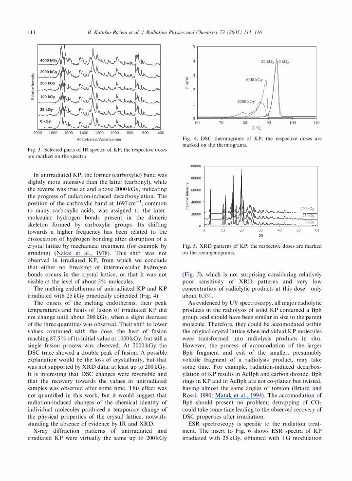
Fig. 5. XRD patterns of KP; the respective doses are marked
on the roentgenograms.
B. Katugin-Razem et al. / Radiation Physics and Chemistry 73 (2005) 111–116114
In unirradiated KP, the former (carboxylic) band was
slightly more intensive than the latter (carbonyl), while
the reverse was true at and above 2000 kGy, indicating
the progress of radiation-induced decarboxylation. The
position of the carboxylic band at 1697 cm�1, common
to many carboxylic acids, was assigned to the inter-
molecular hydrogen bonds present in the dimeric
skeleton formed by carboxylic groups. Its shifting
towards a higher frequency has been related to the
dissociation of hydrogen bonding after disruption of a
crystal lattice by mechanical treatment (for example by
grinding) (Nakai et al., 1978). This shift was not
observed in irradiated KP, from which we conclude
that either no breaking of intermolecular hydrogen
bonds occurs in the crystal lattice, or that it was not
visible at the level of about 3% molecules.
The melting endotherms of unirradiated KP and KP
irradiated with 25 kGy practically coincided (Fig. 4).
The onsets of the melting endotherms, their peak
temperatures and heats of fusion of irradiated KP did
not change until about 200 kGy, when a slight decrease
of the three quantities was observed. Their shift to lower
values continued with the dose, the heat of fusion
reaching 87.5% of its initial value at 1000 kGy, but still a
single fusion process was observed. At 2000 kGy the
DSC trace showed a double peak of fusion. A possible
explanation would be the loss of crystallinity, but that
was not supported by XRD data, at least up to 200 kGy.
It is interesting that DSC changes were reversible and
that the recovery towards the values in unirradiated
samples was observed after some time. This effect was
not quantified in this work, but it would suggest that
radiation-induced changes of the chemical identity of
individual molecules produced a temporary change of
the physical properties of the crystal lattice, notwith-
standing the absence of evidence by IR and XRD.
X-ray diffraction patterns of unirradiated and
irradiated KP were virtually the same up to 200 kGy
(Fig. 5), which is not surprising considering relatively
poor sensitivity of XRD patterns and very low
concentration of radiolytic products at this dose—only
about 0.3%.
As evidenced by UV spectroscopy, all major radiolytic
products in the radiolysis of solid KP contained a Bph
group, and should have been similar in size to the parent
molecule. Therefore, they could be accomodated within
the original crystal lattice when individual KP molecules
were transformed into radiolysis products in situ.
However, the process of accomodation of the larger
Bph fragment and exit of the smaller, presumably
volatile fragment of a radiolysis product, may take
some time. For example, radiation-induced decarbox-
ylation of KP results in AcBph and carbon dioxide. Bph
rings in KP and in AcBph are not co-planar but twisted,
having almost the same angles of torsion (Briard and
Rossi, 1990; Matak et al., 1994). The accomodation of
Bph should present no problem; detrapping of CO2could take some time leading to the observed recovery of
DSC properties after irradiation.
ESR spectroscopy is specific to the radiation treat-
ment. The insert to Fig. 6 shows ESR spectra of KP
irradiated with 25 kGy, obtained with 1G modulation

ARTICLE IN PRESSB. Katugin-Razem et al. / Radiation Physics and Chemistry 73 (2005) 111–116 115
amplitude at 100 kHz with 0.4mW (lower trace) and
1mW microwave power (upper trace).
Room temperature microwave saturation features of
this signal were studied in the range 0–150mW
microwave power and a complex behaviour was
observed, indicating the presence of more than one
radical species. The identification of these species was
not attempted in the present work, but likely candidates
would be: KP� and/or Bph ketyl radical, dCO�2 radical
ion, and possibly peroxyl radicals.
1 100 1000 10000
Num
ber
of s
pins
/g
342 349
1 mW
Dose /kGy
10
Magnetic field/mT
335
EPR
inte
nsity
0.4 µW
1E+19
1E+18
1E+17
1E+16
Fig. 6. Radiolytic accumulation of KP free radicals with dose
at room temperature. Insert: ESR spectra of KP irradiated with
25 kGy; lower trace: 0.4mW; upper trace: 1mW.
Table 1
Number of revertants by unirradiated and irradiated KP and by KP ra
Salmonella typhimurium TA98 and TA100 his� strains without (S9) a
Chemicals (mg /plate) No. of TA98 revertants/plate
Without S9a With
Unirradiated KP
100 3872 5971000 4072 5275000 2371 427
Irradiated KP (3000kGy)
100 5171 5471000 4773 5375000 2774 317
Radiolytic products only (3000 kGy)
100 4472 5671000 4372 4675000 3671 15710,000 1072 77
Solvent control
(DMSO) 4476 417
Positive controls 1150720 21787(1,2,3) (1) (2)
S9—metabolic activation with microsomal fraction from rat liver hom
1. Daunomycin hydrocloride (10mg/plate), 2. 2-aminofluorene (2-AF)aMean7SD of three plates.
The build-up of free radicals with the dose is also
shown in Fig. 6. The concentration increases linearly up
to 500 kGy, and in this range the radiation chemical
yield was G(free radicals)=2.3� 10�9 radicals J�1. The
decay of free radicals followed a complex kinetics, the
decay being faster at lower dose (not shown), which
indicates that recombination was not responsible
for the decay. From the above radiation chemical
yield, the saturation dose of about 1000 kGy and KP
density 1.28 g cm�3 (Liversidge, 1981), the radius of
the sphere of radical migration could be estimated as
about 5 nm. This gives a very large recombination
volume encompassing 860molecular packing units, so
that it is inconceivable that a radical could visit them in
solid. The upper value of the recombination radius
critical for stable trapping in solid is considered to be
about 1 nm per radical (Krushev et al., 1994). These
considerations then also support the view that recombi-
nation is not involved in the decay of free radicals in
irradiated KP.
All tested substances gave a negative response to both
strains, TA98 and TA100 in the Ames mutagenicity test
in comparison with known mutagens (positive controls)
as shown in Table 1. The number of revertants per plate
produced by the test substances was the same as the
number of spontaneous revertants on the negative
control plates. Both unirradiated, as well as irradiated
diolytic products in the Salmonella plate incorporation test using
nd with (+S9) metabolic activation, respectively
No.of TA100 revertants/plate
S9a Without S9a With S9a
1 160720 13574
2 14578 14276
5 134710 13574
5 13574 13078
4 134710 13878
8 13674 142710
1 172720 15374
2 12278 11776
5 152710 11374
4 13072 11474
8 150714 152712
23 2000730 1560748
(3) (2)
ogenate.
(10mg/plate), 3. Methyl methanesulphonate (625 mg/plate).

ARTICLE IN PRESSB. Katugin-Razem et al. / Radiation Physics and Chemistry 73 (2005) 111–116116
KP were not mutagenic to either of the two strains,
and that was also true for the isolated radiolytic
products of KP irradiated with 3000 kGy. There was
no indication of dose dependent responses to any
substance.
These data corroborate the published data on the
absence of mutagenicity of unirradiated KP (Philipose
et al., 1997). The new information is represented
here by the data on irradiated KP and its radiolytic
products; these substances were not investigated
before, and pertinent data do not significantly deviate
from those of unirradiated KP. This strongly supports
the conclusion on the absence of mutagenicity of
irradiated KP.
4. Concluding remarks
The feasibility of radiation sterilization of solid KP
was demonstrated almost up to 100 kGy, which
is well beyond any real needs. In fact, it is likely
that the sterilization dose much lower than the
standard 25 kGy, a routine value until recently, will be
required if the bioburden, its radiation sensitivity
and sterility assurance level were considered. The
high resistance to irradiation of KP in the dry powder
form, established in this work, enables the use of the
pre-sterilized drug in the preparation of sterile
solutions by aseptic processing and filling, which
is the second best option to terminal sterilization. The
study of radiation chemical mechanism of radiolytic
decomposition of neat KP may lead to better under-
standing of and practical solutions for the radiation
sterilization of liquid formulations of KP in final
packages.
Acknowledgement
The fellowship for training in Radiation Steriliza-
tion of Pharmaceuticals (ALG 02017 P) by the Inter-
national Atomic Energy Agency to K.H. is gratefully
acknowledged.
References
Briard, P., Rossi, J.C., 1990. Ketoprofene. Acta Crystallogr. C
46, 1036–1038.
European Pharmacopeia, 2001. Fourth ed. Directorate for
the Quality of Medicines, Council of Europe, Strasbourg,
pp. 1432–1433.
Krushev, V.V., Koizumi, H., Ichikawa, T., Yoshida, H.,
Shibata, H., Tagawa, S., Yoshida, Y., 1994. Relation
between track structure and LET effect on free radical
formation for ion beam-irradiated alanine dosimeter.
Radiat. Phys. Chem. 44, 521–526.
Liversidge, G.G., 1981. Ketoprofen. In: Florey, K. (Ed.),
Analytical Profiles of Drug Substances, vol. 10. Academic
Press, New York, pp. 443–471.
Maron, D.M., Ames, B.N., 1983. Revised methods for the
Salmonella mutagenicity test. Mutation Res. 113, 173–215.
Matak, D., Vinkovic, M., Dumic, M., 1994. 1-(3-Benzoylphe-
nyl)ethanone(I) and 3-benzoyl-a-methylbenzeneacetamidemethylene chloride solvate (2/1) (II). Acta Crystallogr. C
50, 1339–1342.
Nakai, Y., Nakajima, S.I., Yamamoto, K., Terada, K., Konno,
T., 1978. Effects of grinding on physical and chemical
properties of crystalline medicinals with microcrystalline
cellulose. III. Infrared spectra of medicinals in ground
mixtures. Chem. Pharm. Bull. 26, 3419–3425.
Philipose, B., Singh, R., Khan, K.A., Giri, A.K., 1997.
Comparative mutagenic and genotoxic effects of three
propionic acid derivatives ibuprofen, ketoprofen and
naproxen. Mutation Res. 393, 123–131.