Radiation Therapy and Hearing Loss
15
RADIATION THERAPY AND HEARING LOSS Niranjan Bhandare, M.S. * , Andrew Jackson, Ph.D. † , Avraham Eisbruch, M.D. ‡ , Charlie C. Pan, M.D. ‡ , John C. Flickinger, M.D. § , Patrick Antonelli, M.D. ║ , and William M. Mendenhall, M.D. * * Department of Radiation Oncology, University of Florida College of Medicine, Gainesville, Florida ║ Department of Otolaryngology, University of Florida College of Medicine, Gainesville, Florida † Department of Medical Physics, Memorial Sloan-Kettering Cancer Center, New York, NY ‡ Department of Radiation Oncology, University of Michigan § Department of Radiation Oncology, University of Pittsburgh Medical Center Abstract A review of literature on the development of sensorineural hearing loss after high-dose radiation therapy for head-and-neck tumors and stereotactic radiosurgery or fractionated stereotactic radiotherapy for the treatment of vestibular schwannoma is presented. Because of the small volume of the cochlea a dose–volume analysis is not feasible. Instead, the current literature on the effect of the mean dose received by the cochlea and other treatment- and patient-related factors on outcome are evaluated. Based on the data, a specific threshold dose to cochlea for sensorineural hearing loss cannot be determined; therefore, dose–prescription limits are suggested. A standard for evaluating radiation therapy–associated ototoxicity as well as a detailed approach for scoring toxicity is presented. Keywords Radiotherapy; Sensorineural hearing loss; Ototoxicity; Auditory; Ear; QUANTEC 1. CLINICAL SIGNIFICANCE Radiation therapy (RT) may damage the cochlea and/or acoustic nerve, leading to sensorineural hearing loss (SNHL) (1–4), with resultant long-lasting compromise in the quality of life. This report focuses on RT-induced SNHL in adults who have received fractionated RT, stereotactic radiosurgery (SRS), and fractionated stereotactic RT (FSRT) for head-and-neck cancers and vestibular schwannomas (VS). 2. ENDPOINTS SNHL is traditionally defined as a clinically significant increase in bone conduction threshold (BCT) at the key human speech frequencies (0.5–4.0 kHz), as seen in pure-tone audiometry. However, reports of SNHL after fractionated RT vary in terms of: (a) the frequencies evaluated (e.g., 2 or 4 kHz alone (5,6) and/or pure tone average [PTA] of © 2010 Elsevier Inc. Reprint requests to: William M. Mendenhall, M.D., PO Box 100385, Gainesville, FL 32610. Tel: (352) 265-0287; Fax: (352) 265-7045; [email protected]. Conflict of interest: None. NIH Public Access Author Manuscript Int J Radiat Oncol Biol Phys. Author manuscript; available in PMC 2012 April 4. Published in final edited form as: Int J Radiat Oncol Biol Phys. 2010 March 1; 76(3 Suppl): S50–S57. doi:10.1016/j.ijrobp.2009.04.096. NIH-PA Author Manuscript NIH-PA Author Manuscript NIH-PA Author Manuscript
-
Upload
independent -
Category
Documents
-
view
0 -
download
0
Transcript of Radiation Therapy and Hearing Loss

RADIATION THERAPY AND HEARING LOSS
Niranjan Bhandare, M.S.*, Andrew Jackson, Ph.D.†, Avraham Eisbruch, M.D.‡, Charlie C.Pan, M.D.‡, John C. Flickinger, M.D.§, Patrick Antonelli, M.D.║, and William M. Mendenhall,M.D.**Department of Radiation Oncology, University of Florida College of Medicine, Gainesville, Florida║Department of Otolaryngology, University of Florida College of Medicine, Gainesville, Florida†Department of Medical Physics, Memorial Sloan-Kettering Cancer Center, New York, NY‡Department of Radiation Oncology, University of Michigan§Department of Radiation Oncology, University of Pittsburgh Medical Center
AbstractA review of literature on the development of sensorineural hearing loss after high-dose radiationtherapy for head-and-neck tumors and stereotactic radiosurgery or fractionated stereotacticradiotherapy for the treatment of vestibular schwannoma is presented. Because of the smallvolume of the cochlea a dose–volume analysis is not feasible. Instead, the current literature on theeffect of the mean dose received by the cochlea and other treatment- and patient-related factors onoutcome are evaluated. Based on the data, a specific threshold dose to cochlea for sensorineuralhearing loss cannot be determined; therefore, dose–prescription limits are suggested. A standardfor evaluating radiation therapy–associated ototoxicity as well as a detailed approach for scoringtoxicity is presented.
KeywordsRadiotherapy; Sensorineural hearing loss; Ototoxicity; Auditory; Ear; QUANTEC
1. CLINICAL SIGNIFICANCERadiation therapy (RT) may damage the cochlea and/or acoustic nerve, leading tosensorineural hearing loss (SNHL) (1–4), with resultant long-lasting compromise in thequality of life. This report focuses on RT-induced SNHL in adults who have receivedfractionated RT, stereotactic radiosurgery (SRS), and fractionated stereotactic RT (FSRT)for head-and-neck cancers and vestibular schwannomas (VS).
2. ENDPOINTSSNHL is traditionally defined as a clinically significant increase in bone conductionthreshold (BCT) at the key human speech frequencies (0.5–4.0 kHz), as seen in pure-toneaudiometry. However, reports of SNHL after fractionated RT vary in terms of: (a) thefrequencies evaluated (e.g., 2 or 4 kHz alone (5,6) and/or pure tone average [PTA] of
© 2010 Elsevier Inc.Reprint requests to: William M. Mendenhall, M.D., PO Box 100385, Gainesville, FL 32610. Tel: (352) 265-0287; Fax: (352)265-7045; [email protected] of interest: None.
NIH Public AccessAuthor ManuscriptInt J Radiat Oncol Biol Phys. Author manuscript; available in PMC 2012 April 4.
Published in final edited form as:Int J Radiat Oncol Biol Phys. 2010 March 1; 76(3 Suppl): S50–S57. doi:10.1016/j.ijrobp.2009.04.096.
NIH
-PA Author Manuscript
NIH
-PA Author Manuscript
NIH
-PA Author Manuscript

frequencies between 0.5–3.0 kHz) (7–9); (b) the control/standard used for comparison (e.g.,pre-RT BCT of same ear (10) or post-RT BCT of the contralateral ear (5), or age-specificstandard (4)); and (c) the change in BCT (ΔBCT) that is defined as clinically significant(e.g., 20 dB(5,6), 15 dB(7,8), 10 dB(5)). The degree of hearing loss after RT for head-and-neck cancer is worse at higher frequencies, as presented in Figures 1a–c (5–8,10–12).Although early changes in hearing can be reversible, persistent hearing loss (HL) continuesto increase with time (11). Selected studies on SNHL after head-and-neck radiation therapyare shown in Table 1.
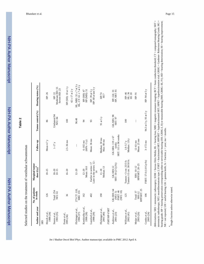
Hearing status after SRS for VS is evaluated using the Gardner-Robertson hearing grade(GRHG) scale, which includes both PTA and speech discrimination scores (SDS) (13). HLafter SRS for VS is commonly presented as pre-RT to post-RT variation in GRHG as: (a)pretreatment hearing preservation (HP) in terms of (i) serviceable hearing (SH), as hearingthat is useful with or without a hearing aid, or (ii) measurable hearing (MH), as any hearingwith detectable audiometric responses; and (b) improvement or loss in hearing expressed aschange in GRHG. Selected studies on the treatment of vestibular schwannomas are shown inTable 2.
Acute SNHL has been reported after SRS (14), but not after fractionated RT. Hearingimpairment has been reported within 3 to 24 months after single-fraction SRS (13,15), witha median time to onset of 4 months (15,16). Although it can occur as early as 3 months aftercompleting fractionated RT, the median latency is 1.5–2.0 years (10,11).
3. CHALLENGES DEFINING VOLUMESComputed tomography (CT)-magnetic resonance imaging fusion is helpful in defining theinner ear. Its small size and location (embedded deep in the temporal bone) make itchallenging to delineate on CT scans and requires the appropriate bone window, level, andimage thickness (preferably ≤1.0 mm). The cochlea is a conical structure with its baseresting anterior to the internal auditory canal and its apex pointed anteriorly, inferiorly, andlaterally, toward the carotid artery. The vestibule is located posterior to the cochlea andlateral to the internal auditory canal. The internal auditory canal is a readily apparentlandmark for identification of the cochlea and vestibule on CT (Figure 2). The volume ofcochlea can be defined on axial CT images as the net volume defined by the bony labyrinth.In adults, the reported average volume of the cochlea using CT varies from 0.13 mL (range,0.11–0.15 mL) (17) to 0.56 mL (range, 0.15–0.91 mL) (5).
4. REVIEW OF DOSE–VOLUME DATAStandard fractionated RT for head-and-neck cancer
A dose–volume analysis is impractical for the cochlea due to its small volume and thelimitations associated with its delineation. Several studies have attempted to relate mean ormedian cochlear dose to persistent hearing loss (6,10,18).
Pan (5) prospectively studied BCTs in 31 patients 1–36 months after unilateral RT withstandard fractionation using changes seen in the contralateral ear as standard (0.25–8 kHz).ΔBCTs >10 dB were rarely seen unless the corresponding difference in mean cochlear dosewas ≥45 Gy. The doses to the contralateral cochlea varied between 0.5 and 31.3 Gy (mean,4.2 Gy).
Honore (10) retrospectively estimated mean cochlear doses in 20 patients with head-and-neck cancer (1.8–4.3 Gy/fraction) and observed ΔBCT 7–79 months post-RT. Doses werereconstructed from patient-specific CT scans or proxy phantoms. A dose-responserelationship was observed for ΔBCT >15 dB at 4 kHz, but not at other frequencies.
Bhandare et al. Page 2
Int J Radiat Oncol Biol Phys. Author manuscript; available in PMC 2012 April 4.
NIH
-PA Author Manuscript
NIH
-PA Author Manuscript
NIH
-PA Author Manuscript

Chen (6) retrospectively studied 22 patients treated with RT for nasopharyngeal cancer (withfraction sizes from 1.6–2.3 Gy and concurrent/adjuvant chemotherapy) and studied ΔBCT12–79 months post-RT. A significant increase in hearing loss (ΔBCT of ≥20 dB at onefrequency or ≥10 dB at two consecutive frequencies) was observed for all frequencies (0.5–4 kHz) when the mean dose received by the cochlea was >48 Gy.
Van der Putten (12) retrospectively evaluated ΔBCT 2–7 years after RT in 21 patients withunilateral parotid tumors (fraction sizes 1.8–3.0 Gy). Using the contralateral ear as a control,SNHL (ΔBCT >15 db difference in ≥three frequencies between 0.25–12 kHz) was seenwhen mean doses received by the cochlea were >50 Gy.
Oh (8) prospectively studied ΔBCTs (0.25–4 kHz) 3–12 months post-RT in 25 patients withnasopharyngeal cancer (fraction size 2 Gy). In this study, the inner ear doses were high (63–70 Gy), and hearing loss (ΔBCT≥15 db from baseline) was associated with total dosereceived by the inner ear.
SRS for vestibular schwannomasVolume–length effect—A dose–volume analysis is not feasible because of the smallnerve diameter, lack of visibility on CT, and variable thickness. Nevertheless, the locationand length of the cochlear nerve involved with tumor and the prescription/marginal tumordose reflect the dose received by the cochlear nerve (16,19). For example, the cochlear nervemay receive less radiation if it lies on the tumor surface vs. if it passes through the core. SRSwas found to be more likely to preserve hearing in patients with small VS (<3 cm) vs. largerlesions (20). When SRS is used to treat intracanalicular VS with an irradiated nerve lengthof 4–12 mm, neither the tumor position in the canal (lateral vs. medial) nor the length of thenerve correlated with long-term hearing preservation. However, the marginal/prescriptiondose to the tumor was significant as was the dose extending beyond the tumor volume insidethe canal was the most important factor responsible for cochlear nerve injury in SRS patients(13). Intracanalicular tumor volume (<100 mm3 vs.≥100 mm3) and intracanalicularintegrated dose (dose × volume) are also thought to influence hearing loss (21).
Total dose effect—In one SRS study, patients receiving a mean maximum cochlearnucleus dose in the brain stem of 6.9 Gy and mean cochlear dose of 9.1 Gy retained usefulhearing, whereas those in patients with hearing declines received 11.1 Gy and 7.8 Gy (22).In another study, serviceable hearing was preserved in 100% of the patients receivingmarginal tumor doses ≤14 Gy but dropped to 20% in those receiving >14 Gy (13). Otherstudies noted increased hearing preservation with marginal tumor doses of 10–16 (vs. 25)Gy (23), and 12–14 (vs. 16–20) Gy (24,25).
5. FACTORS AFFECTING RISKTreatment-related factors
1. The mean total dose to the cochlea during fractionated RT, or to the eighth cranialnerve in SRS for VS, is a dominant factor in post-RT hearing status (see Review ofDose–volume Data).
2. The effect of dose per fraction (≤or >2.0 Gy) has not been thoroughly described.
3. The one study comparing once-daily vs. twice-daily fractionation observed noeffect (4). Some studies suggest that the patients treated for VS with FSRT have abetter chance of maintaining serviceable hearing when compared with those treatedby SRS (23–25). Hypofractionated RT with four fractions of 5 Gy, or five fractionsof 4 Gy, may have less toxicity than SRS in fractions of 10–12 Gy (26).
Bhandare et al. Page 3
Int J Radiat Oncol Biol Phys. Author manuscript; available in PMC 2012 April 4.
NIH
-PA Author Manuscript
NIH
-PA Author Manuscript
NIH
-PA Author Manuscript

4. The possible synergistic toxicity of chemotherapy combined with RT has beenstudied prospectively (5,7,8,11,18), and retrospectively (4,6,10,12). Cisplatin isknown to cause hearing loss (24). Increased toxicity has been observed in patientstreated with both adjuvant and concurrent cisplatin-RT (4,6,18). Low (18) reportedresults at 1 and 2 years after RT delivered with concurrent and adjuvant cisplatinand found significant increases both in BCT at 4 kHz and in BCTs averaged over0.5, 1, and 2 kHz. Conversely, no such increase has been seen in patients treatedwith neoadjuvant cisplatin followed by RT (i.e., without concurrent cisplatin/RT)(7,8,11).
Patient-related factors1. The rate of post-RT SNHL appears to increase with age (>50) (4,5,7,10,11,27).
Grau (28) found a significant relationship between higher patient age and increasedrisk of hearing loss, but, when corrected for dose, the correlation disappeared.Higher rates of post-RT SNHL have been reported in males compared with females(7,11). Other studies have not observed any difference in the incidence of SNHLbetween sexes or races (4).
2. Greater post-RT hearing losses (i.e., greater thresholds) have been associated withbetter pre-RT hearing (i.e., lower thresholds) (5,10).
3. Post-RT otitis media has been associated with an increased risk of SNHL (4,7,11).
4. Compared with sporadic VS, VS secondary to neurofibromatosis (NF2) after SRSor FSRT exhibits lower hearing preservation and increased hearing deterioration(23,29,30).
5. Cerebral spinal fluid shunt has been suggested to increase the risk of HL after RTin children and perhaps adults (31).
6. MATHEMATICAL/BIOLOGICAL MODELSThe values of TD5/5 = 60 Gy, TD50/5 = 70 for SNHL suggested by Emami (34) are notsupported in the literature and should not be utilized in treatment planning. Nevertheless, theinformation on dose–response modeling for post-RT SNHL remains limited.
Pan (5) constructed a linear model demonstrating the differences between pre-RT and post-RT BCTs (corresponding to frequencies varying from 0.25 to 8 kHz) for the ipsilateral andcontralateral ears and their association with relative dose scale, age, test frequency, andbaseline (i.e., pre-RT) BCT and presented these differences in the form of nomograms.Because of its complexity, the details of the model cannot be presented here (5). In brief,hearing loss was found to depend on frequency tested, age, baseline hearing, and dose toinner ear.
Honore (10) presented a logistic model of the probability of post-RT hearing loss ≥15 dB at4 kHz, including only dose, which indicated that D50 = 48 Gy (95% confidence interval notreported) and γ50 = 0.70 (range, 0.22–1.18). Adjusting for patient age and pretreatmenthearing level revealed a steeper dose-response curve with γ50 = 3.4 (95% confidenceinterval, 0.3–6.5).
Their multivariate logistic regression model is presented.
(1)
Bhandare et al. Page 4
Int J Radiat Oncol Biol Phys. Author manuscript; available in PMC 2012 April 4.
NIH
-PA Author Manuscript
NIH
-PA Author Manuscript
NIH
-PA Author Manuscript

Where x1 = dose in Gy, x2 = pretreatment hearing threshold in dB, x3 = observation time inyears, b0 = −24.9, b1 = 0.30 Gy−1 (0.03–0.56), b2 = −0.44 dB−1 (−0.86–0.01), and b3 =0.46 year−1 (0.02–0.90) with a p value of <0.05. Honore (10) also modeled a post-RTincrease in BCT at 4 kHz with multiple linear regressions. Dose, age, and pretherapeutichearing level were significant (p < 0.05), with the coefficients (95% confidence intervals):0.31 (±0.15) dB/Gy, 0.53 (±0.21) dB/year, and −0.28 (±0.22) dB/dB, respectively. Theconstant shift in hearing level in this model, −21.6 (±11.2) dB, was relatively large.
Chen (6) constructed linear models for post-RT changes in BCTs at frequencies between 0.5and 4 kHz and found that dose was significant at all frequencies. In a multivariate linearmodel, RT dose, number of cycles of cisplatin, and time to post-RT hearing test weresignificant at 4 kHz. At 2 and 3 kHz, RT dose and time to posttreatment hearing test weresignificant. At 1 kHz, only RT dose was significant. In addition, hearing loss in the oppositeear was seen to be highly significant, which may provide additional evidence of the toxicityof concurrent plus adjuvant cisplatin.
Van der Putten (12) fitted an NTCP model to the incidence of asymmetrical SNHL (with aminimum of three frequencies from 0.25–12 kHz) as a function of mean dose to theipsilateral inner ear and obtained D50 = 53.2 Gy with γ50 of 2.74 and D10 = 42 Gy.
The incidence of hearing loss at 4 and 2 kHz as reported by Honore (10), Chen (6), and Pan(5) are shown in Figures 1a and 1b. The data of Van der Putten (12), on hearing loss atcombined frequencies, are shown for comparison in Figure 1c. The sources for these dataand caveats concerning the comparisons implied by these plots are given in the figurelegend. It is clear that the response seen by Pan (5) is considerably smaller than that seen bythe other studies. This could be due to a number of factors, the most obvious being therelative endpoint and relative dose scale used by Pan, and the influence of chemotherapy inChen (6). However, the complication rate seen by Honore (10) (in patients treated withoutchemotherapy) is of the same order as that of Chen (6).
Flickinger (19) modeled the effects of minimum tumor dose Dmin and transverse tumordiameter (Td) with multivariate logistic regression analysis (equation 1) for the risk ofacoustic neuropathy (defined as any variation in either PTA or SDS resulting in decline inGRHG for patients with at least Class IV hearing) in patients treated with SRS for VS in twodatasets. The coefficients b1 (1/Gy) for Dmin were 0.166, 0.158 (with respective p =0.00745, 0.1084; SEcoeff, 0.091, 0.097). The coefficients b2 (1/cm) for Td were 0.752, 0.818(with respective p = 0.0079, 0.039; SEcoeff, 0.276, 0.276). The constants b0 were −4.57,−4.48 (with respective p = 0.0044, 0.0076; SEcoeff, 1.56, 1.64).
In addition to the limited information on modeling SNHL, there remain several limitationsin both prospective and retrospective studies in the current literature, such as a relativelysmall number of patients, variation in the standard for HL, frequencies evaluated, and otherapproximations (e.g., the use of a proxy phantom in retrospective studies), thereby makingthe choice of any specific model for routine clinical utilization difficult.
7. SPECIAL SITUATIONS1. Data on cisplatin-RT suggest that radiation doses to the cochlea should be strictly
limited when delivered with cisplatin.
2. Data presented may not be applicable to fractionation schedules beyond the rangesstudied.
3. Data presented in this review apply to adult patients only; for data on pediatricpatients, see Hua et al. (32).
Bhandare et al. Page 5
Int J Radiat Oncol Biol Phys. Author manuscript; available in PMC 2012 April 4.
NIH
-PA Author Manuscript
NIH
-PA Author Manuscript
NIH
-PA Author Manuscript

4. Data for hearing response after SRS or FSRT for sporadic tumors may not berepresentative of the patients with VS secondary to NF2.
8. RECOMMENDED DOSE–VOLUME LIMITS (WHERE POSSIBLE WHILERETAINING THE DESIRED TARGET COVERAGE)
1. For conventionally fractionated RT, to minimize the risk for SNHL, the mean doseto the cochlea should be limited to ≤45 Gy (5,6) (or more conservatively ≤35 Gy)(10). Because a threshold for SNHL cannot be determined from the present data, toprevent SNHL the dose to the cochlea should be kept as low as possible.
2. For SRS for VS, the prescription dose should be limited to 12–14 Gy for hearingpreservation (24,25,33).
3. A suggested hypofractionation schedule for VS, to provide likely tumor control andpreserve hearing, is a total prescription dose of 21–30 Gy in 3–7 Gy per fractionover 3–10 days, though data on this schedule are limited.
9. FUTURE TOXICITY STUDIES1. Larger single and multi-institutional prospective trials utilizing pre- and
posttreatment hearing tests are required to establish absolute hearing loss as afunction of frequency and the absolute radiation dose received by each cochlea, andverify the reported observations regarding SNHL after RT for head-and-neckcancers.
2. The response of SNHL to chemoradiation needs to be determined in prospectivetrials as a function of both cisplatin and radiation doses as well as chemo-regimen(neoadjuvant, concurrent, or adjuvant).
3. In the treatment of VS, the effects of fractionation (SRS vs. FSRT with standardfractionation and hypofractionation), the location and length of the acoustic nerverelative to the tumor, and doses received by it, require systematic prospectiveinvestigation.
10. TOXICITY SCORINGExisting scoring systems (e.g., Radiation Therapy Oncology Group, Late Effects on NormalTissues / Subjective, Objective, Management and Analytic, National Cancer InstituteCommon Terminology Criteria for Adverse Events) have limitations. We make thefollowing recommendations for coding toxicity.
SNHL after fractionated RT for head-and-neck cancers1. Hearing loss should be determined through pre- and post-RT audiometric
evaluations of the same ear. In retrospective studies, if pre-RT audiometricevaluations for the ipsilateral ears are not available, the contralateral ear may bepreferable to an age-specific standard, but both should be viewed as substandardrelative to pre-RT ipsilateral data.
2. To avoid transient post-RT hearing fluctuations, hearing should be tested starting 6months post-RT and at least biannually thereafter.
3. SDS and four-frequency (0.5, 1.0, 2.0, and 3.0 kHz) bone conduction pure toneaverage should be used, as endorsed by the American Academy of Otolaryngology-Head and Neck Surgery Committee on Hearing and Equilibrium (9).
Bhandare et al. Page 6
Int J Radiat Oncol Biol Phys. Author manuscript; available in PMC 2012 April 4.
NIH
-PA Author Manuscript
NIH
-PA Author Manuscript
NIH
-PA Author Manuscript
4. For high-frequency HL, 6 kHz bone conduction thresholds should be measured,because a) the basal turn of the cochlea (i.e., highest frequencies) are the first to beaffected, b) 6 kHz is highest frequency bone conduction threshold measured withstandard bone conducting transducers, and c) bone conduction thresholds minimizethe influence of concomitant middle and external ear pathology.
5. Additionally, a measurement at 4 kHz may facilitate comparison with the presentdatasets.
6. “Clinically significant hearing loss” should be considered as an increase in thethreshold of 10 dB in post-RT BCT, or a decline of 10% in an SDS evaluation, asassessed by an expert.
7. Clinically significant HL observed in two consecutive PTA evaluations isconsidered as persistent.
Toxicity scoring after RT for VS1. Preservation of pretreatment hearing level: (a) preservation pre-RT GRHG I–IV
hearing or (b) in pre-RT GRHG V patients, with no speech discrimination buttestable PTA, a preservation of PTA scores.
2. SH (corresponding to GRHG I–II); commonly defined as PTA ≤ 0 and SDS ≥50%.
3. MH is any hearing with detectable audiometric response.
4. Either an improvement or loss in hearing expressed as a change in GRHG.
REFERENCES1. Jereczek-Fossa BA, Zarowski A, Milani F, et al. Radiotherapy-induced ear toxicity. Cancer Treat
Rev. 2003; 29:417–430. [PubMed: 12972360]2. Bhide SA, Harrington KJ, Nutting CM. Otological toxicity after postoperative radiotherapy for
parotid tumours. Clin Oncol (R Coll Radiol). 2007; 19:77–82. [PubMed: 17305258]3. Raaijmakers E, Engelen AM. Is sensorineural hearing loss a possible side effect of nasopharyngeal
and parotid irradiation? A systematic review of the literature. Radiother Oncol. 2002; 65:1–7.[PubMed: 12413668]
4. Bhandare N, Antonelli PJ, Morris CG, et al. Ototoxicity after radiotherapy for head and necktumors. Int J Radiat Oncol Biol Phys. 2007; 67:469–479. [PubMed: 17236969]
5. Pan CC, Eisbruch A, Lee JS, et al. Prospective study of inner ear radiation dose and hearing loss inhead-and-neck cancer patients. Int J Radiat Oncol Biol Phys. 2005; 61:1393–1402. [PubMed:15817342]
6. Chen WC, Jackson A, Budnick AS, et al. Sensorineural hearing loss in combined modality treatmentof nasopharyngeal carcinoma. Cancer. 2006; 106:820–829. [PubMed: 16421885]
7. Kwong DL, Wei WI, Sham JS, et al. Sensorineural hearing loss in patients treated fornasopharyngeal carcinoma: A prospective study of the effect of radiation and cisplatin treatment. IntJ Radiat Oncol Biol Phys. 1996; 36:281–289. [PubMed: 8892450]
8. Oh YT, Kim CH, Choi JH, et al. Sensory neural hearing loss after concurrent cisplatin and radiationtherapy for nasopharyngeal carcinoma. Radiother Oncol. 2004; 72:79–82. [PubMed: 15236878]
9. Committee on Hearing and Equilibrium. Otolaryngol Head Neck Surg. Vol. 113. AmericanAcademy of Otolaryngology-Head and Neck Surgery Foundation, Inc.; 1995. Committee onHearing and Equilibrium guidelines for the evaluation of hearing preservation in acoustic neuroma(vestibular schwannoma); p. 179-180.
10. Honore HB, Bentzen SM, Moller K, et al. Sensori-neural hearing loss after radiotherapy fornasopharyngeal carcinoma: Individualized risk estimation. Radiother Oncol. 2002; 65:9–16.[PubMed: 12413669]
Bhandare et al. Page 7
Int J Radiat Oncol Biol Phys. Author manuscript; available in PMC 2012 April 4.
NIH
-PA Author Manuscript
NIH
-PA Author Manuscript
NIH
-PA Author Manuscript

11. Ho WK, Wei WI, Kwong DL, et al. Long-term sensorineural hearing deficit followingradiotherapy in patients suffering from nasopharyngeal carcinoma: A prospective study. HeadNeck. 1999; 21:547–553. [PubMed: 10449671]
12. van der Putten L, de Bree R, Plukker JT, et al. Permanent unilateral hearing loss after radiotherapyfor parotid gland tumors. Head Neck. 2006; 28:902–908. [PubMed: 16783830]
13. Niranjan A, Lunsford LD, Flickinger JC, et al. Dose reduction improves hearing preservation ratesafter intracanalicular acoustic tumor radiosurgery. Neurosurgery. 1999; 45:753–762. [PubMed:10515468]
14. Pollack AG, Marymont MH, Kalapurakal JA, et al. Acute neurological complications followinggamma knife surgery for vestibular schwannoma. Case report. J Neurosurg. 2005; 103:546–551.[PubMed: 16235688]
15. Subach BR, Kondziolka D, Lunsford LD, et al. Stereotactic radiosurgery in the management ofacoustic neuromas associated with neurofibromatosis Type 2. J Neurosurg. 1999; 90:815–822.[PubMed: 10223445]
16. Linskey ME, Lunsford LD, Flickinger JC. Tumor control after stereotactic radiosurgery inneurofibromatosis patients with bilateral acoustic tumors. Neurosurgery. 1992; 31:829–838.[PubMed: 1436407]
17. Pacholke HD, Amdur RJ, Schmalfuss IM, et al. Contouring the middle and inner ear onradiotherapy planning scans. Am J Clin Oncol. 2005; 28:143–147. [PubMed: 15803007]
18. Low WK, Toh ST, Wee J, et al. Sensorineural hearing loss after radiotherapy andchemoradiotherapy: A single, blinded, randomized study. J Clin Oncol. 2006; 24:1904–1909.[PubMed: 16622266]
19. Flickinger JC, Kondziolka D, Lunsford LD. Dose and diameter relationships for facial, trigeminal,and acoustic neuropathies following acoustic neuroma radiosurgery. Radiother Oncol. 1996;41:215–219. [PubMed: 9027936]
20. Pollock BE, Lunsford LD, Kondziolka D, et al. Outcome analysis of acoustic neuromamanagement: A comparison of microsurgery and stereotactic radiosurgery. Neurosurgery. 1995;36:215–229. [PubMed: 7708162]
21. Massager N, Nissim O, Delbrouck C, et al. Role of intracanalicular volumetric and dosimetricparameters on hearing preservation after vestibular schwannoma radiosurgery. Int J Radiat OncolBiol Phys. 2006; 64:1331–1340. [PubMed: 16458446]
22. Paek SH, Chung HT, Jeong SS, et al. Hearing preservation after gamma knife stereotacticradiosurgery of vestibular schwannoma. Cancer. 2005; 104:580–590. [PubMed: 15952200]
23. Andrews DW, Suarez O, Goldman HW, et al. Stereotactic radiosurgery and fractionatedstereotactic radiotherapy for the treatment of acoustic schwannomas: Comparative observations of125 patients treated at one institution. Int J Radiat Oncol Biol Phys. 2001; 50:1265–1278.[PubMed: 11483338]
24. Combs SE, Volk S, Schulz-Ertner D, et al. Management of acoustic neuromas with fractionatedstereotactic radiotherapy (FSRT): Long-term results in 106 patients treated in a single institution.Int J Radiat Oncol Biol Phys. 2005; 63:75–81. [PubMed: 16111574]
25. Williams, J. Fractionated radiotherapy for acoustic neuromas. Congress of Neurological Surgeons:50th Annual Meeting; San Antonio, TX. 2000. p. 155
26. Meijer OW, Vandertop WP, Baayen JC, et al. Single-fraction vs. fractionated linac-basedstereotactic radiosurgery for vestibular schwannoma: A single-institution study. Int J Radiat OncolBiol Phys. 2003; 56:1390–1396. [PubMed: 12873685]
27. Moretti JA. Sensori-neural hearing loss following radiotherapy to the nasopharynx. Laryngoscope.1976; 86:598–602. [PubMed: 1263730]
28. Grau C, Overgaard J. Postirradiation sensorineural hearing loss: A common but ignored lateradiation complication. Int J Radiat Oncol Biol Phys. 1996; 36:515–517. [PubMed: 8892478]
29. Flickinger JC, Lunsford LD, Linskey ME, et al. Gamma knife radiosurgery for acoustic tumors:multivariate analysis of four year results. Radiother Oncol. 1993; 27:91–98. [PubMed: 8356233]
30. Rowe JG, Radatz MW, Walton L, et al. Clinical experience with gamma knife stereotacticradiosurgery in the management of vestibular schwannomas secondary to type 2neurofibromatosis. J Neurol Neurosurg Psychiatry. 2003; 74:1288–1293. [PubMed: 12933938]
Bhandare et al. Page 8
Int J Radiat Oncol Biol Phys. Author manuscript; available in PMC 2012 April 4.
NIH
-PA Author Manuscript
NIH
-PA Author Manuscript
NIH
-PA Author Manuscript

31. Merchant TE, Gould CJ, Xiong X, et al. Early neuro-otologic effects of three-dimensionalirradiation in children with primary brain tumors. Int J Radiat Oncol Biol Phys. 2004; 58:1194–1207. [PubMed: 15001264]
32. Hua C, Bass JK, Khan R, et al. Hearing loss after radiotherapy for pediatric brain tumors: Effect ofcochlear dose. Int J Radiat Oncol Biol Phys. 2008; 72:892–899. [PubMed: 18395355]
33. Flickinger JC, Kondziolka D, Niranjan A, et al. Acoustic neuroma radiosurgery with marginaltumor doses of 12 to 13 Gy. Int J Radiat Oncol Biol Phys. 2004; 60:225–230. [PubMed:15337560]
34. Hirsch A, Noren G. Audiological findings after stereotactic radiosurgery in acoustic neurinomas.Acta Otolaryngol. 1988; 106:244–251. [PubMed: 3051887]
35. Noren G, Greitz D, Hirsch A, et al. Gamma knife surgery in acoustic tumors. Acta NeurochirSuppl (Wien). 1993; 58:104–107. [PubMed: 8109269]
36. Foote RL, Coffey RJ, Swanson JW, et al. Stereotactic radiosurgery using the gamma knife foracoustic neuromas. Int J Radiat Oncol Biol Phys. 1995; 32:1153–1160. [PubMed: 7607937]
37. Flickinger JC, Kondziolka D, Pollock BE, et al. Evolution in technique for vestibular schwannomaradiosurgery and effect on outcome. Int J Radiat Oncol Biol Phys. 1996; 36:275–280. [PubMed:8892449]
38. Kondziolka D, Lunsford LD, McLaughlin MR, et al. Long-term outcomes after radiosurgery foracoustic neuromas. N Engl J Med. 1998; 339:1426–1433. [PubMed: 9811917]
39. Lunsford, LD.; Kondziolka, D.; Flickinger, JC., et al. Acoustic neuroma management: Evolutionand revolution. In: Kondziolka, D., editor. Radiosurgery. 2nd ed.. Basel: Karger; 1998. p. 1-7.
40. Flickinger JC, Kondziolka D, Niranjan A, et al. Results of acoustic neuroma radiosurgery: Ananalysis of 5 years’ experience using current methods. J Neurosurg. 2001; 94:1–6. [PubMed:11147876]
41. Williams JA. Fractionated stereotactic radiotherapy for acoustic neuromas. Int J Radiat Oncol BiolPhys. 2002; 54:500–504. [PubMed: 12243828]
Bhandare et al. Page 9
Int J Radiat Oncol Biol Phys. Author manuscript; available in PMC 2012 April 4.
NIH
-PA Author Manuscript
NIH
-PA Author Manuscript
NIH
-PA Author Manuscript

Fig. 1.Mean dose response for sensorineural hearing loss (SNHL) at (a): 4 kHz; (b): 0.5–2 kHz;and (c): all frequencies (0.25–12 kHz). Data from: Figure 3 of Chen et al. (6) (retrospectivestudy; SNHL defined as a ≥20-dB increase in the bone-conduction threshold at ≥1 year;patients received concurrent and adjuvant cisplatin chemotherapy); Figure 1 of Honore et al.(10) (retrospective study; SNHL defined as 20-dB increase in the bone-conduction thresholdat ~0.5–6.5 years); Figure 2 of Pan et al. (5) (prospective study; SNHL defined as a 20-dBdifference between bone-conduction thresholds for ipsilateral and contralateral ears at 1year; doses are ipsilateral-ear mean doses minus contralateral-ear mean doses); Table 2 ofOh et al. (8) (prospective study; SNHL defined as a 15-dB increase in the bone-conduction
Bhandare et al. Page 10
Int J Radiat Oncol Biol Phys. Author manuscript; available in PMC 2012 April 4.
NIH
-PA Author Manuscript
NIH
-PA Author Manuscript
NIH
-PA Author Manuscript

threshold at 1 year; patients received neoadjuvant and concurrent cisplatin chemotherapy);Table 1 and Table 2 of Kwong et al. (7) (prospective study; SNHL defined as a 15-dBincrease in the bone-conduction threshold at 1 year; patients received neoadjuvant andconcurrent chemotherapy; ears received the full prescription dose; prescriptions wereconverted to biologically effective dose in 2 Gy fractions using α/β = 3 Gy); Fig 2 of van derPutten et al. (12) (retrospective study; SNHL defined as a 15-dB increase in the average ofall pure-tone thresholds at 2–17 years).
Bhandare et al. Page 11
Int J Radiat Oncol Biol Phys. Author manuscript; available in PMC 2012 April 4.
NIH
-PA Author Manuscript
NIH
-PA Author Manuscript
NIH
-PA Author Manuscript
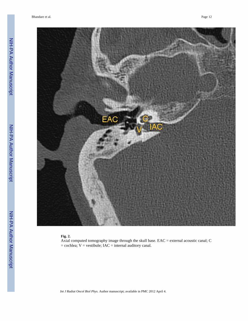
Fig. 2.Axial computed tomography image through the skull base. EAC = external acoustic canal; C= cochlea; V = vestibule; IAC = internal auditory canal.
Bhandare et al. Page 12
Int J Radiat Oncol Biol Phys. Author manuscript; available in PMC 2012 April 4.
NIH
-PA Author Manuscript
NIH
-PA Author Manuscript
NIH
-PA Author Manuscript

NIH
-PA Author Manuscript
NIH
-PA Author Manuscript
NIH
-PA Author Manuscript
Bhandare et al. Page 13
Tabl
e 1
Sele
cted
stud
ies f
or S
NH
L af
ter h
ead-
and-
neck
radi
atio
n th
erap
y
Influ
ence
of v
aria
bles
on
the
outc
ome
Aut
hor
Num
ber
ofpa
tient
s in
stud
y
Mea
n co
chle
ardo
se (G
y)/R
xdo
se (G
y)D
ose
per
frac
tion
(Gy)
Che
mor
adia
tion
(cis
plat
in b
ased
)C
hem
o-ra
diat
ion
Age
Post
-RT
SOM
Gen
der
Tim
e to
hear
ing
test
Pre-
RT
hear
ing
leve
lSt
anda
rd u
sed
for
com
pari
son
End
poin
t for
SNH
L(s
hift
inB
CT
)/fr
eque
ncie
s(k
Hz)
test
ed
Pros
pect
ive
Gra
u et
al.,
1
999
(28)
22N
S/60
–68
2–2.
81N
o, R
T al
one
—N
o*—
—N
oN
oSa
me
ear
Nom
inal
shift
s
in B
CT
(indB
)
repo
rted/
0.5,
1.0
,
2.0
,4.0
Kw
ong
etal
.,
199
6 (7
)
132
NS/
71.3
–85
2–3.
5/2†
Yes
‡ , n
eoad
juva
ntN
oY
es§
Yes
Yes║
Yes
No
Sam
e ea
r15
/avg
. of
0.5,
1, 2
15;
4
Ho
et a
l.,19
99 (1
1)29
470
–91†
/59.
9–70
2–3.
5/2†
Yes
‡ , n
eoad
juva
ntN
oY
es—
—Y
esN
oSa
me
ear
10/a
vg. o
f(0
.5, 1
, 2) 1
0;4¶
Oh
et a
l.,
200
4 (8
)32
54.3
–81.
4/70
2Y
es‡ ,
neo
adju
vant
a
nd c
oncu
rren
tN
oY
es‡*
*Y
esY
es║
Yes
—Sa
me
ear
15/a
vg. o
f(0
.5, 1
, 2) 1
5;4
Pan
et a
l.,
200
5 (5
)22
Ipsi
:‡ 1
4.1–
68.8
C
ontra
: ‡
0.5
–31.
3/40
–70
NS
RT
alon
e (1
8)
Con
curr
ent
c
hem
o. (4
)
—Y
es—
No
No
Yes
††C
ontra
late
ral
e
ar20
/0.2
5, 0
.5,
1, 2
¶ , 4
¶ , 8
Low
et a
l.,
200
6 (1
8)11
5N
S/70
2Y
es‡ ,
con
curr
ent
a
nd a
djuv
ant
Yes
(4 k
Hz)
——
——
—Sa
me
ear
Nom
inal
shift
s
in B
CT
(indB
)
repo
rted/
4,av
g.
of (
0.5,
1.0,
2.0
)
Retr
ospe
ctiv
e
Hon
ore
etal
.,
200
2 (1
0)
207.
1–68
/ 50–
682–
4.3
No,
RT
alon
e—
Yes
——
No
Yes
††Sa
me
ear
15/0
.5, 1
, 2, 4
20; 4
¶
Int J Radiat Oncol Biol Phys. Author manuscript; available in PMC 2012 April 4.

NIH
-PA Author Manuscript
NIH
-PA Author Manuscript
NIH
-PA Author Manuscript
Bhandare et al. Page 14
Influ
ence
of v
aria
bles
on
the
outc
ome
Aut
hor
Num
ber
ofpa
tient
s in
stud
y
Mea
n co
chle
ardo
se (G
y)/R
xdo
se (G
y)D
ose
per
frac
tion
(Gy)
Che
mor
adia
tion
(cis
plat
in b
ased
)C
hem
o-ra
diat
ion
Age
Post
-RT
SOM
Gen
der
Tim
e to
hear
ing
test
Pre-
RT
hear
ing
leve
lSt
anda
rd u
sed
for
com
pari
son
End
poin
t for
SNH
L(s
hift
inB
CT
)/fr
eque
ncie
s(k
Hz)
test
ed
Che
n et
al.,
2
006
(6)
2228
.4–7
01.
6–2.
34Y
es, c
oncu
rren
t
and
adj
uvan
tY
es (4
kH
z)N
oN
o—
Yes
No
Sam
e ea
r20
/0.5
, 1, 2
¶ ,3,
4¶
Van
der
Putte
n
et a
l.,20
06 (1
2)
5229
.2–7
7.3/
50–7
01.
8–3.
0N
o, R
T al
one
——
——
——
Con
trala
tera
l15
/0.2
5–12
fo
r ≥3
ofth
ese
fr
eque
ncie
s
Abbr
evia
tions
: NS
= no
t spe
cifie
d; A
S =
abso
lute
shift
in th
e he
arin
g th
resh
old
repo
rted;
SO
M =
sero
us o
titis
med
ia; R
T =
radi
atio
n th
erap
y; C
T =
bone
con
duct
ion
thre
shol
d; d
b =
deci
bels
; SN
HL
=se
nsor
ineu
ral h
earin
g lo
ss; R
x =
pres
crip
tion.
* Dos
e an
d ag
e co
mpo
nent
of H
L se
para
ted.
† Tota
l dos
es c
alcu
late
d as
BED
in 2
Gy
frac
tions
, with
α/β
= 3
Gy.
‡ The
prim
ary
endp
oint
of a
pro
spec
tive
clin
ical
tria
l.
§ Old
er a
ge fo
und
sign
ifica
nt.
║R
ate
of H
L m
ale
> fe
mal
e.
¶ Dat
a fo
r the
se e
ndpo
ints
reco
nstru
cted
from
figu
res f
or th
is p
aper
.
**Y
oung
er a
ge fo
und
sign
ifica
nt.
††B
ette
r pre
-RT
hear
ing
asso
ciat
ed w
ith w
orse
pos
t RT
HL.
Int J Radiat Oncol Biol Phys. Author manuscript; available in PMC 2012 April 4.
NIH
-PA Author Manuscript
NIH
-PA Author Manuscript
NIH
-PA Author Manuscript
Bhandare et al. Page 15
Tabl
e 2
Sele
cted
stud
ies o
n th
e tre
atm
ent o
f ves
tibul
ar sc
hwan
nom
as
Aut
hor
and
year
No.
of p
atie
nts
in st
udy
Mar
gina
l tum
ordo
se (G
y)*
Follo
w-u
pT
umor
con
trol
(%)
Hea
ring
stat
us (%
)
SRS
Hirs
ch e
t al.,
1
988
(34)
126
18–2
5M
ean
4.7
y86
HP:
26
Nor
en e
t al.,
1
993
(35)
Tota
l: 25
4N
F2: 6
118
–20
10–1
51–
17 y
Uni
late
ral:9
4N
F2: 8
4H
P: 2
2M
oder
ate
HD
: 55
Seve
re H
D: 2
3
Foot
e et
al.,
1995
(36)
3616
–20
2.5–
36 m
o10
0H
P (S
H):
10 a
t 1 y
42 ±
17
at 2
y
Flic
king
er e
t al.,
1
996
(37)
273
CT:
118
,M
RI:
155
12–2
0—
96.4
8H
L, M
RI:
32 ±
7 a
t 3 y
HL,
CT:
61
± 7
at 3
y
Kon
dzio
lka
et a
l.,
199
8 (3
8)16
212
–20
Mea
n: 1
6.6
6–10
2 m
o(6
0% >
5 y)
94H
P (S
H):
47H
P (M
H):
51
Luns
ford
et a
l.,
199
8 (3
9)40
2Ea
rlier
in se
ries:
17
Late
r in
the
serie
s: 1
2–14
Mea
n: 3
6 m
o93
HP:
39
at 5
yH
P: 6
8 at
last
5 y
Flic
king
er e
t al.,
2
001
(40)
190
11–1
8M
edia
n: 1
3M
edia
n: 3
0 m
oM
ax: 8
0 m
o91
at 5
yH
P:74
HI:7
FSRT
/HP-
FSRT
And
rew
s et a
l.,
200
1 (2
3)G
K-S
RS:
64
(NF2
: 5)
FSR
T: 4
6(N
F2: 1
0)
GK
-SR
S:12
SRT:
50
(2 G
y/fx
)G
K-S
RS:
119
± 6
7w
eeks
SRT:
115
± 9
6 w
eeks
GK
-SR
S: 9
8SR
T: 9
7H
P, G
K: 3
3H
P, S
RT:
81
Will
iam
s et a
l.,
200
2 (4
1)12
5Tu
mor
s <3
cm: 2
5/5
fxTu
mor
s ≥3
cm: 3
0/10
fx1.
0–5.
7 y
Med
ian:
1.8
y10
0H
P: 4
6H
L: 3
6H
I: 18
Mei
jer e
t al.,
2
003
(26)
Tota
l: 37
SRS:
12H
PFSR
T: 2
5
SRS:
10–
12H
PFSR
T: 2
0–25
12–6
1 m
oM
ean:
25
mo
—H
P: 9
1
Com
bs e
t al.,
2
005
(24)
106
FSR
T: 5
7.6
(1.8
Gy/
fx)
3–17
2 m
o94
.3 a
t 3 y
, 93
at 5
yH
P: 9
4 at
5 y
Abbr
evia
tions
: SR
S =
ster
eota
ctic
radi
osur
gery
; SO
M =
Ser
ous O
titis
Med
ia; H
L =
hear
ing
loss
; MR
I = m
agne
tic re
sona
nce
imag
ing;
BC
T =
bone
con
duct
ion
thre
shol
d; C
T =
com
pute
d to
mog
raph
y; S
RT
=st
ereo
tact
ic ra
diot
hera
py; N
F2: n
euro
fibro
mat
osis
type
2; F
SRT
= fr
actio
nate
d SR
T; H
PFSR
T =
hypo
frac
tiona
ted
SRT;
HPR
T =
hypo
frac
tiona
tion
trial
; GR
HG
= G
arde
ner-
Rob
erts
on H
earin
g G
rade
; HG
=he
arin
g gr
ade;
HP
= he
arin
g pr
eser
vatio
n co
rres
pond
ing
eith
er to
serv
icea
ble
hear
ing
(SH
; GR
HG
-I, I
I) o
r mea
sura
ble
hear
ing
(MH
; GR
HG
: III
, IV
); H
D =
hea
ring
dete
riora
tion;
HI =
hea
ring
impr
ovem
ent;
NR
= n
ot re
porte
d; U
H =
use
ful h
earin
g; G
K =
gam
ma
knife
; fx
= fr
actio
n; y
= y
ear;
mo
= m
onth
s.
* Sing
le fr
actio
n un
less
oth
erw
ise
stat
ed.
Int J Radiat Oncol Biol Phys. Author manuscript; available in PMC 2012 April 4.