Loss of mitochondrial peptidase Clpp leads to infertility, hearing loss plus growth retardation via...
17
Loss of mitochondrial peptidase Clpp leads to infertility, hearing loss plus growth retardation via accumulation of CLPX, mtDNA and inflammatory factors Suzana Gispert 1 , Dajana Parganlija 1 , Michael Klinkenberg 1 , Stefan Dro ¨ se 2 , Ilka Wittig 2 , Michel Mittelbronn 3 , Pawel Grzmil 4,5 , Sebastian Koob 6 , Andrea Hamann 7 , Michael Walter 8 , Finja Bu ¨ chel 9 , Thure Adler 10,11 , Martin Hrabe ´ de Angelis 10 , Dirk H. Busch 11 , Andreas Zell 9 , Andreas S. Reichert 6 , Ulrich Brandt 2, { , Heinz D. Osiewacz 7 , Marina Jendrach 1 and Georg Auburger 1,∗ 1 Experimental Neurology, 2 Molecular Bioenergetics Group, Center of Biological Chemistry, Cluster of Excellence Macromolecular Complexes and 3 Edinger Institute (Neurological Institute), Goethe University Medical School, 60590 Frankfurt am Main, Germany 4 Institute of Human Genetics, Georg-August-University of Go ¨ ttingen, 37073 Go ¨ ttingen, Germany 5 Department of Genetics and Evolution, Jagiellonian University, 30-387 Krako ´ w, Poland 6 Mitochondrial Biology, Buchmann Institute for Molecular Life Sciences and Mitochondrial Biology, Center for Molecular Medicine, Goethe University, 60438 Frankfurt am Main, Germany 7 Faculty for Biosciences, Molecular Developmental Biology, Cluster of Excellence Macromolecular Complexes, Goethe University, 60590 Frankfurt am Main, Germany 8 Institute for Medical Genetics and 9 Center for Bioinformatics Tuebingen (ZBIT), Eberhard-Karls-University of Tuebingen, 72076 Tu ¨ bingen, Germany 10 German Mouse Clinic, Institute of Experimental Genetics, Helmholtz Zentrum Mu ¨ nchen, Mu ¨ nchen, Germany and 11 Institute for Medical Microbiology, Immunology and Hygiene, Technische Universita ¨ t, Mu ¨ nchen, Germany Received June 4, 2013; Revised and Accepted July 9, 2013 The caseinolytic peptidase P (CLPP) is conserved from bacteria to humans. In the mitochondrial matrix, it multi- merizes and forms a macromolecular proteasome-like cylinder together with the chaperone CLPX. In spite of a known relevance for the mitochondrial unfolded protein response, its substrates and tissue-specific roles are unclear in mammals. Recessive CLPP mutations were recently observed in the human Perrault variant of ovarian failure and sensorineural hearing loss. Here, a first characterization of CLPP null mice demonstrated complete female and male infertility and auditory deficits. Disrupted spermatogenesis already at the spermatid stage and ovarian follicular differentiation failure were evident. Reduced pre-/post-natal survival and marked ubiquitous growth retardation contrasted with only light impairment of movement and respiratory activities. Interestingly, the mice showed resistance to ulcerative dermatitis. Systematic expression studies detected up-regulation of other mitochondrial chaperones, accumulation of CLPX and mtDNA as well as inflammatory fac- tors throughout tissues. T-lymphocytes in the spleen were activated. Thus, murine Clpp deletion represents a faithful Perrault model. The disease mechanism probably involves deficient clearance of mitochondrial compo- nents and inflammatory tissue destruction. † Present address: Radboud University Nijmegen Medical Centre, Nijmegen Centre for Mitochondrial Disorders, Geert Groteplein-Zuid 10, 6525 GA Nijmegen, The Netherlands. ∗ To whom correspondence should be addressed at: Experimental Neurology, Building 89, Goethe University Medical School, Theodor Stern Kai 7, 60590 Frankfurt am Main, Germany. Tel: +49 6963017428; Fax: +49 6963017128; Email: [email protected] # The Author 2013. Published by Oxford University Press. All rights reserved. For Permissions, please email: [email protected] Human Molecular Genetics, 2013, Vol. 22, No. 24 4871–4887 doi:10.1093/hmg/ddt338 Advance Access published on July 12, 2013 at UB Frankfurt/Main on February 17, 2014 http://hmg.oxfordjournals.org/ Downloaded from
-
Upload
independent -
Category
Documents
-
view
1 -
download
0
Transcript of Loss of mitochondrial peptidase Clpp leads to infertility, hearing loss plus growth retardation via...

Loss of mitochondrial peptidase Clpp leads toinfertility, hearing loss plus growth retardationvia accumulation of CLPX, mtDNA andinflammatory factors
Suzana Gispert1, Dajana Parganlija1, Michael Klinkenberg1, Stefan Drose2, Ilka Wittig2, Michel
Mittelbronn3, Pawel Grzmil4,5, Sebastian Koob6, Andrea Hamann7, Michael Walter8, Finja Buchel9,
Thure Adler10,11, Martin Hrabe de Angelis10, Dirk H. Busch11, Andreas Zell9, Andreas S. Reichert6,
Ulrich Brandt2,{, Heinz D. Osiewacz7, Marina Jendrach1 and Georg Auburger1,∗
1Experimental Neurology, 2Molecular Bioenergetics Group, Center of Biological Chemistry, Cluster of Excellence
Macromolecular Complexes and 3Edinger Institute (Neurological Institute), Goethe University Medical School, 60590
Frankfurt am Main, Germany 4Institute of Human Genetics, Georg-August-University of Gottingen, 37073 Gottingen,
Germany 5Department of Genetics and Evolution, Jagiellonian University, 30-387 Krakow, Poland 6Mitochondrial
Biology, Buchmann Institute for Molecular Life Sciences and Mitochondrial Biology, Center for Molecular Medicine,
Goethe University, 60438 Frankfurt am Main, Germany 7Faculty for Biosciences, Molecular Developmental Biology,
Cluster of Excellence Macromolecular Complexes, Goethe University, 60590 Frankfurt am Main, Germany 8Institute for
Medical Genetics and 9Center for Bioinformatics Tuebingen (ZBIT), Eberhard-Karls-University of Tuebingen, 72076
Tubingen,Germany 10GermanMouseClinic, InstituteofExperimentalGenetics,HelmholtzZentrumMunchen, Munchen,
Germany and 11Institute for Medical Microbiology, Immunology and Hygiene, Technische Universitat, Munchen,
Germany
Received June 4, 2013; Revised and Accepted July 9, 2013
The caseinolytic peptidase P (CLPP) is conserved from bacteria to humans. In the mitochondrial matrix, it multi-merizes and forms a macromolecular proteasome-like cylinder together with the chaperone CLPX. In spite of aknown relevance for the mitochondrial unfolded protein response, its substrates and tissue-specific roles areunclear in mammals. Recessive CLPP mutations were recently observed in the human Perrault variant of ovarianfailure and sensorineural hearing loss. Here, a first characterization of CLPP null mice demonstrated completefemale and male infertility and auditory deficits. Disrupted spermatogenesis already at the spermatid stage andovarian follicular differentiation failure were evident. Reduced pre-/post-natal survival and marked ubiquitousgrowth retardation contrasted with only light impairment of movement and respiratory activities.Interestingly, the mice showed resistance to ulcerative dermatitis. Systematic expression studies detectedup-regulation ofothermitochondrialchaperones,accumulationofCLPXandmtDNAaswellas inflammatory fac-tors throughout tissues. T-lymphocytes in the spleen were activated. Thus, murine Clpp deletion represents afaithful Perrault model. The disease mechanism probably involves deficient clearance of mitochondrial compo-nents and inflammatory tissue destruction.
†Present address: Radboud University Nijmegen Medical Centre, Nijmegen Centre for Mitochondrial Disorders, Geert Groteplein-Zuid 10, 6525 GANijmegen, The Netherlands.
∗To whom correspondence should be addressed at: Experimental Neurology, Building 89, Goethe University Medical School, Theodor Stern Kai 7,60590 Frankfurt am Main, Germany. Tel: +49 6963017428; Fax: +49 6963017128; Email: [email protected]
# The Author 2013. Published by Oxford University Press. All rights reserved.For Permissions, please email: [email protected]
Human Molecular Genetics, 2013, Vol. 22, No. 24 4871–4887doi:10.1093/hmg/ddt338Advance Access published on July 12, 2013
at UB
Frankfurt/Main on February 17, 2014
http://hmg.oxfordjournals.org/
Dow
nloaded from

INTRODUCTION
In the cytosol of bacteria, the degradation of proteins andpolypeptides is performed by LON and CLPP (caseinolytic pep-tidase P), among other proteases (1). These two enzymes showhigh phylogenetic conservation, but their complementary rolesare not understood. Mitochondrial LONP1 contains domainswith protease and ATPase activity on the same polypeptide,and is considered as the main factor for proteolysis of solublesubstrates in the matrix compartment. Furthermore, mutantLON/PIM1 yeast strains also exhibit defects in mitochondrialDNA (mtDNA) (2). In contrast, mitochondrial CLPP lacksATPase activity and each subunit contains only the domain fordigestion of small peptides without ATP requirement. Throughassociation with the ATPase and chaperone CLPX, activationof CLPP as serine protease occurs for the ATP-dependent diges-tion of larger proteins (2). Both CLPP and CLPX have beenstudied intensively in Escherichia coli (E. coli) and other prokar-yotes (3). Purification and structural biology studies showed thathexameric rings of the ATPase CLPX or specific other AAA+(ATPases Associated with diverse cellular Activities) proteinsof the HSP100 family (CLPA, CLPC) (4,5) depend on ATP inorder to bind two heptameric CLPP rings at either end, thusforming a barrel-shaped CLPP proteolytic chamber. Unfoldedsubstrates are translocated by CLPX into this cylinder to becleaved in fragments with an average product length of 10 resi-dues. These products are further degraded to free amino acids byexopeptidases (3). CLPX determines substrate specificity of thecomplex. CLPX was found to recognize specific unstructuredpeptide sequences and usually unfold the corresponding pro-teins. The unstructured domains are named degrons, such asthe C-motif 1/ssrA tag (6). Degrons are thought to be exposed,e.g. in nascent peptides on stalled ribosomes, as well as in unas-sembled subunits of ribosomes and other macromolecular com-plexes (3). Several specific degrons were characterized in detail.Individual repair proteins like RecN are synthesized at highlevels during cell stress, but efficiently removed by the CLPXPprotein complex upon return to the unstressed state, to avoiddeleterious effects (7,8). Similarly, a degron in sigmaS, themaster regulator of general stress response in E. coli, is respon-sible for CLPXP recognition and degradation in growing cells(9). With a slightly different mechanism, the degron in RseAensures its degradation by CLPXP in times of cell stress, thusderepressing the sigmaE factor to induce genes responsible forthe envelopestress response (10). Interestingly,CLPXP-mediateddegradation of the bacteriophage repressor and of the O protein asan initiator of phage DNA replication (11,12) vary with growthconditions and thus modulate the bacteriophage lysis-versus-lysogenization decisions, i.e. bacterial immunity.
Eukaryotic cells contain CLPP and CLPX orthologs in themitochondrial matrix. Contrary to the vast knowledge aboutCLPP structure and biochemistry in prokaryotes, relativelylittle is known about the biological role of CLPP in eukaryotes.In good agreement with a CLPP role in cell stress and proteinquality control, its expression is induced by stressors in eukar-yotes as in prokaryotes (13–15). Recent data showed a surpris-ing increase in healthy lifespan due to a deletion of the geneencoding CLPP in the filamentous fungus Podospora anserina(16). In Caenorhabditis elegans, the loss of CLPP modulatesmitochondrial unfolded protein responses (17,18). Mammalian
studies reported the accumulation of unfolded proteins in themitochondrial matrix to induce the expression of CLPP in paral-lel to mitochondrial chaperones such as mtHSP60, mtHSP10 andmtDNAJ, and in dependence on the transcription factors CHOPand C/EBPbeta (13,19,20). Human CLPXP was found to consti-tute a bona fide ATP-dependent protease and assemble similarlyas E. coli CLPXP, but to differ in substrate selection (21).
In humans, recessive CLPP missense and splice-donor muta-tions are the cause of ovarian failure and sensorineural deafnessin Perrault syndrome, as reported during the preparation of ourCLPP-null-mouse manuscript (22). An additional role of CLPPin neurodegenerative diseases is suggested by several observa-tions. CLPP mRNA and protein levels are down-regulated in re-sponse to a dominant negative mutation of mtHSP60 that resultsin a clinical manifestation as spastic paraplegia (23,24). A pro-gressive increase of CLPP protein levels due to the disturbed as-sembly of mitochondrial iron–sulfur clusters results fromfrataxin deficiency in Friedreich’s ataxia (25). CLPXP or othermitochondrial proteases, as HTRA2, PARL, the SPG7/AFG3L2subunits of the m-AAA complex, as well as OPA1, were alsoimplicated in age-associated neurodegeneration syndromes (26)such as Parkinson’s disease (27–29), spinocerebellar ataxia(30,31), spastic ataxia—neuropathy syndrome (32), spastic para-plegia (33) and optic atrophy (34), multiple diseases with estab-lished pathology regarding respiration, lipid organelles, proteinaggregates and neuroinflammation. Thus, the mitochondrialprotein quality control system and in particular CLPXP has an im-portant impact on fertility, survival and neural aging (35–37).
We endeavored to understand CLPP functions in the mitochon-dria and in the stress response of mammalian cells, investigatingfor the first time the Clpp gene ablation effects in mouse and char-acterizing CLPP-dependent phenotypes, histological, biochem-ical, cellular and molecular anomalies. Early, severe andtissue-specific pathologies were observed, which represent aquite faithful model of the human Perrault syndrome. Ovarian dys-genesis was explained by follicular differentiation failure, and inaddition testis pathology due to spermatic differentiation failurewas documented, resulting in complete female and male infertilityin mice. Severe tissue-specific pathology was also observed forauditory responses in analogy to Perrault syndrome. The under-lying chain of molecular eventswas assessed by hypothesis-drivenand unbiased expression studies, demonstrating ubiquitous effectsfor the transcriptional up-regulation of other mitochondrialchaperones, the CLPX accumulation in spite of mRNA down-regulation, the mtDNA accumulation, the induction of an in-flammatory transcriptome signature and the growth impairment.In contrast, the respiratory dysfunction was marginal. Thus, theabsence of CLPP is compensated well by most tissues althoughlight inflammatory stress is triggered by the mitochondrialquality control failure. Profound early pathology occurs ingonadcells thatarecharacterizedbyphysiologicaldramatic reduc-tions of mitochondrial numbers, underscoring the importance ofCLPP for the degradation of mitochondrial components.
RESULTS
Two independent founder lines with successful Clpp geneablation show widespread CLPX protein increase
To characterize the role of CLPP in a mammalian system, weundertook the established approach through constitutive
4872 Human Molecular Genetics, 2013, Vol. 22, No. 24
at UB
Frankfurt/Main on February 17, 2014
http://hmg.oxfordjournals.org/
Dow
nloaded from

mouse mutants (38). The murine Clpp gene was already charac-terized and shown to be highly expressed in skeletal muscle,heart and liver, encoding a mature protein of 34 kDa in size(39). Mouse embryonal stem cells with insertions of the gene-trap construct within the Clpp gene were commercially availablefrom the Texas Institute for Genomic Medicine (TIGM). Embry-onal stem cell lines IST11134F10 (line F) and IST13563G11(line G) with GeneTrap insertions in heterozygous state atClpp intron 1 and 2, respectively (Fig. 1A), were obtained anda reduction in Clpp transcript levels to �50% was demonstratedby real-time RT–PCR (data not shown). Heterozygous breedersfrom each founder line were purchased and mated to study theClpp2/2 mutant and the littermate control offspring in compari-son. Almost complete absence of Clpp transcripts due to theGeneTrap insertion was observed in all tissues studied fromClpp2/2 animals (Fig. 1B). The absence of CLPP protein wasalso confirmed in four representative tissues, i.e. liver (chosenfor high mitochondrial content), testis (maximal mitochondrialfusion within sperm midpiece), ovary (progressive failure in Per-rault syndrome), brain (neural tissue with high respiration andcomplex mitochondrial distribution) and heart (high respiration
and mitochondria surrounding muscular contractile apparatus)(Fig. 1C).
CLPP deficiency resulted in the accumulation of its directinteractor protein CLPX, in comparison with usually lowCLPX levels observed in the corresponding wild-type (WT)tissues (Fig. 1C and Supplementary Material, Fig. S1). ThisCLPX increase was consistently observed in all tissues by theSigma antibody against the N-terminal amino acids 40–132 ofCLPX (Fig. 1C) and by the Abgent antibody against theC-terminal amino acids 452–481 of CLPX (Supplementary Ma-terial, Fig. S1). The relevance of additional bands with CLPXimmunoreactivity cannot be interpreted in absence of aCLPX-knock-out tissue, but it is important to note that two iso-forms of Clpx mRNA were previously described in testis (40).Similar amounts and parallel regulation of CLPP and CLPXwould be expected in view of their cooperative functions andof previous proteomic data reporting similar intermediatelevels in 14 mouse tissues (41), but previous reports on ClppmRNA showed the levels to vary between tissues (39,40,42).In conclusion, we observed an inverse correlation betweenCLPP and CLPX levels in the null mouse, suggesting that
Figure 1. Two independent founder lines with CLPP null mutation show CLPX protein accumulation. (A) Scheme of the Clpp gene exon-intron structure, codingregion (broad bar from ATG) and of the two independent gene-trap insertion sites F (left arrow) and G (arrow further right). (B) Absence of Clpp transcript inbrain hemispheres of 5-month-old line G Clpp2/2 homozygotes. The picture represents RT–PCR products with primers situated in exons 5/6 or 3/4, respectively,with lanes for three Clpp2/2 versus three WT animals. Gapdh served as a loading control. (C) Absent levels of anti-CLPP immunoreactivity contrasted with elevatedlevels of anti-CLPX immunoreactivity (antibody HPA040262 from Sigma) in western blots of all tissues from 5-month-old three line G Clpp2/2 homozygotes versusthree WT animals. Porin represents a loading control of mitochondrial mass.
Human Molecular Genetics, 2013, Vol. 22, No. 24 4873
at UB
Frankfurt/Main on February 17, 2014
http://hmg.oxfordjournals.org/
Dow
nloaded from

CLPX turnover depends directly on CLPP, while indirect effectsor an up-regulation of CLPX translation in response to the CLPPdeletion cannot be excluded.
Clpp2/2 animals exhibit phenotypes of impaired survivaland hearing, absent fertility, resistance to ulcerativedermatitis, growth retardation and motor activity reduction
To test the consequences of the absence of mitochondrial CLPPfor the organism, the animals were aged and all obvious pheno-types documented. The significant underrepresentation of pupswith a Clpp2/2 genotype born from heterozygous matings indi-cated a reduced intrauterine survival (Supplementary Material,Table S1). Clpp2/2 males and females of both lines werecompletely infertile (Fig. 2A), with reduced macroscopic sizeof testes and ovaries already at age 3 months. Following theanimals during our study to their current age of maximally 400days, death from natural causes during postnatal life was signifi-cantly more frequent in the cohort of Clpp2/2 mice in compari-son with their littermate controls (Supplementary Material,Fig. S2).
To assess manifestation of sensorineural deafness as in humanPerrault syndrome, the motor startle response after sudden veryloud acoustic stimuli was recorded on an accelerometer. WhileWT mice with C57BL/6 background also become deaf towardthe end of their natural life span, much earlier deficits were dem-onstrable with high significance in Clpp2/2 mice at ages 12–18months (Fig. 2B). Additional tests involving detection thresh-olds, wave-length differences including ultrasounds, younganimals and sex differences remain to be performed.
Intriguingly, matched WT control mice underwent euthanasiafor ulcerative dermatitis, which is a skin condition well known tooccur spontaneously at age 6–20 months in 5% of all mice withC57BL/6 background (43–45), much more frequently thanClpp2/2 mice. This resistance to inflammatory skin pathologyoccurred in Clpp2/2 mice in spite of sharing the same individu-ally ventilated cages and showing the same pathogen-free statusduring health monitoring as WT mice. An effort to quantify thiseffect examined 20 Clpp2/2 mice versus 25 matched WT con-trols. Ulcerative dermatitis was observed in one Clpp2/2
animal versus nine WT mice, while the related skin pathologyalopecia showed similar frequency during aging, with nineClpp2/2 versus eight WT mice being affected (Fig. 2C).
Clpp2/2 body weight appeared normal until weaning, butlater a reduced weight gain was observed throughout adult life(Fig. 2D) to such a degree that the genotype was obvious fromvisual inspection. This effect was significant for males andfemales of both founder lines. Body length assessment wasmore dependent on variant investigator’s precision and animalcompliance, but reflected essentially the same observation (Sup-plementary Material, Fig. S3).
Analysis of spontaneous motor activity in the open field para-digm at the age of 6 months showed a significant reduction inhorizontal activity (HACT), movement number (MOVNO), ver-tical activity (VACT) (Fig. 2E) as well as total distance, move-ment time, stereotype count, margin distance and centerdistance (data not shown). Analysis of induced motor activityand coordination on a Rotarod showed a trend toward better per-formance of Clpp2/2 mice (Fig. 2F), suggesting that they either
lack the spontaneity to jump off the rotating wheel or that theweight reduction facilitated performance.
To complement these data on Clpp2/2 animals at the level ofcultured Clpp2/2 cells, murine embryonal fibroblast (MEF)lines were established from three Clpp2/2 and three WTanimals and their growth curves compared. Clpp2/2 linesshowed decreased population doubling values and reached rep-licative senescence earlier (Fig. 2G).
These data indicate that CLPP deficiency causes moderatereductions of survival, general growth and motor activity, a com-plete loss of fertility and hearing responses, as well as a remark-able protection against ulcerative dermatitis. Since CLPPmutations were recently identified as the cause of human Perraultsyndrome, it is interesting to note that short stature and neuro-logic abnormalities were discussed by several authors as poten-tial signs of this disorder in addition to the female infertility andsensorineural deafness (46,47).
Respiration in Clpp2/2 tissues shows mild reduction
The bioenergetic deficit in skeletal muscle was studied to deter-mine whether it explained the reduced motor activity of Clpp2/2
mice. The histological assessment of respiratory activitymarkers (COX, SDH and NADH) in type I muscle fibers,which are specialized in slow contractions maintained overlong periods due to mitochondrial abundance and high oxida-tive/low glycolytic capacity, showed a slightly decreased sizeof these muscle cells (Supplementary Material, Fig. S4A).However, signs of marked compensatory mitochondrial prolif-eration such as ragged red fibers or increased lipid dropletswere not detected. In the subsarcolemmal space an increasednumber of enlarged mitochondria, which displayed also irregu-lar cristae membrane organization, was detected upon negativestain electron microscopy. Electron dense granules or paracrys-talline structures were not observed (Supplementary Material,Fig. S4B). Thus, only mild signs of mitochondrial dysfunctionand compensatory effects were observed. Similar unspecificchanges are known to occur after the administration of mito-chondrial toxins (48). Light and electron microscopy analysisof the liver and the brain did not reveal any obvious anomalies(data not shown).
To assess the bioenergetic competence of Clpp2/2 mitochon-dria by respirometry, mitochondria were isolated from skeletaland heart muscle in comparison with the brain. At the age of 4months, the respiratory rates were mostly unchanged. Only forthe complex I-dependent state 3 respiration, a trend for a slightreduction was observed in heart muscle (Supplementary Mater-ial, Fig. S4C). To study assembly/stability of Clpp2/2 OXPHOScomplexes and supercomplexes, heart muscle, liver and brainmitochondria were analyzed by blue native electrophoresis.Mitochondrial complexes in Clpp2/2 brain appeared normal(Supplementary Material, Fig. S4D, left lanes), while a decreasein respiratory supercomplexes was observed in the heart and theliver (Supplementary Material, Fig. S4D, central and rightlanes). Heme stain for complex IV revealed 60% residual mono-meric complex IV in mutant liver mitochondria as measured bydensitometry (data not shown). These data indicate that Clpp2/2
mitochondria have only minor respiratory dysfunction in muscleand liver tissue, whereas they are normal in the brain at youngadult ages.
4874 Human Molecular Genetics, 2013, Vol. 22, No. 24
at UB
Frankfurt/Main on February 17, 2014
http://hmg.oxfordjournals.org/
Dow
nloaded from

Human Molecular Genetics, 2013, Vol. 22, No. 24 4875
at UB
Frankfurt/Main on February 17, 2014
http://hmg.oxfordjournals.org/
Dow
nloaded from

Selective death of spermatids and follicular granulosacells in Clpp2/2 mice
The mild changes in bioenergetic parameters were in stark con-trast with the substantial retardation of general growth and inparticular with the complete infertility of Clpp2/2 animals.Therefore, more detailed analyses focused on testis and ovarytissue. Light microscopy analysis of hematoxylin and eosin(H and E) stained seminiferous tubules demonstrated a completeabsence of spermatids and mature spermatozoa in the testis fromboth Clpp2/2 lines, together with spongiform changes of the sem-iniferousepithelium and a decrease in the number of dividing cellsat the ageof6 months (Fig. 3A). Upon electronmicroscopical ana-lysis, a severe destruction of the cellular organization was evident,and there were no acrosomes and axonemes in Clpp2/2 testes atage 3 months, while they were readily detectable in WT testes(Fig. 3B). However, obvious structural pathologyof mitochondriawas not observed. Thus, the absence of CLPP leads to abortion ofspermatogenesis at an early stage before spermatid generation.
Similarly, in Clpp2/2 ovaries the follicular granulosa celllayers appeared reduced, with a higher amount of apoptoticbodies in comparison with matched controls (Fig. 3C).Overall, the size of ovaries and the ratio of corpora luteaversus follicles before rupture were reduced for Clpp2/2, sug-gesting impaired oogenesis and a disrupted ovarian cycle(Fig. 3D). These findings appeared to be a quite faithful modelof human ovarian failure due to the recently discovered Clppmissense and splice site mutations (22) and provide mechanisticinsights into the pathogenesis of this variant of human Perraultsyndrome, where streak ovaries with few scattered follicleshad previously been described (49,50). Our data suggest a select-ive vulnerability of spermatid and granulosa cell differentiationto CLPP deficiency.
CLPP deficiency triggers ubiquitous induction ofmitochondrial chaperones and of inflammatory factors
To elucidate Clpp2/2 effects on protein levels and mRNA expres-sion in diverse tissues, other pathway components were studiedsystematically (Tables 1 and 2, Supplementary Material,Table S2). Densitometric analyses of immunoblots revealedClpp2/2 to cause ubiquitous accumulation not only of the chaper-one CLPX, but also of the other mitochondrial matrix chaperones.These effects reached significance for mtHSP75/HSPA9/morta-lin, mtHSP10/HSPE1/chaperonin 10 and mtDNAJ/DNAJA3,but not for mtHSP60/HSPD1/chaperonin 60 (Table 1).
In contrast to the down-regulated Clpx transcript levels, thetranscript levels of all other mitochondrial chaperones wereincreased in the more severely affected tissues testis and heartmuscle (Table 2). Additional oligonucleotide-microarray-basedprofiling of mRNAs encoded by the nuclear genome in thesetissues emphasized the severity of the transcriptional dys-regulation in the testis (Supplementary Material, Table S2). Intotal, this transcriptome study revealed 4266 down- and 7985 sig-nificant up-regulations in testis, in comparison with 340 down-and 344 up-regulations in the heart, 418 down- and 310up-regulations in the liver, 155 down- and 125 up-regulations inthe brain at the medium adult age of 9–10 months. Only veryfew transcript dys-regulations affected all tissues consistently.Ubiquitous transcript inductions were observed for the proteaseMmp14, for the antioxidants glutathione S-transferase alphaenzymesGsta1/Gsta2and for the iron-binding transcriptionalcor-egulator Pir (Supplementary Material, Table S2). Specifically inthe testis, a high proportion of subunits of the extra-mitochondrialproteasome and of the ubiquitin-mediated proteolysis pathwaysshowed dys-regulation (Supplementary Material, Fig. S5), pro-viding evidence that nuclear-encoded factors of the cytosolicprotein degradation machinery change their expression levels inresponse to the impaired mitochondrial degradation. Interesting-ly, several subunits of the immunoproteasome were inducedthere (Supplementary Material, Fig. S5). These testis expressiondata jointly suggest that the degradation pathology in the mito-chondrial matrix is perceived by the eukaryotic cell, which trig-gers compensatory degradation efforts with induction ofcytosolic proteases/proteasome subunits.
Surprisingly, all other ubiquitous up-regulations concerned20 oligonucleotide spots that represent transcripts of well-established or putative inflammatory and infection defensefactors, namely Gbp3, Gbp6/Mpa2l, H2-Q6, Ifi27l1, Igtp,Irgm2, Lgals3bp, Oasl2, Rnf213, Rsad2, Rtp4, Stat1, Tgtp1/Tgtp2, Trim30, Usp18 and Xaf1 (Supplementary Material,Table S2) (KEGG pathway alteration illustrated in Supplemen-tary Material, Fig. S6). These findings indicate that the absenceof CLPP in the mitochondrial matrix causes a strong nuclear re-sponse in all tissues, activating well-known mediators of inflam-mation and immunity.
T-lymphocytes in the spleen of Clpp2/2 mice are activated
To document the immunity phenotype of Clpp2/2 mice further,lymphocytes in the spleen were analyzed by multi-color
Figure 2. Clpp2/2 mice show male and female infertility, resistance against dermatitis, reduced survival, growth and motor activity. (A) Numbers of pups born fordifferent combinations of genotypes and sexes demonstrating male and female sterility for Clpp2/2 F and G mouse lines. Clpp+/2 heterozygous mice were abbre-viated as het. (B) Acoustic startle tests showed absent locomotor responses to millisecond 120 dB burst noises in Clpp2/2 F mice at ages 12–18 months (n ¼ 5 versus5). Similarly significant observations were made in aged Clpp2/2 G mice and at 115 dB (data not shown). (C) Skin pathology during a defined aging period is commonin mice with C57BL/6 background and was evaluated in a one-time effort for all mice housed in cages with reported cases. Twenty Clpp2/2 mice (11 F line, 9 G line)and 25 matched WT controls (17 for F, 8 for G) were scored as affected by alopecia, or as affected by ulcerative dermatitis (with skin lacerations and bleeding wounds)or as unaffected. Alopecia showed similar frequencies (F line 7 WT versus 8 Clpp2/2 mice, G line 1 WT versus 1 Clpp2/2), but ulcerative dermatitis exhibited sig-nificantly (P ¼ 0.03) reduced frequencies in Clpp2/2 versus WT mice (F line 4 WT versus 0 Clpp2/2 mice, G line 5 WT versus 1 Clpp2/2). (D) Body weight of micefrom F and G lines showing increasing deficit with advancing age (n varied between 14 and 40 animals studied per line and age; hyperbola regression best-fit comparedby F-test for BMAX difference generated P-values , 0.0001 for each panel in the GraphPad software). (E) Motor analysis in open field paradigm showing signifi-cantly reduced spontaneous activity of Clpp2/2 mice at age 6 months (n ¼ 15 versus 15) for parameters HACT, MOVNO, VACT. (F) Motor coordination analysis onRotarod showing a trend toward improved performance for Clpp2/2 mice at the age of 6 months. (G) Growth curve of MEFs showing reduced population doubling(PD) with premature senescence for the Clpp2/2 genotype (n ¼ 3 Clpp2/2 versus three WT, with all three WT lines showing evident spontaneous immortalization byDay 28, whereas all Clpp2/2 lines went into replicative senescence followed by apoptosis at Day 32). The bar graphs illustrate mean and SEM values.
4876 Human Molecular Genetics, 2013, Vol. 22, No. 24
at UB
Frankfurt/Main on February 17, 2014
http://hmg.oxfordjournals.org/
Dow
nloaded from

flow cytometry. CD4/CD8 subsets of CD3+ T-lymphocytesshowed similar frequency ratios in Clpp2/2 and matched WTmice, but subdividing CD4negativeCD8positive (Fig. 4A) andCD4positiveCD8negative (Fig. 4B) T cells concerning the expres-sion of CD25, CD44, CD62L and Ly6C (presence plotted onX-axes of both Fig. 4 panels) revealed genotype-dependent dif-ferences in the T-cell subset patterns. Among the characteristicsfound were higher frequencies of CD44 and Ly6C co-expressingcells (Fig. 4A, box plot pairs 1 and 3) and lower frequencies ofCD62L-only expressing cells (Fig. 4A, box plot pair 6) in theCD8+ T cell cluster of mutants (Clpp2/2 illustrated in red,WT in blue). The CD4+ T cell cluster showed higher frequen-cies of CD25 co-expressing cells (Fig. 4B, box plot pairs 1, 2,3, 4, 8) and CD25negative CD44, CD62L and Ly6C co-expressingcells (Fig. 4B, box plot pair 9) in mutants. It showed alsolower frequencies of CD62L only expressing cells in mutants(Fig. 4B, box plot pair 14). Ignoring co-expression, mutantCD8+ T-cells had a higher frequency of CD44 (Fig. 4A,columns 1, 2, 3) or Ly6C (Fig. 4A, columns 1, 3, 5) expressingcells but no differences in the frequencies of CD62L expressingcells (data not shown). The mutant CD4+ T-cells had a higherfrequency of CD25 (Fig. 4B, columns 1, 2, 3, 4, 8) and CD44(Fig. 4B, columns 1, 2, 3, 4, 9) expressing cells, but no differ-ences in the frequencies of CD62L expressing cells (data notshown). Similar analyses for B cell subsets revealed no
significant changes concerning the expression pattern ofIgD/IgM/CD24 and MHC class II (data not shown). A parallelanalysis of these biomarkers in a similarly aged mouse mutantwith deficiency in the mitochondrial kinase PINK1 versusmatched controls (51) revealed no significant changes ofsplenocyte subsets (data not shown). This supports the assertionthat CLPP deficiency is special among mitochondrial dysfunc-tions by triggering a strong activation of T-cells and adaptiveimmunity.
CLPP deficiency leads to ubiquitous accumulationof mtDNA
A known link between mitochondria and immunity exists: Theinnate immunity of eukaryotic cells is triggered by exposure toproteins with formyl modifications or to DNA with hypomethy-lated CpG groups that are typical for bacteria or mitochondria(52–55). Furthermore, the accumulation of CLPX levels inClpp2/2 tissues is also linked to mtDNA, since CLPX was re-cently shown to control the condensation and maintenance ofmtDNA (56). In this context, it was remarkable that our system-atic expression analyses had uncovered a ubiquitous significantup-regulation of the transcript levels for the mitochondriallyencoded Cox1, but not for other nuclear encoded subunits ofthe respiratory chain (Table 1, Supplementary Material,
Figure 3. Male and female gonad pathology in Clpp2/2 mice. (A) Light microscopy analysis of testis sections stained by H and E revealed a complete absence ofmature spermatids and spermatozoa from the seminiferous tubules (see arrow) in both lines F and G at the age of 6 months. A section for line F is shown. The sizebar corresponds to 100 mm. (B) Testis electron microscopy detected completed acrosome (arrow above) and axoneme (arrow below) structures of spermatidsonly in wild-type tissue at the age of 3 months, whereas disorganized structures were observed in age-matched Clpp2/2 testis. (C) Ovary light microscopy with Hand E staining revealed a reduced size of the follicular cell layer, with a higher amount of apoptotic bodies in Clpp2/2 mice (see arrows). (D) The overview in Hand E histology underscores the smaller size of Clpp2/2 ovaries. They contained a considerably higher amount of follicles in early stages (primordial, primary, sec-ondary and Graaf follicle), but reduced completion of the ovarian cycle (corpus luteum).
Human Molecular Genetics, 2013, Vol. 22, No. 24 4877
at UB
Frankfurt/Main on February 17, 2014
http://hmg.oxfordjournals.org/
Dow
nloaded from

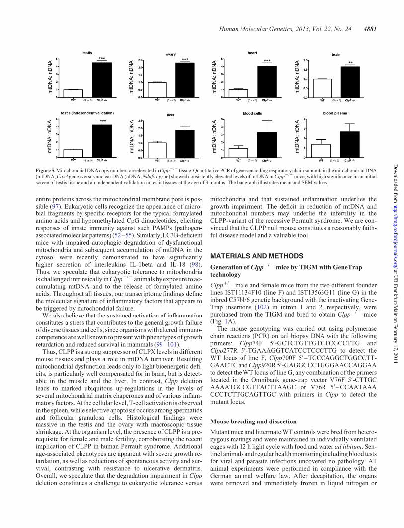
Table S2). To test the hypothesis that abnormal amounts ofmtDNA underlie the elevation of Cox1 transcripts, we quantifiedthe DNA levels of another mitochondrially encoded complex IVsubunit, Cox3, in comparison with the DNA levels of the nuclearencoded complex I subunit Ndufv1. The quantitative real–timePCR (qPCR) results demonstrated several-fold accumulationof mtDNA in Clpp2/2 compared to Clpp+/+ tissues, with highsignificance for the testis, ovary, heart and brain (Fig. 5), andin excellent agreement with the fold-changes of Cox1 transcriptlevels in these Clpp2/2 tissues (Table 2). Thus, the absence ofCLPP affects not only the degradation of proteins, but also themaintenance of mtDNA in the mitochondrial matrix in differenttissues, with effects on transcript levels of mitochondriallyencoded genes.
DISCUSSION
Mitochondria are bacterial endosymbionts that are tolerated ineukaryotic cells since they serve different functions. However,impairment of mitochondrial functions contributes to agingand disease in mammalian organism (57,58). A network of path-ways keeps mitochondria functional over time and regulatestheir mass and dynamics (35–37). Part of this network is thequality control of proteins by different mitochondrial proteasesand chaperones, and the control of mtDNA. In the currentstudy, we investigated the role of the macromolecular CLPXPprotease complex located in the mitochondrial matrix by asses-sing two mouse lines with the deletion of Clpp.
The classical consequences of mitochondrial dysfunctionsuch as apoptosis, respiratory failure combined with oxidativestress, impaired fusion/fission dynamics were not evident inmost tissues. However, strong and specific phenotypes wereobserved in CLPP null mice and can be linked credibly to mito-chondrial pathology. The organism as a whole was found to havesevere growth retardation, diminished spontaneous motor activ-ity, a strong decrease in survival and a marginal respiratorydeficit in several tissues. Similarly, mouse mutants of severalother mitochondrial factors have manifested phenotypes ofgrowth retardation and reduced survival, in particular due tolight respiratory impairment, oxidative stress and mtDNAdamage (59–75). Prominently, Clpp deletion caused selectiveprofound vulnerability of specific cells in testes and ovaries.This is also in agreement with several other mouse and flymutants with mitochondrial dysfunction, which have shown a re-duction of sperm number or motility or impaired spermatogen-esis. Deletions in mtDNA resulted in oligospermia, whiledeletions in the mitochondrial peptidase Immp2l resulted inmale subfertility and female infertility due to defective folliculo-genesis and ovulation (69,76–85).
Ovarian failure and sensorineural deafness are the centralsymptoms of the autosomal recessive human Perrault syndrome,where mutations in the mitochondrial histidyl-tRNA-synthetaseHARS2 (86) and in the steroid metabolism enzyme HSD17B4(87) had previously been observed. A recent publicationreported CLPP missense and splice site mutations (22) in Per-rault syndrome, but CLPP mutations were not yet linked tomale infertility and lack of sperm. Possibly the phenotype in
Table 1. Most Clpp-/- tissues show up-regulation of mitochondrial chaperones
Protein detected by immunoblot densitometry Antibody Testis Heart Liver Brain
Mitochondrial peptidasesCLPP Proteintech 15698-1-AP nd nd nd ndLONP Sigma HPA002192 ns ns ns 4.8× P ¼ 0.0001
ChaperonesCLPX Abgent AP10767b 1.8× P ¼ 0.03 1.9× P ¼ 0.001 1.6× P ¼ 0.01 5.0× P ¼ 0.001mtHSP75/HSPA9/mortalin/GRP75 Oxford Biom Res GR02 2.3× P ¼ 0.01 1.8× P ¼ 0.0009 1.9× P ¼ 0.0007 1.5× P ¼ 0.01mtHSP60/HSPD1/chaperonin 60/SPG13 Santa Cruz sc-13115 ns ns ns nsmtHSP10/HSPE1/chaperonin 10 Epitomics 3106-1 1.4× P ¼ 0.057 1.3× P ¼ 0.04 1.2× P ¼ 0.01 nsmtDNAJ/DNAJA3 Epitomics S2532 1.8× P ¼ 0.0001 1.7× P ¼ 0.004 1.6× P ¼ 0.002 ns
Respiratory chainC-I-20 (ND6) Mitosciences MS 604 0.5× P ¼ 0.004 ns ns 0.9× P ¼ 0.07C-II-30 (FeS) Mitosciences MS 604 ns ns ns nsCx-III-Core2 Mitosciences MS 604 ns ns ns nsCx-IV-I Mitosciences MS 604 0.3× P ¼ 0.04 ns 0.7× P ¼ 0.009 nsC-V-a Mitosciences MS 604 ns ns ns ns
AntioxidantsPRX-III/peroxiredoxin 3 Abfrontier LF-MA0044 2.1× P ¼ 0.0005 1.4× P ¼ 0.0003 ns nsSOD2/superoxide dismutase 2 Santa Cruz sc-30080 ns ns ns ns
ApoptosisAIF/apoptosis inducing factor Santa Cruz sc-9416 ns ns ns ns
Mitochondrial fusionOPA1 (isoform ratio L1 + L2/S3 + S4 + S5) Duvezin-Caubet et al. (2006) ns 0.7× P ¼ 0.01 0.7× P ¼ 0.01 ns
Mitochondrial DNA markersTFAM/mitochondrial transcription factor A Abnova H00007019 ns ns ns ns
Mitochondrial massVDAC1/Porin Calbiochem 529532 Ns ns ns ns
Testis, heart, liver and brain protein levels documented by quantitative immunoblots with densitometry were analyzed with Student’s t-test for significant differences,and the linear fold-change and the P-value were represented in this overview (n ¼ 3 versus 3, mouse age 5 months). Significant up-regulations are highlighted in red,significant down-regulations in green. ns, non-significant; nd, non-detectable.
4878 Human Molecular Genetics, 2013, Vol. 22, No. 24
at UB
Frankfurt/Main on February 17, 2014
http://hmg.oxfordjournals.org/
Dow
nloaded from

mice varies from human, but it is also conceivable that themurine CLPP null mutation has a stronger fertility phenotypethan the human CLPP missense/splice site mutations, whichmay cause only partial CLPP activity loss. Thus, it will be im-portant to screen male patients with a syndrome of azoospermia,short stature and neural dysfunction for CLPP mutations. Con-versely, it will be important to conduct systematic screening ofthe auditory deficit in Clpp2/2 mice assessing the affectedtissue also with histology and molecular profiling. It is wellknown that auditory nerves are exceptionally sensitive to mito-chondrial dysfunction, e.g. to altered import of mtRNA (88,89).
Beyond the severe gonad- and audition- specific pathologies,CLPP absence caused five marked effects that were ubiquitous.First, the accumulation of CLPX; second, the up-regulation ofmitochondrial matrix chaperones; third, the elevated levels ofmtDNA; fourth, the induction of an inflammatory signatureand finally, the growth retardation affecting all tissues. There-fore, it is important to consider what the likely causal chain ofevents between these findings could be. Our data show that theabsence of CLPP leads to CLPX accumulation in spite of down-regulated Clpx transcript levels. The likeliest explanation is thatCLPX degradation is reduced and depends on CLPP, and that thepathologically increased CLPX protein levels are insufficientlycompensated by decreased transcript levels. In contrast, the ele-vated levels of other mitochondrial matrix chaperones throughtranscriptional up-regulation might constitute the crucial com-pensatory effort which prevents earlier pathology in tissueslike the brain. The elevated CLPX levels may be responsible
for mtDNA accumulation, since a recent report showed CLPXto act not only as chaperone for unfolded proteins, but to bealso important for the condensation of mtDNA, similar toTFAM (56). It is well established that the elimination of mito-chondrial number as well as mtDNA reaches its physiologicalmaximum during the maturation of germ cells. During spermato-genesis, the number of mtDNA nucleoids should be reducedfrom 10 000 or 1000 copies per cell to �100 copies per sperm-atozoon. During oogenesis, mitochondrial number would de-crease while going through a quality control bottleneck, beforeexpanding to �100 000 copies per oocyte (90,91). Thereforethe selective and strong testis and ovary pathology in theCLPP null mice might be due to the pathological accumulationof CLPX and mtDNA. While depletion of mtDNA has beendescribed as a consequence of mutations in numerous genes(92), a strong accumulation as in CLPP null tissue has not beendocumented previously. Our data establish CLPP as a modifierfactor for the reduction of mtDNA levels.
The CLPP effect on inflammation and infection defensefactors could be interpreted in the light of two observationsmade in prokaryotes. First, in E. coli CLPP was observed to in-fluence the lysis-lysogeny decision during bacteriophage muinfections (11,93) and second, a report in Staphylococcusaureus showed Clpp deletion to result in poor growth accom-panied by prophage proteome accumulation and increased pro-phage release in some strains (94). Also in mammals theantiviral defense system and interferon induction are modulatedcrucially by mitochondria, through four known factors, namely
Table 2. Clpp-/- testis shows massive affection of transcript levels, while most tissues show moderate anomalies
qPCR target mRNA/gene symbol TaqMan assay Testis Heart Liver Brain
Mitochondrial peptidasesClpp Mm00489940_m1∗ 0.03× P ¼ 0.0003 0.0002× P ¼ 0.0003 0.003× P ¼ 0.01 0.0001× P ¼ 0.0001LonP/Lonp1 Mm01236887_m1 3.6× P ¼ 0.001 3.0× P ¼ 0.01 ns ns
ChaperonesClpx Mm00488586_m1 0.5× P ¼ 0.01 ns ns nsmtHsp75/Hspa9/mortalin/GRP75 Mm00477716_g1 2.0× P ¼ 0.01 2.4× P ¼ 0.0001 ns nsmtHsp60/Hspd1/chaperonin 60/SPG13 Mm00849835_g1∗ 3.2× P ¼ 0.0001 1.6× P ¼ 0.02 2.0× P ¼ 0.008 nsmtHsp10/Hspe1/chaperonin 10 Mm00434083_m1 3.1× P ¼ 0.0005 2.9× P ¼ 0.005 ns nsmtDnaJ/Dnaja3 Mm00469723_m1 2.8× P ¼ 0.0001 2.1× P ¼ 0.006 ns ns
Respiratory chainC-I/NdufvI Mm00504941_m1 1.4× P ¼ 0.004 ns ns nsC-II/Sdhb Mm00458272_m1 1.4× P ¼ 0.001 ns ns nsC-III/Uqcrc2 Mm00445961_m1 2.4× P ¼ 0.001 ns ns nsC-IV/Cox1/Mt-Co1 Mm04225243_g1∗ 5.0× P ¼ 0.0001 2.4× P ¼ 0.006 1.9× P ¼ 0.02 1.7× P ¼ 0.03C-V/Atp5a1 Mm00431960_m1 2.4× P ¼ 0.0004 ns ns ns
AntioxidantsPeroxiredoxin 3/Prdx3 Mm00545848_m1 3.3× P ¼ 0.0001 ns 2.0× P ¼ 0.01 nsSuperoxide dismutase 2/Sod2 Mm01313000_m1∗ ns ns ns ns
ApoptosisApoptosis inducing factor 1/Aifm1 Mm00442540_m1∗ 3.8× P ¼ 0.01 ns ns ns
Mitochondrial fusionOpa1 Mm00453879_m1 1.5× P ¼ 0.001 ns ns ns
Mitochondrial DNA markersTfam Mm00447485_m1 0.11× P ¼ 0.01 ns 1.47× P ¼ 0.004 ns
Mitochondrial massVdac1 Mm00834272_M1 4.9× P ¼ 0.01 ns ns ns
Testis, heart, liver and brain transcript levels documented by quantitative real-time reverse transcriptase PCR (qPCR) through commercial Taqman assays wereanalyzed with the 22DDCt method. The linear fold-change and the unpaired Student’s-test P-value (significance threshold 0.05) were represented in this overview (n ¼3 versus 3, mouse age 5 months). Significant up-regulations are highlighted in red, significant down-regulations in green. ns, non-significant.
Human Molecular Genetics, 2013, Vol. 22, No. 24 4879
at UB
Frankfurt/Main on February 17, 2014
http://hmg.oxfordjournals.org/
Dow
nloaded from

matrix-localized NLRX1 and outer membrane-associatedMAVS, ECSIT and STING (52,53). Thus, in spite of the vast dif-ferences the bacterial and eukaryotic immune defense systems,CLPP deficiency might modulate antiviral defense. In thiscase, a novel functional parallel between the mitochondrialCLPXP machinery and the cytosolic proteasome wouldbecome apparent, since the latter is well known to respond toinfections and inflammations via interferon-gamma exposureby adapting its composition and proteolysis pattern, thusserving as immunoproteasome (95). Thus, although our micewere bred and aged in individually ventilated cages, with theabsence of pathogenic infections being documented in regularhealth screens, we cannot exclude an undetected viral exposureof some mice that might explain our data partially. However, thetranscriptome analyses documented a genotype-dependentelevation of immunity factors in every one of several mice andtissues, and the cellular analyses of splenocytes in independentanimals confirmed this genotype-dependent effect. Furthermore,
almost all mice in both Clpp2/2 lines across their lifespanshowed resistance against ulcerative dermatitis. This skin condi-tion is typical of murine C57BL/6 substrains during aging, doesnot respond to antibiotic or corticosteroid treatment, and isthought to arise from pruritus due to follicular dystrophy, withlater bacterial superinfections by skin commensals (44,96). Thisgeneral reduction of vulnerability of Clpp2/2 animals towardan inflammatory disease is a strong argument that the CLPPeffect on immunity occurs irrespective of infections.
Mitochondrial damage can by itself lead to inflammatoryresponses of the host cells. Our data demonstrate that Clpp2/2
deletion leads not only to the induction of mitochondrial chaper-ones, but also to transcriptional up-regulation of proteasomesubunits and ubiquitin-mediated proteolysis factors, suggestingthat non-degraded CLPP substrates are released from mitochon-dria to the eukaryotic cytosol. This appears possible, since C.elegans CLPP was shown to modulate the peptide efflux fromisolated mitochondria (18) and since retrograde export even of
Figure 4. Activation of T-lymphocytes in the Clpp2/2 spleen. Splenocytes from wild-type (illustrated by blue dots) and mutant (red dots) mice (aged until 18 monthsunder continuous microbial exposure monitoring in shared individually ventilated housing) were characterized using multi-color flow cytometry (n ¼ 8 Clpp2/2
versus 7 WT). (A) Mutant CD8+ T cell subsets analyzed for their expression of CD44/CD62L/Ly6C showed several significant changes. (B) Mutant CD4+T cell subsets analyzed for their expression of CD25/CD44/CD62L/Ly6C also showed several significant changes. Statistical analysis was done by using SPICE(117). Significance (alpha ¼ 5%) in Student’s t-test is indicated by +, significance in the Wilcoxon rank test by #.
4880 Human Molecular Genetics, 2013, Vol. 22, No. 24
at UB
Frankfurt/Main on February 17, 2014
http://hmg.oxfordjournals.org/
Dow
nloaded from
entire proteins across the mitochondrial membrane pore is pos-sible (97). Eukaryotic cells recognize the appearance of micro-bial fragments by specific receptors for the typical formylatedamino acids and hypomethylated CpG dinucleotides, elicitingresponses of innate immunity against such PAMPs (pathogen-associated molecular patterns) (52–55). Similarly, LC3B-deficientmice with impaired autophagic degradation of dysfunctionalmitochondria and subsequent accumulation of mtDNA in thecytosol were recently demonstrated to have significantlyhigher secretion of interleukins IL-1beta and IL-18 (98).Thus, we speculate that eukaryotic tolerance to mitochondriais challenged intrinsically in Clpp2/2 animals by exposure to ac-cumulating mtDNA and to the release of formylated aminoacids. Throughout all tissues, our transcriptome findings definethe molecular signature of inflammatory factors that appears tobe triggered by mitochondrial failure.
We also believe that the sustained activation of inflammationconstitutes a stress that contributes to the general growth failureof diverse tissues and cells, since organisms with altered immuno-competence are well known to present with phenotypes of growthretardation and reduced survival in mammals (99–101).
Thus, CLPP is a strong suppressor of CLPX levels in differentmouse tissues and plays a role in mtDNA turnover. Resultingmitochondrial dysfunction leads only to light bioenergetic defi-cits, is particularly well compensated for in brain, but is detect-able in the muscle and the liver. In contrast, Clpp deletionleads to marked ubiquitous up-regulations in the levels ofseveral mitochondrial matrix chaperones and of various inflam-matory factors. At the cellular level, T-cell activation is observedin the spleen, while selective apoptosis occurs among spermatidsand follicular granulosa cells. Histological findings weremassive in the testis and the ovary with macroscopic tissueshrinkage. At the organism level, the presence of CLPP is a pre-requisite for female and male fertility, corroborating the recentimplication of CLPP in human Perrault syndrome. Additionalage-associated phenotypes are apparent with severe growth re-tardation, as well as reductions of spontaneous activity and sur-vival, contrasting with resistance to ulcerative dermatitis.Overall, we speculate that the degradation impairment in Clppdeletion constitutes a challenge to eukaryotic tolerance versus
mitochondria and that sustained inflammation underlies thegrowth impairment. The deficit in reduction of mtDNA andmitochondrial numbers may underlie the infertility in theCLPP-variant of the recessive Perrault syndrome. We are con-vinced that the CLPP null mouse constitutes a reasonably faith-ful disease model and a valuable tool.
MATERIALS AND METHODS
Generation of Clpp2/2 mice by TIGM with GeneTraptechnology
Clpp+/2 male and female mice from the two different founderlines IST11134F10 (line F) and IST13563G11 (line G) in theinbred C57bl/6 genetic background with the inactivating Gene-Trap insertions (102) in intron 1 and 2, respectively, werepurchased from the TIGM and bred to obtain Clpp2/2 mice(Fig. 1A).
The mouse genotyping was carried out using polymerasechain reactions (PCR) on tail biopsy DNA with the followingprimers: Clpp74F 5′-GCTCTGTTGTCTCGCCTTG andClpp277R 5′-TGAAAGGTCATCCTCCCTTG to detect theWT locus of line F, Clpp700F 5′–TCCCAGGCTGGCCTT-GAACTC and Clpp920R 5′-GAGGCCCTGGGAACCAGGAAto detect the WT locus of line G, any combination of the primerslocated in the Omnibank gene-trap vector V76F 5′-CTTGCAAAATGGCGTTACTTAAGC or V76R 5′ –CCAATAAACCCTCTTGCAGTTGC with primers in Clpp to detect themutant locus.
Mouse breeding and dissection
Mutant mice and littermate WT controls were bred from hetero-zygous matings and were maintained in individually ventilatedcages with 12 h light cycle with food and water ad libitum. Sen-tinel animals and regular health monitoring including blood testsfor viral and parasite infections uncovered no pathology. Allanimal experiments were performed in compliance with theGerman animal welfare law. After decapitation, the organswere removed and immediately frozen in liquid nitrogen or
Figure 5. Mitochondrial DNA copy numbers are elevated in Clpp2/2 tissue. Quantitative PCR of genes encoding respiratorychain subunits in the mitochondrial DNA(mtDNA, Cox3 gene) versus nuclear DNA (nDNA, Ndufv1 gene) showed consistently elevated levels of mtDNA in Clpp2/2 mice, with high significance in an initialscreen of testis tissue and an independent validation in testis tissues at the age of 3 months. The bar graph illustrates mean and SEM values.
Human Molecular Genetics, 2013, Vol. 22, No. 24 4881
at UB
Frankfurt/Main on February 17, 2014
http://hmg.oxfordjournals.org/
Dow
nloaded from

fixed for histological and ultrastructural analyses or processedfor mitochondrial isolation.
Quantitative real-time reverse transcription PCR
Isolation of total RNA was performed with Trizol (Gibco),DNase treatment of RNA with DNAse Amplification Grade(Invitrogen) and reverse transcription with SuperScript III (Invi-trogen), always following the instructions of the manufacturers.
qPCR was performed with Expression Assays (Applied Bio-systems) in cDNA from 20 ng total RNA in 20 ml reactionswith 2× master mix from Roche in a StepOnePlus Real-TimePCR System (Applied Biosystems). The analysis of the datawas carried out with the 22DDCT method (103).
Real-time PCR of two Clpp transcript assays and of Gapdh ashousekeeping normalizer were performed, the products sepa-rated in 2% agarose gels and visualized with ethidiumbromide, to assess the null mutation in brain tissue (Fig. 1B).
Quantitative immunoblots
Frozen tissues were homogenized on ice in a glass-Teflondouncer in RIPA buffer with 50 mM Tris–HCl (pH 8), 150 mM
NaCl, 1% NP-40, 0.5% Na-deoxycholate, 0.1% SDS and prote-ase inhibitor cocktail (Roche). Total lysates were briefly soni-cated on ice, and cell debris was removed by centrifugation.Protein concentration was determined according to the methodof Bradford. SDS–PAGE-separated proteins (20 mg/lane)were blotted onto a PVDF membrane (Bio-Rad) and probedwith the specific primary antibodies (see Table 1) overnight fol-lowed by the incubation with the adequate secondary antibody(GE Healthcare UK Limited, anti-mouse IgG NA931V, anti-rabbit IgG NA934V) for 1 h. The detection was made withSuperSignal West Pico (Thermo Scientific). OPA1 proteolyticprocessing was assessed by calculating the ratio of long isoformsL1 + L2 versus the short isoforms S3 + S4 + S5, using a previ-ously published antibody (104).
Statistical analysis
Quantitative fertility, motor behavior and gene dosage data wereanalyzed with the GraphPad software and illustrated in bargraphs, showing variance with standard error and P-valuesfrom Student’s t-test (∗P , 0.05; ∗∗P , 0.01; ∗∗∗P , 0.001).
Genome-wide mRNA profiling
For expression profiling, 100 ng of total RNA was linearly amp-lified and biotinylated using the GeneChip HT 3′IVT Express Kit(Affymetrix, Santa Clara, CA, USA) according to the manufac-turer’s instructions. Fifteen micrograms of labeled and fragmen-ted cRNA was hybridized onto GeneChip HT MG-430 PMArray Plates (Affymetrix). Hybridization, washing, stainingand scanning were performed automatically in a GeneTitan in-strument (Affymetrix). Scanned images were subjected tovisual inspection to control for hybridization artifacts andproper grid alignment and analyzed with AGCC 3.0 (Affyme-trix) to generate CEL files.
All subsequent data analysis steps were performed on the soft-ware platform R 2.14.0 and Bioconductor 2.14.0 (105). Initially,
the expression data from all chips were background corrected,quantile normalized and summarized with Robust MultichipAverage (106). Owing to the design of the experiment, two para-meters (strain and tissue) have an impact on gene expression.Thus, a combined factor from both parameters was used todesign a linear model that captures the influence on gene expres-sion levels. Comparisons of CLPP homozygous null (2/2) andcorresponding WT samples were defined as contrasts and thelinear model coefficients were estimated. A non-specific filterbased on overall variance was applied to remove non-informative features before the fitting of the linear model wasperformed. Empirical Bayes shrinkage of the standard errorswas used to calculate the moderated F-statistic (107). The result-ing P-values were established and corrected for multiple testingwith ‘Benjamini-Hochberg’ (108). To attribute significant regu-lations to individual contrasts, a decision matrix was generatedbased on the function ‘decide tests’ within the limma package,where significant up- or down-regulations are represented byvalues of 1 or 21, respectively. The lists of differentially regu-lated transcripts were analyzed for over-representation of geneontology terms and KEGG pathways, respectively, using thehypergeometric test.
All transcriptome data were deposited in a public database andare accessible at http://www.ncbi.nlm.nih.gov/geo/query/acc.cgi?acc=GSE40207, last accessed on 17 July 2013.
mtDNA quantification
mtDNA and nuclear DNA copy number were determined byqPCR of the mitochondrial Cox3 gene and the nuclear Ndufv1gene, respectively. For amplification of the mitochondrialCox3 fragment, we used the following primers: Cox3-fwd5′ –TTTGCAGGATTCTTCTGAGC and Cox3-rev 5′ –TGAGCTCATGTAATTGAAACACC. Primer pairs for Ndufv1 wereaccording to a previous publication (109). The qPCR reactionwas initiated by 508C for 2 min and then 948C for 10 min, fol-lowed by 40 cycles of 958C for 10 s, 608C for 60 s, and a finalmelting curve ranging from 858C to 658C was done. All reactionswere run in quadruplicates using a StepOnePlus real-time PCRequipment (Applied Biosystems) and SYBR Green technology.The relative amount of mtDNA to nuclear genome was calcu-lated using the 22DDCt method (103).
Survival curve
Age at natural death was documented for each animal. The numberof mice analyzed was 69 for WT and 79 for Clpp2/2 (both linestogether). For statistical analysis, the IBM SPSS Statistics soft-ware package (19.0.0.) was used and Kaplan–Meier survival esti-mates generated, taking into account that currently a large numberof mice is still alive. Significance was determined with pairwisecomparisons (Log rank, Breslow and Tarone-Ware).
Body weight and length
Mutant animals and matched controls were weighed at regularintervals. The body length was recorded in parallel formatched pairs in manually immobilized and extended animalsfrom the nose to the anus.
4882 Human Molecular Genetics, 2013, Vol. 22, No. 24
at UB
Frankfurt/Main on February 17, 2014
http://hmg.oxfordjournals.org/
Dow
nloaded from

Motor activity
Acoustic startleMaximal amplitude (Vmax) of locomotor responses within a timelag window (50–110 ms) after burst tones interrupting whitenoise at varying time intervals, ranging in loudness between 90and 120 dB were recorded by accelerometer within an SR-LabStartle Reflex System (San Diego Instruments) as previouslypublished (51).
Open fieldSpontaneous motor activity of naive mice was automaticallyrecorded in equipment with Versamax monitors from Accuscan(Columbus) over 5 min sessions.
RotarodInduced motor activity of naive mice was studied on Accelerodequipment (Ugo Basile) over 10 min, recording the latency untilfalling/jumping off or the latency until two consecutive ridesaround hanging onto the beam (110).
MEFs
Generation, culture, mitochondrial dynamics: mouse embryonicfibroblasts (MEFs) were prepared from individual embryos at14.5 days post-coitus following intercrosses of Clpp+/2 mice.MEFs were maintained in Dulbecco’s minimal essentialmedium 4.5 g/l glucose (Invitrogen, Karlsruhe) supplementedwith 15% bovine growth serum (BGS, Thermo Scientific,Schwerte, Germany), l% glutamine, 1% penicillin and strepto-mycin (all Invitrogen, Karlsruhe) at 378C and 5% CO2 in a hu-midified incubator.
Growth curveThree WT MEF lines and three Clpp2/2 MEF lines were culti-vated as described above in flasks coated with 0.2% gelatin at378C. Population densities (PDs) were determined by the follow-ing equation: PD ¼ 3.32 × (log10 UCY 2 log10 I) + X (whereUCY is the number of cells at the end of the passage; I is thenumber of cells that were seeded at the beginning of thepassage and X the previous PD number.
Mitochondrial morphology was analyzed by Mitotracker RedCMX ROS staining (MTR, Invitrogen). 150 000 cells wereseeded onto 24 mm cover slips coated with 0.2% gelatin. After24 h or 48 h the cells were incubated for 1 h with MTR (final con-centration 50 nM). Cells were then fixed with 4% paraformalde-hyde and embedded in Dako Fluorescent Mounting Medium.Mitochondria were visualized by using a Leica TCS SP5 con-focal laser scanning microscope at the appropriate spectral set-tings and equipped with an HCX PL APO lambda blue 63.0×,1.40 OIL UV objective that was controlled by the LAS AFscan software (version 1.8.2, Leica).
Light microscopy
After removal of skeletal muscle and testis, tissue samples wereimmediately frozen in liquid nitrogen. Sections of 10 mm thick-ness were cut using a microtome and mounted on Super FrostPlus slides (Microm International, Walldorf, Germany). Sec-tions were dried and stained for H and E, NADH, COX andSDH according to standard protocols. Additionally, heart andskeletal muscles, testes and ovaries were fixed in buffered
formalin (4% formaldehyde; pH 7.4), embedded in paraffin,cut at 4 mm and stained for H and E.
Electron microscopy
Skeletal muscle and testis tissue samples were fixed for 2 h using2.5% glutaraldehyde buffered in cacodylate buffer. The embed-ding procedure comprised fixation in 1% osmium tetroxide, de-hydration in a graded ethanol series intermingled by anincubation step with uranyl acetate (between the 50 and 90%ethanol step) and finally rinsing in propylene oxide. The speci-mens were then embedded in epoxy resins that polymerizedfor 16 h at 608C. After embedding, first semithin sections(0.5 mm) were cut using an ultramicrotome (Leica UltracutUCT, Deerfield, IL, USA) with a diamond knife. Sectionswere stained with toluidine blue, placed on glass slides andexamined by light microscopy to select appropriate areas forultrathin preparation. Ultrathin sections (50–70 nm) were cutagain using an ultramicrotome. Sections were mounted oncopper grids and contrasted with uranyl acetate for 2–3 h at428C and lead citrate for 20 min at room temperature. Thesesamples were imaged and digital pictures were taken with aFEI Tecnai G2 Spirit Biotwin TEM (Hillsboro, OR, USA) atan operating voltage of 120 kV.
Preparation of mitochondria
Mouse heart mitochondria (MHM) and mouse liver mitochon-dria (MLM) were isolated following the protocol developedfor rat heart mitochondria (111) with the modification that allbuffer volumes and the trypsin and trypsin inhibitor quantitieswere halved. Mouse brain mitochondria (MBM) were isolatedas described previously (112). For the isolation of mitochondriafrom mouse skeletal muscle (MSMM), both rear hind limbs weredissected out and rinsed in ice-cold washing buffer (100 mM
sucrose, 100 mM Tris–HCl, 9 mM EDTA, 1 mM EGTA, 46 mM
KCl, pH 7.4). All following steps were performed at 48C. Themuscle tissue was cut into small pieces, whereas bone piecesand the connective tissue were removed as far as possible. Thedissected muscle tissue was dissolved in 10 ml washing bufferand stirred for 15 min in the presence of 1.25 mg trypsin(dissolved in 1 mM HCl). During incubation, the tissue washomogenized twice in a glass/teflon potter at 500 rpm for1 min. Trypsinization was finally stopped by the addition of6.5 mg trypsin inhibitor (from soy beans). The solution was fil-tered through polyamide filter cloth (pore size 210 mm,A. Hartenstein, Wurzburg) and the remnants of this filtrationwere transferred into 20 ml isolation buffer (100 mM sucrose,100 mM Tris–HCl, 9 mM EDTA, 1 mM EGTA, 46 mM KCl, pH7.4 + 1 mg/ml BSA fatty acid free) and homogenized oncemore at 500 rpm for 1 min. This solution was also filteredthrough the polyamide filter cloth and the two filtrates were com-bined. The combined solution was centrifuged for 15 min at600g, and the supernatant was transferred into a new centrifuga-tion tube. Mitochondria were collected by a high-speed centrifu-gation step (10 min at 5.700g) and re-dissolved in 20 ml isolationbuffer. After a second round of low-speed (15 min at 600g) andhigh-speed centrifugation (10 min at 5700g) the mitochondrialpellet was finally dissolved in 1 ml isolation buffer.
Human Molecular Genetics, 2013, Vol. 22, No. 24 4883
at UB
Frankfurt/Main on February 17, 2014
http://hmg.oxfordjournals.org/
Dow
nloaded from

Measurement of mitochondrial respiration
The rate of mitochondrial respiration was monitored at 258Cusing an Oxygraph-2k system (Oroboros, Innsbruck) equippedwith two chambers and DatLab software version 4.3. MHM(between 90 and 140 mg protein), MBM (between 280 and410 mg protein) or MSMM (between 90 and 120 mg protein)were added to 2 ml of a buffer containing 200 mM sucrose,10 mM potassium phosphate (pH 7.0), 10 mM Tris–HCl,10 mM MgSO4 and 2 mM EDTA. MHM and MBM were fueledby 5 mM malate/5 mM pyruvate, whereas MSMM was fueledby 4.8 mM malate/5.6 mM glutamate. The respiratory controlfactor was determined as the ratio between state 3 and state 2 [fol-lowing the revised nomenclature of (113)] respiration after theaddition of 2 mM ADP. Subsequently, complex I was inhibitedby adding rotenone (5 mM) and the complex II-dependent respir-ation was measured after adding succinate (5 mM). Finally, KCN(2 mM) was added to determine the mitochondria-independentoxygen consumption that was (if present) subtracted from allrates of the respective measurement.
Blue native gel analysis of respiratory complexes
Solubilization of mitochondria and 1-D BNE (blue native elec-trophoresis) was performed as described (114). Briefly isolatedmitochondria from MHM, MBM and MLM were solubilizedwith 6 g/g digitonin/protein in solubilization buffer (50 mM
NaCl, 50 mM imidazole/HCl, 2 mM 6-aminohexanoic acid,1 mM EDTA, pH 7.0). Following 20 min centrifugation at 22000g, supernatants were supplemented with Coomassie BrilliantBlue G-250 suspension in 500 mM 6-aminohexanoic acid, andeach sample was loaded three times onto a 3–13% acrylamidegradient gel with a 3% sample gel on top. One-dimensionalgels were stained with Coomassie Brilliant Blue G-250 or byan in gel complex I activity assay (NADH:NBT reductase activ-ity) or a complex IV heme stain as described in Zerbetto et al.(115) with some modifications (116). The Bio-Rad ChemiDocXRS system was used for densitometric quantification.
Flow cytometric analysis of splenocytes
The flow cytometric analysis of lymphocyte populations in thespleen was based on two 10-parameter antibody panels, coveringmarkers for T-cells (CD3, CD4, CD8 and gdTCR) and furthersubsets (CD44, CD62L, CD25 and Ly6C), and for B-cells(CD19 and B220) and further subsets (CD24, IgD, IgM andMHC class II). In short, after organ collection, the completeorgan was stored at 48C in 2–3 ml of RPMI/2% FCS. Forfurther preparation, a part of each spleen was transferred into acocktail of collagenase II (Serlabo Technologies SARL, Entrai-gues sur la Sorgue, France) and DNase I (Sigma-Aldrich ChemieGmbH, Taufkirchen, Germany) within RPMI/2% FCS and dis-sociated using GentleMACS C tubes (Miltenyi Biotec, BergischGladbach, Germany) and the GentleMACS tissue dissociator(Miltenyi Biotec, Bergisch Gladbach, Germany). Collagenasedigestion at room temperature over 20 min was stopped with a0.1 M EDTA-PBS solution. Then red blood cell lysis was per-formed with RBC lysis solution (eBioscience, Inc., San Diego,USA), cells were washed in PBS buffer (PBS, 0.5% BSA,0.02% sodium azide, pH 7.45), then incubated with Fc block
(anti-mouse CD16/32), fluorescence-conjugated antibodies(BD Biosciences, Heidelberg, Germany), propidium iodide(Sigma-Aldrich Chemie GmbH, Taufkirchen, Germany) andwashed. Cells had been plated on a 96-well plate, and the cellswere acquired with a HyperCyt plate loader in combinationwith a Gallios flow cytometer (Beckman Coulter Germany,Krefeld). Analysis was done with FlowJo (Tree Star, Inc.,Oregon USA); dead cells were eliminated on the basis of theirpropidium iodide signal and subpopulations gated for corre-sponding leukocyte markers.
SUPPLEMENTARY MATERIAL
Supplementary Material is available at HMG online.
ACKNOWLEDGEMENTS
We are grateful to our technical assistants Birgitt Meseck-Selchow, Ilka Siebels and Maximilian Mattil, Tatjana Starzetz,Vanessa Vosseler, Johanna Manz, Elisa Pallasch, the GeneTrapstaff at TIGM, the animal facility staff of the ZFE KlinikumFrankfurt and of MDF Wendelsheim, and to Miguel Barrera(Mitochondrial Biology Buchmann Institute for MolecularLife Sciences, Frankfurt am Main) for technical advice.
Conflict of Interest statement. None declared.
FUNDING
The study was financed by the German Federal Ministry forEducation and Research (BMBF, through the GerontoMitoSysproject under grant number 0315584A, the National Genome Re-search Network NGFN-Plus under grant numbers 01GS08134/01GS08138, and the German Network for Mitochondrial Disor-ders mitoNET under grant number 01GM1113B), by the DeutscheForschungsgemeinschaft (DFG, Sonderforschungsbereich 815,projects A02 and Z1), as well as the Cluster of Excellence Frank-furt Macromolecular Complexes (EXC115).
REFERENCES
1. Sauer, R.T. and Baker, T.A. (2011) AAA+ proteases: ATP-fueledmachines of protein destruction. Annu. Rev. Biochem., 80, 587–612.
2. Voos, W. (2009) Mitochondrial protein homeostasis: the cooperative rolesof chaperones and proteases. Res. Microbiol., 160, 718–725.
3. Baker, T.A. and Sauer, R.T. (2012) ClpXP, an ATP-powered unfolding andprotein-degradation machine. Biochim. Biophys. Acta., 1823, 15–28.
4. Kim, Y.I., Levchenko, I., Fraczkowska, K., Woodruff, R.V., Sauer, R.T.and Baker, T.A. (2001) Molecular determinants of complex formationbetween Clp/Hsp100 ATPases and the ClpP peptidase. Nat. Struct. Biol.,8, 230–233.
5. Stanne, T.M., Pojidaeva, E., Andersson, F.I. and Clarke, A.K. (2007)Distinctive types of ATP-dependent Clp proteases in cyanobacteria. J. Biol.Chem., 282, 14394–14402.
6. Siddiqui, S.M., Sauer, R.T. and Baker, T.A. (2004) Role of the processingpore of the ClpX AAA+ ATPase in the recognition and engagement ofspecific protein substrates. Genes Dev., 18, 369–374.
7. Neher, S.B., Villen, J., Oakes, E.C., Bakalarski, C.E., Sauer, R.T., Gygi,S.P. and Baker, T.A. (2006) Proteomic profiling of ClpXP substrates afterDNA damage reveals extensive instability within SOS regulon. Mol. Cell,22, 193–204.
8. Nagashima, K., Kubota, Y., Shibata, T., Sakaguchi, C., Shinagawa, H. andHishida, T. (2006) Degradation of Escherichia coli RecN aggregates by
4884 Human Molecular Genetics, 2013, Vol. 22, No. 24
at UB
Frankfurt/Main on February 17, 2014
http://hmg.oxfordjournals.org/
Dow
nloaded from

ClpXP protease and its implications for DNA damage tolerance. J. Biol.Chem., 281, 30941–30946.
9. Hengge, R. (2009) Proteolysis of sigmaS (RpoS) and the general stressresponse in Escherichia coli. Res. Microbiol., 160, 667–676.
10. Flynn, J.M., Levchenko, I., Sauer, R.T. and Baker, T.A. (2004) Modulatingsubstrate choice: the SspB adaptor delivers a regulator of theextracytoplasmic-stress response to the AAA+ protease ClpXP fordegradation. Genes Dev., 18, 2292–2301.
11. Czyz, A., Zielke, R. and Wegrzyn, G. (2001) Rapid degradation ofbacteriophage lambda O protein by ClpP/ClpX protease influences thelysis-versus-lysogenization decision of the phage under certain growthconditions of the host cells. Arch. Virol., 146, 1487–1498.
12. O’Handley, D. and Nakai, H. (2002) Derepression of bacteriophage mutransposition functions by truncated forms of the immunity repressor.J. Mol. Biol., 322, 311–324.
13. Zhao, Q., Wang, J., Levichkin, I.V., Stasinopoulos, S., Ryan, M.T. andHoogenraad, N.J. (2002) A mitochondrial specific stress response inmammalian cells. EMBO J., 21, 4411–4419.
14. Gerth, U., Kruger, E., Derre, I., Msadek, T. and Hecker, M. (1998) Stressinduction of the Bacillus subtilis clpP gene encoding a homologue of theproteolytic component of the Clp protease and the involvement of ClpP andClpX in stress tolerance. Mol. Microbiol., 28, 787–802.
15. Zheng, B., Halperin, T., Hruskova-Heidingsfeldova, O., Adam, Z. andClarke, A.K. (2002) Characterization of chloroplast Clp proteins inarabidopsis: localization, tissue specificity and stress responses. Physiol.Plant, 114, 92–101.
16. Fischer, F., Weil, A., Hamann, A. and Osiewacz, H.D. (2013) Human CLPPreverts the longevity phenotype of a fungal ClpP deletion strain. Nat.Commun., 4, 1397.
17. Haynes, C.M., Petrova, K., Benedetti, C., Yang, Y. and Ron, D. (2007) Clppmediates activation of a mitochondrial unfolded protein response in C.
elegans. Dev. Cell, 13, 467–480.18. Haynes, C.M., Yang, Y., Blais, S.P., Neubert, T.A. and Ron, D. (2010) The
matrix peptide exporter HAF-1 signals a mitochondrial UPR by activatingthe transcription factor ZC376.7 in C. elegans. Mol. Cell, 37, 529–540.
19. Aldridge, J.E., Horibe, T. and Hoogenraad, N.J. (2007) Discovery of genesactivated by the mitochondrial unfolded protein response (mtUPR) andcognate promoter elements. PloS ONE, 2, e874.
20. Broadley, S.A. and Hartl, F.U. (2008) Mitochondrial stress signaling:a pathway unfolds. Trends Cell Biol., 18, 1–4.
21. Kang, S.G., Ortega, J., Singh, S.K., Wang, N., Huang, N.N., Steven, A.C.and Maurizi, M.R. (2002) Functional proteolytic complexes of the humanmitochondrial ATP-dependent protease, hClpXP. J. Biol. Chem., 277,21095–21102.
22. Jenkinson, E.M., Rehman, A.U., Walsh, T., Clayton-Smith, J., Lee, K.,Morell, R.J., Drummond, M.C., Khan, S.N., Naeem, M.A., Rauf, B. et al.(2013) Perrault syndrome is caused by recessive mutations in CLPP,encoding a mitochondrial ATP-dependent chambered protease.Am. J. Hum. Genet., 92, 605–613.
23. Hansen, J., Corydon, T.J., Palmfeldt, J., Durr, A., Fontaine, B., Nielsen,M.N., Christensen, J.H., Gregersen, N. and Bross, P. (2008) Decreasedexpression of the mitochondrial matrix proteases Lon and clpP in cells froma patient with hereditary spastic paraplegia (SPG13). Neuroscience, 153,474–482.
24. Bross, P., Naundrup, S., Hansen, J., Nielsen, M.N., Christensen, J.H.,Kruhoffer, M., Palmfeldt, J., Corydon, T.J., Gregersen, N., Ang, D. et al.
(2008) The Hsp60-(p.V98I) mutation associated with hereditary spasticparaplegia SPG13 compromises chaperonin function both in vitro and invivo. J. Biol. Chem., 283, 15694–15700.
25. Guillon, B., Bulteau, A.L., Wattenhofer-Donze, M., Schmucker, S.,Friguet, B., Puccio, H., Drapier, J.C. and Bouton, C. (2009) Frataxindeficiency causes upregulation of mitochondrial Lon and ClpP proteasesand severe loss of mitochondrial Fe-S proteins. FEBS J., 276, 1036–1047.
26. Martinelli, P. and Rugarli, E.I. (2010) Emerging roles of mitochondrialproteases in neurodegeneration. Biochim. Biophys. Acta., 1797, 1–10.
27. Greene, A.W., Grenier, K., Aguileta, M.A., Muise, S., Farazifard, R.,Haque, M.E., McBride, H.M., Park, D.S. and Fon, E.A. (2012)Mitochondrial processing peptidase regulates PINK1 processing, importand Parkin recruitment. EMBO Rep., 13, 378–385.
28. Strauss, K.M.,Martins,L.M.,Plun-Favreau, H., Marx,F.P.,Kautzmann, S.,Berg, D., Gasser, T., Wszolek, Z., Muller, T., Bornemann, A. et al. (2005)Loss of function mutations in the gene encoding Omi/HtrA2 in Parkinson’sdisease. Hum. Mol. Genet., 14, 2099–2111.
29. Jin, S.M., Lazarou, M., Wang, C., Kane, L.A., Narendra, D.P. and Youle,R.J. (2010)Mitochondrial membranepotential regulates PINK1import andproteolytic destabilization by PARL. J. Cell Biol., 191, 933–942.
30. Di Bella,D., Lazzaro, F.,Brusco, A.,Plumari, M.,Battaglia,G., Pastore, A.,Finardi, A., Cagnoli, C., Tempia, F., Frontali, M. et al. (2010) Mutations inthe mitochondrial protease gene AFG3L2 cause dominant hereditary ataxiaSCA28. Nat. Genet., 42, 313–321.
31. Cagnoli, C., Stevanin, G., Brussino, A., Barberis, M., Mancini, C.,Margolis, R.L., Holmes, S.E., Nobili, M., Forlani, S., Padovan, S. et al.
(2010) Missense mutations in the AFG3L2 proteolytic domain account forapproximately 1.5% of European autosomal dominant cerebellar ataxias.Hum. Mutat., 31, 1117–1124.
32. Pierson, T.M., Adams, D., Bonn, F., Martinelli, P., Cherukuri, P.F., Teer,J.K., Hansen, N.F. and Cruz, P., Mullikin For The Nisc ComparativeSequencing Program, J.C., Blakesley, R.W. et al. (2011) Whole-exomesequencing identifies homozygous AFG3L2 mutations in a spasticataxia-neuropathy syndrome linked to mitochondrial m-AAA proteases.PLoS Genet., 7, e1002325.
33. Nolden, M., Ehses, S., Koppen, M., Bernacchia, A., Rugarli, E.I. andLanger, T. (2005) The m-AAA protease defective in hereditary spasticparaplegia controls ribosome assembly in mitochondria. Cell, 123, 277–289.
34. Alexander, C., Votruba, M., Pesch, U.E., Thiselton, D.L., Mayer, S.,Moore, A., Rodriguez, M., Kellner, U., Leo-Kottler, B., Auburger, G. et al.
(2000) OPA1, Encoding a dynamin-related GTPase, is mutated inautosomal dominant optic atrophy linked to chromosome 3q28. Nat.
Genet., 26, 211–215.
35. Fischer, F., Hamann, A. and Osiewacz, H.D. (2012) Mitochondrial qualitycontrol: an integrated network of pathways. Trends Biochem. Sci., 37, 284–292.
36. Baker, B.M. and Haynes, C.M. (2011) Mitochondrial protein qualitycontrol during biogenesis and aging. Trends Biochem. Sci., 36, 254–261.
37. Tatsuta, T. and Langer, T. (2008) Quality control of mitochondria:protection against neurodegeneration and ageing. EMBO J., 27, 306–314.
38. Larsson, N.G. and Rustin, P. (2001) Animal models for respiratory chaindisease. Trends Mol. Med., 7, 578–581.
39. Andresen, B.S., Corydon, T.J., Wilsbech, M., Bross, P., Schroeder, L.D.,Hindkjaer, T.F., Bolund, L. and Gregersen, N. (2000) Characterization ofmouse Clpp protease cDNA, gene, and protein. Mamm. Genome, 11, 275–280.
40. Santagata, S., Bhattacharyya, D., Wang, F.H., Singha, N., Hodtsev, A. andSpanopoulou, E. (1999) Molecular cloning and characterization of a mousehomolog of bacterial ClpX, a novel mammalian class II member of theHsp100/Clp chaperone family. J. Biol. Chem., 274, 16311–16319.
41. Pagliarini, D.J., Calvo, S.E., Chang, B., Sheth, S.A., Vafai, S.B., Ong, S.E.,Walford, G.A., Sugiana, C., Boneh, A., Chen, W.K. et al. (2008) Amitochondrial protein compendium elucidates complex I disease biology.Cell, 134, 112–123.
42. Corydon, T.J., Wilsbech, M., Jespersgaard, C., Andresen, B.S., Borglum,A.D., Pedersen, S., Bolund, L., Gregersen, N. and Bross, P. (2000) Humanand mouse mitochondrial orthologs of bacterial ClpX. Mamm. Genome, 11,899–905.
43. Sundberg, J.P., Taylor, D., Lorch, G., Miller, J., Silva, K.A., Sundberg,B.A., Roopenian, D., Sperling, L., Ong, D., King, L.E. et al. (2010) Primaryfollicular dystrophy with scarring dermatitis in C57BL/6 mouse substrainsresembles central centrifugal cicatricial alopecia in humans. Vet. Pathol.,48, 513–524.
44. Pettan-Brewer, C. and Treuting, P.M. (2011) Practical pathology of agingmice. Pathobiol. Aging Age Relat. Dis., 1, 1–16.
45. Kastenmayer, R.J., Fain, M.A. and Perdue, K.A. (2006) A retrospectivestudy of idiopathic ulcerative dermatitis in mice with a C57BL/6background. J. Am. Assoc. Lab. Anim. Sci., 45, 8–12.
46. Fiumara, A., Sorge, G., Toscano, A., Parano, E., Pavone, L. and Opitz, J.M.(2004) Perrault syndrome: evidence for progressive nervous systeminvolvement. Am. J. Med. Genet. A, 128A, 246–249.
47. Nishi, Y., Hamamoto, K., Kajiyama, M. and Kawamura, I. (1988) ThePerrault syndrome: clinical report and review. Am. J. Med. Genet., 31,623–629.
48. Shah, A.J., Sahgal, V., Muschler, G., Subramani, V. and Singh, H. (1982)Morphogenesis of the mitochondrial alterations in muscle diseases.J. Neurol. Sci., 55, 25–37.
Human Molecular Genetics, 2013, Vol. 22, No. 24 4885
at UB
Frankfurt/Main on February 17, 2014
http://hmg.oxfordjournals.org/
Dow
nloaded from

49. Pallister, P.D. and Opitz, J.M. (1979) The Perrault syndrome: autosomalrecessive ovarian dysgenesis with facultative, non-sex-limitedsensorineural deafness. Am. J. Med. Genet., 4, 239–246.
50. Linssen, W.H., Van den Bent, M.J., Brunner, H.G. and Poels, P.J. (1994)Deafness, sensory neuropathy, and ovarian dysgenesis: a new syndrome ora broader spectrum of Perrault syndrome? Am. J. Med. Genet., 51, 81–82.
51. Gispert, S., Ricciardi, F., Kurz, A., Azizov, M., Hoepken, H.H., Becker, D.,Voos, W., Leuner, K., Muller, W.E., Kudin, A.P. et al. (2009) Parkinsonphenotype in aged PINK1-deficient mice is accompanied by progressivemitochondrial dysfunction in absence of neurodegeneration. PloS ONE,4, e5777.
52. Arnoult, D., Soares, F., Tattoli, I. and Girardin, S.E. (2011) Mitochondria ininnate immunity. EMBO Rep., 12, 901–910.
53. Scott, I. (2010) The role of mitochondria in the mammalian antiviraldefense system. Mitochondrion, 10, 316–320.
54. Manfredi, A.A. and Rovere-Querini, P. (2010) The mitochondrion–aTrojan horse that kicks off inflammation? N. Engl. J. Med., 362, 2132–2134.
55. Zhang, Q., Raoof, M., Chen, Y., Sumi, Y., Sursal, T., Junger, W., Brohi, K.,Itagaki, K. and Hauser, C.J. (2010) Circulating mitochondrial DAMPscause inflammatory responses to injury. Nature, 464, 104–107.
56. Kasashima, K., Sumitani, M. and Endo, H. (2012) Maintenance ofmitochondrial genome distribution by mitochondrial AAA+protein ClpX.Exp. Cell Res., 318, 2335–2343.
57. Wallace, D.C. (2010) Mitochondrial DNA mutations in disease and aging.Environ. Mol. Mutagen., 51, 440–450.
58. Wallace, D.C. (2011) Bioenergetic origins of complexity and disease. ColdSpring Harb. Symp. Quant. Biol., 76, 1–16.
59. Wells, T., Davies, J.R., Guschina, I.A., Ball, D.J., Davies, J.S., Davies, V.J.,Evans, B.A. and Votruba, M. (2012) Opa3, a novel regulator ofmitochondrial function, controls thermogenesis and abdominal fat mass ina mouse model for Costeff syndrome. Hum. Mol. Genet., 21, 4836–4844.
60. Leong, D.W., Komen, J.C., Hewitt, C.A., Arnaud, E., McKenzie, M.,Phipson, B., Bahlo, M., Laskowski, A., Kinkel, S.A., Davey, G.M. et al.(2012) Proteomic and metabolomic analyses of mitochondrial complexI-deficient mouse model generated by spontaneous B2 short interspersednuclear element (SINE) insertion into NADH dehydrogenase (ubiquinone)Fe-S protein 4 (Ndufs4) gene. J. Biol. Chem., 287, 20652–20663.
61. Laker, R.C., Wlodek, M.E., Wadley, G.D., Gallo, L.A., Meikle, P.J. andMcConell, G.K. (2012) Exercise early in life in rats born small does notnormalize reductions in skeletal muscle PGC-1alpha in adulthood.Am. J. Physiol. Endocrinol. Metab., 302, E1221–E1230.
62. Ishii, T., Miyazawa, M., Hartman, P.S. and Ishii, N. (2011) Mitochondrialsuperoxide anion (O(2)(-)) inducible ‘mev-1’ animal models for agingresearch. BMB Rep., 44, 298–305.
63. Leveen, P., Kotarsky, H., Morgelin, M., Karikoski, R., Elmer, E. andFellman, V. (2011) The GRACILE mutation introduced into Bcs1l causespostnatal complex III deficiency: a viable mouse model for mitochondrialhepatopathy. Hepatology, 53, 437–447.
64. Yoda, E., Hachisu, K., Taketomi, Y., Yoshida, K., Nakamura, M., Ikeda,K., Taguchi, R., Nakatani, Y., Kuwata, H., Murakami, M. et al. (2010)Mitochondrial dysfunction and reduced prostaglandin synthesis in skeletalmuscle of Group VIB Ca2+-independent phospholipaseA2gamma-deficient mice. J. Lipid Res., 51, 3003–3015.
65. Zhou, X., Solaroli, N., Bjerke, M., Stewart, J.B., Rozell, B., Johansson, M.and Karlsson, A. (2008) Progressive loss of mitochondrial DNA inthymidine kinase 2-deficient mice. Hum. Mol. Genet., 17, 2329–2335.
66. Paul, E., Cronan, R., Weston, P.J., Boekelheide, K., Sedivy, J.M., Lee, S.Y.,Wiest, D.L., Resnick, M.B. and Klysik, J.E. (2009) Disruption of Supv3L1damages the skin and causes sarcopenia, loss of fat, and death. Mamm.Genome, 20, 92–108.
67. Biederbick, A., Stehling, O., Rosser, R., Niggemeyer, B., Nakai, Y.,Elsasser, H.P. and Lill, R. (2006) Role of human mitochondrial Nfs1 incytosolic iron-sulfur protein biogenesis and iron regulation. Mol. Cell Biol.,26, 5675–5687.
68. Wallace, D.C. (2001) Mouse models for mitochondrial disease. Am. J. Med.Genet., 106, 71–93.
69. Raineri, I., Carlson, E.J., Gacayan, R., Carra, S., Oberley, T.D., Huang, T.T.and Epstein, C.J. (2001) Strain-dependent high-level expression of atransgene for manganese superoxide dismutase is associated with growthretardation and decreased fertility. Free Radic. Biol. Med., 31, 1018–1030.
70. Ibdah, J.A., Paul, H., Zhao, Y., Binford, S., Salleng, K., Cline, M., Matern,D., Bennett, M.J., Rinaldo, P. and Strauss, A.W. (2001) Lack of
mitochondrial trifunctional protein in mice causes neonatal hypoglycemiaand sudden death. J. Clin. Invest., 107, 1403–1409.
71. Will, Y., Fischer, K.A., Horton, R.A., Kaetzel, R.S., Brown, M.K.,Hedstrom, O., Lieberman, M.W. and Reed, D.J. (2000)gamma-glutamyltranspeptidase-deficient knockout mice as a model tostudy the relationship between glutathione status, mitochondrial function,and cellular function. Hepatology, 32, 740–749.
72. Esposito, L.A., Kokoszka, J.E., Waymire, K.G., Cottrell, B., MacGregor,G.R. and Wallace, D.C. (2000) Mitochondrial oxidative stress in micelacking the glutathione peroxidase-1 gene. Free Radic. Biol. Med., 28,754–766.
73. Sligh, J.E., Levy, S.E., Waymire, K.G., Allard, P., Dillehay, D.L.,Nusinowitz, S., Heckenlively, J.R., MacGregor, G.R. and Wallace, D.C.(2000) Maternal germ-line transmission of mutant mtDNAs fromembryonic stem cell-derived chimeric mice. Proc. Natl Acad. Sci. USA, 97,14461–14466.
74. Tanaka, Y., Kanai, Y., Okada, Y., Nonaka, S., Takeda, S., Harada, A. andHirokawa, N. (1998) Targeted disruption of mouse conventional kinesinheavy chain, kif5B, results in abnormal perinuclear clustering ofmitochondria. Cell, 93, 1147–1158.
75. Larsson, N.G., Wang, J., Wilhelmsson, H., Oldfors, A., Rustin, P.,Lewandoski, M., Barsh, G.S. and Clayton, D.A. (1998) Mitochondrialtranscription factor A is necessary for mtDNA maintenance andembryogenesis in mice. Nat. Genet., 18, 231–236.
76. Sampson, M.J., Decker, W.K., Beaudet, A.L., Ruitenbeek, W., Armstrong,D., Hicks, M.J. and Craigen, W.J. (2001) Immotile sperm and infertility inmice lacking mitochondrial voltage-dependent anion channel type 3.J. Biol. Chem., 276, 39206–39212.
77. Graham, B.H., Li, Z., Alesii, E.P., Versteken, P., Lee, C., Wang, J. andCraigen, W.J. (2010) Neurologic dysfunction and male infertility inDrosophila porin mutants: a new model for mitochondrial dysfunction anddisease. J. Biol. Chem., 285, 11143–11153.
78. Narisawa, S., Hecht, N.B., Goldberg, E., Boatright, K.M., Reed, J.C. andMillan, J.L. (2002) Testis-specific cytochrome c-null mice producefunctional sperm but undergo early testicular atrophy. Mol. Cell Biol., 22,5554–5562.
79. Imai, H., Hakkaku, N., Iwamoto, R., Suzuki, J., Suzuki, T., Tajima, Y.,Konishi, K., Minami, S., Ichinose, S., Ishizaka, K. et al. (2009) Depletion ofselenoprotein GPx4 in spermatocytes causes male infertility in mice.J. Biol. Chem., 284, 32522–32532.
80. Schneider, M., Forster, H., Boersma, A., Seiler, A., Wehnes, H., Sinowatz,F., Neumuller, C., Deutsch, M.J., Walch, A., Hrabe de Angelis, M. et al.
(2009) Mitochondrial glutathione peroxidase 4 disruption causes maleinfertility. FASEB J., 23, 3233–3242.
81. Liang, H., Yoo, S.E., Na, R., Walter, C.A., Richardson, A. and Ran, Q.(2009) Short form glutathione peroxidase 4 is the essential isoform requiredfor survival and somatic mitochondrial functions. J. Biol. Chem., 284,30836–30844.
82. Hales, K.G. and Fuller, M.T. (1997) Developmentally regulatedmitochondrial fusion mediated by a conserved, novel, predicted GTPase.Cell, 90, 121–129.
83. Nakada, K., Sato, A., Yoshida, K., Morita, T., Tanaka, H., Inoue, S.,Yonekawa, H. and Hayashi, J. (2006) Mitochondria-related maleinfertility. Proc. Natl Acad. Sci. USA, 103, 15148–15153.
84. Lu, B., Poirier, C., Gaspar, T., Gratzke, C., Harrison, W., Busija, D.,Matzuk, M.M., Andersson, K.E., Overbeek, P.A. and Bishop, C.E. (2008)A mutation in the inner mitochondrial membrane peptidase 2-like gene(Immp2l) affects mitochondrial function and impairs fertility in mice. Biol.
Reprod., 78, 601–610.85. George, S.K., Jiao, Y., Bishop, C.E. and Lu, B. (2011) Mitochondrial
peptidase IMMP2L mutation causes early onset of age-associated disordersand impairs adult stem cell self-renewal. Aging Cell, 10, 584–594.
86. Pierce, S.B., Chisholm, K.M., Lynch, E.D., Lee, M.K., Walsh, T., Opitz,J.M., Li, W., Klevit, R.E. and King, M.C. (2011) Mutations inmitochondrial histidyl tRNA synthetase HARS2 cause ovarian dysgenesisand sensorineural hearing loss of Perrault syndrome. Proc. Natl Acad. Sci.
USA, 108, 6543–6548.87. Pierce, S.B., Walsh, T., Chisholm, K.M., Lee, M.K., Thornton, A.M.,
Fiumara, A., Opitz, J.M., Levy-Lahad, E., Klevit, R.E. and King, M.C.(2010) Mutations in the DBP-deficiency protein HSD17B4 cause ovariandysgenesis, hearing loss, and ataxia of Perrault Syndrome. Am. J. Hum.
Genet., 87, 282–288.
4886 Human Molecular Genetics, 2013, Vol. 22, No. 24
at UB
Frankfurt/Main on February 17, 2014
http://hmg.oxfordjournals.org/
Dow
nloaded from

88. Cacace, A.T. and Pinheiro, J.M. (2011) The mitochondrial connection inauditory neuropathy. Audiol. Neurootol., 16, 398–413.
89. von Ameln, S., Wang, G., Boulouiz, R., Rutherford, M.A., Smith, G.M., Li,Y., Pogoda, H.M., Nurnberg, G., Stiller, B., Volk, A.E. et al. (2012) Amutation in PNPT1, encoding mitochondrial-RNA-import proteinPNPase, causes hereditary hearing loss. Am. J. Hum. Genet., 91,919–927.
90. Rantanen, A. and Larsson, N.G. (2000) Regulation of mitochondrial DNAcopy number during spermatogenesis. Hum. Reprod., 15(Suppl. 2), 86–91.
91. Zhou, R.R., Wang, B., Wang, J., Schatten, H. and Zhang, Y.Z. (2010) Is themitochondrial cloud the selection machinery for preferentially transmittingwild-type mtDNA between generations? Rewinding Muller’s ratchetefficiently. Curr. Genet., 56, 101–107.
92. Suomalainen, A. and Isohanni, P. (2010) Mitochondrial DNA depletionsyndromes–many genes, common mechanisms. Neuromuscul. Disord.,20, 429–437.
93. Ranquet, C., Toussaint, A., de Jong, H., Maenhaut-Michel, G. andGeiselmann, J. (2005) Control of bacteriophage mu lysogenic repression.J. Mol. Biol., 353, 186–195.
94. Frees, D., Andersen, J.H., Hemmingsen, L., Koskenniemi, K., Baek, K.T.,Muhammed, M.K., Gudeta, D.D., Nyman, T.A., Sukura, A., Varmanen, P.et al. (2012) New insights into Staphylococcus aureus stress tolerance andvirulence regulation from an analysis of the role of the ClpP protease in thestrains Newman, COL, and SA564. J. Proteome Res., 11, 95–108.
95. Angeles, A., Fung, G. and Luo, H. (2012) Immune and non-immunefunctions of the immunoproteasome. Front. Biosci., 17, 1904–1916.
96. Sundberg, J.P., Taylor, D., Lorch, G., Miller, J., Silva, K.A., Sundberg,B.A., Roopenian, D., Sperling, L., Ong, D., King, L.E. et al. (2011) Primaryfollicular dystrophy with scarring dermatitis in C57BL/6 mouse substrainsresembles central centrifugal cicatricial alopecia in humans. Vet. Pathol.,48, 513–524.
97. Knox, C., Sass, E., Neupert, W. and Pines, O. (1998) Import intomitochondria, folding and retrograde movement of fumarase in yeast.J. Biol. Chem., 273, 25587–25593.
98. Nakahira, K., Haspel, J.A., Rathinam, V.A., Lee, S.J., Dolinay, T., Lam,H.C., Englert, J.A., Rabinovitch, M., Cernadas, M., Kim, H.P. et al. (2011)Autophagy proteins regulate innate immune responses by inhibiting therelease of mitochondrial DNA mediated by the NALP3 inflammasome.Nat. Immunol., 12, 222–230.
99. Meek, K., Jutkowitz, A., Allen, L., Glover, J., Convery, E., Massa, A.,Mullaney, T., Stanley, B., Rosenstein, D., Bailey, S.M. et al. (2009) SCIDDogs: similar transplant potential but distinct intra-uterine growth defectsand premature replicative senescence compared with SCID mice.J. Immunol., 183, 2529–2536.
100. Robertson, S.A., Care, A.S. and Skinner, R.J. (2007) Interleukin 10regulates inflammatory cytokine synthesis to protect againstlipopolysaccharide-induced abortion and fetal growth restriction in mice.Biol. Reprod., 76, 738–748.
101. Pewe, L., Xue, S. and Perlman, S. (1998) Infection with cytotoxicT-lymphocyte escape mutants results in increased mortality and growthretardation in mice infected with a neurotropic coronavirus. J. Virol., 72,5912–5918.
102. Hansen, G.M., Markesich, D.C., Burnett, M.B., Zhu, Q., Dionne, K.M.,Richter, L.J., Finnell, R.H., Sands, A.T., Zambrowicz, B.P. and Abuin, A.
(2008) Large-scale gene trapping in C57BL/6N mouse embryonic stemcells. Genome Res., 18, 1670–1679.
103. Livak, K.J. and Schmittgen, T.D. (2001) Analysis of relative geneexpression data using real-time quantitative PCR and the 2(-Delta DeltaC(T)) Method. Methods, 25, 402–408.
104. Duvezin-Caubet, S., Jagasia, R., Wagener, J., Hofmann, S., Trifunovic, A.,Hansson, A., Chomyn, A., Bauer, M.F., Attardi, G., Larsson, N.G. et al.
(2006) Proteolytic processing of OPA1 links mitochondrial dysfunction toalterations in mitochondrial morphology. J. Biol. Chem., 281, 37972–37979.
105. Gentleman, R.C., Carey, V.J., Bates, D.M., Bolstad, B., Dettling, M.,Dudoit, S., Ellis, B., Gautier, L., Ge, Y., Gentry, J. et al. (2004)Bioconductor: open software development for computational biology andbioinformatics. Genome Biol., 5, R80.
106. Irizarry, R.A., Bolstad, B.M., Collin, F., Cope, L.M., Hobbs, B. and Speed,T.P. (2003) Summaries of Affymetrix GeneChip probe level data. Nucleic
Acids Res., 31, e15.
107. Smyth, G.K. (2004) Linear models and empirical bayes methods forassessing differential expression in microarray experiments. Stat. Appl.
Genet. Mol. Biol., 3, Article3.108. Benjamini, Y. and Hochberg, Y. (1995) Controlling the false discovery
rate: a practical and powerful approach to multiple testing. J. Royal Stat.
Soc. Ser. B, 57, 289–300.109. Guo, W., Jiang, L., Bhasin, S., Khan, S.M. and Swerdlow, R.H. (2009)
DNA Extraction procedures meaningfully influence qPCR-based mtDNAcopy number determination. Mitochondrion, 9, 261–265.
110. Paylor, R., Hirotsune, S., Gambello, M.J., Yuva-Paylor, L., Crawley, J.N.and Wynshaw-Boris, A. (1999) Impaired learning and motor behavior inheterozygous Pafah1b1 (Lis1) mutant mice. Learn. Mem., 6, 521–537.
111. Drose, S., Brandt, U. and Hanley, P.J. (2006) K+-independent actions ofdiazoxide question the role of inner membrane KATP channels inmitochondrial cytoprotective signaling. J. Biol. Chem., 281, 23733–23739.
112. Hauptmann, S., Scherping, I., Drose, S., Brandt, U., Schulz, K.L., Jendrach,M., Leuner, K., Eckert, A. and Muller, W.E. (2009) Mitochondrialdysfunction: an early event in Alzheimer pathology accumulates with agein AD transgenic mice. Neurobiol. Aging, 30, 1574–1586.
113. Gnaiger, E. (2007) Mitochondrial Pathways and Respiratory Control. InErich Gnaiger (ed), 1st edn. OROBOROS MiPNet Publications, Innsbruck,Austria, pp. 1–95.
114. Wittig, I., Braun, H.P. and Schagger, H. (2006) Blue native PAGE. Nat.
Protoc., 1, 418–428.
115. Zerbetto, E., Vergani, L. and Dabbeni-Sala, F. (1997) Quantification ofmuscle mitochondrial oxidative phosphorylation enzymes viahistochemical staining of blue native polyacrylamide gels.Electrophoresis, 18, 2059–2064.
116. Wittig, I., Carrozzo, R., Santorelli, F.M. and Schagger, H. (2007)Functional assays in high-resolution clear native gels to quantifymitochondrial complexes in human biopsies and cell lines.Electrophoresis, 28, 3811–3820.
117. Roederer, M., Nozzi, J.L. and Nason, M.C. (2011) SPICE: exploration andanalysis of post-cytometric complex multivariate datasets. Cytometry A,79, 167–174.
Human Molecular Genetics, 2013, Vol. 22, No. 24 4887
at UB
Frankfurt/Main on February 17, 2014
http://hmg.oxfordjournals.org/
Dow
nloaded from