female and male infertility in nigeria - KI Open Archive Home
92
From Department of Public Health Sciences Division of International Health (IHCAR) Karolinska Institutet, Stockholm, Sweden FEMALE AND MALE INFERTILITY IN NIGERIA Studies on the epidemiology of infertility in Nigeria with special reference to the role of genital tract infections and sexual and reproductive risk factors Friday Ebhodaghe Okonofua Stockholm 2005
-
Upload
khangminh22 -
Category
Documents
-
view
4 -
download
0
Transcript of female and male infertility in nigeria - KI Open Archive Home

From Department of Public Health Sciences Division of International Health (IHCAR) Karolinska Institutet, Stockholm, Sweden
FEMALE AND MALE INFERTILITY IN NIGERIA
Studies on the epidemiology of infertility in Nigeria with special reference to the role of genital tract infections and
sexual and reproductive risk factors
Friday Ebhodaghe Okonofua
Stockholm 2005

All previously published papers were reproduced with permission from the publisher. Published and printed by Karolinska University Press Box 200, SE-171 77 Stockholm, Sweden © Friday Ebhodaghe Okonofua, 2005 ISBN: 91-7140-355-8

This thesis is dedicated to all Nigerian couples currently grappling with experiences of infertility. It opens an
opportunity for the prevention of similar episodes and the use of evidence-based methods of treatment.
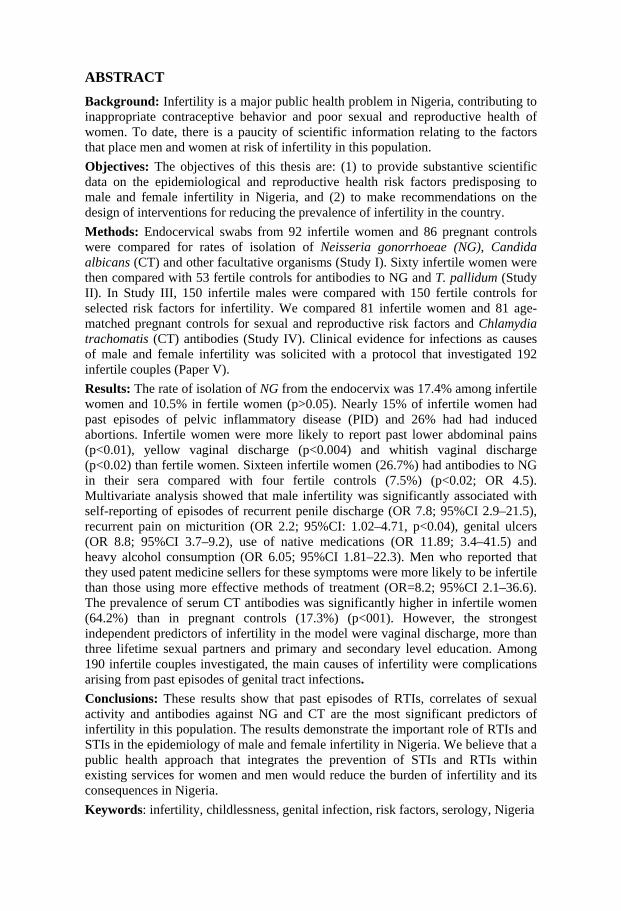
ABSTRACT Background: Infertility is a major public health problem in Nigeria, contributing to inappropriate contraceptive behavior and poor sexual and reproductive health of women. To date, there is a paucity of scientific information relating to the factors that place men and women at risk of infertility in this population. Objectives: The objectives of this thesis are: (1) to provide substantive scientific data on the epidemiological and reproductive health risk factors predisposing to male and female infertility in Nigeria, and (2) to make recommendations on the design of interventions for reducing the prevalence of infertility in the country. Methods: Endocervical swabs from 92 infertile women and 86 pregnant controls were compared for rates of isolation of Neisseria gonorrhoeae (NG), Candida albicans (CT) and other facultative organisms (Study I). Sixty infertile women were then compared with 53 fertile controls for antibodies to NG and T. pallidum (Study II). In Study III, 150 infertile males were compared with 150 fertile controls for selected risk factors for infertility. We compared 81 infertile women and 81 age-matched pregnant controls for sexual and reproductive risk factors and Chlamydia trachomatis (CT) antibodies (Study IV). Clinical evidence for infections as causes of male and female infertility was solicited with a protocol that investigated 192 infertile couples (Paper V). Results: The rate of isolation of NG from the endocervix was 17.4% among infertile women and 10.5% in fertile women (p>0.05). Nearly 15% of infertile women had past episodes of pelvic inflammatory disease (PID) and 26% had had induced abortions. Infertile women were more likely to report past lower abdominal pains (p<0.01), yellow vaginal discharge (p<0.004) and whitish vaginal discharge (p<0.02) than fertile women. Sixteen infertile women (26.7%) had antibodies to NG in their sera compared with four fertile controls (7.5%) (p<0.02; OR 4.5). Multivariate analysis showed that male infertility was significantly associated with self-reporting of episodes of recurrent penile discharge (OR 7.8; 95%CI 2.9–21.5), recurrent pain on micturition (OR 2.2; 95%CI: 1.02–4.71, p<0.04), genital ulcers (OR 8.8; 95%CI 3.7–9.2), use of native medications (OR 11.89; 3.4–41.5) and heavy alcohol consumption (OR 6.05; 95%CI 1.81–22.3). Men who reported that they used patent medicine sellers for these symptoms were more likely to be infertile than those using more effective methods of treatment (OR=8.2; 95%CI 2.1–36.6). The prevalence of serum CT antibodies was significantly higher in infertile women (64.2%) than in pregnant controls (17.3%) (p<001). However, the strongest independent predictors of infertility in the model were vaginal discharge, more than three lifetime sexual partners and primary and secondary level education. Among 190 infertile couples investigated, the main causes of infertility were complications arising from past episodes of genital tract infections. Conclusions: These results show that past episodes of RTIs, correlates of sexual activity and antibodies against NG and CT are the most significant predictors of infertility in this population. The results demonstrate the important role of RTIs and STIs in the epidemiology of male and female infertility in Nigeria. We believe that a public health approach that integrates the prevention of STIs and RTIs within existing services for women and men would reduce the burden of infertility and its consequences in Nigeria. Keywords: infertility, childlessness, genital infection, risk factors, serology, Nigeria

LIST OF PUBLICATIONS This thesis is based on the following papers I. Okonofua FE, Ako-Nai KA, Dighitoghi MD: Lower genital tract
infections in infertile Nigerian women compared with controls. Genitourinary Medicine 1995; 71: 163–8.
II. Okonofua FE, Snow RC, Alemnji GA, Okoruwa A, Ijawere CO:
Serological and clinical correlates of gonorrhoea and syphilis in fertile and infertile Nigerian women. Genitourinary Medicine, 1997; 73: 194–197.
III. Okonofua FE, Menakaya U, Oneumu SO, Omo-Aghoja LO,
Bergstrom S. A case-control study of risk factors for male infertility in Southern Nigeria. Asian Journal of Andrology. (Accepted for Publication)
IV. Omo-Aghoja LO, Okonofua FE, Larsen U, Bergstrom S. Association
of Chlamydia trachomatis serology with tubal infertility in Nigerian women. Sexually Transmitted Infections. (Submitted)
V. Okonofua FE, Menakaya U, Iribhogbe P, Onemu SO, Bergstrom S.
Etiology and outcome of treatment in 190 infertile Nigerian couples. Acta Obstetricia et Gynecologica Scandinavica. (Submitted)
The papers will be referred to by their Roman numerals I–V.

LIST OF CONTENTS
1. INTRODUCTION............................................................................9 1.1 Background ......................................................................................9
1.1.1 Infertility ..................................................................................9 1.1.2 Nigeria – country background................................................10
1.2 Rationale for the studies.................................................................13 1.2.1 Prevalence of infertility in Nigeria.........................................13 1.2.2 Causes and possible risk factors for male and female
infertility ................................................................................14 1.2.3 Implications and social consequences of infertility ...............16 1.2.4 Programmatic issues relating to the prevention and
treatment of infertility ............................................................18
2. AIMS AND OBJECTIVES OF THE STUDY...............................20 Specific objectives .................................................................................20
3. SETTINGS .....................................................................................21
4. SUBJECTS AND METHODS.......................................................24 4.1 Study I............................................................................................24 4.2 Study II...........................................................................................26 4.3 Study III .........................................................................................27 4.4 Study IV.........................................................................................29 4.5 Study V ..........................................................................................31
5. ETHICAL CONSIDERATIONS ...................................................35
6. RESULTS.......................................................................................36 6.1 Lower genital tract infections in fertile and infertile women
(Study I) .........................................................................................36 6.2 Comparison of antibodies to Neisseria gonorrhoeae and
Treponema pallidum and their clinical correlates in fertile and infertile women (Study II)..............................................................40
6.3 Comparison of sexual and reproductive risk factors for male infertility (Study III) ......................................................................42

6.4 The association between Chlamydia trachomatis seropositivity and infertility (Study IV)................................................................51
6.5 Results of management of infertility in 190 couples (Paper V).....57
7. DISCUSSION.................................................................................64 7.1 Association of female infertility with genital tract infectious
morbidity........................................................................................64 7.2 Association of male infertility with genital tract infectious
morbidity........................................................................................68 7.3 Some constraints with epidemiological evidence ..........................69 7.4 Under what social and cultural contexts does genital tract
morbidity lead to high rates of infertility? .....................................71 7.5 From research to service provision for infertility – the need for
a preventive approach ....................................................................72
8. CONCLUSIONS ............................................................................75
9. ACKNOWLEDGEMENT..............................................................77
10. REFERENCES...............................................................................80
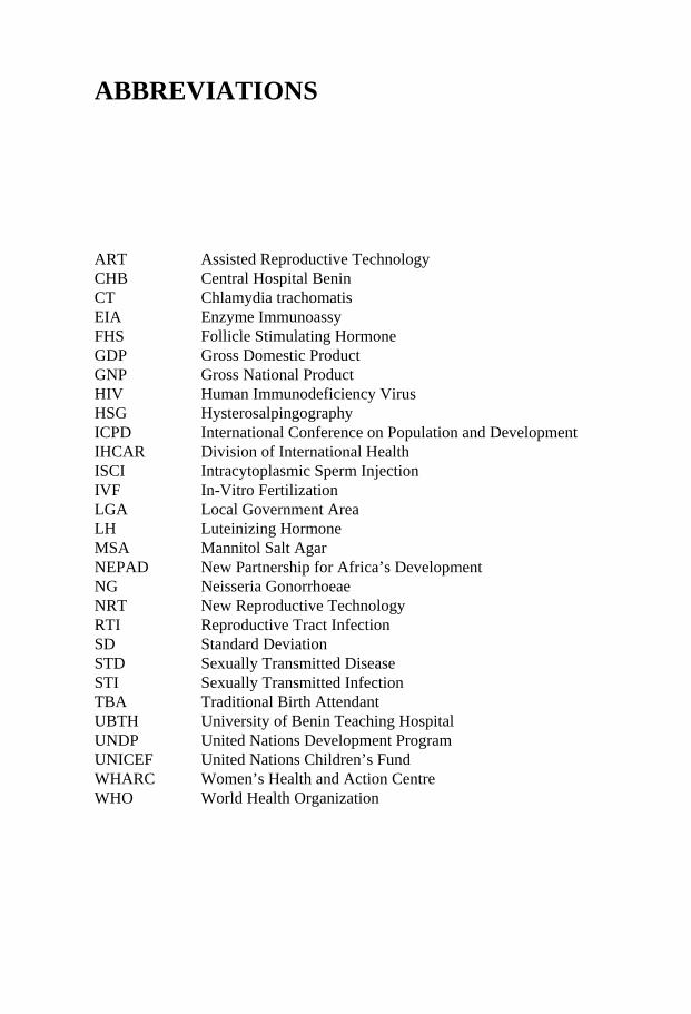
ABBREVIATIONS
ART Assisted Reproductive Technology CHB Central Hospital Benin CT Chlamydia trachomatis EIA Enzyme Immunoassy FHS Follicle Stimulating Hormone GDP Gross Domestic Product GNP Gross National Product HIV Human Immunodeficiency Virus HSG Hysterosalpingography ICPD International Conference on Population and Development IHCAR Division of International Health ISCI Intracytoplasmic Sperm Injection IVF In-Vitro Fertilization LGA Local Government Area LH Luteinizing Hormone MSA Mannitol Salt Agar NEPAD New Partnership for Africa’s Development NG Neisseria Gonorrhoeae NRT New Reproductive Technology RTI Reproductive Tract Infection SD Standard Deviation STD Sexually Transmitted Disease STI Sexually Transmitted Infection TBA Traditional Birth Attendant UBTH University of Benin Teaching Hospital UNDP United Nations Development Program UNICEF United Nations Children’s Fund WHARC Women’s Health and Action Centre WHO World Health Organization

9
1. INTRODUCTION
1.1 Background
1.1.1 Infertility The World Health Organization has recommended the epidemiological definition of infertility as the inability of a couple to conceive within two years of exposure to the risk of pregnancy [1, 2]. Clinical studies frequently use a one-year period of exposure for defining infertility [3], whereas demographic studies use a period of five years [4]. In view of the epidemiological and clinical nature of the studies presented in this report, epidemiological and clinical definitions of infertility will be used interchangeably. Primary infertility is defined as inability to get pregnant [3], while secondary infertility is the inability to get pregnant after an earlier pregnancy [5]. Worldwide, an estimated 580 million people (approximately 5–8% of couples) experience infertility at some point in their reproductive lives [6]. Of these, nearly 372 million persons (about 186 million couples) reside in low- and middle-income countries, with the exclusion of China [3]. Thus, infertility is more prevalent in low-income than in high-income countries. Africa shares by far the largest burden of infertility. Estimates indicate that an average of 10.1% of couples experience infertility in Africa, with a high of 32% in some countries and ethnic groups within Africa [6]. An “infertility belt” spreading through West Africa, through Central Africa to East Africa has been described [4, 7]. In some countries in this belt, up to one-third of women may be childless at the end of their reproductive lives [8]. While infertility is a major problem in Africa, very little has been done at the programmatic and service delivery levels to address the problem. Paradoxically, these regions of Africa with high rates of infertility also have high rates of fertility [3, 9]. Thus, there has been a tendency both locally and internationally to focus exclusively on reducing high fertility rates in Africa, with little or no attention paid to the equally critical problem of high rates of infertility. Throughout much

10
of Africa, fertility is celebrated, while infertility is stigmatising for couples [10]. Available evidence indicates that women disproportionately suffer the social and psychological consequences of infertility in Africa as compared to men [11, 12]. Indeed, women perceive infertility as the most important sexual and reproductive health problem in Africa, and have made efforts to position infertility as an important issue within discourses on gender and sexual and reproductive health and rights [13]. The fear of infertility has been put forward by several advocates and social science researchers in Africa to explain the current low contraceptive prevalence rates in some African countries, as women frequently avoid contraception for fear that it may cause infertility in later life [14–16]. Thus, it is clear that unless the problem of infertility is addressed alongside fertility regulation, there will be little hope of comprehensively and holistically addressing sexual and reproductive health in Africa anytime soon. The ICPD Plan of Action [17] identified the compassionate counseling and treatment of couples with infertility as an important sexual and reproductive health goal, and urged countries to establish programs for the prevention and treatment of infertility. However, whereas many developed countries now have programs that address infertility, very little efforts have been made by African countries in this direction. Clearly, the prevention and treatment of infertility is an important unmet need in sexual and reproductive health programming in Africa, which should be addressed as a basic human and reproductive health right.
1.1.2 Nigeria – country background Nigeria was a colony of Great Britain until October 1960, when it gained independence. Since then, very little progress has been made, with current statistics showing that the country still ranks low in terms of social and economic development even within the context of Africa. Despite being a key player in the African Union and NEPAD, Nigeria’s human development indicators are among the worst in sub-Saharan Africa [18]. Current estimates indicate that Nigeria has a population of about 133 million people [18]. This makes it the most populous country in Africa, accounting for 20% of the population of sub-Saharan African countries. At the current annual growth rate of 2.8%, Nigeria’s population is expected to rise to 155 million by the year 2010, and may double in 25 years if the population growth rate does not decline [19]. The country’s “true fertility rate” is closer to 5.9 or 6.0, making it one of

11
the highest fertility zones in Africa. Contraceptive prevalence is only 8 percent. After several years of military rule, Nigeria finally adopted democratic governance in May 1999. The country presently runs a presidential system of government [20] modeled after the American system, with three tiers of government and a federal structure – a central federal Government, 36 States and 774 Local Government Councils. Each tier of government has an executive arm, a legislative arm and a judiciary. Nigeria is ethnically and socially diverse with more than 350 ethnic nationalities, and two major religions. In order to be able to achieve some generalization, these ethnic groups and states are frequently divided into six geopolitical zones – Northwest, Northeast, North-Central, Southwest, Southeast and South-South geopolitical zones, with a neutral Federal Capital Territory located in Abuja. In view of the considerable social heterogeneity in the country, there is little commonality of purpose, with frequent strife and unrests resulting from inter-ethnic and religious rivalries. Nigeria is undoubtedly endowed with enormous natural resources. However, despite being the fifth largest exporter of crude oil in the world, Nigeria has performed worse in terms of basic social indicators than sub-Saharan Africa as a whole, and much worse than other low-income countries in Asia and Latin America. Despite having earned $320 billion from oil since the 1970s [21, 22], Nigeria still has a stagnant economy, with severe inflationary trends, and some of the worst social, health and education indicators in the low-income world. GNP per capita was only $310 in 1999, making Nigeria a truly low-income country and a collapsing economy. Furthermore, Nigeria has consistently been ranked low in UNDP’s Human Development Index, coming 151st out of 174 countries surveyed in 1998 [23] Estimates indicate that close to 70% of Nigerians live below the poverty line [24], and that the country still services a debt burden of close to US$35 billion. In view of recent increases in oil prices in the international market, from which Nigeria has been a major beneficiary in the past several months, one wonders what has gone wrong. A UNICEF report summarized the problem as follows: “Much of the oil wealth has simply been wasted, poured into misguided public investments or stolen by corrupt public officials” [24]. The health sector has been most badly affected by the negative economic fortunes of Nigeria. Although Nigeria has a national health and population policy [25], very little of the policy is being implemented. The Federal Government of Nigeria has consistently estimated that public
12
funding for health has been in the range of 1–2 percent of GDP [26]. However, the World Bank gave much lower estimates of 0.3 percent of GDP for the period 1990–1996 [22], and 0.2 percent in 1990–1998 [27]. These are low figures compared to averages of 2.6% and 1.5% respectively for Africa for the periods 1990–1996 and 1990–1998. Indeed, the World Bank estimates a higher proportion of private funding of 0.7% of GDP compared to only 0.3% of GDP of public funding of the health sector in Nigeria. Arising from the poor funding of the health sector in Nigeria, most health infrastructures in Nigeria have deteriorated considerably over the years. The country runs a three-level system of health care delivery – primary, secondary and tertiary levels. Recent estimates indicate that there are 18,258 registered PHC facilities across the country, 3,275 secondary facilities, and 29 tertiary facilities [24]. The public sector accounts for 67% of PHC facilities, 25% of secondary facilities and all but one of tertiary facilities. For a vast country such as Nigeria, these facilities are grossly inadequate. The situation has been worsened by internal and external brain drain – internal, because qualified health personnel are concentrated in urban areas to the neglect of rural areas, while external brain drain describes the situation of highly qualified staff who left the country over the past two decades due to poor conditions of work within the country. Brain drain has severely limited the quality of health care available in Nigeria and restricted access to health for millions of Nigerians. Alternative health care providers such as traditional birth attendants (TBAs) and traditional healers now hold sway in the country, providing non-evidence-based health care to a substantial proportion of the population. The combination of social, economic and health problems have resulted in Nigeria having some of the most dismal health indicators in low-income countries. Maternal mortality ratio in Nigeria averages 1,000 per 100,000 live births, accounting for nearly 55,000 maternal deaths annually, and for 10% of global estimates of maternal mortality [28]. An estimated 610,000 unsafe abortions take place in the country annually [29], with high levels of complication, which is responsible for an estimated 20,000 annual maternal deaths [30]. Neonatal, infant and under-five mortality rates are also some of the highest in the low-income countries. HIV sero-prevalence is 5.0%; however, as a result of the huge population of the country, Nigeria currently ranks third in the world (after India and South Africa) in terms of the absolute number of people living with the virus [31]. With such mounting social and economic problems, it is little wonder that infertility is hardly addressed in Nigeria. However, in

13
view of the nature of the background factors that lead to these problems, programs that address infertility will likely benefit other social sectors and health indicators as well.
1.2 Rationale for the studies
1.2.1 Prevalence of infertility in Nigeria The prevalence of infertility in Nigeria has been studied in demographic surveys, epidemiological surveys and through clinical observations. The Nigeria DHS survey for the period 1994–2000 reported a prevalence rate of primary infertility of 22.7% in 15–49-year-old women and 7.1% in 25–49-year-olds [32]. However, demographic surveys of infertility are usually inadequate for programmatic action as they are based on a five-year period of exposure to the risk of pregnancy. By contrast, epidemiological surveys are more useful as they are based on a shorter time frame; however, very few such studies have ever been conducted in Nigeria. A survey of a representative sample of 1,075 ever-married women using a validated WHO protocol revealed a prevalence rate of 20% infertility in Ile-Ife, Southwest Nigeria [33]. As expected, the survey showed a higher prevalence of infertility in the rural parts of the city than in the urban area. A similar survey in a non-representative and smaller sample among predominantly rural residents in Ilorin, North-Central Nigeria, revealed a prevalence rate of infertility of nearly 35% [34]. However, a community-based sample of 400 women in Ilora, a predominantly rural community in Southwest Nigeria, revealed a prevalence rate of 8.7% for infertility [35]. Thus, it is evident that epidemiological surveys have been less than adequate to determine the true prevalence of infertility in Nigeria. However, experiences from actual clinical practice indicate that infertility is a major burden on clinical service delivery in Nigeria. Several reports indicate that infertility is the most frequent reason for gynecological consultation in Nigeria [36]. More than 50% of gynecological caseloads are as a result of infertility consultations [37] and over 80% of laparoscopic investigations are as a result of infertility [38]. Thus, although the scale of infertility has been poorly captured by demographic and epidemiological studies, the size of the clinical problems in health institutions testifies to the enormity of the problem in Nigeria.

14
While most studies have investigated the prevalence of female infertility, very few have studied male infertility. However, it has been suggested that males and females contribute equally to infertility [39]. Nevertheless, reports from various parts of Nigeria have reached different conclusions. While some showed an equal contribution of male and female partners to infertility [40, 41], others showed a disproportionate contribution of male and female partners [42, 43]. However, it is difficult to accurately determine the contribution of males and females to infertility, as fertility is relative and may manifest differently in different couples.
1.2.2 Causes and possible risk factors for male and female infertility Fertility is a complex phenomenon and depends on the normality of an array of important biological systems and mechanisms. Central to normal fertility is the occurrence of normal sexual intercourse between a male partner and female partner. Where any of the biological systems do not work normally in either the male or female partner or both, infertility will be the consequence. Thus, four main categories of causes of infertility are well recognized in clinical practice [10] as follows: (1) male infertility – when infertility is principally due to poor semen parameters; (2) female infertility – when infertility is due to such factors as occlusion of the fallopian tubes, uterine and endometrial abnormalities, abnormal cervix and anovulation in the female partner; (3) infertility in both male and female partners – when factors present in both males and females are responsible for infertility; and (4) normally infertile couples – when both partners are normal, yet they are infertile. The World Health Organization Task Force for the Diagnosis and Treatment of Infertility instituted a standardized protocol for the investigation of infertility, which was adopted in 33 countries across the world [6, 44]. Between 1978 and 1982, a pilot study using this approach examined 8,504 couples and reported on the causes of infertility in 35% of the women and 50% of the infertile men. The results showed that about 85% of women in the African study centers (which included Nigeria) experienced tubal factor infertility compared to about 33% worldwide. Approximately 46% of men in Africa reported a history of sexually transmitted disease, 8% of women reported sexually transmitted disease, while 8% of the women reported abortion complications. The study therefore concluded that infertility in men and women was largely due to genital tract infections when compared to other regions.

15
Several studies from Nigeria have reached the same conclusions on the important role of genital tract infection as causes of male and female infertility. Chukudebelu et al [40] reported on the outcome of investigation of 114 infertile couples in Southeastern Nigeria and concluded that genital tract infections in males and females were the dominant causes of infertility. Reports from various parts of the country including Ilorin [45], Nnewi [46] and Ibadan [47], which represent various geopolitical zones of the country, all confirm the important role of genital tract infections as causes of male and female infertility in Nigeria. While there is general agreement on genital tract infections as principal causes of infertility in Nigeria, very little is known about the nature and determinants of these infections. Such information is critical to designing appropriate interventions for the prevention and treatment of infertility. In women, the most common causes of genital tract infections are sexually transmitted diseases, post-abortion infections and puerperal infections [48, 49]. These infections may produce extensive damage to the fallopian tubes, leading to bilateral tubal occlusion and peritubal adhesions, which prevent ovum pick-up, transluminal transfer and fertilization [10]. By contrast, the most common cause of genital tract infection in males is sexually transmitted diseases (STDs) [50], although other facultative infections such as pyogenic organisms and viruses (mumps virus) are sometimes responsible for male infertility. Both pyogenic organisms and STDs can cause chronic epididymitis, resulting in occlusion of the vas deferens and oligospermia or azoospermia. The principal STDs that produce damage to the genital tract in males and females are Neisseria gonorrhoeae and Chlamydia trachomatis [49]. However, to date, there has been little quantification of their absolute or relative contribution to male or female infertility in Nigeria. Such data are needed at the programmatic level for addressing the high prevalence of infertility in Nigeria. Additionally, only few studies have investigated the epidemiological risk factors for male and female infertility in Nigeria, which are needed to provide critical data for targeting the prevention of infertility. Studies on risk factors for male and female infertility have been conducted in many parts of the world. Such studies have identified smoking [51, 52], caffeine intake [53], high body mass index [54] and alcohol intake [55] as significant risk factors for female infertility in various parts of the world, although the results were not consistent across all study populations. Similarly, studies have shown that occupational factors [56] and smoking [57] are important risk factors associated with

16
male infertility. By contrast, only few such studies have been conducted in Nigeria to identify the factors associated with male and female infertility needed for preventive action. A study in Benin City, Nigeria, reported that dietary exposure to aflatoxin is a significant risk factor for male infertility [58]. Similarly, a hospital-based case-control study has reported unsafe abortion as an important risk factor for female infertility in Nigeria [59]. A study of four countries in Central Africa showed that women married more than once had greater odds of primary and secondary infertility [60]. More studies of this nature are needed to provide important information for counseling and prevention of infertility in Nigeria. Since genital tract infections have been identified as causes of male and female infertility in Nigeria, risk factor analyses are particularly important to determine the predisposing factors to genital tract infections that lead to fertility. Socio-demographic and behavioural risk factors such as patterns of marriage, timing of sexual debut, sexual frequency and numbers of sexual partners, past use of contraception and health-seeking behaviour for genital tract infections are critical sexual and reproductive health issues in Nigeria, for which their association with infertility in later life is critically needed. Such data are needed to support and planned efforts to develop programs for the prevention of infertility in Nigeria.
1.2.3 Implications and social consequences of infertility Evidence from the demographic literature suggests that infertility has important demographic and health implications. It has been suggested that because high infertility has a dampening effect on fertility and the rate of population growth, the treatment of infertility may impede efforts to lower fertility rates. For example, it has been suggested that a reduction in infertility in sub-Saharan Africa to “normal” levels would increase fertility in the region by 15% [61]. A similar demographic report has suggested that infertility accounts for 60% of the variation in total fertility in 18 sub-Saharan African countries and that fertility declines by one birth for each increase in 9 percentage points in women aged 45–49 years who have no children [62]. In Cameroon, a country with an unusually high level of infertility, Larsen and Menken [63] have estimated that the total fertility rate of 5.5 children would rise to 7.3 in the absence of infertility. Such arguments do not necessarily hold for all countries in Africa. In countries such as Nigeria and Mali, there is evidence that the low use of contraception is in part due to the high prevalence of infertility.

17
Sociological and anthropological evidence in these countries [14–16] indicates that women and men are reluctant to use contraceptives for fear that these could lead to infertility, thereby contributing to the low contraceptive prevalence in those countries. Such low contraceptive prevalence rates eventually contribute to high fertility rates in those countries. Thus, demographic analysis cannot be completely correct unless it is combined with an analysis of the sociological consequences of the rising rate of infertility. To date, it is known that although infertility is not a direct cause of mortality in women or men, it is a significant cause of severe socio-psychological morbidity, especially for women. Available evidence from many countries in Africa indicates that the onus of infertility is often placed on women. Infertility has consequences for a woman’s chances of being in a stable marital relationship, whether through lowering her chances of entering into marriage, raising her chances of being divorced or separated, or increasing the chances that her husband will take another wife [3, 11, 64–66]. Furthermore, throughout much of sub-Saharan Africa, several reports indicate that infertility is a cause of physical, social and verbal abuse of women [67, 68] and can lead to social ostracism and denial of inheritance [69]. Infertile men also bear some sociological consequences but to a lesser extent than men. A recent report from South Africa [70] showed how men are perceived not to be men until they have had a child. As a result of the social consequences of infertility, women have been known to manifest various psycho-sexual disorders. While some may have decreased sexual drive [71], some may seek multiple sexual partners [69] in attempts to achieve a desired conception. It is no wonder therefore that increasingly in many parts of Africa, a positive correlation is being found between infertility and higher rates of HIV. Recent reports from Tanzania reported higher rates of HIV in infertile women than in fertile controls [72], while a report from Mozambique showed significantly higher sero-prevalence of syphilis antibodies in infertile women, a finding that may be relevant for a future HIV scenario [73]. No such association has been found for male infertility, but it may exist as well. Higher prevalence of HIV in infertile couples may be due to pre-existing HIV in infertile couples implying that HIV could predispose to infertility. It could also be HIV acquired due to multiple sexual contacts that may be associated in increasing attempts to resolve infertility. The nature of the relationship between HIV and infertility needs to be better understood.

18
1.2.4 Programmatic issues relating to the prevention and treatment of infertility Despite its monumental importance for the promotion of sexual and reproductive health, available evidence suggests that there are only few programs that address the prevention of infertility in many African countries. In many of these countries, infertility is a neglected issue, partly because it does not cause mortality and partly because of perceptions of the relative benefits of addressing infertility versus fertility regulation. Due to a lack of programs to address the prevention of infertility, several reports indicate that women and men in Africa have wrong perceptions of the causes, risk factors and methods of treatment of infertility. Studies across several countries in Africa [74–77] indicate that many people interpret infertility in cultural terms rather than biomedical explanations. Basic counseling and advice of infertile couples are often not available in many African countries [76], and therefore, women often seek inappropriate avenues of treatment based on their limited understanding of the true causes of infertility. Such treatment sources include faith-based treatment, cultural forms of treatment, and non-treatment, many of which may be very expensive and of poor efficacy, since they are not based on scientific evidence [74–77]. Indeed, the lack of programs for the management of infertility is a missed opportunity to promote sexual and reproductive health in Africa. Integrating care of infertility into sexual and reproductive health care will likely increase the acceptance of such care in Africa, and exert multiple beneficial effects on other components of reproductive health [13]. To date, it is known that both conventional and high-technological methods for the treatment of infertility exist in Nigeria [78–80]. Although both types of treatment methods are poorly developed and may be less effective than similar treatment methods in other parts of the world, nevertheless, the availability of a modicum of these methods provides a framework for improving the evidence base for providing effective treatment for infertility in Nigeria. Within the existing fragile nature of the health infrastructure in Nigeria, the argument has been over which public health policy would best address the high rate of infertility in Nigeria. Our view has always been that efforts should concentrate on the prevention of infertility while improving facilities and infrastructure for the conventional treatment of infertility [81]. As part of this argument, we posit that high-technology treatment of infertility would be a heavy burden on the public health sector in a developing economy, and would limit resources for addressing other pressing health problems.

19
Some have countered this argument by stating that high-technology treatment should be provided as a public health effort to provide comprehensive care and to allow low-income countries to match recent advances in knowledge in developed countries [82]. Our response has been that matching scientific advances in developed countries will be a tall order as low-income countries can hardly match the economic requirements for such an exercise [83]. However, low-income countries can seek to locate high-technology treatment of infertility within the private sector since full recovery will be needed to sustain the efforts over time [84]. The public health sector that frequently runs on subsidies in low-income countries can ill afford the economic requirements for sustaining a high-technology treatment service for infertility. We believe that there is a lot to be gained by African countries (including Nigeria) investing in the prevention and conventional treatment of infertility. The emphasis should be on prevention, since the benefits of such a program will benefit other sexual and reproductive health programs in Africa. The development of high-tech treatment of infertility in the public sector should be a long-term venture, when basic health and social issues have been adequately addressed. In summary, the justification for studies of the epidemiological risk factors for infertility in Nigeria was to obtain data to assess the contribution of infectious morbidity and its correlates to infertility in Nigerian men and women. Thus, studies of the carriage of lower genital tract pathogenic organisms and serology for Neisseria gonorrhoeae and Treponema pallidum were undertaken in infertile and fertile women to determine the contribution of these infections to female infertility. A study of the socio-demographic risk factors for male infertility was performed to understand the nature of the infectious determinants of male infertility, so as to seek preventive approaches. Although Chlamydia trachomatis is known to cause infertility worldwide, no studies of its association with infertility have ever been conducted in Nigeria. Thus, we carried out a case-control study of Chlamydia trachomatis serology in tubal infertility, compared to fertile pregnant controls in order to identify the strength of association between tubal infertility and Chlamydia trachomatis infection in Nigeria. Finally, the role of infections can be best assessed through actual clinical management of patients. This was done through the clinical investigation of 192 couples, which enabled us to determine the actual contribution of infectious morbidity to infertility and to evaluate the effects of clinical management.
20
2. AIMS AND OBJECTIVES OF THE STUDY
The general objective of this thesis is to explore the determinants of infectious causes of infertility and make recommendations for improving the prevention and treatment of infertility within the prevailing fragile health infrastructure in Nigeria.
Specific objectives 1. To investigate the possibility that infertile Nigerian women have
higher levels of lower genital tract infections than fertile women and identify the types of organisms that are most commonly associated with infectious lower genital tract morbidity in the women.
2. To test the hypothesis that fertile Nigerian women have higher serum levels of antibodies against Neisseria gonorrhoeae and Treponema pallidum than pregnant fertile controls.
3. To evaluate the association between selected potential socio-demographic and behavioural risk factors and infertility in Nigerian males.
4. To determine the association between tubal infertility and Chlamydia trachomatis seropositivity in Nigerian women.
5. To explore the outcome of investigation and treatment in couples presenting with infertility at a tertiary referral center in Benin City, Nigeria, and explore the role of genital infectious morbidity among these couples.

21
3. SETTINGS
The studies were conducted in two cities in Nigeria – Ile-Ife, in Ife Central Local Government Area of Osun State, Southwest Nigeria; and Benin City in Igor Local Government Area of Edo State, South-South Nigeria. Studies I and II were conducted in Ile-Ife, while studies III, IV and V were conducted in Benin City. Ile-Ife and Benin City are both among the oldest cities in Nigeria with considerable historical and cultural significance. Ile-Ife is ancestral home to the Yoruba ethnic group, both in Nigeria and in the Diaspora, and their natural ruler, the Oni of Ife, is the head of the Yoruba race worldwide. Similarly, Benin City is ancestral home to the legendary Edo-speaking people, whose leader challenged the British Government in the colonial days. The current Oba of Benin is a descendant of the great Benin Kingship dynasty, and continues to hold much of the culture and artistry for which the Benin Empire has always been known and respected worldwide. Ile-Ife is a city of about one million people, consisting of a large urban part and up to 120 surrounding hamlets and villages. The city is approximately 200 km northwest of Lagos. A large university town, with few industries, most residents are petty traders, artisans and farmers. By contrast, Benin City is a large urban cosmopolitan area with a population of 1.2 million residents, about 350 km northeast of Lagos. Although residents of Benin City are mainly Edo-speaking people, the city has a sizeable representation of other ethnic groups as well. Ile-Ife has a large federal university and a federally funded teaching hospital, while Benin City has three universities – one federally funded and two private – and a large federal teaching hospital. Studies I and II were conducted in Ile-Ife. Study I was a hospital-based study conducted at the Obafemi Awolowo University Teaching Hospital in Ile-Ife. The hospital has more than 600 beds, while the Department of Obstetrics and Gynecology of the hospital is a referral unit for obstetrics and gynecology cases in the vicinity. Study II was a community case-control study based on a survey conducted in the urban and rural parts of Ile-Ife. The detailed description of the survey has been presented elsewhere [85]. In brief, the survey was

22
a questionnaire household survey of 1,075 ever-married women, to determine the prevalence of infertility. Infertility was defined in the study as women who had no pregnancy after two years despite regular sexual intercourse without contraception [1]. A validated WHO questionnaire was used, following which women were allocated into four categories of fertility – fertile, primarily infertile, secondarily infertile, and probably infertile. Following these groupings, women classified as infertile were invited to participate in a subset study to determine the presence of antibodies against Neisseria gonorrhea and syphilis in their sera. Infertile women who consented to participate in the study were then age-matched with those identified as being fertile in the community survey. Once an infertile woman was identified, the next fertile woman in the community of the same age as the infertile woman was chosen as control. Both cases and controls were invited to the gynecology clinic of the Obafemi Awolowo University, where the blood samples were taken and the laboratory analysis conducted. Study III was conducted at the Reproductive Health Clinic of the Women’s Health and Action Research Centre (WHARC) in Benin City. WHARC is a non-governmental, non-profit organization established in 1995 with the mission to promote the sexual and reproductive health of women through research, documentation, advocacy, training and service delivery. As part of its service delivery efforts, the Centre runs a reproductive health clinic that is mainly devoted to the reproductive health of adolescents and management of infertility, fertility and sexually transmitted infections. The work of the Centre has been described elsewhere [86, 87]. The clinic manages more than 1,000 infertile couples annually. Study IV was carried out at the University of Benin Teaching Hospital (UBTH) and the Central Hospital (CHB), both located in Benin City. The UBTH is a Federal Government tertiary institution in Benin City, with more than 600 beds. The CHB is a secondary-level health institution managed by the Edo State Government. The hospital is located in the city center and therefore receives a lot of clients for secondary-level obstetrics and gynecological services. Patients attending the infertility clinics of the UBTH and CHB were recruited as cases in the study, while controls were pregnant women attending antenatal clinics in the two hospitals. Study V (the investigation and management of 192 infertile couples) was conducted in WHARC and the UBTH. The patients were initially received in WHARC. However, since WHARC does not have facilities for full investigation of infertility such as laparoscopy and

23
hysterosalpingography, the women were referred to UBTH for these investigations. The same physician who initially managed the patients in WHARC also managed them at the UBTH. Following the completion of investigations at the UBTH, the patients returned to WHARC for follow-up clinic and home-based care.

24
4. SUBJECTS AND METHODS
4.1 Study I This was a case-control study carried out at the Obafemi Awolowo University Hospital, Nigeria between 20 September 1990 and 16 December 1991. The patients were 92 infertile women attending the infertility and endocrinology clinic of the hospital during the period. Infertility was defined according to the WHO current definition of infertility, that is, the inability to establish a pregnancy within two years for couples of reproductive age who are having intercourse without contraception [1]. Primary infertility refers to those who have never been pregnant while secondary infertility refers to those who have had at least one proven pregnancy (including an abortion). The controls were 86 randomly selected pregnant women who attended the antenatal clinic of the hospital. The pregnant women were selected from among those attending the antenatal clinic during the same period as the cases of infertility. To ensure comparability of cases and controls, we selected pregnant women from among women of the same age and educational level as the cases of infertility. This is because we recognized that age and educational level are the two factors most likely to affect the rates of sexually transmitted diseases in the community. Thus, after first identifying an infertile woman, all pregnant women of the same age and educational level were identified in the ensuing antenatal clinic. One of these women would then be randomly selected by lot for inclusion in the study. Using this procedure, eligible controls could not be found for six women from the immediate antenatal clinic, thereby limiting the study to 92 cases and 86 controls. The study was carefully explained to both groups of subjects and they were assured of strict confidentiality of information obtained. All recruited women agreed to participate in the fully explained protocol and were included in the study. At the onset, the women were assisted in completing a structured questionnaire which sought information on their socio-demographic characteristics, their previous pregnancy and infertility history, history of previous treatment of sexually transmitted disease and vaginal discharge
25
and reproductive history including previous use of contraception and abortion. The infertile women were fully investigated with hysterosalpingography, laparoscopy, semen analysis and endometrial biopsy and the probable cause of infertility ascertained. The infertile women had duration of infertility ranging between 2 and 20 years with a median duration of 6 years. Twenty-four (26.1%) of the infertile women had primary infertility while 68 (73.9%) had secondary infertility. After full infertility investigations 15 women were found to have bilateral tubal occlusion; seven had unilateral tubal occlusion and pelvic adhesions; six had uterine fibroids; 16 had semen abnormalities; 18 had ovulatory and menstrual problems; four had problems in both partners; while no cause could be found in 26 couples. Collection and processing of samples in both groups of women, a sterile bivalve speculum examination was carried out by the attending obstetrician/gynecologist and two endocervical samples and one high vaginal sample were taken with the aid of two sterile cotton-tipped applicators (Evepon Industries Ltd., Nigeria). The swab stick was of the endocervix. A wet mount of each sample was prepared by addition of normal saline to the sample on a clean glass slide. The wet mount was then examined under the microscope with x100 for the presence of white blood cells, red blood cells, Candida albicans, Trichomonas vaginalis, bacteria and epithelial cells. Primary isolation for possible anaerobic organisms was performed by inoculating the first swab on freshly prepared cooked meat medium, brain heart infusion broth (BHI) and thioglycolate broth. The fluid media were incubated anaerobically and after growth was observed, each tube was subcultured onto various anaerobic plates. For primary isolation of aerobic organisms, each endocervical swab was streaked onto blood agar, Thayer-Martin agar, mannitol salt agar (MSA), eosin methylene blue agar and sabouraud dextrose agar supplemented with penicillin/neomycin. All plates were subsequently incubated at 37°C for a minimum of five days. All plates were examined daily for growth and based on the gram stain reaction and colonial morphology, each discrete colony was subjected to further characterization (cultural and bio-chemical) using standard microbiological techniques [88]. For example, gram-negative short rods suspected to be enteric organisms were confirmed by the use of the API 10S system (La Balme Les Grotes, 38390 Montalieu Vercieu, France) and Staphylococcus aurous was speciated using coagulase tube test on pooled human plasma. Fungal specimens were inoculated into Sabouraud dextrose agar supplemented with penicillin/neomycin and incubated initially at 29ºC for 3–7 days, and speciation as Candida
26
albicans was done by germ tube production on corn meal agar. Chlamydiospore production in nutritionally deficient media and trypan blue agar was also used for identification of the fungus. Neisseria species were identified initially on Thayer-Martins agar, and subsequently with fermentation of CTA sugars and oxidase test. Antibiotic sensitivity test was performed using the agar diffusion method described by Ericsson and Sherris [89]. Commercially prepared antibiotics (AB Biodisk Pyramidvägen Solna, Sweden) were used with the following concentrations: Ampicillin 10µg, cefataxime 30 µg, streptomycin 30µg, trimethroprim 5 µg, sulfamethoxazole 223.8 µg + trimethoprim 1.2 µg, 25 µg, nalidixic acid 30 µg and sulfisomidine 30µg. Staphylococcus aureus ATCC 25923 and Enterobacter aerogenes ATCC 13042 were run as control organisms. For data analysis the frequency of isolation of the various pathogenic organisms was determined for the infertile and pregnant women and among the subgroups of causes of infertility. Any observed differences between the groups were tested for statistical significance with chi square test and Yates correction as appropriate.
4.2 Study II This study was conducted in Ife Central Local Government Area (LGA) of Southwestern Nigeria between March 1994 and May 1995. The study drew on population-based data to examine a subpopulation of cases and controls. The cases were women with primary and secondary infertility in the LGA who were identified through a comprehensive community survey of the area in 1994. The details of the community survey have been described elsewhere [85]. In brief, 1,075 ever married women aged 15 to 49 years in the area were randomly selected and interviewed in their households with detailed structured and pre-tested questionnaire. The questionnaire solicited information on the socio-demographic profiles of the women and their pregnancy and contraception histories. Inquiries were also made into their gynecological histories with particular reference to their previous experience of symptoms of sexually transmitted infections. Specifically, the women were asked a series of questions as to whether they currently had or had ever had symptoms suggesting an STI or had ever been diagnosed as having a specific STI. The symptoms checklist included repeated lower abdominal pain, yellow vaginal discharge, whitish vaginal discharge, repeated itching in the

27
genital area, repeated painful/burning sensation on micturition, or ulcers in the genital area. After the survey, women were classified by fertility status based on an algorithm designed for the purpose by the World Health Organization [90]. Seventeen women classified as suffering from primary and secondary infertility formed the cases for this study. The controls were women who were classified as being fertile, and they were matched within 2 years by age and resident locality to the infertile cases. Six women in the control group were pregnant at the time of the survey. Both cases and controls were revisited within one month of the initial survey in their households to obtain blood samples for estimating gonococcal and treponemal antibodies. The purpose of the study was fully explained to the women and only those who agreed to participate were finally included into the study. The protocol for the study was approved by the ethics review committees of the Obafemi Awolowo University and Harvard School of Public Health, Boston, USA. The interviews were conducted by trained female interviewers. Women were assured of confidentiality of information and results of the blood samples. A volume of 15 ml of blood was taken from the women in plain tubes, allowed to clot for 2 hours, centrifuged at 3,000 rpm for 15 minutes, and separated into sera. Sera were refrigerated at –20ºC until assayed in batches for antibodies. The presence of gonorrhea anti-bodies was determined with a gonorrhea agglutinotest kit (Istituto Immunologico Italiano, Pomezia, Rome) made up of a stabilized suspension of polystyrene latex particles on which an antigenic extract of strains of Neisseria gonorrhoeae in smooth stage is absorbed. Detection of antibody to pilin proteins has been reported as being valid in diagnosing current and previous gonococcal infection [91, 92]. Serological testing for syphilis was carried out with the rapid plasma reagin method as described by the World Health Organization [93]. For statistical analysis, the proportion of infertile and fertile women demonstrating gonococcal and treponemal antibodies in their sera was determined. The prevalence of symptoms of STIs and previous specific STI diagnoses was also determined in both groups. These variables were then compared using chi square test with Yates correction.
4.3 Study III This was a case-control study conducted at the Women’s Health and Action Research Center, Benin City, Nigeria, between 1 January 1999

28
and 31 December 2003. The cases were 150 males, whose wives presented for investigation and treatment of infertility in the clinic, and who were found to have abnormal semen parameters. The controls were 150 males, whose spouses were pregnant at the time of the study, and who had normal semen parameters. All cases of male infertility were identified from among couples presenting to the clinic requesting investigation and treatment of infertility. A detailed history of infertility was elicited from the couples, and physical and special investigations were carried out. The latter included semen analysis, hysterosalpingography and/or laparoscopy to evaluate tubal factor, hormone assay for ovulation assessment and trans-vaginal ultrasound scan as part of ovulation assessment and to exclude uterine and endometrial abnormalities. Only those who were identified as having semen anomalies as the only cause of their infertility, and who consented to participate in the fully explained protocol, were included as cases in the study. Among the 150 couples identified with male-factor only infertility, 89 (59.3%) had primary infertility, while 61 (40.7%) had secondary infertility, and had infertility duration ranging between 3 and 15 years (median=8.5 years). Thus, all had definite evidence of infertility as defined by the WHO [1]. By comparison, the controls were men with proven fertility. These were men whose wives were recently pregnant, or who delivered within six months of the study. Such women were identified in the clinic and the study was explained to them. The women were asked to inform their partners and to request them to present in the clinic to participate in the study. Among the men who presented, the study was further explained to them in great detail and only those who gave consent were included in the study. Once a case of male infertility was identified, he was matched for age and place of residence with a fertile control. Semen analysis was conducted in both cases and controls after at least three days of abstinence, using established WHO protocols [94]. Semen analyses were done twice at least two weeks apart, in both cases and controls, to eliminate the possibility of a diurnal variability in the reported semen results. Only those with consistent results were included as either cases or controls. Using the WHO criteria [94], normal sperm concentration was accepted as being greater than 20 million/ml; 5–20 million/ml represented oligozoospermia, while less than 5 million/ml was identified as severe oligozoospermia. Motility was described as normal if 50% or more of the sperms were progressively motile within 60 minutes of ejaculation. Sperm morphology describes the number of normal

29
spermatozoa that have an ovoid head, stainable acrosome head and a normal mid-piece and tail. Although the WHO previously accepted 30% as normal, Kruger et al [95] have described strict criteria where less than 14% normal morphology would be abnormal. The 4th edition of the WHO manual in use at the time of the study [94] did not specify a value for morphology and thus, we have not included this in the assessment. All semen samples from both cases and referents were cultured using standard procedures, to determine the presence of pathogenic organisms in the samples. Following these initial assessments, a three-part study protocol was designed to document the findings uniformly in both cases and controls. In the first part of the protocol, we documented detailed information on the socio-demographic characteristics of the respondents – their age, marital status and type of marriage, occupation, religion and educational background. The second part of the protocol obtained information on the respondents’ sexual behaviour and their previous symptoms suggestive of STIs, genital tract infections and other medical/surgical illnesses. If they had experienced these symptoms, we then asked how long the symptoms had lasted and where and how they had been treated. In the second part of the protocol, we also asked whether or not the respondents smoked or used alcohol, and if so the types, duration and amount taken over time. In particular, we distinguished between cigarettes and marijuana smoking, in order to identify possible independent effects of these practices on male fertility. We also asked questions on their use of medications, either routinely or for the indicated medical conditions. In the final part of the protocol, we recorded the results of physical examination and laboratory tests conducted in both cases and controls. In particular, we recorded the presence of the testicles within the scrotal sacs in both cases and controls, and whether or not the subjects had varicocele and urethral discharge at the time of the study. We also recorded the results of semen analysis carried out in both cases and controls.
4.4 Study IV This study was conducted in two hospitals in Benin City, South-South Nigeria – the University of Benin Teaching Hospital and Central Hospital. The study was a case-control study and consisted of cases and controls. The cases were infertile women with proven tubal factor infertility, confirmed by hysterosalpingography and laparoscopy. All

30
patients were investigated for female and male causes of infertility according to established protocols and those found not to have tubal occlusion on laparoscopy were excluded from the study. The controls were pregnant women attending the antenatal clinics of the hospitals during the period of the study. Once a case of infertility was identified, the next woman who presented in the antenatal clinic who was of similar age as the case of infertility was selected as a control. This was due to the high confounding effects of age on the prevalence of chlamydial antibodies. All the identified cases had proven infertility due to tubal occlusion. All had bilateral or unilateral tubal occlusion with or without peritubal adhesions as assessed from laparoscopy and hysterosalpingography. The infertile women had infertility duration ranging between 3 and 17 years, with a median duration of 8 years. By contrast, none of the pregnant women had evidence of tubal infertility and none had previously been investigated for infertility. Upon recruitment, both cases and controls had a data sheet completed that elicited information on socio-demographic variables, sexual and reproductive risk factors. Specifically, the following information was obtained from both cases and controls – age, marital status, educational level and parity. Other information obtained included age at sexual debut, numbers of sexual partners, use of condoms and oral contraceptives, history of a previous ectopic pregnancy, and history suggestive of previous pelvic infections. For Chlamydia antibody assay, 5 ml of venous blood was collected and entered into a clean and sterile plastic container. The blood specimens were allowed to clot, and then centrifuged to obtain sera. The sera were stored frozen at –20°C until they were analyzed in batch for serum Chlamydia antibodies. The serological assay was done using the immunocomb Chlamydia trachomatis IgG kit [96], an indirect solid phase enzyme immunoassay (EIA) test that quantitatively measures antibodies to Chlamydia trachomatis in human serum. The reagent test kit was brought to room temperature, and then 10 ml of pipetted serum was assayed with reagent control samples for Chlamydia trachomatis IgG antibodies. The procedure was then run through a number of timed steps as outlined by the manufacturer [96]. The study was approved by the Human Ethics Committee of the University of Benin Teaching Hospital. The study was carefully explained to the patients and only those who agreed to participate in the fully explained protocol were included.
31
The sample size was calculated based on Chlamydia trachomatis infection prevalence rate of 12% in Benin City reported by Azenabor et al [97], and with the formula by D.W. Taylor [98]. A sample size of 162 for cases and controls was identified as necessary to detect a smallest odds ratio of 2.680, with an estimated value of the smallest proportion of 0.65, at a power of 95% and significance at the 0.05 level. The program EPI-info 2000 was used for data management and the statistical analysis was done using STATA version 8. This consisted of a univariate analysis of socio-demographic characteristics, contraceptive and sexual history, as well as prevalence of chlamydial antibodies, gynecological symptoms and treatment sources for past gynecological problems of infertile cases and pregnant controls. Conditional logistic regression was used to calculate odds ratios and 95% confidence intervals. The characteristics of women with chlamydial antibodies were analyzed in univariate logistic regression models. Multivariate conditional logistic regression was used to determine the association between chlamydial antibodies and infertility controlling for the effects of covariates.
4.5 Study V This study was prospective and consisted of consecutive couples attending the Women’s Health and Action Research Centre (WHARC) in Benin City, Nigeria for infertility treatment between 1 January 2000 and 31 December 2001. WHARC is a non-governmental organization that runs a referral clinic with modern facilities for infertility treatment and fertility management. The Centre is closely associated with the Department of Obstetrics and Gynaecology at the University of Benin Teaching Hospital, where more sophisticated procedures such as laparoscopy are undertaken by the same clinician who first admitted the couple in WHARC. This linkage enables the Centre to provide comprehensive clinical care to infertile couples, while at the same time providing a private setting for their continued confidential counseling and follow-up. At the onset, a protocol adapted from the WHO guidelines for investigating infertility [99] was used to manage all cases of infertility in the Centre. With this protocol, couples were encouraged to December attend the clinic together at their first or second visit, which provided an opportunity to obtain a detailed history from each partner. The history consisted of the duration of infertility, past pregnancy history, menstrual

32
and gynecological history, past medical and surgical history, family, sexual and social history, and history of any previous treatments that may have been undertaken. A detailed history was also taken from the male partners, with special emphasis on the men’s present and past sexual relationships, their past experiences of genital infections and other major illnesses, their occupational, drug and dietary history, and whether or not they engaged in any social habit such as smoking or alcohol intake. Although both partners often attended the clinic together, they were first interviewed together on broad issues relating to their infertility, while answers to more sensitive questions were solicited in separate confidential interviews. Following the history, both partners were clinically examined in efforts to identify any medical conditions that may be associated with the infertility. This consisted of a general physical examination, as well as breasts, cardiovascular, abdominal and pelvic examination in the female. Similarly, the male partners were examined for their general and physical well-being, their cardiovascular health, abdominal organs and their external genitalia including their testes, epididymis and vas deferens. When an abnormality was found in the genital examination, a rectal examination was also done in the male to detect any abnormalities of the prostate gland. Special infertility investigations carried out in the clinic included semen analysis after three days of abstinence using the WHO core methodology [94]. When the initial semen evaluation was abnormal, two more tests were performed two weeks apart before the results were confirmed. Ovulation was ascertained with hormone assay using well validated kits and methods. This consisted of serum progesterone on day 21 of the menstrual cycle and serum FSH, LH, prolactin and testosterone on days 2–3. Transvaginal ultrasonography was also used to check the uterus, uterine lining and ovarian follicles to authenticate the presence or absence of ovulation in any particular menstrual cycle and to monitor the women during treatment. Other tests performed included hysterosalpingography (HSG) on all women presenting with infertility in efforts to exclude tubal pathology. According to the principles outlined in our earlier paper [100], only women with abnormal HSG results were counseled for laparoscopy. Laparoscopy was carried out at the University of Benin Teaching Hospital when required, using established procedures and methods [38, 47, 100]. Other tests done in efforts to comprehensively investigate the causes of infertility included postcoital tests and endometrial biopsy and histology in selected patients,
33
Following investigations, a diagnosis of the pathological causes of infertility was made and summarized in fewer than four broad categories as follows: (1) male cause only, (2) female cause only, (3) causes in both partners, and (4) no cause found in either partner. Treatment was often directed at the identified pathologies in either or both partners and included the following alternatives: (1) treatment in the clinic using conventional methods – counseling, ovulation induction, tubal repair, other gynecological surgical procedures (e.g. myomectomy) and insemination; (2) referral for assisted reproductive technology (ART) procedure either in Nigeria or abroad; and (3) counseling of the couples to accept their infertile status or to adopt a child. WHARC ran a viable adoption clinic side by side with the infertility clinic in collaboration with the Edo State Ministry of Women Affairs and Social Welfare. For ART, couples were referred to the London Fertility Clinic, Harley Street, London or the Bridge Clinic in Lagos, Nigeria, depending on their preference. At the onset, the couples were informed that they would be followed up for one year whether or not they receive specific treatment in the Centre. Thus, informed consent was obtained from the couples for the study, especially to follow them up at home should they fail to report to the clinic. The contact details including telephone numbers (where available) of the couples were kept as part of the clinical records. Two nurses were specially designated and trained in the Centre to conduct these follow-ups and home visits and to maintain confidential records on each couple. The Human Ethics Committee of the University of Benin Teaching Hospital approved the protocol. This report focuses on the results of the investigations to document the medical causes of infertility in the couples and the success rate (in terms of achieved pregnancies) after at least one year of treatment and follow-up. Thus, only simple univariate results have been presented, with very limited attempt made to identify relevant factors that may influence the outcome of specific treatment procedures. Statistical differences between samples were compared using the chi square test with Yates correction as appropriate.

34
Table 1. Characteristics of the studies reported. Paper Topic Study
population Method Period
I A case referent study comparing prevalence of lower genital tract infections in infer-tile women with fertile controls.
92 infertile women and 86 pregnant controls.
Vaginal and endo-cervical swabs cultures for sexually trans-missible infections and facultative organisms.
Sep 1990 to June 1991
II A case referent
study comparing antibodies to N. gonorrhoeae and syphilis and clini-cal correlates in infertile women and fertile controls.
60 infertile women identified from a community survey of 1,075 women and 53 age-matched controls.
Serum assay for gonococcal and treponemal antibodies. Self-reporting of symptoms of genital tract infections.
March 1994 to May 1995
III A case referent
study comparing sexual and reproductive risk factors in fertile and infertile males
162 infertile males and 162 fertile males.
Questionnaire survey
Jan 1999 to Dec 2003
IV A case referent
study comparing prevalence of Chlamydia trachomatis anti-bodies in infertile and fertile women
81 Nigerian women with tubal infertility and 81 pregnant controls.
Serum assay of Chlamydia trachomatis antibodies
Jan 2001 to June 2004
V A cross-sectional
study investigating the etiology and outcome of treat-ment of infertile Nigerian couples.
190 consecutive couples with infertility.
Application of a standardized infertility treatment protocol.
Jan 2000 to Dec 2001

35
5. ETHICAL CONSIDERATIONS
The five studies were approved by the institutional ethics committees of the hospitals in which the study took place, the Obafemi Awolowo University Teaching Hospital and the University of Benin Teaching Hospital. Additionally Study II was approved by the Research Ethics Committee of the Harvard University School of Public Health as the study was conducted in collaboration with the Department of Population and International Health of the Harvard School of Public Health. The study objectives were explained to each participant, and informed consent was obtained prior to questionnaire administration and specimen collection.
36
6. RESULTS
6.1 Lower genital tract infections in fertile and infertile women (Study I)
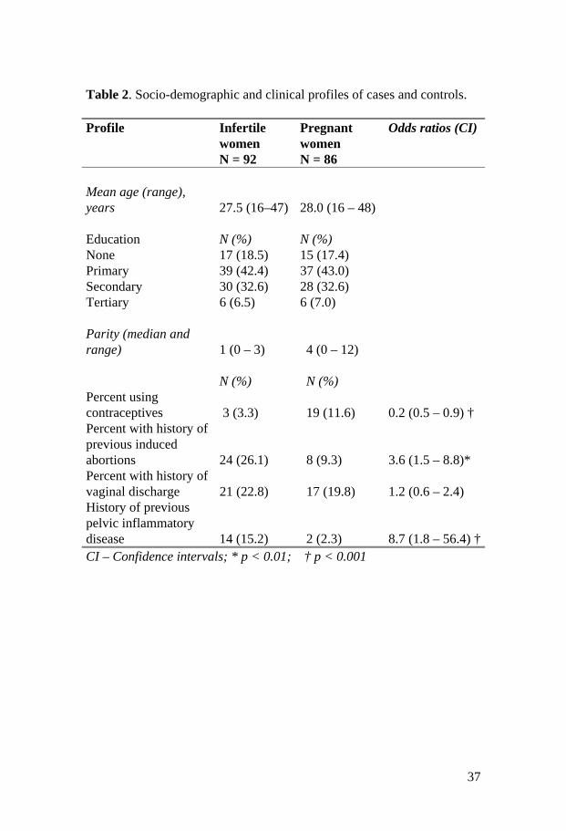
The socio-demographic and clinical profiles of the infertile and pregnant women are shown in Table 2. As indicated, both cases and controls had identical mean age and educational level. The pregnant women were more parous and reported higher “ever use” of contraceptives than infertile women. By contrast, a higher proportion of the infertile women reported previous pregnancy termination. A high proportion of both cases and controls (22.8% vs. 19.8%) admitted experiencing vaginal discharge at the time of the study. However, as shown in Table 2, more infertile women than controls had had symptomatic pelvic inflammatory disease (PID) in the past, which required treatment (p<0.002). Multiple microbial isolates were cultured from the endocervices of the infertile and pregnant women. These included 203 isolates cultured from the endocervix of the 86 infertile women and 164 isolates recovered from the 92 pregnant women. There was apparently no difference in rate of isolation of organisms between pregnant and infertile women. As shown in Table 3, there was also no difference between pregnant and infertile women in the species of organism isolated. Candida albicans was the commonest pathogenic organism isolated in both groups, with 25% of infertile and 29.1% pregnant women harboring the organism in their upper vagina and endocervix (p>0.05). Similarly, there was no significant difference between infertile and pregnant women in the rate of isolation of Neisseria gonorrhoeae, Trichomonas vaginalis and Staphylococcus aureus. The rate of vaginal colonization with pathogenic anaerobes and opportunistic organisms was also identical between pregnant and infertile women.
37
Table 2. Socio-demographic and clinical profiles of cases and controls. Profile Infertile
women N = 92
Pregnant women N = 86
Odds ratios (CI)
Mean age (range), years 27.5 (16–47) 28.0 (16 – 48) Education N (%) N (%) None 17 (18.5) 15 (17.4) Primary 39 (42.4) 37 (43.0) Secondary 30 (32.6) 28 (32.6) Tertiary 6 (6.5) 6 (7.0) Parity (median and range) 1 (0 – 3) 4 (0 – 12) N (%) N (%) Percent using contraceptives 3 (3.3) 19 (11.6) 0.2 (0.5 – 0.9) † Percent with history of previous induced abortions 24 (26.1) 8 (9.3) 3.6 (1.5 – 8.8)* Percent with history of vaginal discharge 21 (22.8) 17 (19.8) 1.2 (0.6 – 2.4) History of previous pelvic inflammatory disease 14 (15.2) 2 (2.3) 8.7 (1.8 – 56.4) † CI – Confidence intervals; * p < 0.01; † p < 0.001

38
Table 3. Rate of isolation of pathogenic organisms from cases (infertile women) and controls (pregnant women) Isolation Infertile
women N = 92 (%)
Pregnant women N = 86 (%)
p*
Pathogenic aerobes Neisseria gonorrhoeae 16 (17.4) 9 (10.5) Ns Staphylococcus aureus 20 (21.7) 10 (11.6) Ns Candida sp 23 (25.0) 25 (29.1) Ns Trichomonas vaginalis 4 (4.3) (2 (2.3) Ns Pathogenic anaerobes Clostridium sp. 3 (3.3) 7 (8.1) Ns Streptococcus sp (beta haemolytic)
7 (7.6) 10 (11.6) Ns
Opportunistic organisms Pseudomonas aeruginosa 3 (3.3) 2 (2.3) Ns Bacillus sp 5 (5.4) 10 (11.6) Ns Proteus vulgaris 3 (3.3) 2 (2.3) Ns Escherichia coli 2 (2.2) 2 (2.3) Ns Ns = Not significant * Chi square test with Yates correction To determine whether the type of infertility has any effect on the pattern of vaginal and cervical colonization by micro-organisms, we compared the proportion of women with pathogenic organisms in those with primary infertility and those with secondary infertility. The results are presented in Table 4. Women with secondary infertility had a higher rate of Neisseria gonorrhoeae (20.5%) than women with primary infertility (8.3%) (p<0.02). Similarly, secondarily infertile women had higher rates of colonization with Candida albicans and Staphylococcus aureus. By contrast, there was no significant difference between the two groups of women in the rates of colonization with Trichomonas vaginalis, Hemophilus sp. and other opportunistic vaginal organisms.

39
Table 4. Rates of microbial isolation in infertile women by type of infertility Isolate Primary
infertility N = 24 (%)
Secondary infertility N = 68 (%)
p*
Pathogenic aerobes Neisseria gonorrhoeae 2 (8.3) 14 (20.5) 0.02 Staphylococcus aureus 2 (8.3 18 (26.5) 0.001 Candida sp 3 (12.5) 20 (29.4) 0.01 Trichomonas vaginalis 1 (4.2) 3 (4.4) Ns Pathogenic anaerobes Clostridium sp. 1 (4.2) 2 (2.9) Ns Opportunistic organisms Hemophilus sp. 2 (8.3) 6 (8.8) Pseudomonas aeruginosa 1 (4.2) 2 (2.9) Ns Bacillus sp. 2 (8.3) 3 (4.4) Ns Proteus vulgaris 1 (4.2) 2 (2.9) Ns Escherichia coli 1 (4.1) 3 (1.5) Ns Ns = Not significant * Chi square test with Yates correction Further analysis shows that of the 16 infertile women, from whom Neisseria gonorrhoeae was isolated, five had a past gynecological history of pelvic inflammatory disease; four had bilateral tubal occlusion; and one had a previous history of chronic cervicitis. No associated risk factors could be identified in the other 10 infertile women from whom Neisseria gonorrhoeae was isolated. To determine the predictive value of a history of vaginal discharge for the rate of isolation of a cervical pathogen among infertile women, we compared the isolates in women with vaginal discharge with those in women without vaginal discharge. Our results indicate that five isolates of Neisseria gonorrhoeae were made from the 38 infertile and pregnant women with vaginal discharge (13.2%) compared with 20 isolates in the 140 women who had no history of vaginal discharge (14.3%). The difference is not statistically significant. There was also no significant difference in the rate of isolation of Candida albicans, Trichomonas vaginalis and Staphylococcus aureus between infertile women with vaginal discharge and those without vaginal discharge. Thus, a history of

40
vaginal discharge did not appear to be a marker for the presence of the micro-organisms in the vagina and endocervices of the women. The antibiotic sensitivity pattern of the isolated organisms is presented in Table 5. Neisseria gonorrhoeae, the most commonly isolated pathogenic organism, was uniformly sensitive to gentamicin and nalidixic acid and uniformly resistant to cefotaxime, ampicillin, spectinomycin and chloramphenicol. Similarly, Staphylococcus aureus, another pathogen, was resistant to many antibiotics and was only moderately sensitive to ampicillin, trimethroprim and sulfamethoxazole-trimethroprim. Table 5. Antibiotic sensitivity pattern of isolated organisms
% of resistant isolates Bacterial isolates GM CT AM SC TS NA TR SU CL
Neisseria gonorrhoeae 0 100 100 100 100 0 50 100 100 Staphylococcus aureus 50 33 75 75 33 42 33 50 58 Pseudomonas aeruginosa 0 13 63 88 13 100 13 75 13 Hemophilus sp 13 13 75 75 63 13 0 88 50 Bacillus sp 17 67 67 83 0 33 50 67 33 Clostridium 0 50 100 100 100 0 100 100 50 Escherichia coli 50 0 100 50 50 0 50 50 50 Proteus vulgaris 0 0 0 100 100 0 100 0 0 AM = ampicillin; CT = cefotaxime; CL = chloramphenicol; GM = gentamicin; NA = nalidixic acid; SC = spectinomycin TS = sulfamethoxazole-trimethoprim; SU = sufisomidine; TR = trimethoprim
6.2 Comparison of antibodies to Neisseria gonorrhoeae and Treponema pallidum and their clinical correlates in fertile and infertile women (Study II)
In the population-based study, 208 women in the sample of 1,075 women (19.4%) were classified as having primary and secondary infertility in Ile-Ife. During subsequent follow-up, 84 cases and 75 age-matched controls were found at home. However, only 60 infertile and 53 fertile women who were approached agreed to participate in the study. Therefore, 60 infertile women and 53 fertile controls met the criteria for

41
inclusion into the study. Among the infertile cases, there were no significant differences between responders and non-responders in mean age, socio-demographic characteristics and type of infertility. However, non-responders had slightly longer mean duration of infertility compared with responders (7.5 years in non-responders versus 6.0 years in responders). Among the infertile women, 59 had secondary infertility while only one had primary infertility. Both cases and controls were similar age and educational status and an equal proportion of the women were resident in the urban and rural areas of the locality. The results of the distribution of reported symptoms of STIs in cases and controls are presented in the Table 6. Consistent with findings from the total population sample of 1,075, infertile cases were significantly more likely than fertile controls to report having had repeated lower abdominal pains, yellow vaginal discharge, and whitish vaginal discharge. By contrast, there was no statistical difference between cases and controls in the proportion reporting repeated itching in the genital area, repeated painful sensation on micturition, and ulcer in the genital area. There was also no difference between both groups in the proportion reporting specific STI diagnoses. Out of six infertile women reporting specific STI diagnosis, two reported candidiasis, two trichomoniasis, while two reported having had gonorrhea. The only woman reporting specific STI in the fertile group admitted having candidiasis. None of the women in the two groups reported infection with syphilis or other STI diagnoses. There were no differences between cases and controls in the number of reported abortions (three in cases and one in controls). Thus, after controlling for number of reported abortions and women’s age, the pattern of observed differences in reported symptoms of previous STIs between cases and controls remained. Regarding the serological results, 16 of the 60 infertile women (26.7%) demonstrated gonococcal antibodies in their sera compared with only four of the 53 fertile controls (7.5%). This difference is statistically significant (X2 test =5.8; p< 0.02; OR 4.5; 95%CI =1.3–19.9). By contrast, there was no significant difference between cases and controls in the proportions showing positive reactivity to treponemal antibodies. Six infertile women (10.0%) compared with nine fertile controls (17.0%) had positive reactivity to treponemal antibodies, a difference that is not statistically significant (X2 test = 0.7; p > 0.5; OR = 0.5, 95%CI= 0.2–1.8).

42
Table 6. Comparison of the distribution of symptoms of sexually transmitted infections (STIs) in infertile women with fertile controls Symptom of STIs Infertile
women N = 60 N (%)
Fertile controls N = 53 N (%)
Odds ratio
p CI
No symptoms 12 (20.0) 32 (60.4) 0.2 0.000 0.1 – 0.4 Repeated lower
abdominal pain 28 (46.7) 12 (22.6) 3.0 0.01 1.3 – 7.4 Yellow vaginal
discharge 25 (41.7) 8 (15.1) 4.0 0.004 1.5 – 11.1 Whitish vaginal
discharge 30 (50.0) 11 (20.8) 3.8 0.002 1.5 – 9.6 Repeated itching in the
genital area 30 (50.0) 18 (34.0) 1.9 Ns 0.9 – 4.5 Repeated painful/bur-
ning sensation on micturition 10 (16.7) 3 (5.7) 3.3 Ns 0.8 –16.3
Ulcer in the genital area 2 (3.3) 0 (0.0) . . .
Previous diagnosis of a specific ST 6 (10.0) 1 (1.9) 5.8 Ns 0.7 – 131.8
The total percentages in both columns exceed 100 because some women admitted having had multiple symptoms. NS – not significant
6.3 Comparison of sexual and reproductive risk factors for male infertility (Study III)
The results of semen analysis performed on fertile and infertile men are shown in Table 7. As expected, infertile men had lower sperm concentra-tion, poorer sperm motility and lower percentage of viable forms of spermatozoa than fertile referents. Twelve (8.0%) infertile men had sperm counts above 20 million, but these also had lower sperm motility (<50%) and lower percentages of viable forms (<30%). Thus, poor motility and abnormal sperm morphology were present as the sole abnormalities in 8.0% of infertile men. Similarly, infertile men were significantly more likely to have bacterial organisms in semen cultures than referents. Sixty-eight of 150 infertile men (45.0%) grew various

43
bacterial organisms in their semen compared to 37/150 (24.7%) fertile referents (p<0.001). The most common organisms grown were Staphylococcus aureus, Streptococcus fecalis, Trichomonas vaginalis and Candida albicans. The socio-demographic characteristics of cases and controls are presented in Table 8: Fertile and infertile men were similar in all respects including age, educational backgrounds, marital status, and types of family, and they were from identical ethnic groups. Their religious and occupational backgrounds were also similar (results not shown). The mean (SD) age of infertile men was 37.3 (4.8) years, whereas fertile men were aged 37.5 (6.1) years; the results were not statistically significant. Table 7: Results of semen analysis by fertility status. Variables
Fertile N = 150
Infertile N = 150
p value
Semen concentration > 20 million/ml 145 (96.7%) 12 (8.0) 10-20 million/ml 5 (3.3) 15 (10.0) 5-10 million/ml – 17 (11.3) < 5 millin/ml – 64 (42.7) Azoospermia – 42 (28.0) 0.001 Motility >50% 147 (98.0) 26 (17.3) 30-50% 3 (2.0) 23 (15.3) <30% – 59 (39.4) Azoospermia – 42 (28.0) 0.001 Viable form 0 – 13 (8.7) >50% 132 (88.0) 25 (16.7) 30-50% 17 (11.3) 35 (23.3) <30% 1 (0.7) 35 (23.3) Azoospermia _ 42 (28.0) 0.001 Semen culture No growth 113 (75.3) 82 (55.0) Positive bacterial growth 37 (24.7) 68 (45.0) 0.001

44
Table 8: Socio-demographic characteristics by fertility status. Frequencies (%) Variables
Fertile N = 150
Infertile N = 150
p value
Age <30 6 (4.9) 8 (5.3) 30-34 19 (12.7) 32 (21.3) 35-39 40 (26.7) 41 (27.3) 40-44 51 (34.0) 48 (32.0) 45-49 27 (18.0) 13 (8.7) 50+ 7 (4.7) 8 (5.3) ns Educational level completed None 11 (7.4) 8 (5.3) Primary 14 (9.3) 19 (12.7) Secondary 40 (26.7) 41 (27.3) Tertiary 85 (56.7) 82 (54.7) ns Marital status Single 4 (2.7) 6 (4.0) Married 138 (92.0) 136 (90.7) Separated 4 (2.7) 3 (2.0) Divorced 2 (1.3) 3 (2.9) Cohabiting 2 (1.3) 2 (1.3) ns Type of family Monogamy 116 (77.3) 122 (81.3) Polygamy 28 (18.7) 21 (14.0) Not stated 6 (4.0) 7 (4.7) ns Ethnic group Urhobo 12 (8.0) 12 (8.0) Estsako 4 (2.7) 9 (6.0) Yoruba 14 (9.3) 13 (8.7) Bini 41 (27.3) 39 (26.0) Ishan 20 (13.3) 27 (18.0) Igbo 39 (26.0) 28 (18.7) Itshekini/Isoko 7 (4.7) 7 (4.7) Others 13 (8.8) 15 (10.0) ns ** Kwale, Hausa, Ijaw, Owan, Idoma, Rivers, Jukun
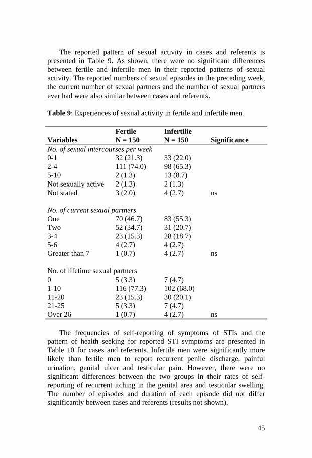
45
The reported pattern of sexual activity in cases and referents is presented in Table 9. As shown, there were no significant differences between fertile and infertile men in their reported patterns of sexual activity. The reported numbers of sexual episodes in the preceding week, the current number of sexual partners and the number of sexual partners ever had were also similar between cases and referents. Table 9: Experiences of sexual activity in fertile and infertile men. Variables
Fertile N = 150
Infertilie N = 150
Significance
No. of sexual intercourses per week 0-1 32 (21.3) 33 (22.0) 2-4 111 (74.0) 98 (65.3) 5-10 2 (1.3) 13 (8.7) Not sexually active 2 (1.3) 2 (1.3) Not stated 3 (2.0) 4 (2.7) ns No. of current sexual partners One 70 (46.7) 83 (55.3) Two 52 (34.7) 31 (20.7) 3-4 23 (15.3) 28 (18.7) 5-6 4 (2.7) 4 (2.7) Greater than 7 1 (0.7) 4 (2.7) ns No. of lifetime sexual partners 0 5 (3.3) 7 (4.7) 1-10 116 (77.3) 102 (68.0) 11-20 23 (15.3) 30 (20.1) 21-25 5 (3.3) 7 (4.7) Over 26 1 (0.7) 4 (2.7) ns The frequencies of self-reporting of symptoms of STIs and the pattern of health seeking for reported STI symptoms are presented in Table 10 for cases and referents. Infertile men were significantly more likely than fertile men to report recurrent penile discharge, painful urination, genital ulcer and testicular pain. However, there were no significant differences between the two groups in their rates of self-reporting of recurrent itching in the genital area and testicular swelling. The number of episodes and duration of each episode did not differ significantly between cases and referents (results not shown).

46
Table 10: Number and percentages reporting symptoms and treatment of STIs by fertility status. Variable
Fertile N = 150
Infertile N = 150
p value
Penile discharge 24 (16.0) 63 (42.0) 0.003 Treated 23 (95.8) 35 (55.6) Not treated 1 (4.2) 28 (44.4) 0.001 Painful urination 51 (34.0) 74 (49.3) 0.05 Treated 50 (98.0) 45 (60.8) Not treated 1 (2.0) 29 (39.2) 0.004 Itching in genital area 48 (32.0) 59 (39.3) ns Treated 45 (93.8) 33 855.9) Not treated 3 (6.2) 26 (44.1) 0.001 Genital ulcer 2 (1.3) 27 (18.0) 0.003 Treated 2 (100) 20 (74.0) Not treated 0 7 (26.0) 0.005 Testicular swelling 0 (0.0) 7 (4.7) ns Treated 0 3 Not treated 0 4 ns Testicular pain 6 (4.0) 18 (12.0) 0.002 Treated 6 (100) 12 (66.7) Not treated 0 6 (33.3) 0.005 The patterns of treatment seeking for reported symptoms of STIs in both groups are also shown in Table 10. In this community, different forms of treatment of STI symptoms have been reported [101]. These include self-treatment, informal sector treatment with traditional medicines and patent medicine dealers and formal sector treatment in hospitals and certified health providers. However, only those reporting treatment with formal health providers have been presented here as “receiving treatment”, while those reporting informal treatments were categorized as “not treated”, since the latter have not be proven to be evidence-based. Overall, infertile men were more likely than fertile men to report treatment with informal sector providers (results not shown). By contrast, as shown in Table 10, fertile men were significantly more likely to report that they received treatment with formal sector providers for
47
their reported penile discharge, painful urination, itching genital area, genital ulcer and testicular pain. Regarding past history of medical and surgical conditions, there were no differences between the two groups in their self-reporting of mumps, diabetes, hypertension, hypothyroidism, hyperthyroidism, cancer, and their use of drugs known to depress testicular function such as antihyper-tensives, antidepressants, cimetidine, nitrofurantoin and sulfasalazine (results not shown). However, 4.1% of fertile men reported native medications repeatedly in the past, compared to 33.8% of infertile men (p<0.002). Eleven infertile and four fertile men reported that they had previous surgical operations in their genital areas. The differences between the groups are not statistically significant. The types of surgical operations (inguinal herniorrhaphies, testicular biopsy, and repair of testicular maldescent, and varicocelectomy) were not statistically different between cases and referents. Finally, we compared the pattern of smoking and alcohol intake in fertile versus infertile men (Table 11). There were no significant differences between fertile and infertile men in the proportions reporting that they had ever smoked; however, infertile men were more likely than fertile referents to report longer duration of smoking. The types of smoking (cigarettes or marijuana) and the frequency of smoking did not differ between the two groups. Both groups also did not differ in the proportions reporting use of alcohol. However, infertile men were more likely to be heavy drinkers than fertile controls. Nearly 42.2% of infertile men reported that they drank one to two glasses of alcohol a day compared to only 12.9% of fertile men (p<0.002).
The results of the multivariate logistic regression of the risk factors for male infertility are presented in Table 12. The model identified 18 variables as possible predictive factors for male infertility in this population. However, after multivariate analysis, the following variables turned out to be important: poor sperm motility and low percentage of viable forms, lack of paternity with current wife or another partner, penile discharge, painful micturition, genital ulcer, use of referral hospital and chemists for treatment of STIs, use of native medications, and excessive intake of alcohol (one or two glasses of alcohol/day). Men who reported having had recurrent penile discharge were nearly eight times more likely to be infertile than those with no such history (OR 7.8; 95%CI 2.9–21.5). Also, men who reported having had recurrent pain on micturition were more likely to be infertile than men who did not give such histories (OR 2.2; 95%CI 1.02–4.71, p<0.04).

48
Table 11: History of cigarette smoking and alcohol consumption in fertile and infertile men. Variable
Fertile N = 150
Infertile N = 150
p value
Ever smoked 52 (34.7) 69 (46.0) ns Duration of smoking (yrs) 1-5 37 (71.2) 22 (34.8) 6-10 11 (19.2) 24 (34.8) 11-15 1 (1.9) 13 (18.8) >16 5 (9.6) 10 (14.5) 0.2 What did you smoke? Cigarettes 41 (78.8) 46 (66.7) Marijuana 6 (11.5) 3 (4.3) Cigarettes and marijuana 4 (7.7) 20 (28.9) Cocaine 1 (1.9) – ns Frequency of smoking Daily 31 (59.6) 55 (79.7) Weekly 10 (19.2) 4 (5.8) Monthly 2 (3.8) 1 (1.4) Occasionally 9 (17.3) 6 (8.7) Stopped – 3 (4.3) ns Taking Alcohol 116 (77.3) 116 (77.3) ns Amount of alcohol consumed 1-2 glasses a day 15 (12.9) 49 (42.2) 1-2 glasses a week 14 (12.1) 24 (20.7) 1-2 glasses a month 12 (10.3) 6 (5.2) Occasionally 75 (64.7) 35 (30.2) Stopped – 2 (1.7) 0.002
Other significant associations in the results are presented in Table 12. Previous genital ulcers and native medications increased the odds of being infertile nine times, and nearly 12 times respectively. Also, men who drank more than 1–2 glasses of alcohol a day were more likely to be infertile than those who drank less alcohol (OR 6.05; 95%CI 1.81–22.3).

49
With respect to healthcare-seeking behaviour for STIs, our results show that men who reported that they used a referral hospital (teaching hospital) for treatment of their symptoms were at least 83% less likely to be infertile than those using other forms of treatment (OR=0.17; 95%CI 0.02–0.86). By contrast, those who reported that they used chemists (patent medicine sellers) were more likely to be infertile than those using other forms of treatment (OR=8.2; 95%CI 2.1–36.6). The other relationships in the logistic regression model were not statistically significant. Table 12: Adjusted odds ratios and confidence intervals for predictors of male infertility. (rc: reference category) Variables Odds ratio CI (95%) Age (rc 45-49) < 30 1.16 0.2-5.0 30-34 1.47 0.4-4.7 35-39 0.89 0.2-2.7 40-44 0.82 0.2-2.4 50+ 0.42 0.1-1.4 Marital status (rc Cohabiting) Single 1.50 0.2-15.5 Married 0.98 0.1-7.1 Separated 0.75 0.1-8.8 Divorced 1.50 0.1-21.3 Religion (rc Anglican) Moslem 825.6 0.0-1.3 Pentecostal 0.61 0.2-1.7 Catholic 0.52 0.2-1.5 Jehovah’s Witness 825.6 0.0-9.6 Type of marriage (rc Polygamy) Monogamy 1.050.3-3.4 Number of years trying to impregnate (rc 1–4) 5-10 0.95 0.5-1.9 11-15 0.18 0.0-0.9 Over 15 years 3.30 0.4-31.0 Semen count (rc > 20 million) 10-20 million 1.000 0.0-0.1 5-10 million 0.000 0.0-0.4 < 5 million 1.000 5.0-112.1
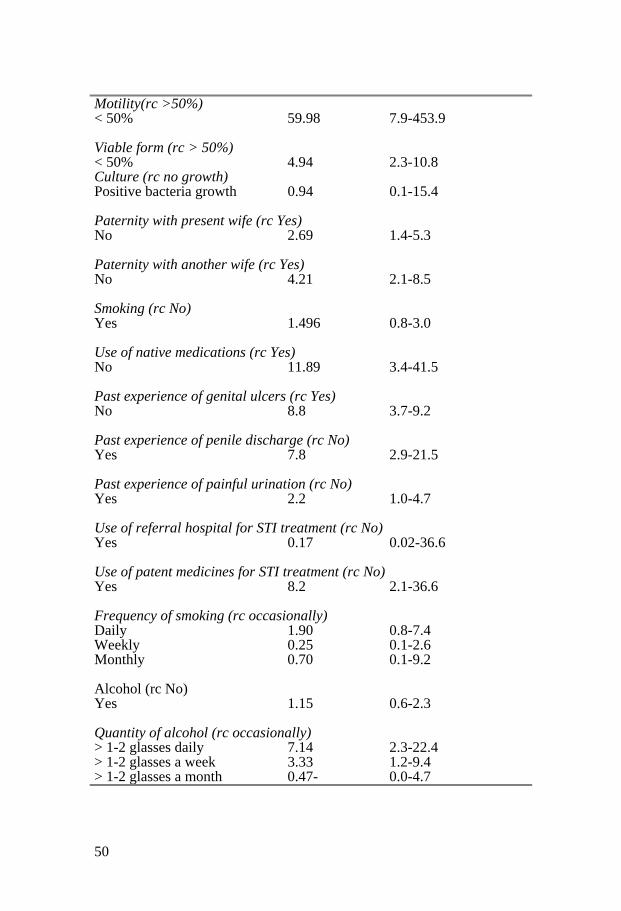
50
Motility(rc >50%) < 50% 59.98 7.9-453.9 Viable form (rc > 50%) < 50% 4.94 2.3-10.8 Culture (rc no growth) Positive bacteria growth 0.94 0.1-15.4 Paternity with present wife (rc Yes) No 2.69 1.4-5.3 Paternity with another wife (rc Yes) No 4.21 2.1-8.5 Smoking (rc No) Yes 1.496 0.8-3.0 Use of native medications (rc Yes) No 11.89 3.4-41.5 Past experience of genital ulcers (rc Yes) No 8.8 3.7-9.2 Past experience of penile discharge (rc No) Yes 7.8 2.9-21.5 Past experience of painful urination (rc No) Yes 2.2 1.0-4.7 Use of referral hospital for STI treatment (rc No) Yes 0.17 0.02-36.6 Use of patent medicines for STI treatment (rc No) Yes 8.2 2.1-36.6 Frequency of smoking (rc occasionally) Daily 1.90 0.8-7.4 Weekly 0.25 0.1-2.6 Monthly 0.70 0.1-9.2 Alcohol (rc No) Yes 1.15 0.6-2.3 Quantity of alcohol (rc occasionally) > 1-2 glasses daily 7.14 2.3-22.4 > 1-2 glasses a week 3.33 1.2-9.4 > 1-2 glasses a month 0.47- 0.0-4.7

51
6.4 The association between Chlamydia trachomatis seropositivity and infertility (Study IV)
The socio-demographic characteristics, contraceptive and sexual history of the cases and controls are presented in Table 13. As cases and controls were systematically matched for age, there was no difference in this variable between the two groups. The mean age was 32.3 + 4.7 years for infertile women and 32.2 + 4.9 years for fertile women. However, as expected, infertile women were significantly more likely to have less than tertiary education, be childless, never used a condom and never used an oral contraceptive compared to the fertile controls. The modal age range at sexual debut for infertile women was 13–17 years (64%) with a mean of 17 ± 3.6 years, which significantly contrasted with controls with a modal age range of 18 years (77%) and a mean of 19.8 + 3.4 (p<0.001). Furthermore, 84% of infertile women reported having had three or more sexual partners in their lifetime, compared with pregnant controls in which 26% reported three or more sexual partners (OR=10.40; 95%CI 4.15–26.04). The difference is statistically significant (p<0.001), and suggests higher level of sexual activity among the infertile group.
The prevalence of chlamydial antibodies, responses of the women concerning their previous experiences of gynecological symptoms and places of treatment are presented in Table 14. As many as 64.2% (52 women) of infertile women had chlamydial antibodies compared to 17.3% (14 women) of the fertile controls (OR=7.33; 95%CI 3.12–17.21). In addition, more infertile women than fertile controls reported that they had previously experienced pain on passing urine, vaginal discharge, lower abdominal pain and abdominal discomfort. This difference is statistically different for each gynecological symptom, while the strongest differential between infertile cases and fertile controls was found for vaginal discharge (OR=11.25; 95%CI 4.05–31.28). The reported places of treatment for these gynecological symptoms are presented in the bottom panel of Table 14. As shown, infertile cases were significantly less likely to report that they used evidence-based methods of treatment in the formal sector (private and public hospitals) than fertile controls (OR=0.37; 95%CI, 0.18–0.73). In contrast, infertile cases were more likely to report the use of chemists (OR=6.33; 95%CI, 2.68–14.98) and traditional healers were used by 32.1% of the infertile women and none by the fertile women.
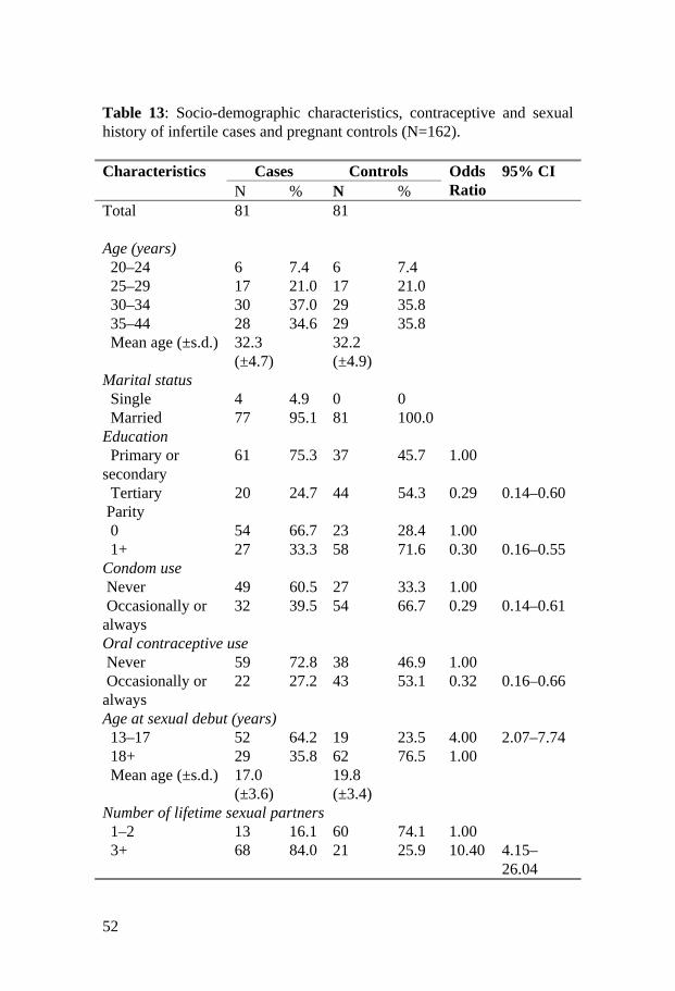
52
Table 13: Socio-demographic characteristics, contraceptive and sexual history of infertile cases and pregnant controls (N=162). Characteristics Cases Controls 95% CI N % N %
Odds Ratio
Total 81 81 Age (years) 20–24 6 7.4 6 7.4 25–29 17 21.0 17 21.0 30–34 30 37.0 29 35.8 35–44 28 34.6 29 35.8 Mean age (±s.d.) 32.3
(±4.7) 32.2
(±4.9)
Marital status Single 4 4.9 0 0 Married 77 95.1 81 100.0 Education Primary or secondary
61 75.3 37 45.7 1.00
Tertiary 20 24.7 44 54.3 0.29 0.14–0.60 Parity 0 54 66.7 23 28.4 1.00 1+ 27 33.3 58 71.6 0.30 0.16–0.55 Condom use Never 49 60.5 27 33.3 1.00 Occasionally or always
32 39.5 54 66.7 0.29 0.14–0.61
Oral contraceptive use Never 59 72.8 38 46.9 1.00 Occasionally or always
22 27.2 43 53.1 0.32 0.16–0.66
Age at sexual debut (years) 13–17 52 64.2 19 23.5 4.00 2.07–7.74 18+ 29 35.8 62 76.5 1.00 Mean age (±s.d.) 17.0
(±3.6) 19.8
(±3.4)
Number of lifetime sexual partners 1–2 13 16.1 60 74.1 1.00 3+ 68 84.0 21 25.9 10.40 4.15–
26.04
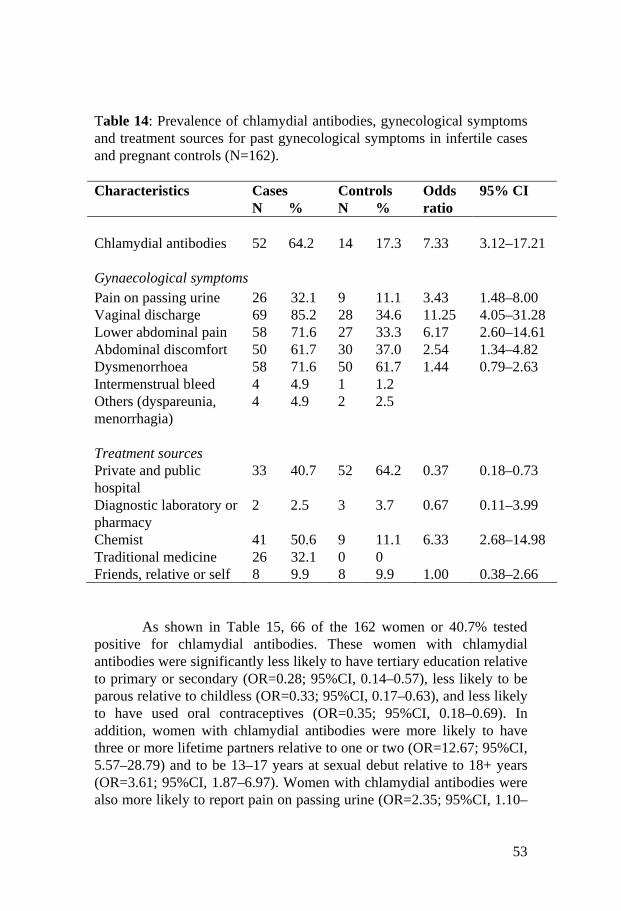
53
Table 14: Prevalence of chlamydial antibodies, gynecological symptoms and treatment sources for past gynecological symptoms in infertile cases and pregnant controls (N=162). Characteristics Cases Controls Odds 95% CI N % N % ratio Chlamydial antibodies 52 64.2 14 17.3 7.33 3.12–17.21 Gynaecological symptoms Pain on passing urine 26 32.1 9 11.1 3.43 1.48–8.00 Vaginal discharge 69 85.2 28 34.6 11.25 4.05–31.28 Lower abdominal pain 58 71.6 27 33.3 6.17 2.60–14.61 Abdominal discomfort 50 61.7 30 37.0 2.54 1.34–4.82 Dysmenorrhoea 58 71.6 50 61.7 1.44 0.79–2.63 Intermenstrual bleed 4 4.9 1 1.2 Others (dyspareunia, menorrhagia)
4 4.9 2 2.5
Treatment sources Private and public hospital
33 40.7 52 64.2 0.37 0.18–0.73
Diagnostic laboratory or pharmacy
2 2.5 3 3.7 0.67 0.11–3.99
Chemist 41 50.6 9 11.1 6.33 2.68–14.98 Traditional medicine 26 32.1 0 0 Friends, relative or self 8 9.9 8 9.9 1.00 0.38–2.66 As shown in Table 15, 66 of the 162 women or 40.7% tested positive for chlamydial antibodies. These women with chlamydial antibodies were significantly less likely to have tertiary education relative to primary or secondary (OR=0.28; 95%CI, 0.14–0.57), less likely to be parous relative to childless (OR=0.33; 95%CI, 0.17–0.63), and less likely to have used oral contraceptives (OR=0.35; 95%CI, 0.18–0.69). In addition, women with chlamydial antibodies were more likely to have three or more lifetime partners relative to one or two (OR=12.67; 95%CI, 5.57–28.79) and to be 13–17 years at sexual debut relative to 18+ years (OR=3.61; 95%CI, 1.87–6.97). Women with chlamydial antibodies were also more likely to report pain on passing urine (OR=2.35; 95%CI, 1.10–

54
5.03), vaginal discharge (OR=4.82; 95%CI, 2.33–9.97), and dysmenorrhoea (OR=3.04; 95%CI, 1.47–6.30). Condom use, abdominal discomfort and lower abdominal pain were not significantly associated with chlamydial antibodies. Table 15: Characteristics of infertile and fertile women with chlamydial antibodies (N=162).
Chlamydial antibodies Characteristics N % OR 95%CI All women 66 40.7 Education Primary or secondary 51 52.0 1.00 Tertiary 15 23.4 0.28 0.14–0.57 Parity 0 42 54.6 1.00 1+ 24 28.2 0.33 0.17–0.63 Oral contraceptive use Never 49 50.5 1.00 Occasionally or always 17 26.2 0.35 0.18–0.69 Condom use Never 37 48.7 1.00 Occasionally or always 29 33.7 0.54 0.28–1.01 Age at sexual debut (years) 13–17 41 57.8 3.61 1.87–6.97 18+ 25 27.5 1.00 Number of lifetime sexual partners 1–2 9 12.3 1.00 3+ 57 64.0 12.67 5.57–28.79 Pain on passing urine Yes 20 57.1 2.35 1.10–5.03 No 46 36.2 1.00 Vaginal discharge Yes 53 54.6 4.82 2.33–9.97 No 13 20.0 1.00 Lower abdominal pain Yes 46 54.1 1.74 0.93–3.29 No 20 26.0 1.00 Abdominal discomfort Yes 38 47.5 1.74 0.92–3.29 No 28 34.2 1.00 Dysmenorrhoea Yes 53 49.1 3.04 1.47–6.30 No 13 24.1 1.00
55
The multivariate analysis of the effects of testing positive for
serum chlamydial antibodies on the risk of being infertile are presented in Table 16. Model 1 presents the effects of chlamydial antibodies on the risk of infertility controlling for vaginal discharge (OR=5.70; 95%CI 2.04–15.96). The effects of chlamydial antibodies were attenuated in this bivariate model controlling for vaginal discharge compared to the univariate model (OR=7.33; 95%CI 3.12–17.21), as presented in Table 14. The association between chlamydial antibodies and infertility was attenuated further and non-significant (OR=1.07; 95%CI 0.22–5. 12) when the effects of vaginal discharge, socio-demographic characteristics, contraceptive and sexual history were controlled, as shown in Model 2. In the multivariate Model 2, vaginal discharge was the strongest predictor of infertility (OR=15.53; 95%CI 2.48–97.31), while the risk of infertility was also elevated for women with three or more lifetime partners (OR=8.30; 95%CI 1.22–56.62), and for women with primary or secondary education (OR=5.27; 95%CI 1.15–24.12). In contrast, the risk of infertility was not significantly associated with parity, oral contraceptive use, condom use or age at sexual debut. In an alternative model, the three variables capturing pain on passing urine, lower abdominal pain and dysmenorrhoea were added to Model 2, but none of these three variables added significantly to the model fit at the 0.05 level of significance based on the likelihood ratio test (results not shown). First-order interactions between chlamydial antibodies and number of sexual partners, vaginal discharge and number of sexual partners, age at sexual debut and number of sexual partners were tested and none of these three interactions was significant at the 0.05 level. In a final model, the variable about chlamydial antibodies was excluded (results not shown). In the latter model, the effects estimates for each variable were almost unchanged, the log likelihood was unchanged at –17.280 (compared to 17.276 in Model 2) and the pseudo R2 was unchanged at.69.

56
Table 16: Multivariate conditional logistic regression models of chlamydial antibodies, vaginal discharge, socio-demographic charac-teristics, contraceptive and sexual history of infertile cases and pregnant controls (N=162). Characteristics
Model 1 OR (95%CI)
Model 2 OR (95%CI)
Chlamydial antibodies Yes 5.70 (2.04–15.96) 1.07 (0.22–5.12) No 1.00 1.00 Vaginal discharge Yes 8.98 (2.88–27.98) 15.53 (2.48–97.31) No 1.00 1.00 Education Primary or secondary 5.27 (1.15–24.12) Tertiary 1.00 Parity 0 1.00 1+ 0.36 (0.09–1.49) Oral contraceptive use Never 1.00 Occasionally or always 1.81 (0.36–9.17) Condom use Never 1.00 Occasionally or always 0.34 (0.06–2.02) Age at sexual debut (years) 13–17 1.34 (0.26–6.84) 18+ 1.00 Number of lifetime sexual partners 1–2 1.00 3+ 8.30 (1.22–56.62) d.f. 2 8 Log likelihood –28.799 –17.276 Pseudo R2 .49 .69

57
6.5 Results of management of infertility in 190 couples (Paper V)
During the two-year period of the study (2000–2001), 211 couples registered for infertility treatment at WHARC. However, at the end of the one-year follow-up period in December 2002, 190 couples met the main criteria for inclusion in the study (i.e. successful completion of infertility work-up and being followed up for one year whether or not they received specific infertility treatment). The results of 21 couples were not included in the analysis because two died before the one-year follow-up period, 5 left the area and could not be traced, 3 had divorced or separated, while 11 failed to complete basic infertility investigations. The mean (SD) age of the female partners was 32 (5.4) years, while the male partners had a mean (SD) age of 39.9 (6.9) years. Other socio-demographic and clinical data on the couples are presented in Table 17. The duration of infertility ranged between 10 months and 20 years, with a median of 8 years. Nearly 19% of the couples had primary infertility, while 81% had secondary infertility. The 154 secondarily infertile couples reported 512 previous pregnancies. The outcomes of these pregnancies are presented in the bottom panel of Table 17. Over 59% of the pregnancies resulted in induced abortions, most often performed by women during their premarital years; 10.4% were spontaneous abortions; while only 20.7% of the pregnancies resulted in live births. Of the 106 live births reported in the sample, 48 women reported one live child; 10 reported 2 children, while 8 had three children. It was of interest that 2 women and one woman had 4 and 6 children, respectively, and yet still presented themselves as being infertile. However, we found that the three women had their children in previous marriages, but recently re-married and wanted children for their current partners. The established causes of infertility as diagnosed after completion of investigations in the couples are presented in Table 18. No apparent cause of infertility could be found in 24 couples (12.6%), while the rest had abnormalities in one or both partners. The most frequently diagnosed individual pathologies were sperm defects, tubal disease and uterine fibroids. Of the 190 males who completed semen analysis, 121 (63.7%) had normal semen parameters, 10 (5.3%) had azoospermia, 35 (18.4%) had severe oligospermia (sperm count < 5 million/ml), while 15 (7.9%) had mild to moderate oligospermia (sperm count 10–15 million/ml). Only 9 (4.7%) of the male partners had poor motility/other sperm defects as the

58
Table 17: Socio-demographic and clinical information on 190 couples presenting with infertility, Benin City, Nigeria 2000–2001. Variables N (%)
Duration of infertility (years) < 1 3 (1.6) 1–5 117 (61.5) 6–10 47 (24.6) 11–15 17 (8.8) 16–20 6 (3.5) Type of infertility Primary 36 (18.9) Secondary 154 (81.1) Previous pregnancies 0 36 (18.9) 1–3 108 (56.8) 4–8 41 (21.6) > 8 5 (2.7) Outcomes of previous pregnancies Live births 106 (20.7) Stillbirths 3 (0.6) Spontaneous abortion 53 (10.4) Induced abortion 303 (59.2) Table 18: Clinically diagnosed causes of infertility in 190 couples in Benin City, Nigeria, 2000–2001. Clinical diagnosis N (%) Ovulation defects (including premature ovarian failure) 30 (15.6) Azoospermia/oligospermia/poor sperm motil 69 (36.3) Tubal disease (no fibroids) 48 (25.3)
- Bilateral tubal occlusion + pelvic adhesions 29 (15.3) - Both tubes patent but with pelvic adhesions 6 (3.2)
Uterine fibroids 33 (17.4) - One or both tubes patent + pelvic adhesions 5 (2.7) - With bilateral tubal occlusion 28 (14.7)
Asherman’s syndrome (endometrial synechiae) 25 (13.1) No cause found in either partner 24 (12.6)

59
only abnormalities. Of the 69 male partners with poor semen, no cause could be found in 50 cases (72.5%). Seven (10.1%) had testicular failure (confirmed by elevated FSH on hormone assay), 4 (5.8%) had varicocele, while 8 (11.6%) had evidence of current and/or past severe genital tract infection. All 190 women were investigated with hysterosalpingography. Of these, 142 (74.7%) were reported as completely normal, and therefore did not require further investigations as dictated by our protocol. The remaining 48 (25.3%) were investigated with laparoscopy, with results showing that 29 (15.3%) had bilateral tubal occlusion, with or without various degrees of pelvic adhesions. By contrast, 13 (6.8%) had unilaterally occluded tubes, with or without pelvic adhesions; while 6 (3.2%) had patent tubes, but with mild to moderate pelvic adhesions. Uterine fibroids were common, with 33 of the 190 female partners presenting with this diagnosis. As shown in Table 18, many women with fibroids had, in addition, bilateral or unilateral tubal occlusion, while only 5 had patent tubes. A common diagnosis was Asherman’s syndrome (endometrial sclerosis/adhesions) occurring in 25 (13.1%) female partners. All of these women reported previous induced abortions. The contributions of male and female partners to infertility in the sample are presented in Table 19. Female causes accounted for only 51.1% of cases, while male causes were found in only 25.8% of cases. In 10.5%, no abnormalities were found in any of the partners that could have accounted for the infertility. Table 19: Contribution of each partner to causes of infertility in 190 Nigerian couples. Category of causes Number of couples (%) Female causes only 97 (51.1) Male causes only 49 (25.8) Causes in both partners 20 (10.5) No cause found in either partner 24 (12.6) Figures exceed 190 and 100% because some couples had multiple causes of infertility.

60
The proportion of couples treated and the kinds of treatment offered are presented in Table 20. All couples presenting with ovulation defects, uterine fibroids with patent tubes (where only the fibroids could have accounted for the infertility), and all women with Asherman’s syndrome were treated. Similarly, more than 60% of couples with poor semen quality, patent tubes with pelvic adhesions and those in which no cause was found were treated. By contrast, only few couples with unilateral and bilateral tubal occlusion and those with severe pelvic adhesions were treated. The treatments offered varied by the causes of infertility and were multiple in many couples. These consisted of treatments using conventional methods – macrosurgical tuboplasty, ovulation induction, uterine adhesiolysis, artificial insemination etc. Additionally, 10 couples were referred for ART procedures. However, only four couples actually underwent the procedures – two at the Bridge Clinic in Lagos, and two at the London Fertility Centre. Three couples were counseled for adoption, but only two consented and one eventually succeeded in adopting a baby. The results of treatment in terms of the number and proportion of women becoming pregnant after treatment are presented in Tables 21 and 22. Among 118 couples receiving one or more treatments, 41 pregnancies (34.7%) in 41 couples were recorded (excluding the one adopted baby) during the one-year after treatment. By contrast, in 72 couples not treated in the center, our follow-up tracking system indicated that 8 (11.1%) women were pregnant after one year. This difference in pregnancy rates between the treated and untreated groups is statistically significant (OR=4.7, 95%CI 1.9–11.8; p<0.0001). Further analysis showed that the non-treated group that later became pregnant tended to be younger, not to have any identified cause for their infertility, and to have a shorter duration of infertility. All pregnancies were confirmed by a pregnancy test and pelvic ultrasound scans. Among the 41 who became pregnant in the treated group, 4 abortions occurred between 8 and 10 weeks, while only one abortion was reported in the non-treated group. Thus, actual rates of continuing pregnancies were 37/118 (31.4%) in the treated group and 7/72 (9.7%) in the untreated group. This difference is still statistically significant (OR=3.2; 95%CI 1.3–8.4, p<0.001).

61
Table 20: Methods of treatment in 190 couples treated for infertility in Benin City, Nigeria, (2000–2001). Clinical diagnosis Total No. N (%) treated Treatment
Ovulation defects 30 30 (100.0) Ovulation induction (28) IVF (1); ISCI with donated eggs (1)
Poor semen 69 45 (65.2) General advice + Mesterolone (32) AID/AIH (9) ISCI (2); varicocelectomy (2)
Bilateral tubal occlusion + Pelvic adhesions
29
6 (20.7)
Tuboplasty (3) IVF (2); ISCI (1)
Unilateral tubal occlusion + Pelvic adhesions 13 4 (30.8) Super ovulation + timed intercourse (2) tuboplasty (2) Both tubes patent but with pelvic adhesions
6
4 (66.7)
Salpingolysis (4)
Fibroids with one or both tubes patent + pelvic adhesions 5 5 (100.0)
Myomectomy + adhesiolysis (5)
Fibroids with both tubes blocked 28
16 (57.1) Myomectomy (12) IVF (1); adoption (3)
Asherman’s syndrome 25 25 (100.0) Uterine adhesiolysis, IUCD insertion and oral estrogen (25)
No cause found in either partner 24 15 (62.5) Super ovulation + timed intercourse (15) Total 229 150 (65.5)
• Figures exceed 190 because some couples received multiple treatments. IVF-ET = in vitro fertilization and embryo transfer; AID = artificial insemination using donor semen; AIH = artificial insemination using husband’s semen; ISCI = intra-cytoplasmic sperm injection

62
As shown in Table 21, the most successful procedures that resulted in pregnancies were induction of ovulation, especially when failure of ovulation was the only cause of infertility, myomectomy when both tubes were patent, superovulation and timed intercourse in couples with no identifiable cause of infertility, and salpingolysis to separate peritubal adhesions when one or both tubes were patent. Treatment outcomes were uniformly poor for male factor infertility, and when there was bilateral tubal occlusion. Treatment outcomes appeared to be good in the 4 couples receiving ART treatment. Of the two couples referred to the London Fertility Center, one became pregnant at the first attempt and delivered a live baby, while the other became pregnant at the second attempt but aborted at 10 weeks. The latter pregnancy was confirmed with pregnancy test and an ultrasound scan at WHARC and evacuation of retained products was also carried out in WHARC. Between the two couples referred to the Bridge Clinic in Lagos, one was a 32-year-old woman who had bilateral tubal occlusion, while her husband had normal semen. She became pregnant at the first attempt but aborted spontaneously at 14 weeks. The other couple treated at the Bridge Clinic was a 46-year-old woman with bilaterally occluded tubes and moderately sized uterine fibroids. They had been infertile for 18 years, while her husband had severe oligospermia. They had ISCI with donated eggs at the Bridge Clinic and became pregnant with triplets at the second attempt. Unfortunately, one of the babies died in utero, while she was delivered by emergency caesarean section following premature labor and fetal distress at 34 weeks. A second baby died in the neonatal period, while only the third baby survived.

63
Table 21: Pregnancy rate by type of treatment given for infertility in 190 couples in Benin City, Nigeria, 2000–2001. Clinical diagnosis
No. treated
N (%) pregnant
No. not treated
N (%) pregnant
Ovulation defects 30 16 (53.3) 0 ovulation induction 28 14 (50.0) 0 – IVF 1 1 (aborted
after second attempt)
ISCI with donated eggs 1 1 (triplets, but only one live birth)
Poor semen 45 5 (11.1) 24 4 (16.7) General advice + Mesterolone
32 3
AID/AIH 9 1 ISCI 2 1 Varicocelectomy 2 0 Bilateral tubal occlusion + Pelvic adhesions
6 2 (33.3) 23 0 (0.0)
tuboplasty 3 0 IVF 2 1 ISCI 1 1 Unilateral tubal occlusion + Pelvic adhesions
4 2(50.0) 9 2 (22.2)
super ovulation + timed intercourse
2 1
tuboplasty 2 1 Both tubes patent but with pelvic adhesions
4 1(25.0) 2 0 (0.0)
tuboplasty 4 1

64
7. DISCUSSION
Infertility has commonly been attributed to several causes including endocrine disorders [102], long-term sequelae of sexually transmitted infections (STIs) [103], puerperal sepsis [104], post-abortion sepsis [105, 106] and congenital malformations [107]. Some of these causes are related to infectious morbidity, while others are not. It has been estimated that up to 80% of infertility in sub-Saharan Africa can be attributed to prior upper reproductive tract infections [108]. The central aim of the studies reported in this thesis was to demonstrate the association between genital tract infectious morbidity and infertility in Nigerian men and women. The results show such an association, and provide evidence for targeting the prevention of genital tract infections as the priority strategy for reducing the prevalence of infertility in Nigeria. Demonstrating a link between infertility and genital tract infections is not easy, especially within the context of low-income countries. Despite the constraints, the results of this thesis have demonstrated an association between past genital infections with Neisseria gonorrhoeae, Chlamydia trachomatis, post-abortal sepsis and puerperal infections and the prevalence of female infertility. While the evidence for Neisseria gonorrhoeae and Chlamydia trachomatis was demonstrated through a direct study, the evidence for post-abortal sepsis and puerperal infections were gleaned from the results of clinical investigations of couples with infertility. By contrast, despite the high prevalence of syphilis in some parts of Nigeria [109, 110], there was no evidence of its association with infertility in the cohort of women studied. We have also provided evidence of an association between episodes of infectious morbidity and male infertility in the Nigerian populations studied.
7.1 Association of female infertility with genital tract infectious morbidity
STIs that may be associated with female infertility include Neisseria gonorrhoeae, Chlamydia trachomatis, vaginal candidosis and Mycoplasma hominis [49, 111, 112]. Although genital tract tuberculosis has been reported in association with infertility in Nigeria [113, 114], the incidence of this

65
appears to be declining [49], and therefore this was not specifically investigated in this report. Our major finding with respect to the role of infectious genital tract infectious morbidity in infertility was the higher prevalence of antibodies against Neisseria gonorrhoeae in infertile women than in fertile controls. This suggests that more infertile women may have had past infections with the organism than fertile women. For years, Neisseria gonorrhoeae has been known to be the most common STI in Nigeria [115]. In 1963, the WHO reported a prevalence rate of Neisseria gonorrhoeae of 4,906 per 10,000 adult populations in Lagos [116], this being the highest rate in the world at the time. Since then, the reported prevalence of Neisseria gonorrhoeae has varied from 9.7% in Sokoto, northern Nigeria [117], to 5% among asymptomatic women in Ibadan [118], 15.8% among commercial sex workers in northern Nigeria [119], to 19% among clinic patients in Lagos, Nigeria [120]. More recently, although the incidence of Neisseria gonorrhoeae has declined considerably in many parts of Nigeria [121], there is still a problem with the emergence of penicillin-resistant strains of the organism [122, 123]. Our use of antibodies as a marker for Neisseria gonorrhoeae ensured that both current and past infections with the organism are captured in both cases and controls. Available evidence indicates that many infections with Neisseria gonorrhoeae may be asymptomatic, especially in women [103]. Thus, it was not surprising that there was poor correlation between the presence of antibodies and self-reporting of genital tract symptoms in both cases and controls. Neisseria gonorrhoeae is principally carried in the crypts of the endocervical canal, with subsequently ascension to the upper genital tract, in particular, the fallopian tubes, causing acute pelvic inflammatory disease and bilateral tubal occlusion [124]. The results of the study showed significantly higher rate of current cervical carriage of Neisseria gonorrhoeae or other pathogenic organisms in cases of secondary infertility than in cases of primary infertility. This suggests progressive carriage of the pathogenic organisms over time, or more recent increased rate of acquisition by secondarily infertile women. Similar serological studies in countries with high community prevalence of Neisseria gonorrhoeae have shown its strong correlation with tubal infertility [125, 126]. Thus, although we have not specifically focused our study on women with tubal infertility, it is evident that the mechanism is through the effects of the organism on the tubal epithelium. Clearly, programs need to be concentrated on reducing the incidence and prevalence of Neisseria gonorrhoeae, in efforts to reduce the prevalence of tubal infertility in Nigeria. The study did not show any significant association between syphilis antibodies and female infertility in this population. We hypothesized that a strong association between syphilis antibodies and infertility would be

66
found, which would further confirm the connection with STIs and proximate correlates of sexual activity. However, contrary to our expectation, we did not find evidence for such an association. This may be due to the declining incidence of syphilis in southern Nigeria, where the study was undertaken [127, 128], or to the non-sterilizing effects of syphilis microorganism. Several reports including a national syphilis sero-prevalence study [129, 130] have consistently shown a higher prevalence of syphilis in northern Nigeria than in southern Nigeria. Perhaps, if the study had been carried out in northern Nigeria, an association would have been found between infertility and syphilis sero-prevalence. Chlamydia trachomatis was also investigated for its association with female infertility by comparing the presence of antibodies in women with tubal infertility and pregnant fertile controls. The results showed that infertile women were seven times more likely to demonstrate the Chlamydia trachomatis antibodies than fertile women. However, the strength of association between chlamydial antibodies and infertility was attenuated when the self-reporting of vaginal discharge, socio-demographic characteristics, contraceptive and sexual history were added to the conditional logistic regression model. This suggests that Chlamydia trachomatis is strongly linked to the behavioural and sexual routes of transmission in the population of women studied. Several studies have reported on the prevalence of Chlamydia trachomatis in Nigeria, but very few have reported its association with infertility. Darougar et al [131] carried out a seroepidemiological survey of chlamydial genital tract infection in Ibadan, Southwest Nigeria, and found a prevalence rate of 26.7% among women attending the STD clinic and 35% among women attending the family planning clinic. Thus, this study demonstrated not only the high prevalence of Chlamydia trachomatis in southern Nigeria, but also its sexual and behavioural modes of transmission. Similar studies from Benin City [97] and Lagos [132] reported similar high rates of sero-prevalence of Chlamydia in clinic populations, with the study from Benin City reporting a prevalence rate of up to 47% among women with genital disease. These studies clearly demonstrate the high prevalence of Chlamydia trachomatis among women in various parts of Nigeria. Only one study had previously investigated the association of Chlamydia with infertility in Nigeria [133]. The study took cervical and urethral swabs from 20 infertile women and 80 pregnant controls and screened them for Chlamydia trachomatis using amplified EIA. The results showed a prevalence of Chlamydia of 11% among pregnant women and 15% in women with demonstrated tubal infertility. The difference may not have been statistically significant due to the fact that only current infections were investigated. Our study was more robust in that both current and past infections were investigated with the use of serological methods. Although our findings were
67
statistically significant at the bivariate level, it nevertheless showed that at the multivariate level of analysis, behavioural risk factors, especially socio-demographic, sexual and contraceptive risk factors, were more important as primary determinants of tubal infertility. Several studies have demonstrated the sexual and behavioural routes of Chlamydia trachomatis [134, 235], a connection that has been further clarified in this study. Clearly, for an infection that is largely asymptomatic [136], but that has devastating effects on future fertility of populations, only a preventive approach would have beneficial effects in controlling the disease and its effects on women’s health in the country. Further evidence of the role of genital tract infections as causes of infertility was provided by the clinical investigation of women presenting with fertility. The results showed that more than 80% of such women had secondary infertility, implying that the majority had one or more pregnancies in the past. This suggests that pregnancy-related complications, which often are infectious in nature, may be the primary causes of infertility in this population. The study showed that among the 192 women, 512 past pregnancies were reported, 59.1% of which had induced abortion, while 21.3% were term pregnancies. Furthermore, more than 25% of the women had laparoscopic evidence of tubal damage from previous tubal infections, while 13% of the women had Asherman’s syndrome. Asherman’s syndrome is a condition characterized by amenorrhea or hypomenorrhea due to scarring and sclerosis of the endometrium, produced by excessive curettage and infection during pregnancy termination [137, 138]. It may also be due to post-partum endometritis arising from complicated pregnancies. Thus, it is evident that reproductive tract infections (RTIs) arising from complicated abortions and pregnancies are an important cause of infertility in Nigeria. Further evidence in support of abortion being an important cause of infertility in this population was provided by the questionnaire survey of women in the first study which showed that up to 25% reported having used abortion in the past compared to 10% of controls. Abortion is legally restricted in Nigeria [137], yet it has been estimated that up to 610,000 abortions take place in the country annually [29]. Many of these abortions are done by unqualified and unskilled persons [139], resulting in high rates of complications [140–143]. Puerperal (post-partum) infections are also a major problem in Nigeria [144–145], due to due high incidence of prolonged obstructed labor [146]. Many of these infections transcend to the upper genital tract, leading to acute and chronic endosalpingitis and perisalpingitis, with resultant infertility and subfertility [49]. Therefore, as suggested elsewhere [147–148] it is evident that the prevention of unwanted pregnancies that lead to abortion, increased access to safe abortion services and proper management of full term pregnancies and their complications are critical measures to reduce the prevalence of infertility in Nigeria.

68
7.2 Association of male infertility with genital tract infectious morbidity
Male infertility may be due to several conditions including congenital maldescent of the testes, testicular varicocele, environmental factors, previous genital tract surgery and hormonal abnormalities [149–150]. However, by far the most important group of causes are those due to RTIs in males. There is evidence that STIs, especially Neisseria gonorrhoeae and Chlamydia trachomatis, account for a significant proportion of cases causing occlusion of the vas deferens and subsequent oligospermia and azoospermia [151–152]. Male infertility may also be due to other infections especially severe epididymo-orchitis either due to pyogenic organisms or to viral agents such as mumps [10]. RTIs in males often manifest with symptoms such as urethral discharge, painful micturition, genital ulcer and testicular pain [153]. While RTIs in females are often asymptomatic, RTIs in males tend to be symptomatic [154] In order to determine the relative contribution of RTIs to male infertility versus other potential causes, we conducted a case-control study of risk factors among males in southern Nigeria. The results showed that bacterial colonies were significantly more likely to be grown in infertile men than in fertile controls. The significant risk factors for male infertility identified in the logistic regression analysis were past history of symptoms of STIs, self-reporting of use of native medications, use of non-evidence-based treatment and moderate to heavy alcohol intake. Men who reported having had repeated episodes of penile discharge, painful micturition, genital ulcers and testicular pain were significantly more likely to be infertile than those not reporting such episodes. As these are recognized to be important proxies for STIs in this population, it suggests greater exposures to STIs in infertile men than in fertile controls, and emphasizes the critical role of STIs as important risk factors for male infertility in Nigeria. Several reports have documented significant associations between STIs, genital tract infections and male infertility in Nigeria [155–159]. However, the strength of this association has not previously been determined because of difficulties in establishing a causal relationship between prior exposure to RTIs and male infertility. This study has undertaken a comprehensive risk factor analysis for male infertility and has attempted to include all socio-demographic and reproductive health variables that may be important as possible determinants of infertility in men. Variables associated with RTIs topped the list as significant risk factors for male infertility in this population. Contrary to our hypothesis, there were no significant differences in the patterns of reported sexual behaviour (i.e. number of sexual partners and sexual frequency) between fertile and infertile men, which suggests that the increased prevalence of STIs in infertile men may be due to other
69
mechanisms rather than increased sexuality. A possible mechanism could be that infertile men may be less likely to practice protective sex (e.g. use of condoms) than fertile men, a relationship that was not explored in this study. Furthermore, the study showed that infertile men were more likely to report the use of non-evidence-based method of treatment for reported STI symptoms (traditional treatment, patent medicine sellers and chemists) rather than evidence-based methods. The delay in using correct and appropriate STI treatment could then lead to damage of the reproductive tract and therefore result in male infertility. Interestingly, while heavy alcohol intake was found to be associated with male infertility in this population, smoking was not found to be a significant factor. In contrast, several lines of evidence suggest that smoking is an important and significant risk factor for male infertility in other populations [57, 160, 161]. This may be due to the fact that Nigeria is not known to be a heavy smoking society [162] or to the fact that smoking may be blurred by sexual risk factors as the two tend to be related. Despite these findings, we recommend that the reduction of smoking and heavy alcohol consumption should both be included in preventive approaches to male infertility in this population.
In sum therefore, our study has demonstrated variables of past RTIs as being the most significant risk factors for male infertility in Nigeria. Thus, we believe that the prevention of STIs and RTIs is critical to reducing the incidence of male infertility in Nigeria.
7.3 Some constraints with epidemiological evidence
Several potential difficulties with use of epidemiological and social data for investigating the risk factors for male and female infertility arise from this report. These include the following: 1) Except for Study II, the other studies in this report were hospital-based rather than community-based. The problem of external validity, i.e. generalizing hospital-based data to community level analysis, therefore arises. However, our use of a case-control design for the studies increased our ability to generalize the findings as both cases and controls originate from the same communities. More importantly, there was no differential bias between cases and controls for elucidating important information relating to risk factors in any of the studies. Thus, any selectivity that would have reduced the reported strength of association of the dependent and independent variables was reduced considerably. Furthermore, as the studies were conducted at the teaching and tertiary level hospitals, it can be argued that the patients attending these hospitals are

70
self-selected and may not represent the true range of patients in the community. However, all patients seeking hospital care for infertility in Nigeria eventually reach the teaching hospitals, as a mechanism exists for referring patients from primary and secondary level facilities to teaching and tertiary hospitals [37, 80]. Our estimates indicate that close to 80% of women with significant infertility who enter the health care delivery system in these parts of Nigeria eventually reach the teaching hospitals. This explains the fact that infertility is currently the most common indication for gynecological consultations in nearly all teaching and tertiary hospitals in Nigeria. 2) Another potential area of concern is that the studies reported in this thesis relied on the use of self-reporting of sexual and reproductive health symptoms rather than on more robust outcome measures. However, it should be understood that within the context of our low-income economy, accurate data are usually not available and therefore it would be impossible to obtain reliable retrospective data on which to base the analysis. Secondly, self-reporting of sexual and reproductive health symptoms have proven to be very useful in several such studies conducted in other parts of Africa [163–165], provided the questions are accurately framed. We believe that our use of a sensitive and systematic approach to the respondents (through the training of interviewers) limited recall bias, and increased the chances of obtaining accurate and truthful answers to the questions. Although a prospective follow-up study will appear to be more accurate for determining the association between infertility and the various socio-demographic and reproductive risk factors, this would be difficult to carry out in a low-income country such as Nigeria, because of the huge costs involved and also the likelihood of a high drop-out rate. Consequently, the case-control method used in this thesis is the most realistic approach, and is sufficiently accurate for making inferences on the role of the association of potential risk factors with infertility. 3) Studies II and IV relied on seroepidemiology to make inferences on past and previous exposures to Neisseria gonorrhoeae, T. Pallidum and Chlamydia trachomatis and their association with female infertility. This relied on the assay of relevant antibodies in both cases and controls as markers of previous exposures. However, as antibodies are known to disappear with time, the use of serology may underestimate the true extent of the infections. Nevertheless, many of the infections are asymptomatic and serology provides the best approach to study their association with infertility. The case-control design also helps to validate the results since whatever attenuation may have occurred due to antibody disappearance will occur equally in cases and controls, with no effects on the net assessment of risk.

71
7.4 Under what social and cultural contexts does genital tract morbidity lead to high rates of infertility?
Overall, the results of the studies reported in this thesis demonstrate a high correlation between genital tract infections and male and female infertility in Nigeria. By contrast, it is known that in middle- and high-income countries, genital tract infections are becoming less important as causes of infertility in males and females [166]. The relevant question therefore, is: “why is infertility so strongly linked with genital tract infections in Nigeria”? In the first place, there can be no doubt that STIs and RTIs are common in Nigeria due to prevailing adverse social and economic circumstances. High levels of poverty, persisting illiteracy, low levels of sexual and reproductive health education and the low social status of women predispose to high levels of premarital, extramarital and commercial sex in various parts of the country [167–170]. Within such adverse socio-economic circumstances, STDs tend to occur [171–173] in males and females, and may lead to severe complications. The low contraceptive prevalence rate in the country [174] despite high levels of sexuality predispose to high levels of unwanted pregnancies [175. 176], which are often aborted [177]. Due to the restrictive abortion law in the country [178], most abortions are unsafe and poorly performed with resulting RTIs and tubal infertility. It is known that pregnant women have poor access to effective antenatal and delivery services and only 30% of deliveries are attended to by skilled birth attendants [179]. The majority of deliveries are unsupervised or are attended to by untrained traditional birth attendants (TBAs) and unskilled persons [180, 181] using unhygienic methods. Consequently, post-partum infections are a common feature of term pregnancies in Nigeria [180] and may result in infertility. When RTIs occur, there is general agreement that the prompt use of appropriate evidence-based method of treatment can reduce the risk of progression to more severe complications and prevent the associated short- and long-term complications [182, 183]. STIs and RTIs result in pelvic infections which may be mild, moderate or severe. Severe pelvic infections may be treated because of associated severe symptoms; however, most mild to moderate pelvic infections may be asymptomatic and may therefore not be easily recognized [184]. The non-recognition of STIs, RTIs and pelvic infections, especially in women, often means that these infections may be left untreated [185]. Even when symptoms are recognized, there is evidence that treatment may be delayed as a result of perceptions relating to cost, confidentiality of treatment and effectiveness of treatment [186]. Adolescents are particularly more likely not to seek accurate treatment for symptoms of STIs and RTIs

72
for these reasons and to seek non-effective methods of treatment. The results of several studies in Nigeria indicate that adolescents and adults are likely to self-treat or to seek STI treatment with non-formal providers such as patent medicine sellers, chemists and traditional healers rather than evidence-based treatment provided in the formal health sector [187, 188]. The delay in treatment leads to persistence of the infection over time and results in long-term complications such as infertility. The differences in treatment-seeking behaviour for RTIs may be an important reason for the differences between low-income and high-income countries in terms of their association with infertility. Apart from the lower incidence of STIs and RTIs in high- and middle-income countries, there is evidence that many pelvic infections in these countries are promptly treated and that effective screening programs exist for the early diagnosis of asymptomatic STIs [189, 190]. Additionally, many high-income countries have effective community-based programs for the prevention of STIs and RTIs that lead to infertility, such as quality family planning, safe abortion, and antenatal and delivery services. Thus, the adverse social and economic circumstances in which RTIs occur in Nigeria and the poor health-seeking behaviour for acquired STIs and RTIs are at the root of the strong correlation of genital tract infections with male and female infertility in Nigeria.
7.5 From research to service provision for infertility – the need for a preventive approach
The results of the studies reported in this thesis support the recommendation that efforts to reduce the prevalence of infertility in Nigeria should be concentrated on preventing the incidence of STIs and RTIs that lead to infertility in males and females. Prevention of infertility in Nigeria rests on the tripod of primary, secondary and tertiary prevention initiatives. As a result of the strong correlation between RTIs, STIs and infertility shown in this study, primary prevention of infertility should involve appropriate measures to prevent STIs and RTIs in men and women. This is by no means an easy task. Current measures to prevent STDs and STIs in Nigeria are based on the behavioural modification intervention initiatives, including the promotion of abstinence, faithfulness among couples and the use of barrier methods of contraception, such as condoms [191]. However, these programs have been poorly entrenched into existing health care delivery services in the country, and are mainly provided by non-governmental organizations with limited donor support [192]. In particular, the promotion of condoms have suffered a serious setback in Nigeria, due to cultural and religious

73
sentiments, which has meant that condoms are hardly promoted within official service delivery outlets in Nigeria [193]. What is needed is an STI primary prevention strategy that is strongly integrated within existing service delivery outlets and that has as one of its ultimate objectives the prevention of infertility in the population. Many public health institutions still have viable family planning clinics, while STD clinics are on their way out [193]. While this may be a good approach to reduce the stigmatizing effects of STD clinics, it has tended to attenuate the quality of programs for the prevention of STIs in health institutions. However, the integration of infertility as an important service delivery endpoint within the context of family planning programs will encourage the participation of a greater number of people in family planning and increase the use of barrier methods versus other methods of contraception. Such increased use of barrier methods will also serve the dual purpose of preventing HIV/AIDS and other sexually transmissible infections with overall beneficial effects on the sexual and reproductive health of men and women. Nigeria is a country with a huge unmet need for contraception [194], which has tended to increase the rates of unwanted pregnancy, unsafe abortion and maternal infection-related morbidity and mortality in the country. Unfortunately, family planning services are still poorly accepted in Nigeria, in part due to perceptions among men and women that contraception may lead to future infertility [195]. Thus, primary prevention initiatives for unwanted pregnancy should involve the active promotion of family planning, and the provision of all forms of birth control methods to reduce the need for recourse to unsafe abortion. It should also include educational programs that dispel myths about causes and treatment methods of infertility, so that family planning programs are not blamed for infertility, and to encourage appropriate healthcare seeking behaviour for both fertility regulation and infertility treatment. The secondary prevention of infertility involves the early recognition and treatment of STIs and RTIs to reduce the prospects that they will proceed to irreversible damage of the reproductive tract system. This involves a policy of active community education, so that people are able to recognize the symptoms of STIs and RTIs, and to seek appropriate and evidence-based treatment. It also includes the improvement and expansion of antenatal and delivery services, so that pregnant women have increased access to safe delivery options. This will reduce the incidence of complicated deliveries that lead to post-partum infections. It includes advocacy activities for the liberalization of the Nigerian abortion law as has been suggested by several reproductive health practitioners in Nigeria [196, 197], so that women have access to safe abortion services, with limited chances of abortion-related infectious complications.
74
The tertiary prevention of infertility consists of provision of community education so that women seek appropriate evidence-based treatment for infertility rather than non-effective treatment. This will reduce the adverse consequences of infertility and promote the reproductive health of women and men. Evidence-based treatment of infertility consists of conventional methods of treatment and the new reproductive technologies [198, 199], both of which are available in Nigeria. However, the results of clinical investigation of 192 infertile couples, as reported in study V, suggest that conventional methods of treatment are of poor efficacy in Nigeria as compared to other settings. Some of the poor results may be due to delayed treatment by the couples. Four couples were treated with NRT, with apparently successful outcomes. These suggest that within the limited resources in Nigeria, both conventional and high-technology treatment of infertility would be effective when appropriately used. However, in view of the high cost of setting up and sustaining high-technology treatment services for infertility, which will drain the resources of any low-income country, the emphasis should be on prevention and conventional treatment. By contrast, high-technology treatment can be located within the private sector, as is currently the case [200], until the economic situation of the country improves.

75
8. CONCLUSIONS
In conclusion, this thesis has demonstrated a strong correlation between episodes of RTIs and infertility in Nigerian women. Self-reported symptoms of RTIs and antibodies against Neisseria gonorrhoeae and Chlamydia trachomatis were demonstrated in the studies to show a strong association with female infertility. By contrast, there was no evidence of an association between syphilis antibodies and infertility in the cohort of women studied. In particular, infertility was strongly associated with past history of abortion, pregnancy complications and socio-demographic and sexual risk factors in women. Similarly, we found a strong correlation between male infertility and various correlates of sexual behaviour, STIs, health-seeking behaviour and heavy alcohol intake in the population of men studied. These results demonstrate the important role for RTIs and STIs in the pathogenesis of male and female infertility in Nigeria. Several factors may explain the high rate of STIs and RTIs as major determinants of male and female infertility in Nigeria. These include a high prevalence of adverse social and cultural factors that increase the vulnerability of men and women to STIs and RTIs. The most important of such factors are: poverty, low status of women, low access to reproductive health services and the fragile health infrastructure in the country. Delayed use of effective treatment for STI symptoms contributes to the problem and increases the risk that acquired STIs will progress to infertility. The results of our studies indicate that men and women who used ineffective treatment methods for RTI and STI treatment were at increased risk of infertility as compared to those who used evidence-based methods. We believe that a public health approach that integrates the prevention of STIs and RTIs within existing services for men and women would reduce the burden of infertility and its consequences in Nigeria. The prevention of infertility in Nigeria should encompass primary, secondary and tertiary prevention initiatives. This should consist of interventions aimed at behavioural and sexual risk modification, early recognition and effective treatment of RTIs and STIs, increased access to safe family planning and abortion services, and improvement of conventional treatment of infertility. The use of high-technology infertility treatment methods should be located within the private sector in view of its high costs, until basic services have been provided for the improvement of women’s health in the public sector.

76
Due to its intermediating factors and correlates, infertility is an important and critical public health and reproductive health problem in Nigeria. The prevention of infertility in Nigeria would lead to improved acceptance of family planning and reproductive health services in the country and is an important contribution to the promotion of sexual and reproductive health. We recommend measures to prevent infertility to policy makers in Nigeria as important interventions worthy of support and active promotion in the coming years.

77
9. ACKNOWLEDGEMENT
It has been a lifetime experience working with Professor Staffan Bergström, whose support has been the single most important factor leading to the completion of this thesis. Since I first met Staffan at a training workshop in Uppsala in 1996, he has been a towering pillar of support to my academic and scholastic aspirations. He has indeed been a wonderful mentor, friend, adviser, associate and benefactor – the very best anyone can hope for in today’s world. He assisted me in every way imaginable throughout the preparation of this thesis, and provided extreme cover, solace and support during my several trips to Sweden in pursuit of this thesis. Words are not sufficient for me to thank him, but I can assure him that he has left an indelible impression in my mind that will guide me in reciprocating to others in similar manner in future. Staffan’s extremely pleasant wife, Birgitta was no less remarkable in her support for me during my visits to Sweden. She often attended to all my small needs including the provision of a “survival kit” to ease my stay whenever I visited Sweden and very warmly received me into her home on numerous occasions. It is with extreme joy and happiness that I thank her and Staffan for their kindness towards me. They supported me in the same way that my Nigerian parents have treated me all through life and therefore, I have no hesitation in calling them my Swedish parents, and behaving to them in that manner from now on. I am now fully a part of the Swedish academic system, and IHCAR is now my second home. I am grateful to all those scholars in Uppsala, Umeå and Stockholm who taught me in the courses I attended leading to this thesis. I have learnt quite a lot, and although I have attended courses in all parts of the world, I am convinced that the Swedish university system offers the best opportunity for comprehensive all-round compassionate growth and development for academics from low-income countries. This thesis was completed with scholarships from the Swedish system, an opportunity, which is difficult to come by in other countries, and for which I am extremely grateful to Karolinska Institutet and the Swedish government. I also deeply appreciate the support of Marie-Louise Thomé, research group secretary, in the preparation of this thesis. She often arranged accommodation and logistics for me during my visits to Sweden, and was an extreme source of strength and support. I thank her and Karin Törnblom,

78
Uppsala, specifically for their time and support and extremely valuable computer skills, particularly in the hectic final formatting of the thesis. I thank all the numerous colleagues I met at IHCAR for their support and encouragement, especially Bah Jainabah, Godfrey Mbaruku, Elizabeth Mathai, Grazyna Alicja Stanczuk and Pius Okong to mention but a few. My friends and colleagues from various parts of the world to whom I mentioned my work on this thesis and who have encouraged me considerably include Professor O A Olatunbosun, Associate Professor Jerker Liljestrand, Dr Kunle Odunsi, Professor Fred Naftolin, Dr Kole Shettimma, Professor Ian Craft, Professor O A Ladipo and Professor Wole Akande – I thank you all. The Harvard School of Public Health first opened my eyes to the international community and to the reproductive health field with the award of the Takemi fellowship in international health in 1992. Since then, I have been appointed a visiting scholar and adjunct to the School on an annual basis. I thank the past Chair of the Department of International Health and Population Studies at Harvard, now director of Harvard’s Centre for Population Studies, Professor Michael Reich, for first admitting me into the program, and for his sustained interest and support for my academic development over these years. He has indeed, been the American version of Staffan, supporting and believing in me with the same passion that Staffan has supported me. I thank him most sincerely for his efforts, which have built up my capacity considerably over the years. I have continued to work with colleagues in Harvard till this day. In particular, I continue to collaborate extensively with Dr Ulla Larsen and Dr Grace Wysack, both in Harvard. I thank them both for their continued support and encouragement over the years. Dr Ulla Larsen offered statistical advice and analysis for some of the studies reported in this thesis, and also co-authored one of the papers. She is indeed, an efficient and reliable research collaborator, and I remain grateful for her extreme kindness and support over the years. The Ford Foundation, New York provided funds for all the studies reported in this thesis through their sustained support for the Women’s Health and Action Research Center (WHARC). Indeed, the first grant for my research in reproductive health given to WHARC in 1995 by the Ford Foundation was for a study into infertility. Since then, the Ford Foundation has provided funding to WHARC for its work in promoting all aspects of sexual and reproductive health. I thank Dr Natalia Kanem of Ford’s office in New York, who first provided funds for the establishment of WHARC and Dr Babatunde Ahonsi, Ford’s Program Officer in Nigeria (a young, purposeful and forthright Nigerian leader), who has continued to support WHARC in unbelievable ways over these years. I thank all staff of WHARC, especially those who worked with me in developing various aspects of this thesis – Becky Tita, Osaretin Aghedo, Dr Uche Menakaya, Dr Loretta Ogboro-Okor, Dr Pius Iribhogbe, Dr Imarengiaye, Dr Steve

79
Ogbonmwan, Usi Osazuwa, Christy Ukpong, Roli Umukoro and Merch Agwu. I am indeed, very grateful to you all. Now to my University of Benin environment - they have equally been very supportive and encouraging. I thank the current Vice-Chancellor of the University, Professor EAC Nwanze, and the immediate Past Vice-Chancellor, Professor Richard A Anao for encouraging me in my efforts to develop this thesis, especially for granting me permission to be away from the University for the several courses I attended in pursuit of the thesis. I thank my colleagues in the College of Medical Sciences and those in the Department of Obstetrics and Gynecology for standing in for me in administrative and technical matters during those inevitable absences from the University. I also thank the authorities of the University of Benin Teaching Hospital and the Obafemi Awolowo University Teaching Hospital for providing the enabling environment for the studies reported in this thesis. All this work would not have been possible without the love, understanding and support of my family. I thank Helen and the children for all the inconveniences they had to bear during the periods of my absenteeism created by my working on this thesis. I thank God Almighty for his support, without which nothing could have been proposed and achieved... Thank you, thank you, and thank you all so much.

80
10. REFERENCES
1 World Health Organization. The epidemiology of infertility. Report of ITS Scientific Group on the Epidemiology of Infertility. Technical Report Series No. 582. Geneva: World Health Organization; 1975.
2 World Health Organization. Reproductive health indicators for global monitoring: Report of the second interagency meeting. Geneva: World Health Organization; 2001 (WHO/RHR/01.19).
3 Rutstein SO, Iqbal HS. Infecundity, Infertility, and Childlessness in Developing Countries. DHS Comparative Reports No. 9. Calverton, Maryland, USA: ORC Macro and Geneva: World Health Organization; 2004.
4 Larsen U. Primary and secondary infertility in sub-Saharan Africa. International Journal of Epidemiology 2000;29:285-91.
5 Mammo A., Morgan SP. Childlessness in rural Ethiopia. Population and Development Review 1986;12:533-46.
6 Gerias AS, Rushman H. Infertility in Africa. Population Sciences 1992;12:25-46.
7 Cates W, Farley TM, Rowe PJ. Worldwide patterns of infertility: is Africa different? Lancet 1985;2:596-8.
8 World Health Organization. Infections, pregnancies and infertility: Perspectives on prevention. Fertility and Sterility 1987;47:964-8.
9 United Nations. World Population Prospects: The 2002 Revision. Vol 2, Sex and Age distribution of populations. New York: United Nations Population Division; 2002 (ST/ESA/SER.A/223).
10 Okonofua FE. Infertility in sub-Saharan Africa. In: Okonofua FE, Odunsi OA (Eds.) Contemporary Obstetrics and Gynecology for Developing Countries. Benin City: Women’s Health and Action Research Center, 2003. p. 128-56.
11 Okonofua FE, Harris D, Odebiyi A. The social meaning of infertility in southwest Nigeria. Health Transition Review 1997;7:205-20.
12 Koster-Oyekan W. Infertility among Yoruba women: Perceptions on causes, treatments and consequences. African Journal of Reproductive Health 1999;3:13-26.
13 Okonofua FE. Infertility and reproductive health in Africa. African Journal of Reproductive Health 1999;3:7-9.
81
14 Otoide V, Okonofua FE. Why Nigerian adolescents seek abortion rather than Contraception: evidence from focus group discussions. International Family Planning Perspectives 2001;27:77-81.
15 Olukoya A, Elias C. Perceptions of reproductive morbidity among Nigerian men and women. Implications for family planning services. Final Project Report. Population Council Contract No. C192-20A. New York: 1999
16 Castle S. Factors influencing young Malian’s reluctance to use hormonal contraceptives. Studies in Family Planning 2003;34:186-99.
17 International Conference of Population and Development (ICPD). Platform of Action, Cairo, 1994.
18 Federal Ministry of Health. Achieving health-related millennium development goals in Nigeria. A report of the Presidential Committee on Achieving Millennium Development Goals in Nigeria. August 9, 2004.
19 Federal Government of Nigeria. Constitution of the Federal Republic of Nigeria. Official Gazette, Vol 86, Federal Republic of Nigeria, Lagos, 5 May 1999.
20 World Bank. World Bank African Database, 1998/99. CD-ROM. Washington D.C.: The World Bank; 1999.
21 World Bank. African Development indicators 2000, Washington, D.C.: The World Bank; 2000.
22 UNDP. Human Development Report 2000. New York: UNDP; 2000. 23 UNICEF/Federal Ministry of Health (FMOH). Children’s and
women’s rights in Nigeria: A wake-up call. Situation Assessment and Analysis. Abuja: UNICEF; 2001.
24 Federal Ministry of Health (FMOH). The National Health Policy and Strategy to Achieve Health for All Nigerians. Abuja: Federal Ministry of Health; 1988.
25 Federal Government of Nigeria. Nigeria’s economic policy and strategy: The way forward. Document prepared for the Consultative Group on Nigeria Abuja: Federal Government of Nigeria; 2000.
26 World Bank. World Development Report 2000. Washington, D.C.: The World Bank; 2000.
27 World Health Organization. Maternal mortality ratios and rates. A tabulation of available information. Geneva: Word Health Organization; 1991 (WHO/MCH/MSM/91.6).
28 Henshaw SK, Singh S, Oye-Adeniran B, Adewole IF. The incidence of induced abortion in Nigeria. International Family Planning Perspectives 1998;24:156-64.
29 Okonofua FE. Preventing unsafe abortion in Nigeria. African Journal of Reproductive Health 1997;1:25-36.

82
30 FMOH. HIV/syphilis Sentinel Sero-Prevalence Survey in Nigeria 1999, National AIDS and STD Control Programme. Abuja: Federal Ministry of Health; 1999.
31 National Population Commission. Nigeria Demographic and Health Surveys, 2000, Abuja: Federal Ministry of Health and Columbia, Maryland, USA: IRD/Macro International Inc.; 2000.
32 Okonofua FE, Snow RW, Kane T, Wysack G. Infertility and reproductive health in southwest Nigeria. Social Science Research on Childlessness in a Global Perspective. SCO: Kohnstaun Institut, 1999. p 175-85.
33 Adetoro OO, Ebomoyi EW. Prevalence of infertility in a rural Nigerian community. African Journal of Medicine and Medical Sciences 1991;20:23-7.
34 Adekunle L, Infertility: a sociological analysis of problems of infertility among women in a rural community in Nigeria. African Journal of Medicine and Medical Sciences 2002;31:263-6.
35 Jimoh AAG. The management of infertility. Nigerian Medical Practitioner 2004;46:4-11.
36 Okonofua FE. Causes and management of infertility in sub-Saharan Africa. Medical Forum International. Gynecology Forum 1999;4:10-4.
37 Otubu JA. Sagay AS, Dauda S. Hysterosalpingography, laparoscopy and hysteroscopy in the assessment of the infertile Nigerian female. East African Medical Journal 1990;67:370-2.
38 Obiechina NJ, Okoye RN, Emelife EC. Seminal fluid indices of men attending infertility clinic at St Charles Borromeo Hospital, Onitsha, Nigeria (1994-1998). Nigerian Journal of Medicine 2002;11:20-2.
39 Chukudebelu WO, Esege N, Megafu U. Etiological factors in infertility in Enugu, Nigeria. Infertility 1979;2:193-200.
40 Alemnji GA, Thomas KD. The need for a balanced emphasis in infertility. East African Medical Journal 1997;74):119.
41 Olatunji AO, Sule-Odu AO. The pattern of infertility cases at a University Hospital. West African Journal of Medicine. 2003;22:205-7.
42 Esimai OA, Orji EO, Lasisi AR. Male contribution to infertility in Ile-Ife, Nigeria. Nigerian Journal of Medicine 2002;11):70-2.
43 World Health Organization. Infections, pregnancies and infertility: perspectives on prevention. Fertility and Sterility 1987;47:964-8.
44 Araoye MP. Epidemiology of infertility: social problems of the infertile couple. West African Journal of Medicine 2003;22:190-6.
45 Ikechebelu JI, Adinma JI, Orie EF, Ikegwuonu SO. High prevalence of male infertility in southeastern Nigeria. Journal of Obstetrics and Gynecology 2003;23:657-9.
83
46 Otolorin EO, Ojengbede O, Falase OA. Laparoscopic evaluation of the tubo-peritoneal factor in infertile Nigerian women. International Journal of Obstetrics and Gynecology 1987;25:47-52.
47 Feldmeier H, Richter J, Helling-Giese G, Krantz I. Reproductive Tract Infections. Lancet 1995;345:868.
48 Opaneye AA. Pelvic Infections. In: Okonofua FE, Odunsi OA (Eds.). Contemporary Obstetrics and Gynecology for Developing Countries. Benin City: Women’s Health and Action Research Center; 2003. p. 54-66.
49 Faro S. Sepsis in obstetrics and gynaecologic patients. Current Clinical Topics in Infectious Diseases 1999;19:60-82.
50 Augood C et al. Smoking and female infertility: a systematic review and meta-analysis. Human Reproduction 1998;13:1532-9.
51 Bolumar F et al. Smoking reduces fecundity: a European multicenter study on infertility and subfecundity. American Journal of Epidemiology 1996;143:578-87.
52 Bolumar F. et al. Caffeine intake and delayed conception: a European multicenter study on infertility and subfecundity. American Journal of Epidemiology 1997;145):324-34.
53 Bolumar F. et al. Body mass index and delayed conception. A European multicenter study on infertility and subfecundity. American Journal of Epidemiology 2000;151:1072-9.
54 Hakim R. et al. Alcohol and caffeine consumption and decreased fertility. Fertility and Sterility. 1998;70:632-7.
55 Chia SE, Ong CN, Lee ST, Tsakok FH. Study of the effects of occupation and industry on sperm quality. Annals of the Academy of Medicine of Singapore. 1994;23:645-9.
56 Chia SE, Lim ST, Tay SK, Lim ST. Factors associated with male infertility: a case control study of 218 infertile and 240 fertile men. British Journal of Obstetrics and Gynaecology 2000;107:55-61.
57 Ibeh IN, Uraih N, Ogonar JI. Dietary exposure to aflatoxin in human male infertility in Benin City, Nigeria. International Journal of Fertility 1994;39:208-14.
58 Okonofua FE: Induced abortion – a risk factor for infertility in Nigerian women. Journal of Obstetrics and Gynecology 1994;14:272-6.
59 Larsen U. Infertility in Central Africa. Tropical Medicine and International Health. 2003;8:354-67.
60 Frank O. Infertility in sub-Saharan Africa: estimates and implications. Population and Development Review 1983;9:137-44.
61 Bongaarts J, Frank O, Lesthaeghe R. The proximate determinants of fertility in sub-Saharan Africa. Working paper, the World Bank 1993.
62 Larsen U, Menken J. Measuring sterility from incomplete birth histories. Demography 1989;26:185-201.

84
63 Sherris JD, Fox G. Infertility and sexually transmitted disease: A public health challenge. Population Reports, Series L. No 4. Population Information Program. Baltimore, Maryland, USA: The Johns Hopkins University; 1983.
64 Dyer SJ, Abrahams N, Hoffman M, van der Spuy ZM. “Men leave me as I cannot have children”: women’s experiences with involuntary childlessness. Human Reproduction 2000;17:1663-8.
65 Tilson D. & Larsen U. Divorce in Ethiopia: the impact of early marriage and childlessness. Journal of Biosocial Science 2000;32:355-72.
66 Richards SC. “Spoiling the womb”: definitions, aetiologies and responses to infertility in northwest province, Cameroon. African Journal of Reproductive Health 2002;6:84-94.
67 Hollos M. Profiles of infertility in southern Nigeria: women’s voices from Amakiri. African Journal of Reproductive Health 2003;7:46-56.
68 Imogie AO, Okonofua FE, Bergstrom S. Healthcare seeking behaviour and perceptions of infertility in rural Nigeria. Afr J Reprod Health, in press.
69 Dyer SJ, Abrahams N, Mokoena NE, van der Spuy ZM. “You are a man because you have children”: Experiences reproductive health knowledge and treatment-seeking behavior among men suffering from couple infertility in South Africa. Human Reproduction 2004;19:960-7.
70 Audu BM. Sexual dysfunction among infertile Nigerian women. Journal of Obstetrics and Gynecology 2002;22:655-7.
71 Favot I, Ngalula Z, et al. HIV infection and sexual behaviour among women with infertility in Tanzania: a hospital-based study. International Journal of Epidemiology 1997;26:414-9.
72 Samucidine M, Baweto J, Folgosa E et al. Infertile women in developing countries at potentially high risk of HIV transmission. African Journal of Reproductive Health 1999;3:98-102.
73 Gerrits T. Social and cultural aspects of infertility in Mozambique. Patient Education and Counselling 1997;31:39-48.
74 Leonard L. Looking for children: the search for fertility among the Sara of southern Chad. Medical Anthropology 2002;21:79-112.
75 Seybold D. Choosing therapies: a Senegalese women’s experience with infertility. Health Care Women International 2002;23:540-9.
76 Sundby J. Infertility in the Gambia: traditional and modern health care. Patient Education and Counselling 1997;31:29-37.
77 Ajayi RA, Parsons JH, Bolton VN. Live births after intra-cytoplasmic sperm injection in the management of oligospermic and azoospermia in Nigeria. African Journal of Reproductive Health 2003;7:121-4.

85
78 Akinrinola OA, Melie NA, Ajayi RA. Poor acceptance rate of semen donors to a private cryobank in Nigeria. African Journal of Reproductive Health 2003;7:12-6.
79 Okonofua F. E. Editorial – “What about us”? Bringing infertility into reproductive health care. Quality/Calidad/Qualit 2002;13,1-2.
80 Okonofua FE. The case against the development of reproductive technology in developing countries. British Journal of Obstetrics and Gynaecology 1996;103:957-62.
81 Edouard L, Olatunbosun OA. The case against new reproductive technologies in developing countries. British Journal of Obstetrics and Gynaecology 1997;104:969, letter.
82 Okonofua FE. The case against new reproductive technologies in developing countries. British Journal of Obstetrics and Gynaecology 1997;104:970. Letter.
83 Okonofua FE. New reproductive technologies and infertility treatment in Africa. African Journal of Reproductive Health 2003;7:7-8.
84 Okonofua FE, Snow RC. Prevalence and risk factors for infertility in Ile-Ife, Nigeria. Report of a study presented to the Ford Foundation, New York, March 1995.
85 Snow RC, Okonofua FE, Kane T, Farley TM, Pinol A. Prevalence and determinants of infertility in Ile-Ife, Nigeria. Contracept Fertil Sex Special No 1 Suppl no 9, 1995;23:S54
86 WHARC: Annual report submitted to the Ford Foundation. Benin City: WHARC; 2004
87 Finegold SM. Anaerobic Infections. Archives on Internal Medicine 1979;139:144
88 Ericsson HM and Sherris JC. Antibiotic sensitivity testing: report of an international collaborative study. Acta Pathology Microbiology Scandinavica; (B) Microbiology and Immunology 1971;217(Suppl 217):1+.
89 World Health Organisation Special Programme of Research, Development and Research Training in Human Reproduction. Task Force on the Prevention and Management of Infertility. Survey of the prevalence of primary and secondary infertility: a core protocol. Geneva: World Health Organization; 1991.
90 Hermodsen MA, Chen KCS, Buchanan TM. Neisseria pili proteins: aminoterminal amino acid sequences and identification of an unusual amino acid. Biochemistry 1978;17:442-5.
91 Reiman K, Odum L, Larsen SO, Lind I. Indirect haemaglutination test using gonococcal pilus antigen: how useful to diagnose gonorrhoea? Genitourinary Medicine 1987;63:250-5.
92 World Health Organization. Bench level laboratory manual for sexually transmitted diseases. Geneva: Word Health Organization 1989. p. 6-24. (WHO/VDT/89. 443).

86
93 World Health Organization. Laboratory manual for the examination of human semen and sperm-cervical inverse interaction. 4th edition. Cambridge: Cambridge University Press; 1999.
94 Kruger TF, Menkveld R, Stander FS, Lombard CJ, Van der Merwe JP, van Zyl JA, Smith K. Sperm morphologic features as a prognostic factor in in-vitro fertilization. Fertility and Sterility 1986;46:118-23.
95 ImmunoCombR Chlamydia trachomatis IgG, Orgenics Code: 50410002 Version: 410/E8 Format 3x12 tests. Oprgenics, New Jersey 1999.
96 Azenabor AA, Eghafona NO (1997). Association of Chlamydia trachomatis antibodies with genital contact disease in women in Benin City, Nigeria. Trop. Med. Int. Health. 2(4)389-92.
97 Taylor DW. The calculation of sample size and power in the planning of experiments. Department of clinical epidemiology and biostatistics Mc Master University, Hamilton, Ontario, Canada, 1994.
98 World Health Organization: Manual for the standardized investigation, diagnosis and management of the infertile male. Cambridge: Cambridge University Press, 1995
99 Okonofua FE, Esen UI, Nimalaraj T. Hysterosalpingography versus laparoscopy in tubal infertility: comparisons based on findings at laparotomy. International Journal of Obstetrics and Gynecology 1989;28:143-7.
100 Okonofua FE, Ogonor JI, Omorodion FI, Coplan PM, Kaufman JA, Heggenhougen KO: Assessment of health services for treatment and prevention of sexually transmitted diseases among Nigerian adolescents. Sexually Transmitted Diseases 1999;26:184-90.
101 Nardo LG. Management of anovulatory infertility associated with polycystic ovary syndrome: tamoxifen citrate an effective alternative compound to clomiphene citrate. Gynecologic Endocrinology 2004;19:235-8.
102 Van de Wijgert J, Elias C. Defining reproductive tract infections and gynaecological morbidities. In: Jejeebhoy S, Koenig M, Elias C (Eds.). Investigating Reproductive Tract Infections and other Gynaecological Disorders. A multidisciplinary Research Approach. Cambridge: Cambridge University Press; 2003. p. 11-29.
103 Moodley P, Stum AW. Sexually transmitted infections, adverse pregnancy outcome and neonatal infection. Seminars in Neonatology 2000;5:255-69.
104 Brabin L, Kemp J, Obunge OK, Ikimalo J, Dollimore N, Odu NN, Hart CA, Briggs ND. Reproductive tract infections and abortion among adolescent girls in rural Nigeria. Lancet 1995;345:300-4.
105 Okumu CV, Kamau RK, Rogo KO. Past reproductive and sexual characteristics of women with tubal infertility at Kenyatta National Hospital. East African Medical Journal 1990;67:864-72.

87
106 Trounson A. Developments in infertility therapy – diagnosis of genetic disease in embryos. Australian Family Physician 2005;34):123-5.
107 Belsey MA. The epidemiology of infertility: a review with particular reference to sub-Saharan Africa. Bulletin of the World Health Organization 1976;54:319-23.
108 Nnoruka EN, Ezeoke AC. Evaluation of syphilis in patients with HIV infection in Nigeria. Tropical Medicine and International Health. 2005;10:58-64.
109 Dada AJ, Ajayi AO, Diamondstone L, Quinn TC, Blatter WA, Biggar RJ. A serosurvey of Haemophilus ducreyi, syphilis, and herpes simplex virus type 2 and their association with human immunodeficiency virus among female sex workers in Lagos, Nigeria. Sexually Transmitted Diseases 1998;25:237-42.
110 Westrom LV, Mardh PA. Acute pelvic inflammatory disease. In: Holmes KK (Ed.). Sexually Transmitted Diseases. New York: McGraw Hill; 1990.
111 Rice PA, Schacter J. Pathogenesis of pelvic inflammatory diseases. Journal of the American Medical Association 1980;138:1017-21.
112 Emembolu JO, Anyanwu DON, Ewa B. Genital tuberculosis in infertile women in northern Nigeria. West African Journal of Medicine 1993;211-2.
113 Ojo OA, Unuigbe JA. Genital tuberculosis in the University of Benin Teaching Hospital. A nine year review. Central African Journal of Medicine. 1987;33:129-32.
114 Ogunbanjo BO. Sexually transmitted diseases in Nigeria. West African Journal of Medicine 1989;8:42-8.
115 World Health Organization. First report of the Expert Committee on Gonococcal Infections. Technical Report Series, No. 262. Geneva: World Health Organization; 1963.
116 Romanowski V. A venereal disease survey in Sokoto town. West African Journal of Medicine 1952;1:166-70.
117 Osoba AO. Epidemiology of urethritis in Ibadan. British Journal of Venereal Diseases. 1972;48:116-20.
118 Bello CS, Elegba OY, Dada YD. Sexually transmitted disease in Northern Nigeria. British Journal of Venereal Diseases 1983;259:202-5.
119 Rotimi VO, Somorin AO. Sexually transmitted diseases in clinic patients in Lagos. British Journal of Venereal Diseases. 1980;56:54-6.
120 Obaseiki-Ebor EE, Oyaide SM, Okpere EE. Incidence of penicillinase producing Neisseria gonorrhoeae (PPNG) strains and susceptibility of gonococcal isolates to antibiotics in Benin City, Nigeria. Genitourinary Medicine 1985;61:367-70.

88
121 Oboho KO. Problems of venereal disease in Nigeria. 1. Gonococcal resistance to antibiotics and treatment of gonorrhoea. Family Practice. 1984;1:219-21.
122 Otubu JA, Imade GE, Sagay AS, Towobola AO. Resistance of recent Neisseria gonorrhoeae isolates in Nigeria and outcome of single-dose treatment with ciprofloxacin. Infection. 1992;20:339-341.
123 Sciarra J. Infertility: an international health problem. International Journal of Obstetrics and Gynaecology 1994;46:155-63.
124 Tjiam KH, zeilmaker GH, Alberda AT,evan Heijst BY, deRoo JC, polka-Vogeizang AA, et al. Prevalence of antibodies to Chlamydia trachomatis, Neisseria gonorrhoea, and Mycoplasma hominis in infertile women. Genitourinary Med 1985;61:175-8.
125 Mabey DCW, Ogbaselassie G, Robertson JN, Heckels JF, Ward ME. Tubal infertility in the Gambia: Chlamydia and gonococcal serology in women with tubal occlusion compared with pregnant controls. Bulletin of the World Health Organization 1986;63:1107.
126 Obisesan KA, Ahmed Y. Routine antenatal syphilis screening – a case against. African Journal of Medicine and Medical Sciences. 1999;28:185-7.
127 Ozumba UC, Oshi DC, Nwokeji CM, Anya SE. Trends in seroreactivity for syphilis among pregnant Nigerian women. Sexually Transmitted Infections. 1999;75:120.
128 Akogun OB. Yaws and syphilis in the Garkida area of Nigeria. Zentralbl Bakteriology 1999;289:101-7.
129 Federal Ministry of Health (FMOH). National HIV and syphilis sero-prevalence sentinel survey 2003. Abuja: Federal Ministry of Health 2004
130 Darouger S, Forsey T, Osoba AO, Dines RJ, Adelusi B, Coker GO. Chlamydial genital infection in Ibadan, Nigeria. A seroepidemiological survey. British Journal of Venereal Disease 1982;58:366-9.
131 Aladesanmi AF, Muntaz g, Mabey DC. Prevalence of cervical Chlamydia infection in antenatal clinic attendees in Lagos, Nigeria. Genitourinary Medicine. 1989;65:130.
132 Walker U, Hofler W. Prevalence of Chlamydia trachomatis in pregnant women and infertility cases in Abeokuta, Nigeria. Tropical Medicine and Parasitology 1989;40:77-8.
133 Duncan ME. Seroepidemiological and socioeconomic studies of genital chlamydial infection in Ethiopians women. Genitourinary Medicine 1992;68:221-7.
134 Scholes D, Stergachis A, Heidrich FE, Andrilla H, Holmes KK, Stamm W. Prevention of pelvic inflammatory diseases by screening for cervical chlamydial infection. North England Journal of Medicine 1996;334:1362-6.

89
135 Moore DE, Cates, W. Sexually transmitted disease and infertility. In: Holmes KK, Mardh PA, Sparling PF, Wiesner PJ, (Eds). Sexually transmitted diseases. New York: McGraw-Hill; 1990. p 763-71.
136 Okonofua FE. Abortion. In: Okonofua FE, Odunsi OA (Eds.) Contemporary Obstetrics and Gynecology for Developing Countries. Benin City: Women’s Health and Action Research Center; 2003. p.179-201.
137 Al-Inany H. Intrauterine adhesions, An update. Acta Obstetrics and Gynecology Scandinavica. 2001;80:986-93.
138 Okonofua FE, Odimegwu C, Ajabor H, et al. Assessing the prevalence and determinants of unwanted pregnancy and unsafe abortion in Nigeria. Studies in Family Planning 1999;39:67-77.
139 Sule-Odu AO, Olatunji AO, Akindele RA. Complicated induced abortion in Sagamu, Nigeria. Journal of Obstetrics and Gynaecology 2002;22:58-61.
140 Oye-Adeniran BA, Umoh AV, Nnatu SN. Complications of unsafe abortion: a case study and the need for abortion law reform in Nigeria. Reproductive Health Matters. 2002;10:18-21.
141 Archibong EI. Illegal induced abortion – a continuing problem in Nigeria. International Journal of Gynecology and Obstetrics 1991;34:261-5.
142 Konje J, Obisesan KA, Ladipo OA. Health and economic consequences of septic abortion. International Journal of Gynecology and Obstetrics 1992;37:193-7.
143 Adetoro O. Maternal mortality from infection at a University hospital in Nigeria. Asia Oceania Journal of Obstetrics and Gynecology 1990;16):101-4.
144 Dare FO, Bako AU, Ezechi OC. Puerperal sepsis: a preventable post-partum complication. Tropical Doctor 1998;28:92-5.
145 Ozumba BC, Uchegbu H. Incidence and management of obstructed labour in eastern Nigeria. Australia and New Zealand Journal of Obstetrics and Gynecology 1991;31:213-6.
146 Okonofua FE. Breaking the silence on prevention of unsafe abortion in Africa. African Journal of Reproductive Health 2004;8,7-10.
147 Adetoro OO, Adeyem KS, Parakoyi B, Oni A, Akure T, Ogunbode O. The application of operational research (OR) procedures to maternal mortality from puerperal sepsis in a rural community. Social Science and Medicine 1991;33:1385-90.
148 Teppa-Garran AD, Palacios-Torres A. Current investigation of male infertility Investigacion clinica 2004;45:355-70.
149 Pflieger-Bruss S, Schppe HC, Schill WB. The male reproductive system and its susceptibility to endocrine disrupting chemicals. Andrologia. 2004;36:337-45.

90
150 Alausa O, Osoba AO. The role of sexually transmitted diseases in male infertility in tropical Africa. Nigerian Medical Journal 1978;8:225-9.
151 Tasdemir I, Tasdemir M, Kodama H, Sekine K, Tanaka T. The relationship between chlamydial infections to male infertility, sperm/anti-sperm antibodies. Archives of STD/HIV Research 1995;9:13-7.
152 Arya OP. Sexually transmitted diseases. In: Lawson JB, Harrison KA, Bergstrom S (Eds.) Maternity Care in Developing Countries. London: RCOG Press; 2001. p. 314-36.
153 World Health Organization. Management of patients with sexually transmitted diseases. Technical Report Series no 810. Geneva: World Health Organization; 1991.
154 Onemu SO, Ibeh IN. Studies on the significance of positive bacterial semen cultures in male infertility in Nigeria. International Journal of Women’s Medicine 2001;46:210-4.
155 Imade GE, Towobola OA, Sagay AS, Otubu JA. Sexually transmitted diseases ad medico-social factors associated with male infertility in Nigeria. Arcives of AIDS Research 1993;7:245-52.
156 Nwabuisi C, Onile BA. Male infertility among sexually transmitted diseases clinic attendees in Ilorin, Nigeria. Nigerian Journal of Medicine 2001;10:68-71.
157 Ekwere PD. Immunological infertility among Nigerian men. Incidence of circulating antisperm autoantibodies and some clinical observations: a preliminary report. British Journal of Urology 1995;76:366-70.
158 Ojengbede OA, Omonria WE, Ladipo OA. Screening for obstruction of the vas deferens in Nigerian men with azoospermia using the alpha-gluosidase reaction in semen. African Journal of Medicine and Medical Sciences. 1992;21:79-81.
159 Bayasgalan G, Naranbat D, Radnaabazar J, Lhagvasuren T, Rowe PJ. Male infertility: risk factors in Mongolian men. Asian Journal of Andrology 2004;6:305-11.
160 Petrelli G, Mantrovani A. Environmental risk factors and male infertility and reproduction. Contraception 2002;65:297-300.
161 Ibeh CC, Ele PH, prevalence of cigerate smoking in young Nigerian Females, African Journal of Medicine and Medical Sciences. 2003;32:335-8.
162 Bhatia JC, Cleland J. Self-reported symptoms of gynaecological morbidity and their treatment in south India. Studies in Family Planning 1995;26:203-16.
163 Younis N, Khattab H, Zurayk H et al. A community based study of gynaecological and related morbidities in rural Egypt. Studies in Family Planning 1995;24:175-86.

91
164 Zurayk H, Khattab H, Younis N et al. Comparing women’s reports with medical diagnoses of reproductive morbidity conditions in rural Egypt. Studies in Family Planning 1995;26:14-21.
165 Tielemans E, Budorf A, te Velde E, Weber R, van Kooij R, Heederik D. Sources of bias in studies among infertility clients. American Journal of Epidemiology 2002;156:86-92.
166 Araoye, Margaret Olabisi and Olurotimi Omoniyi Fakeye. “Sexuality and Contraception among Nigerian Adolescents and Youth”, African Journal of Reproductive Health, 1998;2:142-50.
167 Okonofua FE. Factors associated with teenage pregnancies in rural Nigeria. Journal of Adolescence. 1995;24:419-38.
168 Temin MJ, Okonofua FE, Omorodion FO, Coplan PM. Adolescent perceptions of sexual behavior and sexually transmitted diseases in Midwest Nigeria. International Family Planning Perspectives 1999;25:186-90 & 195.
169 Okonofua FE, Ogbomwan SM, Alutu AN, Okop K, Aghaghowa E. Knowledge, attitudes and experiences of sex trafficking by young women in Benin City, South-south Nigeria. Social Science and Medicine 2004;59,1315-27.
170 Oladepo O, Brieger WR. Sexual attitudes and behaviour of male secondary school students in rural and urban areas of Oyo State, Nigeria. African Journal of Reproductive Health 2000;4:21-34.
171 Fawole OI, Ajayi IO, Babalola TD, Oni AA, Asuzu MC. Socio-demographic characteristics and sexual behavior of adolescents attending the STC, UCH, Ibadan: a 5-year review. West African Journal of Medicine 1999;3:165-69.
172 Lawoyin TO, Walker M, Osinowo H. Socio-demographic factors, condom use and sexually transmitted infections among married men in Ibadan, Nigeria. African Journal of Reproductive Health 2000;4:85-92.
173 Federal Office of Statistics. Nigerian Demographic and Health Surveys 2000. Columbia, MD: IRD/Macro International Inc, 2001.
174 Okonofua FE, Menakaya U. Profile of the sexual and reproductive health of adolescents in Edo State. Project Report Policy Project/USAID, 2002.
175 Okagbue I. Pregnancy termination and the law in Nigeria. Studies in Family Planning 1990;21:197-207.
176 Federal Ministry of Health (FMOH). Nigerian health profile 1992-1993, Department of Planning, Research and Statistics, Abuja: Federal Ministry of Health: 1994.
177 Abioye – Kutey, EA, Elias SO, Famulayi AF, Falaunle A, Akinfolayan K, The role of traditional birth attendance in Atakumosa Nigeria. Journal of Royal Society of Health 2001;121:119-24.

92
178 Itina SM, Characteristics of traditional birth attendants and their beliefs and practices in Offot clan, Nigeria. Bulletin of the World Health Organization 1997;75:563-7.
179 Ibeziako PA. Effects of post-caeserean section sepsis on subsequent fertility. West African Journal of Medicine 1986;5:35-9.
180 Erwin JO. Reproductive tract infections among women in Ado-Ekiti, Nigeria: symptoms, recognition, perceived causes and the treatment choices. Health Transition Review 1993;3(Suppl.):136-49.
181 Akinnawo EO, Oguntimehin F. Health seeking behavior of STD patients in an urban area of southwest Nigeria: an exploratory study. Health Transition Review 1997;7(Suppl.):307-13.
182 Gerbase AC, Rowley JT, Mertens TE. Global epidemiology of sexually transmitted diseases, Lancet 1998;351:S1112-4.
183 Wasserheit JN, Holmes KK. Reproductive tract infections: challenges for international health policy, programs and research. In: Germain A et al, eds Reproductive Tract Infections: Global Impact and Priorities for Women’s Health. New York: Plenum Press: 1992. p. 227-49.
184 Ederal Ministry of Health (FMOH)/NACA. Situational report on STD/HIV/AIDS in Nigeria. Abuja: Federal Ministry of Health and National Action Committee on AIDS, 2001.
185 Araoye MO, Onile BA, Jolayemi J. Sexual behavior and condom acceptance among Nigerian drivers. West African Journal of Medicine 1992;15:6-10.
186 Asuzu MC, Fawole OI. Where have all the STDs gone? African Journal of Medicine and Medical Sciences 1998;27:193-6.
187 Orji EO, Adeyemi AB, Esimai OA. Liberalization of abortion laws in Nigeria: the undergraduates’ perspectives. Journal of Obstetrics and Gynecology 2003;23:63-6.
188 Olatunbosun OA, Lokulo-Sodipe O, Okonofua FE, Oguntade O, Armar NA, Ahuja K. Early experience with in-vitro fertilization, embryo transfer and gamete intrafallopian transfer in a Nigerian hospital. International Journal of Obstetrics and Gynaecology 1990;33:159-62.
189 Onuora VC, Akumabor PN, Orhue AA, Kadiri AI. Subfertile men and severe oligospermia: outcome of management. East African Medical Journal, 1988;65:490-4.