A new mouse mutant of the Cdh23 gene with early-onset hearing loss facilitates evaluation of...
27
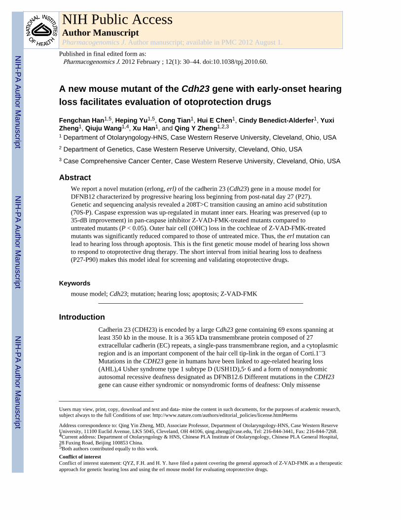
A new mouse mutant of the Cdh23 gene with early-onset hearing loss facilitates evaluation of otoprotection drugs Fengchan Han 1,5 , Heping Yu 1,5 , Cong Tian 1 , Hui E Chen 1 , Cindy Benedict-Alderfer 1 , Yuxi Zheng 1 , Qiuju Wang 1,4 , Xu Han 1 , and Qing Y Zheng 1,2,3 1 Department of Otolaryngology-HNS, Case Western Reserve University, Cleveland, Ohio, USA 2 Department of Genetics, Case Western Reserve University, Cleveland, Ohio, USA 3 Case Comprehensive Cancer Center, Case Western Reserve University, Cleveland, Ohio, USA Abstract We report a novel mutation (erlong, erl) of the cadherin 23 (Cdh23) gene in a mouse model for DFNB12 characterized by progressive hearing loss beginning from post-natal day 27 (P27). Genetic and sequencing analysis revealed a 208T>C transition causing an amino acid substitution (70S-P). Caspase expression was up-regulated in mutant inner ears. Hearing was preserved (up to 35-dB improvement) in pan-caspase inhibitor Z-VAD-FMK-treated mutants compared to untreated mutants (P < 0.05). Outer hair cell (OHC) loss in the cochleae of Z-VAD-FMK-treated mutants was significantly reduced compared to those of untreated mice. Thus, the erl mutation can lead to hearing loss through apoptosis. This is the first genetic mouse model of hearing loss shown to respond to otoprotective drug therapy. The short interval from initial hearing loss to deafness (P27-P90) makes this model ideal for screening and validating otoprotective drugs. Keywords mouse model; Cdh23; mutation; hearing loss; apoptosis; Z-VAD-FMK Introduction Cadherin 23 (CDH23) is encoded by a large Cdh23 gene containing 69 exons spanning at least 350 kb in the mouse. It is a 365 kDa transmembrane protein composed of 27 extracellular cadherin (EC) repeats, a single-pass transmembrane region, and a cytoplasmic region and is an important component of the hair cell tip-link in the organ of Corti.1 – 3 Mutations in the CDH23 gene in humans have been linked to age-related hearing loss (AHL),4 Usher syndrome type 1 subtype D (USH1D),5 , 6 and a form of nonsyndromic autosomal recessive deafness designated as DFNB12.6 Different mutations in the CDH23 gene can cause either syndromic or nonsyndromic forms of deafness: Only missense Users may view, print, copy, download and text and data- mine the content in such documents, for the purposes of academic research, subject always to the full Conditions of use: http://www.nature.com/authors/editorial_policies/license.html#terms Address correspondence to: Qing Yin Zheng, MD, Associate Professor, Department of Otolaryngology-HNS, Case Western Reserve University, 11100 Euclid Avenue, LKS 5045, Cleveland, OH 44106, [email protected], Tel: 216-844-3441, Fax: 216-844-7268. 4 Current address: Department of Otolaryngology & HNS, Chinese PLA Institute of Otolaryngology, Chinese PLA General Hospital, 28 Fuxing Road, Beijing 100853 China. 5 Both authors contributed equally to this work. Conflict of interest Conflict of interest statement: QYZ, F.H. and H. Y. have filed a patent covering the general approach of Z-VAD-FMK as a therapeutic approach for genetic hearing loss and using the erl mouse model for evaluating otoprotective drugs. NIH Public Access Author Manuscript Pharmacogenomics J. Author manuscript; available in PMC 2012 August 1. Published in final edited form as: Pharmacogenomics J. 2012 February ; 12(1): 30–44. doi:10.1038/tpj.2010.60. NIH-PA Author Manuscript NIH-PA Author Manuscript NIH-PA Author Manuscript
Transcript of A new mouse mutant of the Cdh23 gene with early-onset hearing loss facilitates evaluation of...
A new mouse mutant of the Cdh23 gene with early-onset hearingloss facilitates evaluation of otoprotection drugs
Fengchan Han1,5, Heping Yu1,5, Cong Tian1, Hui E Chen1, Cindy Benedict-Alderfer1, YuxiZheng1, Qiuju Wang1,4, Xu Han1, and Qing Y Zheng1,2,3
1 Department of Otolaryngology-HNS, Case Western Reserve University, Cleveland, Ohio, USA2 Department of Genetics, Case Western Reserve University, Cleveland, Ohio, USA3 Case Comprehensive Cancer Center, Case Western Reserve University, Cleveland, Ohio, USA
AbstractWe report a novel mutation (erlong, erl) of the cadherin 23 (Cdh23) gene in a mouse model forDFNB12 characterized by progressive hearing loss beginning from post-natal day 27 (P27).Genetic and sequencing analysis revealed a 208T>C transition causing an amino acid substitution(70S-P). Caspase expression was up-regulated in mutant inner ears. Hearing was preserved (up to35-dB improvement) in pan-caspase inhibitor Z-VAD-FMK-treated mutants compared tountreated mutants (P < 0.05). Outer hair cell (OHC) loss in the cochleae of Z-VAD-FMK-treatedmutants was significantly reduced compared to those of untreated mice. Thus, the erl mutation canlead to hearing loss through apoptosis. This is the first genetic mouse model of hearing loss shownto respond to otoprotective drug therapy. The short interval from initial hearing loss to deafness(P27-P90) makes this model ideal for screening and validating otoprotective drugs.
Keywordsmouse model; Cdh23; mutation; hearing loss; apoptosis; Z-VAD-FMK
IntroductionCadherin 23 (CDH23) is encoded by a large Cdh23 gene containing 69 exons spanning atleast 350 kb in the mouse. It is a 365 kDa transmembrane protein composed of 27extracellular cadherin (EC) repeats, a single-pass transmembrane region, and a cytoplasmicregion and is an important component of the hair cell tip-link in the organ of Corti.1–3Mutations in the CDH23 gene in humans have been linked to age-related hearing loss(AHL),4 Usher syndrome type 1 subtype D (USH1D),5, 6 and a form of nonsyndromicautosomal recessive deafness designated as DFNB12.6 Different mutations in the CDH23gene can cause either syndromic or nonsyndromic forms of deafness: Only missense
Users may view, print, copy, download and text and data- mine the content in such documents, for the purposes of academic research,subject always to the full Conditions of use: http://www.nature.com/authors/editorial_policies/license.html#terms
Address correspondence to: Qing Yin Zheng, MD, Associate Professor, Department of Otolaryngology-HNS, Case Western ReserveUniversity, 11100 Euclid Avenue, LKS 5045, Cleveland, OH 44106, [email protected], Tel: 216-844-3441, Fax: 216-844-7268.4Current address: Department of Otolaryngology & HNS, Chinese PLA Institute of Otolaryngology, Chinese PLA General Hospital,28 Fuxing Road, Beijing 100853 China.5Both authors contributed equally to this work.Conflict of interestConflict of interest statement: QYZ, F.H. and H. Y. have filed a patent covering the general approach of Z-VAD-FMK as a therapeuticapproach for genetic hearing loss and using the erl mouse model for evaluating otoprotective drugs.
NIH Public AccessAuthor ManuscriptPharmacogenomics J. Author manuscript; available in PMC 2012 August 1.
Published in final edited form as:Pharmacogenomics J. 2012 February ; 12(1): 30–44. doi:10.1038/tpj.2010.60.
NIH
-PA Author Manuscript
NIH
-PA Author Manuscript
NIH
-PA Author Manuscript
mutations of CDH23 have been observed in families with nonsyndromic deafness, whereasnonsense, frameshift, splice-site, and missense mutations have been identified in familieswith Usher syndrome (syndromic).7 Age-related hearing loss (AHL) is a characteristic ofthe widely used C57BL/6J mouse strain.8 AHL starts with a moderate hearing impairmentin 1-year-old C57BL/6J mice, and progresses to complete hearing loss with age. Linkagestudies associated AHL with a locus named ahl.9 A single-nucleotide polymorphism in exon7 of Cdh23 was significantly associated with AHL and the deafness modifier mdfw(modifier of deaf waddler). The hypomorphic Cdh23753A (synonymous with Cdh23ahl)allele causes in-frame skipping of exon 7.4 Histological analysis correlated AHL with agradual loss of hair cells, spiral ganglion cells and degeneration of fibrocytes in the spiralligament.10, 11 Most other mouse models of point mutation in the Cdh23 gene arecharacterized by congenital deafness with circling behavior.1, 2, 12, 13 Each of thesemutations leads to the loss of functional domains. In four waltzer alleles (v2J, v6J vAlb, vngt),loss of functional protein has been reported to disrupt the highly organized stereocilia bundleof hair cells in the cochlea and the vestibule during late embryonic/early postnataldevelopment.1, 13, 14 In all previously characterized Cdh23 mouse models, the mice areeither deaf at birth,2 exhibit severe very early onset hearing loss15 or very late-onset hearingloss with slow progression.9 The deaf (v-df) mutant was reported by Deol MS, that v-df micemay be deaf from the beginning or may be able to hear for a few days before weaning butotherwise behave normally16. Although the final pathological outcome is consistentlyobserved as inner ear hair cell loss2, none of these models provide an easily manageabletime interval for evaluating pathophysiological changes and/or for screening and testingdrug therapies. Here, we introduce a new mouse model of DFNB12 which is characterizedby progressive hearing loss starting at P27 and progressing to deafness by p100. This is anideal time window for testing otoprotective drugs. Nevertheless, we have found thatapoptosis plays a major role in hair cell loss in this mouse model. Most importantly, wereport here that a pan-caspase inhibitor not only preserved inner ear hair cells but alsoprevented hearing loss by up to 35 dB in the mutant mice.
Materials and methodsMice, genetic linkage cross, and DNA sequencing
Mice were originally housed in The Jackson Laboratory (Bar Harbor, Maine) researchfacilities and all procedures were approved by the Institutional Animal Care and UseCommittee (protocols 5U01NS041215 & R01DC007392). Mice were then relocated to CaseWestern Reserve University (CWRU, Cleveland, Ohio). Further studies were conducted inaccordance with the principles set forth in the Guide for the Care and Use of LaboratoryAnimals, Institute of Laboratory Animal Resources, and were approved by the Case WesternReserve University of the Health Sciences Institutional Animal Use and Care Committee(R01DC009246). ENU-induced (N-ethyl-N-nitrosourea, C3H6N3O2) mutagenesis wasperformed as part of a large-scale screening program for new mutants at the JacksonLaboratory using predominantly the C57BL/6J (B6) mouse strain (The Jackson LaboratoryNeuromutagenesis Facility website: http://nmf.jax.org). A detailed protocol is described athttp://nmf.jax.org/protocols/genetics_scheme.html. As hearing impairment was the onlymeasurable phenotype of erl mutant mice, we had to trace phenotype by testing hearing withABR thresholds for maintaining colonies and performing all experiments. Genetic intercrossgenerated 13 affected ((B6XC3H/HeJ)F1-erl/+)X((B6XC3H/HeJ)F1-erl/+) F2 progeny withelevated ABR thresholds as shown in Supplementary Table 1. A DNA pooling method forgene mapping was used as previously described.17, 18 Genomic DNA sequencing toidentify the alteration in the erl mutant mouse was performed as follows: Genomic DNAwas prepared from tail tips of mice. Briefly, 2-mm mouse tail tips were digested with 0.3 mlof 50 mM NaOH in a 0.5 ml Eppendorf tube at 95°C for 10 min. 26 μl of 1M Tris-HCl was
Han et al. Page 2
Pharmacogenomics J. Author manuscript; available in PMC 2012 August 1.
NIH
-PA Author Manuscript
NIH
-PA Author Manuscript
NIH
-PA Author Manuscript

then added to each tube. The mixtures were centrifuged at 12,000×g for 5 min and the DNAconcentration in supernatants was measured using a BioPhotometer (Eppendorf AG,Hamburg, Germany). Forty-eight pairs of PCR primers spanning exons of the Cdh23 genewere designed using the Primer3 freeware (http://frodo.wi.mit.edu/primer3/) according tothe exon sequence of the Cdh23 gene in the Ensemble Mouse Genome Server(www.ensembl.org), and synthesized by Integrated DNA Technologies, Inc. (San Diego,CA, USA). PCR for comparative DNA analysis between Cdh23erl/erl and Cdh23ahl/ahl micewas performed according to the Tm of the primers and the expected product sizes. PCRproducts were purified with the QIAquick PCR Purification kit (Qiagen, Inc. Valencia, CA,USA). DNA sequencing was performed using the same primers as for DNA amplificationand then run on an ABI Applied Biosystems 3730 DNA Analyzer (Life Technologies Corp.,Carlsbad, CA, USA).
To confirm the mutation and to identify potential aberrant exon splicing, Cdh23erl/erl andCdh23ahl/ahl mice at 2 weeks of age were used for RNA extraction and RT-PCR. After micewere sacrificed under anesthesia (avertin 5 mg/10 g), the inner ears were quickly removed.Total RNA (DNA-free) was prepared using the pure-LinkTM Micro-to-Midi Total RNAPurification System (Invitrogen, Carlsbad, CA, USA). cDNA synthesis was carried outusing the SuperScript™ First-Strand Synthesis System (Catalog No. 11904-018). PCRprimers (CdhmF and CdhmR in table 2) were designed in exon 2 and exon 4 yielding a 228-bp PCR product containing exon 3 and its flanking regions from exons 2 and 4.
Auditory-evoked brainstem responseABR was measured at various intervals for Cdh23erl/erl and Cdh23ahl/ahl mice (at ages overP14). A computer-aided evoked potential system (Intelligent Hearing Systems, Miami, FL,USA) was used to test mice for ABR thresholds as previously described.8 Briefly, micewere anesthetized and body temperature maintained at 37–38°C by placing them on aheating pad in a sound-attenuating chamber. Subdermal needle electrodes were inserted atthe vertex of (active) and ventrolaterally to (reference) the right ear and to the left ear(ground). Clicks, and 8-, 16- and 32-kHz tone-bursts were respectively channeled throughplastic tubes into the animal’s ear canals. The amplified brainstem responses were averagedby a computer and displayed on a computer screen. Auditory thresholds were obtained foreach stimulus by reducing the sound pressure level (SPL) at 10-dB steps and finally at 5-dBsteps up and down to identify the lowest level at which an ABR pattern could be recognized.ABR threshold values above 55 (for click stimulus), 40 (for 8 kHz), 35 (for 16 kHz), or 60(for 32 kHz) dB SPL were considered to be hearing-impaired.8
Distortion product oto-acoustic emission (DPOAE)To test the function of outer hair cells of different mice at different time points, we used theIHS Smart EP 3.30 USBez Software (Intelligent Hearing Systems, Miami, FL, USA) forDPOAE measurement, which was conducted for pure tones from 2 to 36 KHz.19 AnEtymotic 10B+ (Etymotic Research, Inc., Elk Grove Village, IL, USA) probe was insertedinto the external ear canal and used with two different types of transducers depending on therange of the stimulation frequency. For frequencies ranging from 2 to 16 kHz, an EtymoticER2 stimulator was used and for frequencies ranging from 16 to 30 kHz, an IHS highfrequency transducer was used. Stimulus response signals were sampled at a rate of 128 kHzusing a 16-bit D/A converter; L1 and L2 amplitudes were set to the same level. Frequencieswere acquired with an F2-F1 ratio of 1.22. The stimuli were presented starting from thelowest frequencies tested and increasing to the highest frequencies tested. Five stimulationlevels ranging from 65 to 25 dB SPL in 10-dB steps were used.
Han et al. Page 3
Pharmacogenomics J. Author manuscript; available in PMC 2012 August 1.
NIH
-PA Author Manuscript
NIH
-PA Author Manuscript
NIH
-PA Author Manuscript
Histological analyses of inner earsHistological analyses of inner ears were performed following the methods describedpreviously.20 Briefly, anesthetized mice were perfused through the left ventricle of the heartwith phosphate-buffered saline (PBS) followed by Bouin’s (for H&E staining) or 4%paraformaldehyde (for all others) fixative. For microscopic analysis of cross-sections, innerears from Cdh23erl/erl and Cdh23ahl/ahl mice were dissected, perfused with fixative,immersed in same for 48 h, decalcified with Cal-EX solution for 6 h, and embedded inparaffin. Sections (5 μm) were cut, mounted on glass slides and counterstained inhematoxylin/eosin (H&E).
CytocochleogramsCytocochleograms were obtained by a modified method as described previously.20 Briefly,the organ of Corti was carefully microdissected out and mounted in glycerin on glass slides.The surface preparations were stained for F-actin with Alexa Fluor 568 conjugated tophalloidin to show hair bundles and examined with a fluorescence microscope (LeicaDM4000 B, Leica Microsystems, Wetzlar, Germany). Hair cells were counted as present ifV-shapes of hair bundles were intact. Inner and outer hair cell counts were made bysubdividing the cochlea into 10 regions at 10% distance intervals, beginning at the apex andcontinuing toward the base. Individual cochleograms were constructed to show thepercentage of hair cells missing as a function of distance from the apex.
Semi-Quantitative-RT-PCR for measuring mRNA accumulation levels of apoptosis-relatedgenes
Cdh23erl/erl and Cdh23ahl/ahl mice were sacrificed under avertin anesthesia conditions at 2weeks or 2 months of age. The inner ears and left temporal brain lobes (50 mg) were quicklyisolated for total RNA and cDNA preparation as described in the previous section. One μgof total RNA from each sample was used as template for cDNA synthesis. The 20 μlreaction mixture contained 50 mM KCl, 10 mM Tris-HCl, pH 9.0 (at 25°C), 0.01% TritonX-100, 2 mM MgCl2, 250 nM of each primer (forward and reverse), 200 μM dNTPs, 1 μl ofcDNA and 0.5 U of Taq DNA polymerase (New England BioLabs, Inc., Ipswich, MA,USA). PCR primers are listed in Table 2. PCR was performed in a Bio-Rad PTC-200 PeltierThermal Cycler (Bio-Rad Laboratories, Inc. Hercules, CA, USA). Amplification conditionswere 94°C for 2 min; followed by 28 cycles of 94°C for 30 s, 60°C for 40 s, and 72°C for 50s; followed by 5 min at 72°C. Ten μl of the PCR products were subject to agarose gelelectrophoresis and the gray intensity of each band was digitized using ImageJ software(rsb.info.nih.gov/nih-image/NIH, Bethesda, MD, USA) and corrected by the GAPDHmRNA accumulation level of the same sample.
Caspase-3/7 activity measurementCaspase-3/7 activity was detected in inner ears of 9 Cdh23erl/erl and 7 Cdh23ahl/ahl mice bythe Apo-ONE® Homogeneous Caspase-3/7 Assay (Promega Corp., Madison, WI, USA)according to the manufacturer’s instructions. In brief, the inner ears were isolated andimmersed in 300 μl of 0.1 M phosphate buffer saline (PBS) and homogenized on ice with aTissue Mincer (Fisher Scientific, Pittsburgh, PA, USA). After centrifugation at 12000×g for15 min at 4°C, the supernatant was retained and the protein concentration determined by theLowry method. For the caspase assay, the non-fluorescent caspase substrate (Z-DEVD-R110, diluted 100-fold in the provided buffer) was mixed with an equal volume (20 μl) ofsolution containing the same amount of total inner ear protein (20 μg) for each sample. Theprotein/substrate mixture was incubated at room temperature for 4 h and diluted 300-foldafterward. The fluorescent product was detected using a spectrofluorometer configured to anexcitation wavelength of 499 nm and an emission wavelength of 521 nm. The relative
Han et al. Page 4
Pharmacogenomics J. Author manuscript; available in PMC 2012 August 1.
NIH
-PA Author Manuscript
NIH
-PA Author Manuscript
NIH
-PA Author Manuscript
fluorescence unit as a measure of caspase activity was calculated as the fluorescence countsdivided by the total amount of protein.
Immunostaining for active caspasesA time course immunocytochemistry study of caspase expression was carried out forCdh23erl/erl and Cdh23ahl/ahl. The following antibodies from Cell Signaling Technology,Inc. (Danvers, MA, USA) were used. Cleaved caspase-3 (Asp175) antibody detectsendogenous levels of the large fragment (17/19 kDa) of activated caspase-3 resulting fromcleavage adjacent to Asp175 (This antibody does not recognize full-length caspase-3 orother cleaved caspases.); caspase-8 antibody (mouse-specific) detects endogenous levels ofthe large 18 kDa subunit of active caspase-8; cleaved caspase-9 (Asp353) antibody (mouse-specific) detects endogenous levels of the 37 kDa subunit of mouse caspase-9 only aftercleavage at aspartic acid 353. It does not cross-react with full-length caspase-9 or with othercaspases at endogenous levels. Mice were subjected to ABR testing (age ≥ 14 days) underanesthetizing conditions and then sacrificed. The inner ears were removed and cryosectionswere made and fixed in 4% PFA (diluted in 1× PBS) for 2 h. The sections were washed inPBS at room temperature twice for 5 min and permeabilized in 0.5% Triton X-100 for 30minutes. After being washed twice in 1× PBS for 5 min and blocked in 5% BSA for 1 h, thesamples were immersed in anti-active caspase-3 or caspase-8 or caspase-9 (1:200 dilution)and incubated at 4°C overnight. After being washed twice in 1× PBS for 5 min, the sampleswere immersed in anti-rabbit secondary antibody Alexa 488 (1:500 dilution) for 1 h. Thesamples were also stained with propidium iodide (10 mg/ml in PBS) for 30 min at roomtemperature. The sample mounts were observed under immunofluorescent microscopy.
Intraperitoneal injection and treatment regime optimizationCdh23erl/erl was used as a model for testing anti-apoptotic drug therapy. Sixty Cdh23erl/erl
mice at the age of 7 days (a starting point based on the goal of preventing caspase increases,detected as early as P14 in untreated Cdh23erl/erl mice) were divided into 3 groups: a testgroup, a DMSO group and an untreated group. The following treatment regime was selectedas the best treatment from three sets of preliminary regimes (data not shown). In the testgroup, 22 mice were injected intraperitoneally (IP) under sterile conditions with Z-VAD-FMK (1μg/μl) (Z-Val-Ala-Asp(OMe)-Fluoromethylketone, Alexis Biochemicals,Farmingdale, NY) in PBS-diluted DMSO (1:1) at the dosage21 of 1.5 μg/g mouse weight:firstly, starting at P7, 8 injections, once every other day; secondly, 4 injections, once every 3days; then, one injection every 4 days until the time of euthanization for experimentalprocedures. Eighteen mice in the DMSO group received 1.5 μl of PBS-diluted DMSO (1:1)per gram of mouse weight at the same time points as the test group. ABR and DPOAE weretested at 4 weeks, 6 weeks, 8 weeks and 12 weeks for the mice in each group and 4–5 micefrom each group were sacrificed at each time point for histological investigation.
Statistical methodsThe ANOVA was used for all data analysis except that hair cell loss data were analyzed bythe Chi-Square Test. A value of P < 0.05 was considered significant.
ResultsLinkage analysis and genetic complementation tests demonstrated that erl is a new alleleof the Cdh23 gene
A new hearing-impaired mouse mutant was discovered at the Jackson Laboratory on theC57BL/6J (B6) background initially based on its lack of a Preyer reflex when presented witha calibrated 20 kHz 90 dB SPL (decibel sound pressure level) tone burst from a click box at
Han et al. Page 5
Pharmacogenomics J. Author manuscript; available in PMC 2012 August 1.
NIH
-PA Author Manuscript
NIH
-PA Author Manuscript
NIH
-PA Author Manuscript
two months of age. This mutant was originally generated from the NeuroscienceMutagenesis Facility (http://nmf.jax.org/, was designated #NMF308), but here is namederlong (symbol: erl), meaning hearing impaired in Chinese language. Subsequently, hearingloss (starting at P27, Table 1) was shown to progress to deafness (at P90) by auditorybrainstem response (ABR) threshold testing in the homozygous colony (data not shown). Nobalance defect was observed throughout the lifespan of these mice. Initially, the mutant wasmated with an unrelated B6 mouse to determine the heritability pattern of the mutation.ABR testing revealed no hearing loss in (erl X B6) F1 progeny up to 3 months of age, thusindicating that erl is a recessive hearing loss mutation. B6-erl/erl mice were outcrossed withC3H/HeJ mice to generate the (B6XC3H/HeJ)F1-erl/+ mice. These F1 mice wereintercrossed (F1xF1) and yielded a Mendelian proportion of a single gene recessiveinheritance pattern, generating roughly ¼ (13/51) erl/erl F2 progeny with ABR thresholdsabove 80 dB SPL and ¾ (38/51) ?/+ (¼ +/+ and ½ erl/+) F2 progeny with ABR thresholdsbelow 50 dB SPL at five months of age (see Supplementary Table 1).
Our previous publications showed that, for age-related hearing loss, many strains have aChromosome 10 (Cdh23) effect4, 8, 20, 22–25 that causes hearing loss; therefore, we firsttested this possibility by analyzing the genotype of a marker (D10Mit194, Chr location:46MB) close to the Cdh23 gene (Chr location: 59MB). A PCR assay on pooled genomicDNA from the 13 hearing-impaired F2 progeny showed a distinctive single PCR band forthe B6 allele of D10Mit194, suggesting that erl might be another AHL-related allele of theCdh23 gene on Chr 10 on which we previously worked4, 8, 9, 22, 24, 25 or another locus inthe same region. Then individual DNA samples of the 51 F2 progeny were genotyped withrespect to D10Mit194, D10Mit166 (Chr location: 6MB) and D10Mit42 (Chr location:82MB). The results showed that the candidate interval was between D10Mit194 andD10Mit42 at 29 cM on chromosome 10; whereas, the Cdh23 gene is located at 30.3 cM asshown in the Supplementary Table 1. These results prompted us to do a geneticcomplementation test with mice carrying the most severe Cdh23 allele, waltzer 2 Jackson(abbreviated v-2J; complete designation is C57BL/6J-Cdh23v-2J/J), the deaf and circlingallele1, 2, 25. Complementation tests revealed allelism of the erl locus and the v-2J locusindicating that erl is a new allele of the Cdh23 gene. As shown in Table 1, one litter of 8pups was born (ID 1–8). The 4 erl/v-2J compound-heterozygous mice (ID 1–4) showed ahearing-loss level intermediate (without circling behavior) between that of v-2J/v-2J (ID 9)and erl/erl (ID 10), which suggests a codominant effect. Genotypes of the v-2J and erl miceshown in Table 1 were individually confirmed afterward by sequencing. These results werecorroborated by an analysis of another litter of 9 pups from a B6- erl/erl x B6-v-2J/v-2Jmating that gave a deaf phenotype without any balance defect for all 9 mice at P60.
Identification of a novel mutation in the Cdh23 gene of the hearing impaired mutant mouseGenomic DNA screening to identify the alteration in the Cdh23 gene was performed byPCR and sequencing of all exons of the Cdh23 gene with detailed comparisons betweenmutants and controls using SequencherR 4.0 (genecodes.com; Gene Codes Corporation,Ann Arbor, MI, USA). A point mutation (208T>C) in the middle region of exon 3 (Genbankaccession number: NM_023370) was identified by sequencing the PCR products of primerpair ex3-F1 and ex3-R1 (for three mutant and two control genomics DNA samples, seeTable 2 for primer sequences) and confirmed by sequencing the cDNA of this gene (Figure1a). No other mutations were found in any other exons of this entire gene. The RT-PCRproducts with primers flanking exon 3 and adjacent exons gave a clear single band (of thepredicted size and identical to a normal control, data not shown) upon agarose gelelectrophoresis suggesting that no aberrant splicing is caused by this mutation. The erlmutation did not quantitatively alter the expression level of Cdh23 mRNA (supplementaryFigure 1a). The mutation results in an amino acid (aa) substitution of serine to proline (70S-
Han et al. Page 6
Pharmacogenomics J. Author manuscript; available in PMC 2012 August 1.
NIH
-PA Author Manuscript
NIH
-PA Author Manuscript
NIH
-PA Author Manuscript
P) in the first ectodomain (EC1, aa 38–132). The distinctive cyclic structure of the prolineside chain locks its ϕ backbone dihedral angle at approximately−75°, giving proline anexceptional conformational rigidity compared to other amino acids. Proline acts as astructural disruptor in the middle of regular secondary structure element - alpha helix (aa73–76) and to changes in the surrounding extended strands (specifically, an extended strandformed by aa 65–68 in the wild type mice shifts to that formed by aa 65–67 in the mutant,and another one formed by aa 77–80 in the wild type mice to that formed by aa 76–80 in themutant mice) affecting the secondary structure of the protein when analyzed by the GOR IVsecondary structure prediction method26. The substituted amino acid is not within butnearby a conserved calcium-binding site (aa 59–61, sequence DMD, highlighted in Figure1c). Loss of the alpha helix and changes in the extended strands will probably affect proteinfunction, such as formation of tip-links in the stereocilia or binding efficiency to calciumions. Based on the mutation, a genotyping method was established to distinguish micecarrying the Cdh23erl allele from mice carrying the Cdh23ahl (C57BL/6J) allele by sizedifferences on an agarose gel of PCR products digested with BstN I (Figure 1b). Thisconfirms in another way that the erl mutation was accurately identified. By sequencing, weconfirmed that all of the erl mice carry the ahl allele, thus this allele causes in-frameskipping of exon 7 and may contribute to conformational changes of CDH23.4
DPOAE and ABR testing reveal early onset, progressive hearing loss in Cdh23erl/erl miceDistortion product otoacoustic emissions (DPOAEs) were measured to determine the mutanteffect on OHC function over time in Cdh23erl/erl mice as compared to Cdh23ahl/ahl controls(Fig. 2a). Three-week-old Cdh23erl/erl mice had normal DPOAE amplitudes that wereindistinguishable from those of Cdh23ahl/ahl mice which have normal hearing as determinedby ABR thresholds at 3 weeks (wks). At 7 wks of age, Cdh23erl/erl mice had DPOAEamplitudes 10–20 dB lower than those of Cdh23ahl/ahl at frequencies from 10 to 18 kHz. By22 wks of age, Cdh23erl/erl mice had no detectable DPOAE at any frequency indicatingcomplete deafness.
ABR recordings yielded normal wave patterns (data not shown) but elevated thresholds atevery frequency tested (click, 8, 16, 32 kHz from 30–99 dB, Figure 2b and Table 1) withhigh frequencies affected first, as early as P40, progressing to deafness at P90 and thereafterremaining deaf to at least 9 months of age. The Cdh23erl/erl mice exhibit hearing loss muchearlier (P40) than do mice expressing other Cdh23 alleles such as Cdh23ahl/ahl mice (12-months onset, Figure 2b and Table 1). The vestibular system in these mutant mice appears tobe unaffected by the erl mutation as Cdh23erl/erl mice exhibited normal swimming ability (3Cdh23erl/erl mice at 4 months of age had similar normal swimming as B6 mice) and gaitpattern as observed for all time points examined up to 9 months.
Histologic examination reveals progressive OHC loss that follows hearing loss inCdh23erl/erl mice
To investigate cochlear pathology in Cdh23erl/erl versus Cdh23ahl/ahl control mice, cochleaewere stained for F-actin with Alexa Fluor 568 conjugated to phalloidin to examine all turnsand reveal the presence and morphology of OHCs (Figure 2c). The OHCs of Cdh23ahl/ahl
mice at P100 (Figure 2c, upper panel) showed normal V-shaped arrangement of stereocilia.No OHC loss was observed in any of the cochlear turns of Cdh23ahl/ahl mice, as correlateswith normal ABR thresholds up to 9 months (Figure 2b). In contrast, large segments of OHCloss were evident in the middle cochlear turns of Cdh23erl/erl mice at P100 (Figure 2c, lowerpanel). This OHC loss at P100 follows ABR threshold shifts observed at P27-P90 (Figure2b).
Han et al. Page 7
Pharmacogenomics J. Author manuscript; available in PMC 2012 August 1.
NIH
-PA Author Manuscript
NIH
-PA Author Manuscript
NIH
-PA Author Manuscript

To quantify OHC loss and to determine the developmental time course of OHC loss, wegenerated cytocochleograms from 25 Cdh23erl/erl mice ranging from one to nine months ofage. For each cytocochleogram, we calculated the percentage of missing OHC and IHC bysubdividing the cochlea into 10 regions at 10% distance intervals from the apex of thecochlea. Scatter plots were constructed showing the mean percentage of OHC loss as afunction of age for Cdh23erl/erl mice (Figure 2d). Cdh23erl/erl mice showed no evidence ofOHC loss from P0 to P28. However, OHC loss was observable at 3 months, becameprogressively worse over time, and moved from the cochlear base to the apex in Cdh23erl/erl
mice. OHC loss in the 75–100% region increased to a nearly complete loss after 3 months ofage, corresponding to a high frequency hearing loss (16 and 32 kHz, top two lines in Fig.2b) earlier than that of low frequency hearing loss (8kHz, bottom line, Fig. 2b) as measuredby ABR. Because IHC loss was not significant until after 5 months of age, IHCs wereexcluded from this analysis.
mRNA accumulation and activity of apoptosis-related genes was up-regulated in the innerears of Cdh23erl/erl mice
To determine whether OHC loss might be the result of OHC apoptosis, we examinedexpression of apoptosis-related genes in Cdh23erl/erl mice. Gene transcription levels ofapoptosis-related genes were measured in the inner ears of Cdh23erl/erl and controlCdh23ahl/ahl mice at 2 wks and 2 months of age (Figure 3). The extrinsic caspase-8 andintrinsic caspase-9 initiators, and activator caspase-3 were significantly up-regulated (P <0.05 for each) in Cdh23erl/erl samples relative to the levels measured in control Cdh23ahl/ahl
samples (Figure 3a and 3b) though the intensity for caspase-8 was higher and earlier thanthat of caspase-9, suggesting that the extrinsic pathway was initiated earlier than theintrinsic. At the two-month time point, the expression level of caspase-9 was also up-regulated (P < 0.05) in Cdh23erl/erl samples. However, there was no significant difference inmRNA accumulation levels of caspase-3, caspase-8 and caspase-9 genes in brain tissuebetween Cdh23erl/erl and Cdh23ahl/ahl mice at the ages of 2 weeks or 2 months (P > 0.05,data not shown), indicating that the apoptotic gene expression measured in ear tissue was nota generalized phenomenon.
To confirm whether increased mRNA levels were correlated with changes at the proteinlevel, the Apo-ONE® homogeneous caspase-3/7 assay (Promega) was used to measurecaspase-3 and −7 activities in inner ears of erl mutants and control mice. The relativefluorescence unit is a measure of caspase activity in the homogenized inner ear tissue fromthe mice in each group, upon addition of a caspase substrate to the homogenate. At P14 (2weeks) and P60 (2 months), the relative caspase-3/7 activity was significantly higher inCdh23erl/erl mice than in Cdh23ahl/ahl mice (Figure 3c and 3d).
Immunostaining reveals signs of apoptosis early, increasing toward the 3-month timepoint in the organ of Corti of Cdh23erl/erl mice
To investigate inner ear apoptosis in situ in the Cdh23erl/erl mouse model and determinewhether the observed OHC loss can be explained by apoptosis of the OHCs, we stainedtissue sections with fluorescein-labeled antibodies to detect cells that express caspases. Theantibodies used specifically detect only the active forms of caspases-3, −8 and −9.Cdh23ahl/ahl mice were used as controls. We investigated three time points leading up to thetime of complete hearing loss at 3 months, and beginning at the P14 time point (Figure 4).At P14, caspases-8 and −9 were expressed in the middle turns of cochleae (mainly in theouter hair cells (OHC) and spiral ganglion cells (SG) for caspase-8 and caspase-9) of theCdh23erl/erl mice (Figure 4d, 4e) compared to the same areas of the Cdh23ahl/ahl mice(Figure 4a, 4b). Primary antibody-omitted sham sections of the cochleae of both mousegroups demonstrated that this staining was specific and not an artifact of non-specific
Han et al. Page 8
Pharmacogenomics J. Author manuscript; available in PMC 2012 August 1.
NIH
-PA Author Manuscript
NIH
-PA Author Manuscript
NIH
-PA Author Manuscript
secondary antibody staining (Figure 4c, 4f). Notably, caspase-8 stained OHC stronger thanSG (Figure 4d), whereas caspase-9 stained OHC weaker than SG (Figure 4e).
A time course observation of caspase-3 staining in the middle turns of cochleae ofCdh23erl/erl mice is shown in Figure 5. Anti-caspase-3 (the activated, downstream, effectorcaspase) staining was not visible in any areas of the cochleae at P14 (Figure 5a); however, itwas evident in the cytoplasm of OHCs of Cdh23erl/erl mice by P23 (Figure 5b) and becamestronger by P57 (Figure 5c). Active caspase-3 was also positive in the spiral ligament (SL)at both time points at P57. An enlarged image of OHCs from Figure 5c showed theoverlapping staining (Figure 5f) of PI-stained nuclei (Figure 5e) and anti-caspase-3-stainedcytoplasm (Figure 5d). Anti-caspase-3 signals were not detected in any cochlear turns fromCdh23ahl/ahl mice.
Upregulation of gene expression in the extrinsic (TNF-α) and intrinsic (Calpain) apoptosispathways in Cdh23erl/erl mice
The relative mRNA levels of TNF-α, caspase-12, m-calpain and μ-calpain weresignificantly up-regulated in the Cdh23erl/erl mice at both time points of 2 weeks(Supplementary Figure 1a, c) and 2 months (Supplementary Figure 1b, d), indicatingmultiple apoptosis pathways are involved in the hearing loss of these mutant mice. But TNF-α (Supplementary Fig. 1a) was up-regulated at the younger age (2 weeks) more significantlythan other genes (c), suggesting the extrinsic pathway may play an important role forinitiation of apoptosis. The fact that caspase-8 was up-regulated at the younger age (2weeks) (Figure 3a) more significantly (P<0.01) than other genes in the Cdh23erl/erl mice alsosupports this point becauseTNF-α and caspase-8 are both important indicators of theextrinsic apoptotic pathway. Calpains, caspase-12 (Supplementary Figure 1b, d, P<0.05) andcaspase-9 (Figure 3b, P<0.05) were also up-regulated slightly, but significantly, as indicatorsof intrinsic apoptosis.
OHC protection in Cdh23erl/erl miceCdh23erl/erl was used as a model for testing anti-apoptotic drug therapy. Sixty Cdh23erl/erl
mice were divided into a test group, a diluent (DMSO) control group and an untreatedcontrol group. Each mouse in the test group received Z-VAD-FMK (in dimethyl sulfoxide,DMSO, 1:1 diluted with PBS) intraperitoneally (ip) at a dosage of 1.5 μg/g body weight atdifferent time points: firstly, eight injections, on a schedule of one injection every other day,beginning at age P7; then 4 injections, on a schedule of one injection every 3 days; andlastly, one injection every 4 days until the time of euthanization for analysis at various timepoints. Each mouse in the DMSO group received an equivalent volume of DMSO followingan identical injection schedule to its cohort test group. ABR and DPOAE were tested at 4wks, 6 wks, 8 wks and 12 wks of age. ABR thresholds were significantly lower in theDMSO+Z-VAD-FMK-treated mice than in the DMSO-treated or the untreated Cdh23erl/erl
mice at stimulus frequencies of click, 8 kHz, 16 kHz and 32 kHz and at all time points (P <0.05 at 4 wks; P < 0.01, for the time points of 6 wks, 8 wks and 12 wks) (Figure 6). Therewere also significant differences in ABR thresholds between the untreated and DMSO-treated mice at 12 wks for the stimulus frequency of 16 kHz (P < 0.05) (Figure 6c). Themean amplitude of DPOAE at 16 kHz was significantly higher in the DMSO+Z-VAD-FMK-treated mice than that of the untreated or DMSO-treated group at the age of 8 wks (P< 0.05, respectively, Figure 7). Loss of outer hair cells (OHC) was evident in the middleturns of cochleae of the untreated or DMSO-treated mouse groups at 2 months of age;whereas, no OHC loss was seen in the corresponding areas of the Z-VAD-FMK+DMSO-treated mice (Figure 8). OHC and inner hair cell (IHC) loss was counted and averaged fromapical to basal turn of cochlear surface preparations in the three mouse groups (n=4 mice pergroup) at 2 months of age (Figure 9). The mean percentage of OHC loss was significantly
Han et al. Page 9
Pharmacogenomics J. Author manuscript; available in PMC 2012 August 1.
NIH
-PA Author Manuscript
NIH
-PA Author Manuscript
NIH
-PA Author Manuscript

less in Z-VAD-FMK+DMSO-treated Cdh23erl/erl mice than in the untreated or DMSO-treated mice at the apex, middle or basal cochlear regions. There was also significantly lessOHC loss in the DMSO-treated mice than in the untreated mice at the basal turn (P <0.05).IHC loss was not substantially altered by the treatment. Additionally, we have not observedany adverse effects of Z-VAD-FMK treatment (such as tumor development in the intestine,colon, liver or lungs or i.p injection site) on the treated mice during the experimental periodup to 3–4 months of ages (data not shown).
Z-VAD-FMK treatment prevents OHC loss in Cdh23v-2J/v-2J miceAs represented in Supplementary Figure 2, serial sections demonstrated that Z-VAD-FMKtreatment could protect OHCs from loss in Cdh23v-2J/v-2J mice, but failed to improve thehearing of the mice at any level. The survival of inner ear hair cells may still be important torelease neurotrophic factors for keeping spiral ganglion neurons viable and for responding toelectrical stimuli in cochlear-implanted USH1D patients, despite the inability of rescued haircells to function in hearing. Thus anti-apoptosis treatment in USH1D patients might beuseful in the future, but will require further studies.
DiscussionThe Cdh23erl/erl mice introduced in this study are of the C57BL/6J background and provedto be carrying a new allele of Cdh23. Cdh23erl/erl is characterized by progressive hearingloss starting about 1 month after birth and becoming deaf at 3 months of age. Hair cell lossbegan near the base of the cochlea and spread toward the apical turn, and lesions of thespiral ganglion were also involved at a later stage (P149, data not shown). The Cdh23erl/erl
mouse, therefore, provides a promising model for investigating the pathogenesis ofDNFB12. It is also an ideal mouse model for otoprotection drug therapy and discovery.
Given that the hearing loss in Cdh23erl/erl mice happens before hair cell loss (P60 see Fig.2d) but after caspase signals increase (P14), we hypothesized that apoptosis might be themechanism for causing a physiological deficit to occur before hair cell loss. There aremainly two pathways of cellular apoptosis as shown in Figure 10.27, 28 In response to extra-cellular stimuli, the initiator of the extrinsic pathway (caspase-8) undergoes self-processing,releasing active enzyme molecules into the cytosol and activating caspase-3, caspase-6, andcaspase-7. These activated caspases proteolytically cleave and activate other caspases, aswell as relevant targets in cells. Alternatively, the intrinsic pathway involves release fromthe mitochondrion of pro-apoptotic proteins that activate caspase enzymes, mainlycaspase-9, which ultimately trigger apoptosis by activating caspase-3, caspase-6, andcaspase-7. Therefore, we attempted to determine the role of apoptosis in the pathogenesis ofthe Cdh23erl/erl mouse model by examining expression of apoptosis-related proteins in theinner ears of these mice. Our results showed significantly up-regulated transcriptional levelsof caspase-3, caspase-8 and caspase-9 in the inner ears of Cdh23erl/erl mice at 2 weeks and/or 2 months of age, indicating both extrinsic and intrinsic pathway activation. Though theextrinsic pathway was activated slightly earlier and more strongly than the intrinsic pathway,this presumably was the result of increased TNFα signalling triggered by debris from brokenstereocilia composed of mis-folded CDH23erl/erl protein, due to the ϕ backbone dihedralangle (−75°) caused by the proline from the aa substitution (70S-P). A detailed mechanisticexplanation of why mutated CDH23 increases TNF-α signalling is yet to be determined.Another possibility is that the 70S-P introduced conformational changes in the firstectodomain, ultimately leading to intracellular domain and catenin complex signal changesthat modulate apoptosis.29, 30
Immunostaining showed that caspase-8 was activated in the organ of Corti at P14, thatcaspase-3 was activated by P23 and that the staining for both became more intense at P57.
Han et al. Page 10
Pharmacogenomics J. Author manuscript; available in PMC 2012 August 1.
NIH
-PA Author Manuscript
NIH
-PA Author Manuscript
NIH
-PA Author Manuscript

Additionally, there were signs of apoptosis in the spiral ganglion and the stria vascularis. Incontrast to a previous report in rat of a strong active caspase-3 signal in the lateral wall ofthe cochlear basal turn with drug-induced damage,21 our mouse experiments showed thatcaspase immunoreactivities were first present and strong in the basal turn and middle turnsof the organ of Corti. Based on these results, we conclude that apoptosis plays a major rolein Cdh23erl/erl mice in the death of hair cells, spiral ganglion cells and cells of the spiralligament and stria vascularis in the inner ear, and that the cell death mechanism of drug-induced damage in rat may differ from the mechanism in a genetic mutant such as theCdh23erl/erl mouse. The up-regulated apoptosis is probably localized to the inner ear inCdh23erl/erl mice, as there was no significant difference in mRNA accumulation levels forcaspase-3, caspase-8 or caspase-9 genes in brain tissue between Cdh23erl/erl andCdh23ahl/ahl mice. We acknowledge that other mechanisms may apply for possible causes ofhearing loss in other mutations.
Cadherins are glycoproteins involved in Ca2+-mediated cell-cell adhesion, migration andcompaction.2 CDH23 is an important component of the Ush1 interactome and of the tip-links in the mechanotransduction (MET) system of the organ of Corti.15, 31–33 It isinvolved in regulating the activity of mechanically gated ion channels in hair cells.3, 34, 35It is reported that, of the 21 missense mutations in CDH23 that have been observed inpatients with nonsyndromic hearing loss, 10 occurred within the DXNDN, LDRE and DXDcalcium-binding motifs. These motifs are highly conserved in sequence and spacing and arerequired for cadherin dimerization and Ca2+-binding, suggesting that deficiency of thecalcium-dependent cell adhesion has a major impact on cochlear cell integrity36. Becausecalcium provides rigidity to the elongated structure of cadherin molecules thus enablinghomophilic lateral interaction, mutations in the calcium-binding motifs of CDH23 are likelyto impair homophilic interactions of CDH23 molecules and possibly also interactions withother proteins.37 Though the change in structure in CDH23 protein in the Cdh23erl/erl miceis not located precisely at the Ca2+-binding site, it is nearby. Thus, we speculate that it mayinfluence the efficiency of CDH23 interactions with proteins, Ca2+ and ultimately mayaffect calcium-dependent cell adhesion, leading to apoptosis in the Cdh23erl/erl mutant miceby multiple pathways (Figure 10). (1) The intricate relationship between CDH23 and otherproteins of the USH complex could be disrupted by the erl mutation, causing faulty OHChair bundle formation and extrinsic apoptosis, as the mRNA level of TNF-α wassignificantly higher in the ears of the Cdh23erl/erl mice (Fig s1a,b). This pathway may be theearliest and strongest-affected pathway in our mouse model among all possible involvedpathways. (2) We did find that calpains and caspase 12 were up-regulated at thetranscriptional level in Cdh23erl/erl mutant mice (Fig s1c,d). Calpains are activated by Ca2+
ions 38 and can further activate caspase-12, which subsequently activates caspase-9(intrinsic pathway). (3) As apoptosis was also evident in spiral ganglion, stria vascularis andsupporting cells in the mutant mice, disorder in Ca2+ regulation and in calcium-dependentcell adhesion in the cochlea may be responsible for triggering apoptosis. (4) CDH23 is abona fide type I intrinsic membrane protein and is most likely translated on rough ERmembranes; therefore, the 70S-P substitution may cause CDH23 misfolding resulting inaccumulation of CDH23 in the endoplasmic reticulum (ER), with subsequent ER stressfollowed by apoptosis (Figure 10). (5) It has been reported that a cyclic peptide can perturbcadherin-mediated endothelial cell interactions, resulting in a progressive apoptotic celldeath. This effect depends on cell density, as it is only observed when dense cultures aretreated with the peptide.39 CDH23 is a member of the cadherin family and could have asimilar mechanism of apoptosis. This may also explain why the densely organized OHCs(three rows) were more affected than the more loosely organized IHC (one single row).
It has been documented in drug-induced hearing loss models that hair cell loss can occur byapoptosis, and that such loss can be reversed through treatment with the anti-apoptotic drug,
Han et al. Page 11
Pharmacogenomics J. Author manuscript; available in PMC 2012 August 1.
NIH
-PA Author Manuscript
NIH
-PA Author Manuscript
NIH
-PA Author Manuscript
Z-VAD-FMK (a powerful, irreversible and cell-permeable inhibitor for caspases).21 Wetherefore investigated Z-VAD-FMK as a candidate drug for maintenance of hearing and haircell viability in Cdh23erl/erl mice, though the mechanism of cell death in Cdh23erl/erl micemay differ from that in drug-induced damage. Following Z-VAD-FMK treatment of theCdh23erl/erl mice, there was a significantly reduced amount of OHC loss compared to that ofthe untreated or DMSO-treated mice in all regions of the cochlea. The protective effect of Z-VAD-FMK on OHC was further confirmed by significantly higher DPOAE amplitudes inthe DMSO+Z-VAD-FMK-treated mice than in the untreated or DMSO-treated mice at theage of 8 weeks. Furthermore, ABR thresholds were preserved at levels approximately 15–40dB better in the Z-VAD-FMK-treated group than in the untreated or DMSO-treated groupsof littermate Cdh23erl/erl mice from 6 weeks to 12 weeks of age. The involvement ofapoptosis in the pathology of Cdh23erl/erl inner ears was thus confirmed by anti-apoptoticdrug therapy. Z-VAD-FMK can preserve cells (hair cells, for example) in the inner ears bystopping apoptosis. These results offer the potential that DNFB12 could be preventable inthe future by anti-apoptotic intervention. However, the hearing loss of the Cdh23erl/erl micecould not be completely prevented by Z-VAD-FMK treatment in the time courseobservation, especially at 12 weeks of age, indicating that other factors related to alterationin CDH23 structure may be also involved in the pathology of these mouse inner ears.Additionally, we noticed that DMSO has a minor, but slightly significant, rescue effect onhearing (Figure 6c and Figure 9). This is consistent with another recent report.40
A mouse model of DFNB12 was reported recently, in which the mutation salsa waspredicted to affect Ca2+ binding by altering the extracellular CDH23 domain, resulting insevere hearing loss at P21.15 The early onset and rapid progression of hearing loss in salsamice may make it difficult to find a time period for drug intervention. Therefore, theCdh23erl mouse model may provide a better platform for further anti-apoptotic drugscreening and testing.
In summary, a point mutation in the Cdh23 gene (208T>C) of C57BL/6J mice results inhearing loss around 1 month after birth. Given the strain background on which the erlmutation arose, the Cdh23erl mice also carries the ahl mutation; thus, the combination ofmutations on the C57BL/6J background produces an accelerated hearing loss phenotype thatis well-suited for an experimental model. The erl mutation influences cell fate by increasingcellular apoptosis which may be attenuated by anti-apoptotic drug therapy. These resultsmay provide avenues for improving hearing in patients with DFNB12 by an anti-apoptosisapproach in the near future. Anti-apoptosis drugs could be adapted for preventive pre-clinical application after detailed evaluation of the primary effects and the side effects undervarious treatment regimens in this mouse model. Because this is a preventive drug, wesuggest that only after a patient is clearly genetically diagnosed as DFNB12 younger than adetermined age of hearing loss should a patient be included for trial of this drug.
Supplementary MaterialRefer to Web version on PubMed Central for supplementary material.
AcknowledgmentsThis work was supported by NIH R01DC007392 and R01DC009246 to QYZ. The mutagenesis program at theJackson Laboratory was funded by NIH (U01NS041215). We wish to thank Shengli Li and Baiya Li for technicalassistance, Louise Dionne for initial colony management and Lucy Rowe for her genotyping Service. Hui E Chen issupported by CWRU Summer Program in Undergraduate Research (SPUR).
Han et al. Page 12
Pharmacogenomics J. Author manuscript; available in PMC 2012 August 1.
NIH
-PA Author Manuscript
NIH
-PA Author Manuscript
NIH
-PA Author Manuscript

References1. Di Palma F, Holme RH, Bryda EC, Belyantseva IA, Pellegrino R, Kachar B, et al. Mutations in
Cdh23, encoding a new type of cadherin, cause stereocilia disorganization in waltzer, the mousemodel for Usher syndrome type 1D. Nat Genet. 2001; 27(1):103–107. [PubMed: 11138008]
2. Di Palma F, Pellegrino R, Noben-Trauth K. Genomic structure, alternative splice forms and normaland mutant alleles of cadherin 23 (Cdh23). Gene. 2001; 281(1–2):31–41. [PubMed: 11750125]
3. Siemens J, Lillo C, Dumont RA, Reynolds A, Williams DS, Gillespie PG, et al. Cadherin 23 is acomponent of the tip link in hair-cell stereocilia. Nature. 2004; 428(6986):950–955. [PubMed:15057245]
4. Noben-Trauth K, Zheng QY, Johnson KR. Association of cadherin 23 with polygenic inheritanceand genetic modification of sensorineural hearing loss. Nat Genet. 2003; 35(1):21–23. [PubMed:12910270]
5. Bolz H, von Brederlow B, Ramirez A, Bryda EC, Kutsche K, Nothwang HG, et al. Mutation ofCDH23, encoding a new member of the cadherin gene family, causes Usher syndrome type 1D. NatGenet. 2001; 27(1):108–112. [PubMed: 11138009]
6. Bork JM, Peters LM, Riazuddin S, Bernstein SL, Ahmed ZM, Ness SL, et al. Usher syndrome 1Dand nonsyndromic autosomal recessive deafness DFNB12 are caused by allelic mutations of thenovel cadherin-like gene CDH23. American journal of human genetics. 2001; 68(1):26–37.[PubMed: 11090341]
7. Astuto LM, Bork JM, Weston MD, Askew JW, Fields RR, Orten DJ, et al. CDH23 mutation andphenotype heterogeneity: a profile of 107 diverse families with Usher syndrome and nonsyndromicdeafness. Am J Hum Genet. 2002; 71(2):262–275. [PubMed: 12075507]
8. Zheng QY, Johnson KR, Erway LC. Assessment of hearing in 80 inbred strains of mice by ABRthreshold analyses. Hear Res. 1999; 130(1–2):94–107. [PubMed: 10320101]
9. Johnson KR, Erway LC, Cook SA, Willott JF, Zheng QY. A major gene affecting age-relatedhearing loss in C57BL/6J mice. Hear Res. 1997; 114(1–2):83–92. [PubMed: 9447922]
10. Shnerson A, Devigne C, Pujol R. Age-related changes in the C57BL/6J mouse cochlea. II.Ultrastructural findings. Brain research. 1981; 254(1):77–88. [PubMed: 7272774]
11. Hequembourg S, Liberman MC. Spiral ligament pathology: a major aspect of age-related cochleardegeneration in C57BL/6 mice. Journal of the Association for Research in Otolaryngology. 2001;2(2):118–129. [PubMed: 11550522]
12. Bryda EC, Ling H, Flaherty L. A high-resolution genetic map around waltzer on mousechromosome 10 and identification of a new allele of waltzer. Mamm Genome. 1997; 8(1):1–4.[PubMed: 9021139]
13. Wada T, Wakabayashi Y, Takahashi S, Ushiki T, Kikkawa Y, Yonekawa H, et al. A pointmutation in a cadherin gene, Cdh23, causes deafness in a novel mutant, Waltzer mouse niigata.Biochem Biophys Res Commun. 2001; 283(1):113–117. [PubMed: 11322776]
14. Lefevre G, Michel V, Weil D, Lepelletier L, Bizard E, Wolfrum U, et al. A core cochlearphenotype in USH1 mouse mutants implicates fibrous links of the hair bundle in its cohesion,orientation and differential growth. Development. 2008; 135(8):1427–1437. [PubMed: 18339676]
15. Schwander M, Xiong W, Tokita J, Lelli A, Elledge HM, Kazmierczak P, et al. A mouse model fornonsyndromic deafness (DFNB12) links hearing loss to defects in tip links of mechanosensoryhair cells. Proc Natl Acad Sci U S A. 2009; 106(13):5252–5257. [PubMed: 19270079]
16. Deol M. A gene for uncomplicated deafness in the mouse. J Embryol Exp Morphol. 1956; 4(2):190–195.
17. Johnson KR, Gagnon LH, Webb LS, Peters LL, Hawes NL, Chang B, et al. Mouse models ofUSH1C and DFNB18: phenotypic and molecular analyses of two new spontaneous mutations ofthe Ush1c gene. Hum Mol Genet. 2003; 12(23):3075–3086. [PubMed: 14519688]
18. Taylor BA, Navin A, Phillips SJ. PCR-amplification of simple sequence repeat variants frompooled DNA samples for rapidly mapping new mutations of the mouse. Genomics. 1994; 21(3):626–632. [PubMed: 7959741]
Han et al. Page 13
Pharmacogenomics J. Author manuscript; available in PMC 2012 August 1.
NIH
-PA Author Manuscript
NIH
-PA Author Manuscript
NIH
-PA Author Manuscript

19. Polak M, Eshraghi AA, Nehme O, Ahsan S, Guzman J, Delgado RE, et al. Evaluation of hearingand auditory nerve function by combining ABR, DPOAE and eABR tests into a single recordingsession. J Neurosci Methods. 2004; 134(2):141–149. [PubMed: 15003380]
20. Zheng QY, Ding D, Yu H, Salvi RJ, Johnson KR. A locus on distal chromosome 10 (ahl4)affecting age-related hearing loss in A/J mice. Neurobiol Aging. 2009; 30(10):1693–1705.[PubMed: 18280008]
21. Mizutari K, Matsunaga T, Kamiya K, Fujinami Y, Fujii M, Ogawa K. Caspase inhibitor facilitatesrecovery of hearing by protecting the cochlear lateral wall from acute cochlear mitochondrialdysfunction. J Neurosci Res. 2008; 86(1):215–222. [PubMed: 17722114]
22. Johnson KR, Zheng QY, Erway LC. A major gene affecting age-related hearing loss is common toat least ten inbred strains of mice. Genomics. 2000; 70(2):171–180. [PubMed: 11112345]
23. Johnson KR, Zheng QY, Noben-Trauth K. Strain background effects and genetic modifiers ofhearing in mice. Brain Res. 2006; 1091(1):79–88. [PubMed: 16579977]
24. Zheng QY, Johnson KR. Hearing loss associated with the modifier of deaf waddler (mdfw) locuscorresponds with age-related hearing loss in 12 inbred strains of mice. Hear Res. 2001; 154(1–2):45–53. [PubMed: 11423214]
25. Zheng QY, Yan D, Ouyang XM, Du LL, Yu H, Chang B, et al. Digenic inheritance of deafnesscaused by mutations in genes encoding cadherin 23 and protocadherin 15 in mice and humans.Hum Mol Genet. 2005; 14(1):103–111. [PubMed: 15537665]
26. Garnier J, Gibrat JF, Robson B. GOR method for predicting protein secondary structure fromamino acid sequence. Methods Enzymol. 1996; 266:540–553. [PubMed: 8743705]
27. Yin XM. Signal transduction mediated by Bid, a pro-death Bcl-2 family proteins, connects thedeath receptor and mitochondria apoptosis pathways. Cell Res. 2000; 10(3):161–167. [PubMed:11032168]
28. Creagh EM, Conroy H, Martin SJ. Caspase-activation pathways in apoptosis and immunity.Immunological reviews. 2003; 193:10–21. [PubMed: 12752666]
29. George SJ, Beeching CA. Cadherin:catenin complex: a novel regulator of vascular smooth musclecell behaviour. Atherosclerosis. 2006; 188(1):1–11. [PubMed: 16438974]
30. Zhu W, Leber B, Andrews DW. Cytoplasmic O-glycosylation prevents cell surface transport of E-cadherin during apoptosis. Embo J. 2001; 20(21):5999–6007. [PubMed: 11689440]
31. Adato A, Michel V, Kikkawa Y, Reiners J, Alagramam KN, Weil D, et al. Interactions in thenetwork of Usher syndrome type 1 proteins. Hum Mol Genet. 2005; 14(3):347–356. [PubMed:15590703]
32. Kazmierczak P, Sakaguchi H, Tokita J, Wilson-Kubalek EM, Milligan RA, Muller U, et al.Cadherin 23 and protocadherin 15 interact to form tip-link filaments in sensory hair cells. Nature.2007; 449(7158):87–91. [PubMed: 17805295]
33. Yap AS, Brieher WM, Gumbiner BM. Molecular and functional analysis of cadherin-basedadherens junctions. Annu Rev Cell Dev Biol. 1997; 13:119–146. [PubMed: 9442870]
34. Howard J, Hudspeth AJ. Compliance of the hair bundle associated with gating ofmechanoelectrical transduction channels in the bullfrog’s saccular hair cell. Neuron. 1988; 1(3):189–199. [PubMed: 2483095]
35. Jia S, Dallos P, He DZ. Mechanoelectric transduction of adult inner hair cells. J Neurosci. 2007;27(5):1006–1014. [PubMed: 17267554]
36. Chan DK, Lieberman DM, Musatov S, Goldfein JA, Selesnick SH, Kaplitt MG. Protection againstcisplatin-induced ototoxicity by adeno-associated virus-mediated delivery of the X-linked inhibitorof apoptosis protein is not dependent on caspase inhibition. Otol Neurotol. 2007; 28(3):417–425.[PubMed: 17211286]
37. de Brouwer AP, Pennings RJ, Roeters M, Van Hauwe P, Astuto LM, Hoefsloot LH, et al.Mutations in the calcium-binding motifs of CDH23 and the 35delG mutation in GJB2 causehearing loss in one family. Hum Genet. 2003; 112(2):156–163. [PubMed: 12522556]
38. Azam M, Andrabi SS, Sahr KE, Kamath L, Kuliopulos A, Chishti AH. Disruption of the mousemu-calpain gene reveals an essential role in platelet function. Mol Cell Biol. 2001; 21(6):2213–2220. [PubMed: 11238954]
Han et al. Page 14
Pharmacogenomics J. Author manuscript; available in PMC 2012 August 1.
NIH
-PA Author Manuscript
NIH
-PA Author Manuscript
NIH
-PA Author Manuscript
39. Erez N, Zamir E, Gour BJ, Blaschuk OW, Geiger B. Induction of apoptosis in cultured endothelialcells by a cadherin antagonist peptide: involvement of fibroblast growth factor receptor-mediatedsignalling. Exp Cell Res. 2004; 294(2):366–378. [PubMed: 15023527]
40. Melki SJ, Heddon CM, Frankel JK, Levitt AH, Momin SR, O’Brien RG, et al. PharmacologicalProtection of Hearing Loss in the Mouse Model of Endolymphatic Hydrops. The Laryngoscope.2010 In press.
41. Chen M, Ona VO, Li M, Ferrante RJ, Fink KB, Zhu S, et al. Minocycline inhibits caspase-1 andcaspase-3 expression and delays mortality in a transgenic mouse model of Huntington disease. NatMed. 2000; 6(7):797–801. [PubMed: 10888929]
42. Zender L, Hutker S, Liedtke C, Tillmann HL, Zender S, Mundt B, et al. Caspase 8 small interferingRNA prevents acute liver failure in mice. Proc Natl Acad Sci U S A. 2003; 100(13):7797–7802.[PubMed: 12810955]
43. Ehret G. Masked auditory thresholds, critical ratios, and scales of the basilar membrane of thehousemouse (Mus musculus). J Comp Physiol. 1975; 130:329–341.
Han et al. Page 15
Pharmacogenomics J. Author manuscript; available in PMC 2012 August 1.
NIH
-PA Author Manuscript
NIH
-PA Author Manuscript
NIH
-PA Author Manuscript

Figure 1.Identifying the mutation in Cdh23erl/erl mice. (a) cDNA sequence traces of the Cdh23 genefrom positions 205 to 213 (in the middle of exon 3) show a T>C transition mutation atposition 208 (arrowhead) in Cdh23erl/erl mice (n=3) (right panel) compared to controlC57BL/6J (Cdh23ahl/ahl) mice (left panel). An amino acid substitution from serine to proline(70S-P) is highlighted in red. (b) Genotyping of Cdh23 variants. Lane M, 100 bp DNAladder; middle two lanes: an erl/erl-introduced BstN I restriction site cuts the 219 bphomozygous mutant fragment (from PCR products with primer pairs ex3m-F and ex3m-R inTable 2) into two distinct (152 bp and 68 bp) fragments. In contrast, Cdh23ahl/ahl showedone band (219 bp, left two lanes next to the ladder); and Cdh23ahl/erl showed three (219 bp,152 bp and 68 bp, right two lanes). (c) Analysis of the conservation of serine (S, in red) andits flanking regions across species. The conserved calcium-binding site (DMD) in EC1 ishighlighted in the red box. The S is conserved in mammals. However, it is changed toproline (P) in Cdh23erl/erl mice.
Han et al. Page 16
Pharmacogenomics J. Author manuscript; available in PMC 2012 August 1.
NIH
-PA Author Manuscript
NIH
-PA Author Manuscript
NIH
-PA Author Manuscript
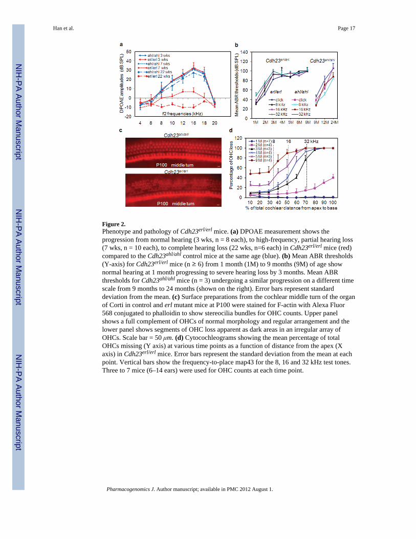
Figure 2.Phenotype and pathology of Cdh23erl/erl mice. (a) DPOAE measurement shows theprogression from normal hearing (3 wks, n = 8 each), to high-frequency, partial hearing loss(7 wks, n = 10 each), to complete hearing loss (22 wks, n=6 each) in Cdh23erl/erl mice (red)compared to the Cdh23ahl/ahl control mice at the same age (blue). (b) Mean ABR thresholds(Y-axis) for Cdh23erl/erl mice (n ≥ 6) from 1 month (1M) to 9 months (9M) of age shownormal hearing at 1 month progressing to severe hearing loss by 3 months. Mean ABRthresholds for Cdh23ahl/ahl mice (n = 3) undergoing a similar progression on a different timescale from 9 months to 24 months (shown on the right). Error bars represent standarddeviation from the mean. (c) Surface preparations from the cochlear middle turn of the organof Corti in control and erl mutant mice at P100 were stained for F-actin with Alexa Fluor568 conjugated to phalloidin to show stereocilia bundles for OHC counts. Upper panelshows a full complement of OHCs of normal morphology and regular arrangement and thelower panel shows segments of OHC loss apparent as dark areas in an irregular array ofOHCs. Scale bar = 50 μm. (d) Cytocochleograms showing the mean percentage of totalOHCs missing (Y axis) at various time points as a function of distance from the apex (Xaxis) in Cdh23erl/erl mice. Error bars represent the standard deviation from the mean at eachpoint. Vertical bars show the frequency-to-place map43 for the 8, 16 and 32 kHz test tones.Three to 7 mice (6–14 ears) were used for OHC counts at each time point.
Han et al. Page 17
Pharmacogenomics J. Author manuscript; available in PMC 2012 August 1.
NIH
-PA Author Manuscript
NIH
-PA Author Manuscript
NIH
-PA Author Manuscript

Figure 3.Increased apoptotic activity in erlong mice precedes hearing loss and hair cell loss. GAPDH-corrected mean mRNA expression levels of apoptosis-related genes caspase-3, −8, and −9in inner ear tissue from Cdh23erl/erl (n = 4 mice, 8 ears) were higher than those fromCdh23ahl/ahl ( n = 4 mice, 8 ears) mice at 2 weeks (a) and 2 months (b) of age. Caspase-8was expressed earlier and at a higher level than others. Caspase 3/7 activity measured bycleavage of the fluorescent caspase 3/7 substrate Z-DEVD-R110 and indicated as relativefluorescence units (RFLU) in the inner ear homogenates from mice at ages of 2 weeks (n = 9mice for each group) (c) and 2 months (n = 7 mice for each group) (d). Error bars representthe standard error from the mean. *P < 0.05, **P < 0.01.
Han et al. Page 18
Pharmacogenomics J. Author manuscript; available in PMC 2012 August 1.
NIH
-PA Author Manuscript
NIH
-PA Author Manuscript
NIH
-PA Author Manuscript

Figure 4.FITC(Green)-anti- caspase antibody staining. Sections of the middle turns of cochleae fromCdh23ahl/ahl (a, b, c) and Cdh23erl/erl ( d, e, f) mice at P14, stained with FITC-anti-activecaspase-8 (a, d) and caspase-9 (b, e). The signals for caspase-8 and caspase-9 staining inCdh23erl/erl mice were more intense in the outer hair cell (OHC) regions, and spiral ganglioncells (SG) than in the corresponding positions of the sections from Cdh23ahl/ahl mice. Therewere no detectable signals when primary antibody was omitted (c, f). StV, Stria vascularis;RM, Reissner’s membrane. Scale bars = 50μm.
Han et al. Page 19
Pharmacogenomics J. Author manuscript; available in PMC 2012 August 1.
NIH
-PA Author Manuscript
NIH
-PA Author Manuscript
NIH
-PA Author Manuscript

Figure 5.Caspase-3 detection in Cdh23erl/erl mice in situ in a time course observation. Representativesections from the mid-turn of cochleae of Cdh23erl/erl mice were stained for active caspase-3expression (FITC-anti-active-caspase-3, green) at P14 (a), P23 (b) and P57 (c). Caspase-3expression in situ is evident at P23 and P57. (d–f) Enlarged area from panel c to showindividual OHCs in the organ of Corti, with propidium iodide (PI) staining (red) revealingOHC nuclei and tissue ultrastructure in panel e. (f) Merged images of d and e. SL, spiralligament, OHC, outer hair cell. For a–c, scale bars = 50 μm. d–f, scale bars = 20 μm.
Han et al. Page 20
Pharmacogenomics J. Author manuscript; available in PMC 2012 August 1.
NIH
-PA Author Manuscript
NIH
-PA Author Manuscript
NIH
-PA Author Manuscript

Figure 6.ABR thresholds to evaluate hearing in Cdh23erl/erl mice with no treatment or after treatmentwith diluents of DMSO or DMSO+Z-VAD-FMK, over a period of 11 weeks. Treatmentswere initiated at mouse age P7 (1 wk). The number of mice tested was 14, 14, and 19 at 4wks; 10, 11 and 14 at 6 wks; 10, 7 and 9 at 8 wks; and 6, 4 and 6 at 12 wks, for no treatment,DMSO and DMSO+Z-VAD-FMK groups, respectively. ABR thresholds were measured ateach time point (indicating mouse age) on the X axis. (a–d) ABR thresholds were measuredat the frequencies indicated on the Y axis of each plot. Each point represents the mean ABRthreshold for a group, with error bars indicating standard error from the mean. The resultsshow that ABR thresholds were significantly lower in the DMSO+Z-VAD-FMK-treatedmice than those of the DMSO-treated or the untreated mice at stimulus frequencies of click,8 kHz, 16 kHz and 32 kHz at all time points ( *P < 0.05 at 4 wks; **P < 0.01, respectively,for 6 wks, 8 wks and 12 wks). There were also significant differences for the ABRthresholds between the untreated and DMSO-treated mice at 12 wks at the stimulusfrequency of 16 kHz (P < 0.05).
Han et al. Page 21
Pharmacogenomics J. Author manuscript; available in PMC 2012 August 1.
NIH
-PA Author Manuscript
NIH
-PA Author Manuscript
NIH
-PA Author Manuscript

Figure 7.A time course observation in the three treatment-based Cdh23erl/erl mouse groups ofDPOAEs to stimulus frequency of 16 kHz (f2) at 4 weeks (wks), 6 wks, 8 wks and 12 wks.DPOAE amplitudes (dB SPL) gradually decreased with time in the untreated group (n = 8,10, 10 and 8, respectively, at each time point), the DMSO-treated group (n = 11, 7, 7 and 6)and DMSO+Z-VAD-FMK-treated group (n = 14, 9, 9 and 6). The mean amplitude wassignificantly higher in the DMSO+Z-VAD-FMK-treated mice than in the untreated (P =0.03294) or DMSO-treated (P = 0.03955) group at the age of 8 wks. There were nosignificant differences for the DPOAE amplitudes between the untreated group and DMSO-treated group at any time points. * P < 0.05. Error bars indicate standard error from themean.
Han et al. Page 22
Pharmacogenomics J. Author manuscript; available in PMC 2012 August 1.
NIH
-PA Author Manuscript
NIH
-PA Author Manuscript
NIH
-PA Author Manuscript

Figure 8.Whole-mount phalloidin-stained preparations from the mid-turn of cochleae reveal normalappearance of OHC morphology and arrangement in Z-VAD-FMK-treated Cdh23erl/erl miceat P56. Left panel, untreated mice; middle panel, DMSO-diluent-treated mice; right panel,Z-VAD-FMK-treated mice. White arrows indicate areas of OHC loss, apparent in bothcontrols. Scale bars = 10 μm.
Han et al. Page 23
Pharmacogenomics J. Author manuscript; available in PMC 2012 August 1.
NIH
-PA Author Manuscript
NIH
-PA Author Manuscript
NIH
-PA Author Manuscript

Figure 9.Hair cell losses in the cochleae of the Z-VAD-FMK-treated Cdh23erl/erl mice at the age of 2months. Surface preparations of 4 cochleae from 4 mice in each mouse group wereexamined. The mean percentages of outer hair cell (OHC) loss in the apex, middle and basalregions of the cochleae of the Z-VAD-FMK-treated mice (black bars) were significantlylower than that of the untreated (white bars) and DMSO-treated (gray bars) mice. Therewere also significant differences in OHC loss between the untreated and DMSO-treatedmice at the basal regions (P < 0.05). No significant difference in inner hair cell loss occurredbetween the three mouse groups.* P < 0.05; ** P < 0.01. Error bars represent standard errorfrom the mean.
Han et al. Page 24
Pharmacogenomics J. Author manuscript; available in PMC 2012 August 1.
NIH
-PA Author Manuscript
NIH
-PA Author Manuscript
NIH
-PA Author Manuscript

Figure 10.Conceptual diagram of possible pathways of apoptosis involved in hair cell loss in theCdh23erl/erl mice and the sites of Z-VAD-FMK blockage (indicated by circled minus signs).The extrinsic caspase-8 and intrinsic caspase-9 initiators, and activator caspase-3 aresignificantly up-regulated by the Cdh23erl/erl mutation. TNF-a, caspase-12 and calpainsincluding m-calpain and μ-calpain were also up-regulated in this study. The interactionsbetween Cdh23 and Pcdh15 (Zheng et al 2005) or between Cdh23 and PMCA2 (Zheng et al2001) were demonstrated in our previous reports. Solid lines show pathways for which thereis direct evidence. Dash lines are proposed links.
Han et al. Page 25
Pharmacogenomics J. Author manuscript; available in PMC 2012 August 1.
NIH
-PA Author Manuscript
NIH
-PA Author Manuscript
NIH
-PA Author Manuscript

NIH
-PA Author Manuscript
NIH
-PA Author Manuscript
NIH
-PA Author Manuscript
Han et al. Page 26
Tabl
e 1
AB
R th
resh
olds
show
ing
a po
sitiv
e co
mpl
emen
tatio
n te
st th
at re
veal
ed a
llelis
m o
f the
erl
and
v-2
J lo
ci (a
bnor
mal
val
ues a
re in
bol
d)
Pare
ntal
mat
ing
IDA
ge (d
ays)
AB
R (d
B S
PL)
gait
geno
type
s
Clic
k8
kHz
16 k
Hz
32 k
Hz
ahl/v
2J x
erl
/erl
F1
126
5050
7080
norm
aler
l/v2J
(cod
omin
ant)
ahl/v
2J x
erl
/erl
F1
226
4540
6090
norm
aler
l/v2J
(cod
omin
ant)
ahl/v
2J x
erl
/erl
F1
326
4040
4070
norm
aler
l/v2J
(cod
omin
ant)
ahl/v
2J x
erl
/erl
F1
426
4050
4070
norm
aler
l/v2J
(cod
omin
ant)
ahl/v
2J x
erl
/erl
F1
526
4040
2040
norm
alah
l/erl
ahl/v
2J x
erl
/erl
F1
626
4040
2040
norm
alah
l/erl
ahl/v
2J x
erl
/erl
F1
726
4040
2040
norm
alah
l/erl
ahl/v
2J x
erl
/erl
F1
826
4040
2050
norm
alah
l/erl
C57
BL/
6J-v
2J/v
2J9
2610
010
010
010
0ci
rcle
rv2
J/v2
J
C57
BL/
6J-e
rl/e
rl10
2640
5020
50no
rmal
erl/e
rl
C57
BL/
6J-e
rl/e
rl11
4040
4070
90no
rmal
erl/e
rl
v2J/
v2J
X C
57B
L6J-
ahl/a
hl12
9045
4020
55no
rmal
ahl/v
2J
Pharmacogenomics J. Author manuscript; available in PMC 2012 August 1.
NIH
-PA Author Manuscript
NIH
-PA Author Manuscript
NIH
-PA Author Manuscript
Han et al. Page 27
Table 2
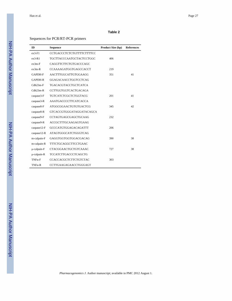
Sequences for PCR/RT-PCR primers
ID Sequence Product Size (bp) References
ex3-F1 CCTGACCCTCTCTGTTTTCTTTTCC
ex3-R1 TGCTTACCCAATGCTACTCCTGGC 406
ex3m-F CAGGTTCTTCTGTGACCCAGC
ex3m-R CCAAAAGATGGTGAGCCACCT 219
GAPDH-F AACTTTGGCATTGTGGAAGG 351 41
GAPDH-R GGAGACAACCTGGTCCTCAG
Cdh23m-F TGACACGTACCTGCTCATCA
Cdh23m-R CCTTGGTGGTCACTGACAGA
caspase3-F TGTCATCTCGCTCTGGTACG 201 41
caspase3-R AAATGACCCCTTCATCACCA
caspase8-F ATGGCGGAACTGTGTGACTCG 345 42
caspase8-R GTCACCGTGGGATAGGATACAGCA
caspase9-F CCTAGTGAGCGAGCTGCAAG 232
caspase9-R ACCGCTTTGCAAGAGTGAAG
caspase12-F GCCCATGTGGAGACAGATTT 206
caspase12-R ATAGTGGGCATCTGGGTCAG
m-calpain-F GAGGTGGTGGTGGACGACAG 390 38
m-calpain-R TTTCTGCAGGCTTCCTGAAC
μ-calpain-F CTACGGAACTGCTGTCAAAC 727 38
μ-calpain-R TCCATCTTGACCCTCAGCTG
TNFα-F CCACCACGCTCTTCTGTCTAC 303
TNFα-R CCTTGAAGAGAACCTGGGAGT
Pharmacogenomics J. Author manuscript; available in PMC 2012 August 1.