Early-onset Dementia with Lewy Bodies
11
Early-onset Dementia with Lewy Bodies—Takao et al 137 INTRODUCTION Mutations (A30P, E46K and A53T) in the α-Synuclein gene and triplication of the region of chromosome 4 that contains the α-Synuclein gene cause familial forms of Parkinson’s disease (PD) (33, 44, 47, 59). Moreover, α-synuclein is the major com- ponent of Lewy bodies and Lewy neurites (48,49), the defining neuropathological characteristics of PD and dementia with Lewy bodies (DLB). is has suggested that dysfunction of α-synuclein may play a cen- tral role in all cases of PD and DLB (20). An additional locus associated with Parkin- son disease (PARK-3) has been mapped to chromosome 2p13 (15,57), but the under- lying genetic defect is not yet known. Mutations in the Parkin (31) and DJ-1 (1) genes have been identified in cases of juvenile autosomal-recessive parkinsonism. Most cases with Parkin mutations appear to exhibit degeneration of dopaminergic nerve cells of the substantia nigra in the absence of Lewy bodies and Lewy neurites (24, 39, 50, 53). However, Lewy body pathology has been described in one individual with familial parkinsonism and a compound heterozygous mutation in Parkin (10). e neuropathological features associated with mutations in the DJ-1 gene are not yet known. It remains to be established whether the mutant forms of α-synuclein, parkin and DJ-1 cause disease by interact- ing in a common pathological pathway, or whether there are separate mechanisms by which they lead to disease. Mutations in the α-Synuclein gene result in a gain of toxic function, whereas most mutations in the Parkin and DJ-1 genes result in a loss of function. us far, PARK-5 has been found associated with only one mutation in the Ubiquitin carboxy-terminal hydrolase L1 (UCH-L1) gene. is was found in individuals from a small family who had a clinical diagnosis of PD, but no neuro- pathological corroboration (37). It remains to be firmly established that UCH-L1 is a gene for PD. DLB is the second most common form of degenerative dementia of old age, after Alzheimer disease (AD) (26). Neuropatho- logically, it is characterized by the presence of large numbers of Lewy bodies and Lewy neurites in the neocortex, limbic system and brainstem. A variable amount of AD-type pathology in the form of neuritic plaques and neurofibrillary lesions is also present in the majority of patients with DLB. Here we present the clinical, neuro- pathological and genetic characterization of a Japanese individual who developed rap- idly progressive parkinsonism and demen- tia at 13 years of age. Neuropathologically, numerous widespread α-synuclein-positive Lewy bodies and Lewy neurites were pres- ent throughout the cerebral cortex, subcor- tical nuclei and brainstem; there was no ev- idence of AD-type pathology. e severity and distribution of α-synuclein deposition exceeded that of typical DLB. MATERIALS AND METHODS Clinical examination of the proband. e proband developed neurological signs at 13 years of age. roughout the 2-year- long course of the disease, she underwent several neurological, neuro-ophthalmologi- cal, electrophysiological and neuroradio- logical examinations. Autopsy. An autopsy of the proband was carried out. Tissue samples of the heart, lung, liver, pancreas, spleen, esophagus, stomach, small and large intestines, kid- ney, thyroid, adrenal glands and ovaries were fixed with 10% formalin. Following formalin fixation the tissue samples were dehydrated in graded alcohols, cleared in RESEARCH ARTICLE Early-onset Dementia with Lewy Bodies Masaki Takao 1,2 ; Bernardino Ghetti 1 ; Hirotaka Yoshida 3 ; Pedro Piccardo 1 ; Yolanda Narain 4 ; Jill R. Murrell 1 ; Ruben Vidal 1 ; Bradley S. Glazier 1 ; Ross Jakes 3 ; Miho Tsutsui 4 ; Maria Grazia Spillantini 4 ; R. Anthony Crowther 3 ; Michel Goedert 3 ; Atsuo Koto 2,5 1 Department of Pathology and Laboratory Medicine, Indiana University School of Medicine, Indianapolis, Ind. 2 Department of Neurology, Keio University School of Medicine, Tokyo, Japan. 3 MRC Laboratory of Molecular Biology, Cambridge, United Kingdom. 4 Centre for Brain Repair and Department of Neurology, Cambridge University, United Kingdom. 5 Faculty of Nursing and Medical Care, Keio University, Tokyo, Japan. Corresponding author: Dr Bernardino Ghetti, Department of Pathology and Laboratory Medicine, Indiana University School of Medicine, 635 Barnhill Drive, B029D, Indianapolis, IN 46202 USA (E-mail: [email protected]) The clinical and neuropathological characteristics of an atypical form of dementia with Lewy bodies (DLB) are described. The proband experienced difficulties in her school performance at 13 years of age. Neurological examination revealed cognitive dysfunction, dysarthria, parkinsonism and myoclonus. By age 14 years, the symptoms had worsened markedly and the proband died at age 15 years. On neuropathological examination, the brain was severely atrophic. Numerous intracytoplasmic and intra- neuritic Lewy bodies, as well as Lewy neurites, were present throughout the cerebral cortex and subcortical nuclei; vacuolar changes were seen in the upper layers of the neocortex and severe neuronal loss and gliosis were evident in the cerebral cortex and substantia nigra. Lewy bodies and Lewy neurites were strongly immunoreactive for α-synuclein and ubiquitin. Lewy bodies were composed of filamentous and granular material and isolated filaments were decorated by α-synuclein antibodies. Immunohis- tochemistry for tau or β-amyloid yielded negative results. The etiology of this atypical form of DLB is unknown, since there was no family history and since sequencing of the exonic regions of α-Synuclein, β-Synuclein, Synphilin-1, Parkin, Ubiquitin C-terminal hydrolase L1 and Neurofilament-M failed to reveal a pathogenic mutation. This study provides further evidence of the clinical and pathological heterogeneity of DLB. Brain Pathol 2004;14:137-147.
-
Upload
independent -
Category
Documents
-
view
1 -
download
0
Transcript of Early-onset Dementia with Lewy Bodies

Early-onset Dementia with Lewy Bodies—Takao et al 137
INTRODUCTIONMutations (A30P, E46K and A53T) in
the α-Synuclein gene and triplication of the region of chromosome 4 that contains the α-Synuclein gene cause familial forms of Parkinson’s disease (PD) (33, 44, 47, 59). Moreover, α-synuclein is the major com-ponent of Lewy bodies and Lewy neurites (48,49), the defining neuropathological characteristics of PD and dementia with Lewy bodies (DLB). is has suggested that dysfunction of α-synuclein may play a cen-tral role in all cases of PD and DLB (20). An additional locus associated with Parkin-son disease (PARK-3) has been mapped to chromosome 2p13 (15,57), but the under-lying genetic defect is not yet known.
Mutations in the Parkin (31) and DJ-1 (1) genes have been identified in cases of juvenile autosomal-recessive parkinsonism. Most cases with Parkin mutations appear to exhibit degeneration of dopaminergic nerve cells of the substantia nigra in the absence of Lewy bodies and Lewy neurites (24, 39,
50, 53). However, Lewy body pathology has been described in one individual with familial parkinsonism and a compound heterozygous mutation in Parkin (10). e neuropathological features associated with mutations in the DJ-1 gene are not yet known. It remains to be established whether the mutant forms of α-synuclein, parkin and DJ-1 cause disease by interact-ing in a common pathological pathway, or whether there are separate mechanisms by which they lead to disease. Mutations in the α-Synuclein gene result in a gain of toxic function, whereas most mutations in the Parkin and DJ-1 genes result in a loss of function. us far, PARK-5 has been found associated with only one mutation in the Ubiquitin carboxy-terminal hydrolase L1 (UCH-L1) gene. is was found in individuals from a small family who had a clinical diagnosis of PD, but no neuro-pathological corroboration (37). It remains to be firmly established that UCH-L1 is a gene for PD.
DLB is the second most common form of degenerative dementia of old age, after Alzheimer disease (AD) (26). Neuropatho-logically, it is characterized by the presence of large numbers of Lewy bodies and Lewy neurites in the neocortex, limbic system and brainstem. A variable amount of AD-type pathology in the form of neuritic plaques and neurofibrillary lesions is also present in the majority of patients with DLB.
Here we present the clinical, neuro-pathological and genetic characterization of a Japanese individual who developed rap-idly progressive parkinsonism and demen-tia at 13 years of age. Neuropathologically, numerous widespread α-synuclein-positive Lewy bodies and Lewy neurites were pres-ent throughout the cerebral cortex, subcor-tical nuclei and brainstem; there was no ev-idence of AD-type pathology. e severity and distribution of α-synuclein deposition exceeded that of typical DLB.
MATERIALS AND METHODS
Clinical examination of the proband. e proband developed neurological signs at 13 years of age. roughout the 2-year-long course of the disease, she underwent several neurological, neuro-ophthalmologi-cal, electrophysiological and neuroradio-logical examinations.
Autopsy. An autopsy of the proband was carried out. Tissue samples of the heart, lung, liver, pancreas, spleen, esophagus, stomach, small and large intestines, kid-ney, thyroid, adrenal glands and ovaries were fixed with 10% formalin. Following formalin fixation the tissue samples were dehydrated in graded alcohols, cleared in
RESEARCH ARTICLE
Early-onset Dementia with Lewy Bodies
Masaki Takao1,2; Bernardino Ghetti1; Hirotaka Yoshida3; Pedro Piccardo1; Yolanda Narain4; Jill R. Murrell1; Ruben Vidal1; Bradley S. Glazier1; Ross Jakes3; Miho Tsutsui4; Maria Grazia Spillantini4; R. Anthony Crowther3; Michel Goedert3; Atsuo Koto2,5
1 Department of Pathology and Laboratory Medicine, Indiana University School of Medicine, Indianapolis, Ind.2 Department of Neurology, Keio University School of Medicine, Tokyo, Japan.3 MRC Laboratory of Molecular Biology, Cambridge, United Kingdom.4 Centre for Brain Repair and Department of Neurology, Cambridge University, United Kingdom.5 Faculty of Nursing and Medical Care, Keio University, Tokyo, Japan.
Corresponding author: Dr Bernardino Ghetti, Department of Pathology and Laboratory Medicine, Indiana University School of Medicine, 635 Barnhill Drive, B029D, Indianapolis, IN 46202 USA (E-mail: [email protected])
The clinical and neuropathological characteristics of an atypical form of dementia with Lewy bodies (DLB) are described. The proband experienced difficulties in her school performance at 13 years of age. Neurological examination revealed cognitive dysfunction, dysarthria, parkinsonism and myoclonus. By age 14 years, the symptoms had worsened markedly and the proband died at age 15 years. On neuropathological examination, the brain was severely atrophic. Numerous intracytoplasmic and intra-neuritic Lewy bodies, as well as Lewy neurites, were present throughout the cerebral cortex and subcortical nuclei; vacuolar changes were seen in the upper layers of the neocortex and severe neuronal loss and gliosis were evident in the cerebral cortex and substantia nigra. Lewy bodies and Lewy neurites were strongly immunoreactive for α-synuclein and ubiquitin. Lewy bodies were composed of filamentous and granular material and isolated filaments were decorated by α-synuclein antibodies. Immunohis-tochemistry for tau or β-amyloid yielded negative results. The etiology of this atypical form of DLB is unknown, since there was no family history and since sequencing of the exonic regions of α-Synuclein, β-Synuclein, Synphilin-1, Parkin, Ubiquitin C-terminal hydrolase L1 and Neurofilament-M failed to reveal a pathogenic mutation. This study provides further evidence of the clinical and pathological heterogeneity of DLB.
Brain Pathol 2004;14:137-147.

138 Early-onset Dementia with Lewy Bodies—Takao et al Early-onset Dementia with Lewy Bodies—Takao et al 139
xylene and embedded in paraffin. Eight-µm-thick sections were cut and stained with hematoxylin and eosin or processed for α-synuclein immunohistochemistry.
Macroscopic neuropathology. From the brain of the proband, 50 g of tissue from the superior frontal gyrus and cingulate gy-rus were dissected and stored at -70°C. e remainder of the brain and the entire spinal cord were fixed in 10% formalin.
Histology and immunohistochemistry. Samples from the superior frontal gyrus, cingulate gyrus, superior temporal gyrus, precentral gyrus, postcentral gyrus, calca-rine cortex, hippocampus, caudate nucleus, putamen, claustrum, globus pallidus, thala-mus, amygdala, hypothalamus, cerebellar cortex, dentate nucleus, midbrain, pons, medulla and spinal cord were dehydrated in a graded alcohol series, cleared in xylene and embedded in paraffin. Eight-µm-thick
sections were stained with hematoxylin-eosin, the Heidenhain-Woelcke method for myelin, the Bodian silver staining for axons, the Perls method for ferric iron and thioflavin S.
Immunohistochemical analysis of archi-val and newly generated paraffin-embed-ded tissue blocks was carried out using an antigen retrieval method, to maximize the labeling (37). Antibodies specific for α-synuclein, β-synuclein, γ-synuclein, parkin, DJ-1, synphilin-1, ubiquitin, tau, β-amyloid, prion protein, neuroserpin, neurofilaments and glial fibrillary acidic protein (GFAP) were used (Table 1). e anti-DJ-1 serum was raised in a rabbit by using a synthetic peptide corresponding to amino acids 174-189 of human DJ-1 as the immunogen. By Western blot analysis, the antibody strongly labeled recombinant GST-DJ-1 at 1:1000 dilution (not shown). e signal from polyclonal antibodies was visualized using avidin-biotin, with
goat anti-rabbit immunoglobulin as the secondary antibody, followed by horse-radish peroxidase-conjugated streptavidin and the chromogens diaminobenzidine or tetramethylbenzidine. e signal from monoclonal antibodies was detected using avidin-biotin, with goat anti-mouse immu-noglobulin as the secondary antibody, fol-lowed by alkaline phosphatase-conjugated streptavidin. Sections were counter-stained with hemotoxylin.
Semi-quantitative analysis. e fre-quency and distribution of intracytoplas-mic and intraneuritic Lewy bodies were assessed semi-quantitatively. A subjective scale was used to represent the numbers of Lewy bodies in a field observed through a 10× objective (0 = none; 1 = 1-4; 2 = 5-10; 3 = >10). In addition, we evaluated the severity of Lewy neurite pathology in each area and the degree of neuronal loss and gliosis.
Electron microscopy on tissue sections. Frontal cortex, substantia nigra and locus coeruleus of the proband were fixed in 4% formaldehyde and post-fixed with 1% osmium tetroxide, dehydrated in a graded alcohol series, cleared in propylene oxide and embedded in Epon. One-µm-thick sections were stained with toluidine blue. Ultra-thin sections were contrasted with uranyl acetate and lead citrate and scanned by electron microscopy.
Extraction and characterization of iso-lated α-synuclein filaments. α-Synuclein filaments were extracted from the superior frontal gyrus of the proband, as described previously for cases of DLB (49). Aliquots of the dispersed filament preparations were placed on carbon-coated 400-mesh grids and stained with 1% lithium phospho-tungstate. Micrographs were recorded at a nominal magnification of ×40 000 on a Philips model EM208S microscope. Proce-dures for immunoelectron microscopy were as previously described (5). e following 7 antibodies were used at 1:100 dilution: PER4, which recognizes residues 121-140 of α-synuclein (49); PER1, which recog-nizes residues 11-34 of α-synuclein (29); SYN36, which recognizes the carboxy-ter-minus of α-synuclein (38); SYN303, which recognizes residues 2-12 of α-synuclein (9); SYNP129, which recognizes α-synuclein
Antibody Type Dilution Source and Reference
Anti-α-synuclein
PER1 (α-synuclein 11-34
) r 1:200 M. Goedert (30)
PER4 (carboxy-terminal) r 1:1000 M. Goedert (49)
PER7 (α-synuclein 1-120
) r 1:1000 M. Goedert (30)
SYNP129 r 1:200 T. Iwatsubo (14)
α-synuclein 119-137
r 1: 300 B. Ghetti (43)
Anti β-synuclein
PER3 r 1:200 M. Goedert (29)
Anti γ-synuclein
PER5 r 1:500 M. Goedert (49)
Anti-parkin m 1:200 Chemicon
Anti-DJ-1 r 1:500 M. Goedert (unpublished)
Anti-synphilin-1 r 1:400 Chemicon
Anti-ubiquitin r 1:100 Dako Corporation
Anti-neurofilament
SMI-31 (p-NF200kDa) m 1:2000 Sternberger Monoclonals
SMI-32 (non p-NF200kDa) m 1:2000 Sternberger Monoclonals
AB1981 (NF150kDa) r 1:200 Chemicon
AB1983 (NF68kDa) r 1:200 Chemicon
Anti-tau
AT8(p-tau Ser202/Thr205
) m 1:400 Polymedco, Inc.
Alz50 m 1:20 P. Davies (3)
Anti-β-amyloid 10D5 m 1:100 Elan Pharmaceuticals
Anti-prion protein 3F4 m 1:200 Signet
Anti-neuroserpin r 1:2000 R. Davis (7, 51)
Anti-GFAP r 1:100 Dako Corporation
Table 1. Antibodies used for immunohistochemistry. Abbreviations: p-NF = phosphorylated neurofilament, non P-NF = non-phosphorylated neurofilament, p-tau = phosphorylated tau, GFAP = glial fibrillary acidic protein, m = mouse monoclonal, r = rabbit polyclonal.

138 Early-onset Dementia with Lewy Bodies—Takao et al Early-onset Dementia with Lewy Bodies—Takao et al 139
phosphorylated at serine residue 129 (14); anti-tau antibody BR134 (19) and anti-ubiquitin antibody 1310 (Chemicon).
Sequential extraction of α-synuclein. One hundred mg of tissue of the superior frontal gyrus from the proband and from a 45-year-old control individual were extracted using a modification of the pro-cedure described in (14). e tissues were homogenized in 400-µl buffer A (50 mM Tris-HCl, pH 7.4, 1 mM EDTA, 1 mM DTT, containing a cocktail of protease inhibitors [Complete™, Roche]). A 20-minute centrifugation at 543 000g gave supernatant TBS-sol and pellet. e pellet was homogenized in 400-µl buffer B (buf-fer A with 10% sucrose and 0.5 M NaCl) containing 1% Triton X-100, incubated for 30 minutes at 37°C, followed by a 20-min-ute centrifugation at 543 000g, giving su-pernatant TX-sol and pellet. e pellet was homogenized in 400-µl buffer B containing 1% sarkosyl, incubated for 30 minutes at 37°C, followed by a 20-minute centrifuga-tion at 543 000g, giving supernatant Sark-sol and pellet. e pellet was extracted in 8M urea in buffer A and sonicated (Urea-extr). For immunoblot analysis, 5 µl of each extract was run on SDS-PAGE and labeled with anti-α-synuclein antibody SYN208, which recognizes human α-synuclein, but not β- or γ-synuclein (16). Enhanced che-miluminescence (ECL, Amersham Phar-macia) was used to detect the signal.
Genetic analysis. Genomic DNA was extracted from frozen brain tissue of the proband and blood of 50 controls from the Japanese population using the QIAamp DNA Mini Kit (QIAGEN). e DNA was analyzed by direct sequencing of exons 1-6 of the α-Synuclein gene (GenBank: AH005859), exons 1-6 of the β-Synuclein gene (GenBank: AF053134), exons 1-10 of the Synphilin-1 gene (GenBank: AH009850), exons 1-12 of the Parkin gene (GenBank: XM_051780) and exons 1-3 of the Neurofilament medium polypeptide gene (NF-M) (GenBank: XM_005158). Am-plification of exons 1-6 of the α-Synuclein gene made use of the following primers: Exon 1: 5´-GAGAA GGAGG AGGAC TAGGA GG-3´ and 5´-CGGTG TTCTC CAGGA TTTC-3´. Exon 2: 5´-CACTT TGGAG GGTTT CTCAT G-3´ and 5´-ATACC TCTGA CTCAG TCCAC
ICLB INLB LN Neuronal Loss/Gliosis
Cerebrum
Frontal cortex 3 3 severe severe/severe
Cingulate cortex 3 3 severe severe/severe
Temporal cortex 3 3 severe severe/severe
Insular cortex 3 3 severe severe/severe
Parietal cortex 3 3 severe severe/severe
Motor cortex 3 3 severe moderate/severe
Sensory cortex 3 3 severe moderate/severe
Insular cortex 3 3 severe severe/severe
Occipital cortex 2 2 severe moderate/severe
Amygdala 3 3 severe severe/severe
Hippocampus 2 3 severe moderate/severe
Dentate gyrus 1 3 mild mild/mild
Subiculum 2 3 severe moderate/severe
Entorhinal cortex 3 3 severe severe/severe
Caudate nucleus 3 3 severe severe/severe
Putamen 2 2 moderate moderate/moderate
Claustrum 2 2 severe moderate/moderate
Globus pallidus 0 0 mild mild/mild
Thalamus 1 1 mild moderate/moderate
Hypothalamus 3 2 severe mild/mild
Substantia innominata 2 1 moderate moderate/moderate
Suprachiasmatic nucleus 2 2 severe moderate/mild
Cerebellum
Cortex 0 0 none mild/moderate
Dentate nucleus 0 0 none moderate/moderate
Midbrain
Periaqueductal gray 3 1 severe moderate/moderate
Oculomotor nerve nuclei 3 1 severe mild/mild
Substantia nigra 3 3 severe severe/severe
Pons
Locus coeruleus 3 2 severe severe/severe
Raphe nucleus 3 2 severe mild/mild
Basis pontis 1 0 mild moderate/severe
Medulla
Hypoglossal nerve nucleus 0 1 mild mild/mild
Dorsal motor nucleus of vagus 3 1 severe moderate/moderate
Vestibular nucleus 1 0 moderate mild/mild
Lateral reticular nucleus 3 1 moderate mild/mild
Inferior olivary nucleus 0 0 mild moderate/moderate
Accessory olivary nucleus 0 0 mild mild/mild
Gracile and cuneate nuclei 0 0 mild mild/mild
Spinal cord
Anterior gray horn 0 0 mild mild/mild
Intermediolateral nucleus 1 0 mild mild/mild
Dorsal nucleus of Clarke 1 0 mild mild/mild
Table 2. Lewy bodies, Lewy neurites, neuronal loss and gliosis.Abbreviations. ICLB = Intracytoplasmic Lewy body, INLB = intraneuritic Lewy body, LN = Lewy neurite. Semi quantitative scale 0 = none, 1 = 1-4, 2 = 5-10, 3 = >10. Number of lesions in a field observed through a 10x objective.

140 Early-onset Dementia with Lewy Bodies—Takao et al Early-onset Dementia with Lewy Bodies—Takao et al 141
C-3´. Exon 3: 5´-GTGGT GGTTA GGAGT TCCTT C-3´ and 5´-GTTCT TAGAT GCTCA GTGAT TG-3´, Exon 4: 5´-ATGGC TAGTG GAAGT GGAAT G-3´ and 5´-CACAG TAAGT ATCTT GCTCC TG-3´. Exon 5: 5´-GTGGC CAACA TCCCT ATATG-3´ and 5´-GAGAA ATGTG ACAAT GACAG G-3´. Exon 6: 5´-TTTTA GTGTA AGTGG GGAGC CATTT C-3´ and 5´-CATTG GAACT GAGCA CTTGT AC-3´.
Amplification of the exons encoding the other genes examined made use of previ-ously described primers (11,31,34,35,56). PCR was carried out with 20 ng/µl ge-nomic DNA as the template using the high fidelity expanded PCR system (Roche Molecular Biochemicals). PCR conditions were as follows: denaturation, 94°C, one minute; annealing, 56°C, one minute; extension, 72°C, one minute, for a total of 35 cycles. For the amplification of exon 3 of the UCH-L1 gene (GenBank: XM-051780) the GC-rich PCR system (Roche Molecular Biochemicals) was used (96°C,
30 seconds; 55°C, 30 seconds; 72°C, 30 seconds, for a total of 40 cycles).
Total RNA was extracted from frozen brain tissue using the ToTALLY RNA kit (Ambion Inc.). Complementary DNA was produced by reverse transcription of RNA using random primers and the ProSTAR kit (Stratagene). For the amplification of α-synuclein cDNA, PCR was carried out us-ing primers 5´-GTGTA AAGGA ATTCA TTAGC-3´ and 5´-ATCTC AAGAA ACTGG GAG-3´ (42). For the amplifi-cation of parkin cDNA, the first round PCR used primers 5´-GCCGC CACCTA CCCAG TGACC AT-3´ and 5´-AGCAC CACTC GAGCC TGCAC T-3´, corre-sponding to nucleotides 81-103 and 1440-1420 of the published sequence (GenBank: AB009973). Two sets of primers were used for the second round PCR: 5´-CCACC TACCC AGTGA CCATG ATA-3´ and 5´-CCACA CAAGG CAGGG AGTAG CCAA-3´, corresponding to nucleotides 85-109 and 972-949, and 5´-AACAA ATAGT CGGAA CATCA CTTGC A-
3´ and 5´-GCGGA CACTT CATGT GCATG CA-3´, corresponding to nucleo-tides 791-816 and 1413-1392. Both sets of primers amplified a DNA sequence con-taining codon 272. PCR was done at 94°C for one minute, 60°C for one minute and 72°C for 2 minutes for a total of 40 cycles.
Amplified products were gel-purified using the QIAquick gel extraction kit (Qia-gen). Sequence analysis was carried out using the quick start CEQ dye terminator cycle sequencing kit on a CEQ 2000XL DNA sequencer (Beckman Coulter).
RESULTS
Clinical findings. Clinical history. e proband, a Japanese female, began to expe-rience difficulties at school at age 13 years, as manifested by a lack of concentration and problems with dressing herself and handwriting. During the same year, she developed dysarthria, monotonous speech and tremor in her upper and lower extremi-ties. A neurological examination docu-mented a cognitive impairment, a stooped posture, bradykinesia, tremor and rigidity in all extremities, as well as micrographia, myoclonus and ataxia. Pharmacological treatment with L-dopa and amantadine failed to improve the neurological symp-toms. e proband was hospitalized at 14 years of age because of aspiration pneu-monia. At that time, she only responded to sensory stimuli and exhibited decor-ticate rigidity and frequent myoclonus. e tendon reflexes were normal and no pathological reflexes were noted. A neuro-ophthalmological examination failed to show conjunctival teleangiectasia, corneal or lens opacity, a cherry-red spot, macu-lar or pigmentary degeneration, or optic atrophy. Routine blood cell counts, blood chemistry and urine analysis were normal, except for an increase in the level of creatine kinase. e level of mucopolysaccharides in the urine was normal. Vacuolated lympho-cytes were not observed. Serum levels of amino acids, iron, copper, ceruloplasmin and β-lipoprotein were normal. Lysosomal enzymes involved in storage diseases, such as GM1 and GM2 gangliosidoses, Gaucher disease, α-mannosidosis and mucopolysa-charidoses IIID and IVB were normal. e titers of antibodies directed against measles virus, rubella virus, cytomegalovirus, herpes simplex virus, Japanese encephalitis virus,
Figure 1. Proband’s brain. A. Computed tomography scans taken 6 months apart show progressive cerebral atrophy and a marked enlargement of the lateral ventricles. B, C. The brain shows cortical atrophy and a widening of the sulci. D, E, F. Axial sections of cerebrum (D), midbrain (E) and pons (F). Severe cortical atrophy is present in the cerebrum; the lateral ventricles are enlarged. The substantia nigra and locus coeruleus show mild depigmentation.

140 Early-onset Dementia with Lewy Bodies—Takao et al Early-onset Dementia with Lewy Bodies—Takao et al 141
poliovirus, cocksackievirus, echovirus and parainfluenza virus were normal. Exami-nation of the cerebrospinal fluid for cells, proteins, glucose and antibodies against measles virus was also unremarkable. e patient died of bronchopneumonia at 15 years of age.
Family history. Based on clinical records, there was no neurological disease in any of the proband’s relatives. ere was no evidence suggesting consanguineous mar-riage in any relative. e karyotype of the proband was not examined.
Electrophysiology. An electroencephalo-gram (EEG) was carried out at 14 years of age and showed diffuse slow waves. Nerve conduction studies and a needle electromy-ography were carried out at 14 years of age and revealed no abnormalities.
Neuroradiology. Brain computed tomo-graphic (CT) scans were carried out when the proband was 13- and 14-years-old. No abnormalities were reported at 13 years, but a progressive diffuse brain atrophy was observed on repeated CT scans at 14 years (Figure 1A).
Biopsy. At 14 years of age, the proband underwent biopsies of quadriceps femoris muscle, sural nerve and rectum. No Lafora bodies or deposits characteristic of storage diseases or metabolic disorders were pres-ent. At the time of this study, biopsy tissue was not available for reexamination.
Autopsy. Analysis of multiple sections of the lung revealed the presence of bron-chopneumonia. No other pathologic al-terations were found in extraneural tissues. α-Synuclein immunohistochemistry of sections obtained from the gastrointestinal tract yielded negative results.
Macroscopic neuropathology. e pro-band’s brain weighed 780 g. A severe gen-eralized atrophy and an enlargement of the sulci were present (Figure 1B, C). On hori-zontal slices, the cortical grey matter was much thinner than normal and the lateral ventricles were enlarged (Figure 1D). e head of the caudate nucleus was moderately atrophic. e substantia nigra and the locus coeruleus were pigmented (Figure 1E, F). e degree of pigmentation was less than that expected in an adult. It was not pos-sible to determine how the degree of pig-mentation would compare to age, gender and race-matched normal controls.
Microscopic neuropathology. ree types of α-synuclein deposits were widespread throughout the cerebral cortex and the brainstem: intraperikaryal Lewy bod-ies, intraneuritic Lewy bodies and Lewy neurites (Figure 2; Table 2). In nerve cell perikarya, Lewy bodies had an eosinophilic and hyaline appearance with or without a dense core. Most were round, but some had a highly irregular shape. Intraneuritic Lewy bodies were eosinophilic, hyaline and mildly argentophilic. Some α-synuclein deposits extended from nerve cell bodies into processes. Most Lewy neurites were argentophilic; they had either a thread-like or torpedo-like shape. Lewy bodies and Lewy neurites were strongly labeled by anti-α-synuclein antibodies PER1, PER4, PER7, SYNP129 and α-synuclein 119-137 (Figures 2-6). Most α-synuclein-positive
structures were also ubiquitin-immunore-active (Figure 6D). Some Lewy bodies and Lewy neurites were labeled by antibodies directed against NF-M (Figure 4C, D). Occasional deposits were weakly labeled by an anti-parkin antibody. Lewy bodies and Lewy neurites were not labeled by antibod-ies directed against DJ-1, β-synuclein, γ-synuclein, synphilin-1, the low- and high-molecular weight neurofilament subunits, tau, β-amyloid, prion protein, neuroserpin or GFAP (data not shown).
e neocortex showed severe neuronal cell loss and gliosis, with extensive vacuolar changes in the upper layers of the frontal, cingulate and temporal cortices (Figure 3). Intraperikaryal Lewy bodies were fre-quently observed in small to medium-sized neurons of the deeper cortical layers, with smaller numbers in the upper layers. How-
Figure 2. Sections of the substantia nigra (A, B, F, G), locus coeruleus (C), temporal cortex (D, E) and frontal cortex (H). A. Neuronal cell loss and gliosis are evident. B, C. Brainstem Lewy bodies. D, E. Cortical Lewy bodies. F, G, H. Lewy bodies and Lewy neurites. A-E. Hematoxylin-eosin; F-H, Immunohistochemistry using antibodies SYNP129 (F, G) and α-synuclein (119-137) (H). Scale bars: A, F = 100 µm; B-E, G-H = 10µm.

142 Early-onset Dementia with Lewy Bodies—Takao et al Early-onset Dementia with Lewy Bodies—Takao et al 143
ever, a clear laminar distribution was not observed. Intraneuritic Lewy bodies were most numerous in cortical layers II-IV. Lewy neurites were present throughout all cortical layers. Lewy bodies were hematoxy-lin-eosin and silver positive. Most cortical Lewy bodies lacked a dense core.
Neuronal cell loss and gliosis were moderate in hippocampal pyramidal cells, subiculum and entorhinal cortex. By con-trast, the transentorhinal cortex and the inferior temporal cortex showed severe neuronal loss, gliosis and vacuolar changes. Numerous Lewy bodies and Lewy neurites were present in the hippocampus and su-biculum (Figure 4). ey were particularly abundant in the stratum oriens, stratum pyramidale and stratum radiatum. Lewy bodies and Lewy neurites were also present in the dentate gyrus of the hippocampus. In the subcortical white matter, scattered α-synuclein-immunoreactive-glial-cell inclusions were present, akin to coiled bod-ies. A mild loss of myelinated nerve fibers
and a moderate to severe gliosis were also present.
Lewy bodies and Lewy neurites were abundant in the caudate nucleus, putamen, claustrum and thalamus (Figure 5). ey were particularly numerous in the caudate nucleus, where neuronal cell loss and glio-sis were also very severe. Lewy bodies and Lewy neurites were present in hypothalam-ic nuclei and the substantia innominata, but not the globus pallidus. e substantia nigra contained abundant Lewy bodies and Lewy neurites. Nerve cell loss and gliosis were severe and neuromelanin was found extracellularly and/or within macrophages. (Figure 2A, B). Only a few pigmented neurons remained in the medial part of the substantia nigra. Numerous Lewy bod-ies and Lewy neurites were present in the oculomotor nerve nucleus, a region where nerve cell loss was only minimal. e peri-
aqueductal grey matter was also affected, but not the red nucleus.
e locus coeruleus contained numerous Lewy bodies and Lewy neurites in spite of the severe nerve cell loss (Figures 2C,5D). e raphe nucleus also contained many Lewy bodies and Lewy neurites; however, there was relatively little nerve cell loss (Fig-ure 5F). e basis pontis showed moder-ate nerve cell loss and gliosis (Figure 5E). Lewy bodies were only occasionally seen, but Lewy neurites were numerous in the pontine nuclei. Moderate nerve cell loss and gliosis in the dorsal motor nucleus of the vagus nerve and in the inferior olivary nucleus were noted. Varying numbers of Lewy bodies and Lewy neurites were seen in the dorsal motor nucleus of the vagus, in the lateral reticular nucleus and inferior olivary nucleus (Figure 5G, H). In other anatomical areas, Lewy bodies and Lewy neurites were also present with various
Figure 3. Sections of the frontal cortex. A. Layers I-VI, showing numerous α-synuclein-immunoreactive Lewy bodies and Lewy neurites. B. Silver-positive inclusions, with numerous segmental swellings in dendrites; the inset shows argentophilic inclusions in the soma and dendrite belonging to the same neuron. C. α-Synuclein-immunoreactive segmental dendritic swelling. A, C. Immunohistochemistry using anti-α-synuclein (119-137) antibody; B. Bodian silver stain. Scale bars: A, B =100 µm; C = 10 µm.
Figure 4. Sections of the insular cortex (A), visual cortex (B), CA1 layer of the hippocampus (C, D), parahippocampus (E) and granule cell layer of the hippocampus (F). A, B. Numerous α-synuclein-positive Lewy bodies and Lewy neurites. C, D. Neurites are double-labeled for α-synuclein (pink) and neurofilaments (brown). E. Numerous α-synuclein-positive neurites. F. α-Synuclein deposits in granule cell bodies. A, B, E, F. Immunohistochemistry using anti-α-synuclein (119-137) antibody. C, D. Double-labeling immunohistochemistry using the polyclonal anti-α-synuclein (119-137) antibody and polyclonal antibodies specific for NF-M. Scale bars: A, B = 100 µm; C-D, F = 10 µm; E = 50 µm.

142 Early-onset Dementia with Lewy Bodies—Takao et al Early-onset Dementia with Lewy Bodies—Takao et al 143
degrees of abundance, as summarized in Table 2. ey were scarce in the spinal cord (Figure 5I).
Neurofibrillary lesions and neuritic plaques were not present in any of the sections sampled. Moreover, there were no tau-, β-amyloid- or prion protein-immu-noreactive deposits. A few Hirano bodies were present in the hippocampus. Granu-lovacuolar degeneration was not seen. Iron accumulation and spheroids were absent from substantia nigra and basal ganglia.
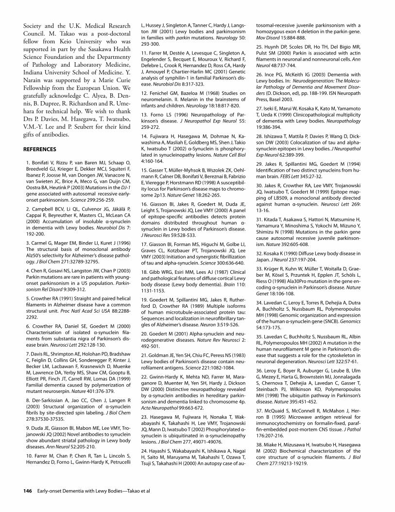
Electron microscopy. In the cerebral cor-tex, most intraperikaryal and intraneuritic Lewy bodies lacked the dense core that is characteristic of brainstem Lewy bodies (Figure 7). ey were composed of dense granular material and randomly arranged filaments with a diameter of 7 to 20 nm. Occasionally, cellular organelles were pres-ent among the filaments. When a core was present, its electron density was variable. Filaments of the core were more densely packed than those of the peripheral zone, which were loosely arranged in a radial pattern.
Characterization of isolated filaments. Filaments were extracted from frontal cortex tissue using a method that enriches for sarkosyl-insoluble filaments. e resus-pended pellets were tested with α-synucle-in, tau and ubiquitin antibodies (Figure 8). e labeled structures corresponded mostly to single filaments, though occasionally small clumps of filaments were seen. All fil-aments were straight and unbranched with a length of 200 to 600 nm. e width was 5 to 10 nm, with some filaments showing a variation in width along their length. e filaments were labeled all along their length by antibodies PER4, SYN36, SYN303 and SYNP129, with SYN36 labeling fewer filaments than the other antibodies. A quite different pattern was observed with antibody PER1, which only ever labeled one end of each filament. Occasional fila-ments were also labeled at only one end by antibody SYN36. No specific labeling was observed with anti-tau and only occasional weak labeling with anti-ubiquitin antibod-ies.
Sequential extraction of α-synuclein. α-Synuclein was sequentially extracted from frontal cortex tissue of a control individual
(c) and the proband (p) using TBS, 1% Triton X-100, 1% sarkosyl and 8M urea (Figure 9). A protein with an apparent molecular mass of 17 kDa was detected with anti-α-synuclein antibody SYN208 in the TBS, Triton X-100 and sarkosyl-sol-uble fractions from both brains. is band, which was much stronger in the TBS- and Triton X-100-soluble fractions than in the sarkosyl-soluble fraction, corresponds to normal α-synuclein. When the sarkosyl-insoluble material was treated with urea, a major, broad band centered at 17 kDa with a smear to lower molecular weight, a weak
band at 22 to 24 kDa and a high-molecular weight smear were visible in the extract from the proband’s brain, but not in the extract from the control brain. is mate-rial corresponds to abnormal aggregates of α-synuclein.
Genetic analysis. No mutations or poly-morphisms were found upon sequencing of exons 1-6 of the proband’s α-Synuclein gene. A silent nucleotide change (CTT to CTC) was present on one allele at codon 38 in exon 2 of β-Synuclein. Sequencing of ex-ons 1-12 of Parkin showed a heterozygous
Figure 5. Sections of the caudate nucleus (A), putamen (B), suprachiasmatic nucleus (C), locus coeruleus (D), basis pontis (E), raphe nucleus (F), dorsal motor nucleus of the vagus (G), inferior olivary nucleus (H) and thoracic spinal cord (I). Numerous α-synuclein-immunoreactive deposits are seen. A-I. Immunohistochemistry using anti-α-synuclein (119-137) antibody. Scale bars: A-I = 50 µm.
144 Early-onset Dementia with Lewy Bodies—Takao et al Early-onset Dementia with Lewy Bodies—Takao et al 145
nucleotide change (CTC to ATC) at codon 272 in exon 7, resulting in a Leu to Ile substitution. is substitution is probably a polymorphism, since it was also present in one out of 50 controls from the Japa-nese population (a 67-year-old male, with no family history of neurological disease). No additional changes were found when cDNAs encoding parkin were sequenced, ruling out deletion mutations on the other allele. e sequence of exons 1-10 of the
proband’s Synphilin-1 gene was normal, except for 2 heterozygous, silent nucleotide changes at codons 144 (GAC to GAT) and 244 (AAA to AAG) in exon 3. Sequencing of the exons encoding NF-M showed a homozygous nucleotide change (GCT to GTT) at codon 482, resulting in an alanine to valine change, which has previously been reported as a polymorphism (NCBI SNP CLUSTER ID: rs1063314). No nucleotide changes were detected in the exons encod-ing the proband’s UCH-L1 gene.
DISCUSSIONWe describe a case of DLB with progres-
sive dementia, parkinsonism and myoclo-nus, and an onset of clinical signs at 13 years of age. e disease progressed rapidly until the patient died at 15 years of age, which is an earlier age of death than pre-viously reported in cases of DLB (18, 27, 32, 58). In addition to the severe cortical atrophy, a finding of interest was that the pigment observed macroscopically in the substantia nigra was more than expected based on the severe loss of neurons observed microscopically. In a study of pigmentation of the substantia nigra in 64 Japanese individuals between the ages of 2 and 27 years, it was reported that pigmentation was visible by naked eye by the age of 16 years (40). In another study of 44 children between the ages of 34 weeks and 16 years, it was reported that the pigment could be
identified macroscopically in preadolescent individuals (12). us, it would appear that the level of pigmentation of the substantia nigra is variable during adolescence. e pigmentation observed in the case reported here may have been due to the few remain-ing neurons, extracellular neuromelanin and/or the presence of numerous neu-romelanin-containing macrophages.
Abundant and widespread Lewy bodies and Lewy neurites were present throughout the central nervous system; there was an absence of neuritic plaques and neurofi-brillary lesions. e Lewy bodies had the typical electron microscopic appearance of brainstem and cortical Lewy bodies (13). is case was atypical because of the early age at onset of clinical signs and the severity of the α-synuclein pathology. e latter was extensive throughout the cerebral cortex and much of the brainstem. e large number of Lewy bodies in the basal ganglia, inclusions in hippocampal granule cells and Lewy neurites in the basis pontis and inferior olivary nucleus were particularly striking. Nerve cell loss and gliosis were extensive in cerebral cortex and brainstem, probably accounting for the cognitive and motor symptoms of the proband. e extent of α-synuclein pathol-ogy was more severe than in typical cases of DLB. A similarly extensive pathology has been only described in the Iowa kindred with a hereditary form of parkinsonism and dementia caused by the triplication of the region of chromosome 4 that contains the α-Synuclein gene (22, 47).
Intracytoplasmic and intraneuritic Lewy bodies, as well as Lewy neurites, were strongly reactive with anti-α-synuclein an-tibodies whose epitopes spanned the length of the protein. ey were also labeled by an antibody specific for α-synuclein phos-phorylated at S129. e inclusions were not labeled by antibodies specific for β- or γ-synuclein. Most inclusions were ubiqui-tin-immunopositive. ese labeling charac-teristics are identical to those of the Lewy pathology of PD and DLB (14, 48, 49). Some inclusions were labeled by antibodies specific for NF-M, in accordance with the known association between neurofilament labeling and Lewy body pathology (21). Published reports on the labeling of Lewy bodies and Lewy neurites for parkin and synphilin-1 have been inconsistent, ranging from extensive to little or no co-localization
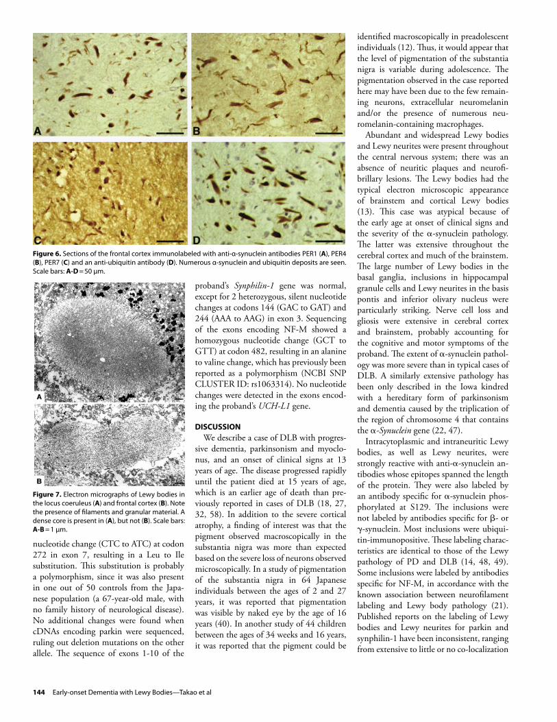
Figure 6. Sections of the frontal cortex immunolabeled with anti-α-synuclein antibodies PER1 (A), PER4 (B), PER7 (C) and an anti-ubiquitin antibody (D). Numerous α-synuclein and ubiquitin deposits are seen. Scale bars: A-D = 50 µm.
Figure 7. Electron micrographs of Lewy bodies in the locus coeruleus (A) and frontal cortex (B). Note the presence of filaments and granular material. A dense core is present in (A), but not (B). Scale bars: A-B = 1 µm.

144 Early-onset Dementia with Lewy Bodies—Takao et al Early-onset Dementia with Lewy Bodies—Takao et al 145
(25, 41, 46, 54). In the proband’s brain, we found a small number of inclusions that were weakly immunoreactive for parkin. Inclusions were not labeled for synphilin-1. Nothing is known about the presence of DJ-1 in Lewy bodies and Lewy neurites. We show here that an antibody raised against the C-terminus of DJ-1 failed to label the inclusions. Recent studies have reported that tau staining is frequently associated with Lewy bodies and that α-synuclein as-semblies can induce tau filament formation (17, 28). However, in the present case, we failed to observe co-localization of tau and α-synuclein. Furthermore, by immuno-electron microscopy, there was no evidence of tau filaments.
Isolated filaments from the proband’s brain were 5 to 10 nm in diameter and strongly decorated by α-synuclein anti-bodies. Antibody PER4, which recognizes the C-terminus of α-synuclein, decorated filaments along their entire length. is contrasted with antibody PER1, which was raised against residues 11-34 of α-synuclein. It labeled only one end of the filaments. ese findings are identical to what we have described previously for α-synuclein filaments from DLB and PD brains (6, 49). Two antibodies (SYN36 and SYN303), which are specific for the N-terminus (residues 1-10 and 2-12, respec-tively) of α-synuclein, decorated filaments, as did the antibody specific for α-synuclein phosphorylated at S129. In some filaments, SYN36 labeled only one end. is indicates that full-length α-synuclein was present in the filaments and that the amino- and car-boxy-termini were exposed on the surface of most filaments, in agreement with re-ports showing that the core of synthetic α-synuclein filaments extends approximately from residues 30-110 (8, 38). Filaments were only very weakly decorated by an anti-ubiquitin antibody, indicating that ubiqui-tination is not necessary for assembly into filaments. is is consistent with the view that ubiquitination of α-synuclein is a late event (45, 49, 52). Sequential biochemical extraction showed the presence of insoluble α-synuclein in the proband’s brain, but not in a control brain. is is consistent with previous studies on DLB (2, 52). In West-ern blot analysis, the insoluble α-synuclein appears as a broad band corresponding to the monomer of α-synuclein and its degra-
dation products, a weak 22 to 24 kDa band and a high-molecular weight smear.
e etiology of this form of DLB is unknown, since the family history was negative for neurological disease and the exon sequences of α-Synuclein were normal. Moreover, the exonic regions of β-Synuclein, UCH-L1, Synphilin-1 and NF-M showed no pathogenic mutations. At present, we cannot exclude the possibil-ity of gene dosage effects or the presence of mutations in the promoter regions or the introns of these genes. When exons 1-12 of Parkin were sequenced, a heterozygous leucine to isoleucine change was found at codon 272 in exon 7. It is located in the first ring finger domain, a functionally im-portant part of the molecule, suggesting a possible pathogenic role. It is also located close to the R275W mutation in exon 7 of Parkin, which has been described in a pa-tient with familial parkinsonism and Lewy body pathology (10). e latter patient was a compound heterozygote, with a 40 bp deletion in exon 3 of Parkin on the other allele. In our proband, sequencing of par-kin cDNAs confirmed the L272I change, but failed to reveal a mutation on the other allele. It remains possible that a second mutation was present, but that it went undetected, because it consisted of a large inversion or duplication, or because it was located in regions that were not sequenced, such as the promoter and the introns. Al-though it has not been reported before (see recent listing in [55]), the L272I change is probably not pathogenic on its own, as it was also found in a 67-year-old control individual from the Japanese population, indicating that it can exist as a polymor-phism. Recently, a heterozygous R271S
change in exon 7 of Parkin was reported in a cohort of North American individuals free of neurological signs (4). is supports the existence of polymorphisms in this func-tionally important region of the protein. e etiology of this form of DLB remains unresolved.
ACKNOWLEDGMENTSis study was supported in part by PHS
P30 AG10133, PHS R01 NS14426, PHS U01 AG16976, the U.K. Alzheimer’s Re-search Trust, the U.K. Parkinson’s Disease
Figure 8. Filaments isolated from the frontal cortex of the proband’s brain were labeled with anti-α-synuclein antibodies PER4, PER1, SYN36, SYN303 and SYNP129 (A-E), and an anti-ubiquitin antibody (F). The gold particles conjugated to the second antibody appear as black dots. Scale bar = 100 nm.
Figure 9. Sequential extraction of α-synuclein from the frontal cortex of a control brain (c) and the proband’s brain (p). The tissues were sequentially extracted with Tris-HCl (TBS), Triton X-100 (Tx), sarkosyl (Sark) and urea, followed by immunoblotting with anti-α-synuclein antibody SYN208.
146 Early-onset Dementia with Lewy Bodies—Takao et al Early-onset Dementia with Lewy Bodies—Takao et al 147
Society and the U.K. Medical Research Council. M. Takao was a post-doctoral fellow from Keio University who was supported in part by the Sasakawa Health Science Foundation and the Departmenty of Pathology and Laboratory Medicine, Indiana University School of Medicine. Y. Narain was supported by a Marie Curie Fellowship from the European Union. We gratefully acknowledge C. Alyea, B. Den-nis, B. Dupree, R. Richardson and R. Ume-hara for technical help. We wish to thank Drs P. Davies, M. Hasegawa, T. Iwatsubo, V.M.-Y. Lee and P. Seubert for their kind gifts of antibodies.
REFERENCES
1. Bonifati V, Rizzu P, van Baren MJ, Schaap O, Breedveld GJ, Krieger E, Dekker MCJ, Squiteri F, Ibanez P, Joosse M, van Dongen JW, Vanacore N, van Swieten JC, Brice A, Meco G, van Duijn CM, Oostra BA, Heutink P (2003) Mutations in the DJ-1 gene associated with autosomal recessive early-onset parkinsonism. Science 299:256-259.
2. Campbell BCV, Li QL, Culvenor JG, Jäkälä P, Cappai R, Beyreuther K, Masters CL, McLean CA (2000) Accumulation of insoluble α-synuclein in dementia with Lewy bodies. Neurobiol Dis 7:192-200.
3. Carmel G, Mager EM, Binder LI, Kuret J (1996) The structural basis of monoclonal antibody Alz50’s selectivity for Alzheimer’s disease pathol-ogy. J Biol Chem 271:32789-32795.
4. Chen R, Gosavi NS, Langston JW, Chan P (2003) Parkin mutations are rare in patients with young-onset parkinsonism in a US population. Parkin-sonism Rel Disord 9:309-312.
5. Crowther RA (1991) Straight and paired helical filaments in Alzheimer disease have a common structural unit. Proc Natl Acad Sci USA 88:2288-2292.
6. Crowther RA, Daniel SE, Goedert M (2000) Characterisation of isolated α-synuclein fila-ments from substantia nigra of Parkinson’s dis-ease brain. Neurosci Lett 292:128-130.
7. Davis RL, Shrimpton AE, Holohan PD, Bradshaw C, Feiglin D, Collins GH, Sonderegger P, Kinter J, Becker LM, Lacbawan F, Krasnewich D, Muenke M, Lawrence DA, Yerby MS, Shaw CM, Gooptu B, Elliott PR, Finch JT, Carrell RW, Lomas DA (1999) Familial dementia caused by polymerization of mutant neuroserpin. Nature 401:376-379.
8. Der-Sarkissian A, Jao CC, Chen J, Langen R (2003) Structural organization of α-synuclein fibrils by site-directed spin labeling. J Biol Chem 278:37530-37535.
9. Duda JE, Giasson BI, Mabon ME, Lee VMY, Tro-janowski JQ (2002) Novel antibodies to synuclein show abundant striatal pathology in Lewy body diseases. Ann Neurol 52:205-210.
10. Farrer M, Chan P, Chen R, Tan L, Lincoln S, Hernandez D, Forno L, Gwinn-Hardy K, Petrucelli
L, Hussey J, Singleton A, Tanner C, Hardy J, Langs-ton JW (2001) Lewy bodies and parkinsonism in families with parkin mutations. Neurology 50:293-300.
11. Farrer M, Destée A, Levesque C, Singleton A, Engelender S, Becquet E, Mouroux V, Richard F, Defebre L, Crook R, Hernandez D, Ross CA, Hardy J, Amouyel P, Chartier-Harlin MC (2001) Genetic analysis of synphilin-1 in familial Parkinson’s dis-ease. Neurobiol Dis 8:317-323.
12. Fenichel GM, Bazeloa M (1968) Studies on neuromelanin. II. Melanin in the brainstems of infants and children. Neurology 18:18:817-820.
13. Forno LS (1996) Neuropathology of Par-kinson’s disease. J Neuropathol Exp Neurol 55:259-272.
14. Fujiwara H, Hasegawa M, Dohmae N, Ka-washima A, Masliah E, Goldberg MS, Shen J, Takio K, Iwatsubo T (2002) α-Synuclein is phosphory-lated in synucleinopathy lesions. Nature Cell Biol 4:160-164.
15. Gasser T, Müller-Myhsok B, Wszolek ZK, Oehl-mann R, Calner DB, Bonifati V, Bereznai B, Fabrizio E, Vieregge P, Horstmann RD (1998) A susceptibil-ity locus for Parkinson’s disease maps to chromo-some 2p13. Nature Genet 18:262-265.
16. Giasson BI, Jakes R, Goedert M, Duda JE, Leight S, Trojanowski JQ, Lee VMY (2000) A panel of epitope-specific antibodies detects protein domains distributed throughout human α-synuclein in Lewy bodies of Parkinson’s disease. J Neurosci Res 59:528-533.
17. Giasson BI, Forman MS, Higuchi M, Golbe LI, Graves CL, Kotzbauer PT, Trojanowski JQ, Lee VMY (2003) Initiation and synergistic fibrillization of tau and alpha-synuclein. Science 300:636-640.
18. Gibb WRG, Esiri MM, Lees AJ (1987) Clinical and pathological features of diffuse cortical Lewy body disease (Lewy body dementia). Brain 110:1131-1153.
19. Goedert M, Spillantini MG, Jakes R, Ruther-ford D, Crowther RA (1989) Multiple isoforms of human microtubule-associated protein tau: Sequences and localization in neurofibrillary tan-gles of Alzheimer’s disease. Neuron 3:519-526.
20. Goedert M (2001) Alpha-synuclein and neu-rodegenerative diseases. Nature Rev Neurosci 2:492-501.
21. Goldman JE, Yen SH, Chiu FC, Peress NS (1983) Lewy bodies of Parkinson’s disease contain neu-rofilament antigens. Science 221:1082-1084.
22. Gwinn-Hardy K, Mehta ND, Farrer M, Mara-ganore D, Muenter M, Yen SH, Hardy J, Dickson DW (2000) Distinctive neuropathology revealed by α-synuclein antibodies in hereditary parkin-sonism and dementia linked to chromosome 4p. Acta Neuropathol 99:663-672.
23. Hasegawa M, Fujiwara H, Nonaka T, Wak-abayashi K, Takahashi H, Lee VMY, Trojanowski JQ, Mann D, Iwatsubo T (2002) Phosphorylated α-synuclein is ubiquitinated in α-synucleinopathy lesions. J Biol Chem 277, 49071-49076.
24. Hayashi S, Wakabayashi K, Ishikawa A, Nagai H, Saito M, Maruyama M, Takahashi T, Ozawa T, Tsuji S, Takahashi H (2000) An autopsy case of au-
tosomal-recessive juvenile parkinsonism with a homozygous exon 4 deletion in the parkin gene. Mov Disord 15:884-888.
25. Huynh DP, Scoles DR, Ho TH, Del Bigio MR, Pulst SM (2000) Parkin is associated with actin filaments in neuronal and nonneuronal cells. Ann Neurol 48:737-744.
26. Ince PG, McKeith IG (2003) Dementia with Lewy bodies. In: Neurodegeneration: The Molecu-lar Pathology of Dementia and Movement Disor-ders (D. Dickson, ed), pp. 188-199. ISN Neuropath Press, Basel 2003.
27. Iseki E, Marui W, Kosaka K, Kato M, Yamamoto T, Ueda K (1999) Clinicopathological multiplicity of dementia with Lewy bodies. Neuropathology 19:386-394.
28. Ishizawa T, Mattila P, Davies P, Wang D, Dick-son DW (2003) Colocalization of tau and alpha-synuclein epitopes in Lewy bodies. J Neuropathol Exp Neurol 62:389-399.
29. Jakes R, Spillantini MG, Goedert M (1994) Identification of two distinct synucleins from hu-man brain. FEBS Lett 345:27-32.
30. Jakes R, Crowther RA, Lee VMY, Trojanowski JQ, Iwatsubo T, Goedert M (1999) Epitope map-ping of LB509, a monoclonal antibody directed against human α-synuclein. Neurosci Lett 269:13-16.
31. Kitada T, Asakawa S, Hattori N, Matsumine H, Yamamura Y, Minoshima S, Yokochi M, Mizuno Y, Shimizu N (1998) Mutations in the parkin gene cause autosomal recessive juvenile parkinson-ism. Nature 392:605-608.
32. Kosaka K (1990) Diffuse Lewy body disease in Japan. J Neurol 237:197-204.
33. Krüger R, Kuhn W, Müller T, Woitalla D, Grae-ber M, Kösel S, Przuntek H, Epplen JT, Schöls L, Riess O (1998) Ala30Pro mutation in the gene en-coding α-synuclein in Parkinson’s disease. Nature Genet 18:106-108.
34. Lavedan C, Leroy E, Torres R, Dehejia A, Dutra A, Buchholtz S, Nussbaum RL, Polymeropoulos MH (1998) Genomic organization and expression of the human α-synuclein gene (SNCB). Genomics 54:173-175.
35. Lavedan C, Buchholtz S, Nussbaum RL, Albin RL, Polymeropoulos MH (2002) A mutation in the human neurofilament M gene in Parkinson’s dis-ease that suggests a role for the cytoskeleton in neuronal degeneration. Neurosci Lett 322:57-61.
36. Leroy E, Boyer R, Auburger G, Leube B, Ulm G, Mezey E, Harta G, Brownstein MJ, Jonnalagada S, Chernova T, Dehejia A, Lavedan C, Gasser T, Steinbach PJ, Wilkinson KD, Polymeropoulos MH (1998) The ubiquitin pathway in Parkinson’s disease. Nature 395:451-452.
37. McQuaid S, McConnell R, McMahon J, Her-ron B (1995) Microwave antigen retrieval for immunocytochemistry on formalin-fixed, paraf-fin-embedded post-mortem CNS tissue. J Pathol 176:207-216.
38. Miake H, Mizusawa H, Iwatsubo H, Hasegawa M (2002) Biochemical characterization of the core structure of α-synuclein filaments. J Biol Chem 277:19213-19219.

146 Early-onset Dementia with Lewy Bodies—Takao et al Early-onset Dementia with Lewy Bodies—Takao et al 147
39. Mori H, Kondo T, Yokochi M, Matsumine H, Nakagawa-Hattori Y, Miyake T, Suda, K, Mizuno Y (1998) Pathologic and biochemical studies of juvenile parkinsonism linked to chromosome 6q. Neurology 51:890-892.
40. Morimatsu Y, Satoh J, Nagata J, Kubota Y, Hayashi M, Sakamoto K, Shinohara T (1987) Pathology of neuromelanin in the development disabilities. Kouseishour Shinkei Shikkan Kenkyu Itaku Jigyou (Japanese) 1:268-282.
41. Murray IVJ, Medford MA, Guan HP, Rueter SM, Trojanowski JQ, Lee VMY (2003) Synphilin in normal human brain and in synucleinopathies: studies with new antibodies. Acta Neuropathol 105:177-184.
42. Ozawa T, Takano H, Onodera O, Kobayashi H, Ikeuchi T, Koide R, Okuizumi K, Shimohata T, Wakabayashi K, Takahashi H, Tsuji T (1999) No mutation in the entire coding region of the α-synuclein gene in pathologically confirmed cases of multiple system atrophy. Neurosci Lett 270:110-112.
43. Piccardo P, Mirra SS, Young K, Gearing M, Dlouhy SR, Ghetti B (1998) α-Synuclein accumu-lation in Gerstmann-Sträussler-Scheinker disease (GSS) with prion protein gene mutation F198S. Neurobiol Aging 19:S172.
44. Polymeropoulos MH, Lavedan C, Leroy E, Ide SE, Dehejia A, Dutra A, Pike B, Root H, Rubinstein J, Boyer R, Stenroos ES, Chandrasekharappa S, Athanassiadou A, Papapetropoulos T, Johnson WG, Lazzarini AM, Duvoisin RC Di Iorio G, Golbe LI, Nussbaum RL (1997) Mutation in the α-synu-clein gene identified in families with Parkinson’s disease. Science 276:2045-2047.
45. Sampathu DM, Giasson BI, Pawlyk AC, Tro-janowski JQ, Lee VMY (2003) Ubiquitination of α-synuclein is not required for formation of pathological inclusions in α-synucleinopathies. Am J Pathol 163:91-100.
46. Schlossmacher MG, Frosch MP, Gai WP, Me-dina M, Sharma N, Forno L, Ochiishi T, Shimura H, Sharon R, Hattori N, Langston JW, Mizuno Y, Hyman BT, Selkoe DJ, Kosik KS (2002) Parkin lo-calizes to the Lewy bodies of Parkinson disease and dementia with Lewy bodies. Am J Pathol 160:1655-1667.
47. Singleton AB, Farrer M, Johnson J, Singleton A, Hague S, Kachergus J, Hulihan M, Peuralinna T, Dutra A, Nussbaum R, Lincoln S, Crawley A, Hanson M, Maraganore D, Adler C, Cookson MR, Muenter M, Baptista M, Miller D, Blancato J, Hardy J, Gwinn-Hardy K (2003) α-Synuclein locus triplication causes Parkinson’s disease. Science 302:841.
48. Spillantini MG, Schmidt ML, Lee VMY, Tro-janowski JQ, Jakes R, Goedert M (1997) α-Synu-clein in Lewy bodies. Nature 388, 839-840.
49. Spillantini MG, Crowther RA, Jakes R, Hasega-wa M, Goedert M (1998) α-Synuclein in filamen-tous inclusions of Lewy bodies from Parkinson disease and dementia with Lewy bodies. Proc Natl Acad Sci USA 95:6469-6473.
50. Takahashi H, Ohama E, Suzuki S, Horikawa Y, Ishikawa A, Morita T, Tsuji S, Ikuta F (1994) Famil-
ial juvenile parkinsonism: clinical and pathologic study in a family. Neurology 44:437-441.
51. Takao M, Benson MD, Murrell JR, Yazaki M, Piccardo P, Unverzagt FW, Davis RL, Holohan PD, Lawrence DA, Richardson R, Farlow MR, Ghetti B (2000) Neuroserpin mutation S52R causes neuroserpin accumulation in neurons and is as-sociated with progressive myoclonus epilepsy. J Neuropathol Exp Neurol 59:1070-1078.
52. Tofaris GK, Razzaq A, Ghetti B, Lilley K, Spill-antini MG (2003) Ubiquitination of α-synuclein in Lewy bodies is a pathological event not associ-ated with impairment of proteasome function. J Biol Chem 278:44405-44411.
53. Van de Warrenburg BPC, Lammens M, Lück-ing CB, Denefle P, Wesseling P, Booij J, Praamstra P, Quinn N, Brice A, Horstink MWIM (2001) Clini-cal and pathologic abnormalities in a family with parkinsonism and parkin gene mutations. Neu-rology 56:555-557.
54. Wakabayashi K, Engelender S, Yoshimoto M, Tsuji S, Ross CA, Takahashi H (2000) Synphilin-1 is present in Lewy bodies in Parkinson’s disease. Acta Neuropathol 47:521-523.
55. West A, Périquet M, Lincoln S, Lücking CB, Nicholl D, Bonifati V, Rawal N, Gasser T, Lohmann E, Deleuze JF, Maraganore D, Levey A, Wood N, Dürr A, Hardy J, Brice A, Farrer M (2002) Complex relationship between parkin mutations and Par-kinson’s disease. Am J Med Genet 114:584-591.
56. Wintermeyer P, Krüger R, Kuhn W, Müller T, Woitalla D, Berg D, Becker G, Leroy E, Poly-meropoulos M, Berger K, Przuntek H, Schöls L, Epplen JT, Riess O (2000) Mutation analysis and association studies of the UCHL1 gene in Ger-man Parkinson’s disease patients. Neuroreport 11:2079-2082.
57. Wzolek ZK, Gwinn-Hardy K, Wszolek EK, Muenter MD, Pfeiffer RF, Rodnitzky RL, Uitti RJ, McComb RD, Gasser T, Dickson DW (2002) Neuro-pathology of two members of a German-Ameri-can kindred (Family C) with late onset parkinson-ism. Acta Neuropathol 103:344-350.
58. Yoshimura N, Yoshimura I, Asada M, Hayashi S, Fukushima Y, Sato T, Kudo H (1988) Juvenile Parkinson’s disease with widespread Lewy bod-ies in the brain. Acta Neuropathol 77:213-218.
59. Zarranz JJ, Alegre J, Gomez-Esteban JC, Lez-cano E, Ros R, Ampuero I, Vidal L, Hoenicka J, Rodriguez O, Atares B, Llorens V, Gomez Tortosa E, del Ser T, Munoz DG, de Yebenes JG (2004) The new mutation, E46K, of alpha-synuclein causes Parkinson and Lewy body dementia. Ann Neurol 55:164-173.