Rapidly progressive dementia and intractable diarrhea
20
Vol.:(0123456789) 1 3 https://doi.org/10.1007/s10072-021-05844-5 REVIEW ARTICLE Rapidly progressive dementia and intractable diarrhea: a teaching case report and a systematic review of cognitive impairment in Whipple’s disease Arianna Manini 1 · Giacomo Querzola 1 · Carlo Lovati 2 · Leonardo Pantoni 1 Received: 11 November 2021 / Accepted: 22 December 2021 © Fondazione Società Italiana di Neurologia 2022 Abstract Objective Whipple’s disease (WD) is a systemic, chronic, relapsing disease caused by Tropheryma whipplei, which can mimic signs and symptoms of various clinical entities. Typical manifestations are represented by gastrointestinal and systemic symptoms, among which neurological ones are frequent. We present the case of a patient with WD and rapidly progressive cognitive impairment and a review of literature aimed to report epidemiological, clinical, neuroimaging, and laboratory findings of cognitive impairment associated with WD. Methods A systematic review of medical literature published until November 22, 2020, was performed. Full-text, peer- reviewed case reports and series in English language presenting patients with WD and cognitive impairment were included. Data concerning demographic, clinical, neuroimaging, and laboratory characteristics were collected and synthesized qualitatively. Results The patient was a 54-year-old male who developed rapidly progressive dementia, fluctuating arousal disturbances, and supranuclear ophthalmoparesis associated with chronic diarrhea and fever spikes. T. whipplei was detected in the cer- ebrospinal fluid, and appropriate antimicrobial therapy was given with progressive clinical benefit. The systematic review of 114 case reports/series identified 147 patients with WD and cognitive impairment; this latter was rarely isolated. Neu- rological symptoms associated with cognitive decline were psychiatric disturbances, supranuclear ophthalmoplegia, hypo- thalamic involvement, and consciousness disorders. Brain imaging and cerebrospinal fluid findings were heterogeneous and nonspecific. Conclusions Cognitive impairment represents one of the most common neurological features associated with WD. The clinical suspicion of this disease in patients with rapidly progressive dementia is crucial to guide diagnostic strategies and proper antimicrobial therapy, which may revert the clinical deterioration. Keywords Whipple’s disease · Tropheryma whipplei · Dementia · Cognitive impairment · Central nervous system Introduction The first description of Whipple’s disease (WD), a rare multi-systemic chronic illness caused by Tropheryma whip- plei [1], dates back to 1895 [2]. In 1907, George Hoyt Whip- ple described a 36-year-old missionary with malabsorptive syndrome due to chronic unexplained diarrhea associated with migratory polyarthritis, cough, and mesenteric lym- phadenopathy [3]. Since then, our knowledge of the patho- genic mechanisms and clinical manifestations of WD has grown, improving our ability of diagnosis and treatment. Nevertheless, different immunopathogenic aspects of the disease remain unclear. Most infected individuals do not develop symptomatic infection, protected by humoral and Arianna Manini and Giacomo Querzola equally contributed to this work * Leonardo Pantoni [email protected] 1 Stroke and Dementia Lab, “Luigi Sacco” Department of Biomedical and Clinical Sciences, University of Milan, Via Giovanni Battista Grassi 74, 20157 Milan, Italy 2 Neurology Unit, “Luigi Sacco” University Hospital, Milan, Italy / Published online: 3 January 2022 Neurological Sciences (2022) 43:907–926
-
Upload
khangminh22 -
Category
Documents
-
view
0 -
download
0
Transcript of Rapidly progressive dementia and intractable diarrhea

Vol.:(0123456789)1 3
https://doi.org/10.1007/s10072-021-05844-5
REVIEW ARTICLE
Rapidly progressive dementia and intractable diarrhea: a teaching case report and a systematic review of cognitive impairment in Whipple’s disease
Arianna Manini1 · Giacomo Querzola1 · Carlo Lovati2 · Leonardo Pantoni1
Received: 11 November 2021 / Accepted: 22 December 2021 © Fondazione Società Italiana di Neurologia 2022
Abstract Objective Whipple’s disease (WD) is a systemic, chronic, relapsing disease caused by Tropheryma whipplei, which can mimic signs and symptoms of various clinical entities. Typical manifestations are represented by gastrointestinal and systemic symptoms, among which neurological ones are frequent. We present the case of a patient with WD and rapidly progressive cognitive impairment and a review of literature aimed to report epidemiological, clinical, neuroimaging, and laboratory findings of cognitive impairment associated with WD.Methods A systematic review of medical literature published until November 22, 2020, was performed. Full-text, peer-reviewed case reports and series in English language presenting patients with WD and cognitive impairment were included. Data concerning demographic, clinical, neuroimaging, and laboratory characteristics were collected and synthesized qualitatively.Results The patient was a 54-year-old male who developed rapidly progressive dementia, fluctuating arousal disturbances, and supranuclear ophthalmoparesis associated with chronic diarrhea and fever spikes. T. whipplei was detected in the cer-ebrospinal fluid, and appropriate antimicrobial therapy was given with progressive clinical benefit. The systematic review of 114 case reports/series identified 147 patients with WD and cognitive impairment; this latter was rarely isolated. Neu-rological symptoms associated with cognitive decline were psychiatric disturbances, supranuclear ophthalmoplegia, hypo-thalamic involvement, and consciousness disorders. Brain imaging and cerebrospinal fluid findings were heterogeneous and nonspecific.Conclusions Cognitive impairment represents one of the most common neurological features associated with WD. The clinical suspicion of this disease in patients with rapidly progressive dementia is crucial to guide diagnostic strategies and proper antimicrobial therapy, which may revert the clinical deterioration.
Keywords Whipple’s disease · Tropheryma whipplei · Dementia · Cognitive impairment · Central nervous system
Introduction
The first description of Whipple’s disease (WD), a rare multi-systemic chronic illness caused by Tropheryma whip-plei [1], dates back to 1895 [2]. In 1907, George Hoyt Whip-ple described a 36-year-old missionary with malabsorptive syndrome due to chronic unexplained diarrhea associated with migratory polyarthritis, cough, and mesenteric lym-phadenopathy [3]. Since then, our knowledge of the patho-genic mechanisms and clinical manifestations of WD has grown, improving our ability of diagnosis and treatment. Nevertheless, different immunopathogenic aspects of the disease remain unclear. Most infected individuals do not develop symptomatic infection, protected by humoral and
Arianna Manini and Giacomo Querzola equally contributed to this work
* Leonardo Pantoni [email protected]
1 Stroke and Dementia Lab, “Luigi Sacco” Department of Biomedical and Clinical Sciences, University of Milan, Via Giovanni Battista Grassi 74, 20157 Milan, Italy
2 Neurology Unit, “Luigi Sacco” University Hospital, Milan, Italy
/ Published online: 3 January 2022
Neurological Sciences (2022) 43:907–926

1 3
cellular immunity [4]. Therefore, detecting T. whipplei in tissues and biological fluids of asymptomatic carriers is not rare [5]. Typical and atypical presentations appear only in a few patients who show genetic predisposition and rarely immune deficits [6, 7]. Classical manifestations are represented by gastrointestinal symptoms, including diar-rhea, weight loss, abdominal pain, nausea and vomit, and systemic features, such as fatigue, migratory arthralgias/arthritis, fever of unknown origin, lymphadenopathy, and skin alterations [8]. Other symptoms are due to localized forms of T. whipplei infection, including the neurological ones. Nervous system involvement produces a broad range of signs and symptoms, whose the most typical is the clas-sic triad of dementia, supranuclear ophthalmoplegia, and myoclonus [9].
Here, we report the case of a patient with WD and pro-gressive cognitive decline and a literature review aimed to clarify epidemiological, clinical, neuroimaging, and labora-tory findings of WD associated with dementia.
Material and methods
Systematic literature review
Two authors (A.M. and G.Q.) performed a systematic review of medical literature by searching two comprehensive medi-cal databases, namely PubMed and Embase, from incep-tion to November 22, 2020. The search query employed was “(whipple disease OR tropheryma whipplei OR tropheryma whippelii) AND (dementia OR central nervous system OR cognitive).” Full-text, peer-reviewed case reports and case series published in English language presenting patients with WD and cognitive impairment were included. All the abstracts were screened independently by the two authors to select full-text articles to be included in the analysis. In case of disagreement, relevant articles were re-reviewed until consensus was reached. The complete list of publica-tions included in our systematic review is available in Sup-plementary Table 1. Data of eligible studies were collected, reported in a dedicated database, and combined, including age at onset and gender of patients; neurological and non-neurological clinical features; neuroimaging features; type of central nervous system (CNS) WD diagnosis (definite or possible) according to Louis et al.’s criteria [10]; and results of CSF examination. Data were qualitatively synthesized, and descriptive analyses were performed using open-source software “Jamovi,” version 1.6 (Sidney, Australia).
Case reports and series were included in the systematic review if the authors used one of the following expressions to describe patient’s clinical condition: “cognitive impair-ment,” “cognitive decline,” “cognitive changes,” “cognitive alterations,” “cognitive abnormalities,” “cognitive disorder,”
“cognitive defects,” “cognitive deterioration,” “cognitive deficits,” “cognitive disturbances,” “cognitive dysfunction,” “cognitive symptoms,” “cognitive complaints,” “cognitive slowness,” “cognitive sequelae,” “neurocognitive features,” “neurocognitive symptoms,” “deterioration in cognition,” “reduced cognition,” “memory loss,” “memory impair-ment,” “decreased memory,” “problems with memory,” “memory lapses,” “memory disturbances,” “memory dif-ficulties,” “memory disorder,” “poor memory,” “memory deficits,” “memory alterations,” “amnesic syndrome,” “dementia,” “demented,” “dementing illness,” and “demen-tial syndrome.” When the authors did not report any of the previous terms, but described an acquired syndrome consist-ing of a loss of several separable but overlapping intellectual abilities that was significant enough to interfere with inde-pendent, daily occupational/domestic/social functioning, then the case was included in the analysis.
Other neurological and non-neurological features asso-ciated with cognitive deficits were also searched for in the publications. Considering other associated neurological features, these were classified in main categories (Supple-mentary Table 2).
After the literature search, we applied the Louis et al.’s criteria [10] for CNS WD for each of the selected cases. According to Louis et al.’s criteria [10], CNS WD is defined as “possible” when at least one out of four systemic symp-toms (fever of unknown origin; gastrointestinal symptoms such as steatorrhea, chronic diarrhea, abdominal distension, or pain; chronic migratory arthralgias or polyarthralgias; unexplained lymphadenopathy, night sweats, or malaise), not due to another known etiology, is associated with at least one out of four neurological signs (supranuclear verti-cal gaze palsy; rhythmic myoclonus; dementia with psychi-atric symptoms; hypothalamic manifestations), not due to another known etiology. CNS WD is otherwise “definite” if at least one of the following criteria is fulfilled: presence of oculomasticatory myorhythmia or oculo-facial skeletal myo-rhythmia; positive tissue biopsy (either periodic acid-Schiff (PAS) positive or bacteria seen on electron microscopy); and positive polymerase chain reaction (PCR) analysis. If histo-logical or PCR analysis is not performed on CNS tissue, then the patient must also have neurological signs. If histological or PCR analysis is performed on CNS tissue, then the patient does not need to have neurological signs.
Results
Case report
The patient was a 54-year-old Caucasian male, professional musicist. Informed consent was given by the patient for the case report publication.
908 Neurological Sciences (2022) 43:907–926
1 3
He had a history of moderate chronic renal failure due to autoimmune membrane-proliferative glomerulonephritis (MPGN), associated with thrombocytopenia, cryoglobu-linemia, and reduction in C3 and C4 fractions. Since the diagnosis in 2013, he had been treated with corticosteroids and immunosuppressive drugs, including cyclophosphamide and rituximab. The remaining history was remarkable only for atrial flutter, previously treated with oral anticoagulant drugs, benign prostatic hypertrophy, and major depressive disorder.
In February 2019, almost 1 month after a 10-day tour in China, the patient developed elevated fever, macrohe-maturia, and diarrhea which led to dehydration and acute chronic renal failure (ACRF). The patient was suspected to have a MPGN relapse, so that renal biopsy was performed, confirming MPGN with hyaline degeneration in about half of the glomeruli. He received treatment with intravenous steroid bolus (methylprednisolone 1 g for 3 days), followed by oral prednisone 50 mg daily and two intravenous infu-sions of rituximab. Renal function partially improved and
macrohematuria disappeared, whereas diarrhea persisted. Metronidazole and piperacillin/tazobactam were admin-istered because of infectious suspicion, with no clinical benefit. Steroid doses were progressively reduced and mycophenolate mofetil was introduced as maintenance immunosuppressive treatment.
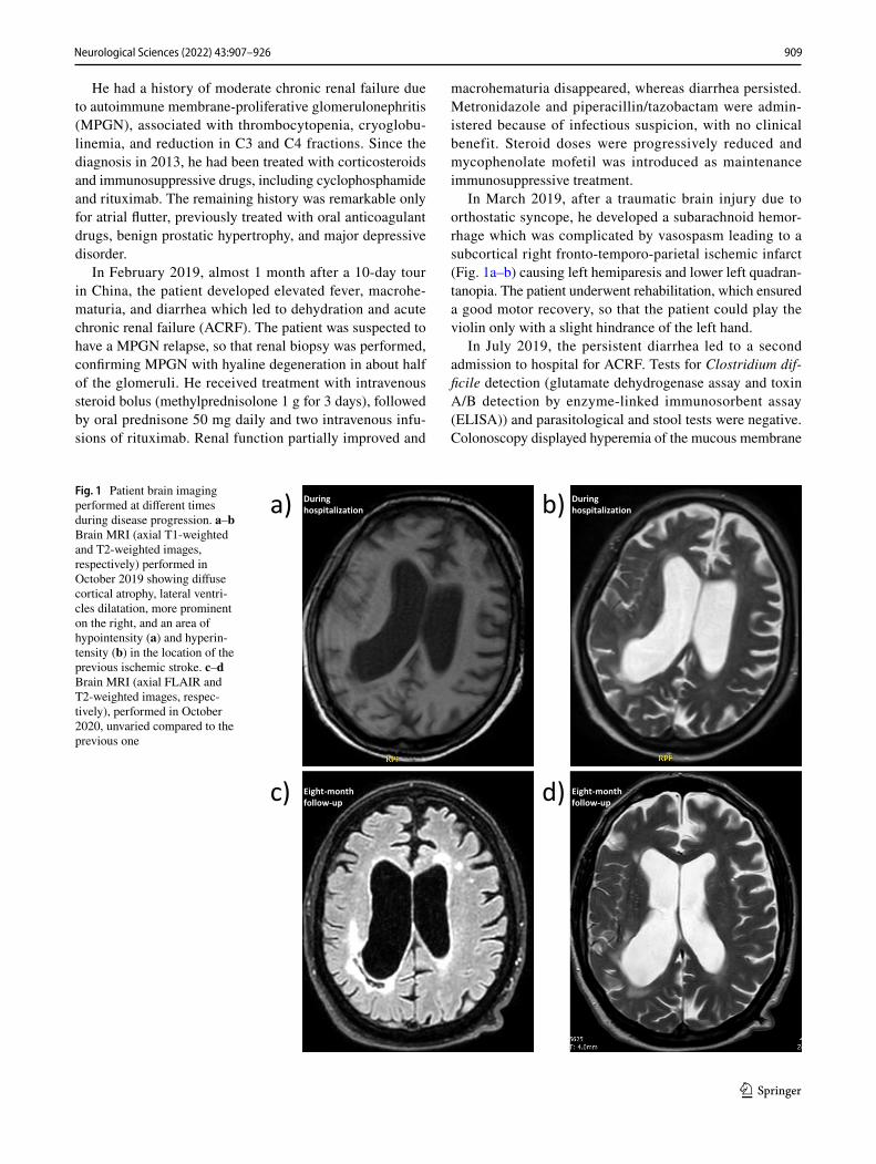
In March 2019, after a traumatic brain injury due to orthostatic syncope, he developed a subarachnoid hemor-rhage which was complicated by vasospasm leading to a subcortical right fronto-temporo-parietal ischemic infarct (Fig. 1a–b) causing left hemiparesis and lower left quadran-tanopia. The patient underwent rehabilitation, which ensured a good motor recovery, so that the patient could play the violin only with a slight hindrance of the left hand.
In July 2019, the persistent diarrhea led to a second admission to hospital for ACRF. Tests for Clostridium dif-ficile detection (glutamate dehydrogenase assay and toxin A/B detection by enzyme-linked immunosorbent assay (ELISA)) and parasitological and stool tests were negative. Colonoscopy displayed hyperemia of the mucous membrane
Fig. 1 Patient brain imaging performed at different times during disease progression. a–b Brain MRI (axial T1-weighted and T2-weighted images, respectively) performed in October 2019 showing diffuse cortical atrophy, lateral ventri-cles dilatation, more prominent on the right, and an area of hypointensity (a) and hyperin-tensity (b) in the location of the previous ischemic stroke. c–d Brain MRI (axial FLAIR and T2-weighted images, respec-tively), performed in October 2020, unvaried compared to the previous one
909Neurological Sciences (2022) 43:907–926

1 3
and erosions in the first 5 cm of the rectal mucosa. The pathological examination showed hyperplasia of glandular epithelium, edema of the lamina propria, exudative inflam-mation, with increase in the number of lymphocytes and plasma cells, and micro-abscesses in descending colon. The physicians hypothesized that diarrhea and pathological alterations were secondary to iatrogenic damage. As a con-sequence, it was decided to interrupt mycophenolate mofetil and to start mesalazine suppositories, which were replaced by beclomethasone dipropionate in August 2019 because of persisting diarrhea.
Soon after that, the patient began complaining of difficul-ties in concentration, especially concerning reading skills. By the end of September, a third ACRF secondary to per-sistent diarrhea led to admission to the Gastroenterology Unit. The use of cholestyramine partially improved diarrhea. Proctoscopy was normal, while small intestine ultrasonog-raphy revealed wall thickening of the last small bowel loop and of descending and sigmoid colon. Digestive endoscopy showed granular aspect of intestinal lining and lymphangi-ectasis of intestinal villi. The research of Helicobacter pylori and Isospora belli did not detect any microorganisms. A wide spectrum screening for infectious diseases was nega-tive, including stool test for Giardia species, Entamoeba histolytica, and Cryptosporidium species and serology for adenovirus, rotavirus, hepatitis B and C viruses, and HIV 1/2. Urinary 5-hydroxyindoleacetic acid was normal, thus excluding the presence of neuroendocrine tumors. Fecal calprotectin was remarkably increased (1304 μg/g; nor-mal value: < 50 μg/g). No altered findings were detected by an autoimmune panel (antinuclear antibodies (ANA), extractable nuclear antigens antibodies (ENA), anti-mito-chondrial antibodies (AMA), anti-alpha-smooth muscle actin antibodies (ASMA), anti-neutrophil cytoplasmic anti-bodies (ANCA), thyrotropin receptor antibodies (TRAB), thyroglobulin antibodies (TgAb), anti-transglutaminase antibodies (ATA), anti-gliadin antibodies (AGA), immuno-globulin G (IgG)). The patient was concerned about the pos-sible repercussions of iodinated contrast on the kidney con-dition and refused an enhanced computerized tomography (CT) of thorax and abdomen, proposed to exclude a possible paraneoplastic genesis of disturbances. Blood tests revealed IgG antibodies deficit. A reduced number of lymphocytes T CD3 + (both CD4 + and CD8 +) and B CD19 + was detected at cytofluorimetry and an ensuing prophylactic therapy with cotrimoxazole on alternate days was initiated.
During hospitalization, he developed intermittent fever and an increase of inflammatory markers. Blood cultures and DNA amplification for Epstein-Barr virus (EBV), cyto-megalovirus (CMV), varicella zoster virus (VZV), and her-pes simplex virus (HSV) 1 and 2 were negative. After the employment of piperacillin/tazobactam, inflammatory mark-ers gradually decreased.
Over about 10 days, the patient underwent a dramatic cognitive deterioration (i.e., he rapidly lost the possibility to speak and write a correct message on the cellular phone with a progressive disruption of grammatical and lexical structure of verbal functions; he was completely disoriented in time and space, and he was not any longer able to inter-act with health workers or family members). Considering the presence of chronic diarrhea and progressive cognitive impairment, an infective or inflammatory involvement of CNS, with the same origin of the gastrointestinal problem, was hypothesized, and the patient was transferred to the Neurology Unit. On admission, neurological examination revealed fluctuating arousal disturbances, attention deficits with difficulty in obeying motor orders, hypophonia, and echolalia. Eye movement examination displayed spontane-ous nystagmus in primary gaze and more sustained in up and right-gaze, and bilateral limitation of ocular motility in horizontal gaze, which evolved in 2 days into ophthalmopa-resis in all directions of gaze. Apparently as a worsening of the consequences of the previous ischemic stroke, the patient showed left hemiparesis, increased spastic tone of the left arm (in contrast with the reduced tone of the other three limbs), left Babinski sign, and extinction of left stimulus on double simultaneous stimulation. Blood tests showed nor-mal level of leukocytes (6180 leukocytes/μL; normal values: 4190–9350 leukocytes/μL), high C-reactive protein levels (132.6 mg/L; normal values: < 10.0 mg/L), normocytic (90.5 fL; normal values: 35–50 g/L) anemia (8.4 g/dL; normal val-ues: 14.2–17.2 g/dL), and hypoalbuminemia (28 g/L; normal values: 35–50 g/L).
At this point, main differential diagnoses included infectious, autoimmune, deficiency, and genetic diseases (Table 1). Supplementation with thiamine did not produce clinical benefits. The absence of characteristic dermatitis consisting of symmetrical erythema in sun-exposed skin made the hypothesis of pellagra unlikely. Prion disease associated with diarrhea and neuropathy appeared doubtful due to the absence of typical autonomic failure and clinical signs of sensory polyneuropathy. The hypotheses of genetic diseases were rejected due to the rapid progression of symp-toms and to the absence of clinical hallmarks (i.e., cobala-min C deficiency and acrodermatitis enteropathica-like; cerebrotendinous xanthomatosis and tendon xanthomas; transthyretin amyloidosis and autonomic dysfunction, car-diac involvement, carpal tunnel syndrome). A subtype of transthyretin amyloidosis called oculoleptomeningeal amy-loidosis, although manifesting with neurological and neu-ropsychiatric symptoms such as dementia, does not produce the typical gastrointestinal picture. The hypothesis of com-plicated celiac disease did not fit with the absence of autoan-tibodies and of pathological hallmarks at duodenal biopsies. WD and anti-dipeptidyl-peptidase-like protein (DPPX) 6, although rare entities, could not be ruled out.
910 Neurological Sciences (2022) 43:907–926

1 3
Tabl
e 1
Diff
eren
tial d
iagn
oses
in p
atie
nts w
ith d
iarr
hea
and
dem
entia
Diff
eren
tial d
iagn
osis
Etio
path
ogen
esis
and
ris
k fa
ctor
sM
ean
age
at o
nset
Clin
ical
feat
ures
Spec
ific
labo
rato
ry te
stsO
ther
tests
Trea
tmen
tRe
fere
nces
Pella
gra
Vita
min
B3
(nia
cin)
de
ficie
ncy
(alc
ohol
-is
m a
nd a
lcoh
ol
with
draw
al, c
arci
noid
tu
mor
, mal
nutr
ition
, dr
ugs)
Varia
ble
Neu
rolo
gica
l and
psy
-ch
iatr
ic fe
atur
es:
Neu
rops
ychi
atric
sym
p-to
ms i
n ea
rly st
ages
Dem
entia
in la
te st
ages
Han
d co
arse
and
resti
ng
trem
orN
euro
path
yM
yocl
onus
Ata
xia
Isol
ated
del
irium
Oth
er c
linic
al fe
a-tu
res:
Sym
met
rical
ery
them
a in
sun-
expo
sed
skin
Intra
ctab
le d
iarr
hea
and
othe
r gas
troin
testi
-na
l sym
ptom
s
Redu
ced
plas
mat
ic
nico
tinic
aci
d an
d ni
cotin
amid
eU
rine
5-H
IAA
(scr
een-
ing
for c
arci
noid
tu
mor
)
EEG
: diff
use
slow
ing,
es
peci
ally
in th
e th
eta
rang
eEG
DS
and
colo
nos-
copy
: muc
osal
infla
m-
mat
ion
thro
ugho
ut th
e ga
stroi
ntes
tinal
syste
m
Nic
otin
amid
eTr
eatm
ent o
f cau
ses
[11–
13]
Thia
min
e de
ficit
Vita
min
B1
(thia
min
e)
defic
ienc
y (a
lcoh
ol-
ism
, mal
nutr
ition
, ba
riat
ric
surg
ery,
pr
egna
ncy,
dru
gs)
Gen
etic
pre
disp
osi-
tion
(i.e.
, SC
L25A
19,
TPK
1, T
HTP
A,
ENTP
D5)
Varia
ble
Neu
rolo
gica
l and
psy
-ch
iatr
ic fe
atur
es:
Mem
ory
defic
itsW
erni
cke
ence
pha-
lopa
thy
Kor
sako
ff sy
ndro
me
Redu
ced
plas
mat
ic
thia
min
e le
vels
MR
I: di
ffuse
and
ban
d-lik
e le
sion
s, es
peci
ally
in
thal
ami,
basa
l ga
nglia
and
fron
tal
lobe
sEE
G: n
orm
al, i
ncre
ased
sl
ow w
aves
or e
pile
p-tic
dis
char
ges
Thia
min
e ad
min
istra
-tio
n ± su
lbut
iam
ine
[14–
18]
911Neurological Sciences (2022) 43:907–926

1 3
Tabl
e 1
(con
tinue
d)
Diff
eren
tial d
iagn
osis
Etio
path
ogen
esis
and
ris
k fa
ctor
sM
ean
age
at o
nset
Clin
ical
feat
ures
Spec
ific
labo
rato
ry te
stsO
ther
tests
Trea
tmen
tRe
fere
nces
Ant
i-DPP
X e
ncep
halit
isA
ntib
odie
s ant
i-dip
ep-
tidyl
-pep
tidas
e-lik
e pr
otei
n 6
(ofte
n B
-cel
l ly
mph
oma)
52 y
ears
(ran
ge 1
3–76
)N
euro
logi
cal a
nd p
sy-
chia
tric
feat
ures
:R
apid
ly p
rogr
essi
ve
dem
entia
Slee
p di
sturb
ance
sH
eada
che
Neu
rops
ychi
atric
sy
mpt
oms
Seiz
ures
Resti
ng a
nd p
ostu
ral
trem
orC
ereb
ella
r sym
ptom
sTr
unca
l dys
toni
a an
d di
ffuse
rigi
dity
Myo
clon
usH
yper
esth
esia
, allo
-dy
nia,
pru
ritus
Dys
phag
iaEy
e m
ovem
ent d
istur
-ba
nces
PER
M-li
ke p
rese
ntat
ion
Aut
onom
ic d
istur
banc
esO
ther
clin
ical
fea-
ture
s:D
iarr
hea
and
othe
r ga
stroi
ntes
tinal
sym
p-to
ms
CSF
ple
ocyt
osis
with
ev
iden
ce o
f int
rath
ecal
prod
uctio
n of
IgG
or
olig
oclo
nal b
ands
Ant
ibod
ies a
gain
st D
PPX
pos
itive
in b
oth
seru
m a
nd C
SF (p
re-
dom
inan
tly Ig
G1
and
IgG
4)
MR
I: pe
riven
tricu
lar
and
subc
ortic
al w
hite
m
atte
r T2/
FLA
IR
hype
rinte
nsiti
es; n
on-
spec
ific
whi
te m
atte
r ch
ange
s; te
mpo
ral
lobe
atro
phy
18F-
FDG
PET
-MR
I: bi
late
ral h
ypom
e-ta
bolis
m o
f cau
date
nu
clei
, fro
ntal
cor
tex,
te
mpo
ral l
obes
and
th
alam
usEE
G: b
ackg
roun
d sl
ow-
ing
and
rare
epi
lept
i-fo
rm d
isch
arge
s
Ster
oids
iv a
nd p
oIm
mun
oglo
bulin
ivR
ituxi
mab
Cyc
loph
osph
amid
e
[19–
23]
912 Neurological Sciences (2022) 43:907–926

1 3
Tabl
e 1
(con
tinue
d)
Diff
eren
tial d
iagn
osis
Etio
path
ogen
esis
and
ris
k fa
ctor
sM
ean
age
at o
nset
Clin
ical
feat
ures
Spec
ific
labo
rato
ry te
stsO
ther
tests
Trea
tmen
tRe
fere
nces
Whi
pple
’s d
isea
seT.
whi
pple
i inf
ectio
nVa
riabl
eN
euro
logi
cal a
nd p
sy-
chia
tric
feat
ures
:D
emen
tiaSu
pran
ucle
ar o
phth
al-
mop
legi
aM
yocl
onus
Ocu
lom
astic
ator
y m
yo-
rhyt
hmia
Ocu
lo-fa
cial
-ske
leta
l m
yorh
ythm
iaPs
ycho
logi
cal a
nd
beha
vior
al a
ltera
tions
Hyp
otha
lam
ic in
volv
e-m
ent
Dis
orde
rs o
f con
scio
us-
ness
Oth
er c
linic
al fe
a-tu
res:
Dia
rrhe
aW
eigh
t los
sA
bdom
inal
pai
nFe
ver
Fatig
ueA
rthra
lgia
s/ar
thrit
isSk
in p
igm
enta
tion/
alte
ratio
ns
T. w
hipp
lei P
CR
PAS-
posi
tive
biop
sies
MR
I: no
rmal
, cer
ebra
l an
d/or
cer
ebel
lar
lesi
ons,
diffu
se c
er-
ebra
l ede
ma,
cor
tical
an
d/or
subc
ortic
al
atro
phy,
hyd
ro-
ceph
alus
, epe
ndym
al
lesi
ons,
intra
cere
bral
he
mor
rhag
e, sp
inal
co
rd le
sion
s
Cef
triax
one
(2 g
twic
e a
day)
for 2
wee
ks, f
ol-
low
ed b
y C
otrim
oxa-
zole
(160
/800
mg
twic
e a
day)
for o
ne
year
[7–9
]
913Neurological Sciences (2022) 43:907–926

1 3
Tabl
e 1
(con
tinue
d)
Diff
eren
tial d
iagn
osis
Etio
path
ogen
esis
and
ris
k fa
ctor
sM
ean
age
at o
nset
Clin
ical
feat
ures
Spec
ific
labo
rato
ry te
stsO
ther
tests
Trea
tmen
tRe
fere
nces
Cob
alam
in C
defi
cien
cyA
utos
omal
rece
ssiv
e (M
MAC
HC
gen
e)Ea
rly-o
nset
(80%
): in
fanc
yLa
te-o
nset
(20%
): ad
oles
cent
or a
dult
Neu
rolo
gica
l and
psy
-ch
iatr
ic fe
atur
es:
Dem
entia
Neu
rops
ychi
atric
sy
mpt
oms
Mye
lopa
thy
Ata
xia
and
myo
clon
ic
jerk
sSe
izur
esN
ysta
gmus
Neu
ropa
thy
Oth
er c
linic
al fe
a-tu
res:
Dia
rrhe
aD
erm
atiti
sTh
rom
boem
bolic
eve
nts
Nep
hrop
athy
and
hem
o-ly
tic u
rem
ic sy
ndro
me
Pulm
onar
y hy
perte
n-si
on
Incr
ease
d pl
asm
atic
an
d ur
inar
y m
ethy
l-m
alon
ic a
cid
Incr
ease
d pl
asm
atic
ho
moc
yste
ine
Incr
ease
d pl
asm
atic
am
mon
iaRe
duce
d pl
asm
atic
met
hion
ine
MR
I: ce
rebr
al, c
er-
ebel
lar a
nd sp
inal
co
rd a
troph
y; w
hite
m
atte
r and
spin
al c
ord
lesi
ons;
hyp
erin
tens
ity
of c
ereb
ellu
mSp
ine
X-r
ay: s
colio
sis
Hyd
roxo
coba
lam
inB
etai
neL-
carn
itine
Vita
min
B6
Folic
aci
d
[24,
25]
914 Neurological Sciences (2022) 43:907–926

1 3
Tabl
e 1
(con
tinue
d)
Diff
eren
tial d
iagn
osis
Etio
path
ogen
esis
and
ris
k fa
ctor
sM
ean
age
at o
nset
Clin
ical
feat
ures
Spec
ific
labo
rato
ry te
stsO
ther
tests
Trea
tmen
tRe
fere
nces
Prio
n di
seas
e as
soci
-at
ed w
ith d
iarr
hea
and
neur
opat
hy
Rar
e PR
NP
varia
nts
(p.Y
163X
, p.Q
160X
)Va
riabl
eN
euro
logi
cal a
nd p
sy-
chia
tric
feat
ures
:D
emen
tiaN
euro
psyc
hiat
ric
sym
ptom
sO
rbito
front
al sy
ndro
me
Cer
ebel
lar a
taxi
aSe
izur
esA
uton
omic
dist
urba
nces
Sens
ory
poly
neur
opat
hyO
ther
clin
ical
fea-
ture
s:C
hron
ic d
iarr
hea
Vom
iting
CSF
ele
vatio
n of
tota
l ta
u, S
100b
pro
tein
and
14
–3-3
pro
tein
Neu
ropa
thol
ogic
al
exam
inat
ion:
cor
tical
am
yloi
d pl
aque
s, ce
rebr
al a
myl
oid
angi
opat
hy, t
auop
a-th
y; c
ortic
al sp
on-
gios
is; p
rion
prot
ein
imm
unor
eact
ivity
of
cra
nial
-ner
ve a
nd
spin
al c
ord
root
sH
istop
atho
logi
cal
stud
ies:
depo
sitio
n of
pr
ion
prot
ein
in d
uo-
denu
m, v
esse
ls, l
ung
alve
oli,
hepa
tic p
orta
l tra
ct, a
roun
d ca
rdia
c m
yocy
tes a
nd k
idne
y tu
bule
sN
euro
phys
iolo
gica
l st
udie
s: pr
ogre
ssiv
e,
pred
omin
antly
sen-
sory
, axo
nal p
olyn
eu-
ropa
thy
MR
I: se
vere
whi
te
mat
ter a
nd o
rbito
fron-
tal c
orte
x at
roph
y,
enla
rged
ven
tricl
es in
th
e te
mpo
ral h
orns
, w
ide
Sylv
ian
fissu
res
EEG
: diff
use
back
-gr
ound
slow
ing
and
atte
nuat
ed c
ereb
ral
activ
ity
Non
e[2
6, 2
7]
915Neurological Sciences (2022) 43:907–926

1 3
Tabl
e 1
(con
tinue
d)
Diff
eren
tial d
iagn
osis
Etio
path
ogen
esis
and
ris
k fa
ctor
sM
ean
age
at o
nset
Clin
ical
feat
ures
Spec
ific
labo
rato
ry te
stsO
ther
tests
Trea
tmen
tRe
fere
nces
Cer
ebro
tend
inou
s xan
-th
omat
osis
Aut
osom
al re
cess
ive
(CYP
27A1
gen
e)Va
riabl
eN
euro
logi
cal a
nd p
sy-
chia
tric
feat
ures
:In
telle
ctua
l dis
abili
ty
and
autis
mB
ehav
iora
l and
psy
chi-
atric
dist
urba
nces
Dem
entia
Pyra
mid
al a
nd c
ereb
el-
lar s
igns
Poly
neur
opat
hyPe
s cav
usO
ptic
neu
ropa
thy
Epile
psy
and
infa
ntile
sp
asm
sPa
rkin
soni
smPa
lata
l myo
clon
usA
taxi
aO
ther
clin
ical
fea-
ture
s:C
hron
ic d
iarr
hea
Juve
nile
bila
tera
l ca
tara
cts
Tend
on x
anth
omas
Prol
onge
d ne
onat
al
chol
esta
tic ja
undi
cePr
emat
ure
oste
opor
osis
Prem
atur
e at
hero
scle
-ro
sis a
nd in
crea
sed
card
iova
scul
ar ri
skC
hole
lithi
asis
Opt
ic d
isk
pale
ness
, pr
emat
ure
retin
al
sene
scen
ce, m
acul
ar
dege
nera
tion
Incr
ease
d pl
asm
atic
ch
oles
tano
lA
ccum
ulat
ion
of
chol
esta
nol a
nd
chol
este
rol i
n tis
sues
(b
rain
, ten
don
xan-
thom
as, b
ile)
Incr
ease
d al
coho
ls in
bi
le, e
xcre
ted
in u
rine
Incr
ease
d gl
ucur
onid
es
in b
ile, u
rine,
and
pl
asm
aC
DCA
abs
ent i
n bi
le
and
low
CD
CA to
ch
olic
aci
d ra
tioIn
crea
sed
CSF
leve
ls
of c
hole
stan
ol,
chol
este
rol,
apol
ipo-
prot
ein
B fr
agm
ents
, ap
olip
opro
tein
-A1,
an
d al
bum
in
MR
I: ce
rebr
al a
nd
cere
bella
r atro
phy;
w
hite
mat
ter l
esio
ns
of th
e sp
inal
cor
d an
d br
ains
tem
; bila
tera
l T2
hyp
erin
tens
ities
/T1
hyp
oint
ensi
ties
of th
e de
ntat
e nu
clei
, su
bsta
ntia
nig
ra, g
lo-
bus p
allid
us, a
djac
ent
whi
te m
atte
r, po
sterio
r an
d la
tera
l col
umns
of
the
spin
al c
ord
MR
spec
tros
copy
: in
crea
sed
peak
s of
chol
ine
EEG
: diff
use
irreg
ular
sl
ow th
eta
and
delta
ac
tivity
with
freq
uent
sh
arp
wav
e di
scha
rges
Che
node
oxyc
holic
aci
d[2
8–38
]
916 Neurological Sciences (2022) 43:907–926

1 3
Tabl
e 1
(con
tinue
d)
Diff
eren
tial d
iagn
osis
Etio
path
ogen
esis
and
ris
k fa
ctor
sM
ean
age
at o
nset
Clin
ical
feat
ures
Spec
ific
labo
rato
ry te
stsO
ther
tests
Trea
tmen
tRe
fere
nces
Tran
sthy
retin
(ATT
R)
amyl
oido
sis
Aut
osom
al d
omin
ant
(TTR
gen
e)Va
riabl
eN
euro
logi
cal a
nd p
sy-
chia
tric
feat
ures
:D
emen
tiaSe
nsor
y-m
otor
pol
yneu
-ro
path
yA
uton
omic
dys
func
tion
Car
pal t
unne
l syn
drom
eTr
ansi
ent i
sche
mic
at
tack
s, ce
rebr
al
isch
emic
and
hem
or-
rhag
ic st
roke
sH
ydro
ceph
alus
Ata
xia
Seiz
ures
Oth
er c
linic
al fe
a-tu
res:
Dia
rrhe
a an
d ot
her
gastr
oint
estin
al sy
mp-
tom
sG
lauc
oma
Car
diac
invo
lvem
ent
Det
ectio
n of
pla
smat
ic
varia
nt T
TR p
rote
in
by m
ass s
pect
rom
etry
Hist
opat
holo
gica
l st
udie
s: am
yloi
d de
posi
ts in
labi
al sa
li-va
ry g
land
, abd
omin
al
subc
utan
eous
adi
pose
tis
sue,
gas
troin
testi
nal
tract
, ner
ve ti
ssue
, an
d ot
her o
rgan
s with
ev
iden
ce o
f inv
olve
-m
ent
MR
I: ce
rebr
al in
farc
-tio
n an
d he
mor
rhag
e,
hydr
ocep
halu
sN
euro
phys
iolo
gica
l st
udie
s: pr
ogre
ssiv
e,
axon
al p
olyn
euro
pa-
thy
pred
omin
antly
aff
ectin
g te
mpe
ratu
re
and
pain
sens
atio
n
Dis
ease
-mod
ifyin
g ta
rget
ed th
erap
y (i.
e.,
liver
tran
spla
ntat
ion,
ta
fam
idis
, difl
unis
al)
Sym
ptom
atic
ther
apy
of se
nsor
imot
or a
nd
auto
nom
ic p
olyn
eu-
ropa
thy
and
card
iac,
re
nal,
and
ocul
ar
inju
ryG
enet
ic c
ouns
elin
g an
d su
ppor
tive
care
[39,
40]
917Neurological Sciences (2022) 43:907–926

1 3
Tabl
e 1
(con
tinue
d)
Diff
eren
tial d
iagn
osis
Etio
path
ogen
esis
and
ris
k fa
ctor
sM
ean
age
at o
nset
Clin
ical
feat
ures
Spec
ific
labo
rato
ry te
stsO
ther
tests
Trea
tmen
tRe
fere
nces
Com
plic
ated
cel
iac
dise
ase
Aut
oim
mun
eVa
riabl
eN
euro
logi
cal a
nd p
sy-
chia
tric
feat
ures
:C
ereb
ella
r ata
xia
Dys
arth
riaC
ortic
ospi
nal s
igns
Eye
mov
emen
t dis
or-
ders
Myo
clon
usN
euro
path
ySe
izur
esH
eada
che
Dem
entia
Neu
rops
ychi
atric
sy
mpt
oms
Oth
er c
linic
al fe
a-tu
res:
Dia
rrhe
a an
d ot
her G
I sy
mpt
oms
Ane
mia
Oste
opor
osis
Oth
er a
utoi
mm
une
con-
ditio
ns (i
.e.,
derm
atiti
s he
rpet
iform
is, a
utoi
m-
mun
e th
yroi
ditis
)
Smal
l bow
el m
ucos
al
villi
atro
phy,
lym
-ph
ocyt
ic in
filtra
tion
and
othe
r typ
ical
pa
thol
ogic
al fe
atur
es
of u
ntre
ated
cel
iac
dise
ase
Plas
mat
ic a
ntib
odie
s to
tTG
(fal
se-n
egat
ive
tests
may
resu
lt)
EEG
: uni
late
ral o
r bi
late
ral s
pike
s or
slow
wav
es, m
ainl
y lo
caliz
ed in
the
occi
pita
l reg
ions
Life
time
diet
ary
glut
en
restr
ictio
n[4
1–43
]
918 Neurological Sciences (2022) 43:907–926

1 3
A cerebral magnetic resonance imaging (MRI) showed diffuse cortical atrophy and lateral ventricles dilatation, more prominent on the right, in addition to signs of the pre-vious traumatic hemorrhage and ischemic stroke (Fig. 1c–d). Serial electroencephalograms (EEG) showed a progressive worsening of diffuse encephalopathy, with symmetric corti-cal electrical activity attenuation and increased slow activity.
As unexplained diarrhea persisted, digestive endoscopy was repeated, confirming a granular aspect of intestinal lin-ing. PAS staining and PCR of T. whipplei on duodenal biop-sies resulted negative.
An extended empiric antimicrobial therapy was initi-ated since an undetected infectious etiology could not be excluded, firstly with piperacillin/tazobactam and subse-quently with meropenem without clinical benefit. Even though an autoimmune origin of the disorder did not seem probable, a therapeutic attempt with intravenous ster-oid bolus (methylprednisolone 500 mg/day) was started and stopped after 3 days, because of severe worsening of symptoms. The clinical picture deterioration after steroids appeared to discredit the hypothesis of an autoimmune encephalitis (i.e., anti-DPPX encephalitis).
As cognitive decline progressed, a lumbar puncture was performed, and cerebrospinal fluid (CSF) analysis dis-played normal cell count (< 2 cells/mm3; normal value: < 5 cells/mm3), glucose at lower level of normal range (42 mg/dL; normal value: > 40 mg/dL), and high proteins level (1318 mg/L; normal value: 150–400 mg/L). Molecular tests aimed to amplify EBV-DNA, HSV1/2-DNA, CMV-DNA, VZV-DNA, enterovirus-RNA, and polyomavirus-JC-DNA were negative. 14.3.3 protein was negative. Given the pres-ence of persistent diarrhea, ophthalmoparesis, and rapidly progressive cognitive impairment, a suspicion of WD was advanced, and PCR assay for T. Whipplei was performed on CSF, which was positive. Appropriate therapy was then started with ceftriaxone (2 g twice a day) for 2 weeks, fol-lowed by cotrimoxazole (160/800 mg twice a day). Shortly after the start of specific antimicrobial therapy, a recovery of neurological deficits initiated; alertness, gaze, and speech were greatly improved in about a week. Physiotherapy could be started, and the patient was transferred to the Rehabilita-tion Unit by the end of December.
The patient was then seen again when the period of lock-down due to COVID-19 pandemic ended. At the first outpa-tient visit (in November 2020, 8 months after discharge), he was alert and oriented times three. His speech was fluent and correct, and he followed multistep commands. Even though complete neuropsychological testing was not performed, cognitive improvement was remarkable. At visual fields examination, the patient showed extinction of left stimu-lus on double simultaneous stimulation. Conjugate right gaze was limited, right-beating nystagmus appeared on left gaze and gaze impersistence was noticed. Vertical gaze was
preserved. Left hemiparesis including central facial palsy and increased spastic tone of both left arm and leg were remarkably reduced. The patient was able to walk with only a single side support. The remaining neurological examina-tion was normal. Brain MRI performed in October 2020 was unchanged. At the last follow-up in April 2021, arousal, speech, and cognition were normal. The patient was now able to live independently, to walk without any support in and outdoor. Even if with a slightly reduced dexterity, he regained the ability to play the violin even in the orchestra, and to perform in public concerts. Neurological examination was further improved, as the patient showed only neglect of left extrapersonal space, nystagmus on bilateral gaze (more pronounced on the right), and left spastic hemiparetic gait. The patient is currently continuing antibiotic therapy.
Systematic literature review
Figure 2 shows the PRISMA flow diagram. Out of 889 records detected by the search strategy, 202 were removed as duplicates. Titles and abstracts of the remaining 687 papers were screened. We excluded articles not written in Eng-lish (n = 101) and not consistent with the aim of the review (n = 279). We considered 307 full-text articles for eligibility, and 193 were excluded (Fig. 2). Finally, we reviewed 114 papers (98 case reports, 16 case series) for a total of 147 patients. The complete list of publications included in the systematic review is reported in Supplementary Table 1.
Demographic characteristics
In 2 and 1 out of 147 patients identified through litera-ture search, age at onset and gender were respectively not reported. For the remaining subjects, mean age at onset was 51.1 years (DS 11.7) and 78.8% patients were males.
Neurological features and accuracy of CNS WD diagnosis
According to Louis et al.’s criteria [10], a “definite” diagno-sis of CNS WD was made in 143/147 patients (97.3%). In the remaining cases, the diagnosis was “possible.”
Most (142/147, 96.6%) of the patients had other neuro-logical signs or symptoms in addition to cognitive decline. The most common neurological features reported included psychological and behavioral alterations (52.4%), supranu-clear ophthalmoplegia (41.5%), hypothalamic involvement (38.1%), and disorders of consciousness (36.7%).
The pathognomonic oculomasticatory myorhythmia and oculo-facial-skeletal myorhythmia were found only in 34/147 (23.1%) patients. Myoclonus, which is considered part of the classic triad of neurological features of CNS WD, was detected in 28/147 (19.0%) patients.
919Neurological Sciences (2022) 43:907–926

1 3
Neurological signs and symptoms are summarized in Table 2.
Non‑neurological features
Table 3 summarizes non-neurological features found in patients with WD and cognitive impairment.
Among gastrointestinal symptoms, 65/147 (44.2%) patients presented weight loss, and 53/147 (36.1%) devel-oped diarrhea. In decreasing order of frequency, abdomi-nal pain (14.3%), nausea (4.8%), and vomit (4.1%) were described.
Common systemic features included arthralgia and/or arthritis (41.5%) and fever (38.1%). Lymphadenopathy (18.4%), anorexia (15.6%), skin alterations (12.2%), and fatigue (6.8%) were reported less frequently. A reduced number of patients showed signs and symptoms involving different organs and apparatus, mainly respiratory, cardiac, endocrinological, and ocular.
Neuroimaging
In the reviewed literature, a wide spectrum of neuroimaging abnormalities, mostly nonspecific, were reported. CT and MRI images were normal in 15 out of 141 (10.6%) cases in which neuroimaging features were reported.
In 19/141 (13.5%) cases, a single brain lesion was described, with a supratentorial localization in 15/16 (93.8%) cases, and an infratentorial one in 1/16 (6.3%). In 3 cases, the location of the single brain lesion was not reported. Out of the reviewed cases reporting a single brain lesion, the imaging investigation showed a pseudotumoral mass in 3/19 (15.8%) and post-gadolinium enhancement in 6/19 (31.6%).
Neuroimaging techniques showed multifocal brain lesions in 79/141 (56.0%) cases, whose localization was reported as only supratentorial in 41/77 (53.2%), only infratentorial in 4/77 (5.2%), and both supra-infratentorial in 32/77 (41.6%) cases. In 2 patients, the localization was not specified. Gado-linium enhancement was present in 33/79 (41.2%) cases. When observed (14/141, 9.9%), hydrocephalus was obstruc-tive in 3/14 (21.4%) and associated with normal pressure in 11/14 (78.6%) cases.
Brain imaging showed cortical and/or subcortical atrophy in 36/141 (25.6%), diffuse cerebral edema in 2/141 (1.4%), ependymal lesions in 3/141 (2.1%), and intracerebral hemor-rhage in 2/141 (1.4%) cases.
Meningeal involvement was reported in 4/141 (2.8%) cases, consisting of diffuse increased contrast enhancement in 2, diffusely increased thickness of meningeal layers in 1, and meningeal infiltrates in 1 case.
Spinal cord involvement was reported in 2/141 (1.4%) cases, both of which as a single lesion.
Fig. 2 PRISMA flow diagram
920 Neurological Sciences (2022) 43:907–926

1 3
Table 4 summarizes brain imaging findings of the cases included in the systematic review.
CSF examination
CSF routine examination disclosed nonspecific results. Cell count was reported in 106 cases, showing mild-to-moderate pleocytosis in 53.8% of them (57/106).
CSF protein levels were almost equally divided between normal (46/94, 48.9%) and increased (45/94, 47.9%), with only a few reports showing reduced levels (3/94, 3.2%).
In most of the cases that reported CSF glucose level, this was normal (68/75, 90.7%). In 3/75 cases, glucose level was increased (4.0%) and in 4/75 reduced (5.3%).
The result of PCR assay against T. whipplei was reported in 35/153 (22.9%) of the reviewed cases, resulting positive in 24 of them (68.6%). In other 5 cases, analysis of CSF showed the presence of T. whipplei with other techniques, including electronic microscopy (2 cases) and PAS-positive stain (3 cases) (Table 5).
Discussion
WD is an infectious, systemic, chronic, and often relaps-ing disease. It represents one of the greatest mimickers of medicine, as it can present with a broad range of signs and symptoms which often lead to misdiagnosis. Neurologi-cal involvement is frequent and is usually combined with systemic features. Notably, cognitive decline is by far the most typical CNS manifestation [44, 45].
This systematic review provides epidemiological, clinical, neuroimaging, and laboratory details of cogni-tive impairment in WD. A quite large number of cases was included for qualitative analysis. Data collected show a predominance of male patients. Psychological and behavioral disturbances, including mood disorders and apathy, accompany cognitive changes in half of the patients with WD. In decreasing order of frequency, supra-nuclear ophthalmoplegia, hypothalamic involvement, and disorders of consciousness are described. In comparison
Table 2 Neurological features in patients with WD and cognitive impairment
Sign and/or symptom N° of cases (%)
Psychological and behavioral alterations 77 (52.4%)Supranuclear ophthalmoplegia 61 (41.5%)Hypothalamic involvement 56 (38.1%)Disorders of consciousness 54 (36.7%)Dizziness AND/OR postural instability AND/OR alterations of gait 46 (31.3%)Cerebellar features 39 (26.5%)Oculomasticatory myorhythmia (OMM) AND/OR oculo-facial-skeletal myorhythmia
(OFSM)34 (23.1%)
Cranial nerves involvement 34 (23.1%)Dysphagia AND/OR dysarthria 34 (23.1%)Extrapyramidal signs AND/OR involuntary movements 31 (21.1%)Seizures 29 (19.7%)Pyramidal signs 29 (19.7%)Myoclonus 28 (19.0%)Eye movement disorders NOT ophthalmoplegia 23 (15.6%)Autonomic dysfunction 21 (14.3%)Headache 19 (12.9%)Symptoms and signs not otherwise classifiable 14 (9.5%)Sensory abnormalities 7 (4.8%)Meningo-encephalitis 6 (4.1%)Neuropathy 6 (4.1%)Myelopathy 2 (1.4%)Myopathy AND/OR muscular dystrophy 2 (1.4%)
921Neurological Sciences (2022) 43:907–926

1 3
Table 3 Non-neurological features in patients with WD and cognitive impairment
Sign and/or symptom N° of cases (%)
Gastrointestinal signs and symptomsWeight loss 65 (44.2%)Diarrhea 53 (36.1%)Abdominal pain 21 (14.3%)Nausea 7 (4.8%)Vomit 6 (4.1%)Gastroenteritis 5 (3.4%)Gastrointestinal bleeding (i.e., hematochezia, hematemesis) 3 (2.0%)Constipation 2 (1.4%)Obesity 2 (1.4%)Weight gain 2 (1.4%)Systemic signs and symptomsArthralgia/arthritis 61 (41.5%)Fever 56 (38.1%)Lymphadenopathy 27 (18.4%)Anorexia 23 (15.6%)Skin pigmentation/alterations 18 (12.2%)Fatigue 10 (6.8%)Sweating 6 (4.1%)Blood cells cytopenia (i.e., anemia, pancytopenia) 6 (4.1%)Hepatosplenomegaly AND/OR hepatitis AND/OR cholestasis 6 (4.1%)Peripheral edema 4 (2.7%)Syncope 3 (2.0%)Bone involvement 1 (0.7%)Respiratory signs and symptomsPneumonia/bronchopneumonia 6 (4.1%)Dyspnea 4 (2.7%)Obstructive sleep apnea 3 (2.0%)Pleuritic chest pain 3 (2.0%)Pleural effusion 2 (1.4%)Cardiac signs and symptomsCardiac valve alterations 10 (6.8%)Pericarditis 4 (2.7%)Congestive heart failure 3 (2.0%)Cardiac hypokinesia/akinesia 2 (1.4%)Cardiomegaly 1 (0.7%)Endocrinological alterations NOT hypothalamicHypogonadism 3 (2.0%)Diabetes mellitus 1 (0.7%)Ocular signs and symptomsUveitis 5 (3.4%)Blurred vision 4 (2.7%)Keratitis 3 (2.0%)Retinal alterations (i.e., hemorrhage, retinitis) 3 (2.0%)Conjunctivitis 2 (1.4%)Vitreitis 1 (0.7%)Dry eyes 1 (0.7%)
922 Neurological Sciences (2022) 43:907–926

1 3
with a recent systematic review of movement disorders and oculomotor abnormalities in WD [45], hypothalamic involvement detection rate was higher in our systematic review (38% vs 19%). Two are the possible explanations of this inconsistency: first, we included a larger number of
cases as we considered all patients with WD and cognitive impairment, which represents the most frequent neurologi-cal manifestation of WD; second, we included sleep dis-turbances under the category “hypothalamic involvement,” while sleep disorders were listed separately from hypo-thalamic dysfunction by Bally et al. [45]. Consistent with previous works on different cohorts [45], oculomastica-tory myorhythmia and oculo-facial-skeletal myorhythmia were reported in almost one quarter of patients with WD and CNS involvement. As a consequence, oculomastica-tory myorhythmia and oculo-facial-skeletal myorhythmia, which are considered pathognomonic for CNS WD [46, 47], are actually found only in a minority of patients with WD and neurological involvement.
A previous review of CNS WD [48] showed that no pathognomonic neuroimaging pattern is associated with CNS WD. Our systematic review confirms that the most common brain imaging finding is represented by T2-weighted hyperintensities, with post-gadolinium enhancement in a significant number of cases. In some patients, brain imaging exhibits atypical patterns, which include pseudotumoral masses [49], cerebral hemorrhages [50], ependymal involvement [51], and spinal cord lesions [52].
A 12-year retrospective study of PCR WD diagnoses in an infectious reference center [53] showed that the num-ber of patients tested for T. Whipplei had significantly increased in the period 2000–2012. Among the 27,923 samples analyzed, 2185 were CSF and a diagnosis was reached in 3.3% cases. In our systematic review, we showed that T. Whipplei PCR had been performed on CSF only in one-fourth of cases. In the remaining cases, CNS WD diagnosis was reached through electronic microscopy or PAS-positive stain on CSF or by the association of a positive T. Whipplei PCR result obtained on a different specimen (i.e., duodenum biopsy) and typical neurologi-cal symptoms.
Although cognitive deterioration is the most frequent neurological manifestation in WD, its neuropsychological pattern is not known. Recently, Knast et al. [54] performed a neuropsychological evaluation of a patient with WD and cognitive dysfunction. Concentration, verbal, and auditory learning; remembering and recognition; and verbal fluency represented the most impaired cognitive domains. Previ-ously, Manzel et al. [55] performed serial neuropsychologi-cal assessments of a patient with CNS WD, who showed deficits in orientation to time and personal information, sus-tained attention, constructional praxis, speed of information processing, and executive function. Unfortunately, we did not have the opportunity to perform an extensive cognitive evaluation because the clinical picture of the patient rap-idly deteriorated after admission, with severe consciousness disturbances.
Table 4 Neuroimaging features in patients with WD and cognitive impairment
Neuroimaging features N° of cases (%)
Normal 15 (10.6%)Single cerebral or cerebellar lesion 19 (13.5%) Pseudotumoral mass 3 (15.8%) Post-gadolinium enhancement 6 (31.6%) Localization Supratentorial 15 (93.8%) Infratentorial 1 (6.3%)
Multifocal cerebral and/or cerebellar lesions 79 (56.0%) Post-gadolinium enhancement 33 (41.8%) Localization Supratentorial 41 (53.2%) Infratentorial 4 (5.2%) Both 32 (41.6%)
Diffuse cerebral edema 2 (1.4%)Cortical and/or subcortical atrophy 36 (25.4%)Hydrocephalus 14 (9.9%) Obstructive 3 (21.4%) Normal pressure 11 (78.6%)
Ependymal lesions 3 (2.1%)Intracerebral hemorrhage 2 (1.4%)Single spinal cord lesion 2 (1.4%)Meningeal involvement 4 (2.8%)
Table 5 Cerebrospinal fluid examination in patients with WD and cognitive impairment
CSF examination N° of cases (%)
Cell count 106 Normal 49 (46.2%) Increased 57 (53.8%)
Protein level 94 Normal 46 (48.9%) Increased 45 (47.9%) Reduced 3 (3.2%)
Glucose level 75 Normal 68 (90.7%) Increased 3 (4.0%) Reduced 4 (5.3%)
T. Whipplei PCR 35 Positive 24 (68.6%) Negative 11 (31.4%)
923Neurological Sciences (2022) 43:907–926

1 3
The patient described in our case report had a history of autoimmune MPGN, associated with reduction in C3 and C4 fractions, had undergone several immunosuppressive thera-pies, and showed IgG antibodies deficit and a reduced num-ber of lymphocytes T CD3 + (both CD4 + and CD8 +) and B CD19 + during hospitalization. The role of immune deficits in WD is controversial. Even though most patients with WD do not usually present a history of immunosuppression and opportunistic infections, some immunological host factors, including defective lymphocytes T helper 1 response [4, 56] and monocyte/macrophage impairment [57], play a role in increasing susceptibility to WD.
Conclusions
Our review confirms the high frequency of cognitive decline as a neurological feature associated with WD and highlights CNS WD heterogeneity in terms of clinical picture, neuro-imaging, and CSF findings. In this scenario, the clinical sus-picion is pivotal to guide correct diagnostic strategies aimed to initiate the proper antimicrobial therapy as soon as pos-sible, to limit and possibly revert the clinical deterioration.
Supplementary Information The online version contains supplemen-tary material available at https:// doi. org/ 10. 1007/ s10072- 021- 05844-5.
Author contribution All authors contributed to the study conception and design. AM and GQ: Material preparation, data collection, and analysis. AM, GQ, and CL: Writing of original draft. LP: Revision of original draft.
Data availability The datasets generated for this study will not be made publicly available. Nevertheless, further analyses might be available from authors by request to the corresponding author.
Declarations
Conflict of interest The authors declare no competing interests.
Statement of human and animal rights The study was performed in accordance with the principles of the Declaration of Helsinki.
Informed consent Informed consent was obtained from the patient.
References
1. Relman DA, Schmidt TM, MacDermott RP, Falkow S (1992) Identification of the uncultured Bacillus of Whipple’s disease. N Engl J Med 327:293–301. https:// doi. org/ 10. 1056/ NEJM1 99207 30327 0501
2. Morgan AD (1961) The first recorded case of Whipple’s disease? Gut 2:370–372. https:// doi. org/ 10. 1136/ gut.2. 4. 370
3. Whipple GH (1907) A hitherto undescribed disease characterized anatomically by deposits of fat and fatty acids in the intestinal
and mesenteric lymphatic tissues. Bull Johns Hopkins Hosp 18:382–393
4. Moos V, Kunkel D, Marth T, Feurle GE, LaScola B, Ignatius R, Zeitz M, Schneider T (2006) Reduced peripheral and mucosal Tropheryma whipplei -specific Th1 response in patients with Whipple’s disease. J Immunol 177:2015–2022. https:// doi. org/ 10. 4049/ jimmu nol. 177.3. 2015
5. Rolain J-M, Fenollar F, Raoult D (2007) False positive PCR detec-tion of Tropheryma whipplei in the saliva of healthy people. BMC Microbiol 7:48. https:// doi. org/ 10. 1186/ 1471- 2180-7- 48
6. Martinetti M, Biagi F, Badulli C, Feurle GE, Müller C, Moos V, Schneider T, Marth T, Marchese A, Trotta L, Sachetto S, Pasi A, De Silvestri A, Salvaneschi L, Corazza GR (2009) The HLA alleles DRB1*13 and DQB1*06 are associated to Whipple’s dis-ease. Gastroenterology 136:2289–2294. https:// doi. org/ 10. 1053/j. gastro. 2009. 01. 051
7. Arnold CA, Moreira RK, Lam-Himlin D, De Petris G, Montgom-ery E (2012) Whipple disease a century after the initial descrip-tion. Am J Surg Pathol 36:1066–1073. https:// doi. org/ 10. 1097/ PAS. 0b013 e3182 5a2fa4
8. El-Abassi R, Soliman MY, Williams F, England JD (2017) Whip-ple’s disease. J Neurol Sci 377:197–206. https:// doi. org/ 10. 1016/j. jns. 2017. 01. 048
9. Schneider T, Moos V, Loddenkemper C, Marth T, Fenollar F, Raoult D (2008) Whipple’s disease: new aspects of pathogenesis and treatment. Lancet Infect Dis 8:179–190. https:// doi. org/ 10. 1016/ S1473- 3099(08) 70042-2
10. Louis ED, Lynch T, Kaufmann P, Fahn S, Odel J (1996) Diagnos-tic guidelines in central nervous system Whipple’s disease. Ann Neurol 40:561–568. https:// doi. org/ 10. 1002/ ana. 41040 0404
11. de Oliveira Alves A, Bortolato T, Bernardes Filho F (2018) Pel-lagra. J Emerg Med 54:238–240. https:// doi. org/ 10. 1016/j. jemer med. 2017. 10. 010
12. Cao S, Wang X, Cestodio K (2019) Pellagra, an almost-forgotten differential diagnosis of chronic diarrhea: more prevalent than we think. Nutr Clin Pract 35:860–863. https:// doi. org/ 10. 1002/ ncp. 10418
13. Oldham MA, Ivkovic A (2012) Pellagrous encephalopathy pre-senting as alcohol withdrawal delirium: a case series and litera-ture review. Addict Sci Clin Pract 7:12. https:// doi. org/ 10. 1186/ 1940- 0640-7- 12
14. Vernau K, Napoli E, Wong S, Ross-Inta C, Cameron J, Bannasch D, Bollen A, Dickinson P, Giulivi C (2015) Thiamine deficiency-mediated brain mitochondrial pathology in Alaskan Huskies with mutation in SLC19A3.1. Brain Pathol 25:441–453. https:// doi. org/ 10. 1111/ bpa. 12188
15. Shang W, Chen X, Li X, Chen H, Tang S, Hong H (2017) Epilep-tic seizures in nonalcoholic Wernicke’s encephalopathy: a case report and literature review. Metab Brain Dis 32:2085–2093. https:// doi. org/ 10. 1007/ s11011- 017- 0106-1
16. Nakamura ZM, Tatreau JR, Rosenstein DL, Park EM (2018) Clini-cal characteristics and outcomes associated with high-dose intra-venous thiamine administration in patients with encephalopathy. Psychosomatics 59:379–387. https:// doi. org/ 10. 1016/j. psym. 2018. 01. 004
17. Gibson GE, Hirsch JA, Fonzetti P, Jordan BD, Cirio RT, Elder J (2016) Vitamin B1 (thiamine) and dementia. Ann N Y Acad Sci 1367:21–30. https:// doi. org/ 10. 1111/ nyas. 13031
18. Nishimoto A, Usery J, Winton JC, Twilla J (2017) High-dose parenteral thiamine in treatment of Wernicke’s encephalopa-thy: case series and review of the literature. In Vivo (Brooklyn) 31:121–124. https:// doi. org/ 10. 21873/ invivo. 11034
19. Zhou Q, Zhu X, Meng H, Zhang M, Chen S (2020) Anti-dipep-tidyl-peptidase-like protein 6 encephalitis, a rare cause of revers-ible rapid progressive dementia and insomnia. J Neuroimmunol 339:577114. https:// doi. org/ 10. 1016/j. jneur oim. 2019. 577114
924 Neurological Sciences (2022) 43:907–926

1 3
20. Boronat A, Gelfand JM, Gresa-Arribas N, Jeong HY, Walsh M, Roberts K, Martinez-Hernandez E, Rosenfeld MR, Balice-Gordon R, Graus F, Rudy B, Dalmau J (2013) Encephalitis and antibodies to dipeptidyl-peptidase-like protein-6, a subunit of Kv4.2 potas-sium channels. Ann Neurol 73:120–128. https:// doi. org/ 10. 1002/ ana. 23756
21. Heine J, Prüss H, Bartsch T, Ploner CJ, Paul F, Finke C (2015) Imaging of autoimmune encephalitis – relevance for clinical prac-tice and hippocampal function. Neuroscience 309:68–83. https:// doi. org/ 10. 1016/j. neuro scien ce. 2015. 05. 037
22. Hara M, Ariño H, Petit-Pedrol M, Sabater L, Titulaer MJ, Mar-tinez-Hernandez E, Schreurs MWJ, Rosenfeld MR, Graus F, Dal-mau J (2017) DPPX antibody–associated encephalitis. Neurology 88:1340–1348. https:// doi. org/ 10. 1212/ WNL. 00000 00000 003796
23. Tobin WO, Lennon VA, Komorowski L, Probst C, Clardy SL, Aksamit AJ, Appendino JP, Lucchinetti CF, Matsumoto JY, Pit-tock SJ, Sandroni P, Tippmann-Peikert M, Wirrell EC, McKeon A (2014) DPPX potassium channel antibody: frequency, clini-cal accompaniments, and outcomes in 20 patients. Neurology 83:1797–1803. https:// doi. org/ 10. 1212/ WNL. 00000 00000 000991
24. Gilson RC, Wallis L, Yeh J, Gilson RT (2018) Dementia, diarrhea, desquamating shellac-like dermatitis revealing late-onset cobala-min C deficiency. JAAD Case Reports 4:91–94. https:// doi. org/ 10. 1016/j. jdcr. 2017. 09. 016
25. Wang S, Yan C, Liu Y, Zhao Y (2018) Late-onset cobalamin C deficiency Chinese sibling patients with neuropsychiatric pres-entations. Metab Brain Dis 33:829–835. https:// doi. org/ 10. 1007/ s11011- 018- 0189-3
26. Mead S, Gandhi S, Beck J, Caine D, Gajulapalli D, Carswell C, Hyare H, Joiner S, Ayling H, Lashley T, Linehan JM, Al-Doujaily H, Sharps B, Revesz T, Sandberg MK, Reilly MM, Koltzenburg M, Forbes A, Rudge P, Brandner S, Warren JD, Wadsworth JDF, Wood NW, Holton JL, Collinge J (2013) A novel prion disease associated with diarrhea and autonomic neuropathy. N Engl J Med 369:1904–1914. https:// doi. org/ 10. 1056/ NEJMo a1214 747
27. Fong JC, Rojas JC, Bang J, Legati A, Rankin KP, Forner S, Miller ZA, Karydas AM, Coppola G, Grouse CK, Ralph J, Miller BL, Geschwind MD (2016) Genetic Prion Disease Caused by PRNP Q160X mutation presenting with an orbitofrontal syndrome, cyclic diarrhea, and peripheral neuropathy. J Alzheimer’s Dis 55:249–258. https:// doi. org/ 10. 3233/ JAD- 160300
28. Mignarri A, Gallus GN, Dotti MT, Federico A (2014) A suspi-cion index for early diagnosis and treatment of cerebrotendinous xanthomatosis. J Inherit Metab Dis 37:421–429. https:// doi. org/ 10. 1007/ s10545- 013- 9674-3
29. Verrips A, Hoefsloot LH, Steenbergen GCH, Theelen JP, Wevers RA, Gabreëls FJM, van Engelen BGM, van den Heuvel LPWJ (2000) Clinical and molecular genetic characteristics of patients with cerebrotendinous xanthomatosis. Brain 123:908–919. https:// doi. org/ 10. 1093/ brain/ 123.5. 908
30. Degos B, Nadjar Y, del Amador MM, Lamari F, Sedel F, Roze E, Couvert P, Mochel F (2016) Natural history of cerebroten-dinous xanthomatosis: a paediatric disease diagnosed in adult-hood. Orphanet J Rare Dis 11:41. https:// doi. org/ 10. 1186/ s13023- 016- 0419-x
31. von Bahr S, Björkhem I, Van’tHooft F, Alvelius G, Nemeth A, Sjövall J, Fischler B (2005) Mutation in the sterol 27-hydroxylase gene associated with fatal cholestasis in infancy. J Pediatr Gastro-enterol Nutr 40:481–486. https:// doi. org/ 10. 1097/ 01. MPG. 00001 50419. 23031. 2A
32. Salen G, Steiner RD (2017) Epidemiology, diagnosis, and treat-ment of cerebrotendinous xanthomatosis (CTX). J Inherit Metab Dis 40:771–781. https:// doi. org/ 10. 1007/ s10545- 017- 0093-8
33. Ly H, Bertorini TE, Shah N (2014) An adult male with progres-sive spastic paraparesis and gait instability. J Clin Neuromuscul Dis 16:98–103. https:// doi. org/ 10. 1097/ CND. 00000 00000 000058
34. Valdivielso P, Calandra S, Duran JC, Garuti R, Herrera E, Gon-zalez P (2004) Coronary heart disease in a patient with cerebro-tendinous xanthomatosis. J Intern Med 255:680–683. https:// doi. org/ 10. 1111/j. 1365- 2796. 2004. 01316.x
35. Dotti MT, Rufa A, Federico A (2001) Cerebrotendinous xan-thomatosis: heterogeneity of clinical phenotype with evidence of previously undescribed ophthalmological findings. J Inherit Metab Dis 24:696–706. https:// doi. org/ 10. 1023/A: 10129 81019 336
36. Kawabata M, Kuriyama M, Mori S, Sakashita I, Osame M (1998) Pulmonary manifestations in cerebrotendinous xanthomatosis. Intern Med 37:922–926. https:// doi. org/ 10. 2169/ inter nalme dicine. 37. 922
37. Fraidakis MJ (2013) Psychiatric manifestations in cerebrotendi-nous xanthomatosis, Transl. Psychiatry 3:e302–e302. https:// doi. org/ 10. 1038/ tp. 2013. 76
38. Vaz FM, Ferdinandusse S (2017) Bile acid analysis in human disorders of bile acid biosynthesis. Mol Aspects Med 56:10–24. https:// doi. org/ 10. 1016/j. mam. 2017. 03. 003
39. Sekijima Y (2015) Transthyretin (ATTR) amyloidosis: clinical spectrum, molecular pathogenesis and disease-modifying treat-ments. J Neurol Neurosurg Psychiatry 86:1036–1043. https:// doi. org/ 10. 1136/ jnnp- 2014- 308724
40. Sekijima Y, Ueda M, Koike H, Misawa S, Ishii T, Ando Y (2018) Diagnosis and management of transthyretin familial amyloid pol-yneuropathy in Japan: red-flag symptom clusters and treatment algorithm. Orphanet J Rare Dis 13:6. https:// doi. org/ 10. 1186/ s13023- 017- 0726-x
41. M. Pennisi, A. Bramanti, M. Cantone, G. Pennisi, R. Bella, G. Lanza, Neurophysiology of the “Celiac Brain”: Disentangling Gut-Brain Connections, Front. Neurosci. 11 (2017). https:// doi. org/ 10. 3389/ fnins. 2017. 00498.
42. S. Yoosuf, G.K. Makharia, Evolving Therapy for Celiac Disease, Front. Pediatr. 7 (2019). https:// doi. org/ 10. 3389/ fped. 2019. 00193.
43. Ciccocioppo R, Kruzliak P, Cangemi G, Pohanka M, Betti E, Lauret E, Rodrigo L (2015) The spectrum of differences between childhood and adulthood celiac disease. Nutrients 7:8733–8751. https:// doi. org/ 10. 3390/ nu710 5426
44. Compain C, Sacre K, Puéchal X, Klein I, Vital-Durand D, Houeto J-L, De Broucker T, Raoult D, Papo T (2013) Central nervous system involvement in Whipple disease. Medicine (Baltimore) 92:324–330. https:// doi. org/ 10. 1097/ MD. 00000 00000 000010
45. Bally JF, Méneret A, Roze E, Anderson M, Grabli D, Lang AE (2018) Systematic review of movement disorders and oculomotor abnormalities in Whipple’s disease. Mov Disord 33:1700–1711. https:// doi. org/ 10. 1002/ mds. 27419
46. Revilla FJ, de la Cruz R, Khardori N, Espay AJ (2008) Teaching neuroimage: oculomasticatory myorhythmia: pathognomonic phe-nomenology of Whipple disease. Neurology 70:e25–e25. https:// doi. org/ 10. 1212/ 01. wnl. 00002 87142. 16160. 0f
47. Schwartz MA, Selhorst JB, Ochs AL, Beck RW, Campbell WW, Harris JK, Waters B, Velasco ME (1986) Oculomasticatory myo-rhythrma: a unique movement disorder occurring in Whipple’s disease. Ann Neurol 20:677–683. https:// doi. org/ 10. 1002/ ana. 41020 0605
48. Black DF, Aksamit AJ, Morris JM (2010) MR imaging of central nervous system Whipple disease: a 15-year review. Am J Neuro-radiol 31:1493–1497. https:// doi. org/ 10. 3174/ ajnr. A2089
49. H. Malikova, J. Peregrin, Primary Whipple disease of the brain: case report with long-term clinical and MRI follow-up, Neuropsy-chiatr. Dis. Treat. (2015) 2461. https:// doi. org/ 10. 2147/ NDT. S92066.
50. Wu L, Wang X, Wei H, Li C, Jia J (2008) Diffuse cortical lesions with hemorrhage in cerebral Whipple’s disease. Clin Neurol Neu-rosurg 110:83–87. https:// doi. org/ 10. 1016/j. cline uro. 2007. 08. 017
51. Vural A, Acar NP, Soylemezoglu F, Oguz KK, Dericioğlu N, Saka E (2015) Isolated central nervous system Whipple’s disease:
925Neurological Sciences (2022) 43:907–926

1 3
two cases. Clin Neurol Neurosurg 139:91–94. https:// doi. org/ 10. 1016/j. cline uro. 2015. 08. 028
52. Benito-León J, Arpa J, Louis ED, Herrera I, De La Loma A (2007) Isolated CNS Whipple disease: acute onset and relapsing-remit-ting course. Scand J Infect Dis 39:623–625. https:// doi. org/ 10. 1080/ 00365 54060 11159 53
53. Edouard S, Fenollar F, Raoult D (2012) The rise of Tropheryma whipplei: a 12-year retrospective study of PCR diagnoses in our reference center. J Clin Microbiol 50:3917–3920. https:// doi. org/ 10. 1128/ JCM. 01517- 12
54. Knast K, Rudzińska M, Dymon I, Tabaka-Pradela J, Dudek D (2017) Neurological and neuropsychological complications in the course of chronic Whipple’s disease - case report. Psychiatr Pol 51:953–961. https:// doi. org/ 10. 12740/ PP/ Onlin eFirst/ 61131
55. Manzel K, Tranel D, Cooper G (2000) Cognitive and behavio-ral abnormalities in a case of central nervous system Whipple
disease. Arch Neurol 57:399. https:// doi. org/ 10. 1001/ archn eur. 57.3. 399
56. Marth T, Kleen N, Stallmach A, Ring S, Aziz S, Schmidt C, Strober W, Zeitz M, Schneider T (2002) Dysregulated peripheral and mucosal Th1/Th2 response in Whipple’s disease. Gastroen-terology 123:1468–1477. https:// doi. org/ 10. 1053/ gast. 2002. 36583
57. Bjerknes R, Laerum OD, Ødegaard S (1985) Impaired bacte-rial degradation by monocytes and macrophages from a patient with treated Whipple’s disease. Gastroenterology 89:1139–1146. https:// doi. org/ 10. 1016/ 0016- 5085(85) 90221-5
Publisher's note Springer Nature remains neutral with regard to jurisdictional claims in published maps and institutional affiliations.
926 Neurological Sciences (2022) 43:907–926