Acute bovine viral diarrhea virus infection inhibits expression ...
12
Biology of Reproduction, 2017, 96(6), 1142–1153 doi:10.1093/biolre/iox056 Research Article Advance Access Publication Date: 12 June 2017 Research Article Acute bovine viral diarrhea virus infection inhibits expression of interferon tau-stimulated genes in bovine endometrium † Zhangrui Cheng 1 , Latta Chauhan 1 , Amy Teresa Barry 1 , Ayimuguli Abudureyimu 2 , Chike F. Oguejiofor 1 , Xing Chen 1, 3 and D. Claire Wathes 1, ∗ 1 Department of Pathobiology and Population Sciences, Royal Veterinary College, North Mymms, Hertfordshire, UK; 2 Life Science and Engineering College, Northwest University for Nationalities, Lanzhou, China and 3 Key Laboratory of Agricultural Animal Genetics, Breeding and Reproduction, Education Ministry of China, College of Animal Science and Technology, Huazhong Agricultural University, Wuhan, China ∗ Correspondence: Department of Pathobiology and Population Sciences, Royal Veterinary College, North Mymms, Hertfordshire AL9 7TA, UK. Email: [email protected] † Grant support: The project was funded by contributions from the Royal Veterinary College (LC, ATB), the China Scholarship Commission (AA, XC) and the Commonwealth Scholarship Commission (CFO). Received 10 February 2017; Revised 30 May 2017; Accepted 9 June 2017 ABSTRACT Bovine viral diarrhea virus (BVDV) can evade host detection by downregulation of interferon signal- ing pathways. Infection of cows with noncytopathic (ncp) BVDV can cause early embryonic mortal- ity. Upregulation of type I interferon stimulated genes (ISGs) by blastocyst-secreted interferon tau (IFNT) is a crucial component of the maternal recognition of pregnancy (MRP) in ruminants. This study investigated the potential of acute BVDV infection to disrupt MRP by modulating endome- trial ISG expression. Endometrial cells from 10 BVDV-free cows were cultured and treated with 0 or 100 ng/ml IFNT for 24 h in the absence or presence of ncpBVDV infection to yield four treat- ment groups: CONT, ncpBVDV, IFNT, or ncpBVDV+IFNT. ncpBVDV infection alone only upregulated TRIM56, but reduced mRNA expression of ISG15, MX2, BST2, and the proinflammatory cytokine IL1B. As anticipated, IFNT treatment alone significantly increased expression of all 17 ISGs tested. In contrast to the limited effect of ncpBVDV alone, the virus markedly inhibited IFNT-stimulated expression of 15 ISGs tested (ISG15, HERC5, USP18, DDX58, IFIH1, IFIT1, IFIT3, BST2, MX1, MX2, RSAD2, OAS1Y, SAMD9, GBP4, and PLAC8), together with ISG15 secreted protein. Only TRIM56 and IFI27 expression was unaltered. IL1B expression was reduced by the combined treatment. These results indicate that acute ncpBVDV infection may decrease uterine immunity and lead to MRP failure through inhibition of IFNT-stimulated endometrial ISG production. This in turn could reduce fertility and predispose cows to uterine disease, while evasion of the normal uterine im- mune response by ncpBVDV may contribute to maintenance and spreading of this economically important disease. Summary Sentence BVDV can interrupt pregnancy recognition and evade host detection in the uterine endometrium by downregulation of interferon signaling pathways 1142 C The Authors 2017. Published by Oxford University Press on behalf of Society for the Study of Reproduction. All rights reserved. For permissions, please e-mail: [email protected] Downloaded from https://academic.oup.com/biolreprod/article/96/6/1142/3866681 by guest on 10 February 2022
-
Upload
khangminh22 -
Category
Documents
-
view
1 -
download
0
Transcript of Acute bovine viral diarrhea virus infection inhibits expression ...

Biology of Reproduction, 2017, 96(6), 1142–1153doi:10.1093/biolre/iox056
Research ArticleAdvance Access Publication Date: 12 June 2017
Research Article
Acute bovine viral diarrhea virus infection
inhibits expression of interferon tau-stimulated
genes in bovine endometrium†Zhangrui Cheng1, Latta Chauhan1, Amy Teresa Barry1,
Ayimuguli Abudureyimu2, Chike F. Oguejiofor1, Xing Chen1,3
and D. Claire Wathes1,∗
1Department of Pathobiology and Population Sciences, Royal Veterinary College, North Mymms, Hertfordshire, UK;2Life Science and Engineering College, Northwest University for Nationalities, Lanzhou, China and 3Key Laboratoryof Agricultural Animal Genetics, Breeding and Reproduction, Education Ministry of China, College of Animal Scienceand Technology, Huazhong Agricultural University, Wuhan, China
∗Correspondence: Department of Pathobiology and Population Sciences, Royal Veterinary College, North Mymms,Hertfordshire AL9 7TA, UK. Email: [email protected]
†Grant support: The project was funded by contributions from the Royal Veterinary College (LC, ATB), the ChinaScholarship Commission (AA, XC) and the Commonwealth Scholarship Commission (CFO).
Received 10 February 2017; Revised 30 May 2017; Accepted 9 June 2017
ABSTRACT
Bovine viral diarrhea virus (BVDV) can evade host detection by downregulation of interferon signal-ing pathways. Infection of cows with noncytopathic (ncp) BVDV can cause early embryonic mortal-ity. Upregulation of type I interferon stimulated genes (ISGs) by blastocyst-secreted interferon tau(IFNT) is a crucial component of the maternal recognition of pregnancy (MRP) in ruminants. Thisstudy investigated the potential of acute BVDV infection to disrupt MRP by modulating endome-trial ISG expression. Endometrial cells from 10 BVDV-free cows were cultured and treated with 0or 100 ng/ml IFNT for 24 h in the absence or presence of ncpBVDV infection to yield four treat-ment groups: CONT, ncpBVDV, IFNT, or ncpBVDV+IFNT. ncpBVDV infection alone only upregulatedTRIM56, but reduced mRNA expression of ISG15, MX2, BST2, and the proinflammatory cytokineIL1B. As anticipated, IFNT treatment alone significantly increased expression of all 17 ISGs tested.In contrast to the limited effect of ncpBVDV alone, the virus markedly inhibited IFNT-stimulatedexpression of 15 ISGs tested (ISG15, HERC5, USP18, DDX58, IFIH1, IFIT1, IFIT3, BST2, MX1, MX2,RSAD2, OAS1Y, SAMD9, GBP4, and PLAC8), together with ISG15 secreted protein. Only TRIM56and IFI27 expression was unaltered. IL1B expression was reduced by the combined treatment.These results indicate that acute ncpBVDV infection may decrease uterine immunity and lead toMRP failure through inhibition of IFNT-stimulated endometrial ISG production. This in turn couldreduce fertility and predispose cows to uterine disease, while evasion of the normal uterine im-mune response by ncpBVDV may contribute to maintenance and spreading of this economicallyimportant disease.
Summary Sentence
BVDV can interrupt pregnancy recognition and evade host detection in the uterine endometriumby downregulation of interferon signaling pathways
1142 C© The Authors 2017. Published by Oxford University Press on behalf of Society for the Study of Reproduction. All rights reserved.For permissions, please e-mail: [email protected]
Dow
nloaded from https://academ
ic.oup.com/biolreprod/article/96/6/1142/3866681 by guest on 10 February 2022

BVDV inhibits bovine endometrial response to IFNT, 2017, Vol. 96, No. 6 1143
Key words: bovine, bovine viral diarrhea virus, endometrium, interferon stimulated genes, pregnancy recognition.
Introduction
Early embryonic death is a major cause of poor reproductive per-formance in cattle, with most losses occurring before day 16 ofpregnancy [1]. There is strong evidence to suggest that infectionwith bovine viral diarrhea virus (BVDV) is one of many potentialcauses of pregnancy failure. BVDV is a single-stranded (ss)RNAvirus in the genus Pestivirus. It causes a significant disease of cattlewhich is endemic in the majority of countries worldwide, leading tomajor economic losses [2]. BVDV is able to replicate in many typesof tissues including the reproductive tract [3, 4]. Reproductive lossesare one of the consequences of this disease: if cows develop an acuteinfection with noncytopathic (ncp) BVDV shortly before or duringthe first 6 months of gestation, then the pregnancy can be adverselyaffected [2, 3]. This includes early embryonic development as con-ception rates fell by up to 44% following experimental infectionseither 9 days before or 4 days after insemination [5]. The review byFray et al. [2] cited many similar results which have been reportedfollowing ncpBVDV infection in the field, in spite of the occasionalreport to the contrary. Acutely infected animals are usually ableto eliminate the virus within 10–14 days post infection [6]. Thereis, however, evidence that transmissible virus can persist for muchlonger periods in some animals which have apparently recovered [7].Bulls can continue to shed virus into semen for some time after ini-tial BVDV infection due to continued viral replication in the seminalvesicles and prostate gland [8], but there is little information avail-able regarding the survival time of BVDV in the female reproductivetract. Bielanski et al. [9, 10] infected heifers with BVDV by i.v. inoc-ulation or by breeding to a persistently infected (PI) bull and detectedvirus in the uterus between 7 and 16 days later, while ncpBVDV wasisolated from the uterocervical mucus of a heifer 24 days after initialinfection [5]. Kirkland et al. isolated BVDV from degenerate fetusesof cows slaughtered approximately 38 days after exposure to BVDV[11]. Firat et al. [12] found BVDV antigen in macrophage-like cells ofthe endometrium in 23% of 65 cows examined in a slaughterhousesurvey, but the previous history of these animals was unknown.
BVDV is known to use a variety of strategies to inhibit host de-fense mechanisms [13–15]. The virus is detected by TLR3 or TLR7/8located in intracellular compartments such as endosomes and the en-doplasmic reticulum or by the pattern recognition receptor DDX58in the cytoplasm which detects ssRNA. The downstream signalingpathway from the TLR involves transcription interferon regulatoryfactor 3 or 7 (IRF3, IRF7) which usually act to upregulate transcrip-tion of type 1 IFN. The BVDV protein Npro acts to target IRF3 to-ward proteasomal degradation, so inhibiting downstream signalingand preventing the rise in IFN. GBP4, an IFN-inducible GTPase, canalso inhibit this pathway by disrupting interactions between TRAF6and IRF7 while leaving NFKB signaling intact [15–18].
We showed previously that infection of cultured bovine endome-trial cells with a noncytopathic strain of BVDV inhibited many ofthe immune pathways normally activated in response to a challengewith bacterial lipopolysaccharide (LPS), including downregulationof many interferon-stimulated genes (ISGs) [19]. This observation ispertinent as pregnancy recognition in cows is initiated by interferontau (IFNT), which is a type I interferon. The conceptus trophecto-derm starts secreting IFNT into the uterine lumen on around day 8 ofgestation, with production increasing dramatically during elongation[20, 21]. When IFNT reaches a sufficient threshold level (normally
by day 16 of pregnancy in cows), the development of oxytocin recep-tors is inhibited, so preventing luteolysis and ensuring maintenanceof the pregnancy [22–24]. Additional actions of IFNT help the uter-ine endometrium to develop a receptive environment for implanta-tion. These include changes in the production and/or localization ofsteroid hormone receptors, cytokines, prostaglandins (PGs), growthhormones and their receptors, and nutrient transporters (reviewed byBazer [25]). Among over 500 uterine endometrial genes which weresignificantly differentially expressed during IFNT-initiated maternalrecognition of pregnancy (MRP) in cows, the greatest upregulationwas in a group of ISGs, including MX2, BST2, RSAD2, ISG15,OAS1, USP18, IFI44, ISG20, SAMD9, EIF4E, and IFIT2, etc. [22,23, 26, 27]. The mechanisms by which these ISGs act on the en-dometrium to ensure MRP are not yet fully understood but they arelikely to have crucial roles in modulation of uterine immunity, stro-mal remodeling, stimulating hyperplasia of the endometrial glands,and development of the uterine vasculature [25, 28]. Their interrup-tion may therefore potentially lead to pregnancy failure.
Investigations to date have failed to determine precisely howncpBVDV can prevent the establishment of a successful pregnancy.As both IFNT and BVDV alter the expression of multiple ISGs,we hypothesized that infection with BVDV might interfere with thenormal pregnancy recognition signaling pathways. In support of this,we demonstrated previously that acute ncpBVDV infection inhibitedthe stimulatory effect of IFNT on uterine PGE2 production [29]. Ithas been shown previously that an increase in PGE2 production isimportant for MRP, both by enhancing IFNT production and byhaving direct luteotrophic actions on the corpus luteum [27, 30].
In the present study, we therefore investigated the effect of anacute ncpBVDV infection on IFNT-induced signaling mechanismsin bovine endometrial cells. We utilized mixed cultures of epithelialand stromal cells as both cell types are important in the innate im-mune response. The epithelium constitutes the first line of defense topathogens that invade the uterus while the stroma is exposed to virusreaching the uterus via the circulation. Interaction between epithe-lium and stroma is essential for the MRP in response to IFNT [19, 25,31, 32]. Both cell types express pattern recognition receptors such asTLRs to enable detection of both bacteria and viruses [33]. The viralinfection in our experimental model (Supplementary Figure S1) wasestablished before the IFNT treatment, in order to mimic the mostlikely in vivo situation in which a cow becomes infected before orduring mating. While previous studies by us and others have inves-tigated the effects of IFNT and BVDV alone this is, to the best ofour knowledge, the first to determine how these two treatments mayinteract in regulating ISG expression. Seventeen candidate ISGs wereselected. ISG15, HERC5, TRIM56, and USP18 are all involved inubiquitin-like modification of target proteins through processes suchas ISGylation [34–36]. DDX58 and IFIH1 (also known as RIG1 andMDA5, respectively) are both cytosolic sensors of viral RNA [16].IFIT1, IFI27, IFIT3, MX1, MX2, RSAD2, SAMD9, GBP4, OAS1Y,and BST2 are all regulators of immunity with known antiviral ac-tivity [37]. PLAC8 is upregulated by IFNT in the endometrium withvarious potential roles described relating to immunity, differentia-tion, and/or proliferation [38]. IL1B was also included. This is notan ISG but represented a major proinflammatory cytokine which isthought to participate in signaling between the conceptus and en-dometrium during MRP in cows [39].
Dow
nloaded from https://academ
ic.oup.com/biolreprod/article/96/6/1142/3866681 by guest on 10 February 2022
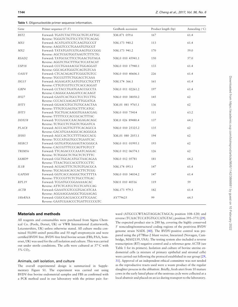
1144 Z. Cheng et al., 2017, Vol. 96, No. 6
Table 1. Oligonucleotide primer sequence information.
Gene Primer sequence (5′-3′) GenBank accession Product length (bp) Annealing (◦C)
BST2 Forward: TGATCTACTTCGCTGTCATTGC XM 871 059.6 167 61.4Reverse: TGGGTCTGTTCCTTCTTCAGAG
MX1 Forward: ACATGATCGTCAAGTGCCGT NM 173 940.2 113 61.4Reverse: AAGGTCCCTGAAATGTGCGT
MX2 Forward: TATATGATCGTGAAGTGCCGGG NM 173 941.2 170 59.0Reverse: AGCTCGGTGGTAAGTCTTTCTG
RSAD2 Forward: TATGCGCTTCCTGAACTGTAGA NM 0 010 45941.1 150 57.0Reverse: AGGTCTGCTTTGCTCCATACAT
USP18 Forward: CCCTGAAAACGCTGGAGGAT NM 0 010 17940.1 133 61.4Reverse: GGCAGATGGGTCAGTGTCAA
OAS1Y Forward: CTCACAGAGTTCGGGTGTCC NM 0 010 40606.1 226 61.4Reverse: TGCCGTTTCTGGACCTCAAA
ISG15 Forward: AGAAGATCAATGTGCCTGCTTT NM 174 366.1 161 61.4Reverse: CTTGTCGTTCCTCACCAGGAT
GBP4 Forward: CCTACCTGATGAACCGCCTA NM 0 011 02261.2 197 61.4Reverse: CAGGGCAAAGATCCACAAGT
IFI27 Forward: GAATCACTGCCTCCTCCTTG NM 0 010 38050.2 145 62Reverse: CCCACCAAGAGTTTGGATGA
IFIT1 Forward: GGAACGTGCTGTGCAACTAA XM 01 081 9765.1 136 62Reverse: TTTGTCGAGTGCTTTCATGC
IFIT3 Forward: TGCTGACAAGGTGAAACGAG NM 0 010 75414 111 63.2Reverse: TTTTTCCCACCGCACTTTAC
DDX58 Forward: TCCGAACCAACAGAGACAGC XM 0 026 89480.4 137 64.2Reverse: TCTGCCTCTGGTCTGGATCA
PLAC8 Forward: ACCCAGTTGTTTCACAGCCA NM 0 010 25325.2 135 62Reverse: GACATGAAAGGCACAGGGGA
IFIH1 Forward: AGCCACTCCTTTTAGCCACG XM 01 080 2053.1 194 62Reverse: TCCCATGGTGCCTGAATCAC
HERC5 Forward: GGTGATGGAAAGTACGGGCA NM 0 011 01995.1 193 62Reverse: CACCGTTTCCCAGTTGTCCT
TRIM56 Forward: TTCAGACCCCAAATCAGGAC NM 0 012 06574.1 126 62Reverse: TCTGGGCTCTGCTCTCTTTC
SAMD9 Forward: CGCTGGACATGCTAACAGAA NM 0 012 05781 181 64.2Reverse: TTAACTGCCACGTTCCCTTC
IL1B Forward: ACGAGTTTCTGTGTGACGCA NM 174 093.1 147 61.4Reverse: TGCAGAACACCACTTCTCGG
GAPDH Forward: GGTCACCAGGGCTGCTTTTA NM 0 010 34034.2 147 61.4Reverse: TTCCCGTTCTCTGCCTTGAC
RPL19 Forward: TCGATGCCGGAAAAACAC NM 0 010 40516 119 59Reverse: ATTCTCATCCTCCTCATCCAG
ACTB Forward: GAAATCGTCCGTGACATCAA NM 173 979.3 182 61.4Reverse: AGGAAGGAAGGCTGGAAGAG
18SrRNA Forward: CGGCGACGACCCATTCGAAC AY779625 99 64.5Reverse: GAATCGAACCCTGATTCCCCGTC
Materials and methods
All reagents and consumables were purchased from Sigma Chem-ical Co. (Poole, Dorset, UK) or VWR International (Lutterworth,Leicestershire, UK) unless otherwise stated. All culture media con-tained 50,000 units/l penicillin and 50 mg/l streptomycin and werecertified BVDV free. BVDV-free fetal bovine serum (FBS; PAA, Som-erset, UK) was used for the cell isolation and culture. This was carriedout under sterile conditions. The cells were cultured at 37◦C with5% CO2.
Animals, cell isolation, and cultureThe overall experimental design is summarized in Supple-mentary Figure S1. The experiment was carried out usingBVDV-free bovine endometrial samples and FBS as confirmed witha PCR method used in our laboratory with the primer pair: for-
ward (ATGCCCWTAGTAGGACTAGCA; position 108–128) andreverse (TCAACTCCATGTGCCATGTAC; position 395–375) [29].The expected product size is 288 bp, covering the highly conserved5′ noncoding/nonstructural coding regions of the pestivirus BVDVgenome strain NADL [40]. The BVDV-positive control was pre-pared using the pT7Blue-2 blunt vector, linearized (Novagen, Cam-bridge, MA02139, USA). The testing system also included a reversetranscription (RT)-negative control and a reference gene ACTB (seeTable 1 for its primers). Isolation and culture of bovine uterine en-dometrial cells (a mixture of primary epithelial and stromal cells)were carried out following the protocol established in our group [29,31]. Approval of an independent ethical committee was not neededas the reproductive tracts used were a waste product of the regularslaughter process in the abbattoir. Briefly, fresh uteri from 10 maturecows in the early luteal phase of the oestrous cycle were collected at alocal abattoir and placed on an ice during transport to the laboratory.
Dow
nloaded from https://academ
ic.oup.com/biolreprod/article/96/6/1142/3866681 by guest on 10 February 2022

BVDV inhibits bovine endometrial response to IFNT, 2017, Vol. 96, No. 6 1145
The cycle stage was estimated by the presence of a newly formed cor-pus hemorrhagicum in one of the ovaries. The uteri appeared healthybased on visual inspection at both collection and dissection. Strips ofintercaruncular endometrium were dissected and put into Dulbecco’sModified Eagle’s Medium/Nutrient Mixture F-12 Ham (DMEM/F12medium) (Sigma). After the strips were chopped into 1 mm3 cubesusing a mechanical tissue chopper (McIIwain Laboratory Engineer-ing, Guilford, Surrey, UK), about 40 g of the chopped tissue wereplaced into two 50 ml sterile vials and mixed with 30 ml digestivesolution containing 100 mg bovine serum albumin (BSA, Sigma), 50mg trypsin III (Worthington, Lakewood, NJ 0 8701, USA), and 50mg collagenase A (Roche, Welwyn Garden City, UK) per 100 ml ofHanks balanced salt solution (HBSS; Sigma). The vials were brieflycentrifuged at 100 × g and 10◦C for up to 1 min to allow the cells tosettle. The supernatant was then removed and replaced with 30 ml ofthe above digestive solution. This significantly increased the effectiveconcentration of the digestion and thus the yield of endometrial cells.After incubation for 90 min at 37◦C with 5% CO2 and manual mix-ing every 30 min, the cell suspension was filtered through a 100 μmmesh into 50 ml falcon vials containing 10% FBS and 3 μg/ml trypsininhibitor (Sigma) with HBSS added to 50 ml and centrifuged at 100 ×g at 10◦C for 10 min. Following two repetitions of the above wash-ing procedures, the cells were suspended with the culture medium(DMEM/F12 medium with 10% FBS) and plated in 24 well IWAKImicroplates (Scitech DIV, Asahi Techno Glass, Japan) at 2 ml perwell containing 0.5 × 10⁵ cells (day 1). Culture medium was changedevery 48 h to allow the cells to grow. The composition of the cellpopulation was confirmed using immunocytochemical staining vali-dated in our laboratory and showed that the stromal and epithelialcells constituted about 10% and 90% respectively of the populationon day 8 of culture [31], when IFNT challenge was carried out, withnegligible presence of immune cells (data not shown).
Infection of bovine endometrial cells with ncpBVDVThe ncpBVDV (Pe515nc strain) was provided by the BVDV ResearchGroup at the Royal Veterinary College, UK. This type 1 strain wasisolated from a cow diagnosed with mucosal disease and virologi-cally cloned as noncytopathogenic virus, and the consensus sequenceof the E2 region was established [41, 42]. The virus stock was prop-agated in BVDV-free Madin-Darby bovine kidney epithelial cells toachieve a 50% tissue culture infective dose (TCID50) of 5 × 10⁵ perml following the method described in detail previously [19, 31]. Thispropagated ncpBVDV was kept at –80◦C until use.
The endometrial cells from 10 cows used in this study were con-firmed to be initially BVDV negative. Cells from each cow were takenas a batch and grown in two 24-well plates as described previously(day 1, see Supplementary Figure S1). After the cells had grown for4 days, reaching about 70% confluence, FBS in the culture mediumwas reduced to 5% (maintenance medium, MM) to prevent over-growth of the cells. One plate was designated as the non-infectedcontrol and the other plate for the ncpBVDV infection. These plateswere subsequently maintained separately to avoid possible cross-contamination. To infect the cells with ncpBVDV, the wells wereinoculated with 0.25 ml of MM containing Pe515nc BVDV at amultiplicity of infection of 0.1 for 3 h. For the cells designated as thenon-infected controls, 0.25 ml MM was added to each well follow-ing the above procedures. The volume in all wells was made up to1 ml with MM, and the medium was changed after 2 days. Detailsof how the treatment times and doses for ncpBVDV infection wereoptimized have been described previously [19, 31]. The infection ofthe endometrial cells with ncpBVDV was confirmed using both the
PCR method described above with the extracted RNA and an in-direct enzyme (alkaline phosphatase) immunostaining procedure asdescribed previously [19, 29].
Treatment of bovine endometrial cells with IFNTIFNT (recombinant ovine IFNT, Cell Sciences, Canton, MA, USA)was prepared in MM at 100 ng/ml. Four days after the ncpBVDVinfection (day 8 of culture), 1 ml of this IFNT enriched medium wasadded to the designated wells and the incubation continued for afurther 24 h. The same procedure, but with 1 ml MM only, wasperformed for the other cells. The cells from each of the 10 replicatecows therefore included four treatment groups: control (CONT),ncpBVDV, IFNT, and IFNT+ncpBVDV. The treated cells were usedfor total RNA extraction (on day 9). The cell viability after expo-sure to the infection and treatment was also examined in a separateexperiment with a CellTiter 96 AQueous One Solution Cell Prolifer-ation Assay (Promega) following the supplied protocol as describedpreviously [31].
ISG15 protein measurementThe culture medium was collected 24 h following IFNT challengeand stored at −20◦C. Concentrations of ISG15 protein in themedium were quantified using an enzyme-linked immunosorbentassay (ELISA) (Insight Biotechnology, Middlesex, UK) following thesupplier’s protocol.
RNA extractionTotal RNA in the treated cells was extracted using RNeasy Mini Kits(Qiagen, Manchester, UK) following the supplier’s protocol. Eachtreatment comprised 6 duplicated wells in each plate and these werepooled for total RNA extraction (one pooled RNA sample for eachof four treatments in each cow). The concentrations and purity ofRNA were measured with a NanoDrop ND-1000 spectrophotometer(NanoDrop Technologies Inc., Wilmington, DE, USA). The RNAwas aliquoted as 1 μg/tube in 0.2 ml PCR tubes and stored at −80◦Cuntil RT.
Primer design and PCRThe primers were designed using a “Primer 3” web-based pro-gram (http://frodo.wi.mit.edu/primer3) with the DNA sequencesobtained from GenBank at NCBI (http://www.ncbi.nlm.nih.gov/Database/index.html). According to the recommendation by PCRBiosystems (London, UK) who supplied the reagents for cDNA syn-thesis (RT) and real time PCR (qPCR), the amplicon length formost of the genes was set to 100–200 bp with the predicted melt-ing temperature of around 60◦C using the default Primer 3 set-tings. Their alignment specificity was checked using the Blast toolat http://www.ncbi.nlm.nih.gov/tools/primer-blast/, and an Amplifytool (http://www.idtdna.com/analyzer/Applications/OligoAnalyzer/Default.aspx) was used to check the primer quality. The detailedinformation of the primers is given in Table 1. The primers weremade by Eurofins MWG Operon (Ebersberg, Germany).
One microgram of total RNA was treated with DNase to elim-inate potential genomic DNA contamination using a RQ1 RNase-Free DNase kit (Promega) and then reverse transcribed into cDNAin a 20 μl reaction volume using a cDNA synthesis kit supplied byPCR Biosystems following the supplier’s protocol. Each 20 μl re-action contained 4 μl 5 × cDNA synthesis mix, 1 μl 20 × RTase,and 1 μg of DNase-treated RNA sample in 15 μl PCR grade water.RT for all samples was performed in one assay to minimize varia-tion. The resulting cDNA was diluted in nuclease-free water up to
Dow
nloaded from https://academ
ic.oup.com/biolreprod/article/96/6/1142/3866681 by guest on 10 February 2022

1146 Z. Cheng et al., 2017, Vol. 96, No. 6
100 μl to achieve a concentration of 10 ng/μl and used for qPCRand conventional PCR. The conventional PCR for the tested geneswas carried out using the G-Storm thermal cycler (G-Storm Ltd,Somerset, UK), Qiagen Multiple PCR kit (Qiagen), and the primerslisted in Table 1 following the methods described previously [29].The specificity of primers was verified using electrophoresis on a2% (w/v) agarose gel. The cDNA amplicon for each gene was pu-rified using a QIAquick PCR purification kit (Qiagen), and theirquality and concentrations were determined with a NanoDrop ND-1000 spectrophotometer (NanoDrop Technologies Inc., Wilminton,USA). It was stored at −80◦C for use in the qPCR standard curveand annealing temperature optimization.
qPCR analysis for gene expressionAn absolute qPCR method was used to quantify the mRNA expres-sion of all selected genes, including 18 target genes (BST2, MX1,MX2, RSAD2, USP18, OAS1Y, ISG15, GBP4, IFI27, IFIT1, IFIT3,DDX58, PLAC8, IFIH1, HERC5, TRIM56, SAMD9, and IL1B)and four potential reference genes (GAPDH, RPL19, ACTB, and18SrRNA) using methods reported previously [43]. A temperaturegradient (55◦C–65◦C) qPCR with 8 identical reactions was carriedout to determine the annealing temperature which produced maxi-mal amplicon and the amplicon-specific melting temperatures of theprimers using a gradient function of the qPCR machine (CFX96Real-Time System DNA, Bio-Rad Laboratories, CA, USA). Each re-action contained 2 ng of the DNA standard, 10 μl Sygreen Mix (PCRBiosystems), 0.8 μl of each 10 μM forward and reverse primer, andnuclease-free water added up to 20 μl. The standard curve contained8 concentrations from 1 to 1 × 10⁻⁷ ng/ml prepared using the pu-rified DNA for each gene. Each qPCR assay contained the standardcurve, no template control, and sample cDNA for a testing genefrom all cows in duplicate. Each qPCR vial contained 5 μl of cDNAstandard or samples, 10 μl Sygreen Mix (PCR Biosystems), 0.8 μl of10μM forward primer, 0.8 μl reverse primer (see Table 1), and 3.4 μlnuclease-free water. Reactions and data acquisition were carried outon CFX96 Real -Time Systems (Bio-Rad) following the protocolsupplied by PCR Biosystems, including an initial Taq activation stepat 95◦C for 2 min followed by 38 cycles of denaturation (95◦C),annealing (the annealing temperatures are given in Table 1), andextension (63◦C). In addition, an amplicon-specific melting temper-ature obtained in the above gradient test was applied to avoid anynoise from smaller nonspecific products, such as dimers, prior to theproduct acquisition. Concentrations of the sample amplicons werecalculated using the standard curve and a semilog regression built inthe CFX Manager software package (Bio-Rad). The limit of quan-tification was 1 × 10⁻⁶ to 1 × 10⁻⁷ng/ml for all tested genes.
Statistical data analysisAmong the four selected reference genes, only the expression ofGAPDH was not significantly altered by the treatments (see Resultssection below). The concentration of DNA for all the target geneswas, therefore, normalized to the expression of GAPDH as describedpreviously [44]. Statistical data analysis was carried out using SPSSV23 (Chicago, IL, USA). Logarithmical transform (BST2, HERC5,ISG15, MX2, OAS1Y, RSAD2, SAMD9, and USP18) was appliedwhere the data were not normally distributed. A linear mixed effectmodel was used for analysis of variance (ANOVA) with random-ized block design, which included treatments (CONT, ncpBVDV,IFNT, and their combination) as fixed effects and cow as randomeffect. The statistical level of significance was set to P < 0.05. WhereANOVA showed statistical significance, Fisher LSD multiple com-
Figure 1. Validation of ncpBVDV infection in bovine endometrial cell culturegroups using PCR with RNA from the cells harvested at the end of the exper-iment. L1: DNA ladder, L2: ncpBVDV positive control, L3: CONT, L4: IFNT, L5:ncpBVDV, and L6: ncpBVDV+IFNT.
parisons based on the least square means were performed to identifythe sources of differences.
Results
The PCR test using the RNA extracted from the uterine tissuesshowed that the uteri used for the experiment were initially free ofBVDV infection. An immunocytochemical procedure as describedpreviously [31] was used to demonstrate that ncpBVDV antigenwas detected in both the epithelial and stromal cells following ex-perimental infection, with no cross-infection in the controls (datanot shown). At the end of culture (day 9 in total and 5 days afterncpBVDV infection), ncpBVDV RNA was also detected by PCRin the endometrial cultures treated with ncpBVDV or ncpBVDV +IFNT while it was not detectable in the CONT or IFNT-treatedsamples (Figure 1). The MTS reduction assay demonstrated that vi-ability was not affected by the individual or combined treatmentsat the doses used. After exposure of the cultured endometrial cellsto ncpBVDV for 4 days and to IFNT at 0 or 100 ng/ml for 24h, the absorbance values at 490 nm in a cell viability assay were1.7 ± 0.07 for CONT, 1.8 ± 0.04 for IFNT, 1.7 ± 0.09 for ncp-BVDV, and 1.7 ± 0.06 for ncpBVDV + IFNT (mean ± SE, n= 6/group, P > 0.05). These results are in accord with the strainof BVDV selected for use which was known to be noncytopathic.
Effect of ncpBVDV, IFNT, and their combination onexpression of the selected reference genes by uterineendometrial cellsAll four potential reference genes (ACTB, RPL19, GAPDH, and18SrRNA) were highly expressed in the cultured bovine endome-trial cells as measured in absolute units using qPCR (Table 2). OnlyGAPDH expression, however, remained stable following treatments(P > 0.05). RPL19 expression was increased by the individual treat-ments with IFNT or ncpBVDV, while expression of ACTB and18SrRNA was significantly lower following the combined treatment(P < 0.05–0.01). For subsequent comparison of the treatment ef-fects, the expression levels of each gene were, therefore, normalizedagainst GAPDH. The absolute expression values of the candidategenes were also shown for the CONT cell cultures. All 18 candi-date genes were detected with a large range in the basal levels ofexpression (Table 3).
Dow
nloaded from https://academ
ic.oup.com/biolreprod/article/96/6/1142/3866681 by guest on 10 February 2022

BVDV inhibits bovine endometrial response to IFNT, 2017, Vol. 96, No. 6 1147
Table 2. Effect of ncpBVDV, IFNT and their combination on expression of the selected reference genes in cultured mixed bovine endometrialcells.
Treatments ACTB RPL19 GAPDH 18SrRNA
CONT 81 ± 8.6a 118 ± 10.3b 159 ± 16.4 27,490 ± 2301.3a
IFNT 99 ± 20.5a 166 ± 19.7a 165 ± 24.8 23,258 ± 2270.5a
ncpBVDV 74 ± 11.7a,b 151 ± 6.0a 170 ± 11.1 26,189 ± 3490.3a
IFNT+ncpBVDV 48 ± 7.3b 138 ± 12.0a,b 170 ± 17.7 15,665 ± 813.2b
Expressed in pg/μg RNA.Within columns, a>b, P < 0.05–0.01.
Table 3. Basal expression values of the candidate genes measured in fg/μg RNA in the CONT cultures of mixed bovine endometrial cellsusing absolute qPCR.
High expression Moderate expression Low expression
PLAC8 9240 ± 2002.7 HERC5 530 ± 90.9 USP18 0.63 ± 0.17IFIT1 4634 ± 771.8 GBP4 306 ± 108.7 OAS1Y 0.14 ± 0.04IFIH1 2144 ± 278.6 IFI27 155 ± 29.0 BST2 0.0014 ± 0.0004SAMD9 1374 ± 329.7 ISG15 96 ± 19.4TRIM56 1094 ± 74.7 RSAD2 92 ± 16.9DDX58 984 ± 42.5 MX1 50 ± 4.8
IL1B 24 ± 5.7MX2 5.2 ± 1.2IFIT3 1.9 ± 0.9
Note that expression levels do not necessarily translate into similar levels of protein production due to further down-stream regulation in processing.
Effect of ncpBVDV and IFNT alone on expression ofcandidate genesncpBVDV infection alone generally had little effect on gene expres-sion (Figure 2). Only one gene, TRIM56, was significantly upreg-ulated 5 days after infection (P < 0.001), whereas expression ofthree ISGs (ISG15, MX2, BST2) together with IL1B was reduced(P < 0.05). As anticipated, IFNT treatment alone significantly in-creased expression of all 17 candidate ISGs tested (P < 0.05–0.0001)but there was a tendency (P = 0.06) for IFNT to decrease expressionof IL1B (Figure 2).
Effect of IFNT on expression of candidate genes inBVDV-infected endometrial cellsIn contrast to the limited effect of ncpBVDV infection alone, thevirus had a profound influence on the ability of IFNT to upregu-late the expression of the candidate ISGs. Of the 17 ISGs tested, thestimulatory effect of IFNT on expression of 15 was significantly in-hibited (P < 0.05–0.001), generally to a value intermediate betweenthat found in the CONT and IFN treated cells (Figure 2). Genesresponding in this way included ISG15, HERC5, USP18, DDX58,IFIH1, IFIT1, IFIT3, BST2, MX1, MX2, RSAD2, OAS1Y, SAMD9,GBP4, and PLAC8. BVDV infection did not, however, alter IFNT-stimulated mRNA expression of two genes (P > 0.05), TRIM56 orIFI27. Whereas ncpBVDV alone caused a small but significant re-duction in IL1B (P < 0.05), expression of this gene was markedlyreduced by the combined treatment (P < 0.001).
ISG15 protein assayISG15 is known to be secreted as a cytokine, so its protein con-centration was measured by ELISA in the medium. This was nearlydoubled following treatment with IFNT alone (P < 0.01). BVDVinfection alone did not change the concentration but in the com-bined treatment BVDV completely inhibited the IFNT stimulation(Figure 3).
Discussion
Many previous publications have described the key influence of IFNTin the MRP in cows, which includes maintenance of the corpus lu-teum and development of a receptive environment in the uterus forsuccessful implantation (reviewed by [25, 27, 28]). During this pro-cess, many ISG are upregulated in the endometrial cells [22, 23, 25],and we confirmed here that expression of all the candidate ISGs in-creased significantly following treatment with IFNT alone. BVDVis a major cause of reduced fertility in many countries [2, 3]. Thevirus is known to have evolved a variety of mechanisms to help itevade detection and eradication by host cells [13–15]. We confirmedhere that ncpBDVD can become established and proliferate withinepithelial and stromal cells of the uterine endometrium. While infec-tion alone produced few changes in gene expression, we show herefor the first time that the ability of endometrial cells to respond toIFNT by increased expression of a variety of ISGs was markedlyinhibited in ncpBDVD-infected cells. This is likely to facilitate theability of the virus to survive within the uterine environment andmay well contribute to the reduced fertility of infected cows.
The model system we developed was based on a mixed cultureof epithelial and stromal cells as both can become infected withvirus, and the interaction between the two cell types is important inthe MRP. Both cell types are also known to exhibit innate immuneactivity [33]. The immune cell population may be important mod-ulators of the response in vivo but were not included in our system[31], allowing us to demonstrate responses of the endometrial cellsalone. We have shown previously that infection of these mixed en-dometrial cultures with ncpBVDV inhibits their immune response toa challenge with bacterial LPS [19]. Further validation of the sys-tem was provided by showing that IFNT treatment alone stimulatedexpression of PTGS1 and synthesis of PGE2 [29]. This is in accordwith the suggested importance of PGE2 as a key signaling molecule inearly pregnancy [27, 30]. The cultured cells expressed PGR, ESR1,and OTR as expected [29] in addition to the TLR receptors whichare important for the detection of RNA viruses (TLR2, −3, −4, −7,
Dow
nloaded from https://academ
ic.oup.com/biolreprod/article/96/6/1142/3866681 by guest on 10 February 2022

1148 Z. Cheng et al., 2017, Vol. 96, No. 6
Figure 2. Effect of CONT, ncpBVDV, IFNT, and ncpBVDV+IFNT treatments on gene expression in bovine endometrial cells. Values are expressed as the ratios toGAPDH after an absolute gene quantification using qPCR and are presented as the mean ± SEM. The columns labeled with different letters were significantlydifferent at P < 0.05–0.0001 (a > b > c > d).
Dow
nloaded from https://academ
ic.oup.com/biolreprod/article/96/6/1142/3866681 by guest on 10 February 2022

BVDV inhibits bovine endometrial response to IFNT, 2017, Vol. 96, No. 6 1149
Figure 3. Effect of CONT, ncpBVDV, IFNT, and ncpBVDV+IFNT treatments onsecretion of ISG15 into the culture medium. Results are presented as themean ± SEM of 10 individual cow endometrial samples tested. The columnslabeled with different letters were significantly different a > b at P < 0.05–0.01.
and −8) [16, 45]. Progesterone is also known to play a key rolein modulating the endometrial response to IFNT. The timing of thepostovulatory progesterone rise co-ordinates the temporal changes inthe endometrial transcriptome. Progesterone downregulates its ownreceptor while inducing a variety of genes whose expression is laterincreased further by IFNT. These include genes involved in transportof glucose and amino acids, cell proliferation, and PG synthesis [28,32]. Progesterone treatment was not included in the current exper-imental design as it would have introduced too many variables totest with adequate replication in the same batch of isolated cells.Its absence may, however, have altered some of the gene expressionprofiles in response to IFNT in comparison with the in vivo situation.
The cells were infected with ncpBVDV several days before theIFNT challenge. This was done to mimic one potential in vivo sit-uation. Cows which have not previously been exposed to BVDVgenerally become infected following a herd breakdown, either di-rectly from the PI animal or from other cows which have recentlybecome acutely infected. This may happen during the calving periodwhen heifers and older cows are often mixed. Infection is also pos-sible (although less likely) with semen from a PI bull or followingintrauterine transfer of infected embryos on day 7 of the oestrouscycle [9, 10, 46]. The length of time the virus can subsequently sur-vive in the uterus has not been well established, but there is evidencethat this can be for several weeks. Depending on the relative tim-ing of infection and mating, this could impair fertilization and veryearly embryo development [47] or disrupt MRP as suggested by thepresent experiment.
One key mechanism by which BVDV can evade the host immuneresponse is by the action of the viral protein Npro which blocks pro-duction of type 1 interferons [13, 19, 48]. On the other hand, therewas upregulation of type I interferon-induced genes in spleen andtracheobronchial lymph nodes of beef calves 5 days after BVDV in-fection [49] and in the blood of transiently infected mid-pregnantheifers and their fetuses [50]. There are a number of possible rea-sons behind these discrepancies. Different cell types may respondto BVDV infection differently and there are differences in virulencebetween strains of type 1 and type 2 ncp BVDV [51]. The absenceof immune cells in most in vitro cultures is also a likely factor. Nev-
ertheless, the lack of any rise in expression of ISGs with the singleexception of TRIM56 following the ncpBVDV treatment alone inthis study implies that the endometrial cells were not exposed to asignificant increase in self-generated type 1 IFN prior to the experi-mental treatment with IFNT.
When faced with a viral attack, the body mobilizes its defensesystems to restrict, neutralize, and remove the virus. Gene targetingstudies have revealed that there are four main effector pathways ofIFN-mediated antiviral responses: an ISG15 ubiquitin-like pathway,an MxGTPase pathway, an OAS1-RNaseL pathway, and a proteinkinase R pathway [52]. Our present study demonstrated that IFNTchallenge activated these pathways to develop a proimmune andantiviral environment in the uterus by stimulating ISG expression.Inhibition of the stimulatory effect of IFN on uterine ISG productionmay, however, facilitate the establishment of life-long persistent in-fection of bovine fetuses with BVDV following intrauterine infectionof the dam in the first trimester of pregnancy [53, 54], so leading tothe birth of PI animals which are a major cause of disease spreadwithin herds [4].
ISG15 is one of the most upregulated genes during pregnancyrecognition in cows. This may facilitate successful conceptus attach-ment or act as a defense strategy against infection [22, 25, 55].Its precise role in this respect has yet to be determined but studiesin mice have shown increased embryo mortality in Isg15− /− dams[56]. In this study, BVDV infection markedly inhibited both basaland IFNT-induced ISG15 expression. Free ISG15 is a cytokine whichinduces natural killer cell proliferation and IFN-γ production andto act as a chemotactic factor for neutrophils [57], and we con-firmed here both its IFNT-stimulated secretion into the medium andthe suppression of the stimulation by BVDV infection. ISG15 alsoacts intracellularly as an ubiquitin-like modifier of many target pro-teins. In a process known as ISGylation, the C-terminus of ISG15is conjugated to lysine residues in the target protein following con-secutive catalysis with three enzymes E1, E2, and E3 [58] (see alsoSupplementary Figure S2). The modified protein may be targeted tolysosomes for destruction or used elsewhere. HERC5 and TRIM56are both E3 ligases. HERC5 does not have substrate specificity soit is able to block the IFN-mediated rise in the total level of IS-Gylated cellular proteins [58]. TRIM56 is a single-RING-finger E3ligase which interacts specifically with the N-terminal protease ofBVDV, Npro, so exhibiting antiviral activity [17]. It was interestingto note that TRIM56 was the only ISG tested which was upregulatedby BVDV alone whereas HERC5 followed the more common pat-tern in which ncpBVDV alone had no effect but the virus markedlyinhibited IFNT-stimulated upregulation.
USP18 is an ubiquitin-specific protease which removes ISG15from the modified protein (De-ISGylation) and can also process theISG15 precursor protein (Supplementary Figure S2) [34–36]. IFN-induced USP18 expression was also inhibited by ncpBVDV. ISGy-lation affects many proteins with important biological functions,including regulation of (1) innate immunity and antiviral/bacterialinfections, (2) proteasome function for protein turnover, (3) apop-tosis and tumorigenesis, and (4) cell proliferation and remodeling[36]. The overall upregulation of ISG15, HERC5, and USP18 byIFNT supports the suggestion that the ISGylation pathway is animportant regulator of protein processing in the endometrium dur-ing MRP [55], with the results of this study implying that this sys-tem can be inhibited by ncpBVDV. On the other hand, upregu-lation of TRIM56 by ncpBVDV shows that the endometrial cellswere still able to offer some immune response, even if this wasmuch reduced.
Dow
nloaded from https://academ
ic.oup.com/biolreprod/article/96/6/1142/3866681 by guest on 10 February 2022

1150 Z. Cheng et al., 2017, Vol. 96, No. 6
Figure 4. Diagrams illustrating the potential effects of an acute ncpBVDV infection on expression of uterine ISGs and pregnancy recognition in the cow. Panel Ashows the points at which cells normally detect and deactivate the virus. (1) ncpBVDV reaches the reproductive tract following infection. (2) The virus attachesto the host cell membrane and is taken up by clathrin-mediated endocytosis. (3) Virus is incorporated into the endosome, where it may be sensed by TLR3or TLR7/8. (4) The endosome buds off multivesicular bodies. While in these ncpBVDV may be protected from detection by cytoplasmic pattern recognitionreceptors (PRR), in particular DDX58 which detects ssRNA. SAMD9 is a facilitator of endosome fusion. (5) Alternatively, virus may be detected and targeted tolysosomes for disposal. This may involve ISGylation. ISG15 may also be secreted as a cytokine. (6) Viral ssRNA is released into the cytoplasm. (7) Viral mRNAis translated into a single polyprotein in the endoplasmic reticulum. This is cleaved into structural proteins (three envelopes and one capsid protein) and abouteight nonstructural proteins, which are involved in replication and assembly. These include the protease Npro. Viral ssRNA is replicated and transcribed. Duringthis process, double-stranded (ds) RNA is produced, which can be detected by the PRR IFIH1. New virions are assembled. The ISGs RSAD2, MX1, MX2, IFIT1,IFIT3, IRF3, and OAS1Y are all able to disrupt viral transcription and/or translation (8) The ncpBVDV virions pass through the Golgi complex. (9) Virions aretransported from the Golgi to the cell surface in small exocytotic vesicles. (10) New virions are released from the cell by exocytosis. Both RSAD2 and BST2are able to inhibit viral egress. ∗IFNT-stimulated ISGs significantly suppressed by ncpBVDV. Panel B illustrates possible consequences of ncpBVDV infection forsignaling by IFNT during the MRP. (1) Infection of bovine endometrium with ncpBVDV and viral intracellular replication. (2) ncpBVDV produces viral factors whichcan counteract the host’s immune response. (3) The early bovine conceptus subsequently secretes IFNT into the uterine lumen which initiates and establishesMRP, normally by day 16 of gestation in cows. (4) IFNT, a type I IFN, signals through binding to the interferon alpha receptors (IFNAR) to stimulate production ofmany ISGs crucial for MRP and uterine immunity. (5, 6, 7) Viral-derived factors inhibit the type I IFN signaling and transcription pathways, leading to decreasedexpression of many IFNT-stimulated ISGs. (8) ncpBVDV infection may, therefore, disrupt MRP and uterine immunity.
Dow
nloaded from https://academ
ic.oup.com/biolreprod/article/96/6/1142/3866681 by guest on 10 February 2022

BVDV inhibits bovine endometrial response to IFNT, 2017, Vol. 96, No. 6 1151
DDX58 (RIGI), IFIH1 (MDA5), IFIT1 (ISG56), MX1, and MX2are all potential targets of ISGylation. DDX58 and IFIH1 are bothDEAD box proteins which are cytosolic sensors of viral RNA. Theyact as RNA helicases and are implicated in a number of cellularprocesses involving RNA binding and alteration of RNA secondarystructure. DDX58 recognizes ssRNA, whereas IFIH1 targets dsRNA.As BVDV is a single-stranded virus, it would only be recognized byIFIH1 during the RNA replication phase [50, 53, 59]. The virus mayevade detection by rapid incorporation into the endosome whichbuds off to form multivesicular bodies [60, 61]. Although these canpotentially target their contents to lysosomes for disposal, multivesic-ular bodies can also shield the viral protein from detection exceptwhen the viral ribonoprotein is released into the cytoplasm beforeuptake into the endoplasmic reticulum for replication (see Figure 4).In contrast to the situation reported here, both DDX58 and IFIH1were upregulated in fetal blood cells in response to persistent infec-tion with ncpBVDV [50, 53, 59].
All the remaining ISGs studied apart from PLAC8 have knownantiviral activity. IFIT1 and IFIT3 (also called retinoic acid-inducedgene G protein, RIGG) encode IFN-inducible proteins which canform a cytoplasmic complex to recognize and destroy viral RNA,acting as inhibitors of viral replication [62, 63]. We found thatIFIT1 was much more highly expressed than IFIT3 in bovine en-dometrial cells, by a factor of over 2000. MX1 and MX2, theMx dynamin-like GTPases, are key effectors in MxGTPase antiviralpathways. They can disturb the transport, transcription, and trans-lation of viruses within the cells [52, 64]. IFI27 (ISG12) is involvedin promoting apoptosis via mitochondrial membrane destabilizationthat may influence the innate immune response of IFNs and cellu-lar metabolism [65]. This and TRIM56 were the only ISGs testedin the present experiment in which upregulation by IFNT was notaffected by ncpBVDV infection. The OAS1 (including OAS1Y) genefamily are the key effectors in the OAS1-RNaseL antiviral path-way [52]. They sense exogenous nucleic acid and activate endori-bonuclease L (RNAseL) which degrades viral RNA [66]. RSAD2(Viperin) interacts with nonstructural viral proteins to inhibit viralRNA replication, and can also limit egress of some viruses fromthe cell [67, 68]. GBP4 is an IFN-induced GTPase which disruptsthe interaction of TNF receptor-associated factor 6 (TRAF6) withIRF7, so reducing virus-mediated induction of type 1 IFNs [18].SAMD9 is a facilitator of endosome fusion which participates in theformation of granules with antiviral properties [69]. BST2 acts asthe “tetherin” molecule to restrict the egress of many viruses, suchas HIV-1 and Ebola virus, reducing their spread within the host[70]. The downregulation of the stimulatory effect of IFNT on allthese genes by ncpBVDV is therefore clearly able to inhibit manyof the mechanisms by which a cell normally combats a viral attack(Figure 4A).
PLAC8 was originally identified in mouse placenta [71]. It hassubsequently been detected in a variety of cell types including bovineendometrium [72] and was the gene with the highest basal expres-sion value in the bovine endometrial cells studied in the presentpaper. It is upregulated by IFNT but its function appears variableaccording to cell type. It can inhibit growth of some cancers byattenuating cell-cycle progression, either induce or inhibit apop-tosis, and inhibit proliferation of endothelial colony-forming cells[38, 73], while PLAC8 deficient mice have defects in innate immu-nity [74]. The role of PLAC8 in bovine implantation remains to bedetermined.
The final gene investigated in this study was the proinflamma-tory cytokine IL1B. In this case, IFNT treatment alone tended to
reduce expression whereas the combined treatment of ncpBVDV +IFN resulted in significant downregulation in the endometrial cells.Its moderate inhibition by IFNT may be beneficial during implan-tation by preventing immune rejection [39], but the much greaterinhibition in the presence of ncpBVDV may lead to decreased uter-ine immunity. In addition to the antiviral effects of uterine ISGs,many of the proteins encoded by these genes also possess antibac-terial functions. Numerous immune genes, including all the ISGsexamined here (ISG15, HERC5, USP18, DDX58, IFIT1, IFIT3,MX2, RSAD2, GBP4, SAMD9, IFI27, OAS1Y, and BST2), wereupregulated following treatment of bovine uterine endometrial cellswith the bacterial endotoxin LPS [31]. This stimulatory effect wasagain significantly inhibited in the presence of ncpBVDV infection[19, 31]. Infection of bovine uterine endometrial cells with ncpBVDValso induced an endocrine switch of PG production from PGF2α toPGE2 [29]. As PGF2α is an immune enhancer and PGE2 is an im-mune suppressor in the uterus [75, 76], this switch would also leadto decreased uterine immunity. These results support the suggestionthat BVDV infection can predispose cows to develop endometritisdue to bacterial infection following calving. This in turn may alsocontribute to early embryonic death and failure of pregnancy estab-lishment [77].
In summary, IFNT challenge significantly stimulated the mRNAexpression of many ISGs in uterine endometrial cells. These ISGsare clearly important for pregnancy recognition and implantationin the cow, although their precise actions in this respect generallyremain to be established. They do, however, have more well definedroles in protecting the host from both viral and bacterial infections.We show here in an in vitro situation that the stimulatory effect ofIFNT on 15 out of 17 ISGs tested was inhibited by prior infectionwith ncpBVDV, as summarized in Figure 4. As outlined above, ncp-BVDV normally evades detection in host cells by downregulation oftype 1 IFN synthesis. As it was still able to downregulate nearly allthe ISGs in the face of exogenous IFNT treatment, the virus clearlyhas additional defense mechanisms available. Future work is war-ranted to determine whether these ISGs are also downregulated invivo in the endometrium of recently mated cows transiently infectedwith BVDV. Previous studies show that if bovine conceptuses havenot elongated sufficiently by day 16 of pregnancy, then their IFNTproduction will be inadequate to prevent onset of the normal lute-olytic mechanism [21, 24]. This provides supporting evidence that afailure of the normal downstream effects of IFNT, as shown here, isalso likely to result in pregnancy failure. Decreased uterine immunityagainst both viral and bacterial infection may provide another mech-anism whereby ncpBVDV infection can cause early embryonic deathand reduced fertility. Finally, the remarkable ability of ncpBVDV toinhibit bovine uterine defense systems may, at least in part, providea mechanism whereby the reproductive system becomes a major sitefor ncpBVDV infection, facilitating maintenance and spread of thisdisease within the cattle population.
Supplementary data
Supplementary data are available at BIOLRE online.Supplementary Figure S1. Summary diagram showing the experi-
mental design used. ncpBVDV, noncytopathic bovine virus diarrheavirus; CONT, control; IFNT, interferon tau.
Supplementary Figure S2. Illustration of the ISGylation pathway.Proteases cleave off the C-terminal extensions from ISG15 precur-sors to generate mature ISG15. This may be secreted as a cytokineor released during cell lysis. Alternatively, ISG15 may be activated
Dow
nloaded from https://academ
ic.oup.com/biolreprod/article/96/6/1142/3866681 by guest on 10 February 2022

1152 Z. Cheng et al., 2017, Vol. 96, No. 6
by the sequential actions of enzymes E1 and E2. It is then linked to atarget protein by the action of an ISG15 E3 ligase [58]. These includeHERC5 and TRIM56. The modified protein may be either targetedto lysosomes for destruction or used elsewhere in the cell. USP18(UBP43) reverses the ISGylation process by cleaving off ISG15 fromthe target protein. It can also act as one of the ISG processing pro-teases. The pathway is upregulated by type 1 interferons. These in-clude IFNα, normally produced in response to a viral infection, andIFNT, produced by the ruminant conceptus during the MRP.
Acknowledgments
The authors would like to thank Professor Joe Brownlie and Miss OliviaAnstaett in the Royal Veterinary College for their generous provision of theBVDV strain.
REFERENCES
1. De Vries A. Economic value of pregnancy in dairy cattle. J Dairy Sci 2006;89:3876–3885.
2. Fray MD, Paton DJ, Alenius S. The effects of bovine viral diarrhoea viruson cattle reproduction in relation to disease control. Anim Reprod Sci2000; 60–61:615–627.
3. Grooms DL. Reproductive consequences of infection with bovine viraldiarrhea virus. Vet Clin North Am Food Anim Pract 2004; 20:5–19.
4. Lanyon SR, Hill FI, Reichel MP, Brownlie J. Bovine viral diarrhoea: patho-genesis and diagnosis. Vet J 2014; 199:201–209.
5. McGowan MR, Kirkland PD, Richards SG, Littlejohns IR. Increased re-productive losses in cattle infected with bovine pestivirus around the timeof insemination. Vet Rec 1993; 133:39–43.
6. Brownlie J, Clarke MC, Howard CJ. Experimental infection of cattle inearly pregnancy with a cytopathic strain of bovine virus diarrhoea virus.Res Vet Sci 1989; 46:307–311.
7. Collins ME, Heaney J, Thomas CJ, Brownlie J. Infectivity of pes-tivirus following persistence of acute infection. Vet Microbiol 2009;138:289–296.
8. Kirkland PD, Richards SG, Rothwell JT, Stanley DF. Replication of bovineviral diarrhoea virus in the bovine reproductive tract and excretion of virusin semen during acute and chronic infections. Vet Rec 1991; 128:587–590.
9. Bielanski A, Algire J, Lalonde A, Garceac A. Embryos produced fromfertilization with bovine viral diarrhea virus (BVDV)-infected semen andthe risk of disease transmission to embryo transfer (ET) recipients andoffspring. Theriogenology 2013; 80:451–455.
10. Bielanski A, Sapp T, Lutze-Wallace C. Association of bovine embryosproduced by in vitro fertilization with a noncytopathic strain of bovineviral diarrhea virus type II. Theriogenology 1998; 49:1231–1238.
11. Kirkland PD, McGowan MR, Mackintosh SG. Factors influencing thedevelopment of persistent infection of cattle with pestivirus. In EdwardsS, (ed.), Proceedings of the 2nd Symposium on Pestiviruses: FondationMarcel Merieux, Lyon 1993, 117–121.
12. Firat I, Ak S, Bozkurt HH, Ak K, Turan N, Bagcigil F. Distribution ofbovine viral diarrhoea virus (BVDV) in the genital system tissues of cattle.Veterinarski Arhiv 2012; 72:235–248.
13. Charleston B, Fray MD, Baigent S, Carr BV, Morrison WI. Establishmentof persistent infection with non-cytopathic bovine viral diarrhoea virus incattle is associated with a failure to induce type I interferon. J Gen Virol2001; 82:1893–1897.
14. Baigent SJ, Goodbourn S, McCauley JW. Differential activation of in-terferon regulatory factors-3 and -7 by non-cytopathogenic and cy-topathogenic bovine viral diarrhoea virus. Vet Immunol Immunopathol2004; 100:135–144.
15. Chen Z, Rijnbrand R, Jangra RK, Devaraj SG, Qu L, Ma Y, Lemon SM, LiK. Ubiquitination and proteasomal degradation of interferon regulatoryfactor-3 induced by Npro from a cytopathic bovine viral diarrhea virus.Virology 2007; 366:277–292.
16. Jensen S, Thomsen AR. Sensing of RNA viruses: a review of innate im-mune receptors involved in recognizing RNA virus invasion. J Virol 2012;86:2900–2910.
17. Wang J, Liu B, Wang N, Lee YM, Liu C, Li K. TRIM56 is a virus- andinterferon-inducible E3 ubiquitin ligase that restricts pestivirus infection.J Virol 2011; 85:3733–3745.
18. Hu Y, Wang J, Yang B, Zheng N, Qin M, Ji Y, Lin G, Tian L, Wu X, WuL, Sun B. Guanylate binding protein 4 negatively regulates virus-inducedtype I IFN and antiviral response by targeting IFN regulatory factor 7. JImmunol 2011; 187:6456–6462.
19. Oguejiofor CF, Cheng Z, Abudureyimu A, Anstaett OL, Brownlie J,Fouladi-Nashta AA, Wathes DC. Global transcriptomic profiling of bovineendometrial immune response in vitro. II. Effect of bovine viral diarrheavirus on the endometrial response to lipopolysaccharide. Biol Reprod2015; 93:101.
20. Kimura K, Spate LD, Green MP, Murphy CN, Seidel GE, Jr, Roberts RM.Sexual dimorphism in interferon-tau production by in vivo-derived bovineembryos. Mol Reprod Dev 2004; 67:193–199.
21. Robinson RS, Fray MD, Wathes DC, Lamming GE, Mann GE. In vivoexpression of interferon tau mRNA by the embryonic trophoblast anduterine concentrations of interferon tau protein during early pregnancy inthe cow. Mol Reprod Dev 2006; 73:470–474.
22. Forde N, Carter F, Spencer TE, Bazer FW, Sandra O, Mansouri-Attia N,Okumu LA, McGettigan PA, Mehta JP, McBride R, O’Gaora P, Roche JFet al. Conceptus-induced changes in the endometrial transcriptome: howsoon does the cow know she is pregnant? Biol Reprod 2011; 85:144–156.
23. Lonergan P, Forde N. Maternal-embryo interaction leading up tothe initiation of implantation of pregnancy in cattle. Animal 2014;8(Suppl 1):64–69.
24. Mann GE, Lamming GE, Robinson RS, Wathes DC. The regulation ofinterferon-tau production and uterine hormone receptors during earlypregnancy. J Reprod Fertil Suppl 1999; 54:317–328.
25. Bazer FW. Pregnancy recognition signaling mechanisms in ruminants andpigs. J Anim Sci Biotechnol 2013; 4:23.
26. Dorniak P, Bazer FW, Wu G, Spencer TE. Conceptus-derivedprostaglandins regulate endometrial function in sheep. Biol Reprod 2012;87:1–7.
27. Spencer TE, Forde N, Dorniak P, Hansen TR, Romero JJ, LonerganP. Conceptus-derived prostaglandins regulate gene expression in the en-dometrium prior to pregnancy recognition in ruminants. Reproduction2013; 146:377–387.
28. Hansen PJ. The immunology of early pregnancy in farm animals. ReprodDomest Anim 2011; 46(Suppl 3):18–30.
29. Cheng Z, Abudureyimu A, Oguejiofor CF, Ellis R, Barry AT, Chen X,Anstaett OL, Brownlie J, Wathes DC. BVDV alters uterine prostaglandinproduction during pregnancy recognition in cows. Reproduction 2016;151:605–614.
30. Arosh JA, Banu SK, McCracken JA. Novel concepts on the roleof prostaglandins on luteal maintenance and maternal recognitionand establishment of pregnancy in ruminants. J Dairy Sci 2016;99:5926–5940.
31. Oguejiofor CF, Cheng Z, Abudureyimu A, Fouladi-Nashta AA, WathesDC. Global transcriptomic profiling of bovine endometrial immune re-sponse in vitro. I. Effect of lipopolysaccharide on innate immunity. BiolReprod 2015; 93:100.
32. Spencer TE, Forde N, Lonergan P. The role of progesterone and conceptus-derived factors in uterine biology during early pregnancy in ruminants. JDairy Sci 2016; 99:5941–5950.
33. Sheldon IM, Owens SE, Turner ML. Innate immunity and the sensingof infection, damage and danger in the female genital tract. J ReprodImmunol 2017; 119:67–73.
34. Kamanova J, Sun H, Lara-Tejero M, Galan JE. The salmonella effectorprotein sopA modulates innate immune responses by targeting TRIM E3ligase family members. PLoS Pathog 2016; 12:e1005552.
35. Takeuchi T, Inoue S, Yokosawa H. Identification and Herc5-mediatedISGylation of novel target proteins. Biochem Biophys Res Commun 2006;348:473–477.
Dow
nloaded from https://academ
ic.oup.com/biolreprod/article/96/6/1142/3866681 by guest on 10 February 2022

BVDV inhibits bovine endometrial response to IFNT, 2017, Vol. 96, No. 6 1153
36. Zhang D, Zhang DE. Interferon-stimulated gene 15 and the protein ISGy-lation system. J Interferon Cytokine Res 2011; 31:119–130.
37. Schoggins JW, Rice CM. Interferon-stimulated genes and their antiviraleffector functions. Curr Opin Virol 2011; 1:519–525.
38. Kaistha BP, Lorenz H, Schmidt H, Sipos B, Pawlak M, Gierke B, KreiderR, Lankat-Buttgereit B, Sauer M, Fiedler L, Krattenmacher A, Geisel Bet al. PLAC8 localizes to the inner plasma membrane of pancreatic cancercells and regulates cell growth and disease progression through criticalcell-cycle regulatory pathways. Cancer Res 2016; 76:96–107.
39. Correia-Alvarez E, Gomez E, Martin D, Carrocera S, Perez S, Otero J,Peynot N, Giraud-Delville C, Caamano JN, Sandra O, Duranthon V,Munoz M. Expression and localization of interleukin 1 beta and inter-leukin 1 receptor (type I) in the bovine endometrium and embryo. J ReprodImmunol 2015; 110:1–13.
40. Vilcek S, Herring AJ, Herring JA, Nettleton PF, Lowings JP, Paton DJ.Pestiviruses isolated from pigs, cattle and sheep can be allocated into atleast three genogroups using polymerase chain reaction and restrictionendonuclease analysis. Arch Virol 1994; 136:309–323.
41. Collins ME, Desport M, Brownlie J. Bovine viral diarrhea virus quasis-pecies during persistent infection. Virology 1999; 259:85–98.
42. Stokstad M, Brownlie J, Collins ME. Analysis of variation of bovine viraldiarrhoea virus E2 sequence following transplacental infection of cattle.Vet Microbiol 2004; 102:141–145.
43. Cheng Z, Abayasekara DR, Ward F, Preece DM, Raheem KA, Wathes DC.Altering n-3 to n-6 polyunsaturated fatty acid ratios affects prostaglandinproduction by ovine uterine endometrium. Anim Reprod Sci 2013;143:38–47.
44. Vandesompele J, De Preter K, Pattyn F, Poppe B, Van Roy N, De PaepeA, Speleman F. Accurate normalization of real-time quantitative RT-PCRdata by geometric averaging of multiple internal control genes. GenomeBiol 2002; 3: RESEARCH0034.
45. Oguejiofor C. Effects of viral disease factors on the innate immune func-tion of the endometrium in dairy cattle. London, UK: Royal VeterinaryCollege, University of London; 2015. PhD thesis.
46. Gard JA, Givens MD, Marley MS, Galik PK, Riddell KP, Edmondson MA,Rodning SP. Intrauterine inoculation of seronegative heifers with bovineviral diarrhea virus concurrent with transfer of in vivo-derived bovineembryos. Theriogenology 2010; 73:1009–1017.
47. Kafi M, McGowan MR, Kirkland PD. In vitro maturation and fer-tilization of bovine oocytes and in vitro culture of presumptive zy-gotes in the presence of bovine pestivirus. Anim Reprod Sci 2002;71:169–179.
48. Schweizer M, Peterhans E. Noncytopathic bovine viral diarrhea virus in-hibits double-stranded RNA-induced apoptosis and interferon synthesis.J Virol 2001; 75:4692–4698.
49. Palomares RA, Walz HG, Brock KV. Expression of type I interferon-induced antiviral state and pro-apoptosis markers during experimentalinfection with low or high virulence bovine viral diarrhea virus in beefcalves. Virus Res 2013; 173:260–269.
50. Smirnova NP, Bielefeldt-Ohmann H, Van Campen H, Austin KJ, Han H,Montgomery DL, Shoemaker ML, van Olphen AL, Hansen TR. Acutenon-cytopathic bovine viral diarrhea virus infection induces pronouncedtype I interferon response in pregnant cows and fetuses. Virus Res 2008;132:49–58.
51. Peterhans E, Bachofen C, Stalder H, Schweizer M. Cytopathic bovine viraldiarrhea viruses (BVDV): emerging pestiviruses doomed to extinction. VetRes 2010; 41:44.
52. Sadler AJ, Williams BR. Interferon-inducible antiviral effectors. Nat RevImmunol 2008; 8:559–568.
53. Smirnova NP, Webb BT, Bielefeldt-Ohmann H, Van Campen H, Anto-niazzi AQ, Morarie SE, Hansen TR. Development of fetal and placentalinnate immune responses during establishment of persistent infection withbovine viral diarrhea virus. Virus Res 2012; 167:329–336.
54. Brock KV. The persistence of bovine viral diarrhea virus. Biologicals 2003;31:133–135.
55. Hansen TR, Pru JK. ISGylation: a conserved pathway in mammalian preg-nancy. Adv Exp Med Biol 2014; 759:13–31.
56. Henkes LE, Pru JK, Ashley RL, Anthony RV, Veeramachaneni DN, GatesKC, Hansen TR. Embryo mortality in Isg15-/- mice is exacerbated byenvironmental stress. Biol Reprod 2015; 92:36.
57. Bogunovic D, Boisson-Dupuis S, Casanova JL. ISG15: leading a doublelife as a secreted molecule. Exp Mol Med 2013; 45:e18.
58. Jeon YJ, Yoo HM, Chung CH. ISG15 and immune diseases. BiochimBiophys Acta 2010; 1802:485–496.
59. Hansen TR, Smirnova NP, Webb BT, Bielefeldt-Ohmann H, Sacco RE,Van Campen H. Innate and adaptive immune responses to in utero in-fection with bovine viral diarrhea virus. Anim Health Res Rev 2015;16:15–26.
60. Linnane B, Robinson P, Ranganathan S, Stick S, Murray C. Role of high-resolution computed tomography in the detection of early cystic fibrosislung disease. Paediatr Respir Rev 2008; 9:168–174; quiz 174-165.
61. Schmeiser S, Mast J, Thiel HJ, Konig M. Morphogenesis of pestiviruses:new insights from ultrastructural studies of strain Giraffe-1. J Virol 2014;88:2717–2724.
62. Xiao S, Li D, Zhu HQ, Song MG, Pan XR, Jia PM, Peng LL, Dou AX,Chen GQ, Chen SJ, Chen Z, Tong JH. RIG-G as a key mediator of the an-tiproliferative activity of interferon-related pathways through enhancingp21 and p27 proteins. Proc Natl Acad Sci USA 2006; 103:16448–16453.
63. Zhou X, Michal JJ, Zhang L, Ding B, Lunney JK, Liu B, Jiang Z. Interferoninduced IFIT family genes in host antiviral defense. Int J Biol Sci 2013;9:200–208.
64. Haller O, Staeheli P, Schwemmle M, Kochs G. Mx GTPases: dynamin-like antiviral machines of innate immunity. Trends Microbiol 2015; 23:154–163.
65. Cheriyath V, Leaman DW, Borden EC. Emerging roles of FAM14 fam-ily members (G1P3/ISG 6–16 and ISG12/IFI27) in innate immunity andcancer. J Interferon Cytokine Res 2011; 31:173–181.
66. Choi UY, Kang JS, Hwang YS, Kim YJ. Oligoadenylate synthase-like(OASL) proteins: dual functions and associations with diseases. Exp MolMed 2015; 47:e144.
67. Helbig KJ, Beard MR. The role of viperin in the innate antiviral response.J Mol Biol 2014; 426:1210–1219.
68. Seo JY, Yaneva R, Cresswell P. Viperin: a multifunctional, interferon-inducible protein that regulates virus replication. Cell Host Microbe 2011;10:534–539.
69. Liu J, McFadden G. SAMD9 is an innate antiviral host factor with stressresponse properties that can be antagonized by poxviruses. J Virol 2015;89:1925–1931.
70. Douglas JL, Gustin JK, Viswanathan K, Mansouri M, Moses AV, Fruh K.The great escape: viral strategies to counter BST-2/tetherin. PLoS Pathog2010; 6:e1000913.
71. Galaviz-Hernandez C, Stagg C, de Ridder G, Tanaka TS, Ko MS, Sch-lessinger D, Nagaraja R. Plac8 and Plac9, novel placental-enriched genesidentified through microarray analysis. Gene 2003; 309:81–89.
72. Mansouri-Attia N, Aubert J, Reinaud P, Giraud-Delville C, Taghouti G,Galio L, Everts RE, Degrelle S, Richard C, Hue I, Yang X, Tian XCet al. Gene expression profiles of bovine caruncular and intercaruncularendometrium at implantation. Physiol Genomics 2009; 39:14–27.
73. Blue EK, Sheehan BM, Nuss ZV, Boyle FA, Hocutt CM, Gohn CR, Var-berg KM, McClintick JN, Haneline LS. Epigenetic regulation of placenta-specific 8 contributes to altered function of endothelial colony-formingcells exposed to intrauterine gestational diabetes mellitus. Diabetes 2015;64:2664–2675.
74. Ledford JG, Kovarova M, Koller BH. Impaired host defense in mice lack-ing ONZIN. J Immunol 2007; 178:5132–5143.
75. Herath S, Lilly ST, Fischer DP, Williams EJ, Dobson H, Bryant CE, Shel-don IM. Bacterial lipopolysaccharide induces an endocrine switch fromprostaglandin F2alpha to prostaglandin E2 in bovine endometrium. En-docrinology 2009; 150:1912–1920.
76. Lewis G. Role of ovarian progesterone and potential role of prostaglandinF2alpha and prostaglandin E2 in modulating the uterine response to in-fectious bacteria in postpartum ewes. J Anim Sci 2003; 81:285–293.
77. Gilbert RO. The effects of endometritis on the establishment of pregnancyin cattle. Reprod Fertil Dev 2011; 24:252–257.
Dow
nloaded from https://academ
ic.oup.com/biolreprod/article/96/6/1142/3866681 by guest on 10 February 2022